Embed Size (px)
Citation preview
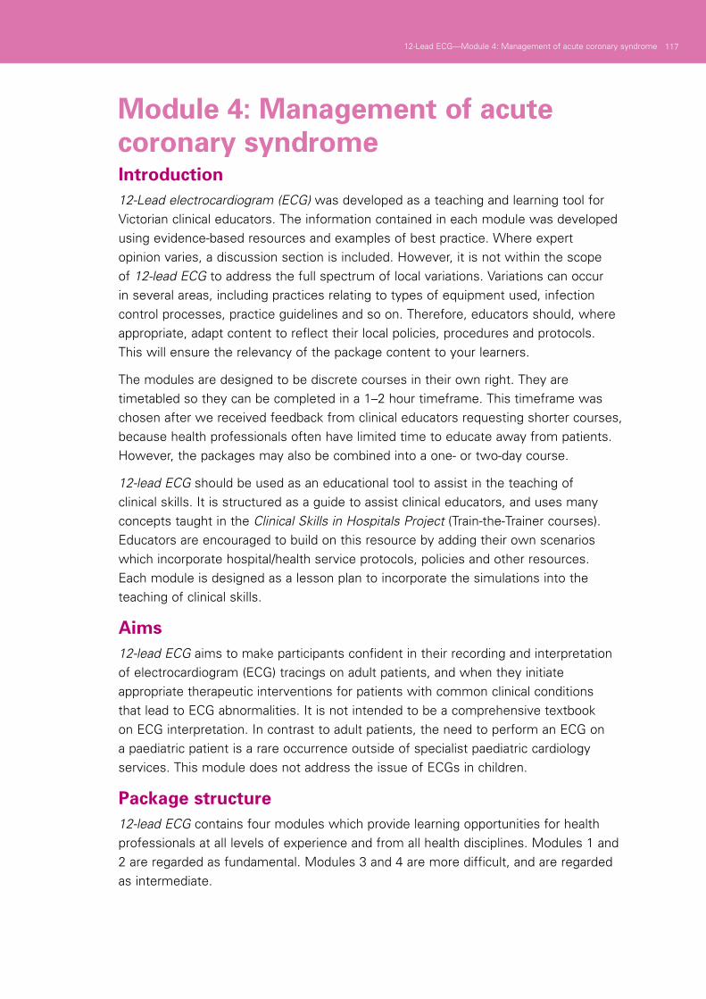
Module 4: Management of acute coronary syndromeIntroduction12-Lead electrocardiogram (ECG) was developed as a teaching and learning tool for Victorian clinical educators. The information contained in each module was developed using evidence-based resources and examples of best practice. Where expert opinion varies, a discussion section is included. However, it is not within the scope of 12-lead ECG to address the full spectrum of local variations. Variations can occur in several areas, including practices relating to types of equipment used, infection control processes, practice guidelines and so on. Therefore, educators should, where appropriate, adapt content to reflect their local policies, procedures and protocols. This will ensure the relevancy of the package content to your learners.
The modules are designed to be discrete courses in their own right. They are timetabled so they can be completed in a 1–2 hour timeframe. This timeframe was chosen after we received feedback from clinical educators requesting shorter courses, because health professionals often have limited time to educate away from patients. However, the packages may also be combined into a one- or two-day course.
12-lead ECG should be used as an educational tool to assist in the teaching of clinical skills. It is structured as a guide to assist clinical educators, and uses many concepts taught in the Clinical Skills in Hospitals Project (Train-the-Trainer courses). Educators are encouraged to build on this resource by adding their own scenarios which incorporate hospital/health service protocols, policies and other resources. Each module is designed as a lesson plan to incorporate the simulations into the teaching of clinical skills.
Aims12-lead ECG aims to make participants confident in their recording and interpretation of electrocardiogram (ECG) tracings on adult patients, and when they initiate appropriate therapeutic interventions for patients with common clinical conditions that lead to ECG abnormalities. It is not intended to be a comprehensive textbook on ECG interpretation. In contrast to adult patients, the need to perform an ECG on a paediatric patient is a rare occurrence outside of specialist paediatric cardiology services. This module does not address the issue of ECGs in children.
Package structure12-lead ECG contains four modules which provide learning opportunities for health professionals at all levels of experience and from all health disciplines. Modules 1 and 2 are regarded as fundamental. Modules 3 and 4 are more difficult, and are regarded as intermediate.
11712-Lead ECG—Module 4: Management of acute coronary syndrome

118 12-Lead ECG—Module 4: Management of acute coronary syndrome
Level of complexity Package structure
ComplexFor participants with more than 4 years experience or who have completed Modules 1–4
IntermediateFor participants in postgraduate years 3–4 or who have completed Modules 1 and 2
FundamentalFor participants in postgraduate years 1–2
12-lead ECG was designed to develop participants’ knowledge, skills and behaviours in ECG interpretation, and to expose them to increasingly complex scenarios aimed at testing their ability to combine these skills, work as a team and problem solve in more difficult situations.
Educators delivering these modules should be aware of participants’ level of experience and choose appropriate modules. Modules presume an increasing level of knowledge as they progress, ranging from a fundamental knowledge of anatomy and physiology for the fundamental modules, up to detailed knowledge of arrhythmia and acute coronary syndrome management for the more complex modules. Novice participants (such as first-year graduates) are expected to start with the fundamental modules, and only move onto intermediate and more complex modules as they demonstrate proficiency. More experienced participants may start at the intermediate level if the educator is satisfied that they have the prior knowledge and skills. Individual educators are responsible for assessing each participant’s baseline knowledge and determining which modules they need to complete. While the intermediate modules contain considerable medical detail, non-medical participants can still gain valuable experience from these modules by focusing on their roles and expectations in these scenarios. If the group contains no medical staff, facilitators may need to play the medical roles. More specific descriptions of presumed knowledge are outlined in each module.
Management of acute coronary syndrome
Management of arrhythmias
Abnormal ECGsBasic ECG recording and interpretation

11912-Lead ECG—Module 4: Management of acute coronary syndrome
The design of these packages presumes that the clinical educators using them have knowledge and expertise in current best practice regarding the teaching of clinical skills and conducting facilitated discussions. Knowledge and expertise are presumed commensurate with the Department of Human Services’ basic and advanced Train-the-Trainer Programs. Clinical educators are encouraged to refer to Department of Human Services’ Clinical Skills Facilitators Manual for theory on:
Peyton’s model for teaching clinical skills1.
leading small group discussions2.
giving feedback3.
crisis resource management skills.4.


12112-Lead ECG—Module 4: Management of acute coronary syndrome
Module 4: Management of acute coronary syndromeAuthors: Dr Stuart Dilley, Ms Debbie Paltridge
AimsThis module allows participants to practise recognising common electrocardiogram (ECG) abnormalities associated with acute coronary syndromes (ACS) (angina and myocardial infarction) and to initiate appropriate management.
Presumed knowledge
This module is targeted to health professionals with some experience in ECG interpretation and management of ACS, such as middle-grade doctors and senior nursing staff. They are also expected to have an intermediate level of knowledge and skills relevant to:
the ECG appearances commonly seen in ACS1.
manual defibrillation2.
common drugs used in the setting of ACS, for example, aspirin, heparin, 3.thrombolytic agents and anti-arrhythmic agents
initial blood investigations for ACS, including cardiac enzymes, electrolytes and full 4.blood examination.
Participants should have already practised these skills and had an opportunity to apply them in clinical simulation scenarios contained in other packages (ALS and BLS) in this project. If participants do not yet feel confident with the individual skills, they should be redirected to these other modules.
ObjectivesBy the end of this module, participants should have:
reviewed the common ECG appearances of ACS1.
discussed the appropriate early pharmacological and non-pharmacological 2.management of ACS
practised the recognition and management of ACS on a simulated patient (manikin)3.
applied their knowledge of pharmacological interventions in ACS to a simulated 4.patient (manikin).
Background information for educatorsThe main purpose of this module is to allow participants to practise recognising ACS on ECGs in a simulated environment, and make decisions about appropriate management. The ECG appearances of ACS were covered in 12-lead ECG—Module 2: Abnormal electrocardiograms. The focus of Module 4: Management of acute coronary syndrome leading into the simulations should be on appropriate management of these conditions. What constitutes appropriate management depends on the background

122 12-Lead ECG—Module 4: Management of acute coronary syndrome
of the participants involved. Medical staff are expected to recognise and manage these conditions. Nursing and allied health staff may be expected to recognise these conditions, seek assistance from more experienced staff and then help provide ongoing care to the patient.
The initial management of patients with suspected ACS requires a coordinated response which produces the simultaneous stabilisation of the patient, investigation of symptoms and treatment.
Initial management
A presumptive diagnosis of ACS can usually be made on history and examination. Once the diagnosis of ACS is likely, initial management should include:
■ aspirin 300 mg orally (unless contraindicated)
■ oxygen 15 L per minute by mask
■ analgesia IV aliquots of morphine 2.5 mg as required
■ glyceryl-trinitrate sublingual or IV as required
■ ECG within 5 minutes of arrival.
InvestigationsElectrocardiogram (ECG)
ECG should be performed early. It is the sole test used to identify patients for reperfusion, whether thrombolysis or acute angioplasty. However, a normal ECG does not exclude ACS.
Current practice is to continue cardiac monitoring of potential ACS patients with normal initial ECGs, and this is recommended by Australian authorities1. Other studies identify that patients who are pain free on assessment and have normal or non-specific ECG findings on initial ECG are at extremely low risk of arrhythmias2. Therefore, these limited resources might be used more efficiently by moving such low-risk patients from cardiac monitoring while continuing to investigate their symptoms.
Cardiac enzymes (CE)
Australian guidelines recommend the measurement of troponin on arrival, repeated 8 hours after the last episode of pain if initially normal. CKMB should be measured if troponin assays are not available. Serial monitoring of CEs should be undertaken with creatine kinase (CK).

12312-Lead ECG—Module 4: Management of acute coronary syndrome
Other blood tests
Australian guidelines also recommend the following tests; however, except for potassium measurement in electrolytes, few are useful in the acute setting:
■ full blood examination (FBE)
■ electrolytes, particularly potassium (K)
■ serum lipids within 24 hours
■ blood glucose.
Chest X-ray (CXR)
CXR is useful in assessing cardiac size and seeking alternative diagnoses, but should not delay definitive treatment for ACS.
ST elevation myocardial infarction (STEMI)
STEMI is defined as presentation with clinical symptoms consistent with an acute coronary syndrome, with ECG features including any of the following:
■ persistent ST segment elevation of > 1 mm in two contiguous limb leads
■ ST segment elevation of > 2 mm in two or more contiguous chest leads
■ left bundle branch block not known to be old.
Patients with STEMI who present within 12 hours should have a reperfusion strategy, percutaneous coronary intervention (PCI) or thrombolysis implemented urgently. Other adjunctive therapies are also indicated.
Anti-platelet therapy
Aspirin 300 mg should be given to all patients with STEMI, if not already done so, unless contraindicated. Evidence exists that clopidogrel 300 mg should be given in addition to aspirin for all patients undergoing fibrinolysis or angioplasty and stent insertion. However, clopidogrel should not be given if there is a reasonable chance of the patient going on to have coronary artery bypass graft (CABG) surgery acutely.
Anti-thrombin therapy1, 3
Anti-thrombin therapy should be used in all patients with STEMI undergoing PCI or thrombolysis. If the patient is undergoing PCI, the dose of unfractionated heparin will depend on whether abciximab is also used. When fibrinolysis is performed, a loading dose of 60 units per kg body weight up to a maxium of 4000 units is recommended.
Enoxaparin 0.75–1 mg per kg body weight subcutaneously is suggested as an alternative therapy to unfractionated heparin, but its effects cannot be as easily tritrated.

124 12-Lead ECG—Module 4: Management of acute coronary syndrome
Glycoprotein (GP) IIb/IIIa inhibitors
GP inhibitors should not be used with thrombolysis, due to the increased risk of bleeding. Abciximab may be beneficial in the setting of PCI; however, the data conflicts regarding benefit.
Beta-blockers
Oral beta-blockers should be administered for ACS of all types unless contraindications are present4. Contraindications include:
■ moderate to severe left ventricular failure and pulmonary oedema
■ bradycardia—less than 60 bpm
■ hypotension—less than 100 mmHg systolic
■ signs of poor peripheral perfusion
■ second- or third-degree heart block
■ significant airways disease.
Reperfusion therapy
The choice of reperfusion therapy depends on several factors:
■ time delay to PCI or thrombolysis
■ time from symptom onset
■ contraindications to thrombolysis
■ presence of cardiogenic shock.
For patients who present very early, that is, within 1 hour of symptom onset, thrombolysis could be considered ahead of PCI. In those presenting between 1–3 hours of symptom onset, PCI is preferred if this can be achieved in a timely manner. For those presenting after three hours, PCI is regarded as superior to thrombolysis. PCI is the preferred option in the setting of cardiogenic shock.
Percutaneous coronary intervention (PCI)
PCI is probably the better reperfusion strategy if performed promptly by an experienced interventional cardiologist in an appropriate institution with sufficient exposure to the procedure. Where PCI is not available or delayed, thrombolytic therapy should be given. A time delay of 90 minutes from first medical contact to balloon inflation is the maximum desirable; otherwise, thrombolysis should be given.
Thrombolysis
Thrombolysis is still the most common method available for coronary reperfusion outside tertiary hospitals and major referral centres in Australia. The agents available for use in Australia at present and their method of administration are outlined in the following:

12512-Lead ECG—Module 4: Management of acute coronary syndrome
■ streptokinase (SK) 1.5 million units over 30–60 minutes
■ retaplase (rPA) two 10-unit boluses, 30 minutes apart
■ alteplase (rt-PA) three weight dependent doses over 90 minutes
■ tenecteplase (TNK) single bolus dose based on body weight.
Absolute contraindications to thrombolysis include:
■ active bleeding or bleeding disorder
■ significant closed head injury or facial trauma
■ any prior intracranial haemorrhage
■ ischaemic stroke within three months
■ known structural cerebral vascular lesion
■ known malignant intracranial neoplasm.
Relative contraindications to thrombolysis include:
■ current use of anticoagulants
■ non compressible vascular punctures
■ recent major surgery (within three weeks)
■ recent internal bleeding
■ active peptic ulcer disease
■ severe uncontrolled hypertension on presentation (systolic BP > 180 mmHg)
■ pregnancy.
Bleeding is the most common adverse event associated with the administration of thrombolytic agents. This may be obvious or concealed bleeding. Concealed intra-cranial bleeding may present as a cerebrovascular accident. Concealed gastrointestinal bleeding may initially only present as a patient in shock.
Non-ST elevation acute coronary syndrome (NSEACS)
NSEACS patients have a history consistent with ACS, but do not show ST elevation on their ECG. This includes a group of patients who may have suffered a non-ST segment elevation myocardial infarction (non-STEMI). Patients in this group need to be risk stratified and treated accordingly.
High-risk patients
These patients have symptoms at rest or prolonged pain, and show non-ST elevation ECG changes and/or troponin rises. They should be treated in the same way as patients with STEMI, including the use of beta-blockers. Thrombolysis should not be used. Arrangements should be made for coronary angiography.

126 12-Lead ECG—Module 4: Management of acute coronary syndrome
Intermediate-risk patients
These patients have symptoms at rest or prolonged pain, but show no ECG abnormalities or troponin rise. They require further investigation to further risk stratify them into high- or low-risk groups (for example, stress testing).
Low-risk patients
Low risk patients can be discharged and further investigated on an outpatient basis after optimising medical therapy.
Right ventricular (RV) infarction
RV infarction may occur in up to 50% of patients with inferior STEMI. The in-hospital mortality for patients with RV dysfunction is 25–30%. Patients with RV dysfunction and infarction depend on adequate filling pressures to maintain cardiac output. This should be achieved by ensuring adequate fluid resuscitation and avoiding vasodilating agents such as nitrate and ACE inhibitors, along with diuretics.
Learning activitiesSuggested learning activities and timetables are outlined below. Timetable 1 is designed for 12 participants working in two groups of six. Timetable 2 is designed for six participants working together.
Timetable 1
Timing Activity Objective
30 minutes Facilitated discussion 1, 2, 3
10 minutes Simulation 1 Simulation 2 3, 4
25 minutes Debrief Debrief 2, 3, 4
10 minutes Simulation 2 Simulation 1 3, 4
25 minutes Debrief Debrief 2, 3, 4
10 minutes Summary All
5 minutes Evaluation
Total time = 2 hours

12712-Lead ECG—Module 4: Management of acute coronary syndrome
Timetable 2
Timing Activity Objective
30 minutes Facilitated discussion 1, 2, 3
10 minutes Simulation 1 3, 4
25 minutes Debrief 2, 3, 4
10 minutes Simulation 2 3, 4
25 minutes Debrief 2, 3, 4
10 minutes Summary All
5 minutes Evaluation
Total time = 2 hours
Facilitated discussion
The facilitator should lead a discussion amongst participants to refresh or clarify issues relating to ECG recognition of ACS as covered in 12-lead ECG—Module 2: Abnormal electrocardiograms. The discussion should then be expanded to cover the appropriate initial management of ACS, and to introduce the simulation training to follow. This should not be a comprehensive lecture on ACS.
Facilitators should be aware of the different professional groups that may be present during this discussion (particularly if they include junior nursing staff or allied health professionals), and allow these groups to discuss their roles in managing patients with arrhythmias. Where the group comprises a mix of participants, facilitators may split them into smaller groups to discuss a clinical scenario and report back to the group. A selection of scenarios is provided with this module for this purpose; however, educators also may wish to develop their own. Thus, participants can concentrate on issues relevant to their craft group and skill level while informing other craft groups of their capabilities and limitations.
The discussion should not go beyond the 30 minutes alotted, in order to keep the module to time. Facilitators are reminded that debriefing time is also a valuable opportunity to clarify or further discuss the management of these clinical conditions.
PowerPoint slides are available for the facilitator to use to summarise these main points at the end of the discussion, or as triggers if participants have not identified the major issues.
The facilitator should ensure these major issues are covered:
■ important ECG findings suggestive of ACS
■ differential diagnosis of ST elevation in the patient with chest pain

128 12-Lead ECG—Module 4: Management of acute coronary syndrome
■ appropriate management of the patient with ACS: anti-platelet therapy, anti-coagulation and revascularisation
■ appropriate reperfusion strategies (PCI and or choice of thrombolytic agent) relevant to each participant’s particular hospital.
Scenario 1
You are conducting a ward round (medical, nursing or allied health) and arrive to assess Mr Jones, a 60-year-old patient on the medical ward for investigation of syncope. You note that looks pale and is short of breath. He states that he has had chest pain for the last hour: central, heavy and radiating to the left arm. What is your role in his initial management (medical, nursing, allied health)?
Provide participants with an ECG that demonstrates an anterior STEMI and ask them to discuss their role in management with respect their health professional group.
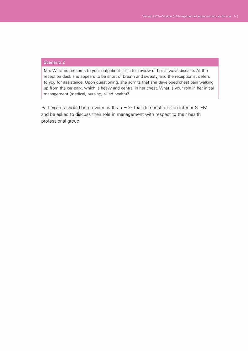
Scenario 2
Mrs Williams presents to your outpatient clinic for review of her airways disease. At the reception desk she appears to be short of breath and sweaty, and the receptionist defers to you for assistance. Upon questioning, she admits that she developed chest pain walking up from the car park. She describes her pain as heavy and central in her chest. What is your role in her initial management (medical, nursing, allied health)?
Provide participants with an ECG that demonstrates an inferior STEMI and ask them to discuss their role in management with respect to their health professional group.
Scenario 3
Mr Nguyen is visiting his wife, a patient you are assessing in the emergency department. He appears to be having chest pains, but does not speak English particularly well. He is pale and sweaty. What is your role in his initial management (medical, nursing, allied health)?
Provide participants with an ECG that demonstrates inferior T wave inversion and ask them to discuss their role in management with respect to their health professional group.
Simulation session
This exercise allows participants to practise their ECG interpretation skills and management of ACS as a team in a simulated environment.
The program assumes two facilitators for 12 participants. Participants should be divided into two groups of six (Timetable 1). Three participants each participate in one scenario and observe a second. Those not participating in the scenario observe and participate in the debriefing session. The debriefing period should include all six participants—that is, the active participants and their observers. These scenarios can be run with smaller groups. If only six participants are present, the scenarios can be run consecutively (Timetable 2).

12912-Lead ECG—Module 4: Management of acute coronary syndrome
With less-experienced participants, this module can extend beyond the expected timeframe. In that instance, it is reasonable to omit the second scenario and offer it as a stand-alone session at a later date, along with a modified version of the facilitated discussion.
If there are no medical participants, facilitators should fill these roles and allow nursing or allied health participants to contribute to the scenarios as they would in real life.
These scenarios can be run on low-fidelity simulators (for example, Resus Anne), but are also quite suitable for more sophisticated simulators (for example, Sim Man, HPS METI).
In these scenarios, there may be considerable difference between institutions in terms of resources available and standard management (for example, availability of cardiac cath labs for acute PCI). Facilitators should instruct participants to request and utilise resources as if they are available to them in their parent institutions. Institution-specific protocols for interventions such as thrombolysis and heparinisation should be followed. This information is expected to be used throughout the facilitated discussion.
Simulation 1: Acute myocardial infarctionScenario design
In this scenario, a 60-year-old woman on the medical ward complains of chest pain. An ECG reveals that she is having an acute anterior STEMI. Participants need to recognise this, initiate immediate treatment, deal with any complications and formulate a management plan.
Case history
Patient details
Sex Female
Age 60 years old
Past history Smoker, diet-controlled diabetic
Hypertension, prescribed irbesartan 150 mg daily
Social history Lives at home with husband
History of present illness Current inpatient on medical ward for investigation of anaemia; now post transfusion
No GIT bleeding has been identified, but you are still awaiting endoscopy
Presenting symptoms Central, heavy chest pain for one hour
Radiates to jaw and left arm is ‘heavy’
Associated nausea and sweatiness

130 12-Lead ECG—Module 4: Management of acute coronary syndrome
Resources
General
Setting/environment Hospital medical ward
Patient attire Hospital gown
Monitoring ECG applied by ward nurse
Supporting documentation required Hospital charts
Equipment
Equipment Number Sourced from
Manikin capable of cardiac rhythm generation
1
Trolley/bed 1
Gown 1
Blanket/pillow 1 each
Oxygen mask, tubing, source 1 set
IV cannulae, tubing, fluids Various size cannulae, several bags of IV fluid and lines
ECG machine 1
Prop ECG anterior AMI 1
Non-invasive BP 1
Drug props: aspirin, GTN, morphine, metoclopramide, heparin, beta-blockers, thrombolytic agent
1 set
Agents specific to individual institution
Roles
Participant 1
You are a health professional working on the medical ward of your hospital. You are asked to review a 60-year-old lady previously admitted for investigation of anaemia because she has developed chest pain. You have a nurse to help you with equipment and medications, and two colleagues to assist you if needed.
Faculty role play (optional)
You are a senior clinician working on the medical ward. At the end of the scenario, you may arrive to take handover of the patient. If the participants have difficulty with the scenario, it may be appropriate to enter the scenario early and offer advice.
Participants 2 and 3
You are health professionals working on the medical ward of your hospital. Your colleague (Participant 1) has gone to review a patient on the ward. You may be asked to assist.

13112-Lead ECG—Module 4: Management of acute coronary syndrome
Faculty role play: nurse
You are a competent nurse working on the medical ward, looking after a 60-year-old lady who is being investigated for anaemia. She complains of chest pain and tells you she has had it for over an hour. You have obtained ECG monitoring and arranged for a 12-lead ECG to be done. You call on Participant 1 to review the patient. You may assist the participants in finding and administering medications, but do not prompt them about appropriate treatment.
Simulator programming considerations
System Baseline state Resolution
CVS HR 80 bpm, sinus rhythm with regular ventricular ectopic beats
BP 110/90 mmHg
Skin sweaty, cold
ST elevation on ECG
HR 80 bpm, sinus rhythm with regular ventricular ectopic beats
BP 130/70 mmHg
Skin warm and dry
ST elevation on ECG if still waiting for PCI
ST elevation resolving if thrombolysed
Respiratory RR 24 per minute
Oxygen saturation 95% room air
RR 18 per minute
Oxygen saturation 98% on oxygen
Neurologic GCS 15 GCS 15
Response to participant intervention
Remain in baseline state till all the following have been done or discussed:
■ IV access
■ aspirin
■ oxygen
■ GTN
■ morphine
■ reperfusion
■ heparin
■ beta-blockers
■ transfer to appropriate ward/cath lab
■ involvement of appropriate personnel

132 12-Lead ECG—Module 4: Management of acute coronary syndrome
Debriefing points:
■ recognition of ACS—that is, anterior STEMI
■ differential diagnosis of chest pain/ST elevation
■ indications and roles of aspirin, oxygen, GTN, morphine and beta-blockers
■ discussion of reperfusion strategy: thrombolysis versus PCI.
Simulation 2: Right ventricular infarctScenario design
In this scenario, a 50-year-old man presents to the emergency department with an inferior AMI with right ventricular involvement. Participants need to recognise this along with the issues that specifically relate to managing right ventricular dysfunction—that is, hypotension.
Case history
Patient details
Sex Male
Age 50
Past history Smoker, high cholesterol, family history of heart disease
‘Never goes to the doctor’
Social history Works long hours driving interstate truck
Lives at home with wife and teenage kids
History of present illness Developed ‘indigestion’ after big meal at home
Brought to hospital ED by wife
Presenting symptoms Pain in lower chest and epigastrium for last hour
Sweaty, pale
Not relieved by antacids
Resources
General
Setting/environment Hospital emergency department
Patient attire and appearance Truckie’s clothes
Monitoring ECG, non-invasive BP, pulse oximetry
Supporting documentation required ED observation and medication charts

13312-Lead ECG—Module 4: Management of acute coronary syndrome
Equipment
Equipment Number Sourced from
Manikin capable of cardiac rhythm generation
1
ED trolley 1
Truckie’s clothes: shorts, blue singlet
1 set
Blanket, pillow 1 each
Oxygen mask, tubing, oxygen source
1 set
IV cannulae, tubing, fluids Various size cannulae, several bags of IV fluid and lines
ECG machine 1
Prop ECG inferior/RV AMI 1
Non-invasive BP 1
Drug props: aspirin, GTN, morphine, metoclopramide, heparin, beta-blockers and thrombolytic agents
1 set
Agents specific to individual institution
Roles
Participant 1
You are a health professional working in the emergency department of your hospital. You have been asked to assess a 50-year-old man in the resuscitation cubicle, complaining of ‘indigestion’. You have a resuscitation nurse with you to help with equipment and medications. You have two colleagues on which to call should you need assistance.
Participants 2 and 3
You are two health professionals working in the emergency department of your hospital. Your colleague has gone to the resuscitation cubicle to attend a 50-year-old man with ‘indigestion’. Your assistance may be required. There is a resuscitation nurse on hand to assist with equipment and medications.
Faculty role play: nurse
You are a competent nurse working in the emergency department, looking after a 50-year-old man who presented with ‘indigestion’. He tells you he has had this pain for over an hour. You have obtained ECG monitoring and arranged for a 12-lead ECG to be done. You call on Participant 1 to review the patient. You may assist the participants in finding and administering medications, but do not prompt them about appropriate treatment.
134 12-Lead ECG—Module 4: Management of acute coronary syndrome
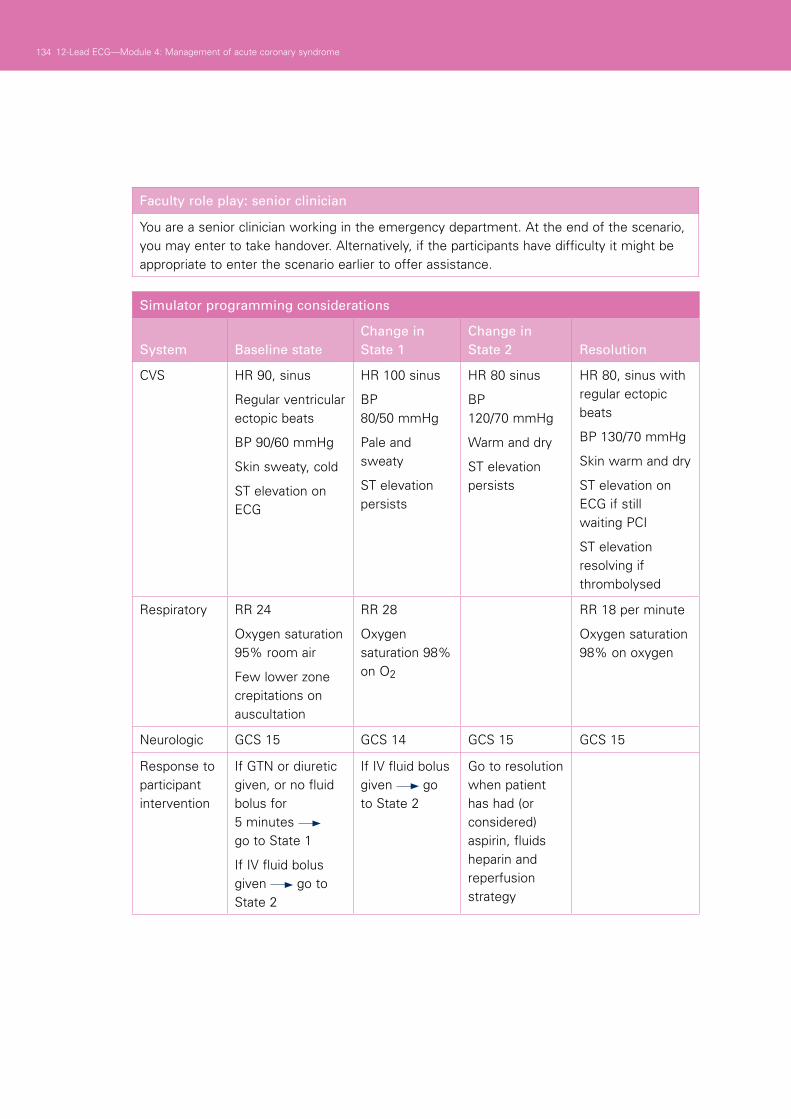
Faculty role play: senior clinician
You are a senior clinician working in the emergency department. At the end of the scenario, you may enter to take handover. Alternatively, if the participants have difficulty it might be appropriate to enter the scenario earlier to offer assistance.
Simulator programming considerations
System Baseline stateChange in State 1
Change in State 2 Resolution
CVS HR 90, sinus
Regular ventricular ectopic beats
BP 90/60 mmHg
Skin sweaty, cold
ST elevation on ECG
HR 100 sinus
BP 80/50 mmHg
Pale and sweaty
ST elevation persists
HR 80 sinus
BP 120/70 mmHg
Warm and dry
ST elevation persists
HR 80, sinus with regular ectopic beats
BP 130/70 mmHg
Skin warm and dry
ST elevation on ECG if still waiting PCI
ST elevation resolving if thrombolysed
Respiratory RR 24
Oxygen saturation 95% room air
Few lower zone crepitations on auscultation
RR 28
Oxygen saturation 98% on O2
RR 18 per minute
Oxygen saturation 98% on oxygen
Neurologic GCS 15 GCS 14 GCS 15 GCS 15
Response to participant intervention
If GTN or diuretic given, or no fluid bolus for 5 minutes go to State 1
If IV fluid bolus given go to State 2
If IV fluid bolus given go to State 2
Go to resolution when patient has had (or considered) aspirin, fluids heparin and reperfusion strategy
13512-Lead ECG—Module 4: Management of acute coronary syndrome
Debriefing points:
■ recognition of ACS—that is, inferior and RV STEMI
■ indications and roles of aspirin, oxygen, GTN, morphine and beta-blockers, particularly in reference to RV infarction.
Summary
The summary session reinforces content covered in the learning activities, and is an opportunity for participants to reflect on what they have covered. No new material should be introduced.
Points to cover in the summary include:
■ indications of ACS on ECG
■ appropriate first-line management and pharmacological interventions for ACS
■ awareness of hospital resources and limitations in the management of ACS.
Participants should be encouraged to read the literature on ACS recognition and management in their own time to reinforce the skills acquired in this module. They should be offered access to equipment and educators in the future if they need to practise or improve their skill level or confidence.
Resource listThe following resource list assumes two facilitators for every 12 participants, a ratio of 1:6. As a minimum, the following resources are needed to conduct this module.
Resource Quantity Additional comments
Equipment and resources as listed in scenario requirements above
PowerPoint presentation 1 set Provided with module
136 12-Lead ECG—Module 4: Management of acute coronary syndrome
EvaluationA formal evaluation has been specifically developed for this module. It incorporates the objectives of the module and the perceptions of the participants about whether they have increased their understanding by working through the module. It is highly recommended that this formal evaluation be copied and completed by all participants at the completion of the module.
A range of informal evaluation tools may also be used in conjunction with this evaluation throughout the module, including those available in the Department of Human Services’ Clinical Skills Facilitators Manual from the basic course conducted in 2007.
References Acute Coronary Syndrome Guidelines Working Group 2006 Guidelines for the 1.
management of acute coronary syndromes. MJA 184: S1–S30
Gatlen M., Perry J., Stiell I., Wielgosz A. and Lee J. 2007 A clinical decision rule to 2.identify which chest pain patients can be safely removed from cardiac monitoring in the emergency department. Ann Emerg Med 50: 136–143
Aroney C., Aylward P., Chew D. et al. 2008 Addendum to the National Heart 3.Foundation of Australia/Cardiac Society of Australia and New Zealand Guidelines for the management of acute coronary syndromes 2006. MJA 188: 303–303
American Heart Association Guidelines for Cardiopulmonary Resuscitation and 4.Emergency Cardiovascular Care 2005 Part 8: Stabilization of the patient with acute coronary syndromes. Circulation 2005 112: IV89–IV110

13712-Lead ECG—Module 4: Management of acute coronary syndrome
ResourcesFacilitator feedback form
The following form should be used to assist you in giving feedback after each participant has practised their ECG skills at the skill station.
Feedback using the Pendleton model
Pendleton’s model of feedback assists learners to maximize their potential at different stages of training, raise their awareness of strengths and areas for improvement, and identify actions to be taken to improve performance. Pendleton’s rules are structured in such a way that the learner identifies the positives first, in order to create a safe environment. This is followed by the facilitator or group reinforcing these positives and discussing skills to achieve them. Different techniques are then suggested. The advantage of this method is that the learner’s strengths are discussed first. Avoiding a discussion of weaknesses right at the beginning prevents defensiveness and allows reflective behaviour in the learner.
Below is a series of questions to assist you in this technique:
1. Ask the learner how they feel.
2. Ask the learner what went well and why (this can be combined with question 1 and 3).
3. Tell the learner what went well and why.
4. Ask the learner what could have been done better and why.
5. Tell the learner what could have been done better and why.
6. Summarise the learner’s strengths and identify up to three things to concentrate on.
Note: This form does not need to be given to the participant — it is a guide for you, the group facilitator.

138 12-Lead ECG—Module 4: Management of acute coronary syndrome
Module 3: Management of acute coronary syndrome —evaluationThank you for participating in this module. As part of our commitment to quality improvement the following questionnaire will be used to plan future implementation of this module. We appreciate your time completing this evaluation.
1. Overall
How would you rate this module?
poor fair good very good outstanding
2. Learning objectives
Please consider whether this module was successful in meeting the following learning objectives:
12-lead ECG
Learning objectives of Module 4: Management of acute coronary syndrome S
tro
ng
ly
dis
agre
e
Dis
agre
e
Slig
htl
y ag
ree
Ag
ree
Str
on
gly
ag
ree
Reviewed the common ECG appearances of ACS
Discussed the appropriate early pharmacological and non-pharmacological management of ACS
Practised the recognition and management of ACS on a simulated patient (manikin)
Applied their knowledge of pharmacological interventions in ACS to a simulated patient (manikin)
3. Important learning outcomes
What are the three most important things you have learned from this module?

13912-Lead ECG—Module 4: Management of acute coronary syndrome
4. Module implementation
Please indicate to what extent you agree or disagree with each of the following statements in relation to the implementation of the module.
Str
on
gly
d
isag
ree
Dis
agre
e
Slig
htl
y ag
ree
Ag
ree
Str
on
gly
ag
ree
The facilitator respected my experience
The facilitator encouraged my participation
I was able to ask the facilitator questions
The facilitator was able to answer my questions
The feedback I received was clear
The feedback I received will assist me my future performance
There was adequate time for the skills stations
There was adequate time for the facilitated discussions
There was adequate time for the simulations
I have increased my confidence in interpreting 12-lead ECGs
I have identified future learning needs in this topic area
5. Future module implementation
Do you think the module should be altered in any way? yes no
If yes, what recommendations do you have?
Thank you

PowerPoint presentation
1. 2.
3.
140 12-Lead ECG—Module 4: Management of acute coronary syndrome

Appendix 1: Clinical scenariosScenario 1
You are conducting a ward round (medical, nursing or allied health) and arrive to assess Mr Jones, a 60-year-old patient on the medical ward for investigation of syncope. You note that looks pale and is short of breath. He states that he has had chest pain for the last hour, central, heavy and radiating to the left arm. What is your role in his initial management (medical, nursing, allied health)?
14112-Lead ECG—Module 4: Management of acute coronary syndrome

142 12-Lead ECG—Module 4: Management of acute coronary syndrome
Figu
re 1
: Clin
ical
sce
nario
s 1
EC
G
14312-Lead ECG—Module 4: Management of acute coronary syndrome
Scenario 2
Mrs Williams presents to your outpatient clinic for review of her airways disease. At the reception desk she appears to be short of breath and sweaty, and the receptionist defers to you for assistance. Upon questioning, she admits that she developed chest pain walking up from the car park, which is heavy and central in her chest. What is your role in her initial management (medical, nursing, allied health)?
Participants should be provided with an ECG that demonstrates an inferior STEMI and be asked to discuss their role in management with respect to their health professional group.

144 12-Lead ECG—Module 4: Management of acute coronary syndrome
Figu
re 2
: Clin
ical
sce
nario
s 2
EC
G

14512-Lead ECG—Module 4: Management of acute coronary syndrome
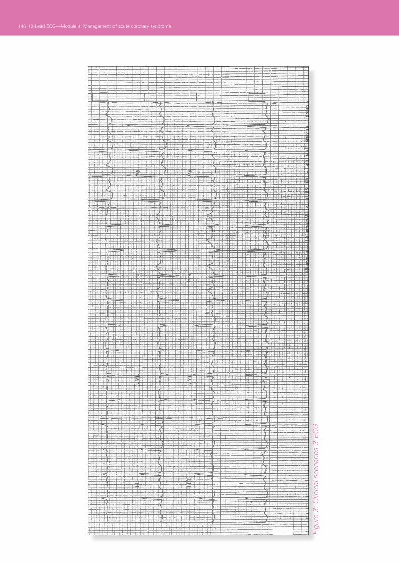
Scenario 3
Mr Nguyen is visiting his wife, a patient you are assessing in the Emergency Department. He appears to be having chest pains, but does not speak English particularly well. He is pale and sweaty. What is your role in his initial management (medical, nursing, allied health)?
Participants should be provided with an ECG that demonstrates inferior T-wave inversion and be asked to discuss their role in management with respect to their health professional group.
146 12-Lead ECG—Module 4: Management of acute coronary syndrome
Figu
re 3
: Clin
ical
sce
nario
s 3
EC
G

14712-Lead ECG—Module 4: Management of acute coronary syndrome
Figu
re 4
: Sim
ulat
ion
1 12
-lead
EC
Gs
App
endi
x 2:
Sim
ulat
ion
scen
ario
Sim
ula
tio
n 1

12-Lead ECG—Module 4: Management of acute coronary syndrome
Figu
re 5
: Sim
ulat
ion
2 C
linic
al s
cena
rios
3 E
CG
s
Sim
ula
tio
n 2
148