Embed Size (px)
Citation preview
MODIFIED CONSTRAINT INDUCED MOVEMENT THERAPY TO IMPROVE
UPPER EXTREMITY FUNCTION POST-STROKE IN CANADIAN NEUROLOGICAL REHABILITATION
by
Alana Bethany Fleet
Submitted in partial fulfilment of the requirements for the degree of Master of Science
at
Dalhousie University Halifax, Nova Scotia
August 2013
© Copyright by Alana Bethany Fleet, 2013

ii
TABLE OF CONTENTS
LIST OF TABLES ........................................................................................................... vi
LIST OF FIGURES ....................................................................................................... vii
ABSTRACT .................................................................................................................... viii
LIST OF ABBREVIATIONS USED ............................................................................ ix
GLOSSARY ...................................................................................................................... xi
ACKNOWLEDGEMENTS ............................................................................................ xiii
CHAPTER 1 INTRODUCTION .................................................................................. 1
CHAPTER 2 BACKGROUND ..................................................................................... 4
CHAPTER 3 RESEARCH OBJECTIVES AND HYPOTHESES ......................... 13
3.1 STUDY 1: CIMT SURVEY ................................................................................ 13
3.2 STUDY 2: SYSTEMATIC REVIEW ..................................................................... 14
3.3 STUDY 3: MCIMT TRIAL .................................................................................. 14
3.4 SUMMARY TO CHAPTER 3 AND TRANSITION TO CHAPTER 4 ........................ 15
CHAPTER 4 STUDY 1: CIMT SURVEY ............................................................... 17
4.1 INTRODUCTION ................................................................................................ 17
4.2 METHODOLOGY ................................................................................................ 19
4.2.1 Participants and Survey Distribution .............................................. 19
4.2.2 Survey Development and Composition ......................................... 20
4.2.3 Data Analysis ........................................................................................... 23
4.3 RESULTS ........................................................................................................... 24
4.3.1 Participants ............................................................................................... 24
4.3.2 CIMT Usage ............................................................................................... 26
4.3.3 Therapist-Related Factors and CIMT Use ...................................... 32
4.3.4 CIMT Effectiveness and Barriers to Use ........................................ 32
4.4 DISCUSSION .................................................................................................... 33

iii
4.4.1 CIMT Use and Parameters .................................................................. 34
4.4.2 Therapist-Related Factors and CIMT Use ...................................... 35
4.4.3 Barriers to CIMT Use ............................................................................. 37
4.5 CONCLUSION ................................................................................................... 39
4.6 KEY MESSAGES ............................................................................................... 40
4.6.1 What is Already Known on this Topic ............................................. 40
4.6.2 What this Study Adds ........................................................................... 40
4.7 SUMMARY TO CHAPTER 4 AND TRANSITION TO CHAPTER 5 ........................ 41
CHAPTER 5 SYSTEMATIC REVIEW .................................................................... 43
5.1 INTRODUCTION ................................................................................................. 43
5.1.1 Description of the Condition .............................................................. 43
5.1.2 Description of the Intervention (mCIMT) ..................................... 43
5.1.3 How the Intervention Might Work ................................................... 46
5.1.4 Why it is Important to do this Review ........................................... 48
5.1.5 Objectives .................................................................................................. 51
5.2 METHODOLOGY ................................................................................................. 51
5.2.1 Criteria for Selecting Studies for Review ...................................... 51
5.2.2 Methods for Identification, Data Collection and Analysis of
Studies .................................................................................................................... 53
5.3 RESULTS ............................................................................................................ 56
5.3.1 Levels of Evidence.................................................................................. 56
5.3.2 Study Demographics ............................................................................. 57
5.3.3 Intervention .............................................................................................. 61
5.3.4 Outcome Measures ................................................................................ 66
5.4 DISCUSSION ..................................................................................................... 71
5.4.1 Effectiveness Across the Stages of Recovery .............................. 72
5.4.2 Interpreting the Level of Evidence: Limitations ......................... 72
5.4.3 Incorporating mCIMT in Clinical Practice ...................................... 74
5.4.4 Conclusion ................................................................................................. 74
iv
5.5 SUMMARY TO CHAPTER 5 AND TRANSITION TO CHAPTER 6 ........................ 75
CHAPTER 6 STUDY 3: mCIMT TRIAL................................................................ 77
6.1 METHODOLOGY ................................................................................................. 77
6.1.1 Groups and Subjects ............................................................................. 77
6.1.2 Assessments ............................................................................................. 79
6.1.3 Primary outcome measures ............................................................... 79
6.1.4 Secondary Outcome Measures .......................................................... 80
6.1.5 Analyses ..................................................................................................... 82
6.1.6 Therapy ...................................................................................................... 84
6.1.7 Thesis Work .............................................................................................. 86
6.1.8 Other Methodological Considerations ............................................. 91
6.1.9 Case Description ..................................................................................... 91
6.2 RESULTS ............................................................................................................ 97
6.2.1 Primary Outcomes ............................................................................. 97
6.2.2 Secondary Outcomes ........................................................................ 99
6.3 SUMMARY TO CHAPTER 6 AND TRANSITION TO CHAPTER 7 ...................... 100
CHAPTER 7 DISCUSSION ................................................................................... 102
7.1 CASE STUDY FINDINGS ..................................................................................... 102
7.1.2 Study Limitations ................................................................................ 103
7.2 CHALLENGES AND BARRIERS TO MCIMT IMPLEMENTATION .......................... 104
7.2.1 Patient Challenges ............................................................................... 104
7.3 CONCLUSION ..................................................................................................... 113
REFERENCES .............................................................................................................. 115
APPENDIX A Copyright Permission Contract .............................................. 124
APPENDIX B Survey Description...................................................................... 125
APPENDIX C Reported Components of CIMT .............................................. 127
APPENDIX D Search Phases .............................................................................. 128
v
APPENDIX E Search Strategy ........................................................................... 133
APPENDIX F Baseline Assessment Measures .............................................. 135
APPENDIX G Task Database .............................................................................. 137
vi
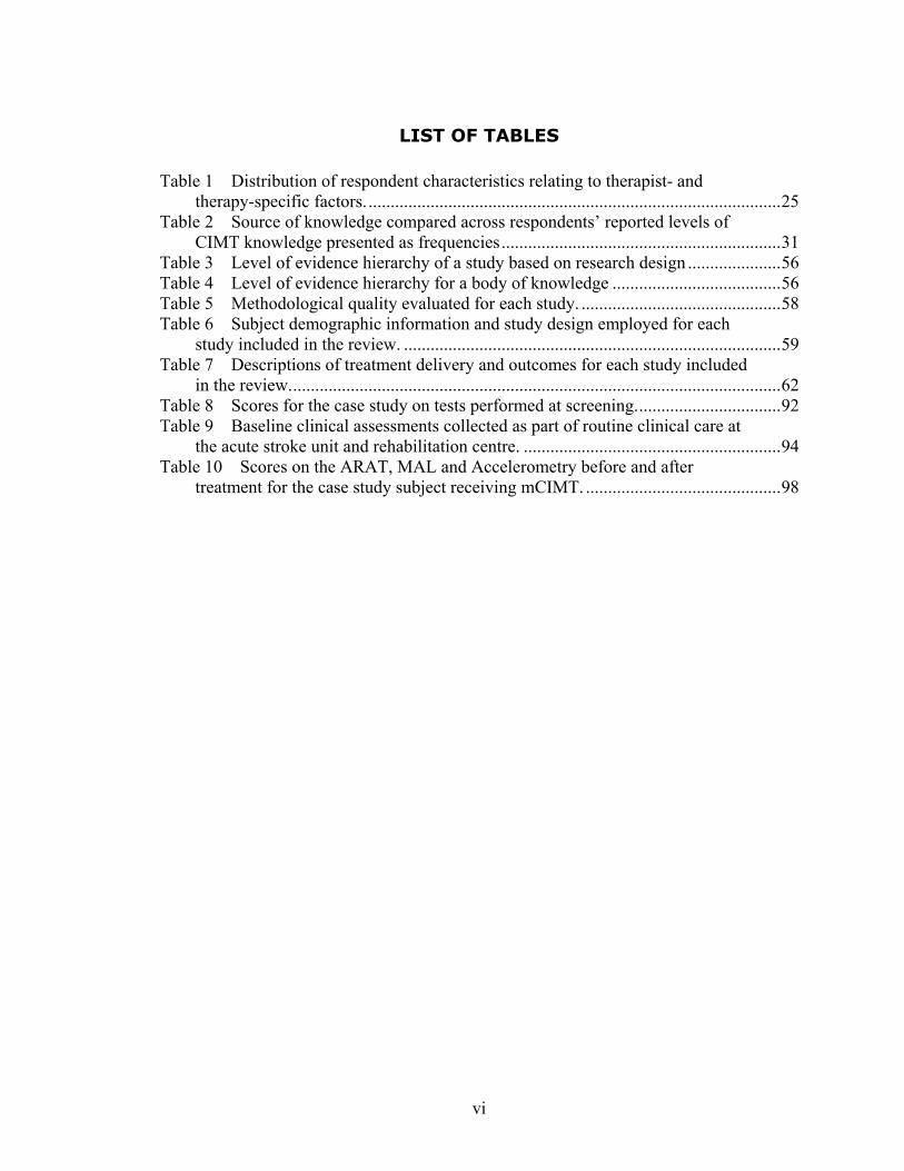
LIST OF TABLES
Table 1 Distribution of respondent characteristics relating to therapist- and therapy-specific factors. ............................................................................................. 25
Table 2 Source of knowledge compared across respondents’ reported levels of CIMT knowledge presented as frequencies ............................................................... 31
Table 3 Level of evidence hierarchy of a study based on research design ..................... 56 Table 4 Level of evidence hierarchy for a body of knowledge ...................................... 56 Table 5 Methodological quality evaluated for each study. ............................................. 58 Table 6 Subject demographic information and study design employed for each
study included in the review. ..................................................................................... 59 Table 7 Descriptions of treatment delivery and outcomes for each study included
in the review. .............................................................................................................. 62 Table 8 Scores for the case study on tests performed at screening. ................................ 92 Table 9 Baseline clinical assessments collected as part of routine clinical care at
the acute stroke unit and rehabilitation centre. .......................................................... 94 Table 10 Scores on the ARAT, MAL and Accelerometry before and after
treatment for the case study subject receiving mCIMT. ............................................ 98
vii
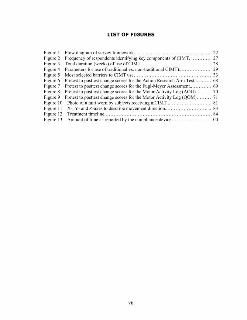
LIST OF FIGURES
Figure 1 Flow diagram of survey framework..………………………………………. 22 Figure 2 Frequency of respondents identifying key components of CIMT. ………… 27 Figure 3 Total duration (weeks) of use of CIMT. …………………………………… 28 Figure 4 Parameters for use of traditional vs. non-traditional CIMT)..……………… 29 Figure 5 Most selected barriers to CIMT use………………………………………… 33 Figure 6 Pretest to posttest change scores for the Action Research Arm Test..……… 68 Figure 7 Pretest to posttest change scores for the Fugl-Meyer Assessment..………… 69 Figure 8 Pretest to posttest change scores for the Motor Activity Log (AOU).……… 70 Figure 9 Pretest to posttest change scores for the Motor Activity Log (QOM)……… 71 Figure 10 Photo of a mitt worn by subjects receiving mCIMT...…………………….. 81 Figure 11 X-, Y- and Z-axes to describe movement direction..…………………….... 83 Figure 12 Treatment timeline.………………………………………………………… 84 Figure 13 Amount of time as reported by the compliance device………………….. 100

viii
ABSTRACT
Recovery of upper extremity (UE) function after stroke is often incomplete. Incorporating evidence-based treatments early in rehabilitation can promote better recovery. One intervention, constraint induced movement therapy (CIMT), has been shown to promote UE recovery after stroke better than usual care. While research has examined CIMT effectiveness, there are gaps in the literature regarding how the therapy is being used and implemented, as well as reviews summarizing the evidence in support of any one CIMT protocol. Also, there is a need to examine the feasibility and effectiveness of CIMT in the acute and subacute stage of stroke recovery in the context of the healthcare system in which it is intended to be delivered. To address these gaps, this research examines the clinical utilization of CIMT, derives the level of evidence in support of its use, and lastly examines, in a preliminary manner, the effectiveness and feasibility of CIMT acutely post-stroke.
While CIMT appears within the literature to be a viable treatment option, little is known about how therapists use it, nor what therapist characteristics predict who would use it. A national survey of therapists working in neurological rehabilitation identified CIMT parameters of treatment and barriers to use (including therapist lack of knowledge and a lack of institutional resources). Methods to overcome barriers to CIMT use are addressed in order to increase its clinical application.
A systematic review of the mCIMT literature, one of the most researched
protocols that follows a distributed practice schedule, showed an intermediate level of evidence in support of its use. Specifically, mCIMT appears to be effective at improving UE function, reducing impairment and increasing activity. While these treatment effects were observed across all stages of recovery, most of the literature is based on chronic stroke populations. Summarizing a body of literature related to a treatment is important for clinicians as it helps evaluate the evidence in support of the therapy, aiding with treatment decisions.
Lastly, preliminary findings of the clinical trial (based on a case study) support the effectiveness of mCIMT to improve UE function acutely post-stroke. Post-treatment, the subject receiving mCIMT demonstrated clinically significant improvements in UE function and activity, and maintained these changes at the 6-month follow-up. While the results may be promising, a number of challenges (for patients and therapists) to mCIMT implementation are discussed along with possible mechanisms to overcome them.
Identifying barriers to mCIMT use is a first step to developing administrative-, education-, and intervention-based solutions to improve clinical utilization. Solutions may be to alter the personnel delivering treatment, providing resource materials to inform clinical practice, and to investigate the minimum required components of mCIMT. If shown that mCIMT is effective and feasible to use in Canada, we can increase its use, in-turn improving the recovery of patients who have had a stroke.

ix
LIST OF ABBREVIATIONS USED
ADL Activity of daily living
ARAT Action Research Arm test
AACPDM American Academy of Cerebral Palsy Developmental Medicine
AOU Amount of use
CAOT Canadian Association of Occupational Therapy
CI Confidence interval
CPA Canadian Physiotherapy Association
CIMT Constraint Induced Movement Therapy
FM Fugl-Meyer Assessment
fMRI Functional Magnetic Resonance Imaging
ICF International Classification of Functioning, Disability and Health
mCIMT Modified Constraint Induced Movement Therapy
LOE Level of evidence
MAL Motor Activity Log
M1 Primary motor cortex
OT Occupational therapy
PT Physical therapy
QOM Quality of movement
RCT Randomised control trial
ROM Range of motion
RTP Repetitive task practice
UE Upper extremity

x
US United States of America
VAS Visual Analogue Scale
WMFT Wolf Motor Function Test
xi
GLOSSARY
Activity: individual performance of an action or task
Behavioural training: one of the main components of mCIMT that may include
the use of short- and long-term goals, identifying and
modifying tasks appropriately to meet those goals, applying
shaping principles to the tasks so patients can be successful
in their performance, problem solving sessions to address
challenges to affected limb use in ADLs.
Body function: body systems’ physiological and psychological function
Body structure: anatomical body part (and components)
Disability: an umbrella term applied to describe the negative
interaction between a person’s health condition as well as
environmental and personal factors. It includes
impairments, activity limitations and participation
restrictions.
Effectiveness: ability of a treatment to lead to recovery (as assessed
through particular outcome measures)
Feasibility: the degree to which a treatment can be implemented
realistically in a particular environment.
Functioning: an umbrella term applied to describe the positive or neutral
interaction between a person’s health condition as well as
environmental and personal factors. It includes body
function, body structures, activities and participation.
xii
Impairment: loss or significant deviation in body function or structure
Learned nonuse: learning phenomenon involving conditional suppression of
movement and consequently, nonuse of a particular limb
Participation: involvement in all areas of life, of a person as a member of
society
Repetitive task practice: one of the main components of CIMT; It involves the
repetition of meaningful functional activities.
Shaping: behavioural technique applied to repetitive task practice;
shaping refers to approaching a task using small,
measurable progressions (which typically increase in
difficulty level) in order to eventually complete/achieve the
task as a whole. During shaping, subjects receive positive
feedback related to their task performance.

xiii
ACKNOWLEDGEMENTS
There are a number of people I would like to thank who helped me complete my
graduate studies. Firstly, I would like to thank the greatest supervisor in the world, Dr.
Shaun Boe, for dealing with my own special brand of crazy. He went above and beyond
expectations to support me through my Master’s work and I would not have been
successful without him. Thank you to my committee members, Dr. Gail Eskes and Dr.
Marilyn MacKay-Lyons for their input and guidance. Additionally, a special thanks is
extended to the members of the Laboratory for Brain Recovery and Function, in
particular Marion Che, Janet Green and Megan Freeman, for their assistance with various
parts of my thesis projects. I would also like to thank my family and friends for ensuring
my sanity over the past two years.
Included herein is a study published in Physiotherapy Canada, reprinted with
permission from University of Toronto Press (www.utpjournals.com).

1
CHAPTER 1 INTRODUCTION
Stroke is the leading cause of neurologically related death and disability in North
America, affecting approximately 50,000 Canadian adults every year.1 This figure is
likely to increase due to the aging population and the escalating percentage of individuals
with risk factors for stroke.2 Unfortunately, full recovery after stroke is elusive; of those
who survive their stroke, approximately two-thirds will experience residual neurological
deficits that impair daily function.3 Specifically, recovery of upper extremity (UE)
function is a major challenge for survivors of stroke, with only 5% regaining full
function.3, 4 As healthcare delivery improves to more effectively treat stroke acutely,
more Canadians are living with its long-term effects. Functional limitations resulting
from UE deficits negatively impact on performance of daily activities, resulting in
decreased levels of physical activity and participation, and increased risk of further health
problems.5-7
People with post-stroke impairments most often receive rehabilitation therapy to
address any loss of function. For instance, an occupational or physical therapist may
work with a patient to improve balance and UE function, address deficits in strength, and
engage in sensory and gait re-training. Specifically, treatments used to address UE
functional deficits as part of usual clinical care include a task-oriented approach to
functional activities (whereby the patient practices particular motor skills), active and
passive range of motion (ROM) exercises, stretching and strength training.8 Treatments
that are considered part of usual care help with UE recovery after a stroke, but they have
limited effectiveness. The limited effectiveness of the current, usual care practice is
evident in the high rates of disability, prevalence of secondary disease, and incomplete
2
functional recovery of the UE observed after stroke.9, 10 To improve UE function after
stroke and reduce the long-term disability related to poor functional recovery, therapeutic
interventions that are both evidence-based and clinically feasible are needed. Increased
use of evidence-based therapies in rehabilitation can improve functional abilities post-
stroke, decreasing health care costs and greatly improving overall quality of life.
Constraint induced movement therapy (CIMT) is a treatment that has been shown
to facilitate UE functional recovery post-stroke, with numerous studies showing it to be
better than usual care.11, 12 CIMT combines (1) repetitive task practice (RTP) in
conjunction with shaping, during which patients engage in meaningful functional
activities with measurable progressions for which they receive positive feedback as the
activities become increasingly more difficult; (2) behavioural training to keep patients
motivated and engaged throughout the therapy; and (3) restraint of the unaffected UE in
order to promote use of affected UE.13-15 A number of different CIMT protocols have
been developed and applied in post-stroke rehabilitation, including a ‘traditional’ or
massed practice approach and a modified or distributed practice approach.11, 12, 16, 17
Regardless of the protocol, all CIMT interventions include the 3 components listed
above.
Although much research has been performed examining the effectiveness of
CIMT as an intervention to promote UE functional recovery post-stroke, there are gaps in
the literature that relate to how the therapy is being implemented and used, and the
potential barriers to its use. With regard to implementation and use, there is a lack of
literature summarizing the evidence in support of any one CIMT protocol. Lastly, there is
a need to examine the feasibility and effectiveness of CIMT in the acute stage of stroke
3
recovery in the context of the healthcare system in which it is intended to be delivered.
Thus the focus of this thesis is to take the first steps towards addressing these gaps,
including examining the utilization of CIMT in clinical practice, summarizing the
evidence in support of one form of CIMT, and lastly examining, in a preliminary manner
via a case study, the effectiveness and feasibility of CIMT acutely post-stroke.
4
CHAPTER 2 BACKGROUND
Derived from basic studies in animals, CIMT is based on principles of
behavioural psychology. In the initial studies that led to the development of CIMT, Taub
and colleagues were interested in studying the role of sensory feedback in movement and
motor learning. In these studies, the UEs of a group of monkeys were deafferented in
order to abolish the sensory pathways while preserving the motor pathways, thereby
disrupting sensorimotor integration.18, 19 Consequently, the animals exhibited poor UE
function; when the monkeys attempted to use the deafferented limb, they encountered
aversive effects (such as movement inaccuracy) and, as a result, stopped using the limb.
This pattern of behaviour was deemed “learned nonuse.”18, 19 In subsequent studies, Taub
et al. explored the behavioural consequences of UE motor impairment and found that
over time, learned nonuse could be overcome by restraining the monkeys’ non-affected
limbs.13, 18 Thus, by forcing the use of their deafferented (affected) UE, the monkeys
engaged their deafferented limb in daily activities (eg. grooming, feeding, etc.). Over
time, increased use of the deafferented or affected limb ultimately led to improved limb
function.13, 18
An additional behavioural technique used to overcome learned nonuse and
induce use of the affected UE is training and practice. Thus, while restraint of the
unaffected limb helps to overcome learned nonuse, the characteristics of the tasks utilized
were also determined to be important elements. Nudo et al. demonstrated that along with
functional UE improvements, animals repetitively engaging in novel motor tasks after
experiencing an ischemic infarct experienced plastic changes in the brain characterized
by a task-dependent reorganization of primary motor cortex.20 Repetitive training

5
produced shifts in the movement representation in primary motor cortex (M1), that
related to the acquisition of new motor skills. Motor skill acquisition was indicated by the
development of new movement patterns and increased efficiency of the tasks. Cortical
representations (for the affected UE) reorganized to reflect this skill acquisition.21, 22
Notably, the neurophysiological and functional changes were observed when animals
engage in novel, rather than simple repetitive tasks. Related studies examining simple,
repetitive movements, showed no task-related changes in cortical representation of M1
after training, compared to pre-training.22 In comparing these two groups (task oriented
training vs. simple repetition), it appears that “repetitive motor activity alone does not
produce functional reorganization of cortical maps… Motor skill acquisition, or motor
learning is a prerequisite factor in driving representational plasticity in M1.”22
Researchers examining CIMT have applied this evidence to combat learned nonuse, and
have employed conditioned-response techniques that allow animals to learn and practice
different movements.13, 18 While some improvements in UE function were observed,
these movements were not generalized to natural settings, prompting the application and
use of an additional behavioural training technique.18
The third behavioural technique investigated by Taub et al. to overcome
learned nonuse is termed ‘shaping’. Shaping is based on operant conditioning principles
and approaches a particular behavioural goal in small, incremental steps to promote
success.13, 23 When shaping principles were applied to UE task practice, animals learned
to use their UE not only in training situations but also in their normal environment as
well.24, 25
6
Learned nonuse of a limb was upheld as a learning phenomenon involving the
conditioned suppression of movement.13 Since the learned nonuse mechanism is
essentially behavioural, Taub et al. reasoned that it should be independent of the source
of injury and applicable to other conditions, so long as the appropriate reinforcements
exist early after injury. This led to investigation into the appearance of learned nonuse in
humans, and research into overcoming the phenomenon.13, 19, 26 Learned nonuse can be
distinguished from poor recovery in humans by comparing a person’s functional ability
with their observed UE activity levels. If a person is able to perform a particular
movement using their affected UE when directed, but chooses not to use that limb in
practice during everyday activities, then learned nonuse is present.
Given the results observed in this series of animal-based studies, coupled with the
observation of learned nonuse in individuals with UE functional deficits, Taub and
colleagues applied the principles observed in the animal studies to a human population.
The principles were applied primarily towards people with stroke-related UE functional
deficits. Initially, studies investigated UE interventions involving restraint27, 28 and task
training29 separately. In 1993, Taub et al. combined restraint and task training30;
however, the goal of this particular study was to over train the affected UE so the tasks
were given without any explicit training program (thus excluding any shaping principles).
While subjects who received the restraint and training program experienced greater
functional gains compared to a control group (receiving therapy to focus attention on the
affected UE), Taub looked to maximize these improvements further. In animals, shaping
and restraint had independently been used to overcome learned nonuse and improve
motor function in a deafferented UE. Since restraint and task practice (without explicit

7
training) had been combined successfully, it was reasoned that adding shaping techniques
could provide complementary, if not compounded, results.13 Thus, in addition to wearing
a restraint on their unaffected UE, Taub and colleagues had subjects engage in RTP
(including shaping) using their affected UE.12 Shaping was established as a key principle
that contributed to the functional gains observed in these human studies.13, 15 Together,
restraint, RTP and behavioural techniques (including shaping) comprise CIMT.
The original protocol for CIMT was performed over a 2-week period and
combined restraint of the unaffected limb for 90% of waking hours with functionally
oriented RTP using the affected UE.11, 12 The task practice component (which applies
shaping principles to task training) was performed 6 hours each weekday over the 2-week
treatment period. Additionally, the therapy incorporated behavioural training techniques
to help patients transfer any functional gains into real world activities.11, 31, 32 Behavioural
training includes the application of shaping principles, as well as the use of a behavioural
contract to promote therapy compliance, goal-setting setting, and problem-solving
sessions to address challenges subjects may face in their ADLs. Many different studies
have examined the original CIMT protocol and support its effectiveness to improve UE
functional outcomes post-stroke. A large randomized controlled trial (RCT), the
EXCITE trial, followed 222 subjects 3-9 months post-stroke. Subjects who engaged in
CIMT demonstrated statistically and clinically significant improvements in UE function
and use compared to the control group of usual care.11 The control group received ‘usual
and customary care’, ranging from no treatment to regular therapy [including the
applications of orthotics, occupational (OT) and physical (PT) therapy home care, day
treatments or hospital outpatient programs].33 Specifically, subjects in the CIMT group

8
demonstrated greater improvements compared to the control group pre-post on the Wolf
Motor Function Test (WMFT), a laboratory based measure of UE motor ability
(function). Importantly, subjects in the CIMT group also had greater gains in the amount
of use of the affected UE compared to the control group, as measured using the Motor
Activity Log (MAL), a measure of activity and subjective scale of real world limb use.11
In addition to observing functional improvements in the affected UE, studies have
demonstrated parallel neuroanatomical and neurophysiological changes in the brain post-
CIMT treatment. Liepert et al. (2000), using transcranial magnetic stimulation, mapped
the ‘hand area’ of primary motor cortex in subjects with chronic stroke before and after a
12-day CIMT intervention. Results of the study demonstrated that after CIMT, the
representation of the hand area in primary motor cortex significantly increased in size.34
Furthermore, cortical representation for the hand shifted to neighbouring regions in motor
cortex, indicating a functional alteration in the somatotopic organization of motor cortex.
The cortical changes paralleled increases in UE activity (tested using the MAL) observed
after the CIMT intervention. This study provided evidence that CIMT can induce plastic
anatomical and functional brain changes corresponding to improved UE movements.
Since Taub’s initial work, a number of different CIMT protocols have been
developed and applied in post-stroke rehabilitation that differ from the ‘traditional’ or
massed practice approach including a modified or distributed practice approach.11, 12, 16, 17
Other protocols have also been described that are variations of these two approaches (for
example see 35-37). The most studied of these distributed practice approaches is termed
‘modified Constraint Induced Movement Therapy’ (mCIMT) whereby therapy sessions
are performed for 30 minutes, 3 times per week over a 10-week schedule.17, 38, 39 Despite

9
differences in the frequency and overall duration of treatment, similar principles are
utilized across the spectrum of these CIMT protocols, including mCIMT. The original
CIMT approach as developed by Taub offers a compressed, but intense treatment. While
the original protocol has been demonstrated to be effective, anecdotal evidence suggests
that it can be difficult to implement clinically due to its high resource demand (eg. time,
availability of therapists, and equipment). Contrary to CIMT, mCIMT distributes therapy
sessions over a longer duration with a considerable reduction in treatment time and thus a
decreased use of resources. As highlighted above, treatment in mCIMT is performed
over 10 weeks and the UE is restrained for 5 hours/weekday (rather than 90% of waking
hours under the original protocol).17
Despite the use of a distributed practice schedule and thus reduced intensity of the
overall treatment, some evidence suggests that mCIMT is better than usual care and as
efficacious as CIMT in promoting UE functional recovery post-stroke.16, 40-42 Page et al.
(2008) demonstrated that mCIMT was superior not only to a control group receiving no
therapy but also to a dose-matched program of usual care in improving UE function in
chronic stroke.17 The usual care group received a time-matched rehabilitation program
consisting mostly of proprioceptive neuromuscular facilitation (PNF) techniques
emphasizing functional tasks, as well as stretching and compensatory techniques when
needed. The duration, frequency and content of usual care sessions were consistent with
typical therapy provided to patients at the same stage of recovery.17 There were
significant improvements for the mCIMT group in both functional UE performance and
amount of use, that mirrored changes observed with the original CIMT protocol.
Specifically, subjects in the mCIMT group had greater improvements on the Action

10
Research Arm test (ARAT), an objective measure of UE function, and on the MAL.17 As
with the traditional CIMT protocol, neuroanatomical and physiological changes have
been demonstrated to underlie the clinical treatment effect observed with mCIMT.
Szaflarski et al. (2006) used functional magnetic resonance imaging (fMRI) to
demonstrate use-dependent cortical re-organization that occurred with mCIMT in
subjects with chronic stroke.40 Subjects who exhibited increases in affected UE function
and use correspondingly demonstrated shifts in their brain activation patterns to the
subcortical and cortical structures in the affected hemispheres. The neuroimaging results
were positively correlated with the observed functional improvements in the affected UE.
Almost exclusively, mCIMT studies have applied the treatment to a sub-acute and
chronic patient population. However, there is considerable evidence suggesting that
initiating rehabilitation in the acute stage of recovery post-stroke is associated with
greater functional outcomes.43, 44 A large, multi-centre prospective study of stroke
recovery in the United States (US) demonstrated that introducing rehabilitation early,
including engaging patients in higher level activities such as UE functional tasks, was
related to greater functional improvements and shorter length of stay in in-patient
rehabilitation.45 Page et al. performed one of the few studies examining the use of
mCIMT in the acute stage of stroke recovery. The study demonstrated that mCIMT
administered acutely (<14 days) post-stroke was associated with significantly greater
improvements in UE function (observed with changes in ARAT scores) compared to a
control group receiving dose-matched traditional UE therapy.16 Given the potential
benefits of early intervention, a more in-depth investigation of acute mCIMT outcomes is
warranted to confirm these findings in a larger and more diverse sample population.

11
Testing the effectiveness of mCIMT when initiated in the acute stage of stroke recovery
can take advantage of the body’s heightened responsiveness to treatment and further
establish evidence in support of mCIMT.
To date, most of the evidence supporting the effectiveness of CIMT has been
generated in the US, and as such it is not known how the treatment will translate into the
publically (vs. privately in the US) Canadian healthcare system. Given limited resources
within the publically funded Canadian healthcare system, implementing the original
CIMT protocol may prove challenging.9, 10 mCIMT may be a more appropriate
alternative to use clinically because the distributed practice schedule aligns better with
the amount of therapy time allocated per patient; typically a patient receives one hour
each of OT and PT per weekday with additional interdisciplinary therapies (eg, speech
language pathology) as indicated.8 Since mCIMT has not been researched in Canada, it is
not known if the treatment is being used clinically or if there are barriers associated with
the treatment that prevent it from being implemented clinically. Given the lack of
research about CIMT use in Canada, it is not understood to what degree CIMT is being
implemented in Canada and if so, how often it is being used and in what form (eg. a
traditional vs. modified protocol). Furthermore, it is not known if mCIMT is an effective
and feasible treatment when implemented in an acute post-stroke patient population
within the Canadian healthcare system. Thus, the purpose of my thesis is to investigate
these research questions through a national survey of therapists, a systematic review of
mCIMT, and preliminary findings of a clinical trial investigating mCIMT in an acute and
sub-acute stroke rehabilitation setting. If mCIMT is shown to be effective and feasible to
use in this patient population in Canada, we can increase its use and improve the recovery

12
of patients who have had a stroke. The overall personal, social and economic burden of
stroke on Canadians can be minimized with improved rehabilitation and corresponding
level of functional UE recovery.
13
CHAPTER 3 RESEARCH OBJECTIVES AND HYPOTHESES
Given the lack of studies examining mCIMT in Canadian healthcare, coupled with a
lack of knowledge about CIMT utilization in rehabilitation, the focus of this research
includes: 1) identifying utilization patterns and barriers related to CIMT use in Canadian
neurological rehabilitation; 2) establishing the level of evidence (LOE) in support of
mCIMT to promote UE recovery post-stroke; and 3) examining the effectiveness and
feasibility of mCIMT to improve UE function of patients with stroke.
3.1 STUDY 1: CIMT SURVEY
Despite evidence of CIMT effectiveness, questions abound regarding its clinical
feasibility.46-48 Prior articles have highlighted that despite CIMT being recommended as
a treatment for UE hemi-paresis in national stroke care guidelines, it is not being
implemented as standard practice for stroke care when appropriate.49, 50 Specifically,
studies have identified several barriers to the implementation of CIMT, including
resource intensity and therapist-/patient-related factors.
Given the lack of studies examining CIMT utilization, empirical knowledge is
needed regarding clinicians’ perceptions, actual application, and perceived barriers to its
implementation. This knowledge would inform research regarding the clinical feasibility
of CIMT and continuing education initiatives to facilitate its translation into clinical
practice. Thus, the purpose of this component of my thesis work is to explore utilization
patterns of CIMT amongst occupational and physical therapists practicing in adult
neurological rehabilitation in terms of frequency of use, parameters of treatment and

14
barriers to use. Additionally, respondent characteristics are examined to identify factors
related to CIMT utilization. Hypotheses related to study 1 include:
1. Despite evidence related to its effectiveness, CIMT is not routinely being used as
a primary treatment for UE hemi-paresis by occupational and physical therapists
practising in adult neurological rehabilitation
2. When CIMT is being employed clinically, it is not being delivered as outlined in
the literature in terms of time of delivery and the key components of treatment.
3.2 STUDY 2: SYSTEMATIC REVIEW
The second component of this thesis evaluates the evidence in support of a
specific CIMT protocol. Specifically, the goal is to establish the LOE for a modified
version of the therapy (mCIMT). Performance of a systematic review such as this one is
important as they summarize and evaluate the literature. Summarizing a body of
literature related to a treatment is important for clinicians as it helps them to evaluate the
evidence in support of the therapy, aiding with treatment decisions.51 Thus, the primary
objective of the review is to investigate the LOE in support of mCIMT to promote UE
recovery post-stroke. The data results generated by outcome measures that assess pre- to
post-treatment changes in UE function, impairment and real-world use will be used to
evaluate treatment effectiveness.
3.3 STUDY 3: MCIMT TRIAL
The last component of this thesis investigates, in a preliminary manner, the
effectiveness and feasibility of a mCIMT protocol in an acute stroke population in

15
Canada. While much work has supported the effectiveness of various CIMT protocols in
American healthcare settings, limited work has been done in Canada. Executing a clinical
trial examining mCIMT will demonstrate if it is still effective when applied in a
publically funded healthcare system given the constraints on resource availability and the
capacity to deliver the treatment in its evidence-based form. Additionally, the trial
examines if the therapy is feasible to implement into usual Canadian clinical practice.
Specifically, the objectives of my thesis work within the trial focus on the development
and execution of the mCIMT intervention. The purpose of my research work will be to
explore initial findings based on a single case study. To this end I will identify and
discuss key features and challenges of task development and mCIMT implementation by
means of the case study. Additionally, I will investigate the preliminary results to
identify potential trends relating to mCIMT’s effectiveness and feasibility as a treatment
option in Canada. The hypothesis for Study 3:
1. Subjects engaging in mCIMT therapy will demonstrate greater UE functional
recovery compared to those in the dose-matched control group. These
improvements will be observed in terms of UE function (Action Research Arm
test), amount of use (accelerometry and Motor Activity Log) and quality of use
(Motor Activity Log)
3.4 SUMMARY TO CHAPTER 3 AND TRANSITION TO CHAPTER 4
An important first step to evaluating mCIMT and to understanding its potential
impact was to establish if it is being used in Canadian rehabilitation and if so, how it is
being used and what the potential barriers to use are. To address this first step, the first
16
project was a national survey of therapists investigating current practices as they relate to
CIMT use in Canada. This work, presented in the following chapter (4), has been
accepted for publication and is currently ‘in-press’ (Appendix A). The manuscript
presented in Chapter 4 has not gone through the entire editorial process at the time of this
thesis completion. Thus, the official version of the article will be published in
Physiotherapy Canada 2014; 66(1).

17
CHAPTER 4 STUDY 1: CIMT SURVEY
4.1 INTRODUCTION
Stroke is the leading cause of neurology-related disability and death in North
America, affecting approximately 50,000 Canadian and 795,000 American adults each
year.1, 52 Recovery of upper-extremity function is a major problem for survivors of stroke,
with only 5% regaining full function.3
CIMT is one intervention that has been shown to facilitate UE functional recovery
in a particular subset of patients after stroke. Derived from basic studies in animals,
CIMT combines RTP with shaping,13 during which participants engage in meaningful
functional activities with measurable progressions for which they receive positive
feedback as the activities become increasingly more difficult; behavioural training (e.g.,
behavioural contract, problem solving to address barriers to affected limb use); and
restraint of the unaffected UE.13, 53 Several CIMT protocols have been developed,
including a “traditional” or massed practice approach, a “non-traditional” or distributed
practice approach,11, 13, 17, 41, 54 and variations on these two approaches.35, 37 Traditional
CIMT involves 6 hours/day of RTP combined with restraint of the unaffected UE for
90% of waking hours over 10 consecutive weekdays (i.e., therapy sessions do not occur
on weekends).11, 13 Conversely, modified CIMT, a non-traditional protocol, involves RTP
for 30 minutes/day, 3 times/week, combined with 5 hours/day of unaffected UE restraint,
over a 10-week period.17, 41 Regardless of the specific protocol used, the client is
generally required to have a degree of movement in the affected UE that, at a minimum,
includes 10° of active wrist extension with 10° extension of the thumb and at least two

18
fingers.11, 17 The need for this level of function and the corresponding capacity for active
engagement in the treatment limits the number of people for whom CIMT is an
appropriate intervention.55
Despite evidence of CIMT’s effectiveness for people who meet the criteria for
treatment, 11, 17, 56-58 questions abound regarding its clinical feasibility.46-48 Prior articles
have highlighted the fact that, even though CIMT is recommended for treating UE hemi-
paresis in national stroke care guidelines,8 it is not being implemented as standard
practice for stroke care.49, 50 The authors identify several barriers to the implementation of
CIMT, including resource intensity and therapist- or patient-related factors. In a study
examining therapists’ opinions of CIMT, Page and colleagues reported that 74% of
occupational and physical therapist respondents (n = 85) believed that their institutions
lacked the resources necessary to provide traditional CIMT.46 Our research team’s
observations suggest that CIMT is not routinely used in clinical practice and that when it
is used, not all CIMT components are implemented.
Given the lack of studies examining use of CIMT, empirical knowledge is needed
on clinicians’ perceptions, actual application, and perceived barriers to implementation.
This knowledge would inform research into the clinical feasibility of CIMT and
educational initiatives to facilitate its translation into clinical practice. The purpose of this
study, therefore, was to explore usage patterns of CIMT among occupational and physical
therapists practising in adult neurological rehabilitation in terms of frequency of use,
parameters of treatment and barriers to use. We also examined respondent characteristics
to identify factors related to CIMT use.

19
4.2 METHODOLOGY
Our study employed a non-experimental, quantitative research design using an
online survey (Opinio version 6.5.1, ObjectPlanet Inc., Oslo, Norway). The study
received approval from the Capital District Health Authority Research Ethics Board.
4.2.1 Participants and Survey Distribution
A total of 588 occupational therapists and 1968 physical therapists who are
licensed to practise in Canada and who practise in adult neurological rehabilitation were
invited to participate. Neurological practice was defined as engagement in treating
people with stroke, traumatic or acquired brain injury, cerebral palsy, multiple sclerosis
or dystonia. All participants were members of the Canadian Association of Occupational
Therapists (CAOT) or the Canadian Physiotherapy Association (CPA).
We recruited occupational therapists directly via emails with a link to the online
survey, using a list of email addresses, purchased from CAOT, for occupational therapists
who self-identified as being involved in neurological practice and who had agreed to be
contacted for research. Physical therapists were recruited through a national e-mail
newsletter distributed by CPA to all its members (approximately 10,600), which included
a brief description of the study and a link to the online survey, and invited participation
from physical therapists who self-identified as practising in neurological rehabilitation.
At the time of survey distribution, 1968 CPA members were actively involved in this area
of practice. Follow-up reminders were sent to both occupational and physical therapists at
2 and 3 weeks after the initial invitation in the manner outlined above, and participants
had 3 months to complete the survey. Respondents provided informed consent by
completing and returning the survey.

20
4.2.2 Survey Development and Composition
Questions were related to two broad categories: respondent profile and CIMT
usage pattern. Survey content complied with three criteria: (1) questions were relevant to
the study’s purpose; (2) the wording was not leading (i.e., did not provide the “correct”
response for subsequent questions); and (3) completion time was <15 minutes.
Five “content experts” (3 occupational and 2 physical therapists) involved in
neurological rehabilitation services in Canada, including CIMT, independently assessed
the content and face validity of the penultimate draft of the survey. Their feedback was
used to refine the final survey items.
The final version of the survey contained 48 questions. The majority were “close-
ended”, with a list of response choices; seven included “other, please specify” to allow
for a written answer. Three questions relating to the respondents knowledge of CIMT,
experience with CIMT, and perceived effectiveness of CIMT, used a 5-point Likert-type
scale. For example, level of CIMT knowledge was coded as follows: 1 = not very
knowledgeable, 2 = minimally knowledgeable, 3 = moderately knowledgeable, 4 =
knowledgeable, and 5 = very knowledgeable). Ten questions required a typed response
(e.g., “Based on your knowledge of CIMT, please list what the key components of CIMT
are:”). For analysis of this question in particular, two researchers independently reviewed
the responses and grouped them according to the themes that emerged (e.g., inclusion
criteria, treatment duration and schedule, type of treatment). A third researcher resolved
any discrepancies. The frequencies of responses per theme were tallied. Three categories
described the components of CIMT: (1) restraint, (2) RTP, and (3) behaviour/shaping; a
fourth category, “identified no components”, was used for blank responses and those that
21
did not meet criteria for the three key component categories. Respondents also identified
their practice location as “rural” or “urban”, and population size of practice location was
determined from the first three digits of the postal code corresponding to the practice
location. Finally, a free-response question at the end of the survey invited participants to
comment on their clinical use of CIMT.
Depending on the responses to questions about CIMT usage patterns (e.g., “Are
you aware of CIMT as a treatment option for upper limb hemi-paresis?”), participants
branched into different arms of the survey (see Figure 1). This funnelling pattern
screened participants so that responses to certain survey questions came from those who
practiced in neurological rehabilitation and who had used CIMT clinically in the past two
years. Participants who used CIMT were asked for characteristics of the protocol they
employ. Responses from those participants who report not using CIMT were also
collected in order to identify barriers to CIMT use in this group. The survey was
structured in such a way that participants responded to a maximum of 37 questions.
Further description of the survey is found in Appendix B.

22
Figure 1 Flow diagram of survey framework designed to identify participants who met
the inclusion criteria and had used CIMT clinically in the past two years and to further classify respondents according to their use of CIMT.

23
4.2.3 Data Analysis
Responses were treated as categorical variables. Because of the funnelling nature
of the survey, the number of respondents was different for each question; we therefore
report adjusted relative frequencies (%) and number of respondents who answered the
question (n) throughout. For some questions, the adjusted relative frequencies do not sum
to 100% because respondents could choose multiple responses. Throughout, we have
grouped occupational and physical therapist responses for analysis. The rationale
underlying this grouped approach is that, first, essential competencies for both
professions include an expectation that therapists practice in an evidence-informed
manner, including incorporating relevant and current knowledge into their practice;59, 60
and, second, that although differences exist between professions related to specific areas
of practice, the assessment and treatment of UE dysfunction post-stroke is a shared area
of practice.61-63 The grouped approach is appropriate because it is reasonable to think that
both occupational and physical therapist respondents have the potential to know about
CIMT and the ability to use it in their practice.
To investigate practice setting size and CIMT use, we matched postal-code data to
the corresponding geographic region using householder counts and map information
available from Canada Post (http://www.postescanada.ca/cpc2/addrm/hh/default-e.asp);
populations for these regions was then obtained from 2011 Statistics Canada data.64
To investigate therapist-related factors and CIMT use, we applied a binary logistic
regression using a forward stepwise (Wald) model (SPSS version 19, IBM Canada Ltd.,
Markham, ON). Number of years in practice, practice location, primary practice setting,
and level of CIMT knowledge were chosen as predictor variables. The threshold for
24
statistical significance was set at p < 0.05. To examine variables that did not prove to be
predictors of CIMT use through regression analysis, we cross-tabulated each variable
against CIMT use. Using row percentages of the categories within each variable, we
calculated the odds ratios and corresponding 95% CIs65 of CIMT use between groups.
For primary practice setting, we compared the odds of using CIMT for each setting
category to the odds of using CIMT in an in-patient acute general setting. We defined the
stages of stroke rehabilitation as follows: acute, 0-3 weeks; sub-acute, 3 weeks to 3
months; and chronic, >3 months post-stroke.
4.3 RESULTS
4.3.1 Participants
Our total response rate was 13.2% (338 responses out of a possible 2556). Of the
338 respondents, 39.9% (135/588, response rate of 23%) practiced as occupational
therapists and the remaining 60.1% (229/1968, response rate of 10.3%) practiced as
physical therapists. The therapists were 89.5% (229) women and represented all
provinces and territories except the Northwest Territories and Nunavut; the majority
(51.9%) practised in Ontario. Respondent characteristics are described in Table 1. Nearly
two-thirds (65.9%, 208) reported a bachelor’s degree as their highest level of education.
While respondents worked in both in- and outpatient settings, the greatest number
(22.1%, 208) reported working primarily in general out-patient rehabilitation. Of all
neurological diagnoses, stroke was the most commonly treated (88.6%, 236). A majority
of respondents (75%, 136) practiced in areas with a population greater than 55,000
people.

25
Table 1 Distribution of respondent characteristics relating to therapist- and therapy-specific factors.
Absolute Frequency Adjusted Relative
Frequency (%)
Years Practicing (n = 225)
0 – 5 25 11.1
6 – 10 31 13.8
11 – 15 27 12.0
16 – 20 45 20.0
21 – 25+ 97 43.1
Practice Location (n = 213)
Urban 170 79.8
Rural 43 20.2
Primary Practice Setting (n = 208)
In-patient acute general 23 11.1
In-patient rehabilitation general 30 14.4
Out-patient rehabilitation general
46 22.1
Private practice 29 13.9
In-patient stroke unit (acute/rehabilitation)
30 14.4
Other* 50 24.0
Treatment Approach/Intervention for Upper Limb Hemi-paresis (n = 204)
Stretching 181 88.7
Strengthening 175 85.8
Motor learning/repetitive task practice
166 81.4
Modalities† 158 77.5
Imagery/Mirror therapy 150 73.5

26
Absolute Frequency
Adjusted Relative
Frequency (%)
NDT/Bobath 141 69.1
Sensory Re-training 82 40.2
CIMT 78 38.2
Bilateral movement therapy 65 31.9
PNF 61 29.9
Other 15 7.4
Level of CIMT Knowledge (n = 185)
1 – Not knowledgeable 10 5.4
2 – Minimally knowledgeable 38 20.5
3 – Moderately knowledgeable 72 38.9
4 – Knowledgeable 48 25.9
5 – Very knowledgeable 17 9.2
Level of CIMT Experience (n = 78)
1 – Little experience 9 11.5
2 – Minimally experienced 22 28.2
3 – Moderately experienced 28 35.9
4 – Experienced 13 16.7
5 – Very experienced 6 7.7
* Includes skilled nursing/restorative care facility, community setting, home care and other. †Includes biofeedback, neuromuscular electrical stimulation, virtual reality, robotics and serial casting NDT = Neurodevelopmental Treatment; CIMT = Constraint-Induced Movement Therapy; PNF = Proprioceptive Neuromuscular Facilitation
4.3.2 CIMT Usage
Some 92% of respondents (202) were aware of CIMT as a treatment option for
UE hemi-paresis; however, only 42.9% (182) said they had used CIMT in the last two

27
years, and only 19.4% (72) used CIMT as a primary treatment (when indicated) for UE
hemi-paresis. CIMT was most commonly used in the chronic (74.0%, 77) and sub-acute
(59.7%) stages of rehabilitation (vs. 7.8% in the acute stage) and was most often used for
people with stroke (89.7%, 78). When asked to rate their level of experience with CIMT
on a 5-point scale (1 = little experience, 5 = very experienced), a majority of therapists
(35.9%, 78) chose 3 (see Table 1).
Figure 2 Frequency of respondents identifying key components of CIMT who do (n = 69) and do not (n =98) use CIMT. ‘Identified no components’ refers to respondents who did not identify any of the key components of CIMT. RTP = Repetitive Task Practice
When asked to name the key components of CIMT, the majority of therapists
using CIMT (88.4%, 69) did not name all three components (see Figure 2). Overall,
however, this group did identify all three components more frequently than those who
reported not using CIMT (11.6% of users vs. 9.2% of non-users). Surprisingly, 40.6% of
CIMT users (vs. 21.4% of non-users) were unable to identify any of the key components.
Common responses for CIMT components that were not categorized as

28
behaviour/shaping, RTP, or restraint included treatment duration and schedule (12 users,
27 non-users), inclusion criteria (5 users, 24 non-users), and type of treatment (7 users, 14
non-users). A complete list of responses is available in Appendix C.
Figure 3 Total duration (weeks) of traditional (n = 8) vs. non-traditional (n = 61) use of CIMT.

29
Figure 4 Parameters for use of traditional vs. non-traditional CIMT respectively, including: (A) days/week of restraint (n = 8, n = 60); (B) days/week of RTP/shaping (n = 8, n = 60); (C) hours/day of restraint (n = 8, n = 60); (D) hours/day of RTP/shaping (n = 8, n = 58). RTP = Repetitive Task Practice
The majority of CIMT users reported using a non-traditional approach (88.4%,
69) rather than a traditional approach (11.6%, 69) (see Figures 3 and 4). The most
commonly reported approach involved using CIMT for fewer hours per day over a longer
duration than the traditional approach (see Figures 3 and 4B). There was considerable
variability in the parameters reported for the delivery of non-traditional CIMT; for
instance, hours of restraint per day varied from 0 to >7 (see Figure 3C), while days of
RTP/shaping per week ranged from 1 to 7 (see Figure 4B).
When asked about their level of knowledge related to CIMT, 38.9% of
respondents (185) said they were moderately knowledgeable (rating of 3 on a 5-point
30
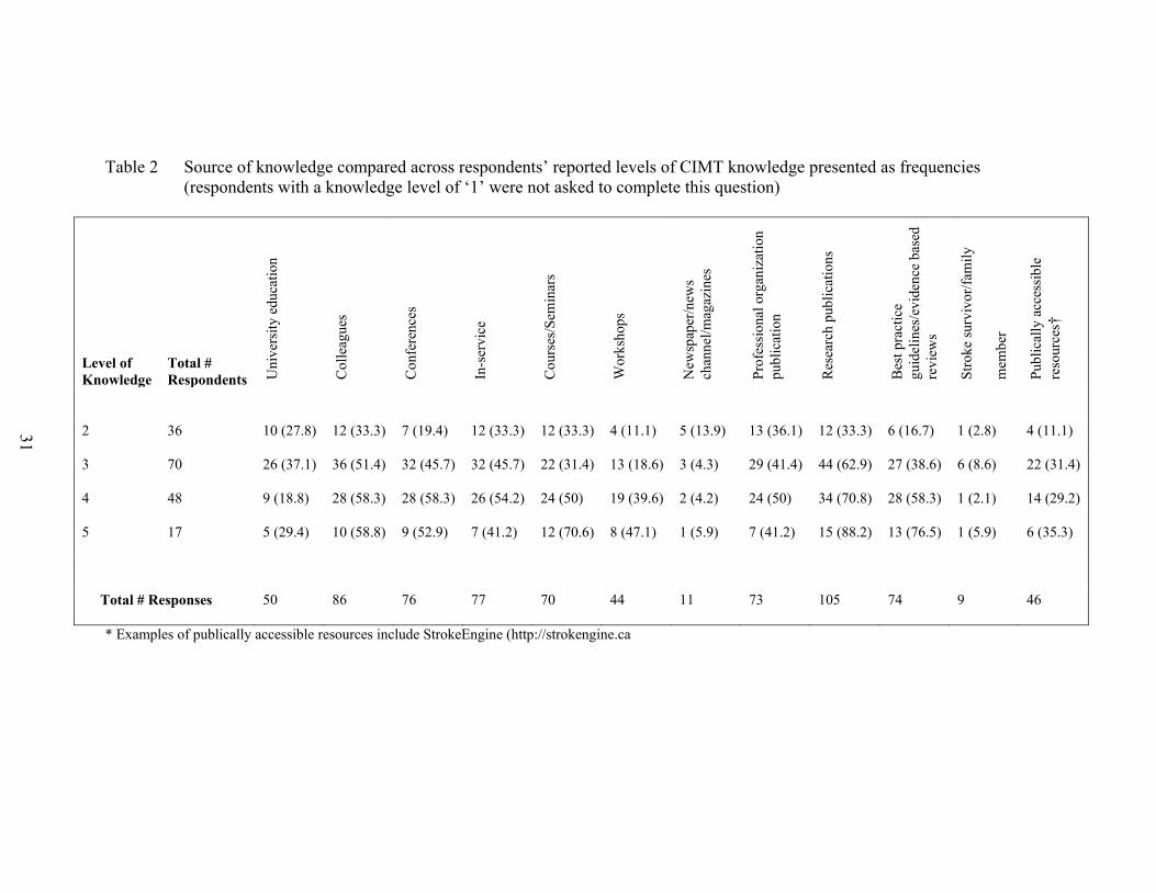
Likert-type scale). More than half (60.7%, 173) reported obtaining their knowledge from
research publications; other sources of knowledge are outlined in Table 2.
31
Table 2 Source of knowledge compared across respondents’ reported levels of CIMT knowledge presented as frequencies (respondents with a knowledge level of ‘1’ were not asked to complete this question)
Uni
vers
ity e
duca
tion
Col
leag
ues
Con
fere
nces
In-s
ervi
ce
Cou
rses
/Sem
inar
s
Wor
ksho
ps
New
spap
er/n
ews
chan
nel/m
agaz
ines
Prof
essi
onal
org
aniz
atio
n pu
blic
atio
n
Res
earc
h pu
blic
atio
ns
Bes
t pra
ctic
e gu
idel
ines
/evi
denc
e ba
sed
revi
ews
Stro
ke su
rviv
or/fa
mily
mem
ber
Publ
ical
ly a
cces
sibl
e re
sour
ces†
Level of Knowledge
Total # Respondents
2 36 10 (27.8) 12 (33.3) 7 (19.4) 12 (33.3) 12 (33.3) 4 (11.1) 5 (13.9) 13 (36.1) 12 (33.3) 6 (16.7) 1 (2.8) 4 (11.1)
3 70 26 (37.1) 36 (51.4) 32 (45.7) 32 (45.7) 22 (31.4) 13 (18.6) 3 (4.3) 29 (41.4) 44 (62.9) 27 (38.6) 6 (8.6) 22 (31.4)
4 48 9 (18.8) 28 (58.3) 28 (58.3) 26 (54.2) 24 (50) 19 (39.6) 2 (4.2) 24 (50) 34 (70.8) 28 (58.3) 1 (2.1) 14 (29.2)
5 17 5 (29.4) 10 (58.8) 9 (52.9) 7 (41.2) 12 (70.6) 8 (47.1) 1 (5.9) 7 (41.2) 15 (88.2) 13 (76.5) 1 (5.9) 6 (35.3)
Total # Responses 50 86 76 77 70 44 11 73 105 74 9 46
* Examples of publically accessible resources include StrokeEngine (http://strokengine.ca
31
32
4.3.3 Therapist-Related Factors and CIMT Use
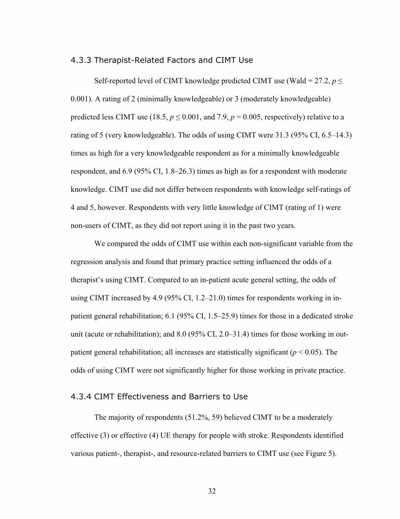
Self-reported level of CIMT knowledge predicted CIMT use (Wald = 27.2, p ≤
0.001). A rating of 2 (minimally knowledgeable) or 3 (moderately knowledgeable)
predicted less CIMT use (18.5, p ≤ 0.001, and 7.9, p = 0.005, respectively) relative to a
rating of 5 (very knowledgeable). The odds of using CIMT were 31.3 (95% CI, 6.5–14.3)
times as high for a very knowledgeable respondent as for a minimally knowledgeable
respondent, and 6.9 (95% CI, 1.8–26.3) times as high as for a respondent with moderate
knowledge. CIMT use did not differ between respondents with knowledge self-ratings of
4 and 5, however. Respondents with very little knowledge of CIMT (rating of 1) were
non-users of CIMT, as they did not report using it in the past two years.
We compared the odds of CIMT use within each non-significant variable from the
regression analysis and found that primary practice setting influenced the odds of a
therapist’s using CIMT. Compared to an in-patient acute general setting, the odds of
using CIMT increased by 4.9 (95% CI, 1.2–21.0) times for respondents working in in-
patient general rehabilitation; 6.1 (95% CI, 1.5–25.9) times for those in a dedicated stroke
unit (acute or rehabilitation); and 8.0 (95% CI, 2.0–31.4) times for those working in out-
patient general rehabilitation; all increases are statistically significant (p < 0.05). The
odds of using CIMT were not significantly higher for those working in private practice.
4.3.4 CIMT Effectiveness and Barriers to Use
The majority of respondents (51.2%, 59) believed CIMT to be a moderately
effective (3) or effective (4) UE therapy for people with stroke. Respondents identified
various patient-, therapist-, and resource-related barriers to CIMT use (see Figure 5).
33
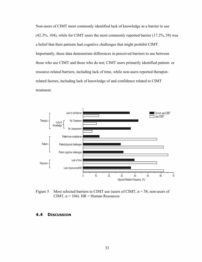
Non-users of CIMT most commonly identified lack of knowledge as a barrier to use
(42.3%, 104), while for CIMT users the most commonly reported barrier (17.2%, 58) was
a belief that their patients had cognitive challenges that might prohibit CIMT.
Importantly, these data demonstrate differences in perceived barriers to use between
those who use CIMT and those who do not; CIMT users primarily identified patient- or
resource-related barriers, including lack of time, while non-users reported therapist-
related factors, including lack of knowledge of and confidence related to CIMT
treatment.
Figure 5 Most selected barriers to CIMT use (users of CIMT, n = 58; non-users of CIMT, n = 104). HR = Human Resources
4.4 DISCUSSION
34
Although evidence shows CIMT to be an effective therapy for UE hemi-paresis in
patients who meet the criteria for treatment, and although the majority of respondents
know about CIMT, less than half reported using it. Based on our findings, discrepancies
between awareness of CIMT and clinical use of this therapy suggest that both lack of
knowledge and lack of resources are barriers to the implementation and use of CIMT in
clinical practice.
4.4.1 CIMT Use and Parameters
Respondents most often reported using CIMT for fewer hours/day for a longer
duration (i.e., using a non-traditional protocol), which suggests that more intense (i.e.,
traditional) protocols may be perceived as less clinically feasible. The treatment
parameters reported by therapists who use non-traditional CIMT (see Figures 3 and 4)
indicate variability in its delivery, which may reflect the integration of studies examining
CIMT across the continuum from traditional to non-traditional protocols, resulting in
limited consensus as to the most effective and feasible protocol.56, 57, 66 While useful in
determining overall effectiveness, this approach becomes problematic when therapists try
to implement a specific CIMT protocol clinically. The variability we observed suggests
that many therapists opt to develop their own method by integrating different evidence-
based protocols. The discrepancy between CIMT treatment parameters reported in the
research literature and those noted in our study suggests a problem in translating CIMT
knowledge from research to clinical practice, although this conclusion should be
considered in the context of the current sample. In light of these results, we must also
consider the notion that CIMT protocols, in their current form, do not reflect the
constraints of clinical practice and that these constraints should be taken into account as
35
protocols are further studied and modified – that is, that clinical practice should drive
research.
To assess respondents’ knowledge about CIMT, we asked them to list its key
components. Predictably, non-users of CIMT could not list all three components; more
unexpectedly, however, the majority of CIMT users were also unable to identify all three
components – for instance, less than 32% were able to identify the RTP and
shaping/behavioural components of CIMT. Shaping in conjunction with RTP is
considered one of the core components of treatment, and consequently a critical part of
any CIMT protocol.15, 32 Thus, when CIMT is used clinically, therapists may not be
implementing the treatment components that have been shown empirically to be effective
including the use of shaping (e.g., 11, 13, 17, 53). This inability to identify CIMT’s
fundamental components implies a lack of crucial knowledge of the therapy, at least
among respondents to our current study. This finding reveals knowledge barriers not only
among therapists who do not use CIMT but also among those who do. Continuing
clinical education may be a means to target and reduce knowledge-related barriers for
both users and non-users, with the goal of delivering CIMT protocols in their evidence-
based form.
4.4.2 Therapist-Related Factors and CIMT Use
4.4.2.1 Level of Knowledge
Among respondents in our study, level of CIMT knowledge was a significant
predictor of its use. Therapists who reported being very knowledgeable about CIMT had
greater odds of using it in their clinical practice than those who reported minimal or
moderate knowledge. The inability to distinguish CIMT use between very knowledgeable

36
and knowledgeable therapists supports our conclusion that having only some knowledge
of CIMT (i.e., minimal or moderate knowledge) is not sufficient to implement CIMT in
clinical practice, at least within this sample of respondents. Notably, a majority of
respondents (59.4%, 130) reported having only some knowledge about CIMT, which may
explain the discrepancy between the number of people who know about CIMT and the
number who actually use it clinically. As highlighted below and in Figure 5, non-users of
CIMT reported lack of knowledge as the primary barrier to CIMT use. Interestingly,
while a similar number of respondents across all knowledge levels identified non-
empirical sources of knowledge, a higher percentage of “very knowledgeable or
knowledgeable” respondents than of those with minimal or moderate knowledge
identified more research-based sources of knowledge (Table 2). The latter group tended
to rely more on their entry-level education or on non-empirical sources of information.
While these findings are specific to our small sample of therapists, they parallel prior
observations on barriers to CIMT use and therapists’ knowledge.49, 50, 67
4.4.2.2 Practice Setting
Our findings show that therapists in our sample rarely use CIMT for their clients
with acute stroke. Rather, they most often employ CIMT in out- and in-patient
rehabilitation settings, likely because their clients in these settings are in the sub-acute to
chronic stage of rehabilitation – the predominant patient groups in which CIMT’s
effectiveness has been examined in the literature.11, 39, 41 Moreover, there is a tendency
for patients in the acute stage of recovery to be excluded from treatment because they do
not meet the criteria for CIMT. Excluding these patients also helps to explain our
findings: if fewer patients are eligible for treatment, therapists may be less likely to focus

37
on CIMT, instead investing their time in interventions more appropriate to the acute stage
of stroke recovery. While there is evidence that some forms of CIMT are effective in the
acute phase, Brunner and colleagues suggest that CIMT not be used before 4 weeks post-
stroke, since rapid improvement appears to occur during the first month of standard
rehabilitation.55
4.4.3 Barriers to CIMT Use
Therapists who use CIMT and those who do not reported different types of
barriers to CIMT use: non-users more frequently identified therapist-related barriers (see
Figure 5), while CIMT users more frequently identified barriers related to their patients
(e.g., physical/cognitive challenges) and institutions (e.g., lack of resources). Given their
lack of experience using CIMT, one might expect that non-users would be unaware of
patient barriers and thus would not report them; the finding that non-users identified
therapist-related factors as barriers to CIMT use supports the notion that increasing CIMT
knowledge through training and education may increase clinical use of CIMT.
Conversely, the fact that users of CIMT tended not to report barriers related to
themselves (knowledge and confidence), and instead cited external barriers (related to
their patients and institutional resources), suggests that in the absence of increased
funding for in- and out-patient stroke rehabilitation, CIMT protocols may need further
adaptation within the constraints of clinical practice.68 Specifically, researchers need to
engage clinicians in conversations about evidence-based treatments to better align
research with what is feasible in a clinical setting.
With respect to patient and institutional barriers, our findings indicate not only
that a lack of resources can prevent CIMT use but also that patient non-compliance and
38
physical and cognitive characteristics may be major barriers to implementing CIMT (see
Figure 5, patient-related factors). Similarly, Page and colleagues have suggested that
when offered the traditional CIMT protocol, many patients with stroke do not participate,
preferring a less intensive CIMT protocol.46 It is important to note that in identifying
barriers to use of CIMT, we did not distinguish between traditional and non-traditional
protocols; our conclusions on the feasibility of traditional CIMT are drawn from the
frequency with which therapists report using it relative to non-traditional CIMT.
Therapists who report patient and institutional barriers to CIMT use may be doing so in
the context of either a traditional or a non-traditional approach. Regardless of the
protocol, the fact that CIMT users identified patient and institutional barriers suggests a
need to further develop a CIMT protocol that is both effective and clinically feasible. It
should also be noted that if therapists primarily treat people who do not meet the criteria
for CIMT, they will not report using it, even though they may be knowledgeable and able
to implement it.
Given regional differences in delivery of health care (e.g., publicly vs. privately
funded services), barriers to CIMT use, and specifically patient and institutional barriers
(e.g., patient populations and resources available to therapists), may vary from those
identified in our study. Irrespective of these differences, however, previous articles
commenting on barriers to CIMT implementation in multiple countries47, 49, 50 have
consistently identified therapist knowledge as a barrier to CIMT use. The results obtained
from our sample of therapists reinforce prior observations that increasing therapists’
knowledge of CIMT can contribute to more frequent use in clinical practice.
39
Our study has several limitations. First, the size of our sample resulted in low
statistical power and potential for bias in the data. Although 338 therapists responded, not
all completed every question. The lower number of respondents was problematic for
questions near the end of the survey (due in part to the funnelling nature of the survey)
and those with multiple levels. Further, because we specifically targeted therapists
practising in neurological rehabilitation, our sample may not be representative of the
Canadian occupational therapy/physical therapy population as a whole. Our results and
subsequent discussion should therefore be framed in the context of our sample. For
instance, relatively wide CIs reflect the possible variability of the results, which should
be taken into consideration when interpreting the applicability of the findings to the
larger OT/PT population. Furthermore, the low response rate suggests a possible self-
selection bias. Finally, because so few respondents (n = 8) reported using traditional
CIMT, we were unable to investigate whether certain factors could predict the type of
CIMT protocol used.
4.5 CONCLUSION
Although research has shown CIMT to be an effective therapy for UE hemi-
paresis post-stroke for patients who meet the criteria for treatment, a discrepancy exists
between the high level of awareness of CIMT and its low clinical use in our sample of
occupational and physical therapists. Our findings regarding lack of knowledge about
CIMT among practising therapists in our sample underscore the need for continuing
education. Of equal importance, the number of therapists reporting patient and
institutional barriers suggests a need to further modify current CIMT protocols to ensure
40
that they fit with clinical practice while remaining effective. Therapists’ perceptions of
CIMT can inform recommendations for educational initiatives and the development of
clinical guidelines. Furthermore, these results can guide future research, which should
focus on achieving a balance between the clinical feasibility of CIMT and its
effectiveness. This objective may be accomplished by investigating treatment dosage to
find a quantity that is clinically feasible while eliciting optimal rehabilitation outcomes.
4.6 KEY MESSAGES
4.6.1 What is Already Known on this Topic
Many people experience UE impairment following a stroke, and few regain full
function. There is evidence that CIMT is effective in improving UE function after stroke;
however, it is not known whether and how CIMT is being implemented in stroke
rehabilitation. Although a few studies subjectively report barriers to CIMT use,46, 49, 50 no
empirical studies have examined CIMT use, specifically clinicians’ perceptions, actual
clinical use, and perceived barriers to implementation.
4.6.2 What this Study Adds
This study is the first to report data from practicing therapists relating to their use
and perception of CIMT in clinical practice. Although awareness of CIMT was high
amongst the sample of therapists, many do not use it clinically. Lack of knowledge about
CIMT was the most commonly reported barrier to its implementation amongst therapists
who do not use CIMT. Institutional resources and patient barriers were frequently cited
by all respondents, but particularly by therapists who report using CIMT. These data
41
underscore the need for 1) educational initiatives to improve knowledge related to CIMT;
and 2) increased consultation between researchers and clinicians to optimize CIMT
protocols for clinical practice. Both of these initiatives have the potential to improve the
clinical use of CIMT.
4.7 SUMMARY TO CHAPTER 4 AND TRANSITION TO CHAPTER 5
A survey investigated CIMT use in Canadian neurological occupational and
physical therapy in terms of participant practices, perceptions and opinions related to
their use of CIMT in their clinical practice. While many therapists knew of the therapy,
less than half reported using it. Additionally, those who did use CIMT most often
employed a non-traditional or distributed protocol. A number of barriers to CIMT use
were identified, including lack of knowledge about the treatment and institutional
resources to support its use.
While the lack of institutional resources is important, for the purposes of my thesis I
am focusing on the amount of knowledge regarding the therapy as a barrier to use of
CIMT. In identifying knowledge as a barrier to use, a logical next step is to determine
how to address it and to direct efforts to overcome it. The survey study presented in
Chapter 4 identified the need for educational initiatives to improve therapist knowledge.
Importantly, therapist respondents that were most knowledgeable about CIMT tended to
acquire their information from research publications and best practice guidelines. Thus,
these are resources that can be targeted to address this knowledge barrier by describing
the treatment and outlining the evidence that supports its use. Specifically, systematic
reviews are important tools for clinicians because they summarize and evaluate the
literature, allowing clinicians to make evidence-informed treatment decisions. The

42
following chapter reports a systematic review of the literature whose purpose is to
establish the LOE in support of mCIMT.
43
CHAPTER 5 SYSTEMATIC REVIEW
5.1 INTRODUCTION
5.1.1 Description of the Condition
Stroke is the leading cause of neurological related death and disability in North
America, affecting approximately 50,000 Canadian and 795,000 American adults every
year.1 This figure is likely to increase due to the aging population and the escalating
percentage of individuals with risk factors for stroke.2 Unfortunately, full recovery after
stroke is elusive; of those who survive their stroke, approximately two-thirds will
experience residual neurological deficits that impair daily function. Specifically,
recovery of UE function is a major challenge for survivors of stroke, with only
approximately 5% regaining full function.3 As healthcare improves to more effectively
treat stroke acutely (e.g., through improvements to thrombolytic drugs), more North
Americans are living with its effects. Functional limitations resulting from UE deficits
negatively impact on performance of daily activities, resulting in decreased levels of
participation and physical activity, and increased risk of further health problems.5, 7
5.1.2 Description of the Intervention (mCIMT)
Post-stroke, patients often receive rehabilitation therapy to address functional
deficits. For instance, an occupational or physical therapist may work with a patient to
improve balance and motor function, address deficits in strength, and engage in sensory
and gait re-training. Specifically, treatments used to address UE functional deficits as
part of usual clinical care can include a task-oriented approach to functional activities,
active and passive range of motion (ROM) exercises, stretching and strength training.8
44
Treatments that are considered part of usual care help with UE recovery but have
limited effectiveness, evident in the high rates of disability, prevalence of secondary
disease, and incomplete functional recovery of the UE observed after stroke.9, 10 To
improve UE function after stroke and reduce the long-term disability related to poor
functional recovery, effective, evidence-based therapeutic interventions are needed.
Increased use of evidence-based therapies in rehabilitation can improve functional
abilities post-stroke, decreasing health care costs and greatly improving overall quality of
life.
Constraint induced movement therapy (CIMT) is a treatment that has been shown
to promote UE recovery post-stroke in a particular subset of patients, with numerous
studies showing it to be more effective than usual care.11, 17, 57 CIMT combines RTP
training, behavioural training techniques, and restraint of the unaffected UE.11, 69 In
CIMT, RTP is delivered in conjunction with shaping, during which patients engage in
meaningful functional activities with measurable progressions for which they receive
positive feedback as the activities become increasingly more difficult.13 Generally
patients are required to have some movement in the affected UE, which at a minimum
includes 10 degrees of active wrist extension, with 10 degrees extension of the thumb and
at least 2 fingers. The need for this level of function and corresponding ability to actively
engage in the treatment limits the number of patients for which CIMT is an appropriate
intervention.
As developed by Taub et al., the traditional form of CIMT follows a massed
practice schedule, and is performed over a 2-week period.11, 12 During this 2-week
treatment period, restraint of the unaffected limb for 90% of waking hours is combined
45
with 6 hours of functionally oriented RTP using the affected UE each weekday.
Behavioural training techniques are also employed to help patients transfer functional
gains into real world activities.
Numerous studies have examined this traditional CIMT protocol and support its
effectiveness at improving UE function post-stroke. For instance, the EXCITE trial, a
large RCT, followed 222 subjects 3-9 months post-stroke.11, 33 Subjects randomised to
the CIMT group demonstrated statistically and clinically significant improvements in UE
function and use compared to a usual care control group. Specifically, subjects in the
CIMT group demonstrated greater improvements pre-post treatment on the WMFT, a
laboratory based measure of UE function. Importantly, subjects in the CIMT group also
had greater gains in the amount of use of the affected UE compared to the control group,
as measured using the MAL, a subjective scale of real world limb use.
Since the initial description of the traditional CIMT protocol, a number of
different CIMT protocols have been developed and applied in post-stroke rehabilitation,
including those that more closely resemble the traditional or massed practice approach, as
well as those using a modified or distributed practice approach.11, 12, 16, 17 Other protocols
have also been described that are variations of these two approaches (for example see 35-37).
Importantly, the CIMT protocols developed to date comprise the same 3 key components:
restraint, repetitive task practice, and the application of behavioural techniques including
shaping.
The most studied of the distributed practice approaches, developed by Page and
colleagues, is termed mCIMT. In mCIMT, therapy sessions are performed for 30
minutes, 3 times per week over a 10-week schedule.16, 17 Despite the use of a distributed
46
practice schedule and thus a reduction in the overall treatment time, evidence suggests
that mCIMT is as efficacious as CIMT in promoting UE functional recovery post-stroke.
Page et al. demonstrated that mCIMT was superior to a dose-matched program of usual
care in improving UE function in chronic stroke.17 Significant improvements were
observed for the mCIMT group in both functional UE performance and amount of use.
Specifically, subjects in the mCIMT group had greater improvements on the ARAT, an
objective measure of UE function, and increased affected arm use as measured by the
MAL.
Despite differences in the frequency and overall duration of treatment, similar
principles are utilized across the spectrum of these CIMT protocols, including mCIMT.
The traditional CIMT approach as developed by Taub offers a compressed, but time-
intensive treatment. While the traditional protocol has been demonstrated to be effective,
evidence suggests that it can be difficult to implement clinically due to its high resource
demand (eg. time, availability of therapists, and equipment).46, 49, 70 Contrary to CIMT,
mCIMT distributes therapy sessions over a longer duration with a considerable reduction
in treatment time and thus a decreased use of resources, making the treatment easier to
manage clinically.71 As highlighted above, treatment in mCIMT is performed over 10
weeks and the UE is restrained for 5 hours/weekday (rather than 90% of waking hours
under the traditional protocol).
5.1.3 How the Intervention Might Work
Derived from basic studies in monkey’s, Taub and colleagues observed a pattern
of behaviour they termed “learned nonuse”18, 19; that is, removal of sensory feedback

47
from the UE resulted in the animals encountering aversive effects when using it and, as a
result, stopped using the limb.
In subsequent studies, it was found that learned nonuse could be overcome by
restraining the monkeys’ non-affected limb thereby forcing use of the deafferented limb
in daily activities (e.g., grooming, feeding). Over time, increased use of the deafferented
limb ultimately led to improved limb function.13, 18 Given the results observed in this
series of animal-based studies, coupled with the observation of learned nonuse in
individuals with UE functional deficits, Taub and colleagues applied the principles
observed in the animal studies to rehabilitation of individuals post-stroke.13, 19, 26
The treatment components of CIMT are grounded in sound neurophysiological
evidence. It is generally accepted that the basis for functional recovery post-stroke is the
repetitive practice of novel, skilled movements. Research in animals has clearly
demonstrated that repetitively engaging in novel motor tasks after an ischemic infarct
results in plastic changes in the brain, characterized by a task-dependent reorganization of
primary motor cortex.20, 22, 34, 72 Similar observations have been made in humans post-
stroke, with evidence from neuroimaging studies repeatedly demonstrating task-
dependent changes in the brain that correlate with UE functional recovery. 27, 6132 Thus,
in addition to wearing a restraint on their unaffected UE, subjects in CIMT engage in
RTP using their affected UE. Task selection is believed to be an important aspect of the
therapy, such that tasks utilized should be of value and interest to the subjects. Lastly,
shaping and other behavioural techniques have been established as key principles that
contribute to the functional gains observed in human studies.13, 32, 47
48
5.1.4 Why it is Important to do this Review
Owing to the number of studies examining CIMT in recent years, systematic
reviews have evaluated the effectiveness of the treatment in promoting UE recovery post-
stroke.57, 58, 66, 73-77 While useful in gauging the overall effectiveness of CIMT protocols
collectively, there are 2 major issues with the current reviews that limit their ability to
portray the evidence in support of CIMT. These issues include: (1) they only focus on
RCTs; and (2) they group treatment protocols of different durations.
5.1.4.1 Focus on RCTs
Although RCTs are valuable in that they provide the strongest LOE to support
causality (i.e., a given outcome is related to the experimental treatment or intervention),
the number of large RCTs (N ≥ 100) examining any of the CIMT protocols is small in
number, and as such the majority of systematic reviews draw conclusions regarding
treatment efficacy based on smaller (N < 100) RCTs. Of note, inclusion of the EXCITE
trial, the largest RCT examining CIMT, may well introduce bias into meta-analyses with
potential for skewed data resulting from overemphasizing the effects of CIMT compared
to a control group. The therapy received by the control group was not consistent, with
subjects receiving no therapy throughout the study. As the number of studies examining
the efficacy of the various CIMT protocols increase, systematic reviews have become
more specific to include only those with dose-matched treatment groups. For instance,
Stevenson et al. (2012) performed a meta-analysis comparing CIMT to dose-matched
interventions, showing CIMT to be superior to usual care or standard treatment. Owing to
the use of a comparator group which for the most part did not receive any treatment, the
EXCITE trial is most often excluded from these types of reviews. Overall, inclusion of

49
RCTs only in systematic reviews disregards a large body of literature that provides
evidence with regard to the efficacy of CIMT in promoting UE functional recovery post-
stroke. There is support for systematic reviews that are based on the comprehensive body
of literature including various study designs.78 Inclusion of non-RCT study designs would
provide a comprehensive picture of the evidence for or against the use of CIMT post-
stroke.
5.1.4.1 Grouping of Different Treatment Protocols
As highlighted previously there is considerable variability in the duration and
schedule of treatment sessions along the continuum of CIMT protocols. Accordingly,
there is a large body of literature reporting on the effectiveness of these different
treatment protocols, which has led to the publication of numerous systematic reviews
summarizing their findings.58, 73, 74, 76 While these systematic reviews have proved useful
to measure the effectiveness of CIMT in broad terms, it is difficult to draw conclusions
regarding the effectiveness of a specific CIMT protocol, as the different protocols are
grouped and examined collectively (eg.57, 75-77). Owing to the differences relating to both
the duration of treatment sessions and the overall treatment schedule (i.e., massed vs.
distributed practice), it may be more appropriate to perform a systematic review and
subsequently assign a LOE to a ‘body of literature’ that relates to a specific CIMT
protocol. Performing a systematic review of the literature relating to a single CIMT
protocol would allow a clinician to make decisions about implementing a therapy based
on the evidence in support of a specific protocol (not a number of protocols grouped
together), with consideration of how that protocol suits their clinical practice.
50
Data from a recent study (survey study – Chapter 4) examining utilization patterns
of CIMT by practicing occupational and physical therapists supports the notion that
clinicians do not relate a particular treatment schedule/session duration with a given
CIMT protocol.70 For instance, amongst therapists who report using a ‘non-traditional’
CIMT protocol (i.e., distributed practice greater than 2 weeks in duration), there was
considerable variability in both the treatment schedule (number of therapy sessions/week)
and session duration (therapy time/day). These results suggest that clinicians within this
sample appear not to be following the treatment parameters of any one CIMT protocol as
it is reported in the literature. While it understood that variations in the delivery of a
given CIMT protocol may be made to facilitate its clinical use, it is not known how
deviations from its ‘evidence-based form’ will impact on its effectiveness. Furthermore,
results of the study showed that no one protocol was being used with greater frequency
compared to others. As suggested by Mulrow, systematic reviews are an important tool
for clinicians to keep up-to-date on the current literature and to make informed decisions
regarding evidence-based practice.51 Prior studies have reported that a large proportion
of therapists obtain knowledge related to interventions from research publications. Thus,
the grouping of protocols within the systematic review literature may be a factor related
to the variability observed in the delivery of CIMT in the clinical setting.
An alternative to grouping CIMT protocols with distinct treatment parameters
would be to focus on a single protocol in order to establish its LOE with regard to
promoting UE recovery. Coupling a LOE with a clearly defined treatment protocol would
better facilitate its translation to clinical practice in the form in which the evidence
supporting its use was generated. One of the more frequently examined CIMT protocols

51
is the modified version developed by Page and colleagues (mCIMT: 3 times/week for 10
weeks as described previously).16, 17, 38 Owing to the distributed nature of its treatment
schedule, mCIMT offers considerable promise in terms of its clinical feasibility and ease
of implementation for clinicians, particularly in comparison to traditional protocols.
Establishing the LOE with regard to mCIMT independent of massed practice protocols
would allow one to discern its effectiveness at promoting UE recovery post-stroke.
5.1.5 Objectives
The primary objective of this review was to investigate the LOE in support of
mCIMT (i.e., a CIMT protocol with a distributed practice schedule of 3 sessions/weekday
for 10 weeks) to promote UE recovery post-stroke. Effectiveness was determined based
on outcome measures that assess pre-post treatment changes in UE function, impairment
and activity.
5.2 METHODOLOGY
5.2.1 Criteria for Selecting Studies for Review
All study designs were eligible for inclusion. For a study to be included it must
have included adult subjects (≥ 18 years) with a clinical diagnosis of stroke and who had
experienced UE impairment.
The experimental intervention employed in the selected studies followed a
distributed therapy schedule and in most instances reflected the mCIMT protocol of Page
et al.16, 17, 38 Therapy had to be performed 3 times per week over a 10-week period. The
amount of therapy time per session and amount of restraint per day could vary. Because

52
there was no restriction with regard to study design, the comparator interventions vary
across studies (eg. usual care control, no treatment control, no comparator group).
The outcome measures used to assess treatment effectiveness were not part of the
inclusion criteria for the study review. Measures of interest included UE function (the
anatomical and physiological parts of the body’s systems), as assessed by the ARAT; UE
impairment (the loss or significant deviation from full functioning body systems), as
assessed by the Fugl-Meyer Assessment of Motor Recovery After Stroke (FM); and UE
activity (the performance of an action at the level of an individual), as assessed by the
MAL.
The ARAT is an evaluative measure for use in individuals with hemi-paresis
resulting from stroke to assess UE function.79 The measure includes 19 items grouped
into four sub-scales including grasp, grip, pinch and gross movement.80 Each sub-scale
item is graded on a four point ordinal scale (0 = cannot perform any part of the test, 1 =
performs test partially, 2 = completes test, but in abnormally long time, and 3 = performs
the test normally) for a total possible score of 0-57. The UE portion of the FM is a
performance-based assessment of UE motor impairment.81, 82 A total score of 66 points
can be obtained as each item is scored on an ordinal scale of 0 (cannot perform) to 2 (can
perform fully). The FM uses direct observation to assess a series of voluntary limb
movements (at times using a variety of small objects). The MAL assesses people with
moderate-mild hemiparesis resulting from stroke and their affected UE activity.83, 84 It is
a semi-structured interview that measures the amount of use (AOU) and quality of
movement (QOM) of a patient using the UE to complete 14 ADLs. Responses are scored

53
along a 6-point scale and can range from 0 (did not use affected UE) to 5 (use affected
UE the same as pre-stroke).
5.2.2 Methods for Identification, Data Collection and Analysis of
Studies
Identification, selection and initial analysis of the studies to be included in the
review were performed in 3 phases by two independent reviewers (including myself). A
multi-phase approach ensured identification of all relevant sources and allowed the
review to begin with a broad, comprehensive literature search, followed by identification
and selection of articles that matched the reviews specific inclusion criteria (outlined
below and in Appendix D). The initial search was conducted on June 20, 2011 by one of
the study authors and a reference librarian who specializes in evidence-based searching,
and included electronic databases for empirical research studies and the grey literature.
Electronic databases searched included PubMed, CINAHL, Embase, the
Cochrane Library, Web of Science, ProQuest and OpenGrey from inception to present.
The search followed a multi-step process and initially included key words and medical
subject headings (MeSH; e.g., cerebrovascular disease; upper extremity; exercise
therapy), exploded to include related terms (Appendix E). Limiters (Humans; English;
and Adults) were then added to the search to filter results to match the reviews inclusion
criteria. Individual citations were then exported to a reference manager database
(EndNote X4; Thomson Reuters, New York, NY) for each database searched and
duplicates were removed. The full text version of each citation was then retrieved and
added to the reference manager database. The comprehensive search of electronic

54
databases culminated in the identification of 486 articles. Thirteen of the citations
obtained following the literature search were removed before the first phase of article
selection because the full text was not accessible in print or electronic copy.
5.2.2.1 Phase 1 – Refining the Literature
The two independent reviewers received the reference manager database that
included all 473 citations and the PDF full text of the article. An Excel spreadsheet was
used to record information related to the selected studies including the first author, date
of publication, journal, and reason for exclusion (if excluded). Article selection in this
phase was based on information gathered from the title and abstract; criteria for inclusion
was the presence of content related in any way to CIMT, or the use of alternate terms
(e.g., forced use; constraint induced therapy). Any disagreement between the two
reviewers related to inclusion of specific articles was resolved by an additional reviewer.
Following phase 1, 151 articles were identified for further review.
5.2.2.2 Phase 2 – Identifying Articles Examining mCIMT
Articles identified in phase 1 were subjected to further review to select those that
utilized the mCIMT (distributed CIMT) protocol. Article selection was based on review
of the abstract and full text. A standard checklist form was used to evaluate the studies
and record the pertinent data (study design, population, intervention studied, target of
intervention, comparator intervention, and primary outcome measures). Studies were
selected based on the following inclusion criteria: (1) mCIMT intervention following a
distributed practice schedule (3 times/weekday for 10 weeks); (2) adult subjects (≥ 18
yrs); (3) clinical diagnosis of stroke (ischemic or hemorrhagic); and (4) the UE was the
target of the intervention. As in phase 1, any disagreement with regard to article inclusion

55
was resolved by an additional reviewer. Fifteen studies were identified after the second
phase.
5.2.2.3 Phase 3 – Assessing and Assigning the Level of Evidence
The articles selected for review following phase 2 were categorized based on
study design for analysis. Both assessors reviewed the identified articles and assessed the
LOE of each single study. The LOE was based on the American Academy of Cerebral
Palsy Developmental Medicine (AACPDM) methodology to develop systematic reviews
of treatment intervention.85 A standardized form was used to extract information related
to the subjects (mean age, time since stroke, number of subjects in intervention or control
groups) and a basic description of the intervention. Conduct questions on the data
collection form were used to assess the risk of bias and methodological quality.
Collectively the studies were reviewed to assess and assign the LOE to the body of
knowledge on mCIMT.
To examine the body of evidence related to mCIMT in this systematic review, all
study designs were included. Thus, the LOE assigned to single studies was based on
research design and categorized levels 1 through 5. As shown in Table 3, in descending
order the study designs are decreasingly able to demonstrate causality, that is, that the
intervention was responsible for UE improvements. The LOE for the body of knowledge
looks at all the included studies and can be labelled A to D (Table 4).
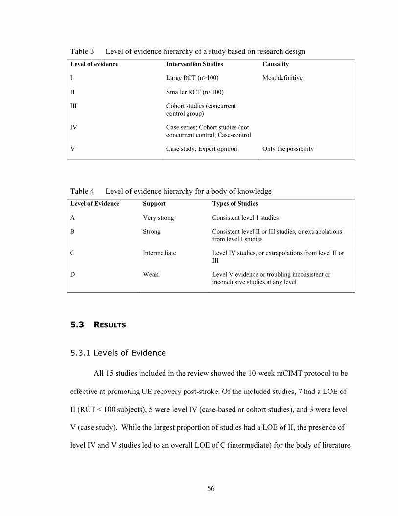
56
Table 3 Level of evidence hierarchy of a study based on research design Level of evidence Intervention Studies Causality
I Large RCT (n>100) Most definitive
II Smaller RCT (n<100)
III Cohort studies (concurrent control group)
IV Case series; Cohort studies (not concurrent control; Case-control
V Case study; Expert opinion Only the possibility
Table 4 Level of evidence hierarchy for a body of knowledge Level of Evidence Support Types of Studies
A Very strong Consistent level 1 studies
B Strong Consistent level II or III studies, or extrapolations from level I studies
C Intermediate Level IV studies, or extrapolations from level II or III
D Weak Level V evidence or troubling inconsistent or inconclusive studies at any level
5.3 RESULTS
5.3.1 Levels of Evidence
All 15 studies included in the review showed the 10-week mCIMT protocol to be
effective at promoting UE recovery post-stroke. Of the included studies, 7 had a LOE of
II (RCT < 100 subjects), 5 were level IV (case-based or cohort studies), and 3 were level
V (case study). While the largest proportion of studies had a LOE of II, the presence of
level IV and V studies led to an overall LOE of C (intermediate) for the body of literature
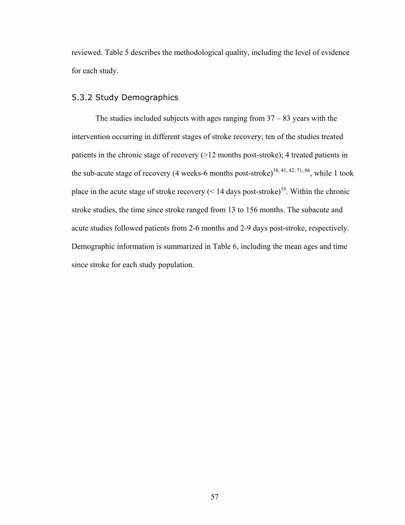
57
reviewed. Table 5 describes the methodological quality, including the level of evidence
for each study.
5.3.2 Study Demographics
The studies included subjects with ages ranging from 37 – 83 years with the
intervention occurring in different stages of stroke recovery; ten of the studies treated
patients in the chronic stage of recovery (>12 months post-stroke); 4 treated patients in
the sub-acute stage of recovery (4 weeks-6 months post-stroke)38, 41, 42, 71, 86, while 1 took
place in the acute stage of stroke recovery (< 14 days post-stroke)16. Within the chronic
stroke studies, the time since stroke ranged from 13 to 156 months. The subacute and
acute studies followed patients from 2-6 months and 2-9 days post-stroke, respectively.
Demographic information is summarized in Table 6, including the mean ages and time
since stroke for each study population.
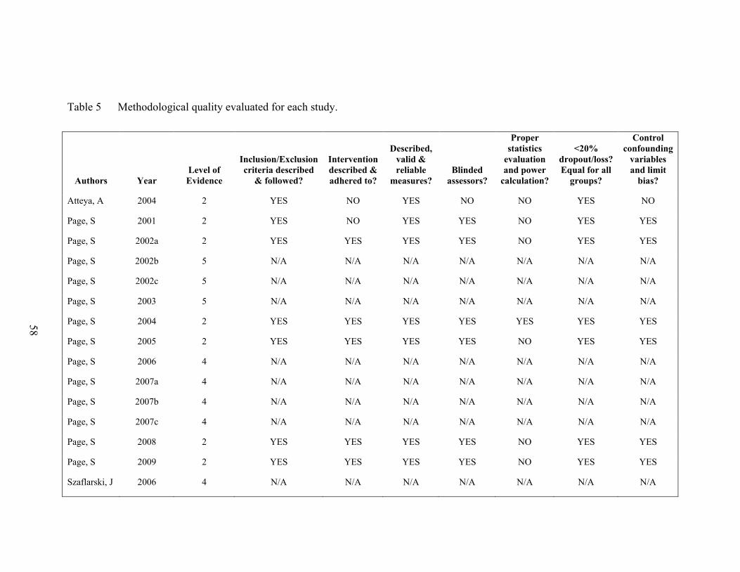
58
Table 5 Methodological quality evaluated for each study.
Authors Year Level of Evidence
Inclusion/Exclusion criteria described
& followed?
Intervention described & adhered to?
Described, valid & reliable
measures? Blinded
assessors?
Proper statistics
evaluation and power
calculation?
<20% dropout/loss? Equal for all
groups?
Control confounding
variables and limit
bias?
Atteya, A 2004 2 YES NO YES NO NO YES NO
Page, S 2001 2 YES NO YES YES NO YES YES
Page, S 2002a 2 YES YES YES YES NO YES YES
Page, S 2002b 5 N/A N/A N/A N/A N/A N/A N/A
Page, S 2002c 5 N/A N/A N/A N/A N/A N/A N/A
Page, S 2003 5 N/A N/A N/A N/A N/A N/A N/A
Page, S 2004 2 YES YES YES YES YES YES YES
Page, S 2005 2 YES YES YES YES NO YES YES
Page, S 2006 4 N/A N/A N/A N/A N/A N/A N/A
Page, S 2007a 4 N/A N/A N/A N/A N/A N/A N/A
Page, S 2007b 4 N/A N/A N/A N/A N/A N/A N/A
Page, S 2007c 4 N/A N/A N/A N/A N/A N/A N/A
Page, S 2008 2 YES YES YES YES NO YES YES
Page, S 2009 2 YES YES YES YES NO YES YES
Szaflarski, J 2006 4 N/A N/A N/A N/A N/A N/A N/A
58
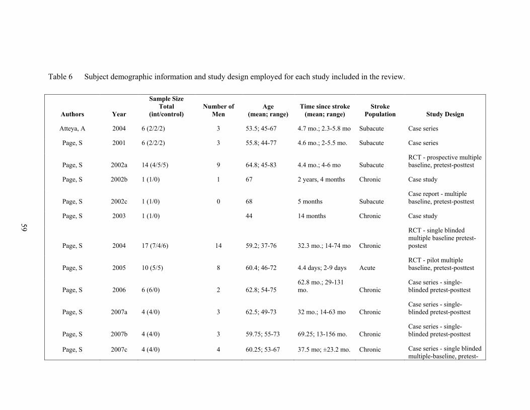
59
Table 6 Subject demographic information and study design employed for each study included in the review.
Authors Year
Sample Size Total
(int/control) Number of
Men Age
(mean; range) Time since stroke
(mean; range) Stroke
Population Study Design
Atteya, A 2004 6 (2/2/2) 3 53.5; 45-67 4.7 mo.; 2.3-5.8 mo Subacute Case series
Page, S 2001 6 (2/2/2) 3 55.8; 44-77 4.6 mo.; 2-5.5 mo. Subacute Case series
Page, S 2002a 14 (4/5/5) 9 64.8; 45-83 4.4 mo.; 4-6 mo Subacute
RCT - prospective multiple baseline, pretest-posttest
Page, S 2002b 1 (1/0) 1 67 2 years, 4 months Chronic Case study
Page, S 2002c 1 (1/0) 0 68 5 months Subacute Case report - multiple baseline, pretest-posttest
Page, S 2003 1 (1/0) 44 14 months Chronic Case study
Page, S 2004 17 (7/4/6) 14 59.2; 37-76 32.3 mo.; 14-74 mo Chronic
RCT - single blinded multiple baseline pretest-postest
Page, S 2005 10 (5/5) 8 60.4; 46-72 4.4 days; 2-9 days Acute RCT - pilot multiple baseline, pretest-posttest
Page, S 2006 6 (6/0) 2 62.8; 54-75 62.8 mo.; 29-131 mo. Chronic
Case series - single-blinded pretest-posttest
Page, S 2007a 4 (4/0) 3 62.5; 49-73 32 mo.; 14-63 mo Chronic Case series - single-blinded pretest-posttest
Page, S 2007b 4 (4/0) 3 59.75; 55-73 69.25; 13-156 mo. Chronic Case series - single-blinded pretest-posttest
Page, S 2007c 4 (4/0) 4 60.25; 53-67 37.5 mo; ±23.2 mo. Chronic Case series - single blinded multiple-baseline, pretest-
59
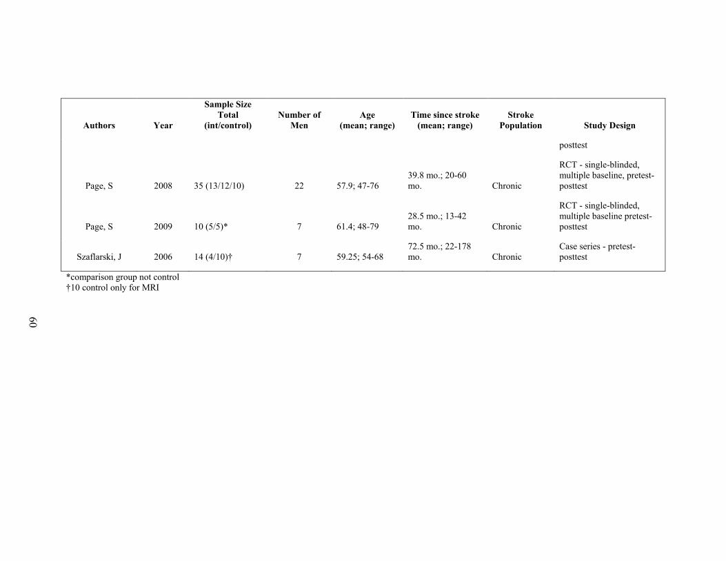
60
Authors Year
Sample Size Total
(int/control) Number of
Men Age
(mean; range) Time since stroke
(mean; range) Stroke
Population Study Design
posttest
Page, S 2008 35 (13/12/10) 22 57.9; 47-76 39.8 mo.; 20-60 mo. Chronic
RCT - single-blinded, multiple baseline, pretest-posttest
Page, S 2009 10 (5/5)* 7 61.4; 48-79 28.5 mo.; 13-42 mo. Chronic
RCT - single-blinded, multiple baseline pretest-posttest
Szaflarski, J 2006 14 (4/10)† 7 59.25; 54-68 72.5 mo.; 22-178 mo. Chronic
Case series - pretest-posttest
*comparison group not control †10 control only for MRI
60

61
5.3.3 Intervention
Consistent with the stated inclusion criteria, all of the studies followed a
distributed practice schedule with treatment sessions occurring 3 times/week for 10-
weeks. The majority of the studies (10/15) employed thirty-minute therapy sessions,
while the remainder of the studies employed sixty minute therapy sessions (30 minutes
each of OT and PT). Each study performed mCIMT, including the 3 key components
that are consistently described in the literature and across the different CIMT protocols
(restraint, RTP and behavioural techniques including shaping). In many instances (6/15
studies), the mCIMT intervention was compared to a control group, which consisted of
either usual care (e.g., proprioceptive neuromuscular facilitation techniques focusing on
functional tasks, stretching and compensatory techniques using the less affected UE as
needed)17 or no therapy. Of the six studies that used a control group, all compared
mCIMT to a dose-matched control group receiving usual care therapy, with five of those
studies also comparing mCIMT to a no therapy control group. While all 15 studies
included performed pre- and post-treatment assessments (baseline and at 10 weeks), 3
studies also included a 3-month follow-up assessment.39, 87, 88 Information about the
treatment delivered in each study is summarized in Tables 7.

62
Table 7 Descriptions of treatment delivery and outcomes for each study included in the review.
Authors Year Outcome Measures Interventions Training time Restraint time Control group Outcomes Follow-up
Atteya, A 2004 FM ARAT WMFT MAL
mCIMT 1 hour total (0.5 hour each with OT & PT)
5 days/week for 5 hours (UE and lower extremity)
(1) traditional therapy (2) no therapy
mCIMT: substantial increases on all measures Traditional/no therapy: few improvements
Page, S 2001 FM ARAT WMFT MAL
mCIMT 1 hour total (0.5 hour each with OT & PT)
5 hrs/weekday (1) traditional therapy (2) no therapy
mCIMT: substantial increases on all measures
Traditional/no therapy: no improvements
Page, S 2002a FM ARAT MAL
mCIMT 1 hour total (0.5 hour each with OT & PT)
5 hrs/weekday (1) traditional therapy (2) no therapy
mCIMT: substantial increases on all measures
Traditional/no therapy: few improvements
Page, S 2002b FM ARAT MAL
mCIMT 1 hour total (0.5 hour each with OT & PT)
5 hrs/weekday none Increases on all measures Activity monitor data
3 months post-intervention
62
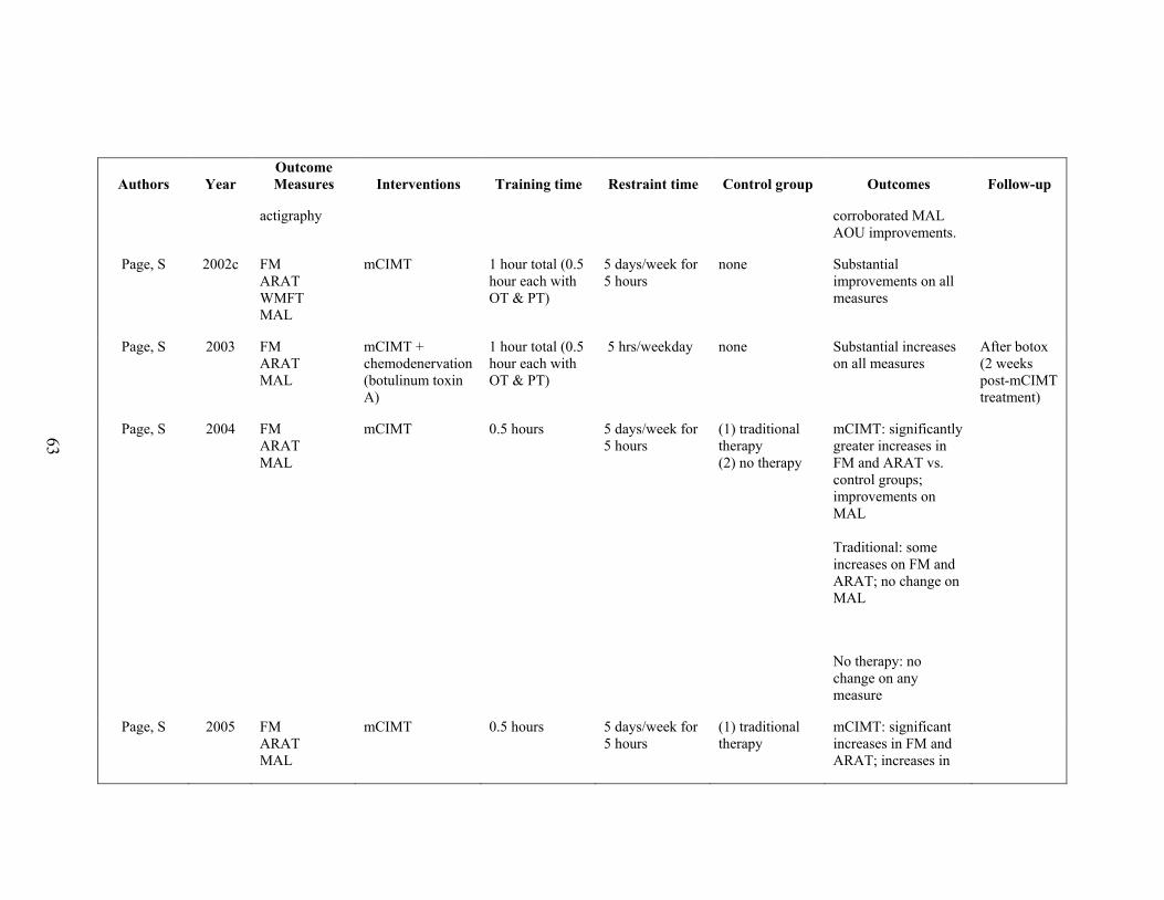
63
Authors Year Outcome Measures Interventions Training time Restraint time Control group Outcomes Follow-up
actigraphy corroborated MAL AOU improvements.
Page, S 2002c FM ARAT WMFT MAL
mCIMT 1 hour total (0.5 hour each with OT & PT)
5 days/week for 5 hours
none Substantial improvements on all measures
Page, S 2003 FM ARAT MAL
mCIMT + chemodenervation (botulinum toxin A)
1 hour total (0.5 hour each with OT & PT)
5 hrs/weekday none Substantial increases on all measures
After botox (2 weeks post-mCIMT treatment)
Page, S 2004 FM ARAT MAL
mCIMT 0.5 hours 5 days/week for 5 hours
(1) traditional therapy (2) no therapy
mCIMT: significantly greater increases in FM and ARAT vs. control groups; improvements on MAL Traditional: some increases on FM and ARAT; no change on MAL
No therapy: no change on any measure
Page, S 2005 FM ARAT MAL
mCIMT 0.5 hours 5 days/week for 5 hours
(1) traditional therapy
mCIMT: significant increases in FM and ARAT; increases in
63
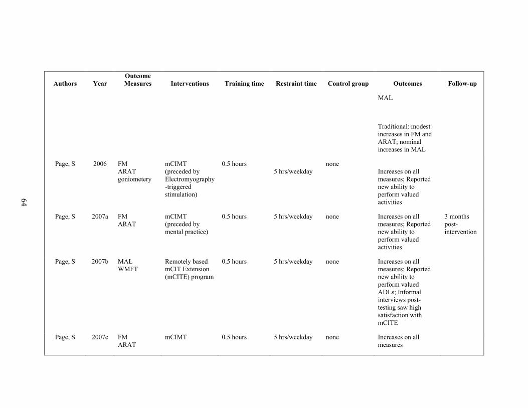
64
Authors Year Outcome Measures Interventions Training time Restraint time Control group Outcomes Follow-up
MAL
Traditional: modest increases in FM and ARAT; nominal increases in MAL
Page, S 2006 FM ARAT goniometery
mCIMT (preceded by Electromyography-triggered stimulation)
0.5 hours 5 hrs/weekday
none Increases on all measures; Reported new ability to perform valued activities
Page, S 2007a FM ARAT
mCIMT (preceded by mental practice)
0.5 hours 5 hrs/weekday none Increases on all measures; Reported new ability to perform valued activities
3 months post-intervention
Page, S 2007b MAL WMFT
Remotely based mCIT Extension (mCITE) program
0.5 hours 5 hrs/weekday none Increases on all measures; Reported new ability to perform valued ADLs; Informal interviews post-testing saw high satisfaction with mCITE
Page, S 2007c FM ARAT
mCIMT 0.5 hours 5 hrs/weekday none Increases on all measures
64

65
Authors Year Outcome Measures Interventions Training time Restraint time Control group Outcomes Follow-up
MAL
Page, S 2008 FM ARAT MAL
mCIMT 0.5 hours 5 hrs/weekday (1) traditional therapy (2) no therapy
mCIMT: significant increases on ARAT and MAL; Treatment effect across all 3 groups for ARAT traditional therapy: few improvements on ARAT and FM
No therapy: no change
Page, S 2009 FM ARAT
mCIMT 0.5 hours 5 hrs/weekday Comparison group: mCIMT and 30 min mental practice sessions
Increases on all measures for both groups
3 months post-intervention
Szaflarski, J 2006 FM ARAT MAL - AOU fMRI at 4T
mCIMT 0.5 hours 5 days/week for 5 hours
Volunteers provided data re: typical activation patterns for fMRI
For 3 subjects, substantial increases on ARAT and MAL; modest increases on FM; Reported new ability to perform valued ADLs; fMRI data suggests cortical reorganisation 4th subject saw minimal changes on any measures
65
66
A number of studies incorporated additional elements beyond the standard
mCIMT protocol. When this was the case, the studies utilized scores reported for
assessments performed pre-post mCIMT, independent from any other treatment. For
instance, mCIMT was combined with another therapy in 3 studies. Two of these studies
involved combining mCIMT with mental practice. Page et al. had all subjects receive
mCIMT, however one group also received 30 minutes of mental practice following each
mCIMT session.86 This differed from a 2007 study that began delivering mCIMT a week
after the mental practice therapy post-testing.17 A case study performed in 2003 by Page
et al. followed mCIMT with chemodenervation, using botulinum toxin A.87
5.3.4 Outcome Measures
To identify the directionality of the LOE we looked to evaluate the effectiveness
of mCIMT to improve UE function, impairment and use post-stroke. Across the fifteen
studies, common assessment tools used included the ARAT, FM, and MAL. While both
measure motor changes, the ARAT measures UE function while the FM measures
changes in UE impairment. The MAL evaluates participation through two subscales that
measure the amount of use, and quality of movement. Since the MAL is a self-report tool
and vulnerable to subject bias, the MAL can also be administered to the primary
caregiver.84 This was done in 4 of the included studies. In addition to the clinical
outcome measures, 2 studies incorporated other means to quantify treatment-related
changes. Szaflarski et al. used functional magnetic resonance imaging (fMRI) to
examine cortical reorganization resulting from treatment (patients receiving mCIMT
were compared to 10 non-disabled subjects who demonstrated the typical brain activation
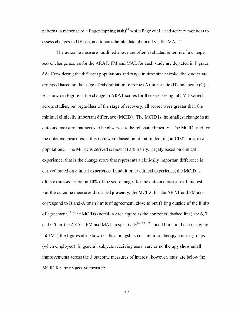
67
patterns in response to a finger-tapping task)40 while Page et al. used activity monitors to
assess changes in UE use, and to corroborate data obtained via the MAL.39
The outcome measures outlined above are often evaluated in terms of a change
score; change scores for the ARAT, FM and MAL for each study are depicted in Figures
6-9. Considering the different populations and range in time since stroke, the studies are
arranged based on the stage of rehabilitation [chronic (A), sub-acute (B), and acute (C)].
As shown in Figure 6, the change in ARAT scores for those receiving mCIMT varied
across studies, but regardless of the stage of recovery, all scores were greater than the
minimal clinically important difference (MCID). The MCID is the smallest change in an
outcome measure that needs to be observed to be relevant clinically. The MCID used for
the outcome measures in this review are based on literature looking at CIMT in stroke
populations. The MCID is derived somewhat arbitrarily, largely based on clinical
experience; that is the change score that represents a clinically important difference is
derived based on clinical experience. In addition to clinical experience, the MCID is
often expressed as being 10% of the score ranges for the outcome measure of interest.
For the outcome measures discussed presently, the MCIDs for the ARAT and FM also
correspond to Bland-Altman limits of agreement, close to but falling outside of the limits
of agreement.82 The MCIDs (noted in each figure as the horizontal dashed line) are 6, 7
and 0.5 for the ARAT, FM and MAL, respectively82, 83, 89. In addition to those receiving
mCIMT, the figures also show results amongst usual care or no therapy control groups
(when employed). In general, subjects receiving usual care or no therapy show small
improvements across the 3 outcome measures of interest; however, most are below the
MCID for the respective measure.

68
Figure 6 Pretest to posttest change scores for the Action Research Arm Test (ARAT)
with a dashed line to represent the minimal clinically important difference. Atteya (2004) also has a ‘no therapy’ group, but does not report the scores. A – chronic, B – subacute, C – acute; inverted triangle – mCIMT, square – traditional therapy, circle – no therapy.
A similar trend is observed for the FM and MAL scores (Figures 7-9) in that the
mCIMT group in most studies improved more than the MCID, while the usual care and
no therapy control groups did not surpass the MCID. However, the FM results differ in
that the mCIMT group scores fall below the MCID in 3 studies investigating a chronic
stroke population. For the MAL (AOU) scores, the studies examining patients in the
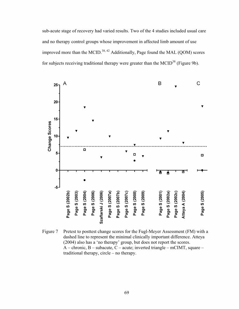
69
sub-acute stage of recovery had varied results. Two of the 4 studies included usual care
and no therapy control groups whose improvement in affected limb amount of use
improved more than the MCID.38, 42 Additionally, Page found the MAL (QOM) scores
for subjects receiving traditional therapy were greater than the MCID38 (Figure 9b).
Figure 7 Pretest to posttest change scores for the Fugl-Meyer Assessment (FM) with a
dashed line to represent the minimal clinically important difference. Atteya (2004) also has a ‘no therapy’ group, but does not report the scores. A – chronic, B – subacute, C – acute; inverted triangle – mCIMT, square – traditional therapy, circle – no therapy.
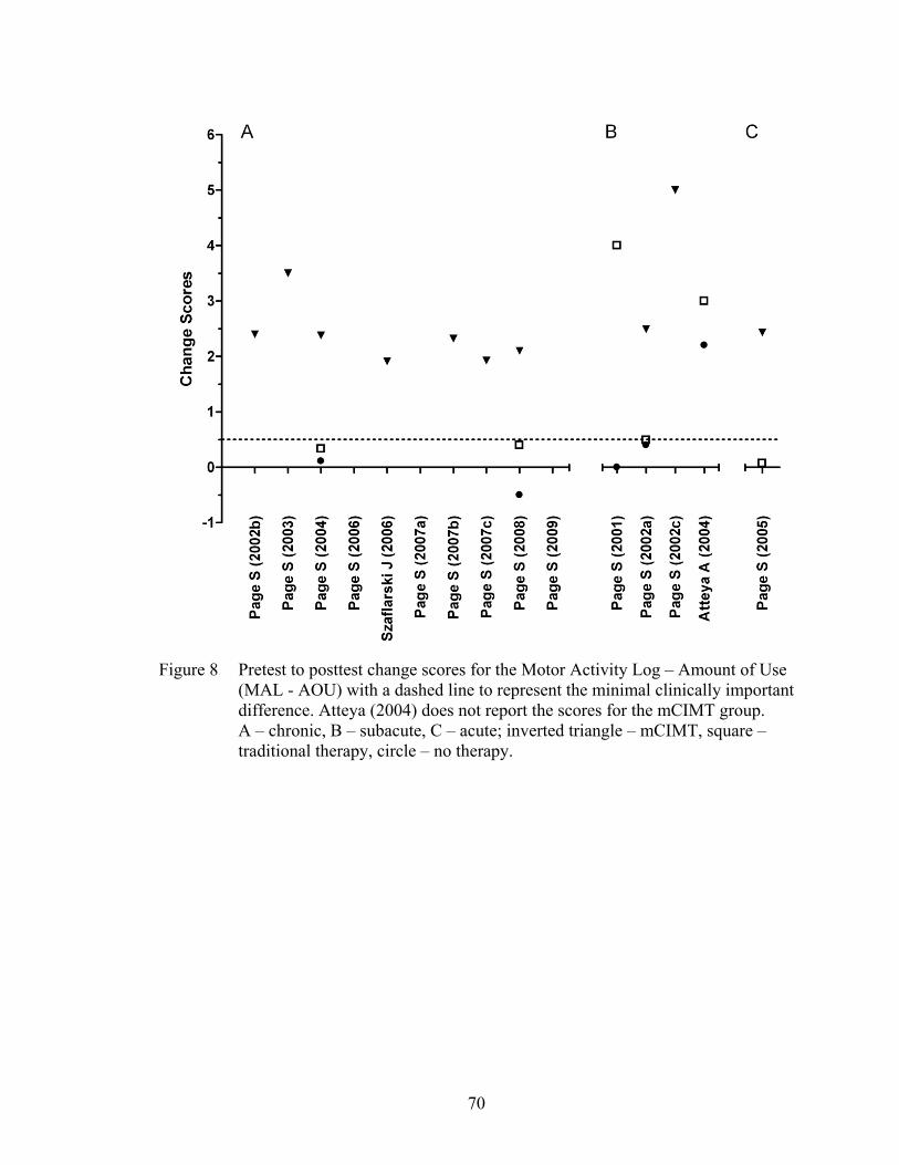
70
Figure 8 Pretest to posttest change scores for the Motor Activity Log – Amount of Use
(MAL - AOU) with a dashed line to represent the minimal clinically important difference. Atteya (2004) does not report the scores for the mCIMT group. A – chronic, B – subacute, C – acute; inverted triangle – mCIMT, square – traditional therapy, circle – no therapy.
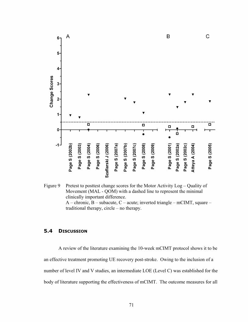
71
Figure 9 Pretest to posttest change scores for the Motor Activity Log – Quality of
Movement (MAL - QOM) with a dashed line to represent the minimal clinically important difference. A – chronic, B – subacute, C – acute; inverted triangle – mCIMT, square – traditional therapy, circle – no therapy.
5.4 DISCUSSION
A review of the literature examining the 10-week mCIMT protocol shows it to be
an effective treatment promoting UE recovery post-stroke. Owing to the inclusion of a
number of level IV and V studies, an intermediate LOE (Level C) was established for the
body of literature supporting the effectiveness of mCIMT. The outcome measures for all
72
studies demonstrated a positive improvement for subjects who received mCIMT with
regard to UE function, impairment and use (Figures 1-4). Importantly, in most of the
studies the degree of change was greater than the MCID. Thus, those receiving mCIMT
observed a degree of UE recovery that is clinically meaningful. This differs from
subjects in the control groups who may have experienced some change in outcome
measure scores; however these changes generally were not clinically meaningful.
5.4.1 Effectiveness Across the Stages of Recovery
The improvements noted for subjects receiving mCIMT were observed across all
stages of recovery, indicating a 10-week mCIMT intervention can be applied effectively
amongst acute, sub-acute or chronic patients who have had a stroke. While this evidence
suggests that mCIMT can be beneficial for patients across the recovery continuum, it is
important to consider that the bulk of evidence comes from a chronic stroke population.
The changes observed amongst the acute stroke population16, while significantly different
between the mCIMT and usual care therapy groups, should be considered cautiously
since the study only engaged 8 subjects.
5.4.2 Interpreting the Level of Evidence: Limitations
While the 10-week mCIMT intervention appears to be an effective tool for
rehabilitation of the UE post-stroke, the lack of large RCTs (≥ 100) or greater number of
small RCTs reduce its overall LOE (i.e., for the body of literature). Additionally, it is
difficult to run a comprehensive statistical or meta-analysis on the data from the current
collection of studies as they differ in methodology. The inability to perform a meta-
analysis is a necessary trade-off that allows for a comprehensive review of the mCIMT

73
literature and the assignment of a LOE to a specific CIMT protocol. Similarly, there are
challenges comparing studies with patient populations ranging across the stages of
rehabilitation. To circumvent this to some degree, we sub-divided the data into chronic,
sub-acute and acute populations. Sub-dividing the data in this manner allows clinicians to
determine the effectiveness of the mCIMT intervention in relation to the number and
quality of the studies examining a given stage of recovery. While we applied a consistent
MCID for the respective outcome measures across all stages of recovery, research
suggests that the MCID can change depending on the stage of recovery. In particular the
degree of change would be larger in more acute stages of recovery. For instance Lang et
al. estimate the MCID for the ARAT in acute stroke populations to vary between 12 and
17 points, depending if the affected UE is dominant or non-dominant, respectively.90
Importantly, these values were determined from a study that applied the original 2-week
CIMT protocol. The post-treatment assessment was conducted an average of 25.9 days
post-stroke. Comparatively, Page et al. also recruited subjects within 14 days of stroke
but applied a 10-week mCIMT protocol.16 Thus, post-stroke assessments ranged from
10-12 weeks (70-84 days) post-stroke. Considering the much shorter post-stroke
assessment time period, the MCID values suggested by Lang et al. may not be
appropriate for the purposes of this review.
The outcome measures and their generalizability can also prove challenging for
researchers. While both the ARAT and FM assess similar movements, they include
differing features. The FM tends to focus less on the skills performed and practiced
during mCIMT. This differs from the ARAT which tests movements that more closely
resemble those practiced in mCIMT. Hence, the ARAT tends to have greater

74
responsiveness (more sensitive to change) for UE improvements compared to the FM in
patients post-stroke.82, 91 The ARAT may be a better tool (relative to the FM) to measure
changes in UE function for subjects receiving mCIMT. This can help to explain the
discrepancies between the two measures, as depicted in Figures 1 and 2. Since the
ARAT is more likely to find changes in subjects who received mCIMT, it is not
surprising that the mCIMT group across all of the included studies had ARAT changes
scores greater than the MCID. The less responsive FM, however, had variable changes
scores; most, though not all, studies were above the MCID.
5.4.3 Incorporating mCIMT in Clinical Practice
It is important for therapists and clinicians to know the LOE of specific therapy
protocols, in this instance mCIMT, in order to make informed decisions about using it in
their clinical practice. The majority of mCIMT evidence focuses on chronic stroke
populations and supports mCIMT use at this stage of recovery. Studies examining
patients in the subacute stage of recovery, though fewer in number, also support mCIMT
as an effective intervention for the UE post-stroke. Although it appears promising, the
evidence is limited to support mCIMT for people with acute stroke. Clinicians should
consider their patients’ stage of recovery, if they meet CIMT criteria, and which protocol
is reasonable to implement for their particular practice setting, in the context of the
evidence supporting a given CIMT protocol.
5.4.4 Conclusion
Overall there is an intermediate LOE in support of mCIMT as an effective
treatment for UE hemi-paresis post-stroke. Future research including large RCTs would
75
strengthen the LOE for mCIMT. Additional investigation into the effectiveness of
mCIMT in acute and sub-acute stroke populations is warranted. A specific MCID should
also be established for each stage of rehabilitation. In-turn, a stronger LOE for mCIMT
can provide additional support for its inclusion in best practice guidelines for stroke care,
further promoting its use in clinical practice. Publications outlining in detail the way in
which a particular therapy is delivered can augment systematic reviews summarizing the
evidence for a given protocol. For instance, Page et al. provide a detailed account of the
‘ingredients’ necessary for implementing mCIMT in clinical practice.54 Such methods-
based papers can act as a valuable resource for therapists. By improving clinical use of
mCIMT we can improve the rehabilitation outcomes for people who have experienced a
stroke.
5.5 SUMMARY TO CHAPTER 5 AND TRANSITION TO CHAPTER 6
As outlined previously, mCIMT is a 10-week protocol that can be used as a
treatment option for the hemiparetic UE post-stroke. Overall there is an intermediate
LOE in support of mCIMT as an effective UE treatment. Specifically, subjects receiving
mCIMT most often see UE recovery in terms of improved function, reduced impairment
and increased activity. While a number of studies have been performed using mCIMT,
they primarily include case series and small RCTs examining its effectiveness in the
chronic stage of post-stroke rehabilitation. A number of studies however have emerged
evaluating mCIMT amongst sub-acute and acute post-stroke populations. Importantly,
subjects receiving mCIMT saw greater UE functional recovery compared to usual care
and no therapy groups across all stages of recovery.
76
Through examination of the current literature and understanding how researchers
have investigated mCIMT, we can evaluate the evidence in support of the therapy. One
question that arises based on this review is whether mCIMT would be clinically feasible
and effective to implement in Canada. This question represents the focus of the final
component of this thesis, which was completed as part of a clinical trial examining the
application of mCIMT in acute stroke.

77
CHAPTER 6 STUDY 3: mCIMT TRIAL
The effectiveness and feasibility of mCIMT was investigated through a clinical
trial that implements a mCIMT protocol within an acute and sub-acute post-stroke
rehabilitation setting. This trial was a pilot, single centre, randomized single-blinded
parallel group study. The following sections describe the methodology of the trial as a
whole; however, as this thesis includes a single case study of a subject that has completed
the trial, subsequent sections will outline methodology specific to this case and my role in
the delivery of mCIMT. Subsequent sections will discuss challenges to the
implementation of mCIMT as well as a discussion of its feasibility and effectiveness (in a
preliminary manner) based on the results of a case study.
6.1 METHODOLOGY 6.1.1 Groups and Subjects
The study compared rehabilitation outcomes of an experimental group to a control
group. All subjects received usual care as part of their normal therapy sessions, as
designated by their healthcare team. The experimental group was enrolled in a mCIMT
intervention that was additional to usual care. The control group received additional
usual care as part of the research study that was dose-matched to the mCIMT therapy
received by the experimental group. Matching the dose of therapy between groups
ensured the same exposure to therapy throughout the treatment phase of the study.
Subjects were selected from admissions to the acute stroke service at the Queen
Elizabeth Health Sciences Centre (QEII). To participate, subjects must have experienced
their first symptomatic stroke (ischemic or hemorrhagic) resulting in UE hemi-paresis

78
within 2 weeks of recruitment. Additional inclusion criteria were: (1) ability to perform a
two-step command, (2) ≥ to 18 years old, (3) resided within a 75km radius of the QEII,
(4) met standard mCIMT criteria16, 17, 40 including (i) ≥10° of active wrist extension (ii)
≥10° of active thumb abduction/extension (iii) ≥10° of active extension in two additional
digits (iv) ability to perform the movements 3 times in 1 minute. Exclusion criteria were:
(1) excessive pain in the affected UE, defined as > 4 on a 10 cm visual analogue scale
(VAS), (2) orientation score of < 6, (3) diagnosis of pre-stroke dementia, (4) diagnosis of
terminal illness, life-threatening co-morbidity, or concomitant neurological or psychiatric
illness, (5) excessive tone in the upper limb, > 3 on the modified Ashworth Scale (MAS),
(6) MAL score > 2.5 on the amount of use sub-scale. The VAS is a 10 cm line anchored
by 0 (far left) and 10 (far right) whereby subjects were able to mark the severity of pain
in their UE ranging from no pain (equivalent to 0) to pain equal to the worst pain they
have experienced (equivalent to 10). The MAS provides a means of quantifying
hypertonicity. To perform the MAS, the extemity being examined was moved passively
through the joint ROM, with the degree of tone assigned a score along a 6-point scale
ranging from 0 (no increase in tone) to 4 (rigid in flexion and extension).92
Subjects were randomized into the experimental or control groups following
consent and the pre-treatment assessment. Subjects were stratified by pre-treatment
ARAT scores [≤ 24 (lower UE function) or > than 24 (higher UE function)] and subject
age (≤ 60 or > 60 years)], into one of four groups including (1) younger with lower UE
function; (2) younger with higher UE function; (3) older with lower UE function; and (4)
older with higher UE function.

79
6.1.2 Assessments
Within 2 weeks of admission to the acute stroke service, subjects agreeing to
enrol and who provided written, informed consent underwent baseline measures (only
collected once and described in Appendix F). Assessments were performed at 4 time
points: (1) pre-treatment (T=0); (2) immediately post-treatment (T=10 weeks); (3) at 6
months from the last treatment session (T=36 weeks); and (4) at 12 months from the last
treatment session (T=62 weeks). Outcome measures were assessed at all time points for
both experimental and control subjects to facilitate comparison between the two groups.
The assessor was blinded to group assignment throughout the trial.
6.1.3 Primary outcome measures The ARAT is a functional test used to assess UE movement. Changes in ARAT
scores over time can assess UE functional recovery. Particularly, the measure is used to
assess UE functional changes in individuals post-stroke with hemiparesis.79 The test
contains 19 items that are grouped to test different qualities of UE function by the
following 4 sub-scales: grasp, grip, pinch and gross movement. Each subscale is graded
on a 4 point scale where ‘0’ = cannot perform any part of the movement; 1 = performs
the test partially; 2 = completes the test, but in an abnormally long time; and 3 = performs
the test normally. Thus, there is a total possible score of 57 points, with a score of 57
indicative of good UE function. The ARAT is commonly used in studies examining UE
recovery and function because it requires less than 10 minutes to complete and has been
shown to be a valid and reliable measure of UE function for individuals recovering from
stroke.80, 93 It is sensitive and responsive to change resulting from treatment in the stroke
population.91, 94, 95 On the ARAT, a change of 6 points identifies the MCID.82, 89
80
Amount of use and quality of use of the affected UE were assessed using the
MAL and through accelerometry. The MAL is a 14-item questionnaire used for patients
with mild-moderate hemiparesis to measure ‘real world’ use of the affected UE (‘activity’
as per the ICF).83, 84 Fourteen ADLs are assessed based on AOU and QOU subscales to
ascertain how much and how well the affected UE is used. Each activity is evaluated on
a 6 point scale whereby 0 = ‘did not use my weaker arm’ and 5 = ‘used my weaker arm
as often as before the stroke.’ The MAL has been used extensively to evaluate stroke
rehabilitation treatments because of a large responsiveness ratio (>3) and sensitivity to
changes pre-post CIMT.83 A change of 0.5 points indicates 10% of the scale’s total range
and is often used to identify a clinical difference.83, 84 Accelerometry (Gulf Coast Data
Concepts, LLC) was used to quantitatively measure the amount of use of the UEs during
a 3-day period prior to each assessment (thus 4 times in total). The equipment is a small
(10 x 2.5 x 2.5 cm; 55g), 3-axis unit that acquires and stores time-stamped acceleration
data. The units are placed inside custom-made bands that are worn on each wrist. The
devices capture daily use of the affected and unaffected UEs with the memory and battery
capacity to record and store data for 3 days. It provides a valid, quantitative measure of
functional UE recovery post-stroke among the subjects.83, 96, 97
6.1.4 Secondary Outcome Measures
Feasibility of the mCIMT intervention was assessed by patient compliance to the
regimen. Compliance was measured in the following ways: 1) the percentage of patients
enrolled in the experimental group who complete all therapy sessions; and 2) the
percentage of prescribed time that patients wear the restraint, as quantified by a
compliance device. The second method of measuring compliance assigns a quantitative
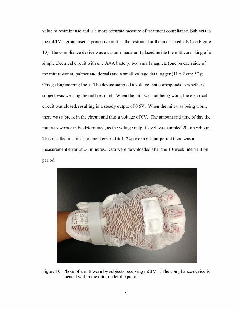
81
value to restraint use and is a more accurate measure of treatment compliance. Subjects in
the mCIMT group used a protective mitt as the restraint for the unaffected UE (see Figure
10). The compliance device was a custom-made unit placed inside the mitt consisting of a
simple electrical circuit with one AAA battery, two small magnets (one on each side of
the mitt restraint, palmer and dorsal) and a small voltage data logger (11 x 2 cm; 57 g;
Omega Engineering Inc.). The device sampled a voltage that corresponds to whether a
subject was wearing the mitt restraint. When the mitt was not being worn, the electrical
circuit was closed, resulting in a steady output of 0.5V. When the mitt was being worn,
there was a break in the circuit and thus a voltage of 0V. The amount and time of day the
mitt was worn can be determined, as the voltage output level was sampled 20 times/hour.
This resulted in a measurement error of ± 1.7%; over a 6-hour period there was a
measurement error of ±6 minutes. Data were downloaded after the 10-week intervention
period.
Figure 10 Photo of a mitt worn by subjects receiving mCIMT. The compliance device is located within the mitt, under the palm.

82
6.1.5 Analyses
For the purpose of my thesis, only data based on a preliminary subset of subjects
was investigated. Given the number of subjects enrolled, and the number randomized to
the experimental (mCIMT) group, subsequent sections report methods and data as they
relate to a single subject. Information regarding individual subject data is presented to
characterize the subject (e.g., demographic data, type of stroke experienced, and baseline
measures). Changes in UE function and activity were assessed using accelerometry,
MAL and ARAT scores. Custom software applied to the accelerometry data quantified
UE movement as a composite score using parameters equivalent to those previously
reported.98 Specifically, time-stamped accelerometer data were examined in 2-second
epochs (equivalent to 20 samples) for each of the X, Y and Z-axes. The resultant 2-
second epochs were then examined to determine the number of epochs that exceed an
absolute threshold value of 2, which is representative of a movement in that particular
axis. The resulting composite score was the sum of the number of 2-second epochs,
expressed in seconds, which exceed the threshold value. For instance, if 2000 epochs
were found to exceed the threshold value, the resultant composite score would be 4000
seconds (i.e., for that axis, movement was detected during the data collection period for
4000 seconds). Movement along the x-axis (moving front-to-back as shown in Figure
11) is reported and used for analysis as it is has been shown to be most reflective of
movement.98 Where appropriate, comparisons were made within the same UE from pre
to post-treatment, and between the unaffected and affected UE at baseline and at post-
treatment. As outlined previously, the MAL is a quantitative, but subjective, report of
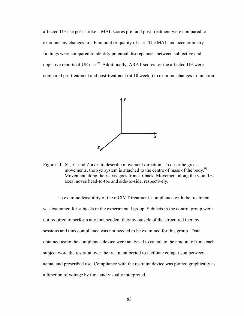
83
affected UE use post-stroke. MAL scores pre- and post-treatment were compared to
examine any changes in UE amount or quality of use. The MAL and accelerometry
findings were compared to identify potential discrepancies between subjective and
objective reports of UE use.39 Additionally, ARAT scores for the affected UE were
compared pre-treatment and post-treatment (at 10 weeks) to examine changes in function.
Figure 11 X-, Y- and Z-axes to describe movement direction. To describe gross movements, the xyz system is attached to the centre of mass of the body.99 Movement along the x-axis goes front-to-back. Movement along the y- and z- axes moves head-to-toe and side-to-side, respectively.
To examine feasibility of the mCIMT treatment, compliance with the treatment
was examined for subjects in the experimental group. Subjects in the control group were
not required to perform any independent therapy outside of the structured therapy
sessions and thus compliance was not needed to be examined for this group. Data
obtained using the compliance device were analyzed to calculate the amount of time each
subject wore the restraint over the treatment period to facilitate comparison between
actual and prescribed use. Compliance with the restraint device was plotted graphically as
a function of voltage by time and visually interpreted.

84
6.1.6 Therapy
As highlighted above, subjects were enrolled within 2 weeks of admission and
treatment continued for 10 consecutive weeks. Thus, treatment spans from acute
inpatient care at the QEII to in- or out-patient rehabilitation at the Nova Scotia
Rehabilitation Centre (NSRC) (see Figure 12 for a detailed timeline). The same
occupational or physical therapist administered treatment for subjects in the experimental
and control groups. The team members received training in mCIMT at a workshop
performed prior to the onset of the study.
Figure 12 Treatment timeline. Patients meeting the inclusion criteria and who are willing to provide informed consent will be assessed [baseline measures and pre-treatment testing (T=0)] and then randomized within two weeks of admission. Treatment will occur for 10 weeks as either an in- or out-patient. Assessment of the outcome measures will occur at the end of treatment (post-treatment; T = 10) and following 6 months, (1st retention test) and 12 months (2nd retention test).
6.1.6.1 Usual Care
All patients enrolled in the trial (control and experimental) received usual care.
Usual care within in-patient settings typically consisted of 2 hours/day, one hour each of
OT and PT. It focused on a variety of therapeutic techniques, including ROM activities,
strengthening, manual dexterity exercises and general aerobic conditioning. Once a
85
patient began outpatient therapy, usual care often consisted of 1 hour of group therapy, 2
times/week emphasizing the same components of in-patient usual care. Documentation
was collected throughout the study as per usual clinical practice and reviewed. This
helped to ensure that therapies were consistent among subjects.
6.1.6.2 Control Group
Subjects randomized to the control group received a total of 1.5 hours/week of
additional usual care as part of the trial. These sessions were typically broken down into
30-minute sessions performed 3 times/week. Since the control group performed the same
volume of treatment as the experimental group, any changes in outcome measures could
be attributed to the difference in treatment (mCIMT plus usual care vs. dose-matched
usual care alone) rather than simply receiving more therapy.
6.1.6.3 Experimental Intervention (mCIMT) Subjects randomized to the experimental group received a total of 1.5 hours/week
of mCIMT. This treatment consisted of 30-minute therapy sessions performed 3
times/week.16, 17 As described earlier, the therapy sessions incorporated shaping
techniques as subjects performed various functional tasks. Furthermore, the tasks were
performed primarily using the affected UE while the unaffected UE was restrained in a
mitt. Additional to the therapy sessions, subjects undergoing mCIMT treatment aimed to
wear the mitt restraint on the unaffected extremity 5 hours/weekday. This may have
occurred in a 5-hour block or smaller intervals throughout the day that summed to 5
hours. I worked in collaboration with the treating physical therapist in the preparation
and documentation of mCIMT therapy sessions. Time was spent before each treatment
session and outside of normal sessions to prepare the tasks and outline the focus for the

86
session. Subjects also received homework tasks to complete outside of the structured
therapy sessions. Records related to the homework assigned and degree to which they
were completed were filled out by myself and the subject. The homework tasks were
reassessed at each therapy session to ensure the activities are appropriate for each subject
(i.e. the task difficulty matches each person’s functional ability).
Each therapy session involved performing and practicing 4-5 functionally relevant
tasks. Subjects spent about 5-7 minutes per task, with rest breaks provided as needed.12
Tasks included as part of the therapy must have been functional and able to adhere to
shaping principles. Tasks must have had a performance component that allows for
feedback to the subject. For instance, tasks could be performed in timed trials (eg.
complete X within a 30 second trial). Furthermore, the tasks must have been able to be
modified so they could be progressive and measureable (Appendix G for examples).
6.1.7 Thesis Work
Within the context of the larger clinical trial, this aspect of my thesis work
focused on the development and execution of the mCIMT intervention. This was
inclusive of task development, mCIMT session planning and execution, behavioural
techniques, and homework activities. The case study that follows provides an example of
how I planned and executed sessions, and offers a context for a discussion as it relates to
the delivery of mCIMT. Additionally the case study provides a preliminary indication of
the study effectiveness and feasibility
87
6.1.7.1 Task Development and mCIMT Session Planning
In preparing therapy sessions, tasks could be selected from a pre-determined
database of tasks or new tasks could be created that addressed a subject’s motor deficits
or aligned with their personal interests. A component of my thesis work involved
designing tasks and structuring the mCIMT therapy sessions. Task design and selection
were based on the considerations outlined above (i.e., amenable to shaping) and those
established in prior work on CIMT.12, 13 Subjects identified 4-5 major goals and
functional tasks they enjoy (eg. gardening, paddling or fishing). These goals were broken
down into functional movements, and then further reduced to their component
movements (eg. elbow flexion, wrist extension). Task selection focused on the
movements necessary to achieve the larger functional movement as well as the
movements that may have had the greatest potential for improvement. As alluded to
above, tasks should not only be based on a subject’s functional ability, but also should
reflect the subjects’ interests. The subject valued the activities, either as an identified
goal or a subcomponent of the functional movement.
As outlined above, task selection was guided by the ability to develop shaping
parameters for that particular task. For each task, a series of potential shaping
progressions was established to modify the task and progressively increase task
difficulty.13, 47 These shaping steps matched subjects’ functional abilities to promote
success. Creating a shaping program allowed the therapist and I to modify task difficulty
in small amounts to better match improvements in motor function and task performance.
The shaping progressions developed also provided a means to give the subject positive

88
feedback related to their performance, which is a key component of the behavioural
aspect of mCIMT.13, 32
6.1.7.2 mCIMT Therapy Sessions
A key piece of this aspect of my thesis work involved attending all of the mCIMT
therapy sessions to document the overall treatment session, document and implement
feedback throughout the sessions, and lastly, document the subject’s performance on the
individual tasks utilized. This documentation included measures of performance (eg.
number of task repetitions performed correctly, number of repetitions in 30 seconds etc.).
As outlined above, documentation of performance could be used to provide immediate
positive feedback (eg. in reference to throwing a ball to a target: “you completed all 5
throws successfully in half the amount of time”). Documenting performance was
important in order to titrate task difficulty as performance improved.12, 13 For instance,
task difficulty could be altered by changing the object used in the task (eg. heavier or
larger diameter object), the distance to a target, temporal aspects of the task (eg.
decreased time to complete), or the number of successful repetitions in a given time
period (eg. complete 5 times correctly in 30 seconds). Although the typical progression
was to increase task difficulty with time, ultimately it was most important to match task
difficulty to a subject’s functional ability. Task difficulty increased when a task was
performed successfully (ie. a particular shaping parameter is achieved). For example, if a
subject was asked to repeat a task 5 times accurately in 1 minute and accomplished this
goal, the task would then subsequently be altered to be more difficult (more repetitions in
the same amount of time, increased distance to perform the task etc.). On the other hand,
if a subject was not successful in a task, it was modified to decrease the difficulty level
89
and help to ensure success on the next trial. (See Appendix G for examples of tasks with
potential shaping steps).
Additionally during therapy sessions, I led discussions with subjects regarding
challenges they faced in their daily lives and issues specific to the mCIMT treatment.
These conversations were opportunities to problem solve with subjects and address
functional tasks that could targeted.32
6.1.7.3 Additional Components of mCIMT
Prior to beginning the mCIMT treatment, subjects reviewed and were asked to
sign a behavioural contract, in conjunction with the treating therapist. The intent of this
contract was to give subjects another opportunity to review the treatment requirements to
fully understand their significance and to help ensure its adherence.17, 48 The rationale of
including a behavioural contract was to allow subjects to ‘buy-in’ to the therapy and be
motivated to participate. Additionally, if compliance was low the contract acts as a
reinforcement tool for the treating therapist to remind subjects about their commitment.
Beyond the practice of tasks within structured therapy sessions, subjects within
the mCIMT group were to wear the restraint on their unaffected UE for 5 hours per
weekday. Subjects wore a protective safety mitt (Stevens Company Ltd.) during a time
of frequent use, as identified by each subject. I consulted with the subject and their
caregiver (if applicable) to recognize appropriate times for wear. Depending on the
subject’s lifestyle, times for wearing the restraint could be broken into smaller intervals
rather than a continuous 5-hour block (for example, 2 hours after breakfast, 2 hours after
lunch, and 1 hour after dinner). It was preferable to wear the mitt for intervals lasting at
least one hour, however individual subjects’ preferences could be accommodated. Times

90
of frequent use could include recreational activity or usual therapy sessions. Safety
considerations related to mitt use (eg. when taking a shower or using a walker) were
discussed with subjects and their caregivers. Subjects were also given a document
outlining these safety considerations. I followed up with subjects throughout the 10-
week intervention period to re-evaluate the appropriate times for wearing the restraint.
Compliance with wearing the restraint was measured using a device that was embedded
within the padded surface of the mitt (as described on page 77).
6.1.7.4 mCIMT Homework Sessions
During the time that the mitt was worn outside of therapy sessions, subjects were
to engage in homework tasks. These tasks were similar to those performed during
therapy sessions and were structured such that subjects could perform them
independently. At the end of each therapy session, I would plan the homework tasks in
consultation with each subject. Tasks could be, though were not necessarily, the same as
those performed during the therapy session. Equipment could be loaned to subjects to
help them perform homework tasks. Furthermore, subjects were given written/photo
instructions for each task (when needed) and were asked to complete a form to record any
homework activities they partook in. During the next therapy session, I would re-assess
if the homework tasks were still appropriate and interesting to the subject, and would
alter as necessary. To complete this assessment, forms were developed that allowed
subjects to report information regarding homework tasks (eg. number of times performed,
difficulty level, different shaping parameters). Additionally, post-homework discussions
with each subject at the subsequent therapy sessions allowed for a review of the previous
homework tasks, any other activities subjects engaged in, and tasks that had been

91
assigned and planned for the following days. Participants could perform the same task
for homework over several days, modifying the tasks based on the shaping parameters
provided to them to ensure the tasks remained challenging.
6.1.8 Other Methodological Considerations
One of the underlying goals of this aspect of my thesis work was to highlight
challenges encountered in the preparation and implementation of mCIMT. Of primary
interest was identifying the challenges of delivering mCIMT in an acute clinical setting.
Furthermore, the research focus involved the development of individualized treatment
plans that make up the tasks and skills component of mCIMT. To this end, I have
developed a database of tasks for use in the mCIMT trial (see Appendix G). Moreover,
an additional role was to collect information from each subject and their caregiver in
order to apply tasks to be used in that individual’s treatment. Overall, I led the
development of the mCIMT interventions that were used in the trial. Recording and
eventually publicizing the mCIMT ‘task database’ will be essential to more widespread
implementation of mCIMT in clinical rehabilitation.
As highlighted previously, the following sections will include the presentation
and interpretation of data from a single subject enrolled in the mCIMT group as part of
the larger randomized control trial. Presentation of this case study will provide a
contextual backdrop to permit discussion related to the implementation and effectiveness
(evaluated in a preliminary manner) of mCIMT in this particular setting.
6.1.9 Case Description

92
The subject was a right-handed 60-year-old white man who experienced a right
pontine infarct on April 27, 2012. Upon written consent, the subject was screened on
May 1, 2012. The scores for these tests can be found in Table 8. The subject reported
some pain in his shoulder and quantified it as 4/10 on the VAS. Because he reported no
pain in his arm, he described his overall VAS pain score for the UE as 2/10. The MAS
finding of 0 in both the wrist and elbow indicates no tone about the joints. Additionally,
the subject met the minimum ROM criteria and presented with deficit in cognitive
function according to our initial screen. The subject was also tested on the MAL, using
the AOU subscale as another screening tool (see 6.1.8.2 below for information regarding
MAL scores). He tested below the exclusion score of 2.5 points.
Table 8 Scores for the case study on tests performed at screening.
VAS Modified Ashworth Scale
Orientation Active ROM Aphasia screen
Wrist Elbow Wrist
2/10 0/4 0/4 12/12 14° 5/5
Following the initial screen, it was determined that the subject met the inclusion
criteria and thus underwent randomization. The subject, who was part of the lower
functioning (he scored ≤24 on the ARAT at the baseline assessment; see 6.1.8.2 below)
and younger (≤ 60 yrs old) stratification group, was randomized into the intervention
group receiving mCIMT.
93
Prior to the baseline assessment, the subject wore accelerometers on each wrist to
measure activity over a 3-day period (see section 6.1.3 for information regarding
accelerometry).
6.1.9.1 Baseline Assessments
Baseline testing occurred on May 4, 2013. As highlighted previously, the subject
was right-handed and experienced reduced function in his left UE owing to the right-side
infarct. Prior to his stroke, the subject was experiencing a number of medical conditions
including osteoarthritis, peripheral vascular disease, diabetes, circulatory problems, and
high blood pressure. Additionally, the subject was a regular smoker. The subject was not
married or living with another person at the time of stroke. The subject was able to talk
and was oriented in time and space. Gross movement of the UE was intact (i.e., he was
able to lift both arms off his bed), however his mobility was impaired, requiring
assistance to walk.
A number of baseline measures, as outlined in Appendix G, are performed as part
of routine clinical care and collected for the trial as a means of characterizing the subjects
at entry into the trial (e.g., baseline assessment). Table 9 outlines these measures and the
date each was performed. On admission (the day of his stroke), the subject’s affected UE
was quite impaired, with a CMSA score of 1 for the arm and hand (denoting flaccid
paralysis). One day post-stroke the subject had a global impairment rating of ‘moderate’
based on the SSS (impairment in two of the assessed domains). Five days following his
stroke he had a Barthel Index of 33 (whereby 100 is completely independent). Thirty-five
days post-stroke the subject has little to no cognitive impairment based on the cognitive
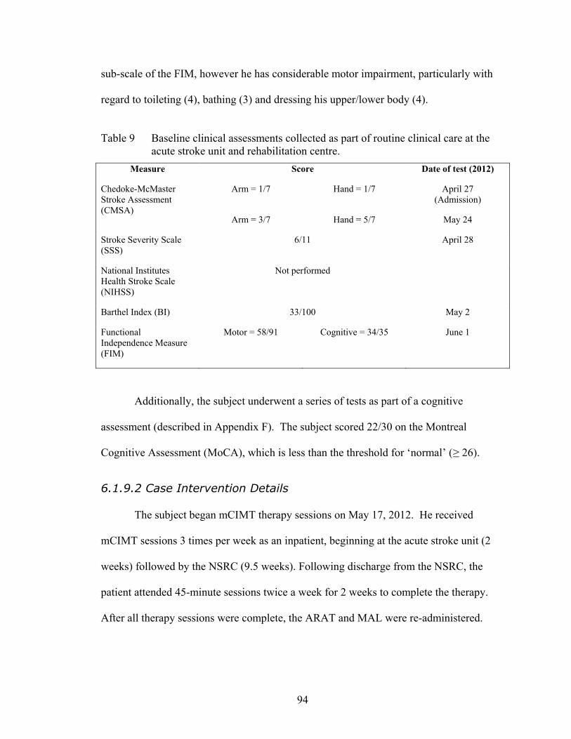
94
sub-scale of the FIM, however he has considerable motor impairment, particularly with
regard to toileting (4), bathing (3) and dressing his upper/lower body (4).
Table 9 Baseline clinical assessments collected as part of routine clinical care at the
acute stroke unit and rehabilitation centre. Measure Score Date of test (2012)
Chedoke-McMaster Stroke Assessment (CMSA)
Arm = 1/7 Hand = 1/7 April 27 (Admission)
Arm = 3/7 Hand = 5/7 May 24
Stroke Severity Scale (SSS)
6/11 April 28
National Institutes Health Stroke Scale (NIHSS)
Not performed
Barthel Index (BI) 33/100 May 2
Functional Independence Measure (FIM)
Motor = 58/91 Cognitive = 34/35 June 1
Additionally, the subject underwent a series of tests as part of a cognitive
assessment (described in Appendix F). The subject scored 22/30 on the Montreal
Cognitive Assessment (MoCA), which is less than the threshold for ‘normal’ (≥ 26).
6.1.9.2 Case Intervention Details
The subject began mCIMT therapy sessions on May 17, 2012. He received
mCIMT sessions 3 times per week as an inpatient, beginning at the acute stroke unit (2
weeks) followed by the NSRC (9.5 weeks). Following discharge from the NSRC, the
patient attended 45-minute sessions twice a week for 2 weeks to complete the therapy.
After all therapy sessions were complete, the ARAT and MAL were re-administered.
95
The subject again wore accelerometers on each wrist over a 3-day period following the
post-treatment assessment.
Prior to initiating mCIMT, the patient was asked to identify major goals that he
wanted to achieve via mCIMT, as well as tasks that he would like to focus on as part of
his therapy. The subject reported his two favourite pastimes were hunting and fishing and
thus were the main activities he wished to get back to doing. Other ADLs he identified as
being of importance to him were putting on clothing (in particular doing up buttons),
playing cards, reading, throwing and catching balls, and doing laundry. These activities
formed the basis of our therapy session and homework tasks. To accomplish his goals
over the course of therapy, each goal was broken down into component movements.
Tasks performed during mCIMT were selected to address these motor skills, evolving
from basic to more complex movement patterns as the subject recovered. For instance,
the subject’s goal with regard to hunting was to be able to hold a rifle with his left UE.
This task requires shoulder flexion (with maintenance of position at approximately 90° of
flexion), elbow flexion, gross hand movement (power grip) and isolated use of the digits.
For fishing, there are a number of movements required: shoulder mobility as well as
shoulder and elbow coupling for casting, gross hand movement (power grip) to hold the
rod, and fine finger movements for baiting the hook and gutting fish.
For each mCIMT session, a number of tasks were selected to focus on different
component movements (eg. shoulder flexion) that were part of these larger goals, with
each being shaped according to the subject’s ability. For instance, one task involved
pinning clothespins onto different rungs of a baking rack. This task involved finger
movement (pincer grasp), elbow extension, shoulder flexion and shoulder mobility.
96
During one session the task was modified as follows for each trial: (1) with the rack held
horizontally on the far left side of the table, 5 pins were to be placed anywhere on the
rack, as fast as possible (completed in 1 min, 25 sec); (2) repeat the task, with a goal of
completing it in under 1 min, 25 seconds (completed in 1 min, 35 sec); (3) place 5 pins in
the centre column only of the top half of the rack; (4) place 5 pins along the top of the
rack, focusing on the quality of the movement – aiming to rotate his wrist to pronate the
hand (completed successfully); (5) place 5 pins along the top half of the middle column
faster than previous trials (completed in 1 min). The subject was challenged to maintain
accuracy while increasing his speed. Additionally, placing the clothespins on higher
rungs was more challenging for the subject as it involved increased shoulder flexion and
shoulder stability. He was required to maintain hand accuracy (by properly placing the
pins on the rack rungs) with increased shoulder ROM. This task was performed during
subsequent therapy sessions and was further shaped by increasing the number of clothes
pins placed in a given amount of time (speed-accuracy), decreasing the space available to
place the pins (eg. on one row only), and placing the rack further away and higher up
from the subject (requiring increased elbow extension, shoulder flexion and shoulder
stability).
It is important to note that throughout the first 7.5 weeks of the mCIMT
intervention the subject struggled to stay motivated and his compliance with the mCIMT
intervention (in addition to usual care treatment) fluctuated week by week. A number of
therapy sessions were cancelled, rescheduled and altered (in terms of length) to
accommodate both the subject’s and therapist’s schedules. Missed sessions were
rescheduled but in many instances time would be added onto subsequent sessions (i.e., 45

97
vs. 30 min sessions) if the schedule did not allow for additional sessions. In total, the
mCIMT intervention for this particular subject spanned 13.5 weeks, with the same
amount of therapy hours (compared to the typical 10 week intervention) being delivered.
6.2 RESULTS
6.2.1 Primary Outcomes
At baseline the subject attained the maximum score (57) on the ARAT for his
unaffected UE. For his affected UE, the subject scored 0 on the ARAT, indicating that he
could not perform any part of the assessment tasks. Likewise, the subject scored 0 on
both subscales of the MAL (AOU and QOM) indicating that he did not use his affected
limb for any of the 14 ADLs assessed over the past week (performed during the screening
stage).
Scores on both the ARAT and MAL improved over the course of
treatment. Table 10 displays the subject’s scores for the ARAT and MAL at baseline,
post-treatment and the follow-up. The ARAT score for the affected UE improved by 34
points, well above the MCID of 6 points. MAL scores also increased from pre- to post-
treatment, with increases of 2.83 and 2.33 points for the AOU and QOM subscales,
respectively. The change in MAL scores was also greater than the MCID of 0.5 points,
indicating a clinical difference in use. Upper extremity function as measured by the
ARAT remained unchanged at the 6-month follow-up assessment for both the unaffected
(score of 57) and affected (score of 34) UE. There were slight decreases in the MAL
scores between the post-treatment and 6-month follow-up assessment, however the
decreases were minimal and likely did not reflect a clinically significant difference.
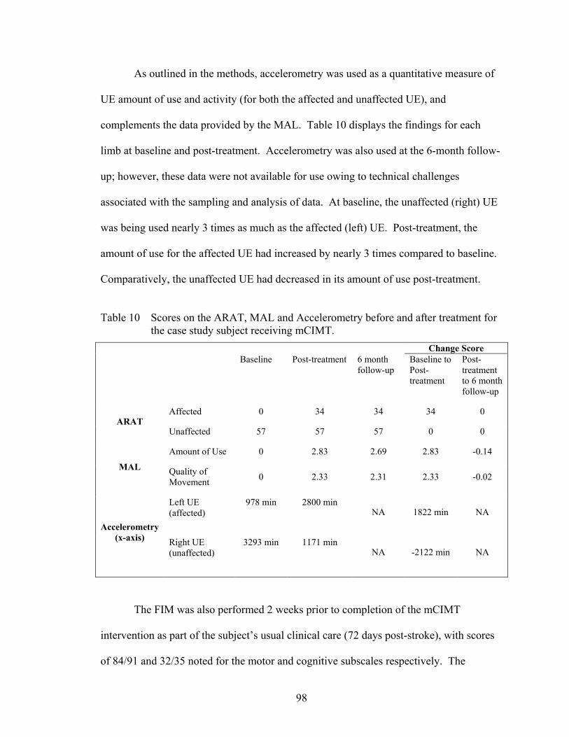
98
As outlined in the methods, accelerometry was used as a quantitative measure of
UE amount of use and activity (for both the affected and unaffected UE), and
complements the data provided by the MAL. Table 10 displays the findings for each
limb at baseline and post-treatment. Accelerometry was also used at the 6-month follow-
up; however, these data were not available for use owing to technical challenges
associated with the sampling and analysis of data. At baseline, the unaffected (right) UE
was being used nearly 3 times as much as the affected (left) UE. Post-treatment, the
amount of use for the affected UE had increased by nearly 3 times compared to baseline.
Comparatively, the unaffected UE had decreased in its amount of use post-treatment.
Table 10 Scores on the ARAT, MAL and Accelerometry before and after treatment for
the case study subject receiving mCIMT. Change Score Baseline Post-treatment 6 month
follow-up Baseline to Post-treatment
Post-treatment to 6 month follow-up
ARAT Affected 0 34 34 34 0
Unaffected 57 57 57 0 0
MAL
Amount of Use 0 2.83 2.69 2.83 -0.14
Quality of Movement 0 2.33 2.31 2.33 -0.02
Accelerometry (x-axis)
Left UE (affected)
978 min
2800 min
NA 1822 min NA
Right UE (unaffected)
3293 min
1171 min
NA -2122 min NA
The FIM was also performed 2 weeks prior to completion of the mCIMT
intervention as part of the subject’s usual clinical care (72 days post-stroke), with scores
of 84/91 and 32/35 noted for the motor and cognitive subscales respectively. The
99
subject’s score on the motor subscale represented an increase of 26 points from pre- to
post-treatment, indicating greater independence on the specific tasks assessed. For
instance, in the self-care domain, which he scored the lowest during his first assessment
(toileting, bathing and dressing his upper/lower body), he improved and reached the
maximal score (7) for each task. Locomotion remained as the only domain for which he
required assistance (6: supervising or cueing).
6.2.2 Secondary Outcomes The subject’s compliance to treatment was measured as a means of assessing the
clinical feasibility of mCIMT in this particular setting. As described in sections 6.1.3 and
6.1.5.3, the subject wore a mitt containing a compliance device to measure the amount of
time he wore the restraint. As noted in the methods, when the mitt was being worn the
compliance device would output and measure a voltage, with no voltage produced (and
thus detected) when the mitt was not being worn. Figure 13 depicts when the mitt was
being worn over the treatment time period from the first to last therapy session. Based on
the data obtained from the compliance device, it appears that the mitt was worn
extensively throughout the mCIMT intervention. For instance, from the end of June to
mid-July there appears a positive voltage, indicating that the mitt was being worn.
Interestingly the data obtained from the compliance device conflict with the subject’s
self- report of mitt use and the therapist’s (and my own) observations. Thus, the results
obtained from the compliance device may not be valid. The subject was regularly
reminded of the importance of wearing the restraint and encouraged to engage his
affected UE in ADLs and homework activities. After a series of missed and rescheduled
therapy sessions, the therapist reminded the subject of the behavioural contract he signed,
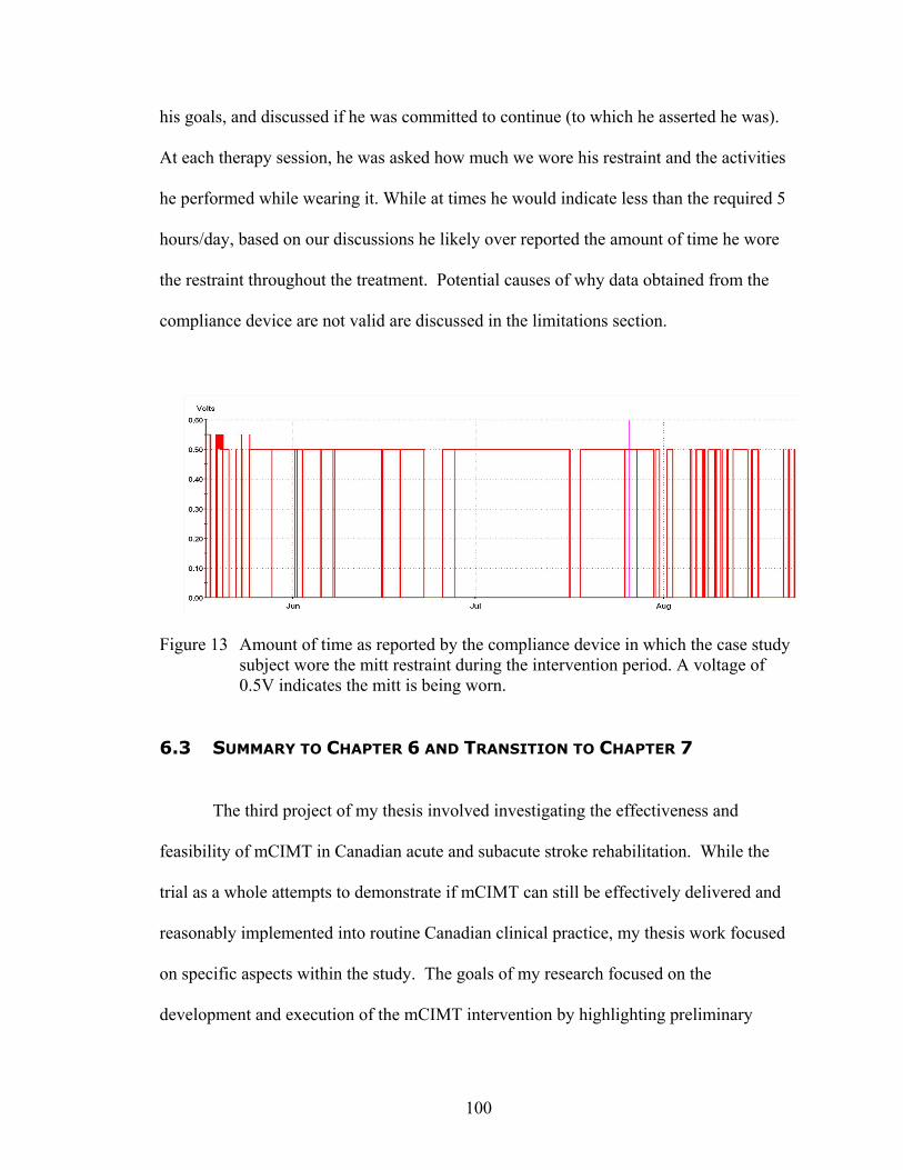
100
his goals, and discussed if he was committed to continue (to which he asserted he was).
At each therapy session, he was asked how much we wore his restraint and the activities
he performed while wearing it. While at times he would indicate less than the required 5
hours/day, based on our discussions he likely over reported the amount of time he wore
the restraint throughout the treatment. Potential causes of why data obtained from the
compliance device are not valid are discussed in the limitations section.
Figure 13 Amount of time as reported by the compliance device in which the case study subject wore the mitt restraint during the intervention period. A voltage of 0.5V indicates the mitt is being worn.
6.3 SUMMARY TO CHAPTER 6 AND TRANSITION TO CHAPTER 7
The third project of my thesis involved investigating the effectiveness and
feasibility of mCIMT in Canadian acute and subacute stroke rehabilitation. While the
trial as a whole attempts to demonstrate if mCIMT can still be effectively delivered and
reasonably implemented into routine Canadian clinical practice, my thesis work focused
on specific aspects within the study. The goals of my research focused on the
development and execution of the mCIMT intervention by highlighting preliminary

101
findings regarding effectiveness and feasibility. Furthermore, the purpose of my thesis
work was to discuss key features of task development and challenges of mCIMT
implementation. These goals are addressed in the context of the case study in the
following chapter. Findings and observations from the case study are further discussed
more globally in terms of barriers to mCIMT implementation and how to overcome them.

102
CHAPTER 7 DISCUSSION
7.1 CASE STUDY FINDINGS
Following the mCIMT intervention, the subject experienced clinically significant
increases in function, amount of use and quality of movement in his affected UE. These
changes are supported by the increase in his ARAT and MAL scores from pre- to post-
treatment. The accelerometry data complement and support the MAL findings,
indicating that use of his affected UE increased over the course of treatment and was
comparatively active to his right UE post-treatment. Furthermore, these clinically
relevant changes were sustained over a 6-month period following treatment with mCIMT.
Considering the subject was enrolled during the acute stage of recovery and thus was
receiving usual in-patient and outpatient therapy, it is not surprising that there was some
amount of functional recovery. Page et al. performed a RCT within an acute stroke
population to deliver a similar mCIMT intervention.16 As can be seen in Figures 6, 8 and
9 (Chapter 5), the group receiving mCIMT had significantly greater increases in ARAT
and MAL scores compared to the dose-matched usual care control group. While
improvements were made, the control group did not exceed the MCID on any measure.
The mCIMT group, however, far exceeded the MCID with assessment scores that nearly
quadrupled those of the control group (21.7, 2.43 and 2.19 for the ARAT, MAL (AOU),
and MAL (QOM) respectively). Comparatively, the subject described in this case study
experienced changes on the ARAT that exceeded those of the mCIMT group in the Page
study, with similar changes observed for the MAL. This comparison to the results of the
Page study provide indirect evidence that the mCIMT treatment received by the case

103
study subject contributed to his functional gains and increased activity. Additionally,
Lang et al. suggest that the MCID for ARAT scores differs when assessing individuals in
the acute stage of post-stroke recovery.90 Depending on the side affected, the suggested
MCID raw scores are 12 (for the dominant side) and 17 (for the non-dominant side).
Further research is needed to determine if the suggested MCID is still appropriate when
measuring score changes after a 10-week mCIMT intervention (as discussed in chapter
5). Nevertheless, the case study subject demonstrated UE functional improvements
greater than the MCID of 17 points as suggested by Lang et al. Thus, the subject’s
increases in functional ability appear to be clinically meaningful.
It should be noted however that because the subject also received usual care as
part of his routine clinical treatment, some amount of UE recovery was expected.
Without direct comparison to a dose-matched control, it is not possible to definitively
conclude that the mCIMT intervention led to his functional recovery.
7.1.2 Study Limitations Although the trial is still underway, there were limitations specific to this case
study. As noted above, data from the compliance device may not be a valid
representation of the amount of time the subject wore the restraint device. There are two
likely causes of this limitation. Firstly, the magnetic reed switch, and in-turn the capacity
to generate a voltage output, is controlled by the position of the magnets on the dorsal
surface of the mitt. If the device is not oriented properly in the mitt, is it possible for the
magnets to not make contact with the reed switch, thus keeping the reed switch in the
‘on’ mode and thus continually outputting a voltage (indicating the mitt is being worn).
In this instance the device would falsely record the mitt as being worn when in fact it is
104
not. Additionally, this subject (and other subjects in the trial) have reported that an
object, such as a water bottle, can be placed inside the mitt, thus mimicking the position
of the hand, in-turn making it appear as if it the mitt is being worn. The data obtained
from the compliance device, thus, needs to be interpreted in the context of these
limitations.
Additionally, the accelerometers are used as a means to assess the amount of UE
movement, and thus changes that may result from the intervention. With that said, the
data obtained from the accelerometers are not necessarily an absolute reflection of UE
movement. Since the accelerometers are placed on subjects’ wrists, certain movements
will not be reflected in the accelerometer data (for example, finger movements while
typing on a computer keyboard since the wrist stays relatively static). Moreover, the use
of a threshold value (in this case 2) to determine if a movement occurred or not can lead
to an over-estimation of the amount of movement. However, important findings can still
be discerned in relative comparisons of the data given that the same conditions applied
across limbs and assessment time-points. Thus, while the composite scores from the
accelerometer data do not provide an absolute measure of UE movement, the difference
between limbs and/or change across time provides valuable information regarding UE
recovery.
7.2 CHALLENGES AND BARRIERS TO MCIMT IMPLEMENTATION
7.2.1 Patient Challenges
In addition to the specific limitations encountered in the case study, a number of
challenges and barriers relating to the global delivery of the mCIMT intervention were

105
apparent across subjects in the mCIMT group (which to date included 3 subjects). One of
the key aspects to successful rehabilitation, and a vital component of mCIMT, is keeping
subjects motivated.12, 32 As noted in the case study, and observed across subjects,
keeping a high level of motivation proves to be challenging. The behavioural component
of mCIMT is intended to help address this challenge, as its focus is to keep subjects
motivated through the use of short- and long-term goals, identifying and modifying tasks
appropriately to meet those goals, applying shaping principles to the tasks so subjects can
be successful in their performance, and lastly working with the subject to address
challenges to affected limb use. While these aspects of mCIMT helped the case study
subject during structured therapy sessions, outside of the structured sessions it became
more difficult to keep the subject engaged. Gillot et al. explored patient experiences and
perceptions of CIMT and the factors that influence success with the treatment.100 Factors
related to personal motivational and expectations emerged as a major theme that
generates a subject’s drive to participate in the therapy. While the case study appeared to
have positive experiences during the therapy sessions and asserted as such, a lack of
motivation was evidenced by variable compliance with treatment. Not only did the
subject need constant reminders to wear his restraint, but in turn it became difficult to
ensure that homework activities (or other ADLs) were being performed independently,
and for the subject to keep a record of it. Thus, personal motivation needs to be
encouraged to ensure proper delivery of mCIMT and to improve the chance of treatment
success (and in turn, recovery).
Furthermore, the subject struggled to adhere to the treatment schedule; a number
of therapy sessions had to be rescheduled and adjusted for time due to missed sessions

106
and conflicting appointments. The subject often had busy weekdays including other tests
and appointments and would make excuses that he was too tired, too busy, or simply did
not want to do it that day. While many people in acute and sub-acute care face similar
challenges, this subject in particular seemed to struggle to balance his schedule and meet
the everyday demands of an in-patient. The subject also missed his regular therapy
sessions and was not consistently compliant with that treatment either (based on
conversations with his occupational and physical therapist). In contrast, the two other
subjects who have since received mCIMT in the study consistently made their scheduled
therapy sessions and were proactive to reschedule as soon as a conflict arose. Another
noticeable difference was in how the subjects prioritized therapy sessions. The two recent
subjects seemed to schedule their other appointments and activities around the mCIMT
sessions, rather than the other way around (as seemed to be so with the case study
subject). Nevertheless, all subjects have commented on the challenge of the mCIMT
schedule and the number of activities we ask them to do, in and outside of therapy
sessions. Additionally, the subjects were commonly fatigued during therapy sessions and
treatment would often be modified to address this.
The time and resource intensity of CIMT interventions are often cited as a
challenge to implementation, not only for therapists but for subjects as well.46, 47, 49, 70
While some aspects of treatment are diluted with the distributed mCIMT protocol, many
of the demands are still felt by subjects (as observed and subsequently reported by the
trial subjects).
The difficulties with treatment compliance and motivation exhibited by the case
study subject reflect challenges that may be tied more generally to working in an acute
107
and sub-acute rehabilitation setting. Although mCIMT is designed to keep subjects
motivated, patients dealing acutely with a stroke are often subject to many other pressures
that can affect their emotional state.3, 7 For instance, the case study subject had to deal
with the likelihood that he could not return to his job, that he could not return home right
away, and that he would need to rely on distant family members. He would often discuss
these issues during therapy sessions and comment on his feelings; when treatment began
he was still trying to accept that he had experienced a stroke and the repercussions of it
on his daily life. Unlike in later stages of recovery, patients who are acutely post-stroke
have had less time to deal with the practical consequences and the emotional trauma of
their stroke. Thus, when dealing with a number of life changes it is not surprising that a
subject’s motivation to engage in therapy could fluctuate day to day. The challenges
associated with environmental and psychosocial factors likely affect all rehabilitation
therapies at this stage of recovery, although they may become more prominent with
mCIMT because of the associated time demands and commitment.
7.2.2 Therapist Challenges
Beyond the pressures experienced by patients in acute stroke rehabilitation,
therapists also face barriers to the implementation of mCIMT.70 mCIMT has many
different aspects to the treatment and requires a particular attentiveness on the part of the
therapist to ensure all the different components are being addressed.49, 101 A number of
the barriers highlighted by the survey study (see Chapter 4) became apparent while
working in the mCIMT trial. The issue of therapist knowledge is particularly prevalent
as the study therapist received additional training to deliver mCIMT and was constantly

108
challenged to employ those skills. Furthermore, I acted as an additional knowledge
resource for planning and executing therapy sessions, as well as played a practical role
assisting during sessions and following-up with subjects outside of therapy sessions.
During therapy sessions, I would record notes to be able to provide immediate
feedback for different tasks being carried out. This is an important part of shaping, but
requires time and forces a therapist to attend to several tasks simultaneously if mCIMT is
delivered independently. Outside of therapy sessions, a lot of time is spent reviewing the
results of the preceding session, and preparing/ planning for the next session. One of my
roles with regard to the therapy sessions was to select tasks and have a plan for shaping
modifications. A therapist would not normally have someone to assist with these aspects
of the therapy, and as such would increase the demands on the therapist’s time.
Furthermore, weekly I spent time outside of therapy sessions putting together homework
tasks, following up on subjects’ progress, as well as addressing everyday challenges and
how to overcome those barriers. Again, these aspects of the therapy are time consuming
and not necessarily a part of routine practice.46, 49 Ploughman et al. noted therapists
concerns about the time required outside of scheduled sessions for planning and
organizing equipment and the need for ongoing collaboration, at the very least between
therapist and subject.101 Without additional assistance, delivery of mCIMT outside of a
research study may need modification to ensure its feasibility.
Thus, not only is it important for the therapist to be knowledgeable regarding the
treatment, but also having resources in place (personnel and materials) helps ensure the
therapy can be carried out as it is intended. With the heavy workloads already facing
therapists, it may prove difficult for a therapist to deliver mCIMT independently.46, 49, 101
109
7.2.3 How to Address the Challenges and Limitations
In identifying that a therapist’s ability to independently deliver mCIMT may be a
barrier to implementation, two potential resources come about: the relationship between
complementary disciplines (i.e., OT and PT), and the role of a caregiver. Within the
literature it appears appropriate that either an occupational or physical therapist could
deliver mCIMT. Practically within acute post-stroke rehabilitation, patients often receive
both OT and PT. What a therapist focuses treatment on may depend on what other
treatments the patient is receiving, as well as other areas of need. Considering the
potential for both professions to deliver mCIMT, it is plausible that a patient’s
occupational and physical therapist could work in tandem to deliver the therapy and share
the workload. Ploughman et al. performed a case study applying CIMT, with the
delivery of CIMT sessions shared between a pair of occupational and physical
therapists.101 In examining if CIMT was a feasible treatment option, the researchers
decided to split the therapy into 4 sessions, alternating between therapists each session.
The therapists noted that while the treatment was resource intensive and required daily
communication between therapists, they felt competent to deliver the treatment in their
practice. It was stressed that the collaborative approach was vital; the shared workload
and family participation likely led to the success of the program. As alluded to above, the
subject’s caregiver (family) can be an additional resource in therapy sessions, or as a
resource at home. How active a role a caregiver is able to play may prove to be an
important measure of success. Gillot et al. noted that while personal motivation is critical
to participation in CIMT, environmental demands (including family support) could
influence the drive to participate and recover.100 As an example within the case study,
110
once the subject had a regular caregiver he no longer missed a therapy session, reported
increased compliance with regard to wearing his mitt, and regularly reported the activities
he performed at home. Additionally, Ploughman et al. noted that not only could family
members influence motivation, but also their willingness to participate was fundamental
to the treatment’s success. Thus, beyond simply motivating subjects to stay engaged, a
caregiver can play an active role helping a subject with homework task and has the
potential to assist a therapist within therapy sessions.
Aside from additional personnel, resource materials can provide therapists with
the tools to more effectively use mCIMT, independently or within a health care team.
Systematic reviews that critically analyze individual CIMT protocols are relevant and
needed so clinicians have access to robust sources of evidence when making treatment
decisions that are specific to their patient population.51 In turn, with a protocol guide in
place, a task database can help with clinical implementation by acting as a knowledge
source and increasing the efficiency of treatment delivery. A task database can help with
mCIMT implementation by forming the basic foundation of a treatment package with the
tasks, equipment needed, and some preliminary shaping parameters, progressions, and
suggestions for feedback, set forth as a guideline for therapists. While useful for all
therapists no matter the level of mCIMT experience, a database may particularly target
therapists newer to the therapy. Additionally, a task database can help to address issues
related to the time required to plan, organize and deliver treatment with regard to
choosing activities and potential shaping parameters. Having access to a collection of
tasks and their component parts can help a therapist more quickly prepare treatment plans

111
and maximize therapy time; it would make mCIMT easier to implement as therapists
could consult the resource for new ideas and to ensure proper delivery of treatment.
There may be cases though where a therapist does not have access to additional
personnel (in terms of a colleague or family member), and access to knowledge
resources, such as a task database, and thus cannot overcome the time and resource
demands. Given these additional requirements that make mCIMT feasible to deliver
clinically, future research should perhaps focus on examining the therapy’s component
parts to determine what components are critical to treatment and what aspects of therapy
can be reduced or removed altogether. Uswatte et al. investigated in a preliminary
manner the contribution of shaping and restraint components of CIMT in affecting
treatment outcomes.15 Groups in this particular study used different types of restraint and
the training varied to include and exclude shaping. Even though one group did not use a
restraint and only received RTP with shaping, no between group differences were found
at post-treatment. Functional gains were observed after receiving CIMT even in subjects
that were not restrained. Similarly, Sterr et al. were interested in the clinical benefits of a
shaping-only training protocol and performed a series of studies to examine this.26, 47
Treatment effects were found to be specific for subjects receiving RTP in conjunction
with shaping, but not for those who received dose-matched RTP training, highlighting the
importance of shaping principles for clinical outcomes. It was asserted that “clinical
improvement is neither determined simply by amount of therapy nor the intensity with
which it is applied, but by what the patient does in the treatment sessions, that is the
therapeutic principles applied.”47 Hence, functional improvements can be gained even
without the restraint; shaping-only protocols may provide a reasonable alternative when
112
use of a restraint is not feasible. It should be noted however that subsequent findings by
Sterr et al. suggest that for patients who experience learned nonuse, constraining the
unaffected UE is still most likely the most powerful tool to overcome this behaviour.47
In addition to shaping principles, Gauthier et al. investigated the other behavioural
training techniques (termed the ‘transfer package’ which included eg. daily monitoring of
UE use in ADLs and problem solving sessions with the therapists to overcome perceived
barriers to UE use) and their role in treatment outcomes.32 Comparing two subject
groups, one that received all aspects of CIMT (including the transfer package) and
another that did not receive the transfer package, no between-group differences were
found in scores for laboratory-based measures of motor ability (WMFT). While both
therapies were equally effective at yielding significant improvements with regard to
function, subjects receiving the transfer package saw significantly greater improvements
in real-world amount of use (MAL), compared to subjects who did not receive the
transfer package. Thus, the behavioural component of CIMT appears to play an
important role to promote a highly successful transfer of functional gains into real-world
activity. While the restraint appears to be somewhat important, the behavioural and
shaping component to treatment has emerged as key aspects of CIMT. Unfortunately,
reducing or removing the use of a restraint does not overcome the barriers related to time
and resource intensity of treatment.70 Thus, not only is there a need to identify the most
important aspects of mCIMT, but research is also needed to investigate the minimal
combination of components necessary for functional gains. Research needs to establish
the dose-response of mCIMT and balance efficacy with an amount of treatment that is
still clinically feasible to deliver.
113
In considering treatment feasibility, it should be considered if the therapeutic
benefits of mCIMT outweigh the costs (financial and otherwise). Initial attempts by the
Ontario Ministry of Health to examine the cost-effectiveness of CIMT (in this instance
the 2 week or ‘traditional’ protocol) suggest that while positive outcomes are reported,
they cannot overcome the financial pressures associated with its delivery.68 As noted
above however, this report only looked at massed practice protocols and focused on
short-term outcomes. There needs to be further investigation into the costs of distributed
protocols and if these may be more feasible to deliver in a cost-effective manner.
Additionally, investigations must consider short and long-term treatment outcomes as
many upfront costs could be offset by long-term benefits associated with better functional
recovery.
7.3 CONCLUSION
Based on the findings from the case study reported above, mCIMT appears to
have potential as a treatment option post-stroke. While the patient and therapist faced
challenges in implementing the therapy, ultimately the subject saw marked improvements
in his affected UE recovery in terms of function and activity. As demonstrated in the
literature highlighted by the systematic review, there is intermediate evidence that
supports the use of mCIMT to treat the UE post-stroke. However, findings from the
CIMT survey study reflect many of the practical challenges faced by clinicians when
trying to implement the therapy. Striving to ensure therapies are successful, we may
need to find a better balance between effectiveness and feasibility. Future research
should investigate the effectiveness of treatment in larger studies to strengthen the
114
evidence. Likewise it is also important that research investigates how mCIMT treatments
are being delivered and how each component contributes to recovery. In its present form,
mCIMT may not be feasible to implement in acute rehabilitation; however it should be
investigated if the treatment can be further distilled and simplified while maintaining
effectiveness. By better understanding mCIMT and its potential for UE functional
recovery, clinicians can be more effective and efficient in delivering treatments and in
turn offer better healthcare to their patients.

115
REFERENCES
1. Heart and Stroke Foundation of Canada. Statistics 2012. Available from: URL: http://www.heartandstroke.com/site/c.ikIQLcMWJtE/b.3483991/k.34A8/Statistics.htm#stroke.
2. Reorganizing stroke care in Nova Scotia. Halifax, NS: Nova Scotia Integrated Stroke Strategy Committee 2002.
3. Barreca S. Management of the post stroke arm and hand: Treatment recommendations of the 2001 consensus panel. Heart and Stroke Foundation of Ontario 2001:162.
4. Barreca S, Wolf SL, Fasoli S, Bohannon R. Treatment interventions for the paretic upper limb of stroke survivors: a critical review. Neurorehabil Neural Repair 2003;17(4):220-6.
5. Lai SM, Studenski S, Duncan PW, Perera S. Persisting consequences of stroke measured by the Stroke Impact Scale. Stroke 2002;33(7):1840-4.
6. Mayo NE, Wood-Dauphinee S, Cote R, Durcan L, Carlton J. Activity, participation, and quality of life 6 months poststroke. Arch Phys Med Rehabil 2002;83(8):1035-42.
7. Dobkin BH. Clinical practice. Rehabilitation after stroke. N Engl J Med 2005;352(16):1677-84.
8. Lindsay MP, Gubitz G, Bayley M, Hill MD, Davies-Schinkel C, Singh S et al. Canadian Best Practice Recommendations for Stroke Care (Updated 2010). Ottawa, Ontario, Canada: Canadian Stroke Network; 2010.
9. Teasell RW, Foley NC, Salter KL, Jutai JW. A blueprint for transforming stroke rehabilitation care in Canada: the case for change. Arch Phys Med Rehabil 2008;89(3):575-8.
10. Teasell R, Meyer MJ, Foley N, Salter K, Willems D. Stroke rehabilitation in Canada: a work in progress. Top Stroke Rehabil 2009;16(1):11-9.
11. Wolf SL, Winstein CJ, Miller JP, Taub E, Uswatte G, Morris D et al. Effect of constraint-induced movement therapy on upper extremity function 3 to 9 months after stroke: the EXCITE randomized clinical trial. JAMA 2006;296(17):2095-104.
12. Taub E, Uswatte G, King DK, Morris D, Crago JE, Chatterjee A. A placebo-controlled trial of constraint-induced movement therapy for upper extremity after stroke. Stroke 2006;37(4):1045-9.

116
13. Taub E, Crago JE, Burgio LD, Groomes TE, Cook EW, 3rd, DeLuca SC et al. An operant approach to rehabilitation medicine: overcoming learned nonuse by shaping. J Exp Anal Behav 1994;61(2):281-93.
14. Taub E, Uswatte G, Elbert T. New treatments in neurorehabiliation founded on basic research. Nat Rev Neurosci 2002;3(3):228-36.
15. Uswatte G, Taub E, Morris D, Barman J, Crago J. Contribution of the shaping and restraint components of Constraint-Induced Movement therapy to treatment outcome. NeuroRehabilitation 2006;21(2):147-56.
16. Page SJ, Levine P, Leonard AC. Modified constraint-induced therapy in acute stroke: a randomized controlled pilot study. Neurorehabil Neural Repair 2005;19(1):27-32.
17. Page SJ, Levine P, Leonard A, Szaflarski JP, Kissela BM. Modified constraint-induced therapy in chronic stroke: results of a single-blinded randomized controlled trial. Phys Ther 2008;88(3):333-40.
18. Taub E. Movement in nonhuman primates deprived of somatosensory feedback. Exerc Sport Sci Rev 1976;4:335-74.
19. Taub E, Uswatte G, Mark VW, Morris DM. The learned nonuse phenomenon: implications for rehabilitation. Eura Medicophys 2006;42(3):241-56.
20. Nudo RJ, Milliken GW, Jenkins WM, Merzenich MM. Use-dependent alterations of movement representations in primary motor cortex of adult squirrel monkeys. J Neurosci 1996;16(2):785-807.
21. Nudo RJ, Milliken GW. Reorganization of movement representations in primary motor cortex following focal ischemic infarcts in adult squirrel monkeys. J Neurophysiol 1996;75(5):2144-9.
22. Plautz EJ, Milliken GW, Nudo RJ. Effects of repetitive motor training on movement representations in adult squirrel monkeys: role of use versus learning. Neurobiol Learn Mem 2000;74(1):27-55.
23. Risley T, Baer D. Operant behavior modification: The deliberate development of behavior. Review of child development research 1973;3:283-329.
24. Taub E, Perrella P, Barro G. Behavioral development after forelimb deafferentation on day of birth in monkeys with and without blinding. Science 1973.
25. Taub E, Perrella P, Miller E, Barro G. Diminution of early environmental control through perinatal and prenatal somatosensory deafferentation. Biological psychiatry 1975;10(6):609.
117
26. Sterr A, Freivogel S, Schmalohr D. Neurobehavioral aspects of recovery: Assessment of the learned nonuse phenomenon in hemiparetic adolescents. Archives of Physical Medicine and Rehabilitation 2002;83(12):1726-31.
27. Ostendorf CG, Wolf SL. Effect of forced use of the upper extremity of a hemiplegic patient on changes in function. A single-case design. Phys Ther 1981;61(7):1022-8.
28. Wolf SL, Lecraw DE, Barton LA, Jann BB. Forced use of hemiplegic upper extremities to reverse the effect of learned nonuse among chronic stroke and head-injured patients. Experimental neurology 1989;104(2):125-32.
29. Halberstam JL, Zaretsky HH, Brucker BS, Guttman AR. Avoidance conditioning of motor responses in elderly brain-damaged patients. Arch Phys Med Rehabil 1971;52(7):318-27 passim.
30. Taub E, Miller NE, Novack TA, Cook EW, 3rd, Fleming WC, Nepomuceno CS et al. Technique to improve chronic motor deficit after stroke. Arch Phys Med Rehabil 1993;74(4):347-54.
31. Winstein CJ, Miller JP, Blanton S, Taub E, Uswatte G, Morris D et al. Methods for a multisite randomized trial to investigate the effect of constraint-induced movement therapy in improving upper extremity function among adults recovering from a cerebrovascular stroke. Neurorehabil Neural Repair 2003;17(3):137-52.
32. Gauthier LV, Taub E, Perkins C, Ortmann M, Mark VW, Uswatte G. Remodeling the brain: plastic structural brain changes produced by different motor therapies after stroke. Stroke 2008;39(5):1520-5.
33. Wolf SL, Winstein CJ, Miller JP, Thompson PA, Taub E, Uswatte G et al. Retention of upper limb function in stroke survivors who have received constraint-induced movement therapy: the EXCITE randomised trial. Lancet neurology. 2008. p 33-40.
34. Liepert J, Bauder H, Wolfgang HR, Miltner WH, Taub E, Weiller C. Treatment-induced cortical reorganization after stroke in humans. Stroke 2000;31(6):1210-6.
35. Dromerick AW, Lang CE, Birkenmeier RL, Wagner JM, Miller JP, Videen TO et al. Very Early Constraint-Induced Movement during Stroke Rehabilitation (VECTORS): A single-center RCT. Neurology 2009;73(3):195-201.
36. Wu CY, Chen CL, Tang SF, Lin KC, Huang YY. Kinematic and clinical analyses of upper-extremity movements after constraint-induced movement therapy in patients with stroke: a randomized controlled trial. Arch Phys Med Rehabil 2007;88(8):964-70.
37. Lin KC, Wu CY, Wei TH, Lee CY, Liu JS. Effects of modified constraint-induced movement therapy on reach-to-grasp movements and functional performance after chronic stroke: a randomized controlled study. Clin Rehabil 2007;21(12):1075-86.
118
38. Page SJ, Sisto SA, Levine P, Johnston MV, Hughes M. Modified constraint induced therapy: a randomized feasibility and efficacy study. J Rehabil Res Dev 2001;38(5):583-90.
39. Page SJ, Sisto SA, Levine P. Modified constraint-induced therapy in chronic stroke. Am J Phys Med Rehabil 2002;81(11):870-5.
40. Szaflarski JP, Page SJ, Kissela BM, Lee JH, Levine P, Strakowski SM. Cortical reorganization following modified constraint-induced movement therapy: a study of 4 patients with chronic stroke. Arch Phys Med Rehabil 2006;87(8):1052-8.
41. Page SJ, Sisto S, Johnston MV, Levine P. Modified constraint-induced therapy after subacute stroke: a preliminary study. Neurorehabil Neural Repair 2002;16(3):290-5.
42. Atteya AAA. Effects of modified constraint induced therapy on upper limb function in subacute stroke patients. Neurosciences 2004;9(1):24-9.
43. Maulden SA, Gassaway J, Horn SD, Smout RJ, DeJong G. Timing of Initiation of Rehabilitation After Stroke. Archives of Physical Medicine and Rehabilitation 2005;86(12, Supplement 1):34-40.
44. Paolucci S, Antonucci G, Grasso MG, Morelli D, Troisi E, Coiro P et al. Early versus delayed inpatient stroke rehabilitation: A matched comparison conducted in Italy. Archives of Physical Medicine and Rehabilitation 2000;81(6):695-700.
45. Horn SD, DeJong G, Smout RJ, Gassaway J, James R, Conroy B. Stroke Rehabilitation Patients, Practice, and Outcomes: Is Earlier and More Aggressive Therapy Better? Archives of Physical Medicine and Rehabilitation 2005;86(12, Supplement 1):101-14.
46. Page SJ, Levine P, Sisto S, Bond Q, Johnston MV. Stroke patients' and therapists' opinions of constraint-induced movement therapy. Clin Rehabil 2002;16(1):55-60.
47. Sterr A, Szameitat A, Shen S, Freivogel S. Application of the CIT concept in the clinical environment: hurdles, practicalities, and clinical benefits. Cogn Behav Neurol 2006;19(1):48-54.
48. Stevenson T, Thalman L. A modified constraint-induced movement therapy regimen for individuals with upper extremity hemiplegia. Can J Occup Ther 2007;74(2):115-24.
49. Viana R, Teasell R. Barriers to the implementation of constraint-induced movement therapy into practice. Top Stroke Rehabil 2012;19(2):104-14.
50. Walker J, Pink MJ. Occupational therapists and the use of constraint-induced movement therapy in neurological practice. Aust Occup Ther J 2009;56(6):436-7.

119
51. Mulrow CD. Rationale for systematic reviews. British Medical Journal 1994;309(6954):597-.
52. Roger VL, Go AS, Lloyd-Jones DM, Benjamin EJ, Berry JD, Borden WB et al. Heart disease and stroke statistics--2012 update: a report from the American Heart Association. Circulation 2012;125(1):e2-e220.
53. Taub E, Uswatte G, Elbert T. New treatments in neurorehabilitation founded on basic research. Nat Rev Neurosci 2002;3(3):228-36.
54. Page SJ, Boe S, Levine P. What are the "ingredients" of modified constraint-induced therapy? An evidence-based review, recipe, and recommendations. Restor Neurol Neurosci 2013;31(3):299-309.
55. Brunner IC, Skouen JS, Strand LI. Recovery of upper extremity motor function post stroke with regard to eligibility for constraint-induced movement therapy. Top Stroke Rehabil 2011;18(3):248-57.
56. Peurala SH, Kantanen MP, Sjogren T, Paltamaa J, Karhula M, Heinonen A. Effectiveness of constraint-induced movement therapy on activity and participation after stroke: a systematic review and meta-analysis of randomized controlled trials. Clin Rehabil 2012;26(3):209-23.
57. Shi YX, Tian JH, Yang KH, Zhao Y. Modified constraint-induced movement therapy versus traditional rehabilitation in patients with upper-extremity dysfunction after stroke: a systematic review and meta-analysis. Arch Phys Med Rehabil 2011;92(6):972-82.
58. Stevenson T, Thalman L, Christie H, Poluha W. Constraint-Induced Movement Therapy Compared to Dose-Matched Interventions for Upper-Limb Dysfunction in Adult Survivors of Stroke: A Systematic Review with Meta-analysis. Physiotherapy Canada 2012;64(4):397-413.
59. COTO. Essential Competencies of Practice for Occupational Therapists in Canada. College of Occupational Therapists of Ontario; 2011.
60. CPA. Essential Competency Profile for Physiotherapists in Canada. Canadian Physiotherapy Association; 2009.
61. Shearer B, Burnham J, Wall JC, Turnbull GI. Physical and occupational therapy: what's common and what's not? Int J Rehabil Res 1995;18(2):168-74.
62. Booth J, Hewison A. Role overlap between occupational therapy and physiotherapy during in-patient stroke rehabilitation: an exploratory study. J Interprof Care 2002;16(1):31-40.

120
63. Smith S, Roberts P, Balmer S. Role Overlap and Professional Boundaries: Future Implications for Physiotherapy and Occupational Therapy in the NHS: Forum. Physiotherapy 2000;86(8):397-400.
64. Statistics Canada. Census Profile 2011. Available from: URL: http://www12.statcan.gc.ca/census-recensement/2011/dp-pd/prof/index.cfm?Lang=E.
65. D'Agostino Sr. RB, Sullivan LM, Beiser AS. Introductory Applied Biostatistics. Toronto: Thomson Brooks/Cole; 2006.
66. Corbetta D, Sirtori V, Moja L, Gatti R. Constraint-induced movement therapy in stroke patients: systematic review and meta-analysis. Eur J Phys Rehabil Med 2010;46(4):537-44.
67. Salbach NM, Jaglal SB, Korner-Bitensky N, Rappolt S, Davis D. Practitioner and organizational barriers to evidence-based practice of physical therapists for people with stroke. Phys Ther 2007;87(10):1284-303.
68. Medical Advisory Secretariat HQO. Constraint-Induced Movement Therapy for Rehabilitation of Arm Dysfunction After Stroke in Adults: An Evidence-Based Analysis. In: Services OHQ, editor. Toronto: Ontario Health Technology Assessment Series; 2011. p 1-58.
69. Taub E, Uswatte G, Pidikiti R. Constraint-Induced Movement Therapy: a new family of techniques with broad application to physical rehabilitation--a clinical review. J Rehabil Res Dev 1999;36(3):237-51.
70. Fleet A CM, MacKay-Lyons M, MacKenzie D, Page SJ, Eskes G, McDonald A, Boyce J, Boe S. Examining Utilization of Constraint-Induced Movement Therapy in Canadian Neurological Occupational and Physical Therapy. Physiotherapy Canada 2013;In Press.
71. Page SJ, Sisto S, Johnston MV, Levine P, Hughes M. Modified constraint-induced therapy in subacute stroke: a case report. Arch Phys Med Rehabil 2002;83(2):286-90.
72. Liepert J, Graef S, Uhde I, Leidner O, Weiller C. Training-induced changes of motor cortex representations in stroke patients. Acta Neurol Scand 2000;101(5):321-6.
73. Hakkennes S, Keating JL. Constraint-induced movement therapy following stroke: a systematic review of randomised controlled trials (Structured abstract). Australian Journal of Physiotherapy. 2005. p 221-31.
74. Bjorklund A, Fecht A. The effectiveness of constraint-induced therapy as a stroke intervention: A meta-analysis. Occupational Therapy in Health Care 2006;20(2):31-49.

121
75. Bonaiuti D, Rebasti L, Sioli P. The constraint induced movement therapy: a systematic review of randomised controlled trials on the adult stroke patients. Eura Medicophys 2007;43(2):139-46.
76. Sirtori V, Corbetta D, Moja L, Gatti R. Constraint-induced movement therapy for upper extremities in stroke patients. Cochrane Database of Systematic Reviews. John Wiley & Sons, Ltd; 2009.
77. French B, Leathley M, Sutton C, McAdam J, Thomas L, Forster A et al. A systematic review of repetitive functional task practice with modelling of resource use, costs and effectiveness. Health Technol Assess 2008;12(30):iii, ix-x, 1-117.
78. Finding What Works in Health Care: Standards for Systematic Reviews. The National Academies Press; 2011.
79. Lyle RC. A performance test for assessment of upper limb function in physical rehabilitation treatment and research. Int J Rehabil Res 1981;4(4):483-92.
80. De Weerdt WJG, Harrision MA. Measuring recovery of arm and hand function in stroke patients: a comparision of the Brunnstrom-Fugl-Meyer test and the Action Research Arm test. Physiother Can 1985;37(2):65-70.
81. Fugl-Meyer AR, Jaasko L, Norlin V. The post-stroke hemiplegic patient. II. Incidence, mortality, and vocational return in Goteborg, Sweden with a review of the literature. Scand J Rehabil Med 1975;7(2):73-83.
82. van der Lee JH, Beckerman H, Lankhorst GJ, Bouter LM. The Responsiveness of the Action Research Arm Test and the Fugl-Meyer Assessment Scale in Chronic Stroke Patients. Journal of Rehabilitation Medicine (Taylor & Francis Ltd) 2001;33(3):110-3.
83. Uswatte G, Taub E, Morris D, Vignolo M, McCulloch K. Reliability and validity of the upper-extremity Motor Activity Log-14 for measuring real-world arm use. Stroke 2005;36(11):2493-6.
84. Park SW, Wolf SL, Blanton S, Winstein C, Nichols-Larsen DS. The EXCITE Trial: Predicting a clinically meaningful motor activity log outcome. Neurorehabil Neural Repair 2008;22(5):486-93.
85. O'Donnell M DJ, Adams R, Butler C, Roxborough L, Damiano D. AACPDM methodology to develop systematic reviews of treatment intervention. American Academy of Cerebral Palsy & Developmental Medicine; 2005.
86. Page SJ, Levine P, Khoury JC. Modified constraint-induced therapy combined with mental practice: thinking through better motor outcomes. Stroke 2009;40(2):551-4.
87. Page SJ, Elovic E, Levine P, Sisto SA. Modified constraint-induced therapy and botulinum toxin A: A promising combination. American Journal of Physical Medicine and Rehabilitation 2003;82(1):76-80.

122
88. Page SJ, Levine P, Hill V. Mental practice as a gateway to modified constraint-induced movement therapy: a promising combination to improve function. Am J Occup Ther 2007;61(3):321-7.
89. van der Lee JH, Wagenaar RC, Lankhorst GJ, Vogelaar TW, Deville WL, Bouter LM. Forced Use of the Upper Extremity in Chronic Stroke Patients : Results From a Single-Blind Randomized Clinical Trial. Stroke 1999;30(11):2369-75.
90. Lang CE, Edwards DF, Birkenmeier RL, Dromerick AW. Estimating minimal clinically important differences of upper-extremity measures early after stroke. Arch Phys Med Rehabil 2008;89(9):1693-700.
91. Lang CE, Wagner JM, Dromerick AW, Edwards DF. Measurement of Upper-Extremity Function Early After Stroke: Properties of the Action Research Arm Test. Archives of Physical Medicine and Rehabilitation 2006;87(12):1605-10.
92. Bohannon RW, Smith MB. Interrater reliability of a modified Ashworth scale of muscle spasticity. Phys Ther 1987;67(2):206-7.
93. Platz T, Pinkowski C, van Wijck F, In-Ha K, di Bella P, Johnson G. Reliability and validity of arm function assessment with standardized guidelines for the Fugl-Meyer Test, Action Research Arm Test and Box and Block Test: a multicentre study. Clinical Rehabilitation 2005;19(4):404-11.
94. Rabadi MH, Rabadi FM. Comparison of the Action Research Arm Test and the Fugl-Meyer Assessment as Measures of Upper-Extremity Motor Weakness After Stroke. Archives of Physical Medicine and Rehabilitation 2006;87(7):962-6.
95. Hsueh IP, Hsieh C-L. Responsiveness of two upper extremity function instruments for stroke inpatients receiving rehabilitation. Clinical Rehabilitation 2002;16(6):617-24.
96. Uswatte G, Foo WL, Olmstead H, Lopez K, Holand A, Simms LB. Ambulatory monitoring of arm movement using accelerometry: an objective measure of upper-extremity rehabilitation in persons with chronic stroke. Arch Phys Med Rehabil 2005;86(7):1498-501.
97. Uswatte G, Giuliani C, Winstein C, Zeringue A, Hobbs L, Wolf SL. Validity of accelerometry for monitoring real-world arm activity in patients with subacute stroke: evidence from the extremity constraint-induced therapy evaluation trial. Arch Phys Med Rehabil 2006;87(10):1340-5.
98. Uswatte G, Miltner WH, Foo B, Varma M, Moran S, Taub E. Objective measurement of functional upper-extremity movement using accelerometer recordings transformed with a threshold filter. Stroke 2000;31(3):662-7.
99. Enoka RM. Neuromechanics of Human Movement. Human Kinetics; 2008.
123
100. Gillot AJ, Holder-Walls A, Kurtz JR, Varley NC. Perceptions and experiences of two survivors of stroke who participated in constraint-induced movement therapy home programs. Am J Occup Ther 2003;57(2):168-76.
101. Ploughman M, Shears J, Hutchings L, Osmond M. Constraint-induced movement therapy for severe upper-extremity impairment after stroke in an outpatient rehabilitation setting: a case report. Physiother Can 2008;60(2):161-70.

124
APPENDIX A Copyright Permission Contract
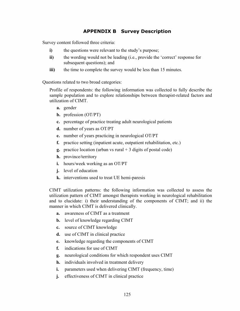
125
APPENDIX B Survey Description
Survey content followed three criteria:
i) the questions were relevant to the study’s purpose; ii) the wording would not be leading (i.e., provide the ‘correct’ response for
subsequent questions); and iii) the time to complete the survey would be less than 15 minutes.
Questions related to two broad categories:
Profile of respondents: the following information was collected to fully describe the sample population and to explore relationships between therapist-related factors and utilization of CIMT.
a. gender b. profession (OT/PT) c. percentage of practice treating adult neurological patients d. number of years as OT/PT e. number of years practicing in neurological OT/PT f. practice setting (inpatient acute, outpatient rehabilitation, etc.) g. practice location (urban vs rural + 3 digits of postal code) h. province/territory i. hours/week working as an OT/PT j. level of education k. interventions used to treat UE hemi-paresis
CIMT utilization patterns: the following information was collected to assess the utilization pattern of CIMT amongst therapists working in neurological rehabilitation and to elucidate: i) their understanding of the components of CIMT; and ii) the manner in which CIMT is delivered clinically.
a. awareness of CIMT as a treatment b. level of knowledge regarding CIMT c. source of CIMT knowledge d. use of CIMT in clinical practice e. knowledge regarding the components of CIMT f. indications for use of CIMT g. neurological conditions for which respondent uses CIMT h. individuals involved in treatment delivery i. parameters used when delivering CIMT (frequency, time) j. effectiveness of CIMT in clinical practice

126
k. barriers to CIMT utilization

127
APPENDIX C Reported Components of CIMT
List of responses (by theme) provided by CIMT users and non-users when asked to identify the key components of CIMT (see methods and results for details).
Theme Frequency (CIMT users)
Frequency (Non CIMT users)
Restraint 31 72 RTP 22 37 Shaping/Behavior 19 9 Use Affected Limb 22 57 Motivation 13 0 Treatment Duration & Schedule 12 27 Repetition 11 10 Type of Restraint 9 13 Safety 9 3 Type of Treatment 7 14 Commitment 7 3 Family Support 7 1 Education 6 0 Inclusion Criteria 5 24 Neuroplasticity 5 8 No Knowledge 4 10 UL (Upper Limb) 4 4 Patient Population 4 3 Team Approach 4 0 Bimanual Movements 3 0 Forced Use 2 6 Learned Disuse 2 3 Sensory/Cognition 2 1 Testing 2 0 FITT 1 0 Consistency 1 0 Team Knowledge 0 2 CVA 0 1 Specific Training 0 1 Hand Dominance 0 1 Motor Tasks 0 1
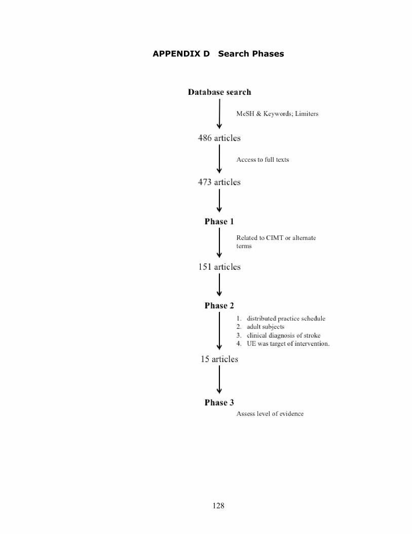
128
APPENDIX D Search Phases

129
PHASE 1 For articles that you are uncertain about, please highlight the row and provide a comment Endnote # First Author; Year
(eg. Doe, J; 2009) Journal Reason for
Exclusion Uncertain (provide comment why uncertain)
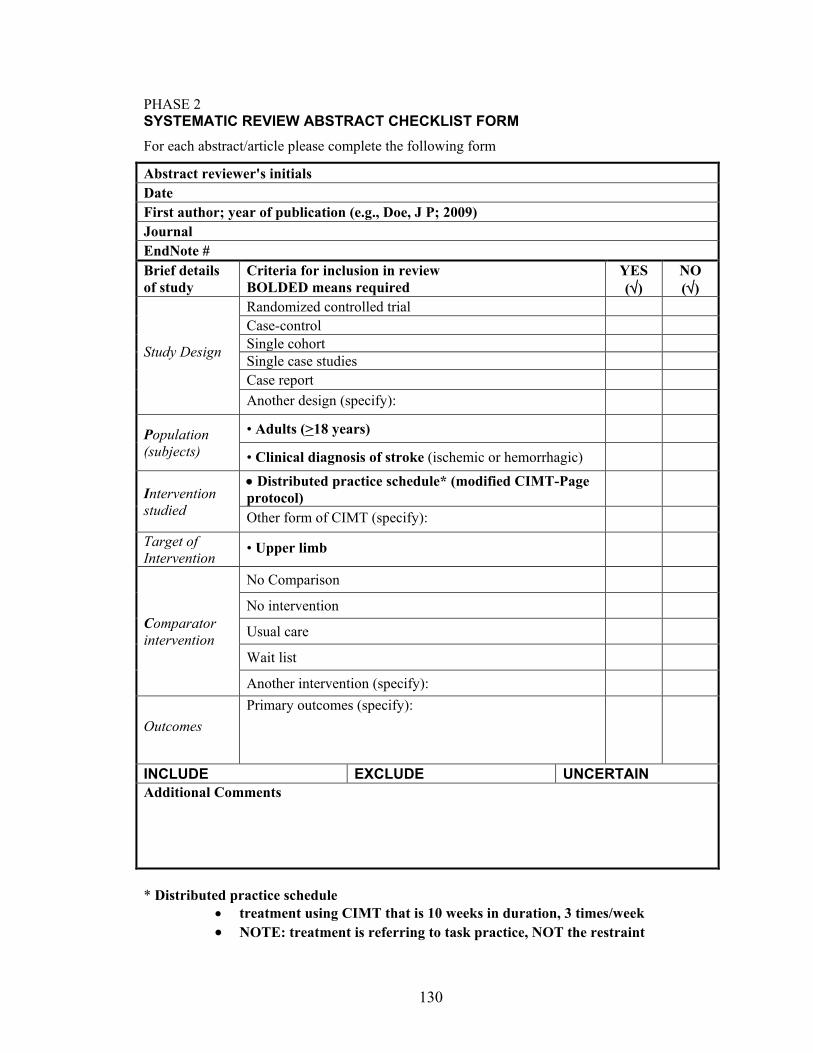
130
PHASE 2 SYSTEMATIC REVIEW ABSTRACT CHECKLIST FORM For each abstract/article please complete the following form
Abstract reviewer's initials Date First author; year of publication (e.g., Doe, J P; 2009) Journal EndNote # Brief details of study
Criteria for inclusion in review BOLDED means required
YES (√)
NO (√)
Study Design
Randomized controlled trial Case-control Single cohort Single case studies Case report Another design (specify):
Population (subjects)
• Adults (>18 years)
• Clinical diagnosis of stroke (ischemic or hemorrhagic)
Intervention studied
• Distributed practice schedule* (modified CIMT-Page protocol)
Other form of CIMT (specify):
Target of Intervention
• Upper limb
Comparator intervention
No Comparison
No intervention
Usual care
Wait list
Another intervention (specify):
Outcomes Primary outcomes (specify):
INCLUDE EXCLUDE UNCERTAIN Additional Comments * Distributed practice schedule
• treatment using CIMT that is 10 weeks in duration, 3 times/week • NOTE: treatment is referring to task practice, NOT the restraint
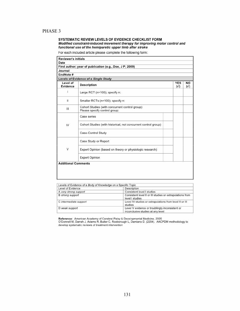
131
PHASE 3
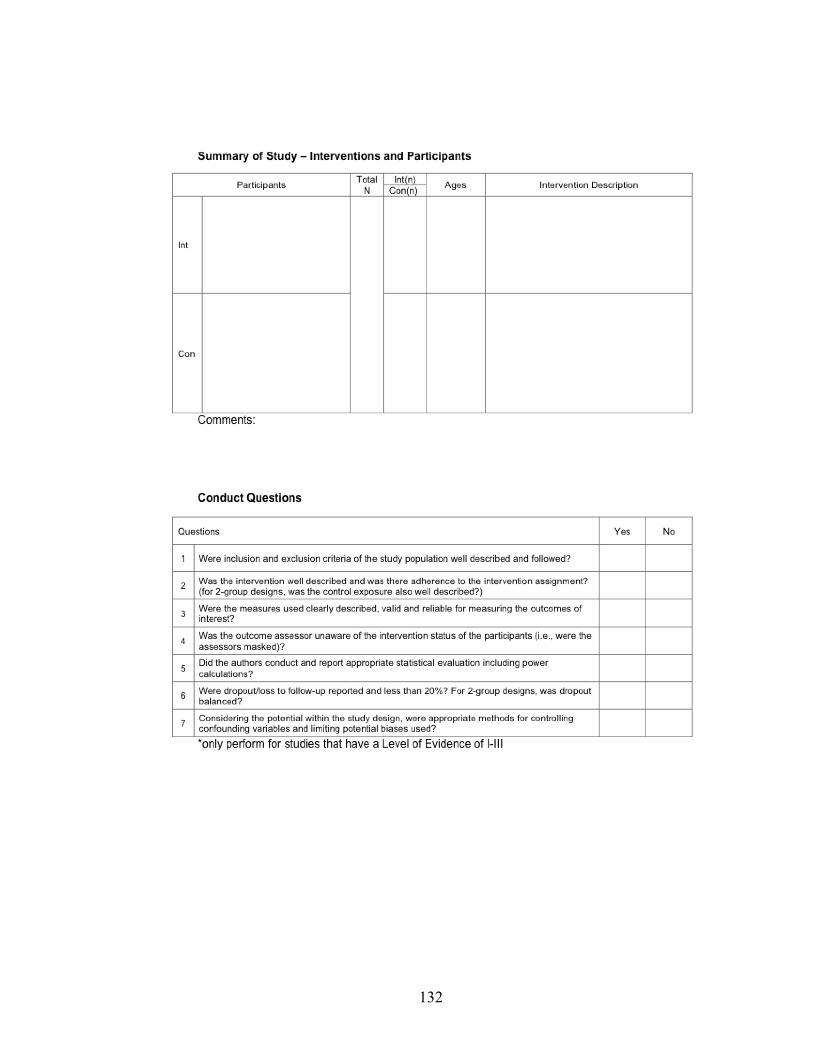
132
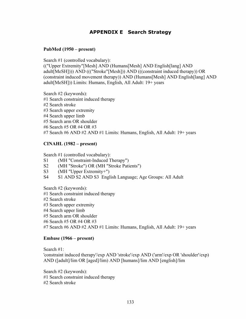
133
APPENDIX E Search Strategy
PubMed (1950 – present) Search #1 (controlled vocabulary): (("Upper Extremity"[Mesh] AND (Humans[Mesh] AND English[lang] AND adult[MeSH]))) AND ((("Stroke"[Mesh])) AND (((constraint induced therapy)) OR (constraint induced movement therapy)) AND (Humans[Mesh] AND English[lang] AND adult[MeSH])) Limits: Humans, English, All Adult: 19+ years Search #2 (keywords): #1 Search constraint induced therapy #2 Search stroke #3 Search upper extremity #4 Search upper limb #5 Search arm OR shoulder #6 Search #5 OR #4 OR #3 #7 Search #6 AND #2 AND #1 Limits: Humans, English, All Adult: 19+ years CINAHL (1982 – present) Search #1 (controlled vocabulary): S1 (MH "Constraint-Induced Therapy") S2 (MH "Stroke") OR (MH "Stroke Patients") S3 (MH "Upper Extremity+") S4 S1 AND S2 AND S3 English Language; Age Groups: All Adult Search #2 (keywords): #1 Search constraint induced therapy #2 Search stroke #3 Search upper extremity #4 Search upper limb #5 Search arm OR shoulder #6 Search #5 OR #4 OR #3 #7 Search #6 AND #2 AND #1 Limits: Humans, English, All Adult: 19+ years Embase (1966 – present) Search #1: 'constraint induced therapy'/exp AND 'stroke'/exp AND ('arm'/exp OR 'shoulder'/exp) AND ([adult]/lim OR [aged]/lim) AND [humans]/lim AND [english]/lim Search #2 (keywords): #1 Search constraint induced therapy #2 Search stroke

134
#3 Search upper extremity #4 Search upper limb #5 Search arm OR shoulder #6 Search #5 OR #4 OR #3 #7 Search #6 AND #2 AND #1 Limits: Humans, English, All Adult: 18+ years Cochrane Library and Web Of Science do not use the same type of controlled vocabulary as the previous three databases, so only keyword searches were used: Cochrane (1993 – present) "constraint induced" NEAR therap*:ti,ab,kw and (stroke):ti,ab,kw and ("upper extremity" OR "upper limb" OR arm OR shoulder:ti,ab,kw) Web Of Science (1900 – present) Topic=("constraint induced therapy" OR "constraint induced movement therapy") AND Topic=(stroke) AND Topic=("upper extremity" OR "upper limb" OR arm OR shoulder) Refined by: Languages=( ENGLISH ) Databases=SCI-EXPANDED, SSCI, A&HCI, CPCI-S, CPCI-SSH Timespan=All Years Proquest Dissertations and Theses (1966-present) Searched two phrases: “constraint induced therapy” OR “constraint induced movement therapy” Open Grey (1997 – present) Searched two phrases: “constraint induced therapy” OR “constraint induced movement therapy”

135
APPENDIX F Baseline Assessment Measures
1. Descriptive information from hospital record a. Date of birth b. Sex c. Pertinent medical and medical history d. Pre-morbid handedness e. Type and location of stroke
2. Stroke severity a. Stroke Severity Scale (SSS)
i. Measure of global impairment b. National Institutes of Health Stroke Scale (NIHSS) – 15 item impairment
scale i. Assesses cognitive, sensory and motor impairments
3. Physical impairment
a. Chedoke-McMaster Stroke Assessment – recovery stage of the arm and hand
b. Modified Ashworth Scale (MAS) i. Measure of muscle tone
4. Disability a. Functional Independence Measure (FIM) – commonly used in acute and
rehab settings to uniformly assess level of disability and associated level of assistance needed to perform activities of daily living
b. Barthel Index – used as a record of what a patient does (rather than capability) and assesses level of independence for a number of self care and mobility activities
5. Cognitive Impairment 30-minute protocol as recommended by Hachinski et al. that assesses language, memory, spatial neglect, attention and executive function, depression and other behavioural changes
a. Montreal Cognitive Assessment (MoCA) b. Fluency test c. Symbol Digit Modalities Test d. Hopkins verbal learning test – revised (HVLT-R) e. Centre for epidemiological studies – depression scale f. Neuropsychiatric inventory – questionnaire g. Trail-making h. Mini Mental State Exam (MMSE) i. Figure cancellation
6. Active ROM
136
a. Of the wrist and fingers via goniometry according to standardized clinical testing methods
* The NIHSS, CMSA and FIM are considered clinical assessments that are performed as part of routine clinical care. ** All of the above measures are only collected once

137
APPENDIX G Task Database
Example equipment list:
o Aluminum foil o Binders o Bingo cards o Bingo dauber o Books o Bottle o Bouncing ball (various sizes)
Small, rubber Tennis ball Volleyball Basketball
o Bowl (various sizes) o Brown paper bags o Buttoned shirt o Calculator o Cans of food o Checkers o Chinese checker board o Cloth o Clothes hangers o Clothespins o Coins o Coin wrappers o Computer mouse o Connect 4 game o Containers
Various sized openings o Cooling rack/wire rack o Cotton balls o Cups o Dirt o Doorknobs/locks o Dominoes o Dried beans
Various sizes Eg. Kidney; white
o Dusting cloth/dusting mitt/dust buster
o Food items/containers o Grocery bag o Iron o Ironing board
o Jenga o Keys o Keyboard o Light socket adapter o Magazine o Marbles (various sizes) o Masking tape o Measuring cup o Mouse pad with pattern
‘Mouse game’ o Napkins o Non-slip mat
to stabilizing objects o Nuts and bolts (variety of sizes) o Operation game o Paint brush o Paint roller o Pegboard o Pennies o Photo album (with photos) o Piggybank o Pillow o Pillow case o Ping pong balls o Pitcher/jug o Placemat o Plastic fruit o Plastic golf balls o Plastic wrap o Plates o Playing cards o Pot holders (larger flower
pots/small plant trays) o Rolodex o Seeds/plants (plastic or beans) o Shoe brush o Shoe polish o Silverware o Styrofoam chips o Spade (different sizes of grips) o Spoons
Teaspon

138
Tablespoon Serving spoon
o Straws (or stir sticks) o Thera-putty (or playdoh) o Towel o Unwired light socket o Wooden blocks (various sizes) o Wooden pegs o Zip lock bags
139
Badminton Use a badminton racquet and birdie. The subject can play alone, with the assistance of the therapist, or against the therapist. The subject uses the racquet to bounce the birdie. Shaping progressions:
• Moving the racquet to the left, right, or further away from body • Bouncing the birdie off the wall • Hitting a birdie thrown by the therapist • Rallying with therapist
Feedback parameters:
• Number of hits in certain amount of time • Number of successful hits without dropping • Time it takes to complete a number of hits without dropping
Movements emphasized:
• Shoulder mobility • Elbow extension • Shoulder-elbow coupling • Wrist flexion/extension • Power grip (cylindrical)
Beans Beans placed in various containers. The subject can pick up and move the beans to a target area. Additionally, objects can be mixed within the beans and selectively targeted. The subject is asked to pick up the objects one at a time and place them on the table, on a target or in a container. Shaping progressions:
• Different sized containers o shallow or deep o small or wide opening
• Different sizes of beans • Placement of container (left, centre, right) • Sitting vs. standing • Distance of container from subject • Distance between bean container and target • Size of target
o Size of container opening, working towards progressively smaller openings
• Size of objects placed in beans • Method to select beans/object
o Different pincer grips o Alternating between fingers
140
o Using a spoon Feedback parameters:
• Time to move a certain number of beans/objects • Time to complete movements without dropping beans/objects
Movements emphasized:
• Shoulder flexion/abduction • Wrist pronation/supination • Wrist flexion/extension • Fine finger movements • Various grasps (eg. pincer grip; tip-to-tip vs. pad-to-pad)
Bingo Use bingo cards and have subject mark off the various spaces. The subject can use a bingo dauber and aims to mark off a space without smearing the ink. The therapist can list off the appropriate spaces to mark. Shaping progressions:
• Size of bingo cards and spaces (smaller are more difficult) • Using a different utensil (eg. a pen to mark off each space) • Distance of bingo card from subject • Height of bingo card
Feedback parameters:
• Number of successfully marked spots (completely marked without ink outside the box)
• Time to complete a row/column or whole card
Book/Magazine/Binder Place reading material on table. The subject turns the pages while concentrating on turning the pages by either pronating or supinating
Shaping Progressions:
• The position of the reading material o Left/centre/right o Distance away from subject
• The number of pages to turn • The size/thickness of the material
Feedback parameters:

141
• Number of pages turned in a set amount of time • Amount of time required to turn a set number of pages
Movements Emphasized:
• Wrist pronation/supination • Pincer or lateral pincer grasp • Shoulder internal and external rotation
Clothes Pins on Rack Place a number of clothespins on a wire rack. The rack can be held vertically by the therapist or placed horizontally on a table. The subject can place the clothespins on the rack and/or remove them. Shaping progressions:
• Distance of rack from subject • Rack placement (left/centre/right) • Target placement of clothespins on rack
o placing to the left/centre/right of the rack) • Height of the rack • Height of clothespin placement • Target size
o Placing clothespins in a particular quadrant or rung • The number of clothespins • The digits used to open or close the clothespins
Feedback parameters:
• Number of clothespins placed in a certain amount of time • Amount of time to place a certain amount of clothespins
Movements Emphasized:
• Pincer grasps (eg. side-to-pad vs. pad-to-pad) • Wrist supination/pronation • Elbow extension • Shoulder flexion
Checkers Checkers are placed on the table. The subject is asked to move the checkers either to stack or to push the checkers by extending certain fingers. The checkers are to be stacked or pushed to a target location. Shaping progressions:
142
• Increase distance required to push the checkers • Increase number of checkers • Change location to stack checkers (left/centre/right) • Increase distance from subject to stack checkers
Feedback parameters:
• Number of checkers subject is able to push to target in a given amount of time • Time it takes to push a number of checkers to a target area, or to stack a given
number of checkers • Number of checkers that can be stacked without the stack falling over
Movements Emphasized:
• Fine finger movements • Elbow extension • Shoulder flexion
Chopsticks The subject uses chopsticks to pick up various objects and place them on a target Shaping progressions:
• Different sizes and weights of objects o Cotton balls o Styrofoam chips o Beans o Play-doh
• Distance between objects and target area • Distance of target area from subject • Height of container holding objects (deeper requires greater shoulder flexion and
longer pinching of object to successfully reach the target area) Feedback parameters:
• Number of objects moved in a certain amount of time • Number of successful movements to target ie. without dropping in certain amount
of time • Number of movements to target without dropping in a row • Amount of time to move a given number of objects
Movements emphasized:
• Fine finger control • Pincer grip • Elbow extension • Shoulder control • Shoulder flexion/abduction

143
Cleaning This includes dusting or any other wiping activity (eg. wiping off a counter). The subject stands at a table (or any other flat surface). The subject uses a cloth/sponge/duster to wipe the table clean. This can be done using a dry cloth, or a wet one in which the subject also wrings out excess water. Shaping progressions:
• Change the height of the wiping surface • Increase the surface area needed to clean
o Greater distance left to right o Increase the distance a subject needs to reach forward o Change the type of cleaning tool used (different grasps) o Different surface types
Vertical surface if have shoulder control Feedback parameters:
• Area wiped clean during a certain time period • Number of wipes it takes to clean a particular area • Time it takes to wipe off a particular area
Movements emphasized:
• Shoulder flexion • Shoulder abduction/adduction • Elbow flexion/extension • Gross hand movements • Various grasps • Finger flexion/extension
Computer mouse The subject uses a mouse that is unattached to computer. The subject practices moving it in different directions and picking it up. A particular pattern can be designed by the therapist to follow using a ‘mouse pad’ with different shapes and colours. Shaping progressions:
• Distance from subject the mouse is placed and designed to be moved • Distance between shapes (on ‘mouse pad’) • Complexity of pattern designed by therapist • Height of mouse
Feedback parameters:
• Time it takes to move through a particular pattern • The number of movements that can be performed in a set time period

144
Movements emphasized:
• Gross hand movement • Power grip (spherical grasp) • Elbow flexion/extension • Shoulder flexion
Cups/Containers A subject can use cups (or other containers) to lift, move and stack them. lifting/moving/stacking. Shaping progressions:
• Difference sized cups and containers • Containers with different handles for changes in grip
o Measuring cup o Pitcher
• Cups with different weights o Plastic o Glass o Mug
• Distance away from subject the cups are placed • Distance between the cups and the target areas • Flipping upside down when stacking or moving
Feedback parameters:
• Amount of time to move a certain number of cups • The number of cups that can be moved in a certain amount of time • The time to complete a number of accurate movements (eg. fully rotating wrist to
move through supination-pronation; holding the cup with all 5 fingers). • Amount of time to move cups a certain distance • Ability to complete a number of movements successfully in a row (eg. flipping
cups completely without having them tip over) • Increase in height to move cups
Movements emphasized:
• Gross hand movements (power grip) • Hand release • Wrist pronation/supination • Elbow flexion/extension • Shoulder movement (flexion/abduction) • Various grasps
Cutting food
145
The subject sits at a table with a knife and plate. Various objects can be used to simulate different food textures and thicknesses (eg. play-doh; cotton balls; Styrofoam chips) Shaping progressions:
• Different types of objects to cut o Eg. Play-doh is softer and easier to cut than foam chips which require
more strength and control • Different thicknesses • Number of slices for a particular sized object • Dividing an object into equal sized pieces
Feedback parameters:
• Accuracy of cutting task (e.g. pieces being of even size) • Number of pieces cut in a particular time period • Time it takes to cut a particular number of pieces • Increased strength by ability to cut tougher objects • Increased strength/grasp as evidenced by cutting putty/food with more resistance
Movements emphasized:
• Power grip • Shoulder control • Shoulder flexion/abduction
Dominoes Dominoes are placed in front of the subject. The subject is asked to reach forward and flip the dominoes using either forearm pronation or supination. The correct movement can be best isolated by asking the subject to rest their forearm on the table during the task. The subject can also be asked to lift and move the dominoes to a target (and stack) Shaping Progressions:
• The number of dominoes used • Increasing the distance to move dominoes • Different sized dominoes • Requiring subject to rest forearm on the table during the task (or requiring subject
to not lean on table if trying to promote shoulder movement) • Increase height to move dominoes
Feedback parameters:
• The number of dominoes moved or flipped in a set period of time • The amount of time required to move a set number of dominoes
Movements Emphasized:
• Lateral pincer grasp

146
• Wrist extension • Forearm supination/pronation (depending on direction of flip) • Shoulder flexion • Elbow flexion/extension
Gardening The subject stands at a table with flower pot(s) centered in front of them. A container of dirt and the plants/seeds to be planted are positioned to one or the other side. The subject uses a spade to scoop dirt into the container. Seed/simulated seeds (eg. beans) can be planted one by one into the dirt Shaping progressions:
• Placement of pot and container relative to subject • Distance between the pot and dirt container • The size of pot and opening • Type of spoon (various grasps) • Number of seeds to be planted • Amount of dirt to be moved
Feedback parameters: • Number of pots filled with dirt in a set time period • Amount of time to move a certain amount of dirt • Number of seeds/plants planted • Number of scoops needed to move a certain amount of dirt • Ability to move a certain amount of dirt without spilling any (accuracy) • Increased use of specific functional movement patterns (e.g. use of supination
when planting ‘seeds’) Movements emphasized:
• Shoulder control • Various grasps • Wrist pronation/supination • Elbow flexion/extension
Grocery packing While sitting or standing, participants lift items (eg. cans, cereal box etc.) from grocery bags and place them on a table/shelf. Items can be repacked from the table/shelf into the grocery bags. Shaping progressions:
• Sitting compared to standing • Height of grocery bag (on floor or table)

147
• Height of table/shelf to place items • Size and weight of grocery items • Shelf size (smaller shelf requires participant to arrange grocery items and
determine the appropriate placement)
• Feedback parameters: • Number of items moved in given time period • Successful movements of objects with different weights and sizes • Height of table/shelves and associated shoulder movement
• Movements emphasized: • Shoulder flexion • Should abduction/adduction • Elbow flexion/extension • Various grasps
Jenga A Jenga game is comprised by a number of wooden pieces. The subject removes the pieces and builds a tower. Once the tower is built, pieces are removed and placed on top of the tower, without having the tower topple over. Can be performed alone or with the therapist Shaping progressions:
• Number of pieces that can be stacked on top of each other before falling over • Building tower using cardboard guide or independently • Placement of tower (distance from subject) • Height of tower • Standing vs. sitting
Feedback parameters:
• Number of pieces stacked in a certain amount of time • Amount of time to stack a given number of pieces • Number of pieces that can be successfully removed and placed on top of tower
without it falling over • Amount of time to build tower of a given height
Keyboard Place keyboard on the table. Have the subject place hand on table and ask him to depress a key repeatedly with one finger at a time. Subject is instructed to isolate the individual finger movements by keeping their hand as flat as possible on the table. Shaping progressions:

148
• Move keyboard farther away from subject • Have subject alternate fingers over trials and within a trial • Increase the amount of time the task is to be performed for • Have subject concentrate on a particular rhythm or pattern
Feedback parameters:
• The number of depressions accomplished in a set period of time • The amount of time required to depress key a set number of times
Movements Emphasized:
• Finger flexion • Finger extension • Wrist flexion/extension
Laundry (folding and sorting) The subjects sits/stands at a table with clothing placed in front (can be in a container such as a laundry basket). There are a number of different items and colours. The clothing is to be sorted (eg. by colour) and folded. Also can be hung on hangers Shaping progressions:
• Number of different ways to sort the clothing (eg. only shirt or pants vs. long-sleeve, short sleeve, shorts, pants, etc.)
• Different sized items (eg. cloth vs. pants) • Distance that clothing needs to be moved
Suggested Feedback: • Number of items sorted and folded in a time period • Quality of folding (e.g. symmetrically)
Movements emphasized:
• Shoulder movement (flexion/extension/abduction) • Elbow flexion • Shoulder/elbow coupling • Gross hand movements (power grasps)
Light bulb An unwired light socket is mounted on a board/wood piece. The piece can be held vertically or horizontally. A light socket adapter is used to twist into the light socket (instead of light bulb due to safety concerns). The subject is asked to either twist the adapter into the light socket, or to twist it out, or both. Shaping progression:
• The position of the light socket (lower or higher up)

149
• The number of times required to twist the adapter • Type of grip on the socket adapter
Feedback parameters:
• Number of times the subject can twist the adapter into/out of the socket in a set period of time
• Amount of time it takes subject to twist adapter into/out of socket a given number of times
Movements Emphasized:
• Various grasps (eg. Cylindrical grasp vs. pincer) • Forearm supination/pronation • Shoulder flexion and control (when socket placed higher)
Locks and doorknobs A number of doorknobs and locks are mounted onto a board/wood piece. The subject locks or unlocks the doorknobs, and twists them. Shaping progressions:
• Height of knobs • Type of lock
o Twist or needing a key • Number of locks
Feedback parameters:
• Number of times can be locked and unlocked in certain time period • Amount of time to unlock a certain number of locks • Number of turns required to turn knob completely • Quality of turning (all the way or not)
Movements emphasized:
• Wrist pronation/supination • Pincer grasp • Power grip (cylindrical grasp) • Shoulder flexion (when placed higher)
Marbles Marbles can be used to balance. Subject must balance marble in a spoon while performing various tasks (eg. moving left to right; sit to stand; walking down the hall; up a stair) Shaping progressions:
• Size of spoon
150
• Distance needing to travel • Size of marble • Number of marbles in spoon (fewer is harder because harder to control) • Distance from body that spoon is held at
Feedback parameters:
• Time it takes to travel a given distance • Performing a certain movement without dropping the marble • Performing a certain movement a given amount of times in a set time period
Movements emphasized:
• Wrist control • Power grip • Shoulder stability • Elbow extension
Nuts and Bolts A variety of nuts and bolts are fastened onto a board. The subject is asked to screw on or unscrew the nuts and washers from the bolts. Additional nuts are kept in a container so can be picked up and placed onto bolts. Shaping progressions:
• Size of nuts and bolts (smaller is more difficult) • Different bolt heights • The position of the board and distance from the subject • The distance between the container and the board
Feedback parameters:
• Number of nuts placed in a certain time period • Amount of time to screw on or unscrew a given number of nuts • Number of turns it takes to screw on or unscrew a given number of nuts • Type of grasp used to turn nuts
Movements Emphasized:
• Pincer grasp (pad-to-pad vs. side-to-pad) • Wrist extension • Elbow flexion/extension
Pegboard (vertical) Participants lift wooden pegs and place them into holes on a pegboard. The pegboard rests vertically on a table.

151
Shaping progressions:
• Pegboard distance from participant • Pegboard location on table (to the right or left of participant) • Height of peg placements on board • Location of peg placements on board (left or right side) • Pegboard rotated horizontally
• Feedback conditions: • Number of pegs in particular board location (accuracy) • Amount of time to place certain number of pegs • Number of pegs placed over given period of time
• Movements emphasized: • Shoulder flexion • Elbow extension • Wrist extension • Forearm pronation/supination • Pincer grasp
Pennies Subject moves pennies and placed them into paper wrappers or to a different target area. Shaping progressions:
• Distance of target from container • Target placement relative to subject (left/centre/right) • Height of target • Size of opening
o Open container o Piggy bank slot
• Number of pennies that can be picked up at once Feedback conditions:
• Time it takes to move a certain number of pennies • Number of pennies that can be moved in a given time period • Number of movements that can be performed accurately (Eg. placing pennies into
piggy bank slot without dropping) Movements emphasized:
• Pincer grasp • Elbow flexion/extension • Shoulder control
152
Ping pong balls Ping pong balls are placed on a table and the subject is asked to pick them up one at a time and place them in a container. If the subject has trouble picking up the balls because they roll too fast, the balls can be placed on a towel to slow them down. Shaping progressions:
• If a towel was used to decrease rolling, remove the towel • Decrease the size of opening of the container • Increase the number of balls • Change the position of the container relative to the subject • Increase the height of the container • Use balls of different weights (eg. golf balls) • Require the subject to use different types of grasps
Feedback parameters:
• Number of balls placed in a container in a set period of time • Amount of time it takes to place a certain number of balls in the container • Number of balls moved accurately into the container without dropping
Movements Emphasized: • Various grasps (eg. pincer grip vs. spherical grip) • Elbow extension • Shoulder flexion
Polishing shoes The subject sits at a table with a shoe in front of them stabilized on a non-slip surface. The subject uses a shoe brush to stimulate applying polish and then “wipes off” the polish with a soft cloth Shaping progressions:
• Different shoes - firm shoes will be easier to polish than softer shoes • Vertical motion (requiring more proximal upper extremity use) can be
incorporated into this activity by using boots instead of shoes • Change in shoe placement relative to subject and height
Feedback parameters: • Number of shoes polished in allotted time period • Amount of surface area polished in a certain time period
Movements emphasized:
• Fine finger movements • Wrist flexion/extension

153
• Wrist pronation/supination • Shoulder movement
Pouring The subject sits at the table with the container(s) in front. Liquid to be poured is placed in one container/cup. Several containers and pouring items can be set up in advance to enable the subject to practice the task more than once. Shaping progressions:
• The size of cups/containers • Pouring items of various consistency
o Beans o Marbles o Liquid
• Change the weights of items being poured • Alter the number of times to pour • Alter how full a cup is when pouring
o Fuller cups are harder to control • The size of container pouring into (size of opening) • Change the type of handle/grip • Distance to move cup before pouring • Perform sitting or standing
Feedback parameters:
• Amount of spillage noted during pouring task • Number of containers filled in the allotted time period • Ability to pick up heavier items
Movements emphasized:
• Wrist pronation/supination • Power grips • Shoulder control • Shoulder abduction/adduction
Scooping Subject sits or stands at the table. Items to be scooped are in a container or on a plate/bowl. The container that the subject scoops into is placed in front of them. The subject scoops items one by one with a spoon from the plate/bowl to the next one. Shaping progressions:
• Altering the type of container to scoop into (size of opening) • Distance between container and target

154
• Type of spoon – alter grasp • Items to be scooped can be graded from easy to scoop (e.g. cubes) to more
challenging (e.g. beans, marbles) Feedback parameters:
• Number of items scooped within the set time period • Number of scoops in a row without dropping • Time it takes to make a number of scoops • Number of scoops required to move a given amount of items • Feedback related to functional movement patterns (e.g. decreased use of
compensatory trunk movements, increased use of forearm and wrist movements) Movements emphasized:
• Power grasps • Shoulder control • Shoulder flexion • Elbow extension • Shoulder-elbow coupling • Wrist pronation/supination
Sticks/Straws into cup Sticks and cup are placed on table. The subject is asked to pick up the sticks/straws and place them in the bottle by either pronating or supinating the forearm. Shaping progressions:
• Move cup farther from subject • Increase height of cup • Use cups/bottles with progressively smaller openings • Increase time or number of stirrers • Change type of grasp required
Feedback parameters:
• Number of straws in cup/target area in set period of time • Amount of time required to get set number of straws into cup/bottle
Movements Emphasized:
• Various grasps • Forearm supination • Forearm pronation • Shoulder flexion • Elbow extension

155
Storing food items The subject sits at the table with simulated food items (e.g. plastic fruit/vegetables) in front of them. Storage items (e.g. aluminum foil/zip lock bags) are placed to the more affected side. Food items are placed/wrapped in storage items one by one. Once all the food items are placed in bags/foil they are removed and placed back into the original container. Storage prep can be combined with the “putting groceries away” task Shaping progressions
• Use of storage items can be graded from least challenging (e.g. brown paper bags) to most challenging (e.g. foil wrapping, closing zip lock bags)
• Height performing activity • Number of items wrapped/stored
Feedback parameters:
• Number of items successfully placed in storage container • Amount of time to wrap a certain number of objects • Use of increasingly more challenging storage containers
Movements emphasized:
• Fine finger movement • Gross hand movement • Wrist movement
Throwing/Catching/Playing with a ball The subject plays with a ball independently or with the therapist. Ball can be bounced, thrown and caught independently, thrown against a wall and caught, or thrown and caught between subject and therapist. Shaping progressions:
• Different sized balls (and weights) • Distance throwing and catching (either wall or therapist) • Performing the task while standing or walking • Using a target area (accuracy) • Changing the distance to target area
Feedback parameters:
• The number of bounces/throws/catches accomplished in a set period of time • The amount of time required to bounce and catch the ball a set number of times • The number of bounces/catches accomplished before subject losses control of the
ball (number of drops) Movements Emphasized:

156
• Massed flexion/extension of hand • Forearm supination and pronation • Shoulder movement (flexion, extension, abduction) • Shoulder-elbow coupling