Embed Size (px)
Citation preview

Chapter 3Sequence Alignment and Homology Modelling
For molecular modeling of proteins in general, the structure of the protein is needed.How can such a structure be obtained? One might consider first a modeling of theprotein structure de novo or ab initio based on the amino acid sequence. There areseveral approaches described in literature (Fleishman et al. 2006; Yarov-Yarovoyet al. 2006; Taylor et al. 2008; Zhang 2008; Barth et al. 2009; Zaki et al. 2010).For small proteins, these techniques result in suitable structures, which are in goodaccordance to experimentally derived structures. But it should be taken into account,that with increasing number of amino acids, thus methods are not longer appropriate,because of an exponentially increasing computational time. Thus, other techniquesare necessary. One is the technique of homology modelling. This is based on theassumption that proteins of on class have a very similar structure. Thus, if the structureof one protein of a distinct class is evaluated by experimental methods, the structuresof all other proteins can be modelled in homology to this experimental template. Thetechnique of homology modelling is used with regard to several GPCRs (Zhang et al.2006), like the NK1 receptor (Evers et al. 2004), the P2Y6 receptor (Costanzi et al.2005), the CB2 receptor (Pei et al. 2008), the NKB and NK3 receptor (Ganjiwaleet al. 2011), the cholecystokinin-1 receptor (Henin et al. 2006), histamine receptors(Jongejan et al. 2005; Preuss et al. 2007; Jongejan et al. 2008; Lim et al. 2008;Igel et al. 2009; Strasser and Wittmann 2010a; Brunskole et al. 2011) and besidesaddresses GPCR oligomerization (Simpson et al. 2010).
3.1 Selection of a Template
To be able to start homology modelling, one has to search for an appropriate templatestructure. A large number of such templates are available at the Protein Data Bank(PDB, http://www.pdb.org). Until end of 2011 a large number of crystal structureswere available (Table 3.1). As illustrated by Table 3.1, most crystal structures con-cern the β1- and β2- adrenergic receptor. These crystal structures are of great interest,since different types of ligands, like inverse agonists, antagonists or (partial) agonistsare bound. Thus, these crystal structures reveal important information with regard
A. Strasser, H.-J. Wittmann, Modelling of GPCRs, 13DOI 10.1007/978-94-007-4596-4_3, © Springer Science+Business Media Dordrecht 2013

14 3 Sequence Alignment and Homology Modelling
Table 3.1 pdb-codes of most important crystal structures related to opsin or GPCRs
GPCR Related pdb-codes
Bovin (rhod)opsin 1F88, 1HZX, 1GZM, 3CAP, 3DQB, 3PQR, 3PXOHuman β2 adrenergic receptor 2RH1, 2R4R, 2R4S, 3D4S, 3NYA, 3NY8, 3NY9, 3KJ6,
3P0G, 3PDS, 3SN6Turkey β1 adrenergic receptor 2VT4, 2YCW, 2YCX, 2YCY, 2YCZ, 2Y00, 2Y01,
2Y02, 2Y03, 2Y04Human dopamine D3 receptor 3PBLHuman histamine H1 receptor 3RZEHuman chemokine CXCR4 receptor 3ODU, 3OE6, 3OE8, 3OE9, 3OE0Human adenosine A2A receptor 3EML, 2YDO, 2YDV, 3QAK, 3PWH, 3REY, 3RFM
to different conformations of the receptors. Recently, the crystal structure of a lig-and bound covalently to the hβ2R was published (3PDS) (Rosenbaum et al. 2011).Besides the crystal structures of adrenergic receptors, 2010 the crystal structure ofthe human dopamine D3 receptor (3PBL) (Chien et al. 2010) and 2011 the crystalstrucuture of the human histamine H1 receptor (3RZE) (Shimamura et al. 2011) waspublished. In addition to the mentioned crystal structures of biogenic amine recep-tors, crystal structures of the human chemokine CXCR4 receptor (Wu et al. 2010)and the human adenosine A2A receptor (Jaakola et al. 2008; Lebon et al. 2011; Xuet al. 2011; Dore et al. 2011) are known (Table 3.1).
Thus, if a GPCR has to be modelled an appropriate template has to be chosen.If one likes to model a biogenic amine receptor by homology modelling, the crystalstructure of a biogenic amine receptor is suggested to be used as template to solve thistask. For modelling of inverse agonists or neutral antagonist in the receptor boundstate, a template, representing the inactive conformation should be chosen, whereas atemplate, representing the active conformation should be used in case of (partial) ag-onists. Furthermore, the homology between the receptor to be modelled and the tem-plate should be as high, as possible. Based on these suggestions, it is the responsibilityof the modeller to choose an appropriate template for homology modelling.
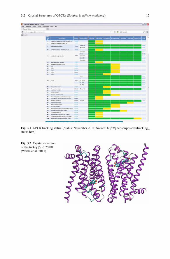
Sometimes, a look onto the homepage of GPCR network (http://cmpd.scripps.edu) is very useful. There, you get information about the tracking status of GPCRswhich will be crystallized in future (Fig. 3.1).
3.2 Crystal Structures of GPCRs (Source: http://www.pdb.org)
In the appendix, the most important information with regard to all crystal structuresof (rhod)opsin or GPCRs is summarized tabular. These tables should give you a fastoverview onto available crystal structures, resolution, structure of a cocrystallizedligand, related UniProtKB entries and corresponding literature. Have a careful lookonto the section “mutation”! Often, not the wild type receptor is crystallized, insteadpoint mutations were introduced. Thus, if you want to model the receptor, whichis crystallized, you may change the amino acids, mutated in the crystal structure,into the corresponding amino acid of the wild type receptor. An overview of thedifferences in crystal structures is given by the Figs. 3.2–3.6.
3.2 Crystal Structures of GPCRs (Source: http://www.pdb.org) 15
Fig. 3.1 GPCR tracking status. (Status: November 2011; Source: http://gpcr.scripps.edu/tracking_status.htm)
Fig. 3.2 Crystal structureof the turkey β2R, 2Y00.(Warne et al. 2011)

16 3 Sequence Alignment and Homology Modelling
Fig. 3.3 Crystal structure of the human β2R, 3PDS. (Rosenbaum et al. 2011)
Fig. 3.4 Crystal structure ofthe human CXCR4, 3ODU.(Wu et al. 2010)

3.2 Crystal Structures of GPCRs (Source: http://www.pdb.org) 17
Fig. 3.5 Crystal structure ofthe human CXCR4, 3OE0.(Wu et al. 2010)
Fig. 3.6 Crystal structure ofthe human A2AR, 3EML.(Jaakola et al. 2008)

18 3 Sequence Alignment and Homology Modelling
3.3 Amino Acid Sequences and Sequence Alignment
Before being able to start the homology modelling, it has to be decided which aminoacid of the template sequence corresponds to an amino acid in the target sequence.Therefore, a sequence alignment has to be performed manually or automatically.Clustal (http://www.clustal.org) for example, is a software for multiple sequencealignment. However, before starting with sequence alignment, the correspondingamino acid sequences have to be obtained.
3.3.1 Amino Acid Sequences – Where to Get From?
There are several sources for amino acid sequences present in the internet. Oneprominent is for example the Expasy Proteomics Server (http://expasy.org) (Fig. 3.7).
Exercise Start your internet browser and open the site http://expasy.org. Nowchoose “UniProtKB” under the section “query”. Then you can type your searchstring into the field on the right.
Now we want to search for the human adrenergic β2 receptor. There aredifferent possibilities for the search string. For example, type “adrenergic” andclick the “Search” button. Now, more than 900 results, related to “adrenergic”are presented. Scroll, until the receptor of your choice is listed. In our case itis the human adrenergic β2 receptor with the accession code “P07550”. If youwant to reduce the number of hits, the search string has to be defined moreexactly. Please try “beta adrenergic receptor”, “beta-2 adrenergic receptor”and “beta-2 adrenergic receptor human”. By defining the search string moreexactly, the number of hits can be significantly reduced and it is easier for youto find the hit, you are searching for.
Now, click, onto the corresponding entry with the accession code “P07550”and you get a lot of very useful information about this receptor, including theamino acid sequence. In the section “Regions”, the amino acids, related withthe N-terminus, C-terminus, intracellular loops, extracellular loops and trans-membrane domains are given. This information is very helpful for the sequencealignment later on. In the section “Sequence” you can find the whole aminoacid sequence of the protein. For further proceeding on with the amio acidsequence like for sequence alignment, it may be easier for you, to downloadthe amino acid sequence as “fasta” format. To do so, please click onto thestring “FASTA”. Now you get the amino acid sequence as simple ascii file.

3.3 Amino Acid Sequences and Sequence Alignment 19
Fig
.3.7
Hom
epag
eof
the
expa
syse
rver
.(ht
tp://
expa
sy.o
rg)

20 3 Sequence Alignment and Homology Modelling
Table 3.2 Highly conserved amino acid according to Ballesteros (Ballesteros et al. 2001) of eachtransmembrane domain of rhodopsin-like GPCRs
TM I TM II TM III TM IV TM V TM VI TM VII
Asn, N Asp, D Arg, R Trp, W Pro, P Pro, P Pro, P
3.3.2 Ballesteros Nomenclature
A careful analysis of the known amino acid sequences of known rhodopsin-likeGPCRs by Ballesteros (Ballesteros et al. 2001) exhibited the most conserved aminoacid within each of the seven transmembrane domains, which is used as a referencefor all other amino acids within the same helix. Within this nomenclature, the termX.YY is used. Therein, X represents the number of the transmembrane domain andYY the position of the residue within the transmembrane domain. The most conservedamino acid within one helix gets the number 50. All other amino acids within thesame helix are numbered relative to that highly conserved position 50. The highlyconserved amino acids of each transmembrane domain of a GPCR, according to theBallesteros nomenclature (Ballesteros et al. 2001) are given in Table 3.2.
In Fig. 3.8, the complete amino acid sequence with the conserved amino acidsaccording to Ballesteros (Ballesteros et al. 2001) of the human adrenergic β2 receptoris presented.
One should pay attention onto the transmembrane regions, as pointed out inFig. 3.8. As already mentioned the amino acids related to the transmembrane regionsare given at http://expasy.org under the corresponding accession code. A comparisonto the corresponding crystal structure – if available – shows sometimes differenceswith regard to the helical region. Let us for example look onto TM III of the humanadrenergic β2 receptor. The transmembrane region is defined from Glu-107 until Val-129 at expasy (Fig. 3.9a). However, a closer look onto the corresponding domainat the crystal structure shows that the helical structure is much longer at both sides(Fig. 3.9b). Thus, the domains are adopted with regard to the amino acid sequencein Fig. 3.9c. Additionally, in Fig. 3.9b, the amio acids Glu-107 and Val-129 are men-tioned Glu3.26 and Val3.48 in the Ballesteros nomenclature. Some additional aminoacids are shown in the Ballesteros nomenclature in Fig. 3.9c. For the termini and theloops no corresponding nomenclature is available.
3.3.3 Amino Acid Sequences – Templates
Before performing an amino acid sequence alignment, one has to decide, whichstructure should be used as template structure for homology modelling. Meanwhilea lot of crystal structures of bovin rhodopsin or GPCRs like the human adrener-gic β2 receptor or turkey adrenergic β1 receptor are available (see Tab. 3.1 andappendix Important Crystal Structures of GPCRs (Source: http://www.pdb.org)). Itcannot be decided overall, which crystal structure should be used as a template for

3.3 Amino Acid Sequences and Sequence Alignment 21
Fig. 3.8 Amino acid sequence of the human adrenergic β2 receptor. The transmembrane domainare presented, as defined at http://expasy.org, accession code P07550. The highly conserved aminoacids, defined by Ballesteros (Ballesteros et al. 2001) are marked by red boxes
Fig. 3.9 Helical structure of a transmembrane domain. a Definition of the TM domain III of thehuman adrenergic β2 receptor at expasy (http://www.expasy.org). b TM III of the human adrenergicβ2 receptor of a crystal structure. c Amino acid sequence of TM domain III, based on the crystalstructure

22 3 Sequence Alignment and Homology Modelling
homology modelling. In general, the crystal structure with highest sequence homol-ogy to the receptor, which is intended be modelled, should be chosen. Besides thatit should be taken into account that different template crystal structures in homologymodelling could lead to differences in the resulting homology model. However, themainly used templates for modelling class A GPCRs are bovine rhodopsin and thehuman adrenergic β2 receptor (see appendix Important Crystal Structures of GPCRs(Source: http://www.pdb.org)).
3.3.4 Sequence Alignment
After retrieving the amino acid sequences of the template structure and the destinationreceptor, the sequence alignment can be performed. There exist several techniques,to perform the sequence alignment. On the one hand, the sequence alignment can beperformed manually. The corresponding steps require some time and concentration.On the other hand, there exist several software products, which allow performingan alignment automatically, like clustal (http://www.clustal.org) (see appendix Sum-mary of Important Internet Resources). However, if software is used, it is definitelynecessary to check to resulting alignment in order to avoid unexpected mistakes orsome inaccuracies.
For a manual sequence alignment, the alignment is performed by several steps:
1. Use the information of the expasy server (http://expasy.org) to get an idea aboutthe amino acids of the seven transmembrane domains for template and targetsequence.
2. Perform the sequence alignment for each transmembrane domain in ascend-ing order. Here, it is necessary, that the highly conserved amino acid of eachtransmembrane domain has the same position in template and target.
3. Now, the alignment for the termini and loops can be performed. There you haveto take into account several points:
– In most crystal structures, the N-terminus and C-terminus are often not com-plete. Thus, there you can perform the alignment of such regions, but there isno real use in homology modelling, since no template structure is given forsuch regions.
– The I1-, E1-, I2- and E3-loop can be aligned easily in most cases to the templatesequence. However it should be taken into account, that corresponding loopsof different GPCRs could differ in their length. This has to be taken carefullyinto account later on in the homology modelling. To declare a vacant positionin amino acid sequence, a hyphen (-) is used in general.
– The I3-loop differs significantly in length (from some ten to some hundredamino acids) within the different GPCRs. Additionally, the I3-loop is not com-pletely present in the crystal structures, available up to now. Thus, a completeI3-loop alignment is useless for homology modelling. However, for MD simu-lations, it will be useful to close the open ends between TM V and TM VI.

3.4 Homology Modelling 23
Fig. 3.10 Manual alignment of the hH4R to the hH1R. green: termini and loops; grey: transmem-brane domains; red boxes: highly conserved amino acids (Ballesteros et al. 2001); yellow: highlyconserved cysteine, establishing a disulfide bridge to the upper part of TM III; -: missing aminoacids; the amino acids of the I3-loop are not shown completely, which is indicated by dots
Therefore, some amino acids of the beginning and end of the I3-loop aremodelled correctly and the gap is closed by an alanine chain.
– The E2-loop has to be aligned very carefully. It has to be taken into account,that there is a highly conserved disulfide bridge between the E2-loop and theupper part of TM III. Thus, the corresponding cysteine has to be positionedcorrectly.
An example for an alignment of the human histamine H4 receptor to the humanhistamine H1 receptor is shown in Fig. 3.10.
3.4 Homology Modelling
3.4.1 Modelling of the Transmembrane Domains
The helical transmembrane domains can be easily modelled straight forward. There-fore, only the amino acid side chains have to be changed into the side chain of thedestination with appropriate modelling software.
3.4.2 Modelling of Loops
In general the transmembrane domains of different GPCRs consist of the same num-ber of amino acids. Thus, the homology modelling of transmembrane domains isquite easy and can be performed straight forward. In case of intra- or extracellu-lar loops, which are connecting the transmembrane domains, differences in numberof amino acids of a loop between different GPCRs can occur. This is the case forthe E2- or E3-loop between hH1R and hH4R (Fig. 3.10). Small gaps can be closedwith “loop search” modules by using appropriate software. For some biogenic amine

24 3 Sequence Alignment and Homology Modelling
Fig. 3.11 Different conformations of the E2-loop based on crystal structures
receptors, an influence of extracellular loops, especially the E2-loop, onto the bind-ing of ligands to the receptor was shown (Lim et al. 2008; Brunskole et al. 2011).Thus, a correct modelling of the loops is very important. Most of the loops are re-solved by crystal structures. However, this is often not the case with regard to theextracellular loop E2 and this is not the case with regard to the intracellular loop I3.
Since the E2-loop is in contact with the binding pocket, the E2-loop has to bemodelled completely. If you look onto different crystal structures with completeE2-loop, you can see different conformations (Fig. 3.11).
Thus, you have to decide carefully, which template is to be used for modellingof the E2-loop. A large number of crystal structures are obtainable for the humanadrenergic hβ2 receptor. But the hβ2R is a special case: There are two disulfidebridges in the E2-loop (Fig. 3.12), whereas in most others GPCRs there is only onedisulfide bridge in the E2-loop, connecting the E2-loop with the upper part of theTM III.
A part of the E2-loop of the hβ2R exhibits a helical structure, but this is not the casefor all other GPCRs. Thus, you have to decide carefully, if it would be appropriateto use two different template structures for homology modelling: one for the E2-loop and one for the remaining parts of the receptor. However, the E2-loops arewidely different in their length, thus, in most cases, the E2-loop cannot be modelledby changing an amino acid side chain of the template into the side chain of thedestination. Thus, you have to use also techniques, like “loop search”. For only oneloop search, the number of amino acids is too long, and you would get bad results.Thus, it is better, to use at least one fixed point. This is the highly conserved cysteine,connecting the E2-loop by a disulfide bridge with the upper part of TM III (Fig. 3.10).

3.4 Homology Modelling 25
Fig. 3.12 Different numbers of disulfide bridges in the E2-loop
3.4.3 Modelling of Internal Water
A detailed analysis of the crystal structures of GPCRs reveals that there are internal,highly conserved water molecules present (Fig. 3.13). Several studies showed thatthese water molecules are involved in the hydrogen bonding within the receptor.Based on the published data, it can be suggested, that these water molecules areessential for stabilizing the receptor or important for receptor activation (Pardo et al.2007). Thus, in order to generate a stable receptor model, the water molecules whichare localized/crystallized within the receptor should be included into the homologymodel.
3.4.4 Modelling of the C-Terminal Part of the Gα Subunitor the Whole Gα Subunit
Based on several studies it is suggested, that a GPCR in its active conformationinteracts in the intracellular part with the Gα subunit. There is only small knowledgeabout the receptor – G protein interaction. However, recently, the crystal structure ofopsin, cocrystallized with eleven amino acids of the C-terminus of the Gα subunit(Scheerer et al. 2008) and a complete GPCR – G protein complex (Rasmussen et al.2011) were published. A detailed analysis of the corresponding crystal structures(3DQB, 3SN6) shows, that the C-terminus of the Gα subunit is deeply bound in apocket between the transmembrane domains. Leaving out this part of the Gα willresult in some problems in subsequent molecular dynamic simulations. In general, ifmolecular dynamic simulations of a receptor are performed, the receptor is embeddedin its natural surrounding. Thus, if the C-terminal part of Gα or the whole Gα is

26 3 Sequence Alignment and Homology Modelling
Fig. 3.13 Crystal structure ofbovin rhodopsin (1GZM)with internal water (redballs). (Li et al. 2004)
missing, the resulting free space is filled with water molecules. Water molecules arehighly polar and thus have completely other (surface) properties than the C-terminalpart of Gα. Thus, leaving out the C-terminal part of Gα and substitution by watermolecules in molecular dynamics can lead to instabilities of the receptor during themolecular dynamic simulation. Thus, it is suggested, to include at the whole Gα orleast the C-terminal part of Gα in a homology model. Be aware, that each GPCRcouples to a distinct Gα subunit (Fig. 2.8).
3.4.5 Refinement of the Receptor Model
After finishing the homology modelling, several checks of the complete model shouldbe performed. A typical error of beginners in molecular modelling is presented inFigs. 3.14–3.16. During homology modelling, some amino acid side chains have tobe mutated into the correct amino acid side chain. Sometimes, especially with regardto long side chains or aromatic rings, collisions between the side chains arise. Thereare two types of collisions: In the first type, two side chains are in close contact,as shown in Fig. 3.14. In most of these cases, energy minimization is sufficient to

3.4 Homology Modelling 27
Fig. 3.14 Close contact between the atoms of a lysine and phenylalanine: Left: before minimization,right: after minimization
Fig. 3.15 Part of a proteinstructure after minimization.What is the problem?
remove the collision and suitable structures might be obtained. The second type ofcollision is a more difficult pitfall, which is illustrated in Figs. 3.15 and 3.16. Lookcarefully onto the Fig. 3.15. Where is the problem?
After a careful look onto the picture you may see, that there is a problem withregard to a lysine and phenylalanine in box B3. This is also illustrated in Fig. 3.16.
Here, a long amino acid side chain, like present in lysine, is located withinan aromatic ring, like present in tyrosine, phenylalanine, tryptophane or histidine.

28 3 Sequence Alignment and Homology Modelling
Fig. 3.16 Wrong close contact between the atoms of a lysine and phenylalanine: Left: beforeminimization, right: wrong structure after minimization
Unfortunately, a large number of modelling software minimizes a protein, containingsuch type of wrong structure. And additionally in most cases the potential energy isnegative. Thus, one might conclude that all is well. However, often during moleculardynamic simulation, problems occur and the simulation stops with an error. If thisis the case, you have to go back to your starting structure and look for the error.Often, an error, similar to that described above (Fig. 3.16) causes the problem. Asimilar problem can occur not only within the protein, but also between proteinand lipid molecules. If there are collisions between amino acid side chains, onehas to decide, how to remove this collisions. In general, there are two possibilities:First, one can simply perform an energy minimization. But in some cases, this couldlead to artefacts, especially, if two aromatic moieties are linked together. Thus, itis suggested, that one looks separately onto each collision and tries to remove thecollisions by carefully changing the corresponding dihedral angles.
After completing these steps, the homology model can be energetically minimized.Here it is suggested, that the energy minimization is performed step by step. Inorder to avoid structural artefacts, induced by minimization, it is important, thatthe backbone of the transmembrane domains is provided with position restraintsduring a first minimization. In a subsequent minimization steps, the receptor can beminimized without any position restraints. Afterwards, the model should be checked,addressing the following items and if everything is correct, one can start with furthermodelling studies, like docking or molecular dynamic simulations.
� Check for the correct amino acid sequence� Check for the presence of the disulfide bridge between the E2-loop and
the upper part of TM III� Check for the correct configuration of the amino acids� Check for collisions or bad contacts between amino acid side chains