Embed Size (px)
Citation preview

Modeling Ewing sarcoma tumors in vitro with3D scaffoldsEliza Li Shan Fonga,1, Salah-Eddine Lamhamedi-Cherradib,1, Emily Burdetta,1, Vandhana Ramamoorthyb,Alexander J. Lazarc, F. Kurtis Kaspera, Mary C. Farach-Carsond, Deeksha Vishwamitrae, Elizabeth G. Demiccof,Brian A. Menegazb, Hesham M. Amine, Antonios G. Mikosa,2, and Joseph A. Ludwigb,2
Departments of aBioengineering and dBiochemistry and Cell Biology, Rice University, Houston, TX 77005; Departments of bSarcoma Medical Oncology,cPathology, and eHematopathology, University of Texas M. D. Anderson Cancer Center, Houston, TX 77030; and fDepartment of Pathology, Mount SinaiMedical Center, New York, NY 10029
Edited by Robert Langer, Massachusetts Institute of Technology, Cambridge, MA, and approved March 12, 2013 (received for review December 8, 2012)
The pronounced biological influence of the tumor microenviron-ment on cancer progression and metastasis has gained increasedrecognition over the past decade, yet most preclinical antineoplasticdrug testing is still reliant on conventional 2D cell culture systems.Although monolayer cultures recapitulate some of the phenotypictraits observed clinically, they are limited in their ability to modelthe full range of microenvironmental cues, such as ones elicited by3D cell–cell and cell–extracellular matrix interactions. To addressthese shortcomings, we established an ex vivo 3D Ewing sarcomamodel that closely mimics the morphology, growth kinetics, andprotein expression profile of human tumors. We observed thatEwing sarcoma cells cultured in porous 3D electrospun poly(e-cap-rolactone) scaffolds not only were more resistant to traditional cy-totoxic drugs than were cells in 2D monolayer culture but alsoexhibited remarkable differences in the expression pattern of theinsulin-like growth factor-1 receptor/mammalian target of rapamy-cin pathway. This 3D model of the bone microenvironment mayhave broad applicability for mechanistic studies of bone sarcomasand exhibits the potential to augment preclinical evaluation of an-tineoplastic drug candidates for these malignancies.
tissue engineering | tumor model | biological therapy | connective tissue
Despite the primacy of the cancer cell’s dysregulated genotype[e.g., a near universal translocation of the Ewing sarcoma
(EWS) breakpoint region 1 gene in EWS cells] as the initial step inmalignant transformation, it has become increasingly apparentthat the overall tumor phenotype is also dictated by the 3D tumormicroenvironment (1–4). Nonetheless, studies of cancer biologyand evaluation of antineoplastic drug efficacy remain heavily de-pendent on conventional 2D cell culture systems despite theirlimited ability to reflect the 3D tumor architecture, extracellularmatrix (ECM), and surrounding cell types that comprise the in vivotumor milieu.To overcome some of these constraints, 3D in vitro models such
as spheroid and gel systems have been extensively studied and,compared with 2D monolayer culture, appear to better mimic theprofound effects that the in vivo 3D environment has upon thehuman tumor phenotype (5–9). For example, malignant cellscultured in 3D exhibit increased chemoresistance (10, 11) anddecreased cell proliferation (12), and assume specific phenotypesinducible only under a 3D context, such as angiogenic capability(13–15). Furthermore, striking differences in signaling pathwaystargeted by proven and experimental therapies have been observedin 3D tumor models (16–18). Accordingly, heightened awarenessof the importance of 3D culture for cancer cells has resulted in theincreasing use of spheroid culture systems for cancer research.However, these non–adhesion-mediated systems provide poorcontrol over the tumor architecture and cell–cell interactions; asa result of culture conditions that prohibit cellular attachmentonto surrounding surfaces, cells autonomously aggregate and formtheir own 3D geometry. An emerging strategy to overcome this in-herent drawback of spheroid culture is to leverage 3D scaffolding
technologies that have been developed for tissue-engineeringapplications (19), to guide tumor tissue formation in vitro withcontrolled and tunable architectural complexity.In the present study, our interdisciplinary team of cancer
biologists, tissue engineers, and clinicians sought to develop andvalidate a drug-testing tool that would elicit in vivo-like drugsensitivity and potentially advance mechanistic studies of diseasebiology through precise control of the in vitro tumor architecture.Focusing specifically on bone sarcomas, we established a 3Dmodel of EWS, the second most common pediatric bone malig-nancy, by culturing TC-71 human EWS cells within electrospunpolymeric scaffolds fabricated from poly(e-caprolactone) (PCL),a biologically inert synthetic polymer previously investigated foruse in bone and other tissue-engineering applications (20–23).These scaffolds have several favorable features, including highporosity, a large surface area-to-volume ratio for cellular attach-ment, tunable fiber diameter, low cost, ease of fabrication andhandling, and high reproducibility (24, 25). With these 3D PCLscaffolds, we observed that ex vivo EWS traits were very similar tothose in vivo with respect to morphology, growth kinetics, andprotein expression, and were remarkably different from theirmonolayer counterparts throughout the insulin-like growth factor-1receptor (IGF-1R)/mammalian target of rapamycin (mTOR)-related signaling cascade. IGF-1R and mTOR, in particular, arepotential therapeutic targets for EWS under active investigationin human clinical trials, as their combined abrogation promotedstriking antineoplastic responses in a subset of patients withchemotherapy-resistant EWS.
Results and DiscussionWe engineered an ex vivo EWS model by culturing TC-71 cellsin a porous 3D electrospun PCL scaffold. At 2 d in culture, TC-71 cells adhered in small, sparse clusters to individual PCLfibers and were located primarily on the top surface of thescaffold. With increased duration of culture (20 d), cells werefound throughout the scaffold, with the majority located withinthe upper one-fifth of the scaffold (Fig. S1). Representativescaffolds collected after short- and long-term culture were imagedusing scanning electronmicroscopy (Fig. 1A). Careful review of theresulting images by an experienced sarcoma pathologist identifiedthe small round-cell morphology characteristic of human EWS
Author contributions: E.L.S.F., S.-E.L.-C., E.B., A.G.M., and J.A.L. designed research; E.L.S.F.,S.-E.L.-C., E.B., V.R., D.V., E.G.D., and B.A.M. performed research; E.L.S.F., S.-E.L.-C., E.B.,A.J.L., F.K.K., M.C.F.-C., H.M.A., A.G.M., and J.A.L. analyzed data; and E.L.S.F., S.-E.L.-C.,and J.A.L. wrote the paper.
The authors declare no conflict of interest.
This article is a PNAS Direct Submission.1E.L.S.F., S.-E.L.-C., and E.B. contributed equally to this work.2To whom correspondence may be addressed. E-mail: [email protected] or [email protected].
This article contains supporting information online at www.pnas.org/lookup/suppl/doi:10.1073/pnas.1221403110/-/DCSupplemental.
www.pnas.org/cgi/doi/10.1073/pnas.1221403110 PNAS Early Edition | 1 of 6
MED
ICALSC
IENCE
SEN
GINEE
RING
tumors. Additionally, we used a well-validated panel of immu-nohistochemical markers routinely used for diagnostic purposesin patients (CD99+, keratin−, and smooth muscle actin−), which
confirmed that our established in vitro 3D tumor model preservesa well-differentiated EWS-like phenotype (Fig. 1B).Paralleling a rapid increase in regulatory approval of biologically
targeted drugs and “omics”-based technologies that have madetumor profiling commonplace, some of the most promising ex-perimental agents being tested in the treatment of EWS in early-phase clinical trials exert their effect by antagonizing IGF-1R,mTOR, or other proteins in their shared signaling cascades (26,27). Although a thorough discussion of the roles of IGF-1R andmTOR in EWS carcinogenesis is beyond the scope of this article,two decades of research indicate that their expression is critical, asthe pathognomonic EWS breakpoint region 1–erythroblast trans-formation specific (EWSR1–ETS) translocation alone is insuffi-cient to promote and maintain EWS oncogenesis, growth, andinvasion (28–31).Despite a wealth of preclinical information and two recent
clinical trials demonstrating marked tumor regression in a sub-set of patients with EWS treated with combined antagonists ofthe IGF-1R/mTOR pathway, we noted a surprising lacklustercellular response to IGF-1R antagonists in conventional 2Dmonolayer culture (32, 33). In seeking to elucidate biomarkersof response and mechanisms of de novo and acquired re-sistance, we hypothesized that this difference in preclinical andclinical response may result from inherent changes in the IGF-1R/mTOR signaling cascade when tumor cells are removedfrom their native microenvironment, which we sought to re-capitulate using our 3D PCL scaffold while maintaining controlover experimental parameters.Striking differences were observed in the expression of proteins
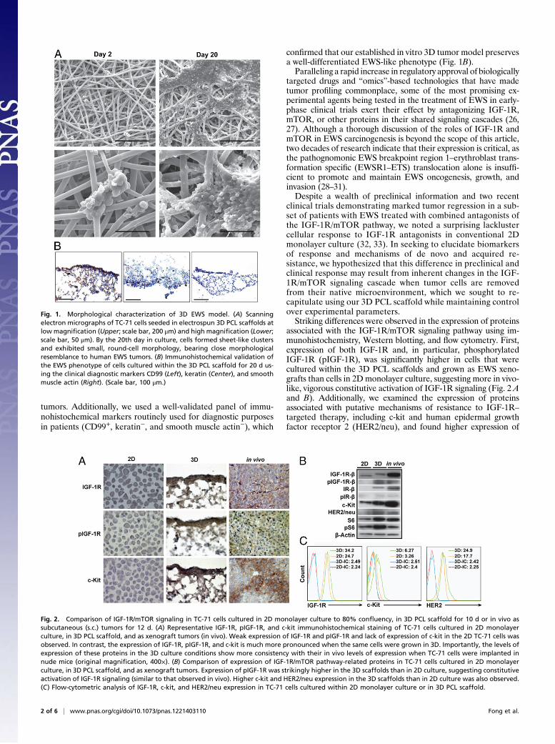
associated with the IGF-1R/mTOR signaling pathway using im-munohistochemistry, Western blotting, and flow cytometry. First,expression of both IGF-1R and, in particular, phosphorylatedIGF-1R (pIGF-1R), was significantly higher in cells that werecultured within the 3D PCL scaffolds and grown as EWS xeno-grafts than cells in 2Dmonolayer culture, suggesting more in vivo-like, vigorous constitutive activation of IGF-1R signaling (Fig. 2 Aand B). Additionally, we examined the expression of proteinsassociated with putative mechanisms of resistance to IGF-1R–
targeted therapy, including c-kit and human epidermal growthfactor receptor 2 (HER2/neu), and found higher expression of
Fig. 1. Morphological characterization of 3D EWS model. (A) Scanningelectron micrographs of TC-71 cells seeded in electrospun 3D PCL scaffolds atlow magnification (Upper; scale bar, 200 μm) and high magnification (Lower;scale bar, 50 μm). By the 20th day in culture, cells formed sheet-like clustersand exhibited small, round-cell morphology, bearing close morphologicalresemblance to human EWS tumors. (B) Immunohistochemical validation ofthe EWS phenotype of cells cultured within the 3D PCL scaffold for 20 d us-ing the clinical diagnostic markers CD99 (Left), keratin (Center), and smoothmuscle actin (Right). (Scale bar, 100 μm.)
Fig. 2. Comparison of IGF-1R/mTOR signaling in TC-71 cells cultured in 2D monolayer culture to 80% confluency, in 3D PCL scaffold for 10 d or in vivo assubcutaneous (s.c.) tumors for 12 d. (A) Representative IGF-1R, pIGF-1R, and c-kit immunohistochemical staining of TC-71 cells cultured in 2D monolayerculture, in 3D PCL scaffold, and as xenograft tumors (in vivo). Weak expression of IGF-1R and pIGF-1R and lack of expression of c-kit in the 2D TC-71 cells wasobserved. In contrast, the expression of IGF-1R, pIGF-1R, and c-kit is much more pronounced when the same cells were grown in 3D. Importantly, the levels ofexpression of these proteins in the 3D culture conditions show more consistency with their in vivo levels of expression when TC-71 cells were implanted innude mice (original magnification, 400×). (B) Comparison of expression of IGF-1R/mTOR pathway-related proteins in TC-71 cells cultured in 2D monolayerculture, in 3D PCL scaffold, and as xenograft tumors. Expression of pIGF-1R was strikingly higher in the 3D scaffolds than in 2D culture, suggesting constitutiveactivation of IGF-1R signaling (similar to that observed in vivo). Higher c-kit and HER2/neu expression in the 3D scaffolds than in 2D culture was also observed.(C) Flow-cytometric analysis of IGF-1R, c-kit, and HER2/neu expression in TC-71 cells cultured within 2D monolayer culture or in 3D PCL scaffold.
2 of 6 | www.pnas.org/cgi/doi/10.1073/pnas.1221403110 Fong et al.

both proteins in the TC-71 xenografts as well as cells that werecultured within our 3D PCL scaffolds, compared with cells in 2Dmonolayer culture (Fig. 2A andB).We confirmed this observationusing flow cytometry, as indicated by the mean fluorescence levelsfor c-kit (3.26 versus 6.27) andHER2/neu (17.7 versus 24.9) in cellsin 2D monolayer culture and the 3D PCL scaffolds, respectively(Fig. 2C). Taken together, these observations suggest that cellscultured in the 3D PCL scaffold may serve as a reliable ex vivosurrogate for xenografts, and potentially human EWS tumors, forevaluating the IGF-1R/mTOR pathway under both physiologicaland perturbed states.To demonstrate that our 3D EWSmodel exhibits tumor growth
characteristics necessary to serve as a reliable platform for testingpromising drug candidates, particularly those that exert theirantineoplastic effect on actively dividing cells, we compared therate of proliferation of TC-71 cells cultured in 3D PCL scaffoldswith that of cells in 2D monolayer culture and human EWSxenografts (Fig. 3A) using DNA content as an index of pro-liferation. In contrast to 2Dmonolayer culture, the proliferationrate of TC-71 cells within the 3D PCL scaffolds was substantiallyslower andmore closely aligned with that of cells grown in vivo asxenografts, which serves as the current preclinical gold standardfor assessing drug efficacy. As determined by flow cytometry, thisfinding can be attributed to both decreased proliferation andincreased apoptosis as evidenced by altered intracellular ex-pression of Ki-67, caspase-3, and cleaved poly(ADP-ribose)polymerase (PARP) protein in TC-71 cells cultured within the3D PCL scaffolds (Fig. 3B).We further characterized the sensitivity of the 3D EWS model
to doxorubicin, a prototypical cytotoxic chemotherapeutic agentwith proven clinical value in EWS patients. Comparable with in-creased doxorubicin resistance observed in TC-71 xenografts thatgrew in CB17/severe combined immunodeficient (SCID) mice[half-maximal inhibitory concentration (IC50), 0.184 μM], TC-71cells cultured in 3DPCL scaffolds demonstrated greater resistancethan cells in 2D monolayer culture (IC50, 2.738 and 0.012 μM,respectively) (Fig. 4A). We expected this result given the slowerproliferation rate associated with growth in the 3D PCL scaffoldsand doxorubicin’s knownmechanism of cytotoxicity. However, thismay also be attributed to restoration of structurally complex invivo-like cell–cell contact in the 3D PCL scaffolds similar to pre-viously reported observations of drug and radiation resistance intumor spheroids (34).To elucidate the potential role of cell–cell contact in doxoru-
bicin resistance in our 3Dmodel, we investigated the effect of celldensity as a surrogate measure of cell–cell contact in the scaffolds.As shown in Fig. 4B, densely seeded cell cultures were associatedwith 8.7-fold greater chemoresistance (IC50, 4.225 μM) than weretheir more sparsely growing counterparts (IC50, 0.485 μM), sug-gesting a negative correlation between cell–cell contact and drugsensitivity. However, the exact signaling mechanisms implicatedin drug resistance are still unclear, and current studies are lookingat whether differential sensitivity is a direct consequence of en-hanced cell–cell contact alone or interplay among other factors(e.g., intracellular changes, paracrine signaling, modifications inthe supporting matrix). The well-recognized phenomenon of che-moresistance previously observed in another spatially complextumormodel (tumor spheroids) results frompoor drug penetrationinto the innermost cell layers, but we found nothing to suggest thatthis occurred in the 3D PCL scaffolds (35, 36). Indeed, as drugtransport and interaction with the scaffold may have an impact onthe efficacy of the drug, we conducted an additional study tocharacterize the potential interaction between doxorubicin andthe 3D PCL scaffold (Fig. S2). Although significantly moredoxorubicin was found to adsorb onto the PCL scaffolds com-pared with tissue culture plastic controls, Fig. 4A shows that,within the concentration range of doxorubicin after adsorptiononto the PCL scaffold, cytotoxicity in 2D is still greater than
90%, indicating that the increased resistance observed in 3D isnot due to decreased availability of the drug after adsorption ontothe scaffold.Given the lower proliferative index of cells in the 3D PCL
scaffolds than in 2D monolayer culture that better mimics humantumor growth, this model may be particularly appropriate forinvestigating the long-term impact of drug exposure on cancercells, which is a challenging endeavor with 2D culture systems,given that confluency limits the duration of culture. Fig. 4C shows
Fig. 3. Growth profile of TC-71 EWS cells in 2D, 3D, and in vivo. (A) EWSgrowth in 2D monolayer culture, in 3D PCL scaffold, or in vivo as xenografts.TC-71 cells proliferated at a substantially slower rate in the 3D scaffolds thanin 2D culture (left axis, fold change in cell number), with the former bearingcloser similarity to the in vivo tumor growth rate (right axis, fold change intumor volume). Data represent the mean fold change in cell number (2D and3D) or tumor volume (in vivo) ± SEM (n = 4 for 2D culture and in vivo, n = 6for 3D scaffolds). (B) Levels of Ki-67, caspase-3, and cleaved-PARP expressionin TC-71 cells in 3D PCL scaffolds (cultured for 10 d) and 2D monolayer cul-ture as measured using flow cytometry. The majority of the cells in the 3DPCL scaffolds were still proliferative even at 10 d in culture, with a smallpercentage of apoptotic cells present.
Fong et al. PNAS Early Edition | 3 of 6
MED
ICALSC
IENCE
SEN
GINEE
RING

that prolonged exposure to doxorubicin ultimately elicited sig-nificant cell death despite negligible short-term antineoplasticeffects of the drug (IC50, 1.397 and 0.051 μM for short and longdoxorubicin exposure, respectively). Hence, in addition to itsgreater fidelity to the in vivo EWS tumor phenotype, our 3DEWSmodel may be an exceptionally useful tool for conducting long-term studies necessary for determining the often subtle anddelayed antineoplastic effects exerted by biologically targetedtherapy. Notably, as the vast majority of cytotoxic and biologicallytargeted therapies exert their antineoplastic effects well within thelong doxorubicin exposure period investigated in this study, wedid not extend this time frame beyond 16 d.As we observed striking differences in the IGF-1R/mTOR
pathway signaling pattern in EWS cells in our 3D PCL scaffoldand 2D monolayer culture, we next sought to investigate whetherwe could elicit more in vivo-like drug sensitivity to inhibitors ofIGF-1R and mTOR. We treated TC-71 cells grown under thethree conditions (2D monolayer, 3D PCL scaffold, and as xeno-grafts) with MK-0646, a humanized IgG1 monoclonal antibodyagainst IGF-1R.We observed an up-regulation of HER2/neu andc-kit expression in the 3D PCL scaffolds, which is in concordancewith the expression pattern in xenografts (Fig. 5 A–C). Addi-tionally, in agreement with published data implicating the insulinreceptor (IR) as a major contributor of resistance to IGF-1R–
targeted therapy (via formation of hybrid IGF-1R/IR-α receptors)(37), our data demonstrated that IGF-1R inhibition led to con-stitutive phosphorylated IR-β protein activation in TC-71 cellscultured in our 3D PCL scaffold and in xenograft tumors but notin 2D monolayer culture (Fig. 5B). Furthermore, treatment withthe small-molecule mTOR inhibitor MK-8669 (ridaforolimus)had no effect on IGF-1R, c-kit, or HER2/neu expression despitesuppressed phosphorylated S6, suggesting that our 3D model isable to mimic the expected in vivo pharmacodynamic response ofmTOR inhibition. Overall, these results offer a unique perspec-tive on IGF-1R/mTOR signaling in a biomimetic 3D preclinicalmodel of EWS.
ConclusionWe developed an in vitro human EWS model that exhibits mor-phological and biochemical features of in vivo tumors in starkcontrast with conventional 2D models that poorly represent invivo EWS tumor biology. The remarkable similarity between theengineered EWS tumor model and in vivo xenograft EWS tumorssuggests that tumor cells cultured within the 3D PCL scaffold may
represent a better model than 2D culture systems for mechanisticstudies of standard chemotherapies and/or biologically targetedtherapies for EWS under preclinical investigation.Accelerated development of antineoplastic drugs is critically
dependent on preclinical models that simulate in vivo tumorgrowth and intracellular signaling as accurately as possible. Two-dimensional culture systems that poorly recapitulate the in vivo3D tumor microenvironment and, hence, in vivo signaling cas-cades may possess limited utility in this regard. Dysregulatedsignaling cascades propagated in 2D culture may lead to erro-neous scientific conclusions that complicate our understandingof cancer biology and mask resistance mechanisms that wouldotherwise be elucidated under 3D culture conditions.With superior fidelity to the in vivo phenotype, our 3Dmodel of
EWS provides the unique opportunity to examine the biology ofthis cancer in a preclinical in vivo-like system that is still amenableto standard experimental techniques. Furthermore, the enhancedconstitutive activation of IGF-1R in this 3D system may facilitatethe identification of critical nodes in the IGF-1R signaling net-work that may be exploited therapeutically. Moreover, becauseour 3D model reproduces several putative mechanisms of drugresistance known to be found clinically (e.g., increased reliance onalternative receptor tyrosine kinases such as c-kit and IGF-1R/IR-α hybrid receptors that activate the mTOR pathway independentof IGF-1R), it may serve as a reliable platform for testing com-bined antagonists of the IGF-1R/mTOR signaling cascade. Last,given the in vivo-like sensitivity of EWS cells to doxorubicin in our3D model, this model may also facilitate the pursuit of combi-national therapeutic strategies involving both IGF-1R blockadeand standard cytotoxic chemotherapy.Although our understanding of the profound influence of 3D
culture conditions on EWS growth and survival has advanced withthe use of the engineered PCL scaffolds, many questions remainto be answered. For example, as the effect of oxygen, nutrient, anddrug concentration gradients on the tumor phenotype and drugefficacy are important considerations in 3D tumormodels, furtherstudies are warranted to elucidate the potential impact of theseparameters. Additionally, given the role of tumor-associated cellsin influencing tumor behavior, a logical progression of the workreported herein is to recapitulate the in vivo cellular cross talk andaddress whether the coculture of EWS cells with other tumor-associated cells (such as stromal and endothelial cells) within our3D PCL scaffold affects the activation status of the IGF-1R sig-naling cascade. Furthermore, is the in vivo-like signaling cascade
Fig. 4. Response of TC-71 EWS cells to doxorubicin. (A) Percentage of viable TC-71 cells in 2D monolayer culture, in 3D PCL scaffold, and in vivo as xenograftsafter doxorubicin exposure. Cells in 3D scaffolds (IC50, 2.738 μM) and in vivo (IC50, 0.184 μM) demonstrated greater drug resistance than did cells in 2D culture(IC50, 0.0122 μM). P < 0.05 for 3D versus 2D; the asterisk (*) indicates P < 0.05 at each respective concentration. (B) Percentage of viable TC-71 cells cultured in3D PCL scaffolds at low and high densities (representing little and extensive cell–cell contact, respectively) after doxorubicin exposure. High-density cultureresulted in greater doxorubicin resistance (IC50, 4.225 μM) than did low-density culture (IC50, 0.485 μM). P < 0.05 for low versus high density; the asterisk (*)indicates P < 0.05 at each respective concentration. (C) Percentage of viable TC-71 cells in 3D PCL scaffolds after short (3 d) or long (16 d) doxorubicin ex-posure. Cells exposed to doxorubicin for a longer duration (IC50, 0.051 μM) exhibited lower drug resistance than did those exposed to it for a shorter duration(IC50, 1.397 μM). P < 0.05 for short versus long duration; the asterisk (*) indicates P < 0.05 at each respective concentration. Data represent the mean per-centage viability (2D and 3D; left axis) and tumor volume (in vivo; right axis) ± SEM normalized against untreated controls (n = 4 for 2D culture and in vivo, n =6 for 3D scaffolds).
4 of 6 | www.pnas.org/cgi/doi/10.1073/pnas.1221403110 Fong et al.

observed in 3D induced in part by altered adhesion of tumor cellsto the scaffold surface via focal adhesion kinases or attributable tochanges in the expression of cell–cell adhesion molecules? Willthe use of flow perfusion culture conditions to mimic the me-chanical aspect of the bone microenvironment affect EWS celldifferentiation and signaling? The answers to these questions andothers must ultimately be derived from the use of a biologicallyrelevant 3D environment.
Materials and MethodsScaffold Preparation. Nonwoven electrospun PCL scaffolds were electrospunusing the electrospinning apparatus as previously described (25). In brief, thesetup consists of a syringe pump, power supply, and a grounded, squarecopper plate. A 30-mL syringe was filled with PCL (inherent viscosity range,1.0–1.3; DURECT Corporation) dissolved in a 5:1 (vol/vol) chloroform/meth-anol solution to 18 wt%, and fitted with a 16-gauge blunt needle. A copperring was placed in-between the needle and the copper plate to stabilize theelectric field. The positive lead from the power supply was split and con-nected to both the needle and the copper ring. Fibers were collected ona glass plate placed in front of the copper plate during the electrospinningprocess. PCL microfiber mats (microfiber diameter, 11.6 ± 1.7 μm) wereelectrospun to a thickness of 1 ± 0.1 mm, and individual 8-mm–diameter PCLdiscs were punched out using a biopsy dermal punch. Scanning electronmicroscopy was used to assess each disk’s fiber morphology and diameter.Before cell seeding, electrospun scaffolds were sterilized in ethylene oxide(AN73; Andersen Products) for 12 h and aerated to remove residual fumes.Scaffolds were then prewetted in an ethanol gradient [100–30% (vol/vol)],rinsed three times with PBS, and soaked in culture medium for 2 d.
Cell Culture and Seeding. The human EWS cell line, TC-71, generously providedby John Trent (University of Texas M. D. Anderson Cancer Center, Houston,TX), was used in all experiments. Cells were maintained in RPMI medium 1640(Mediatech) containing 10% (vol/vol) FBS (Gemini Bio-Products) and anti-biotics (100 IU/mL penicillin and 100 μg/mL streptomycin; Mediatech) ina humidified incubator at 37 °C and in a 5% CO2 atmosphere. The mediumwas replaced every 2 d. For monolayer cell culture, 50,000 TC-71 cells wereseeded onto 12-well plates containing 3 mL of medium per well. Two mil-liliters of the medium was replaced daily. For 3D cell culture, the prewettedscaffolds were press-fitted into individual cylindrical cassettes designed toconfine the cell suspension and placed in 12-well plates for seeding. A totalof 250,000 TC-71 cells in 200 μL of medium was then seeded onto eachprewetted scaffold. The cells were allowed to adhere to the scaffolds for4–5 h before medium was gently added to immerse the cassettes completely.Seeded scaffolds were removed from the cassettes at 24 h and placed in 12-well plates containing 3 mL of medium, which was replaced daily.
Scanning Electron Microscopy. Three-dimensional constructs were fixed in2.5% (vol/vol) glutaraldehyde for at least 30 min and then dehydrated in anethanol gradient [70–100% (vol/vol)]. Constructs were then air-dried over-night, sputter-coated with gold, and imaged using an FEI Quanta 400emission scanning electron microscope (FEI Company).
Cell Proliferation Assay. DNA content was used as an indirect measure of cellnumber in both 2D monolayer and 3D PCL scaffold. The DNA content of cellswas quantified using a Quant-iT PicoGreen dsDNA assay kit (Life Technolo-gies) as described in SI Materials and Methods.
Immunohistochemical Analysis. Constructs were fixed in 10% (vol/vol) neutralbuffered formalin and then immersed in 70% (vol/vol) ethanol before beingembedded in HistoPrep freezing medium (Fisher Scientific). The 7-μm–thickfrozen sections of the constructs were cut using a CM1850 UV cryostat (LeicaBiosystems) and mounted onto Superfrost Plus microslides (VWR). Slideswere allowed to thaw at room temperature after cryosectioning, bakedovernight at 60 °C, and analyzed histologically (SI Materials and Methods).
Flow Cytometry. Cells inmonolayer culture or 3D PCL scaffolds were harvestedusing an enzyme-free cell dissociation buffer (Life Technologies), washed inPBS, and processed as described in SI Materials and Methods.
Doxorubicin Studies. The dose–response for doxorubicin (Pfizer) was evalu-ated in 2D and 3D culture at concentrations of 0, 0.025, 0.05, 0.1, 0.5, and1 μM. Cells were cultured as 2D monolayers for 1 d or in 3D PCL scaffolds for16 d before a 3-d exposure to doxorubicin. In assessing the effect of cell densityon doxorubicin sensitivity, cells were cultured for 3 or 16 d, which representedlow and high density, respectively, before being exposed to doxorubicin for3 d. The effect of drug exposure duration was also investigated, where cellswere cultured for 16 d before being exposed to doxorubicin over a short (3 d)or long (16 d) culture duration. Doxorubicin-containing medium was replacedevery 2 d during the drug exposure period. Percentage cell viability was cal-culated by normalizing the average number of cells at each doxorubicin con-centration against the average number of cells in the untreated controls. Thepercentage tumor volume was calculated by normalizing the average tumor
Fig. 5. Response of TC-71 EWS cells to IGF-1R and mTOR inhibition. (A) Re-verse-phase protein array (RPPA) analysis of selected proteins in the IGF-1R/mTOR pathway (red, increased signal; blue, decreased signal). Protein lysateswere harvested from TC-71 cells in 3D PCL scaffolds, in 2D monolayer culture,or in vivo. RPPA findings were validated using (B) Western blot and (C) flow-cytometric analysis. Similar alterations in the expression of IGF-1R, c-kit,and HER2/neu were observed in cells in the 3D PCL scaffolds and in vivoxenografts.
Fong et al. PNAS Early Edition | 5 of 6
MED
ICALSC
IENCE
SEN
GINEE
RING

volume at each doxorubicin concentration against the average tumor volumein the untreated controls.
IGF-1R and mTOR Studies. Cells in monolayer were either exposed to MK-0646(humanized antibody against IGF-1R; Merck) at concentrations of 0, 15, and150 μg/mL for 3 d, or to MK-8669 (small-molecule inhibitor of mTOR; Merck)at concentrations of 0, 0.4, and 40 μM for 1 d. Three-dimensional constructswere cultured for 10 d, before being exposed to the same MK-0646 and MK-8669 concentrations for 7 subsequent days. Medium change was carried outevery 2 d.
Protein Isolation and Quantification for Western Blot and Reverse-PhaseProtein Array Analysis. Lysis buffer [1% Triton X-100 (vol/vol), 50 mMHepes, pH 7.4, 150 mM NaCl, 1.5 mM MgCl2, 1 mM EGTA, 100 mM NaF,10 mM Na pyrophosphate, 1 mM Na3VO4, 10% (vol/vol) glycerol] containinga freshly added protease inhibitor mixture and phosphatase inhibitors(Roche Applied Science) was used to lyse frozen EWS tumors via homoge-nization (excised tumors), and TC-71 monolayer and 3D constructs via coldincubation. The protein concentration for each scaffold was measured usinga Micro BCA protein assay kit (Thermo Fisher Scientific). Lysates were thenstored at −80 °C until analyzed.
Western Blotting. Proteins were resolved in SDS-polyacrylamide gel elec-trophoresis and transferred to PVDF membranes. The membranes wereblocked using 5% (wt/vol) milk and hybridized with different primary anti-bodies: phosphorylated IGF-1Rβ, IGF-1Rβ, HER2, anti-phosphorylated HER2,phosphorylated IRβ, IRβ, S6 ribosomal protein, phosphorylated S6 ribosomalprotein, c-kit, and β-actin. All primary antibodies were from Cell SignalingTechnology except for phosphorylated HER2 (Millipore) and c-kit (Epito-mics). Signals were captured using horseradish peroxidase-conjugated sec-ondary anti-rabbit IgG and anti-mouse IgG antibodies (Cell Signaling
Technology) and visualized using SuperSignal West Dura chemiluminescentsubstrate (Thermo Fisher Scientific). The level of immunoreactive proteinwas measured using chemiluminescent Hyperfilm ECL (GE Healthcare)and quantified using an ImageQuant TL computing densitometer (GEHealthcare).
Reverse-Phase Protein Array and Bioinformatic Analysis. Reverse-phase pro-tein array experiments were performed as described previously (38). Anal-ysis of 3D constructs, 2D monolayer, and xenograft TC-71 tumor sampleswas performed simultaneously using the same array. Lysates were nor-malized to 1 μg/μL and boiled in a solution containing SDS (90%) and β-mercaptoethanol (10%). Supernatants were manually diluted in fivefoldserial dilution with lysis buffer and processed as described in SI Materialsand Methods.
In Vivo TC-71 Xenograft Tumor Growth. Male nonobese diabetic/SCID−/− andCB17/SCID−/− mice (The Jackson Laboratory) were used to generate s.c. (106
cells injected per animal) TC-71 xenografts. All mice were maintained un-der barrier conditions, and experiments were conducted using protocolsand conditions approved by the University of Texas M. D. Anderson Can-cer Center Institutional Animal Care and Use Committee as outlined in SIMaterials and Methods.
ACKNOWLEDGMENTS. This research is supported in part by University ofTexas M. D. Anderson Cancer Center Support Grant CA016672 and NationalInstitutes of Health Grants R01 AR057083 and R01 CA151533. E.L.S.F.acknowledges funding from the National University of Singapore–OverseasGraduate Scholarship. The contents of this paper are solely the responsibilityof the authors and do not necessarily represent the official views of theNational Institutes of Health.
1. Bissell MJ, Radisky D (2001) Putting tumours in context. Nat Rev Cancer 1(1):46–54.2. Jacks T, Weinberg RA (2002) Taking the study of cancer cell survival to a new di-
mension. Cell 111(7):923–925.3. Weaver VM, et al. (1997) Reversion of the malignant phenotype of human breast cells
in three-dimensional culture and in vivo by integrin blocking antibodies. J Cell Biol137(1):231–245.
4. Fischbach C, et al. (2007) Engineering tumors with 3D scaffolds. Nat Methods 4(10):855–860.
5. Kunz-Schughart LA (1999) Multicellular tumor spheroids: Intermediates betweenmonolayer culture and in vivo tumor. Cell Biol Int 23(3):157–161.
6. Friedrich J, Ebner R, Kunz-Schughart LA (2007) Experimental anti-tumor therapy in 3-D: Spheroids—old hat or new challenge? Int J Radiat Biol 83(11–12):849–871.
7. Weigelt B, Bissell MJ (2008) Unraveling the microenvironmental influences on thenormal mammary gland and breast cancer. Semin Cancer Biol 18(5):311–321.
8. Lawlor ER, Scheel C, Irving J, Sorensen PH (2002) Anchorage-independent multi-cellularspheroids as an in vitro model of growth signaling in Ewing tumors. Oncogene 21(2):307–318.
9. Kang HG, et al. (2007) E-cadherin cell-cell adhesion in ewing tumor cells mediatessuppression of anoikis through activation of the ErbB4 tyrosine kinase. Cancer Res67(7):3094–3105.
10. David L, et al. (2008) Hyaluronan hydrogel: An appropriate three-dimensional modelfor evaluation of anticancer drug sensitivity. Acta Biomater 4(2):256–263.
11. Chitcholtan K, Sykes PH, Evans JJ (2012) The resistance of intracellular mediators todoxorubicin and cisplatin are distinct in 3D and 2D endometrial cancer. J Transl Med10:38.
12. Bates RC, Edwards NS, Yates JD (2000) Spheroids and cell survival. Crit Rev OncolHematol 36(2–3):61–74.
13. Fischbach C, et al. (2009) Cancer cell angiogenic capability is regulated by 3D cultureand integrin engagement. Proc Natl Acad Sci USA 106(2):399–404.
14. Verbridge SS, Chandler EM, Fischbach C (2010) Tissue-engineered three-dimensionaltumor models to study tumor angiogenesis. Tissue Eng Part A 16(7):2147–2152.
15. Liu H, Radisky DC, Wang F, Bissell MJ (2004) Polarity and proliferation are controlledby distinct signaling pathways downstream of PI3-kinase in breast epithelial tumorcells. J Cell Biol 164(4):603–612.
16. Kenny PA, et al. (2007) The morphologies of breast cancer cell lines in three-dimensional assays correlate with their profiles of gene expression. Mol Oncol 1(1):84–96.
17. Weigelt B, Lo AT, Park CC, Gray JW, Bissell MJ (2010) HER2 signaling pathway acti-vation and response of breast cancer cells to HER2-targeting agents is dependentstrongly on the 3D microenvironment. Breast Cancer Res Treat 122(1):35–43.
18. Muranen T, et al. (2012) Inhibition of PI3K/mTOR leads to adaptive resistance inmatrix-attached cancer cells. Cancer Cell 21(2):227–239.
19. Lee SJ, Atala A (2013) Scaffold technologies for controlling cell behavior in tissueengineering. Biomed Mater 8(1):010201.
20. Thibault RA, Scott Baggett L, Mikos AG, Kasper FK (2010) Osteogenic differentiationof mesenchymal stem cells on pregenerated extracellular matrix scaffolds in the ab-sence of osteogenic cell culture supplements. Tissue Eng Part A 16(2):431–440.
21. Liao J, Guo X, Nelson D, Kasper FK, Mikos AG (2010) Modulation of osteogenicproperties of biodegradable polymer/extracellular matrix scaffolds generated witha flow perfusion bioreactor. Acta Biomater 6(7):2386–2393.
22. Mountziaris PM, Tzouanas SN, Mikos AG (2010) Dose effect of tumor necrosis factor-alpha on in vitro osteogenic differentiation of mesenchymal stem cells on bio-degradable polymeric microfiber scaffolds. Biomaterials 31(7):1666–1675.
23. Zhang YZ, Su B, Venugopal J, Ramakrishna S, Lim CT (2007) Biomimetic and bioactivenanofibrous scaffolds from electrospun composite nanofibers. Int J Nanomedicine2(4):623–638.
24. Cipitria A, Skelton A, Dargaville TR, Dalton PD, Hutmacher DW (2011) Design, fabri-cation and characterization of PCL electrospun scaffolds-a review. J Mater Chem21(26):9419–9453.
25. Pham QP, Sharma U, Mikos AG (2006) Electrospun poly(epsilon-caprolactone) micro-fiber and multilayer nanofiber/microfiber scaffolds: Characterization of scaffolds andmeasurement of cellular infiltration. Biomacromolecules 7(10):2796–2805.
26. Olmos D, et al. (2011) Targeting the insulin-like growth factor 1 receptor in Ewing’ssarcoma: Reality and expectations. Sarcoma 2011:402508.
27. Ludwig JA, Lamhamedi-Cherradi S-E, Lee H-Y, Naing A, Benjamin R (2011) Dual tar-geting of the insulin-like growth factor and collateral pathways in cancer: Combatingdrug resistance. Cancers 3(3):3029–3054.
28. Scotlandi K, et al. (1996) Insulin-like growth factor I receptor-mediated circuit inEwing’s sarcoma/peripheral neuroectodermal tumor: A possible therapeutic target.Cancer Res 56(20):4570–4574.
29. Scotlandi K, et al. (1998) Blockage of insulin-like growth factor-I receptor inhibits thegrowth of Ewing’s sarcoma in athymic mice. Cancer Res 58(18):4127–4131.
30. Scotlandi K, et al. (2002) Effectiveness of insulin-like growth factor I receptor anti-sense strategy against Ewing’s sarcoma cells. Cancer Gene Ther 9(3):296–307.
31. Toretsky JA, Kalebic T, Blakesley V, LeRoith D, Helman LJ (1997) The insulin-likegrowth factor-I receptor is required for EWS/FLI-1 transformation of fibroblasts. J BiolChem 272(49):30822–30827.
32. Naing A, et al. (2012) Insulin growth factor-receptor (IGF-1R) antibody cixutumumabcombined with the mTOR inhibitor temsirolimus in patients with refractory Ewing’ssarcoma family tumors. Clin Cancer Res 18(9):2625–2631.
33. Schwartz GK, et al. (2013) Cixutumumab and temsirolimus for patients with bone andsoft-tissue sarcoma: A multicentre, open-label, phase 2 trial. Lancet Oncol, 10.1016/s1470-2045(13)70049-4.
34. Olive PL, Durand RE (1994) Drug and radiation resistance in spheroids: Cell contactand kinetics. Cancer Metastasis Rev 13(2):121–138.
35. Burdett E, Kasper FK, Mikos AG, Ludwig JA (2010) Engineering tumors: A tissue en-gineering perspective in cancer biology. Tissue Eng Part B Rev 16(3):351–359.
36. Sutherland RM, Eddy HA, Bareham B, Reich K, Vanantwerp D (1979) Resistance toadriamycin in multicellular spheroids. Int J Radiat Oncol Biol Phys 5(8):1225–1230.
37. Garofalo C, et al. (2011) Efficacy of and resistance to anti-IGF-1R therapies in Ewing’ssarcoma is dependent on insulin receptor signaling. Oncogene 30(24):2730–2740.
38. Hennessy BT, et al. (2010) A technical assessment of the utility of reverse phaseprotein arrays for the study of the functional proteome in non-microdissected humanbreast cancers. Clin Proteomics 6(4):129–151.
6 of 6 | www.pnas.org/cgi/doi/10.1073/pnas.1221403110 Fong et al.