Embed Size (px)
DESCRIPTION
MODEL STUDIES OF BLOOD FLOW IN BASILAR ARTERY WITH 3D LASER DOPPLER ANEMOMETER. Sergey Frolov, Tambov State Technical University, Russia Sergey Sindeev, Tambov State Technical University, Russia Dieter Liepsch, Munich University of Applied Sciences, Germany - PowerPoint PPT Presentation
Citation preview
MODEL STUDIES OF BLOOD FLOW IN BASILAR ARTERY WITH 3D LASER DOPPLER ANEMOMETER
Biomedical Engineering
Sergey Frolov, Tambov State Technical University, RussiaSergey Sindeev, Tambov State Technical University, Russia
Dieter Liepsch, Munich University of Applied Sciences, GermanyAndrea Balasso, Technical University of Munich, Germany
Sergey Proskurin, Tambov State Technical University, RussiaAnton Potlov, Tambov State Technical University, Russia
Актуальность
Brain aneurysm suffers 3-5 % of adult population
Topicality
More then 60 % die from ruptured aneurysm (stroke)
Biomedical Engineering
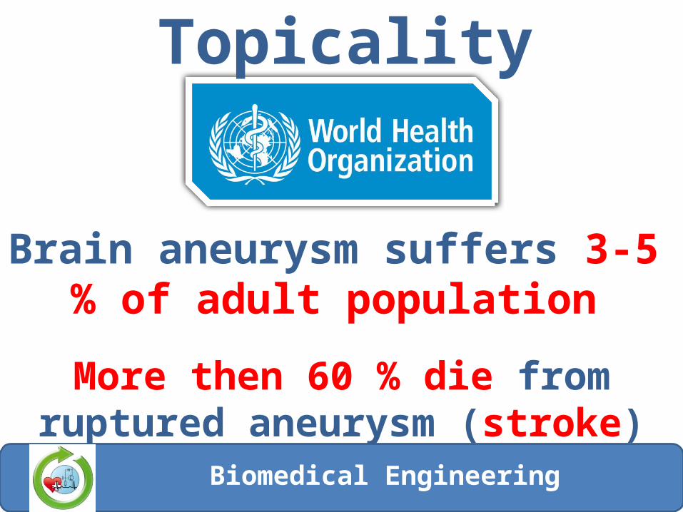
Aneurysm
Кафедра «Биомедицинская техника»
Protrusion of the arterial wall due to its stretching or thinning
Biomedical Engineering
Current stateCauses of aneurysm development and growth are not fully studied
Researchers suggest that key role in aneurysm development play violation of local and global hemodynamics
Biomedical Engineering
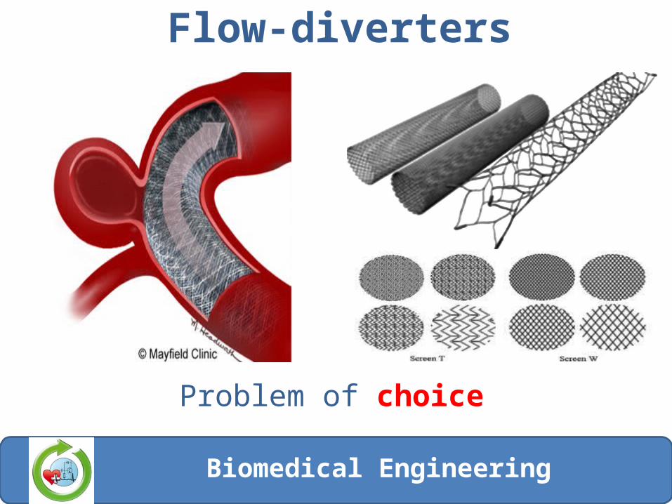
Flow-diverters
Problem of choice
Biomedical Engineering
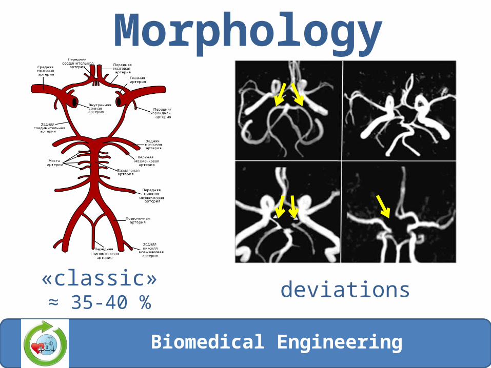
Morphology
«classic»≈ 35-40 % cases
deviations
Biomedical Engineering
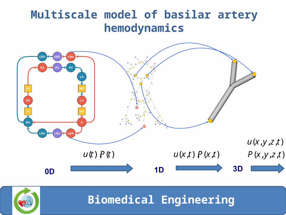
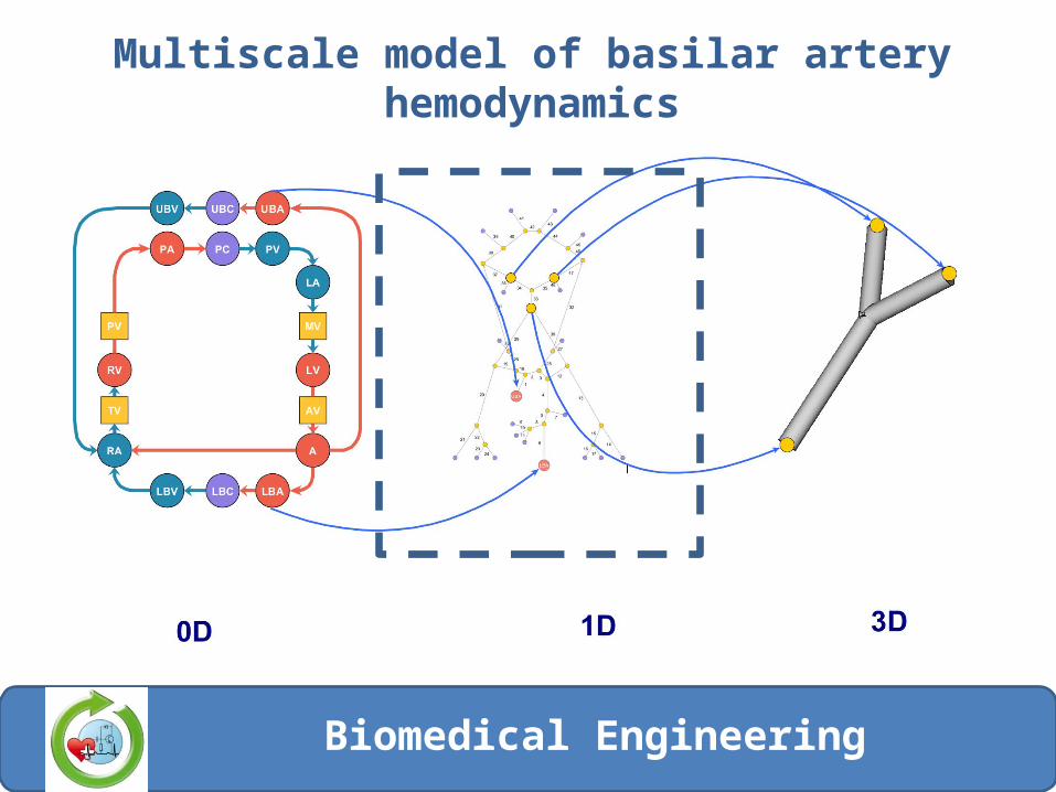
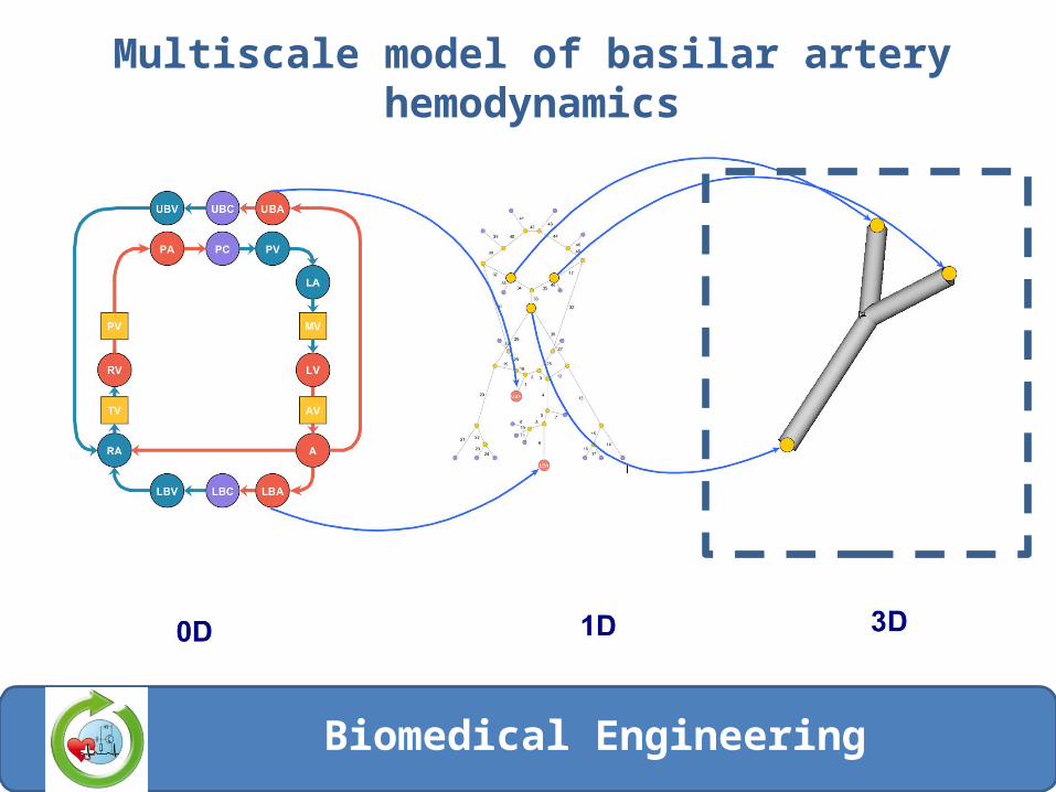
Multiscale model of basilar artery hemodynamics
)(),( tPtu ),(),,( txPtxu ),,,(
),,,,(
tzyxP
tzyxu
Biomedical Engineering
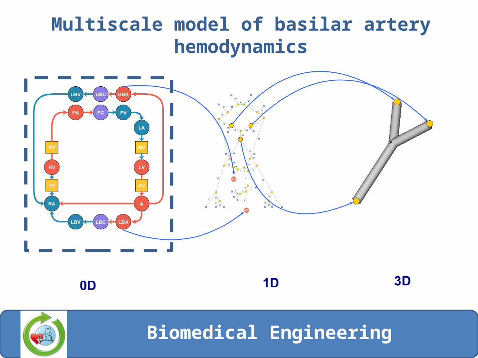
Multiscale model of basilar artery hemodynamics
Biomedical Engineering
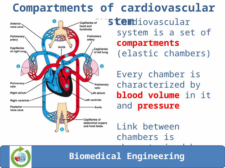
Compartments of cardiovascular systemCardiovascular system is a set of compartments (elastic chambers)
Every chamber is characterized by blood volume in it and pressure
Link between chambers is characterized by blood flow
Biomedical Engineering
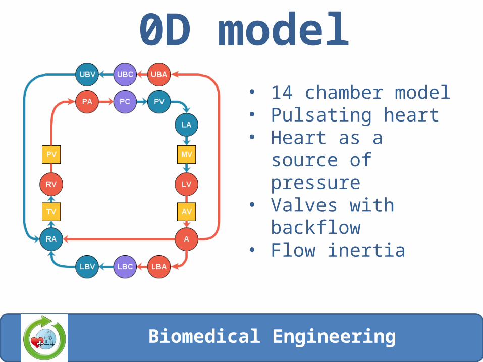
0D model• 14 chamber model• Pulsating heart• Heart as a source of
pressure• Valves with backflow• Flow inertia
Biomedical Engineering
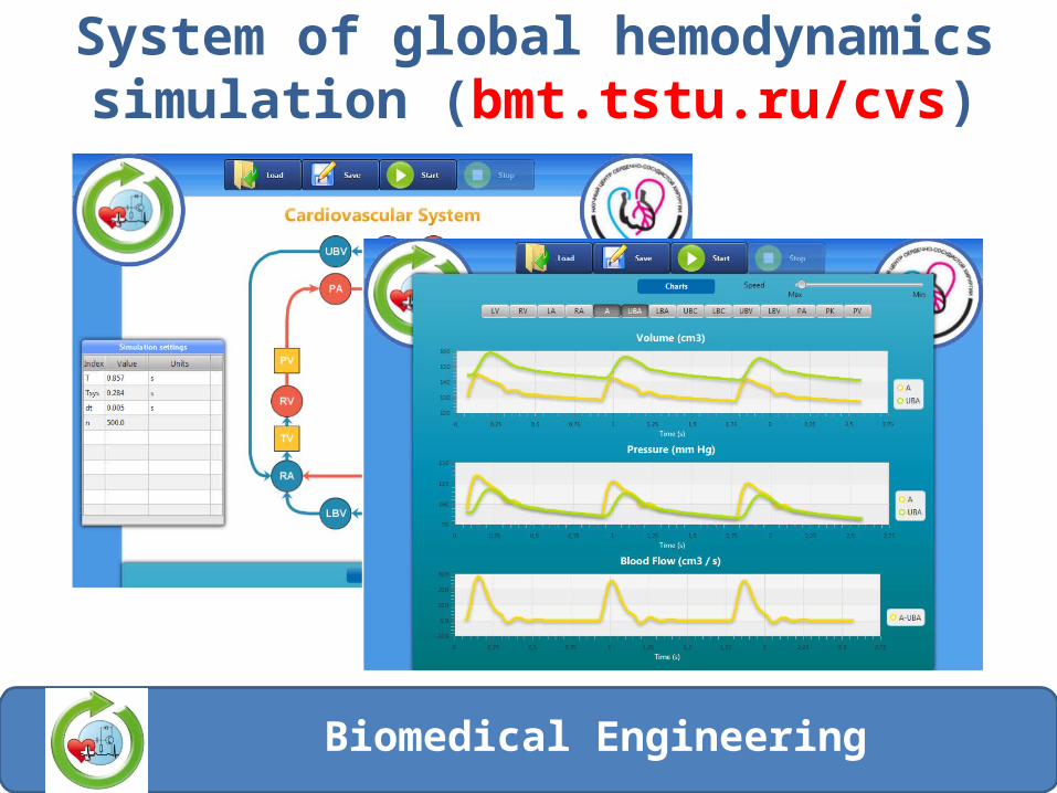
System of global hemodynamics simulation (bmt.tstu.ru/cvs)
Biomedical Engineering
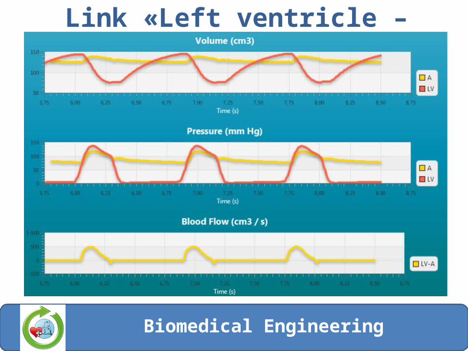
Link «Left ventricle – aorta»
Biomedical Engineering
Multiscale model of basilar artery hemodynamics
Biomedical Engineering
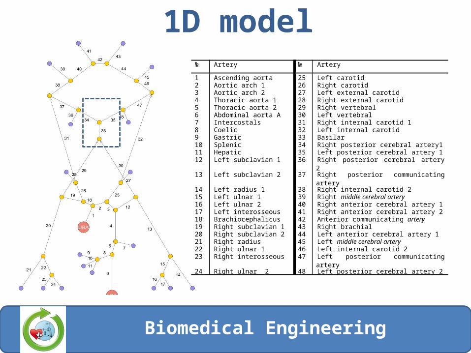
1D model№ Artery № Artery
1 Ascending aorta 25 Left carotid2 Aortic arch 1 26 Right carotid3 Aortic arch 2 27 Left external carotid4 Thoracic aorta 1 28 Right external carotid5 Thoracic aorta 2 29 Right vertebral6 Abdominal aorta A 30 Left vertebral7 Intercostals 31 Right internal carotid 18 Coelic 32 Left internal carotid9 Gastric 33 Basilar10 Splenic 34 Right posterior cerebral artery111 Hepatic 35 Left posterior cerebral artery 112 Left subclavian 1 36 Right posterior cerebral artery 213 Left subclavian 2 37 Right posterior communicating artery14 Left radius 1 38 Right internal carotid 215 Left ulnar 1 39 Right middle cerebral artery16 Left ulnar 2 40 Right anterior cerebral artery 117 Left interosseous 41 Right anterior cerebral artery 218 Brachiocephalicus 42 Anterior communicating artery19 Right subclavian 1 43 Right brachial20 Right subclavian 2 44 Left anterior cerebral artery 121 Right radius 45 Left middle cerebral artery22 Right ulnar 1 46 Left internal carotid 223 Right interosseous 47 Left posterior communicating artery24 Right ulnar 2 48 Left posterior cerebral artery 2
Biomedical Engineering
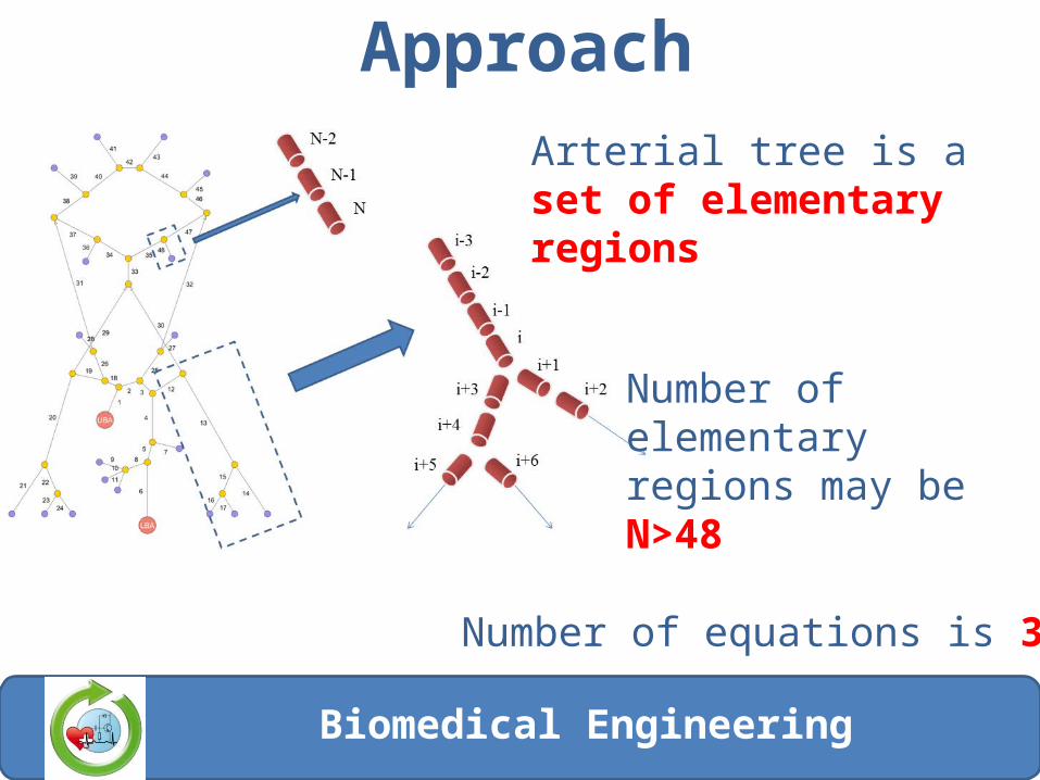
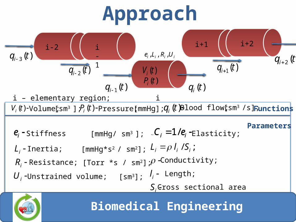
ApproachArterial tree is a set of elementary regions
Number of elementary regions may be N>48
Number of equations is 3*N
Biomedical Engineering
Festdaten
12
i-2 i-1
i
i+1 i+2
)(2 tqi
)(tqi
)(3 tqi )(2 tqi)(1 tqi
)(1 tqi
i – elementary region;
ie
i
i
i
U
R
L
Elasticity;
Inertia;
Resistance;
Unstrained volume;
Stiffness [mmHg/ sm3 ]; ii eС /1
[mmHg*s2 / sm2];
[Тоrr *s / sm2];
[sm3];
iiii URLe ,,,
i
i
iii
S
l
SlL
;/
Conductivity;
Length;
Cross sectional area
);(tVi
)(tPi)(tVi
)(tPi
)(tqiVolume; [sm3 ]; Pressure; [mmHg]; Blood flow; [sm3 /s];
Parameters
Functions
ie ii eС /1
Approach
Biomedical Engineering
Multiscale model of basilar artery hemodynamics
Biomedical Engineering
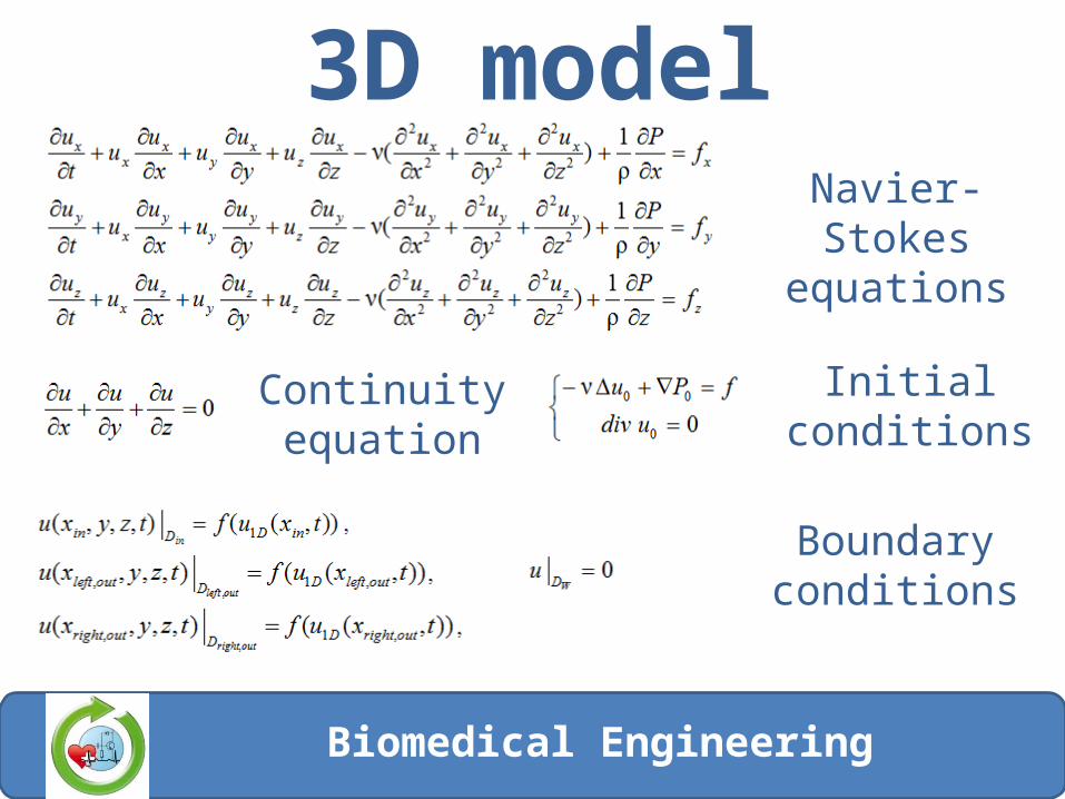
3D modelNavier-Stokes
equations
Continuity equation
Boundary conditions
Initial conditions
Biomedical Engineering
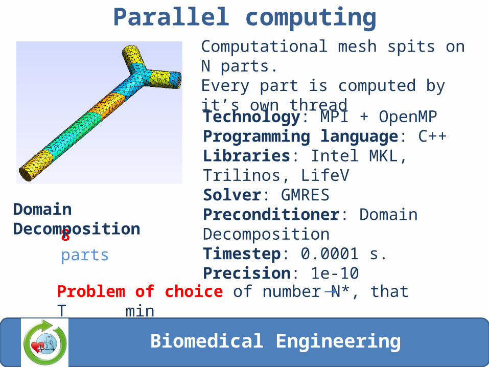
Parallel computing
8 parts
Domain Decomposition
Computational mesh spits on N parts.Every part is computed by it’s own thread
Technology: MPI + OpenMPProgramming language: С++Libraries: Intel MKL, Trilinos, LifeVSolver: GMRESPreconditioner: Domain DecompositionTimestep: 0.0001 s.Precision: 1e-10
Problem of choice of number N*, that T min
Biomedical Engineering
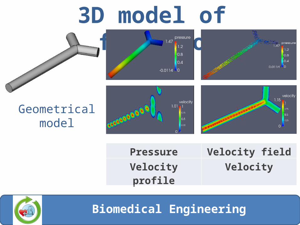
3D model of bifurcations
Geometrical model
Pressure Velocity fieldVelocity profile Velocity
Biomedical Engineering

Experimental setup3D Laser Doppler Anemometer measure blood velocity in elastic vessel model. It gives opportunity to estimate stent influence on blood flow
Biomedical Engineering
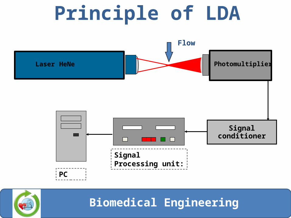
Principle of LDA
Biomedical Engineering
Signalconditioner
Flow
Signal Processing unit:
Laser HeNe Photomultiplier
PC

Measurements
Biomedical Engineering
The flow velocity is measured with a 1-component laser Doppler anemometer system equipped with a 5mW He-Ne laser with a wavelength of 632.8nm. The laser Doppler anemometer does not disturb the flow and is not affected by temperature, pressure or fluid density. It offers a high spatial (focus point less than 70 µm) and temporal (1ms) resolution. Velocity measurements are performed in the model at different cross sections allowing a 2D or a 3D-flow-reconstruction.
Measurements
Biomedical Engineering
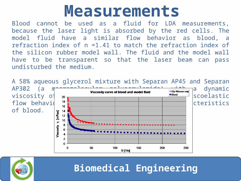
Blood cannot be used as a fluid for LDA measurements, because the laser light is absorbed by the red cells. The model fluid have a similar flow behavior as blood, a refraction index of n =1.41 to match the refraction index of the silicon rubber model wall. The fluid and the model wall have to be transparent so that the laser beam can pass undisturbed the medium.
A 58% aqueous glycerol mixture with Separan AP45 and Separan AP302 (a macromolecular polyacrylamide) with a dynamic viscosity of 8mPas is used. The mixture shows viscoelastic flow behavior similar to the non-Newtonian characteristics of blood.
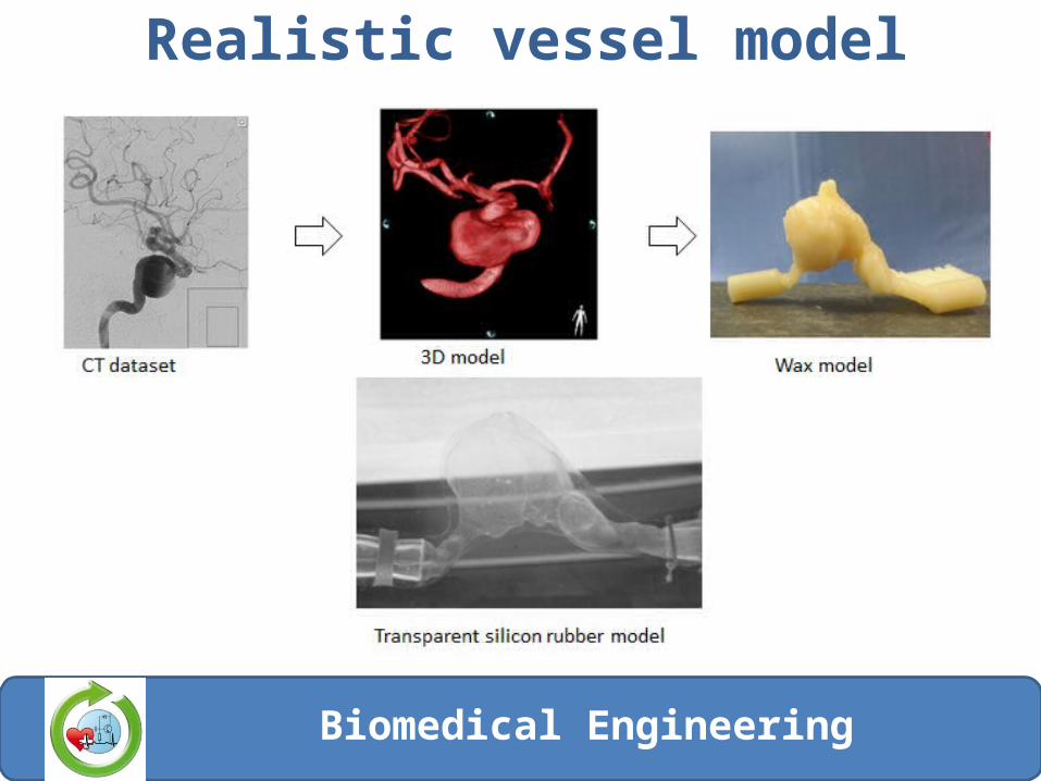
Realistic vessel model
Biomedical Engineering
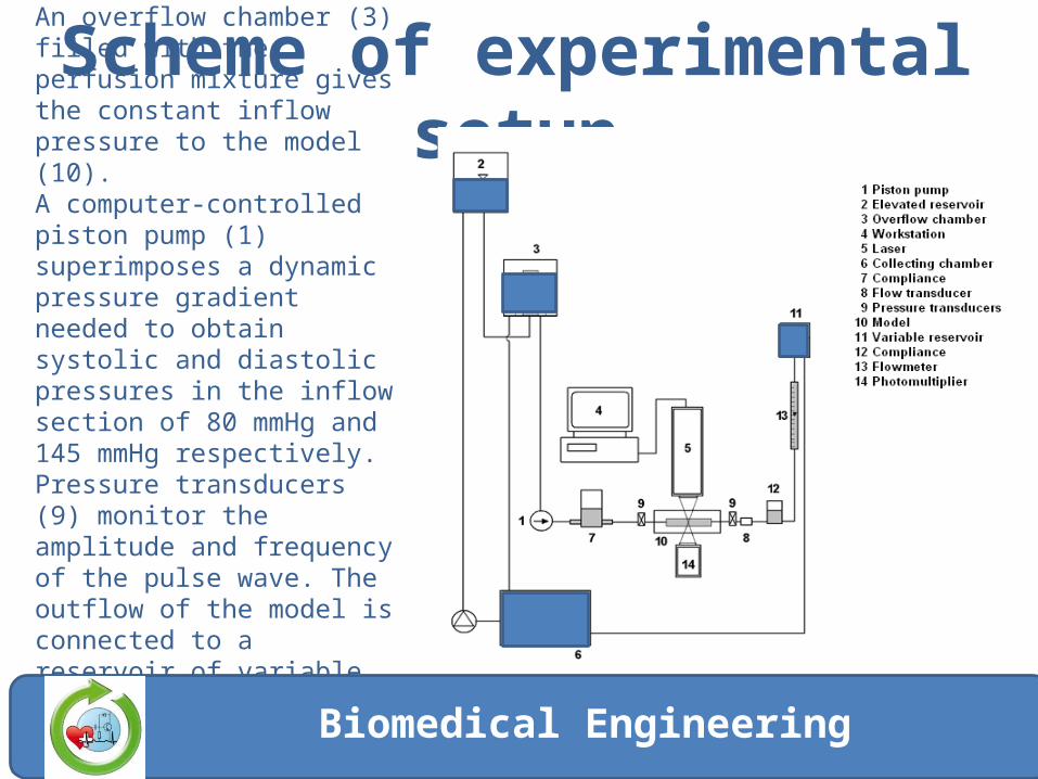
Scheme of experimental setupAn overflow chamber (3) filled with the perfusion mixture gives the constant inflow pressure to the model (10). A computer-controlled piston pump (1) superimposes a dynamic pressure gradient needed to obtain systolic and diastolic pressures in the inflow section of 80 mmHg and 145 mmHg respectively. Pressure transducers (9) monitor the amplitude and frequency of the pulse wave. The outflow of the model is connected to a reservoir of variable height (11) to allow control of the flow volume.
Biomedical Engineering

Thanks for attention
Biomedical Engineering