Embed Size (px)
Citation preview
1521-0103/374/2/252–263$35.00 https://doi.org/10.1124/jpet.120.266122THE JOURNAL OF PHARMACOLOGY AND EXPERIMENTAL THERAPEUTICS J Pharmacol Exp Ther 374:252–263, August 2020Copyright ª 2020 by The Author(s)This is an open access article distributed under the CC BY Attribution 4.0 International license.
MK-8719, a Novel and Selective O-GlcNAcase Inhibitor ThatReduces the Formation of Pathological Tau and AmelioratesNeurodegeneration in a Mouse Model of Tauopathy s
Xiaohai Wang, Wenping Li, Jacob Marcus, Michelle Pearson, Lixin Song, Karen Smith,Giuseppe Terracina, Julie Lee, Kwok-Lam Karen Hong, Sherry X. Lu, Lynn Hyde,Shu-Cheng Chen, David Kinsley, Jerry P. Melchor, Daniel J. Rubins, Xiangjun Meng,Eric Hostetler, Cyrille Sur, Lili Zhang, Joel B. Schachter, J. Fred Hess, Harold G. Selnick,David J. Vocadlo, Ernest J. McEachern, Jason M. Uslaner, Joseph L. Duffy,and Sean M. SmithMRL, Merck & Co., Inc., Kenilworth, New Jersey (X.W., W.L., J.M., M.P., L.S., K.S., G.T., J.L., K.-L.K.H., S.X.L., L.H., S.-C.C.,D.K., J.P.M., D.J.R., X.M., E.H., C.S., L.Z., J.B.S., J.F.H., H.G.S., J.M.U., J.L.D., S.M.S.) and Alectos Therapeutics Inc., Burnaby,British Columbia, Canada (D.J.V., E.J.M.)
Received March 6, 2020; accepted June 1, 2020
ABSTRACTDepositionof hyperphosphorylatedandaggregated tauprotein in thecentral nervous system is characteristic of Alzheimer disease andother tauopathies. Tau is subject to O-linked N-acetylglucosamine(O-GlcNAc) modification, and O-GlcNAcylation of tau has beenshown to influence tau phosphorylation and aggregation. In-hibition of O-GlcNAcase (OGA), the enzyme that removesO-GlcNAc moieties, is a novel strategy to attenuate the forma-tion of pathologic tau. Here we described the in vitro and in vivopharmacological properties of a novel and selective OGA in-hibitor, MK-8719. In vitro, this compound is a potent inhibitor ofthe human OGA enzyme with comparable activity against thecorresponding enzymes from mouse, rat, and dog. In vivo, oraladministration of MK-8719 elevates brain and peripheral bloodmononuclear cell O-GlcNAc levels in a dose-dependent man-ner. In addition, positron emission tomography imaging studiesdemonstrate robust target engagement of MK-8719 in thebrains of rats and rTg4510mice. In the rTg4510mousemodel ofhuman tauopathy, MK-8719 significantly increases brainO-GlcNAc levels and reduces pathologic tau. The reduction in
tau pathology in rTg4510 mice is accompanied by attenua-tion of brain atrophy, including reduction of forebrain volumeloss as revealed by volumetric magnetic resonance imaginganalysis. These findings suggest that OGA inhibition mayreduce tau pathology in tauopathies. However, since hun-dreds of O-GlcNAcylated proteins may be influenced by OGAinhibition, it will be critical to understand the physiologic andtoxicological consequences of chronic O-GlcNAc elevationin vivo.
SIGNIFICANCE STATEMENTMK-8719 isanovel, selective,andpotentO-linkedN-acetylglucosamine(O-GlcNAc)-ase (OGA) inhibitor that inhibits OGA enzymeactivity across multiple species with comparable in vitro po-tency. In vivo, MK-8719 elevates brainO-GlcNAc levels, reducespathological tau, and ameliorates brain atrophy in the rTg4510mouse model of tauopathy. These findings indicate that OGAinhibition may be a promising therapeutic strategy for thetreatment of Alzheimer disease and other tauopathies.
IntroductionNeurofibrillary tangles (NFTs), a pathologic hallmark of
Alzheimer disease (AD), are mainly composed of intracellularhyperphosphorylated and aggregated microtubule-associatedprotein tau (Lee et al., 2001; Ballatore et al., 2007). In additionto AD, NFTs are also present in a family of neurodegenerative
diseases known as tauopathies, including frontotemporaldementia, corticobasal degeneration, Pick disease, and pro-gressive supranuclear palsy (Lee et al., 2001). In frontotem-poral dementia with parkinsonism type-17, tau is causallyrelated to the neurodegeneration and dementia, as demon-strated by the discovery that mutations in the tau gene causethe disease (Hutton et al., 1998). These mutations may affectalternative splicing of taumRNA, decrease the ability of tau tobind microtubules, and increase the propensity of tau to formaggregates (Dayanandan et al., 1999; Gamblin et al., 2000;Lee et al., 2001).
This work was funded by Merck Sharp & Dohme Corp., a subsidiary ofMerck & Co., Inc., Kenilworth, NJ and Alectos Therapeutics Inc.
https://doi.org/10.1124/jpet.120.266122.s This article has supplemental material available at jpet.aspetjournals.org.
ABBREVIATIONS: AD, Alzheimer disease; BID, twice daily; CSF, cerebrospinal fluid; GlcNAc, N-acetylglucosamine; HEX A/B, hexosaminidaseA/B; LMA, locomotor activity; MRI, magnetic resonance imaging; MSD, MesoScale Discovery; NAGLU, N-acetylglucosaminidase; NFT,neurofibrillary tangle; OGA, O-GlcNAcase; O-GlcNAc, O-linked GlcNAc; OGT, O-GlcNAc transferase; PBMC, peripheral blood mononuclear cell;PET, positron emission tomography; SD, Sprague-Dawley; SPR, surface plasmon resonance; vMRI, volumetric MRI; WGA, wheat germ agglutinin.
252
http://jpet.aspetjournals.org/content/suppl/2020/06/03/jpet.120.266122.DC1Supplemental material to this article can be found at:
at ASPE
T Journals on February 4, 2022
jpet.aspetjournals.orgD
ownloaded from
Tau is subject to a variety of post-translational modifications,including phosphorylation, acetylation, sumoylation, isomeriza-tion, ubiquitination, nitration, andO-linkedN-acetylglucosamine(O-GlcNAc)-ylation (Martin et al., 2011). O-GlcNAcylation isa noncanonical glycosylation that involves the attachment ofsingle O-GlcNAc moieties to serine and threonine residues ofhundreds of different proteins found in the nucleus, mitochon-dria, and cytosol (Yang and Qian, 2017). The dynamic cyclingof this protein modification is controlled by a single pair ofenzymes: O-GlcNAc transferase (OGT), which catalyzesthe transfer of a N-acetylglucosamine (GlcNAc) moiety to thetarget serine and threonine residues, and O-GlcNAcase(OGA), which catalyzes the hydrolysis of this sugar modifica-tion (Hart et al., 2007). Cellular levels of O-GlcNAcylationare dependent on availability of the donor substrate forO-GlcNAcylation,UDP-GlcNAc. This donor substrate is amet-abolic product of glucose through the hexosamine biosyntheticpathway, and intracellular UDP-GlcNAc levels have beenshown to tightly correlate with the availability of glucose(Lefebvre et al., 2010). Therefore, O-GlcNAcylation may beconsidered as an intracellular sensor of glucose metabolismstatus (Gong et al., 2016).Impaired brain glucose metabolism has been observed in
multiple neurodegenerative diseases, including AD, and mayplay a critical role in the pathogenesis of these diseases(Hoyer, 2000; Mosconi, 2005; Butterfield and Halliwell,2019). Functional imaging studies have consistently identifieddeficient glucose metabolism throughout the cortex of ADpatients (Mosconi, 2005) and progression from mild cognitiveimpairment to AD tracks with reduction in cortical glucosemetabolism (Drzezga et al., 2003; Mosconi, 2005). Consistentwith these findings, O-GlcNAcylation levels are markedlyreduced in AD brains as compared with healthy controls (Liuet al., 2004, 2009; Pinho et al., 2019). More importantly, thedecrease in O-GlcNAcylation correlates with increasedhyperphosphorylated tau levels in AD brain. This relation-ship suggests that reduced tau O-GlcNAcylation mightcontribute to the progression of tauopathy in AD patients(Liu et al., 2009; Gong et al., 2016).Accumulating evidence indicates that O-GlcNAcylation
may limit tau phosphorylation and aggregation. For example,pharmacological inhibition of OGA elevates O-GlcNAcylationand reduces tau phosphorylation both in vitro and in vivo(Lefebvre et al., 2003; Yuzwa et al., 2008; Hastings et al.,2017). In addition, neuronal specific deletion of OGT leadsto accumulation of hyperphosphorylated tau in the brainand the spinal cord of transgenic animals (O’Donnell et al.,2004). Other than modulating tau phosphorylation, increasedO-GlcNAcylation of tau protein has also been reported tohinder tau aggregation (Yuzwa et al., 2012, 2014). Consistentwith these findings,O-GlcNAcylated tau is primarily detectedin the less phosphorylated soluble tau species but not inhyperphosphorylated insoluble tau (Lefebvre et al., 2003; Liuet al., 2009; Graham et al., 2014; Hastings et al., 2017). Inaddition, reduction of protein aggregation propensity withO-GlcNAcylation has been demonstrated with other proteins.As an example, the polycomb protein polyhomeotic forms largeaggregates in the absence of O-GlcNAcylation both in vitroand in vivo (Gambetta andMuller, 2014).Moreover,O-GlcNAcmodification has been shown to block the aggregation ofa-synuclein protein associated with Parkinson disease (Mar-otta et al., 2015; Levine et al., 2019). These findings suggest
that pharmacological inhibition of OGA could increaseO-GlcNAcylation of tau and reduce the formation of pathologictau and could be explored as a treatment of AD and otherproteinopathies provided that this mechanism does notimpact the function of other essential proteins.In this work, we characterize the in vitro and in vivo
pharmacological properties of a novel and selective OGAinhibitor, MK-8719. This compound demonstrates nanomolarpotency at the OGA enzyme and increases O-GlcNAc levelsboth in vitro and in vivo. In addition, MK-8719 enzymeoccupancy is revealed by positron emission tomography(PET) imaging analysis. In the rTg4510 transgenic mousemodel of tauopathy, MK-8719 reduces brain aggregated tau inthe insoluble fraction of brain homogenates, attenuates NFT-like pathology, and ameliorates brain atrophy.
Materials and MethodsEnzyme Activity Assay. OGA enzymatic reactions were carried
out in a reaction containing 50 mM NaH2PO4, 100 mM NaCl, and0.1% bovine serumalbumin (pH 7.0) using 2mM4-methylumbelliferylN-acetyl-b-D-glucosaminide dihydrate (M2133; Sigma) dissolvedin double distilled H2O, as a fluorogenic substrate. The amount ofpurified human OGA enzyme used in the reaction was 0.7 nM.Test compound of varying concentrations was added to theenzyme prior to initiation of the reaction. The reaction wasperformed at room temperature in a 96-well plate and wasinitiated with the addition of substrate. The generation offluorescent product, 4-methylumbelliferone, was measured every60 seconds for 45 minutes with a Tecan Infinite M200 plate readerwith excitation at 355 nM, and emission was detected at 460 nM.4-methylumbelliferone (M1381; Sigma) was used to producea standard curve. The slope of product production was determinedfor each concentration of compound tested and plotted, usingstandard curve-fitting algorithms for sigmoidal dose-responsecurves. The values for a four-parameter logistic curve fit of thedata were determined. Ki values were determined using theCheng-Prusoff equation.
Hexosaminidase A/B (HEX A/B) enzyme activity assay was per-formed in a similar condition as the OGA enzyme activity assay,except the amount of purified human hexosaminidase enzyme used inthe reaction was 24 nM. N-acetylglucosaminidase (NAGLU) enzymeactivity assay was also conducted in a condition similar to the OGAenzyme activity assay, except the reactionswere carried out in a buffercontaining 200 mM NaH2PO4 and 0.5% bovine serum albumin (pH7.0) using 0.4 mM 4-methylumbelliferyl N-acetyl-b-D-glucosaminidedihydrate (M2133; Sigma) dissolved in double distilled H2O as a sub-strate, and the amount of purified humanNAGLU enzyme used in thereaction was 20 nM.
OGA Cell-Based Assay. Inhibition of cellular OGA was deter-mined by measuring the increase of O-GlcNAcylated protein in PC-12cells. Increased O-GlcNAcylated protein was measured by ELISAusing the RL-2 antibody that binds total O-GlcNAcylated protein. Inthis assay, rat PC-12 cells were plated in 96-well plates withapproximately 10,000 cells/well. Compounds to be tested were dis-solved in DMSO, either 2- or 10-mM stock solution, and then dilutedwith DMSO and water in a two-step process using a Tecan worksta-tion. Cells were treated with diluted compounds for 24 hours to reacha final concentration of inhibitor desired to measure a concentration-dependent response, typically, 10 3-fold dilution steps, starting at10 mM. To prepare a cell lysate, the media from compound-treatedcells were removed, and the cells werewashed oncewith PBS and thenlysed for 5 minutes at room temperature in 50 ml of PhosphoSafereagent (Novagen Inc., Madison, WI) with protease inhibitors andphenylmethylsulfonyl fluoride. The cell lysate was collected andtransferred to a new plate, which was then either coated to assay
MK-8719 Reduces Tauopathy and Brain Atrophy in rTg4510 Mice 253
at ASPE
T Journals on February 4, 2022
jpet.aspetjournals.orgD
ownloaded from

plates directly or frozen at 280°C until used in the ELISA procedure.If desired, the total protein concentration of samples was determinedusing 20 ml of the sample with the BCA method.
The ELISA portion of the assay was performed in a black Maxisorp96-well plate that was coated overnight at 4°C with 100 ml/well of thecell lysate (1:10 dilution of the lysate with PBS-containing proteaseinhibitors, phosphatase inhibitors, and phenylmethylsulfonyl fluo-ride). The day after, the wells were washed three times with 300 ml/well of wash buffer (Tris-buffered saline with 0.1% Tween 20). Thewells were blocked with 100 ml/well blocking buffer (Tris-bufferedsaline with 0.05% Tween 20 and 2.5% bovine serum albumin). Eachwell was then washed two times with 300 ml/well of wash buffer. Theanti–O-GlcNAc antibody RL-2 (Abcam, Cambridge, MA), diluted 1:1000 in blocking buffer, was added at 100ml/well. The plate was sealedand incubated at 37°C for 2 hours with gentle shaking. The wells werethen washed three times with 300 ml/well wash buffer. To detect theamount of RL-2–bound, horseradish peroxidase–conjugated goat anti-mouse secondary antibody (diluted 1:3000 in blocking buffer) wasadded at 100 ml/well. The plate was incubated for 60 minutes at 37°Cwith gentle shaking. Each well was then washed three times with300 ml/well wash buffer. The detection reagent, 100 ml/well of AmplexUltra RED reagent (prepared by adding 30 ml of 10 mMAmplex UltraRED stock solution to 10 ml PBS with 18 ml 3% hydrogen peroxide,H2O2),was added.Thedetection reactionwas incubated for 15minutesat room temperature and then read with excitation at 530 nm andemission at 590 nm.
The amount of O-GlcNAcylated protein, as detected by the ELISAassay, was plotted for each concentration of test compound usingstandard curve-fitting algorithms for sigmoidal dose-response curves.The values for a four-parameter logistic curve fit of the data weredetermined, with the inflection point of the curve being the potencyvalue for the test compound.
Surface Plasmon Resonance Assay. Surface plasmon reso-nance (SPR) analysis was performed on a Biacore T-200 instrument(Biacore; GE Healthcare, Life Sciences). Human OGA protein wascaptured on a series S sensor chip CM5 using amine-coupling method.The methods for immobilization and binding analysis were generatedusing Biacore T200 control software, version 1.0. 1-ethyl-3-(3-dime-thylaminopropyl)carbodiimide hydrochloride/N-hydroxysuccinimidesolutions were used to activate a carboxymethylated dextran matrixon CM5 chip surface to covalently attach humanOGA protein throughexposed primary amines. Usually human OGA protein was diluted to100mg/ml, injected over the surface of the activatedCM5 chip at a flowrate of 10 ml/min for 45 seconds, and then followed by deactivationwith 130 ml of 1.0 M pH 8.5 ethanolamine to achieve the immobiliza-tion level of approximately 3500 resonance units of bound protein. Thebinding assay was performed using the running buffer of 10 mMHepes, pH 7.4; 150 mM NaCl; 0.005% Tween 20; 1 mM dithiothreitol;and 2% DMSO (filtered through a 0.2-mm polyethersulfone mem-brane). The 2X serial dilution of compound MK-8719 was made in100% DMSO at 50X concentration followed by subsequent dilutionand mixing with the running buffer. Eight different concentrations ofMK-8719, from 20 to 0.078 mM in running buffer, were injectedsimultaneously over the immobilized and reference surfaces, and thenet signal was obtained by subtracting the reference signal from thesignal with immobilized enzyme. The association phase for thecompound was followed for 90 seconds, and the dissociation phasewas followed for 1500 seconds. Solvent corrections were runwith eightdifferent DMSO concentrations spanning the range from 1% to3% according to the manufacturer recommendations.
Animals. This study was conducted in strict accordance with theNational Research Council’s Guide for the Care andUse of LaboratoryAnimals. The protocol was approved by the Institutional Animal CareandUse CommitteeMerck&Co., Inc., Kenilworth, NJ.Male Sprague-Dawley (SD) rats (Charles River) weighing 250–300 g were housed inan air-conditioned room on a 12-hour light/dark cycle with food andwater available ad libitum. The rTg4510 mouse line overexpressinghuman 4R0N tau with P301L mutation was licensed from the Mayo
Clinic and was bred and maintained at Taconic Biosciences, Hudson,NY. Briefly, the mice were generated by crossing the activator line(129S6 strain) expressing tetracycline-controlled transcriptional acti-vator driven by the calmodulin-dependent protein kinase II-a pro-moter and the responder line (FVB/N strain) expressing human 4R0Ntau with P301L mutation driven by tetracycline-operon responsiveelement as described previously (Santacruz et al., 2005). Only the F1females with 50%129S6 strain and 50%FVB/N strain were used in thisstudy. The mice were housed under standard laboratory conditions ofcontrolled temperature, humidity, and lighting (12-hour light: 12-hourdark; lights on at 6:00 AM) at Merck & Co., Inc. facility at least 1 weekbefore undergoing behavior testing to allow acclimation.
Drug Administration. MK-8719 was prepared by Merck & Co.,Inc., as described previously (Selnick et al., 2019). MK-8719 wasadministered either by oral gavage (5 ml/kg) or through in-diet dosingas described in Hastings et al. (2017). For oral gavage administrationof MK-8719, dosing solutions were prepared by dissolving MK-8719powder in sterile distilled water. Sterile water was administered asvehicle. After preparation, solutions were stored at 4°C and usedwithin 24 hours. For in-diet dosing, MK-8719 was formulated inmouse chow (D01060501) by Research Diets, Inc, New Brunswick,NJ. The concentration of MK-8719 in mouse chow was calculatedaccording to the average body weight of rTg4510 mice, average dailyfood intake, and dose levels. Concentration ofMK-8719 inmouse chow= (dose levels � body weight)/food intake.
Brain and Peripheral Blood Mononuclear Cell O-GlcNAcylatedProtein Assay. Male SD rats were euthanized by inhalation of CO2
at the indicated time point after administration of vehicle or MK-8719. After euthanasia, 4 to 5ml blood was drawn from the vena cavausing an 18-gauge needle attached to a 5ml syringe. A 4-ml CPT tubeprefilled with density gradient solution and sodium citrate asanticoagulant was filled for peripheral blood mononuclear cells(PBMCs) and plasma preparation. PBMCs were prepared by gener-ation of buffy cell layer according to the manufacturer’s instructions.After centrifugation, 0.2 ml of plasma was transferred to a 96-wellplate and frozen. Then 1.2 ml of the mononuclear cell layer wastransferred to a 1.7-ml microcentrifuge tube. PBMC samples werecentrifuged for 10 minutes at 1000g. Aqueous buffer was aspirated,and the cell pellets and plasma samples were frozen on dry ice andstored at 280°C.
After blood sampling, brain was harvested and dissected. All pieceswere frozen on dry ice and stored at 280°C. From each animal,unilateral pieces of frontal cortex were harvested into 2-ml tubes foranalysis of brain O-GlcNAc levels. One unilateral remnant forebrainsample was placed into a 48-well plate for measurement of compoundexposures. The remaining brain structures were discarded. To eachfrozen brain sample, one 5-mm steel bead and 700 ml of coldPhosphoSafe homogenization buffer were added. Samples werehomogenized by agitation at 30 Hz in a Qiagen TissueLyzer for2minutes. Samples were centrifuged at 15,000g for 15minutes at 4°C.Supernatant was transferred into 96-well plates and used for proteinmeasurements.
A sandwich immunoassay was developed to measure totalO-GlcNAc. Biotinylated wheat germ agglutinin (WGA) (Vector Bi-ology) at 5 mg/ml was used to coat a 96-well avidin plate [MesoScaleDiscovery (MSD)] in 50 ul Dulbecco’s PBS buffer (Fisher) containing5% Blocker A (MSD) and 0.2% Tween 20. After incubation for 1 hour,the platewaswashed three timeswith 0.2%Tween 20 in PBS.Brain orPBMChomogenates in 25ml (∼100mg protein) were added for a 3-hourincubation at room temperature before being washed three times with0.2% Tween 20/PBS. O-GlcNAc antibody RL2 (1:1000 dilution;AbCam) for O-GlcNAc detection and sTAG goat anti-mouse antibody(MSD) were added in 50 ml 5% Blocker A and 0.2% Tween 20, and theplate was incubated at 4°C overnight. The plates were washed threetimes before adding 150 ml Read Buffer T (MSD) and read on a Sector6000 Imager (MSD).
Protein Extraction and AlphaLISA-Based Immunoassaysfor Tau Protein. rTg4510mice were euthanizedwith CO2, and their
254 Wang et al.
at ASPE
T Journals on February 4, 2022
jpet.aspetjournals.orgD
ownloaded from
brains were dissected quickly. After removing the olfactory bulbs andthe cerebellum, the forebrains were weighed and cut in half sagittally.The left hemi-forebrains were fixed in 10% formalin for histology, andthe right hemi-forebrains were immediately frozen on dry ice and keptin 280°C for biochemical analysis. The forebrain was collectedfrom rTg4510 mice for O-GlcNAc and tau protein analysis, whereasfrontal cortex was harvested from SD rats to measure proteinO-GlcNAcylation. The large size of rat frontal cortex tissue wassufficient to provide uniform dissection and pharmacodynamic meas-urements across animals.
To separate the soluble and insoluble tau protein fractions fromrTg4510mouse brain, hemi-forebrains were homogenized using 5-mmmetal beads in a TissueLyzer (Qiagen) in 900 ml preparation buffercontaining 50 mM Tris, pH 8.0; 250 mM NaCl; 5 mM KCl; 2 mMEDTA; 2 mM EGTA; Phospho-Safe extraction buffer (EMD/Novagen)plus Complete EDTA-free Protease Inhibitor Tablet (Roche); PhoStopTablet (Roche); 2 mM Trichostatin A (Sigma-Aldrich); 5 mM nicotin-amide (Sigma-Aldrich); and 1 mM PUGNAc (Sigma-Aldrich). Thehomogenates were centrifuged at 14,000g for 15 minutes to removetissue debris. The supernatants from the first spin were thencentrifuged at 100,000g for 30 minutes. The pellets from second spinwere resuspended in preparation buffer and defined as insolublefraction, whereas the supernatants from the second spin were definedas the soluble fraction. The insoluble fraction was used for aggregatedtau and phosphorylated tau measurements. Protein concentrationswere determined using the BCA assay kit (Pierce).
For the AlphaLISA-based assays, tau protein was captured bya monoclonal antibody that was biotinylated and bound tostreptavidin-coated donor beads (PerkinElmer). Detection was accom-plished either by a monoclonal antibody conjugated to the acceptorbeads directly or by a polyclonal antibody in combination with anti-rabbit IgG-conjugated acceptor beads (PerkinElmer). Assay reactions(25 ml) were carried out in OptiPlate-384 microplates (PerkinElmer)that contained 5 ml of analyte at the specified protein concentration,10 ml of biotinylated capture antibody (final concentration 2 nM), and10 ml of detection antibody-conjugated acceptor beads (final concen-tration 20 mg/ml). After overnight incubation at 4°C, 25 ml ofstreptavidin donor beads were added under subdued light conditions(final concentration 40 mg/ml), and the reactions were incubated atroom temperature for 60 minutes with gentle shaking. The fluores-cent signal was detected on an Envision Plate Reader at 615 nm(PerkinElmer).
To detect aggregated forms of tau, biotin-HT7 was used incombination with HT7-conjugated acceptor beads. This assay detectsany aggregated tau from a dimer to large aggregates of tau. Theaggregated tau assay preferentially detects the 64-kD hyperphos-phorylated species that is enriched in aggregated forms (Song, et al.,2015). Hyperphosphorylated tau species were determined usingbiotin-HT7 as the capture antibody and acceptor beads conjugatedto phosphorylated tau antibodies PHF6 (Covance) or AT8 (ThermoScientific) for detection.
Histology. Hemi-forebrains were immersion-fixed for 48 hours in10% neutral buffered formalin at 4°C, blocked coronally into four3-mm slabs using a brain matrix, and processed and embedded intoparaffin. Immunohistochemistry was performed on 5-mm coronalsections using automated immunostainers (Ventana Medical Sys-tems; Discovery XT) with AT8 mouse monoclonal antibody (Thermo-Fisher) and counterstained with hematoxylin. After coverslipping,slides were digitized using a slide scanner (Aperio ScanScopeXT;Leica), and numbers of NFT positive neurons were counted in theentorhinal cortex region using image analysis software (AperioImageScope) after manual determination of the region of interest.
Mouse Cerebrospinal Fluid Collection. Mice were euthanizedwith CO2 and placed on the stereotaxic frame with head tilted down∼80°. The skin and muscle were carefully removed above the cisternamagna, and dura mater was exposed under a surgical microscope. Asmall opening on dura mater was made with the tip of a 30-G needle,and the cerebrospinal fluid (CSF) was drawn from the cisternamagna
with a 20-ml pipettor. CSF samples were quickly transferred to a 500-ml eppendorf tube and frozen on dry ice. CSF total tau levels wereevaluated with the AlphaLISA assay using biotin-HT7 as the captureantibody and acceptor beads conjugated to total tau antibody BT2(Thermo Scientific) for detection.
Spontaneous Locomotor Activity. Spontaneous locomotor ac-tivity (LMA) was determined using digital activity monitors (KinderScientific, Poway, CA). Each apparatus consisted of a clear plexiglassbox (7 � 15 inches) placed within the activity monitor. All LMA testswere performed during light cycle between 9:00 AM and 11:00 AM. Apotential influence of altered circadian rhythm on LMA was notinvestigated in the current study. During the test, mice were free toambulate and to rear for 30 minutes. LMA was detected by infraredphoto beam breaks, and the number of beam breaks was recorded andanalyzed by a computer connected to the apparatus. LMA wasrecorded in 10-minute segments during the 30-minute test session.Activity chambers were cleaned with a diluted alcohol solution aftereach test run to eliminate residual odor cues.
PET Imaging. [18F]MK-8553, a novel OGA inhibitor PET ligand,has been developed as a preclinical and clinical research tool todetermine target engagement and help dose selection (Li et al., 2016).In vivo occupancy studies were carried out in rTg4510 mice and SDrats from Taconic with MK-8719 to establish a drug plasmaconcentration–OGA enzyme occupancy relationship. [18F]MK-8553(specific activity .1000 Ci/mmol, radiochemical purity .99%) wassupplied by Siemens Biomarker Research, North Wales, PA. PETimaging studies were conducted under the guiding principles of theAmerican Physiologic Society and the Guide for the Care and Use forLaboratory Animals published by the US National Institutes ofHealth (NIH publication 85-23, revised 1985) and were approved bythe Institutional Animal Care andUseCommittee atMerck&Co., Inc.
rTg4510 mice and SD rats were anesthetized using isoflurane(4% to 5% induction, 1%–3%maintenance) through a closed-nose conethroughout the imaging session. Animals were positioned on the PET(Focus220; Siemens Medical Solution, Hoffman Estate, IL) bed side-by-side or in a mouse hotel. The brains were positioned within thecenter of the field of view. Body temperature was maintained at 37°Cvia a heating lamp control by a rectal probe connected with a temper-ature controller. Heart rate and SPO2 were monitored by Pulseoximetry. MK-8719 was orally administered at 4–4.5 hours before[18F]MK-8553 injection (intravenous). PET data were collected for90 minutes starting at the time of radiotracer administration. Bloodsamples were taken before and at the end of the PET scan to measuredrug levels. Tracer kinetic modeling utilizing a metabolite-correctedarterial input function was performed to determine OGA occupancy inrats. In mice, arterial input function cannot be used for tracerquantification because of the large volume of blood required for thisapproach, and an alternative approach was applied by using a Logan-plot analysis and the average cerebellum curve across all full-blockscans as the nondisplaceable binding component. OGA occupancy wascalculated by comparing the group average distribution volume ratioat baseline, after full block, and after drug administration. Theaverage plasma level of MK-8719 during the PET study (rats) or theplasma level of MK-8719 at the end of the PET study (mice) wascorrelated to OGA enzyme occupancy.
Brain Volumetry by Volumetric Magnetic Resonance Imag-ing. Magnetic resonance acquisitions were performed in CharlesRiver Discovery Research Services facility in Finland. A horizontal11.7 T magnet with bore size 160 mm (Bruker Biospin GmbH,Karlsruhe, Germany) equipped with Bruker BGS-9-S/HP gradientset (maximum gradient strength 760mT/m, bore 90mm) interfaced toa Bruker BioSpec console (Bruker Biospin GmbH) was used for thisstudy. A 72-mm volume coil was used for transmission, and mousequadrature surface coil was used for receiving (Bruker Biospin GmbHandRapidBiomedical GmbH,Rimpar, Germany, respectively). Three-dimensional volumetric magnetic resonance imagining (vMRI) datawere acquired using a magnetization transfer–prepared 3D gradientecho sequence-fast low angle shot with the following parameters: echo
MK-8719 Reduces Tauopathy and Brain Atrophy in rTg4510 Mice 255
at ASPE
T Journals on February 4, 2022
jpet.aspetjournals.orgD
ownloaded from
time/repetition time = 2.5/25 milliseconds, flip of 10°, acquisition/reconstruction matrix 3D 128 � 96 � 96, field of view 12.8 � 9.6 �19.2 mm3 with a saturation slice, and number of accumulations = 5.Magnetization transfer preparation was achieved by an 8-millisecondGauss pulse with 768 flip and 4 kHz offset. The acquired 3D magneticresonance images were analyzed in the coronal plane for whole brain,forebrain, cerebellar block, olfactory bulb, hippocampus, cortex, andlateral ventricle volumes using an in-house written analysis programrun under an MATLAB (The MathWorks Inc., Natick, MA)environment.
Statistics. In all figures, data are presented as mean6 S.E.M. Allstatistics were performed with the software Prism (version 7.0;GraphPad). A P value of ,0.05 was considered significant. Thestatistical treatment of each data set is described individually in theresults.
ResultsIn Vitro Characterization of MK-8719. MK-8719 was
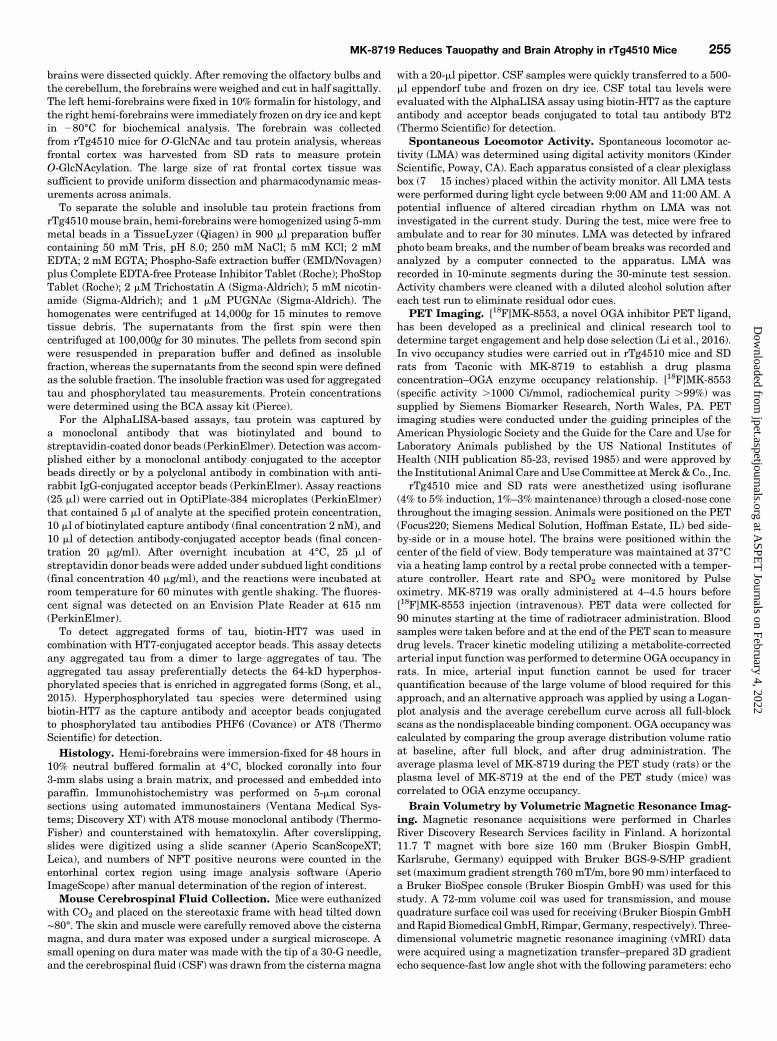
initially identified as a novel OGA inhibitor with excellentbrain permeability (Selnick et al., 2019). The functionalactivity of MK-8719 against human, rat, and dog OGA wasevaluated using fluorescent intensity assays with 2 mM 4-methylumbelliferyl N-acetyl-b-D-glucosaminide dihydrate asa substrate. MK-8719 inhibits the activity of the purifiedhuman OGA enzyme with a Ki of 7.9 6 0.5 nM (Fig. 1A). MK-8719 has similar in vitro potency for rat, dog, and mouse OGAenzymes, with Ki values of 9.76 0.8, 12.16 1.5, and 9.76 0.6nM, respectively. The cellular activity of MK-8719 againstOGA was established by measuring the accumulation ofO-GlcNAc in PC-12 cells that express rat OGA. Thepotency (EC50) of MK-8719 was 52.7 6 7 nM in the PC-12 cell-based O-GlcNAc assay (Fig. 1B). The affinity ofMK-8719 for human OGA was determined using a BiacoreT200 system that employs SPR technology. In this assay,MK-8719 demonstrated a Kd value of 3.1 nM. In addition,the SPR analysis also revealed the reversible nature ofMK-8719 binding to OGA, with a dissociation half-life of46 seconds.The primary counterscreens for MK-8719 were human Hex
A/B and human NAGLU assays. In vitro fluorescence in-tensity assays demonstrated thatMK-8719 up to 1mMhad nomeasurable activity against HEXA/B (Supplemental Table 1).Similarly, MK-8719 at concentrations up to 1 mM did notsignificantly inhibit NAGLU enzyme activity (SupplementalTable 1). In addition to the targeted screens against Hex A/Band NAGLU, MK-8719 was screened for binding activityagainst a panel of 118 known human and animal pharmacol-ogy targets by Ricerca Biosciences to confirm selectivity ofthe compound. This binding assay screen did not detect any
notable findings against a panel of.100 receptor and enzymetargets whenMK-8719 was tested at a concentration of 10 mM(unpublished data).Effects of OGA Inhibition on Brain and PBMC
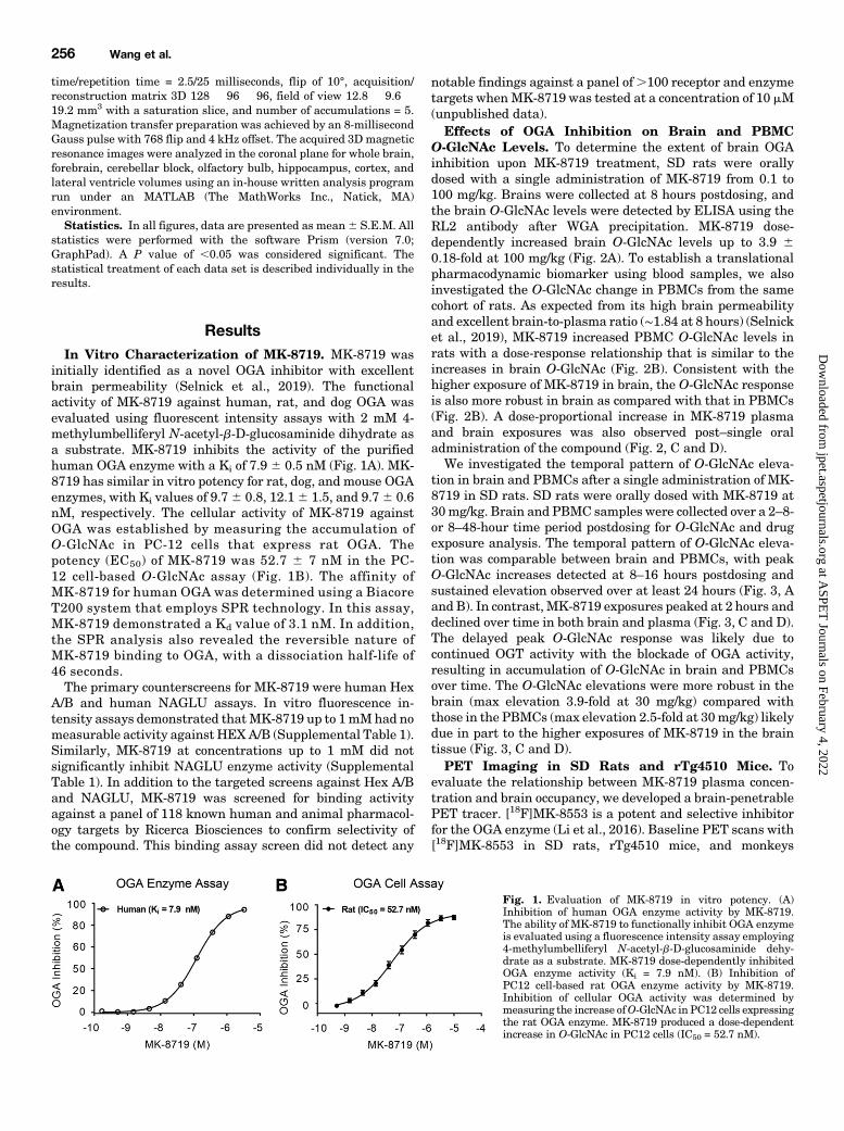
O-GlcNAc Levels. To determine the extent of brain OGAinhibition upon MK-8719 treatment, SD rats were orallydosed with a single administration of MK-8719 from 0.1 to100 mg/kg. Brains were collected at 8 hours postdosing, andthe brain O-GlcNAc levels were detected by ELISA using theRL2 antibody after WGA precipitation. MK-8719 dose-dependently increased brain O-GlcNAc levels up to 3.9 60.18-fold at 100 mg/kg (Fig. 2A). To establish a translationalpharmacodynamic biomarker using blood samples, we alsoinvestigated the O-GlcNAc change in PBMCs from the samecohort of rats. As expected from its high brain permeabilityand excellent brain-to-plasma ratio (∼1.84 at 8 hours) (Selnicket al., 2019), MK-8719 increased PBMC O-GlcNAc levels inrats with a dose-response relationship that is similar to theincreases in brain O-GlcNAc (Fig. 2B). Consistent with thehigher exposure of MK-8719 in brain, the O-GlcNAc responseis also more robust in brain as compared with that in PBMCs(Fig. 2B). A dose-proportional increase in MK-8719 plasmaand brain exposures was also observed post–single oraladministration of the compound (Fig. 2, C and D).We investigated the temporal pattern of O-GlcNAc eleva-
tion in brain and PBMCs after a single administration of MK-8719 in SD rats. SD rats were orally dosed with MK-8719 at30mg/kg. Brain and PBMC samples were collected over a 2–8-or 8–48-hour time period postdosing for O-GlcNAc and drugexposure analysis. The temporal pattern of O-GlcNAc eleva-tion was comparable between brain and PBMCs, with peakO-GlcNAc increases detected at 8–16 hours postdosing andsustained elevation observed over at least 24 hours (Fig. 3, Aand B). In contrast, MK-8719 exposures peaked at 2 hours anddeclined over time in both brain and plasma (Fig. 3, C and D).The delayed peak O-GlcNAc response was likely due tocontinued OGT activity with the blockade of OGA activity,resulting in accumulation of O-GlcNAc in brain and PBMCsover time. The O-GlcNAc elevations were more robust in thebrain (max elevation 3.9-fold at 30 mg/kg) compared withthose in the PBMCs (max elevation 2.5-fold at 30mg/kg) likelydue in part to the higher exposures of MK-8719 in the braintissue (Fig. 3, C and D).PET Imaging in SD Rats and rTg4510 Mice. To
evaluate the relationship between MK-8719 plasma concen-tration and brain occupancy, we developed a brain-penetrablePET tracer. [18F]MK-8553 is a potent and selective inhibitorfor the OGA enzyme (Li et al., 2016). Baseline PET scans with[18F]MK-8553 in SD rats, rTg4510 mice, and monkeys
Fig. 1. Evaluation of MK-8719 in vitro potency. (A)Inhibition of human OGA enzyme activity by MK-8719.The ability of MK-8719 to functionally inhibit OGA enzymeis evaluated using a fluorescence intensity assay employing4-methylumbelliferyl N-acetyl-b-D-glucosaminide dehy-drate as a substrate. MK-8719 dose-dependently inhibitedOGA enzyme activity (Ki = 7.9 nM). (B) Inhibition ofPC12 cell-based rat OGA enzyme activity by MK-8719.Inhibition of cellular OGA activity was determined bymeasuring the increase ofO-GlcNAc in PC12 cells expressingthe rat OGA enzyme. MK-8719 produced a dose-dependentincrease in O-GlcNAc in PC12 cells (IC50 = 52.7 nM).
256 Wang et al.
at ASPE
T Journals on February 4, 2022
jpet.aspetjournals.orgD
ownloaded from
revealed similar regional distribution (Li et al., 2016). Thehighest uptake was in striatum, and significant uptake wasalso observed in the hippocampus and cerebral cortex in rats(Fig. 4A) and rTg4510 mice (Fig. 5A). PET occupancy studiesin animals were conducted 4–4.5 hours after oral dosing ofMK-8719. [18F]MK-8553-specific uptake in the striatum,hippocampus, and cerebral cortex was almost completelyblocked after oral administration of OGA inhibitor MK-8719at 10 mg/kg in SD rats (Fig. 4B) and 100 mg/kg in rTg4510mice (Fig. 5B). The average plasma level of MK-8719 during
the PET study was correlated to brain occupancy in SDrats (Fig. 4C) and rTg4510 mice (Fig. 5C). The plasmaconcentration–occupancy relationship for PET studies fitsa typical sigmoidal equation in SD rats (Hill coefficient of0.696 0.13; Concentration required to produce 50% occupancy= 5.3 6 1.4 nM) and rTg4510 mice (Hill coefficient of 1.55 60.4; Concentration required to produce 50% occupancy = 2.660.6 nM). In addition, the brain O-GlcNAc levels at the endof the study were determined and plotted against occupancyin SD rats (Fig. 4D) and rTg4510 mice (Fig. 5D). The brain
Fig. 2. Dose-dependent effects of MK-8719 treatment onbrain and PBMC O-GlcNAc levels in SD rats. MK-8719dose-dependently increased O-GlcNAc levels in both brain(A) and PBMC (B) samples at 8 hours post–singleadministration by oral gavage. The elevations in PBMCO-GlcNAc were similar to those observed in the brain. (C)Brain exposure of MK-8719 at 8 hours post–single oraladministration. (D) Plasma exposure of MK-8719 at 8 hourspost–single oral administration (*P , 0.05 compared withthe vehicle groups, one-way ANOVA followed by Dunnett’spost hoc test, n = 8/group). BLQ, below the limit ofquantification.
Fig. 3. Time course of brain and PBMC O-GlcNAcelevations after MK-8719 treatment in SD rats. (A and B)Time course of brain and PBMC O-GlcNAc elevation from 2to 8 hours (A) and from 8 to 48 hours (B) after a singleadministration of MK-8719 at 30 mg/kg by oral gavage. (Cand D) Brain and plasma exposures of MK-8719 from 2 to 8hours (C) and from 8 to 48 hours (D) post–single oraladministration of MK-8719 at 30 mg/kg (n = 8/group).
MK-8719 Reduces Tauopathy and Brain Atrophy in rTg4510 Mice 257
at ASPE
T Journals on February 4, 2022
jpet.aspetjournals.orgD
ownloaded from

O-GlcNAc–occupancy relationship for MK-8719 in both SDrats and rTg4510 mice indicated that 80% occupancy corre-lated with a∼1.5-fold change in brainO-GlcNAc response, andOGA occupancy plateaued at levels that produced a ∼2-foldbrain O-GlcNAc elevation.MK-8719 Attenuates Brain Tauopathies in rTg4510
Mice. rTg4510 mice, which overexpress human mutant tau(P301L) in the forebrain structures, recapitulate several keyfeatures of tau pathology in patients with tauopathies. Thefeatures include progressive age-dependent increase in hyper-phosphorylated and aggregated tau species, NFTs, and brainatrophy (Santacruz et al., 2005; Song et al., 2015; Wang et al.,2018). To determine the effects of MK-8719 on brain tauopa-thies in rTg4510mice, we chronically treated themice with in-diet dosing of MK-8719 at 10, 30, and 100 mg/kg from 8 to16 weeks of age. The compound was well-tolerated with noovert adverse effects observed in any treatment group. Theanimals in all groups gained weight as expected, and therewere no significant differences in food intake or body weightbetween the treatment groups (Supplemental Fig. 1). Eightweeks of MK-8719 in-diet administration dose-dependentlyincreased brain O-GlcNAc levels by 2.3-, 3.3-, and 4.7-fold at10, 30, and 100 mg/kg, respectively (Fig. 6A). Brain samplesfrom the baseline group were collected at 8 weeks of agewithout compound treatment. Eight weeks of vehicle admin-istration from 8 to 16 weeks of age had no effect on brainO-GlcNAc levels (P . 0.05) as compared with the baselinegroup (Fig. 6A).The effects of chronic MK-8719 treatment on aggregated
and hyperphosphorylated tau in rTg4510mice were evaluated
by detecting various tau species in the insoluble fraction ofbrain homogeneous. To assess aggregated tau, we developedan HT7:HT7 AlphaLISA assay. In this assay, the total tauantibody HT7 was conjugated to both acceptor beads anddonor beads. Since there is only one HT7-binding site for eachtau protein, this assay only detects aggregated tau but notmonomeric tau (Song et al., 2015). The assay revealed an 8.8-fold increase in aggregated tau levels between the baselineand the vehicle groups suggesting the robust progression oftau pathology in rTg4510 brain from 8 to 16 weeks of age(Fig. 6B). Eight weeks of in-diet treatment with MK-8719dose-dependently reduced brain aggregated tau by 12.8%,22.9%, and 26% at 10, 30, and 100 mg/kg, respectively(Fig. 6B). Significant effects of MK-8719 were observed at 30(P , 0.05) and 100 mg/kg (P , 0.01) (Fig. 6B). To evaluatehyperphosphorylated tau, we developed HT7:PHF6 and HT7:AT8AlphaLISA assays. In these assays, the total tau antibodyHT7 was conjugated to acceptor beads and used as a captureantibody. The hyperphosphorylated tau antibodies PHF6 orAT8 were conjugated to the donor beads as detecting anti-bodies (Song et al., 2015). Compared with the baseline group,brain hyperphosphorylated tau levels in the vehicle groupincreased by ∼12-fold from 8 to 16 weeks (Fig. 6, C and D).Eight weeks of in-diet dosing with MK-8719 reduced brainhyperphosphorylated PHF6 tau by 20.7%, 23.6%, and 31% at10, 30, and 100 mg/kg, respectively (Fig. 6C), with significantreduction observed at 30 mg/kg (P, 0.05) and 100 mg/kg (P,0.01) (Fig. 6C). Similar effects of MK-8719 were found onhyperphosphorylated AT8 tau, with 17.3%, 25%, and 30.5% re-duction at 10, 30, and 100 mg/kg, respectively (Fig. 6D). In
Fig. 4. MK-8719 brain occupancy in SD rats revealed byPET imaging. (A) Summed PET baseline image (upper) andtime-activity curves (lower) showing the retention of [18F]MK-8553 in the striatum, hippocampus,s cerebral cortex,and cerebellum of SD rats. Rat brain region is highlightedwith the black circle in the coronal image. SUV, standard-ized uptake value. (B) [18F]MK-8553–specific uptake in thestriatum and other brain regions was almost completelyblocked after oral administration of 10 mg/kg MK-8719 inthe same animal as revealed by the summed PET image(upper) and the time-activity curves (lower). PO, by mouth.(C) Relationship between MK-8719 plasma concentration(at 6 hours post-treatment) and brain OGA occupancy. Occ,occupancy. (D) Relationship between brain O-GlcNAc level(at 6 hours post-treatment) and brain OGA occupancy asdetermined in vivo by [18F]MK-8553 in SD rats. PD,pharmacodynamics.
258 Wang et al.
at ASPE
T Journals on February 4, 2022
jpet.aspetjournals.orgD
ownloaded from
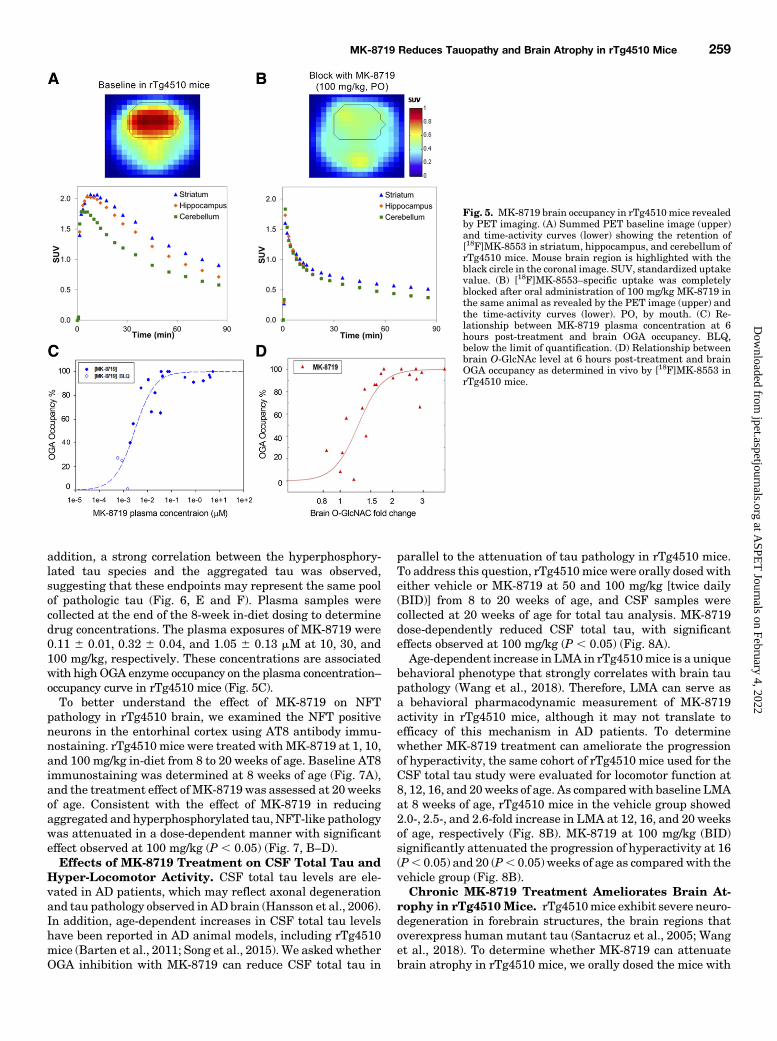
addition, a strong correlation between the hyperphosphory-lated tau species and the aggregated tau was observed,suggesting that these endpoints may represent the same poolof pathologic tau (Fig. 6, E and F). Plasma samples werecollected at the end of the 8-week in-diet dosing to determinedrug concentrations. The plasma exposures of MK-8719 were0.11 6 0.01, 0.32 6 0.04, and 1.05 6 0.13 mM at 10, 30, and100 mg/kg, respectively. These concentrations are associatedwith high OGA enzyme occupancy on the plasma concentration–occupancy curve in rTg4510 mice (Fig. 5C).To better understand the effect of MK-8719 on NFT
pathology in rTg4510 brain, we examined the NFT positiveneurons in the entorhinal cortex using AT8 antibody immu-nostaining. rTg4510mice were treated withMK-8719 at 1, 10,and 100 mg/kg in-diet from 8 to 20 weeks of age. Baseline AT8immunostaining was determined at 8 weeks of age (Fig. 7A),and the treatment effect of MK-8719 was assessed at 20 weeksof age. Consistent with the effect of MK-8719 in reducingaggregated and hyperphosphorylated tau, NFT-like pathologywas attenuated in a dose-dependent manner with significanteffect observed at 100 mg/kg (P , 0.05) (Fig. 7, B–D).Effects of MK-8719 Treatment on CSF Total Tau and
Hyper-Locomotor Activity. CSF total tau levels are ele-vated in AD patients, which may reflect axonal degenerationand tau pathology observed in AD brain (Hansson et al., 2006).In addition, age-dependent increases in CSF total tau levelshave been reported in AD animal models, including rTg4510mice (Barten et al., 2011; Song et al., 2015). We asked whetherOGA inhibition with MK-8719 can reduce CSF total tau in
parallel to the attenuation of tau pathology in rTg4510 mice.To address this question, rTg4510mice were orally dosed witheither vehicle or MK-8719 at 50 and 100 mg/kg [twice daily(BID)] from 8 to 20 weeks of age, and CSF samples werecollected at 20 weeks of age for total tau analysis. MK-8719dose-dependently reduced CSF total tau, with significanteffects observed at 100 mg/kg (P , 0.05) (Fig. 8A).Age-dependent increase in LMA in rTg4510mice is a unique
behavioral phenotype that strongly correlates with brain taupathology (Wang et al., 2018). Therefore, LMA can serve asa behavioral pharmacodynamic measurement of MK-8719activity in rTg4510 mice, although it may not translate toefficacy of this mechanism in AD patients. To determinewhether MK-8719 treatment can ameliorate the progressionof hyperactivity, the same cohort of rTg4510 mice used for theCSF total tau study were evaluated for locomotor function at8, 12, 16, and 20weeks of age. As comparedwith baseline LMAat 8 weeks of age, rTg4510 mice in the vehicle group showed2.0-, 2.5-, and 2.6-fold increase in LMA at 12, 16, and 20 weeksof age, respectively (Fig. 8B). MK-8719 at 100 mg/kg (BID)significantly attenuated the progression of hyperactivity at 16(P, 0.05) and 20 (P, 0.05) weeks of age as compared with thevehicle group (Fig. 8B).Chronic MK-8719 Treatment Ameliorates Brain At-
rophy in rTg4510 Mice. rTg4510mice exhibit severe neuro-degeneration in forebrain structures, the brain regions thatoverexpress human mutant tau (Santacruz et al., 2005; Wanget al., 2018). To determine whether MK-8719 can attenuatebrain atrophy in rTg4510 mice, we orally dosed the mice with
Fig. 5. MK-8719 brain occupancy in rTg4510mice revealedby PET imaging. (A) Summed PET baseline image (upper)and time-activity curves (lower) showing the retention of[18F]MK-8553 in striatum, hippocampus, and cerebellum ofrTg4510 mice. Mouse brain region is highlighted with theblack circle in the coronal image. SUV, standardized uptakevalue. (B) [18F]MK-8553–specific uptake was completelyblocked after oral administration of 100 mg/kg MK-8719 inthe same animal as revealed by the PET image (upper) andthe time-activity curves (lower). PO, by mouth. (C) Re-lationship between MK-8719 plasma concentration at 6hours post-treatment and brain OGA occupancy. BLQ,below the limit of quantification. (D) Relationship betweenbrain O-GlcNAc level at 6 hours post-treatment and brainOGA occupancy as determined in vivo by [18F]MK-8553 inrTg4510 mice.
MK-8719 Reduces Tauopathy and Brain Atrophy in rTg4510 Mice 259
at ASPE
T Journals on February 4, 2022
jpet.aspetjournals.orgD
ownloaded from

vehicle or MK-8719 at 100 mg/kg (BID) from 8 to 32 weeks ofage and periodically examined cortical, hippocampal, andlateral ventricle volumes with longitudinal vMRI. From 16to 32weeks of age, cortical and hippocampal volumes declined,
whereas lateral ventricle volume increased in rTg4510 mice(Fig. 9, A–C). MK-8719 significantly attenuated corticalvolume decline at 24 (P , 0.05) and 32 weeks of age (P ,0.05) (Fig. 9A). In addition, hippocampal volume decline was
Fig. 6. Effects of chronic MK-8719 treatment on brain O-GlcNAc and pathologic tau species in rTg4510 mice. MK-8719 at 10, 30, and 100 mg/kg orvehicle was administered in-diet to rTg4510mice from 8 to 16 weeks of age. Baseline samples were collected at 8 weeks of age without any treatment. (A)MK-8719 increased brain O-GlcNAc level detected by ELISA using the RL2 antibody after WGA precipitation. (B–D) MK-8719 reduced aggregated taudetected by HT7/HT7 AlphaLISA (B) and decreased hyperphosphorylated tau recognized by PHF6 (C) and AT8 (D) antibodies. RFU, relativefluorescence unit. (*P , 0.05; **P , 0.01; ***P , 0.001 compared with the vehicle group, n = 56/group, one-way ANOVA followed by Dunnett’s testcompared with the vehicle). (E and F) Correlations between levels of aggregated tau (HT7/HT7), AT8 tau, and PHF6 tau were determined using datafrom all vehicle- and MK-8719–treated animals (n = 226). Veh, vehicle.
Fig. 7. Effects of MK-8719 on NFT-like pathology in theentorhinal cortex of rTg4510 mice. rTg4510 mice wereadministered MK-8719 (1, 10, and 100 mg/kg) or vehicle viain-diet dosing from 8 to 20 weeks of age. Baseline levels ofAT8 immunoreactivity were evaluated in 8-week-oldanimals, and the effects of MK-8719 were determined in20-week-old rTg4510 mice. (A–C) Representative images ofNFT-like pathology in the entorhinal cortex of rTg4510mice determined by AT8 immunostaining. Scale bar, 100mm. (D) MK-8719 reduced NFTs in a dose-dependentmanner (*P, 0.05, n = 25/group, one-way ANOVA followedby Dunnett’s test compared with the vehicle). Veh, vehicle.
260 Wang et al.
at ASPE
T Journals on February 4, 2022
jpet.aspetjournals.orgD
ownloaded from
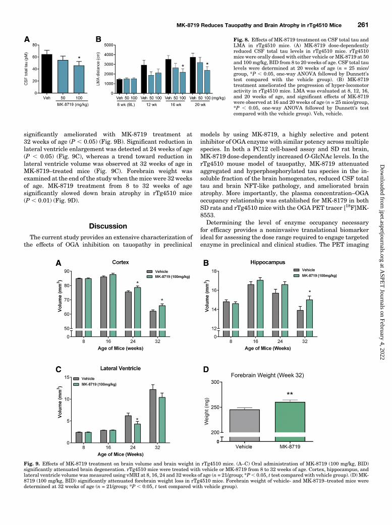
significantly ameliorated with MK-8719 treatment at32 weeks of age (P , 0.05) (Fig. 9B). Significant reduction inlateral ventricle enlargement was detected at 24 weeks of age(P , 0.05) (Fig. 9C), whereas a trend toward reduction inlateral ventricle volume was observed at 32 weeks of age inMK-8719–treated mice (Fig. 9C). Forebrain weight wasexamined at the end of the studywhen themice were 32weeksof age. MK-8719 treatment from 8 to 32 weeks of agesignificantly slowed down brain atrophy in rTg4510 mice(P , 0.01) (Fig. 9D).
DiscussionThe current study provides an extensive characterization of
the effects of OGA inhibition on tauopathy in preclinical
models by using MK-8719, a highly selective and potentinhibitor of OGA enzyme with similar potency across multiplespecies. In both a PC12 cell-based assay and SD rat brain,MK-8719 dose-dependently increased O-GlcNAc levels. In therTg4510 mouse model of tauopathy, MK-8719 attenuatedaggregated and hyperphosphorylated tau species in the in-soluble fraction of the brain homogenates, reduced CSF totaltau and brain NFT-like pathology, and ameliorated brainatrophy. More importantly, the plasma concentration–OGAoccupancy relationship was established for MK-8179 in bothSD rats and rTg4510 mice with the OGA PET tracer [18F]MK-8553.Determining the level of enzyme occupancy necessary
for efficacy provides a noninvasive translational biomarkerideal for assessing the dose range required to engage targetedenzyme in preclinical and clinical studies. The PET imaging
Fig. 8. Effects of MK-8719 treatment on CSF total tau andLMA in rTg4510 mice. (A) MK-8719 dose-dependentlyreduced CSF total tau levels in rTg4510 mice. rTg4510mice were orally dosed with either vehicle or MK-8719 at 50and 100 mg/kg, BID from 8 to 20 weeks of age. CSF total taulevels were determined at 20 weeks of age (n = 25 mice/group, *P , 0.05, one-way ANOVA followed by Dunnett’stest compared with the vehicle group). (B) MK-8719treatment ameliorated the progression of hyper-locomotoractivity in rTg4510 mice. LMA was evaluated at 8, 12, 16,and 20 weeks of age, and significant effects of MK-8719were observed at 16 and 20 weeks of age (n = 25 mice/group,*P , 0.05, one-way ANOVA followed by Dunnett’s testcompared with the vehicle group). Veh, vehicle.
Fig. 9. Effects of MK-8719 treatment on brain volume and brain weight in rTg4510 mice. (A–C) Oral administration of MK-8719 (100 mg/kg, BID)significantly attenuated brain degeneration. rTg4510 mice were treated with vehicle or MK-8719 from 8 to 32 weeks of age. Cortex, hippocampus, andlateral ventricle volume was measured using vMRI at 8, 16, 24 and 32 weeks of age (n = 21/group; *P, 0.05, t test compared with vehicle group). (D) MK-8719 (100 mg/kg, BID) significantly attenuated forebrain weight loss in rTg4510 mice. Forebrain weight of vehicle- and MK-8719–treated mice weredetermined at 32 weeks of age (n = 21/group; *P , 0.05, t test compared with vehicle group).
MK-8719 Reduces Tauopathy and Brain Atrophy in rTg4510 Mice 261
at ASPE
T Journals on February 4, 2022
jpet.aspetjournals.orgD
ownloaded from

studies revealed that high degree of enzyme occupancy isrequired to drive brain O-GlcNAc increases in SD rats andrTg4510 mice. As demonstrated by the relationship betweenMK-8719 brain O-GlcNAc and brain occupancy, 80% enzymeoccupancy correlated with only ∼1.5-fold elevation in brainO-GlcNAc, and near-maximal occupancy is required to achieve.3-fold brain O-GlcNAc increase. This is consistent with ourprevious observations in which 80% knockdown of OGAprotein in the OGA-inducible knockdown mice was onlyassociated with 1.4-fold increase in brain O-GlcNAc, and500 mg/kg dose of the potent OGA inhibitor Thiamet-G wasneeded to produce .3-fold brain O-GlcNAc elevation aftera single oral administration (Hastings et al., 2017). Indeed,a 500-mg/kg dose of Thiamet-G was typically administered intau transgenic models to evaluate the effects of OGA in-hibition on tauopathy (Yuzwa et al., 2012; Borghgraef et al.,2013; Graham et al., 2014; Hastings et al., 2017; Wang et al.,2018). To determine the level of enzyme occupancy requiredfor reducing aggregated and hyperphosphorylated tau inrTg4510 brain, we assessedMK-8179 at 10, 30, and 100mg/kg,the dose levels that are associated with high enzyme occu-pancy on the plasma concentration–occupancy curve inrTg4510 mice. At such high occupancy levels, MK-8719 wasstill able to demonstrate clear dose-dependent effects on brainO-GlcNAc elevation and pathologic tau reduction. In addition,significant effects of MK-8719 on brain NFT-like pathology,CSF total tau level, and hyper-locomotor activity were onlyobserved at 100-mg/kg dose but not at lower doses, suggestingthat extremely high enzyme occupancy is critical toO-GlcNAcchange and attenuation of tauopathy in vivo. [18F]MK-8553can be used as a valuable clinical tool to establish MK-8719plasma concentration–occupancy relationship for transla-tional modeling.Several mechanisms have been proposed to elucidate the
beneficial effects of OGA inhibition in animal models oftauopathy, including reduction of tau phosphorylation uponacute treatment with OGA inhibitors (Yuzwa et al., 2008; Yuet al., 2012; Graham et al., 2014), limiting the formation of tauaggregates (Yuzwa et al., 2012; Hastings et al., 2017) andenhancement of autophagy through an mechanistic target ofrapamycin–independent pathway (Zhu et al., 2018). Byemploying a novel O-GlcNAc detection method involving“click” N-acetylgalactosamine derivatization coupled withtetramethylrhodamine labeling, we have previously demon-strated O-GlcNAc modification of tau after chronic treatmentof rTg4510mice with Thiamet-G, which is structurally similarto MK-8719 (Hastings et al., 2017). One of the most consistentfindings from the tau transgenic models chronically treatedwith the OGA inhibitor Thiamet-G is the reduction ofaggregated and hyperphosphorylated tau species in the in-soluble fraction of brain homogenates (Yuzwa et al., 2012;Graham et al., 2014; Hastings et al., 2017; Wang et al., 2018).In addition, it has been shown that chronic OGA inhibition intau transgenic models has no effect on the phosphorylationstatus of soluble nonpathologic tau species. As such,O-GlcNAcylation may not directly regulate the phosphoryla-tion of tau (Yuzwa et al., 2012; Graham et al., 2014).Moreover,O-GlcNAcylated tau was primarily detected in the solublenonpathologic tau species but not in the insoluble aggregatedtau, suggesting that addition of polar O-GlcNAc moieties mayrender tau less prone to aggregation (Graham et al., 2014;Hastings et al., 2017). Consistent with these findings, the
current study demonstrated reduction in aggregated tau andseveral phosphorylated tau species in the insoluble fraction ofrTg4510 brain upon chronicMK-8719 treatment. Importantly,the phosphorylated tau species closely correlated with theaggregated tau, indicating that these endpoints may rep-resent the same pool of aggregated tau that was attenuatedby OGA inhibition. Overall, these data support the hypoth-esis that O-GlcNAc modification of tau reduces the pro-pensity of tau to form aggregates and therefore amelioratetauopathy.Magnetic resonance imaging (MRI)-based measures of
brain atrophy are regarded as valuable markers of neurode-generative diseases and are increasingly used as outcomemeasures in clinical studies of disease-modifying therapies(Frisoni et al., 2010). In AD patients, vMRI studies haveshown that the rates of brain atrophy in several structuralmeasures, including hippocampus, entorhinal cortex, wholebrain, and ventricle enlargement, correlate closely withchanges in cognitive performance (Fox et al., 1999; Jacket al., 2004; Thompson et al., 2004; Schott et al., 2008; Sluimeret al., 2010). rTg4510 mice develop severe age-dependentatrophy and neuron loss in forebrain structures, the regionsthat overexpress human mutant P301L tau under the controlof the calmodulin-dependent protein kinase II promoter(Ramsden et al., 2005; Santacruz et al., 2005; Yang et al.,2011; Wang et al., 2018). The current vMRI study shows that,between 16 and 32 weeks of age, the cortical and hippocampalvolume of rTg4510 mice decreased by 27.8% and 16.8%,respectively, whereas lateral ventricle volume increased by3.2-fold. Importantly, chronic OGA inhibition with MK-8719can ameliorate brain atrophy in multiple forebrain regions,suggesting that vMRImay serve as translational biomarker toevaluate the efficacy of OGA inhibitors in clinical settings.In summary, the present study characterizes the robust
in vitro and in vivo pharmacological profiles of OGA inhibition.Significant increases in brain and PBMC O-GlcNAc weredetected in response to MK-8719 administration in rodent.With the OGA PET tracer [18F]MK-8553, we were able todemonstrate that high degree of enzyme occupancy is requiredto drive brain O-GlcNAc elevation and ameliorate braintauopathy. The focus of the current studies was to evaluatethe effects of MK-8719 on the target protein tau related toAD. However, OGA inhibition influences O-GlcNAc mod-ification on hundreds of proteins. Therefore, it will beimportant to understand the physiologic and toxicologicalconsequences of prolonged elevation of O-GlcNAcylatedproteins throughout the proteome. These findings providea rationale for testing the hypothesis that OGA inhibitionmay be a promising therapeutic strategy for treatment oftauopathies.
Authorship Contributions
Participated in research design: Wang, Li, Marcus, Pearson, K.Smith, Lee, Chen, Hostetler, Sur, Zhang, Schachter, Hess, Selnick,Vocadlo, McEachern, Uslaner, Duffy, S.M. Smith.
Conducted experiments: Wang, Li, Marcus, Pearson, Song, K.Smith, Terracina, Lee, Hong, Lu, Hyde, Chen, Kinsley, Melchor, Meng.
Performed data analysis: Wang, Li, Marcus, Pearson, Song, K.Smith, Terracina, Lee, Hong, Lu, Hyde, Chen, Melchor, Rubins,Zhang, Hess, S.M. Smith.
Wrote or contributed to the writing of the manuscript: Wang, Li,Marcus, Sur, Zhang, Schachter, Hess, Selnick, Vocadlo, McEachern,Uslaner, Duffy, S.M. Smith.
262 Wang et al.
at ASPE
T Journals on February 4, 2022
jpet.aspetjournals.orgD
ownloaded from

References
Ballatore C, Lee VM, and Trojanowski JQ (2007) Tau-mediated neurodegeneration inAlzheimer’s disease and related disorders. Nat Rev Neurosci 8:663–672.
Barten DM, Cadelina GW, Hoque N, DeCarr LB, Guss VL, Yang L, Sankaranar-ayanan S, Wes PD, Flynn ME, Meredith JE, et al. (2011) Tau transgenic mice asmodels for cerebrospinal fluid tau biomarkers. J Alzheimers Dis 24 (Suppl 2):127–141.
Borghgraef P, Menuet C, Theunis C, Louis JV, Devijver H, Maurin H, Smet-Nocca C,Lippens G, Hilaire G, Gijsen H, et al. (2013) Increasing brain protein O-GlcNAc-ylation mitigates breathing defects and mortality of Tau.P301L mice. PLoS One 8:e84442.
Butterfield DA and Halliwell B (2019) Oxidative stress, dysfunctional glucose me-tabolism and Alzheimer disease. Nat Rev Neurosci 20:148–160.
Dayanandan R, Van Slegtenhorst M, Mack TG, Ko L, Yen SH, Leroy K, Brion JP,Anderton BH, Hutton M, and Lovestone S (1999) Mutations in tau reduce itsmicrotubule binding properties in intact cells and affect its phosphorylation. FEBSLett 446:228–232.
Drzezga A, Lautenschlager N, Siebner H, Riemenschneider M, Willoch F, MinoshimaS, Schwaiger M, and Kurz A (2003) Cerebral metabolic changes accompanyingconversion of mild cognitive impairment into Alzheimer’s disease: a PET follow-upstudy. Eur J Nucl Med Mol Imaging 30:1104–1113.
Fox NC, Scahill RI, Crum WR, and Rossor MN (1999) Correlation between rates ofbrain atrophy and cognitive decline in AD. Neurology 52:1687–1689.
Frisoni GB, Fox NC, Jack CR Jr., Scheltens P, and Thompson PM (2010) The clinicaluse of structural MRI in Alzheimer disease. Nat Rev Neurol 6:67–77.
Gambetta MC and Müller J (2014) O-GlcNAcylation prevents aggregation of thePolycomb group repressor polyhomeotic. Dev Cell 31:629–639.
Gamblin TC, King ME, Dawson H, Vitek MP, Kuret J, Berry RW, and Binder LI(2000) In vitro polymerization of tau protein monitored by laser light scattering:method and application to the study of FTDP-17 mutants. Biochemistry 39:6136–6144.
Gong CX, Liu F, and Iqbal K (2016) O-GlcNAcylation: a regulator of tau pathologyand neurodegeneration. Alzheimers Dement 12:1078–1089.
Graham DL, Gray AJ, Joyce JA, Yu D, O’Moore J, Carlson GA, Shearman MS,Dellovade TL, and Hering H (2014) Increased O-GlcNAcylation reduces patholog-ical tau without affecting its normal phosphorylation in a mouse model of tauop-athy. Neuropharmacology 79:307–313.
Hansson O, Zetterberg H, Buchhave P, Londos E, Blennow K, and Minthon L (2006)Association between CSF biomarkers and incipient Alzheimer’s disease in patientswith mild cognitive impairment: a follow-up study. Lancet Neurol 5:228–234.
Hart GW, Housley MP, and Slawson C (2007) Cycling of O-linked beta-N-acetylglu-cosamine on nucleocytoplasmic proteins. Nature 446:1017–1022.
Hastings NB, Wang X, Song L, Butts BD, Grotz D, Hargreaves R, Fred Hess J, HongKK, Huang CR, Hyde L, et al. (2017) Inhibition of O-GlcNAcase leads to elevationof O-GlcNAc tau and reduction of tauopathy and cerebrospinal fluid tau in rTg4510mice. Mol Neurodegener 12:39.
Hoyer S (2000) Brain glucose and energy metabolism abnormalities in sporadicAlzheimer disease. Causes and consequences: an update. Exp Gerontol 35:1363–1372.
Hutton M, Lendon CL, Rizzu P, Baker M, Froelich S, Houlden H, Pickering-Brown S,Chakraverty S, Isaacs A, Grover A, et al. (1998) Association of missense and 59-splice-site mutations in tau with the inherited dementia FTDP-17. Nature 393:702–705.
Jack CR Jr., Shiung MM, Gunter JL, O’Brien PC, Weigand SD, Knopman DS, BoeveBF, Ivnik RJ, Smith GE, Cha RH, et al. (2004) Comparison of different MRI brainatrophy rate measures with clinical disease progression in AD. Neurology 62:591–600.
Lee VM, Goedert M, and Trojanowski JQ (2001) Neurodegenerative tauopathies.Annu Rev Neurosci 24:1121–1159.
Lefebvre T, Dehennaut V, Guinez C, Olivier S, Drougat L, Mir AM, Mortuaire M,Vercoutter-Edouart AS, and Michalski JC (2010) Dysregulation of the nutrient/stress sensor O-GlcNAcylation is involved in the etiology of cardiovascular dis-orders, type-2 diabetes and Alzheimer’s disease. Biochim Biophys Acta 1800:67–79.
Lefebvre T, Ferreira S, Dupont-Wallois L, Bussière T, Dupire MJ, Delacourte A,Michalski JC, and Caillet-Boudin ML (2003) Evidence of a balance betweenphosphorylation and O-GlcNAc glycosylation of Tau proteins--a role in nuclearlocalization. Biochim Biophys Acta 1619:167–176.
Levine PM, Galesic A, Balana AT, Mahul-Mellier AL, Navarro MX, De Leon CA,Lashuel HA, and Pratt MR (2019) a-Synuclein O-GlcNAcylation alters aggregationand toxicity, revealing certain residues as potential inhibitors of Parkinson’s dis-ease. Proc Natl Acad Sci USA 116:1511–1519.
Li WP, Salinas C, Riffel K, Miller P, Zeng ZZ, Lohith T, Selnick HG, Purcell M,Holahan M, Meng XJ, et al. (2016) The discovery and characterization of [18F]MK-8553, a novel PET tracer for imaging O-GlcNAcase (OGA), in International Sym-posium on Function NeuroReceptor Mapping of the Living Brain (NRM2016);Boston, MA.
Liu F, Iqbal K, Grundke-Iqbal I, Hart GW, and Gong CX (2004) O-GlcNAcylationregulates phosphorylation of tau: a mechanism involved in Alzheimer’s disease.Proc Natl Acad Sci USA 101:10804–10809.
Liu F, Shi J, Tanimukai H, Gu J, Gu J, Grundke-Iqbal I, Iqbal K, and Gong CX (2009)Reduced O-GlcNAcylation links lower brain glucose metabolism and tau pathologyin Alzheimer’s disease. Brain 132:1820–1832.
Marotta NP, Lin YH, Lewis YE, Ambroso MR, Zaro BW, Roth MT, Arnold DB,Langen R, and Pratt MR (2015) O-GlcNAc modification blocks the aggregation andtoxicity of the protein a-synuclein associated with Parkinson’s disease. Nat Chem7:913–920.
Martin L, Latypova X, and Terro F (2011) Post-translational modifications of tauprotein: implications for Alzheimer’s disease. Neurochem Int 58:458–471.
Mosconi L (2005) Brain glucose metabolism in the early and specific diagnosis ofAlzheimer’s disease. FDG-PET studies in MCI and AD. Eur J Nucl Med Mol Im-aging 32:486–510.
O’Donnell N, Zachara NE, Hart GW, and Marth JD (2004) Ogt-dependent X-chro-mosome-linked protein glycosylation is a requisite modification in somatic cellfunction and embryo viability. Mol Cell Biol 24:1680–1690.
Pinho TS, Correia SC, Perry G, Ambrósio AF, and Moreira PI (2019) DiminishedO-GlcNAcylation in Alzheimer’s disease is strongly correlated with mitochondrialanomalies. Biochim Biophys Acta Mol Basis Dis 1865:2048–2059.
Ramsden M, Kotilinek L, Forster C, Paulson J, McGowan E, SantaCruz K, Gui-maraes A, Yue M, Lewis J, Carlson G, et al. (2005) Age-dependent neurofibrillarytangle formation, neuron loss, and memory impairment in a mouse model of hu-man tauopathy (P301L). J Neurosci 25:10637–10647.
Santacruz K, Lewis J, Spires T, Paulson J, Kotilinek L, Ingelsson M, Guimaraes A,DeTure M, Ramsden M, McGowan E, et al. (2005) Tau suppression in a neurode-generative mouse model improves memory function. Science 309:476–481.
Schott JM, Crutch SJ, Frost C, Warrington EK, Rossor MN, and Fox NC (2008)Neuropsychological correlates of whole brain atrophy in Alzheimer’s disease.Neuropsychologia 46:1732–1737.
Selnick HG, Hess JF, Tang C, Liu K, Schachter JB, Ballard JE, Marcus J, Klein DJ,Wang X, Pearson M, et al. (2019) Discovery of MK-8719, a potent O-GlcNAcaseinhibitor as a potential treatment for tauopathies. J Med Chem 62:10062–10097.
Sluimer JD, Bouwman FH, Vrenken H, Blankenstein MA, Barkhof F, van der FlierWM, and Scheltens P (2010) Whole-brain atrophy rate and CSF biomarker levels inMCI and AD: a longitudinal study. Neurobiol Aging 31:758–764.
Song L, Lu SX, Ouyang X, Melchor J, Lee J, Terracina G, Wang X, Hyde L, Hess JF,Parker EM, et al. (2015) Analysis of tau post-translational modifications inrTg4510 mice, a model of tau pathology. Mol Neurodegener 10:14.
Thompson PM, Hayashi KM, Sowell ER, Gogtay N, Giedd JN, Rapoport JL, deZubicaray GI, Janke AL, Rose SE, Semple J, et al. (2004) Mapping cortical changein Alzheimer’s disease, brain development, and schizophrenia. Neuroimage 23(Suppl 1):S2–S18.
Wang X, Smith K, Pearson M, Hughes A, Cosden ML, Marcus J, Hess JF, Savage MJ,Rosahl T, Smith SM, et al. (2018) Early intervention of tau pathology preventsbehavioral changes in the rTg4510 mouse model of tauopathy. PLoS One 13:e0195486.
Yang D, Xie Z, Stephenson D, Morton D, Hicks CD, Brown TM, Sriram R, O’Neill S,Raunig D, and Bocan T (2011) Volumetric MRI and MRS provide sensitive meas-ures of Alzheimer’s disease neuropathology in inducible Tau transgenic mice(rTg4510). Neuroimage 54:2652–2658.
Yang X and Qian K (2017) Protein O-GlcNAcylation: emerging mechanisms andfunctions. Nat Rev Mol Cell Biol 18:452–465.
Yu Y, Zhang L, Li X, Run X, Liang Z, Li Y, Liu Y, Lee MH, Grundke-Iqbal I, Iqbal K,et al. (2012) Differential effects of an O-GlcNAcase inhibitor on tau phosphoryla-tion. PLoS One 7:e35277.
Yuzwa SA, Cheung AH, Okon M, McIntosh LP, and Vocadlo DJ (2014) O-GlcNAcmodification of tau directly inhibits its aggregation without perturbing the con-formational properties of tau monomers. J Mol Biol 426:1736–1752.
Yuzwa SA, Macauley MS, Heinonen JE, Shan X, Dennis RJ, He Y, Whitworth GE,Stubbs KA, McEachern EJ, Davies GJ, et al. (2008) A potent mechanism-inspiredO-GlcNAcase inhibitor that blocks phosphorylation of tau in vivo.Nat Chem Biol 4:483–490.
Yuzwa SA, Shan X, Macauley MS, Clark T, Skorobogatko Y, Vosseller K, and VocadloDJ (2012) Increasing O-GlcNAc slows neurodegeneration and stabilizes tau againstaggregation. Nat Chem Biol 8:393–399.
Zhu Y, Shan X, Safarpour F, Erro Go N, Li N, Shan A, Huang MC, Deen M, HolicekV, Ashmus R, et al. (2018) Pharmacological inhibition of O-GlcNAcase enhancesautophagy in brain through an mTOR-independent pathway. ACS Chem Neurosci9:1366–1379.
Address correspondence to: Dr. Xiaohai Wang, MRL, Merck & Co., Inc., 770Sumneytown Pike, WP14-3393, West Point, PA 19486. E-mail: [email protected]
MK-8719 Reduces Tauopathy and Brain Atrophy in rTg4510 Mice 263
at ASPE
T Journals on February 4, 2022
jpet.aspetjournals.orgD
ownloaded from