Embed Size (px)
Citation preview
[CANCER RESEARCH 53, 5592-5596, December 1,1993]
Advances in Brief
Mitosis-promoting Factor Activity of Inducer Mitotic Cells May Affect Radiation
Yield of Interphase Chromosome Breaks in the Premature Chromosome
Condensation Assay 1
Xinbo Cheng, Gabriel E. Pantelias, 2 Ryuichi Okayasu, Nge Cheong, and George Iliakis 3 Thomas Jefferson University, Department of Radiation Oncology and Nuclear Medicine, Philadelphia, Pennsylvania 19107 [X.C., G.E.P., R.O., N.C., G.I.], and National Center for Scientific Research, Demokritos, Athens 153.10, Greece [G.E.P.]
Abstract
We measured mitosis-promoting factor (MPF) activity in two cell lines, CHO and HeLa, extensively used at mitosis as inducers in the assay of premature chromosome condensation to study the yield and the repair kinetics of radiation damage in interphase chromosomes of diverse cell lines. We found a 2.5-fold higher MPF activity in HeLa as compared to CHO mitotic cells per mg of crude extract protein. HeLa mitotic cells, when used as inducers of premature chromosome condensation, uncov- ered two times more interphase chromosome breaks in irradiated, non- stimulated human lymphocytes as compared to CHO mitotic cells. A 2-fold increase in the yield of interphase chromosome breaks with HeLa mitotics was also observed in G1 cells from plateau-phase CHO cultures. Thus, MPF activity may be a contributing factor of the process that transforms radiation-induced DNA damage to chromosome breaks, and subsequently to other types of lethal chromosome aberrations. We specu- late that the level and the control in the cell cycle of MPF activity may influence the radiosensitivity of calls to killing. The results strongly sug- gest that a direct comparison between the yields of interphase chromo- some breaks measured in different laboratories may not be possible un- less similar inducer cells with similar MPF activity are used.
Introduct ion
Fusion of a mitotic with an interphase cell by treatment with either Sendai virus or PEG 4 induces mitotic events in the interphase nucleus
leading to a nuclear membrane breakdown and premature condensa-
tion of interphase chromatin (1). An activity present in the mitotic cell, termed MPF, is implicated for the events observed in the interphase
nucleus (2). Purified MPF consists of two polypeptide chains identi-
fied as the homologue of the gene product of cdc2 in the fission yeast Schizosaccharomyces pombe, p34 cdr and a cyclin of the B family.
p34 cue2 is a serine threonine kinase and cyclin B acts as a regulatory
subunit in the MPF complex (3-5).
PCC is used extensively not only for studies related to cell cycle control, but also as a means of visualizing and scoring damage in-
duced by radiation and other clastogenic agents in interphase chro- mosomes (6-8). It allows measurement of the yield and the repair
kinetics of chromosome damage in G1 or G2 cells immediately after irradiation. The assay bridges in this way results obtained at the DNA
Received 9/8/93; accepted 10/19/93. The costs of publication of this article were defrayed in part by the payment of page
charges. This article must therefore be hereby marked advertisement in accordance with 18 U.S.C. Section 1734 solely to indicate this fact.
1 This work was supported by National Cancer Institute Grant IRO1 CA42026 awarded from NIH, Department of Health and Human Services.
2 Supported in part by Grant F13P-Cl92-0017 awarded by the Commission of the European Communities, Radiation Protection Programme.
3 To whom requests for reprints should be addressed, at Thomas Jefferson University, Department of Radiation Oncology and Nuclear Medicine, Thompson Building, Room B13, Philadelphia, PA 19107.
4 The abbreviations used are: PEG, polyethylene glycol; MPF, mitosis-promoting factor; PCC, premature chromosome condensation; CHO, Chinese hamster ovary; dsb, double-stranded break(s).
and the cell level, and is helpful in the elucidation of the mechanism that leads to the formation of chromosome aberrations as visualized at metaphase (9).
Since the process of premature chromosome condensation is pre- dominantly the result of MPF acting on interphase chromatin, we
examined the contribution of MPF activity to chromosome breakage.
The experimental approach chosen is based on a preliminary obser- vation we made that mitotic cells from different origins display dif- ferent levels of MPF activity. Here, we show that higher MPF activity in the inducer mitotic cell used for PCC is associated with higher yields of chromosome breaks in the irradiated interphase cell. This finding implicates a cellular activity with a tight regulation throughout
the cell cycle in the transformation of radiation-induced damage to lethal chromosome aberrations.
Materials and Methods
Cell Culture and Irradiation. Experiments were carried out with normal human lymphocytes and CHO cells, strain 10B. Human lymphocytes were obtained from blood freshly drawn from normal subjects into heparinized syringes by venipuncture. The lymphocytes were separated by using Ficoll- Packe, and were incubated in McCoy's medium 5A supplemented with 10% fetal calf serum after washing once in the same medium (10). Experiments were initiated immediately after preparation. CHO cells were grown as mono- layers in McCoy's medium 5A supplemented with 10% fetal calf serum and antibiotics. All experiments were performed with plateau-phase cells obtained by growing 1 • 105 cells in 60-ram dishes (3 ml medium) for 4--5 days without medium change (11). Human lymphocytes rest in Go, and plateau-phase CHO cultures have approximately 95% of cells in G1 (Go-like stage), 2% in S and 3% in Ga + M phases.
Irradiations were carried out with a Siemens therapeutic X-ray machine operated at 250 kVp, 15 mA, with a 2-mm aluminum filter (effective photon energy about 70 keV), at a dose rate of 2.0 Gy/min. Dosimetry was performed with a Victoreen dosimeter. Cells were irradiated on ice, in suspension.
p34 cdc2 Kinase Assay. A synthetic peptide with the sequence ADAQHATP- PKKKRKVEDPKDF derived from simian virus 40 large T-antigen, a known substrate of p34 cdc2 kinase (12), was used to measure MPF activity (GIBCO- BRL). In a mitotic cell the majority of this activity is taken to reflect the activity of MPE The peptide was modified by altering the serine residues at positions 3 and 6 (120 and 123 in the protein) with alanine residues, leaving thus a single phosphorylation site and reducing the probability of phosphory- lation by other protein kinases. The peptide is not phosphorylated by protein kinase C, cyclic cAMP-dependent protein kinase, casein kinases I or II, gly- cogen synthase kinase, phosphorylase b kinase, or calmodulin kinase II (13). The kinase that phosphorylates the peptide can be depleted by using antibodies to p34 cac2, or by immobilized p13, a p34CdC2-binding protein (13). All these findings suggest a high specificity of the peptide as a substrate for p34 cd~2 kinase. The peptide is equivalent to histone H1 as a substrate for the p34 ~d~2 kinase, and has a K,,, value of 74 p.M (13).
To prepare cellular extracts, 3 to 5 million mitotic HeLa or CHO cells [mitotic index about 98%, kept frozen as described previously (11)] were washed twice in Hanks' balanced salt solution and lysed in a buffer containing 50 mM 4-(2-hydroxyethyl)-l-piperazineethanesulfonic acid (pH 7.4), 0.1% Tri- ton X-100, 150 mM NaCI, 15 mM MgC12, 50 mM/3-glycerol phosphate, 1 mM
5592
Research. on June 27, 2018. © 1993 American Association for Cancercancerres.aacrjournals.org Downloaded from
MPF ACTIVITY OF INDUCER MITOTIC C E L L S
dithiothreitol, 20 mM ethyleneglycol bis(j3-aminoethyl ether)N,N,N',N'-tetra- acetic acid, 25 mM NaF, 1 mM phenylmethylsulfonyl fluoride, 1 gg/ml leupep- tin, and 1 gg/ml aprotinin. They were homogenized in a glass Dounce ho- mogenizer with the use of a type B pestle (30 strokes), and centrifuged at 16,000 • g for 15 min. The supematant was transferred to a new tube and was either immediately used, or stored frozen at -85~ The protein concentration of the cell lysates was determined by using a colorimetric protein assay kit (Bio-Rad).
Kinase activity was measured in a 30-/xl reaction mix containing: 365 IXM peptide substrate, 50 mM Tris-HC1 (pH 8.0), 1 mM dithiothreitol, 10 mra MgCI2, 1 "m~ ethyleneglycol bis03-aminoethyl ether)N,N,N',N'-tetraacetic acid, 100 /a,u ATP, 3.33 /xCi of [~/-32p]ATP (6000 Ci/mmol: NEN), and appropriate amounts of cell extract as required by the experimental protocol. The reaction was allowed to take place at 30~ for 20 rain, and was stopped by the addition of 15 gl of 30% trichloroacetic acid. Protein was allowed to precipitate for 15 min on ice, and was pelletted by a 15-min centrifugation at 4~ A 10-/xl aliquot of the supematant was then spotted onto a phosphocellulose disk (GIBCO-BRL) that binds the peptide substrate. Phosphocellulose disks were washed twice for 10 min in 100 mu phosphoric acid, and twice for 5 min in deionized water before counting in a scintillation counter.
Premature Chromosome Condensation. Induction of damage induced at the chromosome level in human lymphocytes was measured by a procedure developed for PEG-mediated cell fusion and PCC induction (10). Briefly, 106 mitotic cells (CHO or HeLa, depending on the experimental protocol) were mixed with an equal number of interphase cells, centrifuged, and resuspended in 0.1 ml of 50% ice-cold PEG solution (Mr 1450; Sigma, prepared in phos- phate-buffered saline, 50% (w/v). The pellet was held in this solution for 1 min and subsequently 2 ml of cold phosphate-buffered saline were slowly added. The cell suspension was centrifuged, the pellet resuspended in 0.7 ml of McCoy's growth medium 5A containing colcemid at 10 -6 M, and cells were incubated at 37~ for 60 rain; in that time, cell fusion and induction of PCC was completed.
Damage induced at the chromosome level in plateau-phase CHO cells, and in some experiments in human lymphocytes fused with HeLa mitotics, was measured by Sendai virus-mediated fusion and induction of PCC (1, 7, 8). Briefly, 106 mitotic cells were suspended with an equal number of interphase cells in serum-free medium. After centrifugation at 200 • g for 5 min, the supernatant was discarded and the pellet was resuspended in 0.7 ml of ice-cold serum-free medium containing 300 hematoglutinating units of UV-inactivated Sendai virus. "l]ais suspension was kept in ice for 15 min to allow for virus attachment. Subsequently, colcemid was added to a final concentration of 10 -6 M and samples were incubated at 37~ for 1 h, in that time, cell fusion and induction of PCC was completed.
After chromosome condensation, cells were treated in hypotonic (0.075 M) KCI solution and fixed in 10 ml methanol:acetic acid (3:1). Cells were washed once more in fixative, dropped on precleaned wet slides, air dried, and stained with 2% Giemsa (Sigma). Analysis for PCC fragments was performed by means of light microscopy. Routinely, 25-30 cells were scored for excess chromosome fragments. The results obtained are presented as mean • SE of three independent experiments. We have previously shown that the choice of the fusogen (PEG v e r s u s Sendai virus) does not affect the yield of interphase chromosome breaks in the PCC assay. 5 The experiments carried out with human lymphocytes as part of the study presented here confirm this observa- tion.
Results
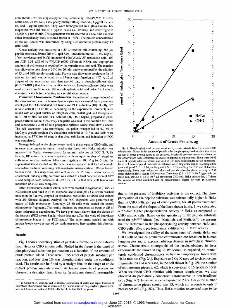
Fig. 1 shows phosphory la t ion of pept ide substrate by crude extracts
f rom HeLa or C H O mitot ic cells. Plot ted in the f igure is the pmol of phosphory la ted substrate per react ion as a funct ion of the amoun t o f
crude prote in added. There were 10.95 nmol of pept ide substrate per
reaction, and less than 1% was phosphory la ted under the condi t ions
used. The results can be fitted by a straight line, in the range of crude
extract protein amounts shown. At higher amoun t s o f protein we observed a deviat ion f rom linearity (results not shown) , p resumably
5 R. Okayasu, N. Cheong, and G. lliakis. Comparison of yields and repair kinetics of interphase chromosome breaks visualized by Sendai-virus or polyethylene glycol-medi- ated cell fusion in irradiated CHO cells, Int. J. Radiat. Biol., in press.
100
90
80
~A
r 70
6O
A
50
40
30
20
10
�9 HeLa
t CHO
0 5 10 15 20
A m o u n t o f C r u d e P r o t e i n , / . t g
Fig. 1. Phosphorylation of enzyme substrate by crude extracts from HeLa and CHO mitotic cells. Plotted is the amount of peptide substrate phosphorylated as a function of the amount of crude protein added to the reaction. Results of one experiment are shown but the observations were confirmed in several independent experiments. There were 10.95 nmol of peptide substrate present and 2.42 • 106 dpm corresponded to the phosphory- lation of 1 nmol of peptide substrate in each reaction. Fitting of the results to a straight line gave a slope of 2.10 • 0.14 pmol//xg and 5.29 +__ 0.35 pmol//xg for ClIO and HeLa cells, respectively. From these values we calculated that the phosphorylation activity is 2.52 times higher in HeLa than in CHO extracts. There were (2.9 • 0.5) • 10 -4/~g protein per HeLa cell, and 2.2 • 0.4 • 10 -4/xg protein per CHO cell. HeLa mitotics had 1.7 times the volume of CHO mitotics based on measurements carried out with an electronic counter.
due to the presence of inhibi tory activities in the extract. The phos-
phoryla t ion of the pept ide substrate was substantial ly higher in HeLa
than in C HO cells, p e r / x g of crude protein, for all points examined.
F rom the ratio o f the slopes of the lines shown in Fig. 1, we calculated
a 2.5-fold higher phosphory la t ion activity in HeLa as compared to
C H O mitot ic cells. Based on the specif ici ty o f the pept ide substrate
used for p34 c'~c2 kinase (see "Materials and Methods") , we assume
that the difference in the phosphory la t ing activity be tween HeLa and
C H O cells reflects p redominan t ly a difference in M P F activity. We invest igated the ability o f the same batch of mitot ic HeLa and
C HO cells to induce premature c h r o m o s o m e condensa t ion in huma n
lymphocy tes and to express radiation damage in interphase chromo-
somes. Characteris t ic micrographs of the results obta ined in these
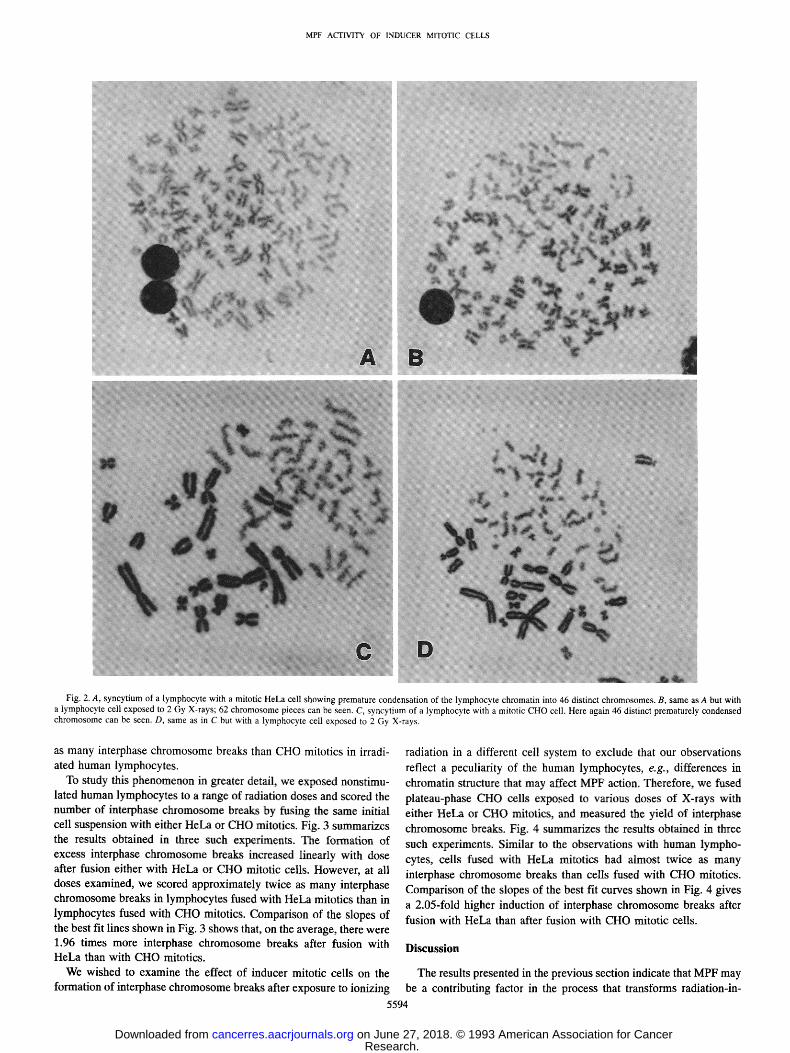
exper iments are shown in Fig. 2. We scored, as expected, 46 prema-
turely condensed c h r o m o s o m e s in h u m a n lymphocy te s fused wi th HeLa mitot ics (Fig. 2,4). Exposure to 2 Gy X-rays led to c h r o m o s o m e
f ragmenta t ion and increased, in the cell shown in Fig. 2B, the number
o f c h r o m o s o m e pieces to 62, which is equivalent to 16 breaks per cell. W h e n we fused C HO mitot ics with h u m a n lymphocytes , we also observed 46 premature ly condensed c h r o m o s o m e s in non-irradiated
cells (Fig. 2C). Howeve r in cells exposed to 2 Gy X-rays the n u m b e r of c h r o m o s o m e pieces scored was 53, which corresponds to only 7 breaks per cell (Fig. 2D). Thus, HeLa mitot ics uncovered over twice
5593
Research. on June 27, 2018. © 1993 American Association for Cancercancerres.aacrjournals.org Downloaded from
MPF ACTIVITY OF INDUCER MITOTIC CELLS
/~!~!i~iiiil/,~ ~ .... ~i :~ �84184 . . . . iii ~::~ i ~ i ~ A/
i
i
D
Fig. 2. A, syncytium of a lymphocyte with a mitotic HeLa cell showing premature condensation of the lymphocyte chromatin into 46 distinct chromosomes. B, same as A but with a lymphocyte cell exposed to 2 Gy X-rays; 62 chromosome pieces can be seen. C, syncytium of a lymphocyte with a mitotic CHO cell. Here again 46 distinct prematurely condensed chromosome can be seen. D, same as in C but with a lymphocyte cell exposed to 2 Gy X-rays.
as many interphase chromosome breaks than CHO mitotics in irradi- ated human lymphocytes.
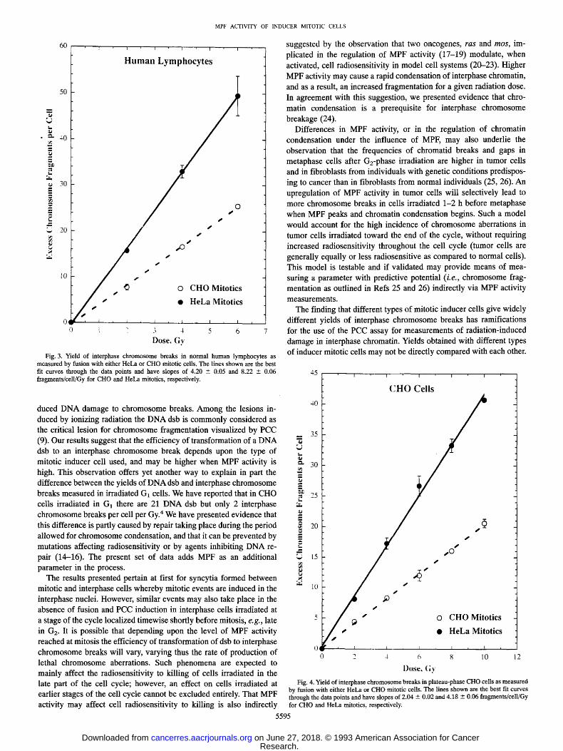
To study this phenomenon in greater detail, we exposed nonstimu- lated human lymphocytes to a range of radiation doses and scored the number of interphase chromosome breaks by fusing the same initial cell suspension with either HeLa or CHO mitotics. Fig. 3 summarizes the results obtained in three such experiments. The formation of excess interphase chromosome breaks increased linearly with dose after fusion either with HeLa or CHO mitotic cells. However, at all doses examined, we scored approximately twice as many interphase chromosome breaks in lymphocytes fused with HeLa mitotics than in lymphocytes fused with CHO mitotics. Comparison of the slopes of the best fit lines shown in Fig. 3 shows that, on the average, there were 1.96 times more interphase chromosome breaks after fusion with HeLa than with CHO mitotics.
We wished to examine the effect of inducer mitotic cells on the formation of interphase chromosome breaks after exposure to ionizing
radiation in a different cell system to exclude that our observations
reflect a peculiarity of the human lymphocytes, e.g., differences in chromatin structure that may affect MPF action. Therefore, we fused plateau-phase CHO cells exposed to various doses of X-rays with either HeLa or CHO mitotics, and measured the yield of interphase chromosome breaks. Fig. 4 summarizes the results obtained in three
such experiments. Similar to the observations with human lympho- cytes, cells fused with HeLa mitotics had almost twice as many interphase chromosome breaks than ceils fused with CHO mitotics. Comparison of the slopes of the best fit curves shown in Fig. 4 gives a 2.05-fold higher induction of interphase chromosome breaks after fusion with HeLa than after fusion with CHO mitotic cells.
Discussion
The results presented in the previous section indicate that MPF may be a contributing factor in the process that transforms radiation-in-
5594
Research. on June 27, 2018. © 1993 American Association for Cancercancerres.aacrjournals.org Downloaded from
M P F A C T I V I T Y O F I N D U C E R M I T O T I C C E L L S
J
: s
2
60
50
40
'i' i i ' 1 . i 1
Human Lymphocytes
w L
o 7
T
s ~
j .
S
s
s
s
o C H O Mitot ics
�9 HeLa Mitot ics
i I * i , 1 : l ~ I , I
l " 3 4 5 6 Dose. (;v
Fig. 3. Yield of interphase chromosome breaks in normal human lymphocytes as measured by fusion with either HeLa or CHO mitotic cells. The lines shown are the best fit curves through the data points and have slopes of 4.20 -+ 0.05 and 8.22 • 0.06 fragments/cell/Gy for CHO and HeLa mitotics, respectively.
duced DNA damage to chromosome breaks. Among the lesions in- duced by ionizing radiation the DNA dsb is commonly considered as the critical lesion for chromosome fragmentation visualized by PCC (9). Our results suggest that the efficiency of transformation of a DNA dsb to an interphase chromosome break depends upon the type of mitotic inducer cell used, and may be higher when MPF activity is high. This observation offers yet another way to explain in part the difference between the yields of DNA dsb and interphase chromosome breaks measured in irradiated G1 cells. We have reported that in CHO cells irradiated in G1 there are 21 DNA dsb but only 2 interphase chromosome breaks per cell per Gy. 4 We have presented evidence that this difference is partly caused by repair taking place during the period allowed for chromosome condensation, and that it can be prevented by mutations affecting radiosensitivity or by agents inhibiting DNA re- pair (14-16). The present set of data adds MPF as an additional parameter in the process.
The results presented pertain at first for syncytia formed between mitotic and interphase cells whereby mitotic events are induced in the interphase nuclei. However, similar events may also take place in the absence of fusion and PCC induction in interphase cells irradiated at a stage of the cycle localized timewise shortly before mitosis, e.g., late in G2. It is possible that depending upon the level of MPF activity reached at mitosis the efficiency of transformation of dsb to interphase chromosome breaks will vary, varying thus the rate of production of lethal chromosome aberrations. Such phenomena are expected to mainly affect the radiosensitivity to killing of cells irradiated in the late part of the cell cycle; however, an effect on cells irradiated at earlier stages of the cell cycle cannot be excluded entirely. That MPF activity may affect cell radiosensitivity to killing is also indirectly
suggested by the observation that two oncogenes, ras and mos, im- plicated in the regulation of MPF activity (17-19) modulate, when activated, cell radiosensitivity in model cell systems (20-23). Higher MPF activity may cause a rapid condensation of interphase chromatin, and as a result, an increased fragmentation for a given radiation dose. In agreement with this suggestion, we presented evidence that chro- matin condensation is a prerequisite for interphase chromosome breakage (24).
Differences in MPF activity, or in the regulation of chromatin condensation under the influence of MPF, may also underlie the observation that the frequencies of chromatid breaks and gaps in metaphase cells after G2-phase irradiation are higher in tumor cells and in fibroblasts from individuals with genetic conditions predispos- ing to cancer than in fibroblasts from normal individuals (25, 26). An upregulation of MPF activity in tumor cells will selectively lead to more chromosome breaks in cells irradiated 1-2 h before metaphase when MPF peaks and chromatin condensation begins. Such a model would account for the high incidence of chromosome aberrations in tumor cells irradiated toward the end of the cycle, without requiring increased radiosensitivity throughout the cell cycle (tumor cells are generally equally or less radiosensitive as compared to normal cells). This model is testable and if validated may provide means of mea- suring a parameter with predictive potential (i.e., chromosome frag- mentation as outlined in Refs 25 and 26) indirectly via MPF activity measurements.
The finding that different types of mitotic inducer cells give widely different yields of interphase chromosome breaks has ramifications for the use of the PCC assay for measurements of radiation-induced damage in interphase chromatin. Yields obtained with different types of inducer mitotic cells may not be directly compared with each other.
45 i �9 i " " I T i i L"
C H O Cel ls
40
_ 35
ez 30
.~ 25 t _
20
--" ~ s ~ 15 / s
" l0
5 O C H O Mitot ics
/ / f f s �9 HeLa Mitot ics
0 L I , . t : , t , I ,
0 2 4 6 8 10 2
Dose, Gy
Fig. 4. Yield of interphase chromosome breaks in plateau-phase CHO cells as measured by fusion with either HeLa or CHO mitotic cells. The lines shown are the best fit curves through the data points and have slopes of 2.04 • 0.02 and 4.18 --- 0.06 fragments/celljGy for CHO and HeLa mitotics, respectively.
5595
Research. on June 27, 2018. © 1993 American Association for Cancercancerres.aacrjournals.org Downloaded from
MPF ACTIVITY OF INDUCER MITOTIC CELLS
Indeed , w i d e var ia t ions have b e e n repor ted in the y ie ld o f in te rphase
c h r o m o s o m e b r e a k s in h u m a n cel ls i r radia ted in G1 and fu sed ei ther
wi th C H O or FleLa mi to t ics (8, 27, 28). It is l ike ly that d i f f e rences in
i nduce r mi to t ic cel ls u sed is at least one o f the causes for the differ-
ences in the resul ts ob ta ined .
I f M P F ac t iv i ty is a con t r ibu t ing fac tor in the c h r o m o s o m e frag-
men ta t i on o b s e r v e d b y m e a n s o f P C C in i r radiated in terphase cel ls
(G1 or G2), pa rame te r s p resen t ly neg l ec t ed w h e n p e r f o r m i n g the
a s say wil l need to be cons ide red . In addi t ion to the total M P F act iv i ty
p resen t in the mi to t i c cell , one shou ld a lso cons ide r the v o l u m e o f the
in terphase cel l and the total v o l u m e o f the s y n c y t i u m genera ted .
Fur ther pa rame te r s wi l l inc lude the p r e sence in the in terphase cell o f
ac t iv i t ies neut ra l iz ing MPF , as we l l as a l tera t ions in ch roma t in that
wil l a f fec t its ac t ion as subs t ra te for M P E Care fu l eva lua t ion o f the
con t r ibu t ion o f all these pa rame te r s in c h r o m o s o m e b r e a k a g e as meas -
ured b y m e a n s o f P C C wil l he lp not on ly in the s tandard iza t ion o f the
assay, bu t a lso in the e luc ida t ion o f the m e c h a n i s m o f fo rma t ion o f c h r o m o s o m e aberra t ions .
Acknowledgments
The authors are indebted to Nancy Mott for secretarial help and for editing the manuscript.
References
1. Rao, P. N., and Johnson, R. T. Mammalian cell fusion: studies on the regulation of DNA synthesis and mitosis. Nature (Lond.), 225: 159-164, 1970.
2. Adlakha, R. C., and Rao, P. N. Molecular mechanisms of the chromosome conden- sation and decondensation cycle in mammalian cells. BioEssays, 5: 100-105, 1986.
3. Mailer, J. L. MPF and cell cycle control. In: Y. Nishizuka et al. (eds.), The Biology and Medicine of Signal Transduction, pp. 323-328, New York: Raven Press, 1990.
4. Nurse, P. Universal control mechanism regulating onset of M-phase. Nature (Lond.), 344: 503-508, 1990.
5. Freeman, R. S., and Donoghue, D. J. Protein kinases and protooncogenes: biochemi- cal regulators of the eukaryotic cell cycle. Biochemistry, 30: 2293-2302, 1991.
6. Hittelman, W. N., and Rao, P. N. Premature chromosome condensation. I. Visualiza- tion of X-ray-induced chromosome damage in interphase cells. Mutat. Res., 23: 251-258, 1974.
7. Hittelman, W. N. Prematurely condensed chromosomes: a model system for visual- izing effects of DNA damage, repair and inhibition at the level of chromosome structure. In: A. Collins, C. S. Downes, and R. T. Johnson (eds.), DNA Repair and Its Inhibition, pp. 341-371, New York: Oxford University Press, 1984.
8. Cornforth, M. N., and Bedford, J. S. Ionizing radiation damage and its early devel- opment in chromosomes. Adv. Radiat. Biol., 17: 423--496, 1993.
9. Iliakis, G. The role of DNA double strand breaks in ionizing radiation-induced killing in eukaryotic cells. BioEssays, 13: 1-8, 1991.
10. Pantelias, G. E., and Maillie, H. D. The use of peripheral blood mononuclear cell prematurely condensed chromosomes for biological dosimetry. Radiat. Res., 99: 140-150, 1984.
11. lliakis, G., Pantelias, G. E., and Seaner, R. Effect of arabinofuranosyladenine on radiation-induced chromosome damage in plateau-phase C'HO cells measured by premature chromosome condensation: implications for repair and fixation of alpha PLD. Radiat. Res., 114: 361-378, 1988.
12. McVey, D., Brizuela, L., Mohr, I., Marshak, D. R., Gluzman, Y., and Beach, D. Phosphorylation of large tumor antigen by cdc2 stimulates SV40 DNA replication. Nature (Lond.), 341: 503-507, 1989.
13. Marshak, D. R., Vandenberg, M. T., Bae, Yi S., and Yu, I. J. Characterization of synthetic peptide substrates for p34 '~ac2 protein kinase. J. Cell. Biochem., 45: 391- 400, 1991.
14. Iliakis, G., Okayasu, R., Varlotto, J., Shemoff, C., and Wang, Y. Hypertonic treatment during premature chromosome condensation allows visualization of interphase chro- mosome breaks repaired with fast kinetics in irradiated CHO cells. Radiat. Res.,135: 160--170, 1993.
15. Okayasu, R., Varlotto, J., and Iliakis, G. Hypertonic treatment does not affect the radiation yield of interphase chromosome breaks in DNA double-strand break repair deficient xrs-5 cells. Radiat. Res., in press, 1993.
16. Okayasu, R., and Iliakis, G. Ionizing radiation induces two forms of interphase chromosome breaks in CHO cells that rejoin with different kinetics and show different sensitivity to treatment in hypertonic medium or 18-araA. Radiat. Res., 136: in press, 1993.
17. Sagata, N., Oskarsson, M., Copeland, T., Brumbaugh, J., and Vande Woude, G. E Function of c-mos proto-oncogene product in meiotic maturation in Xenopus oocytes. Nature (Lond.), 335: 519-525, 1988.
18. Sagata, N., Watanabe, N., Vande Woude, G. F., and lkawa, Y. The c-mos proto- oncogene product is a cytostatic factor responsible for meiotic arrest in vertebrate eggs. Nature (Lond.), 342: 512-518, 1989.
19. Daar, I., Nebreda, A. R., Yew, N., Sass, P., Paules, R., Santos, E., Wiglet, M., and Vande Woude, G. E The ras oncoprotein and M-phase activity. Science (Washington DC), 253: 74-76, 1991.
20. Sklar, M. D. The ras oncogenes increase the intrinsic resistance of NIH 3T3 cells to ionizing radiation. Science (Washington DC), 239: 645-647, 1988.
21. Ling, C. C., and Endlich, B. Radioresistance induced by oncogenic transformation. Radiat. Res., 120: 267-279, 1989.
22. McKenna, W. G., Weiss, M. C., Endlich, B., Ling, C. C., Bakanauskas, V. J., Kelsten, M. L., and Muschel, R. J. Synergistic effect of the v-myc oncogene with H-ras on radioresistance. Cancer Res., 50: 97-102, 1990.
23. Suzuki, K., Watanabe, M., and Miyoshi, J. Differences in effects of oncogenes on resistance to gamma rays, ultraviolet light, and heat shock. Radiat. Res., 129: 157- 162, 1992.
24. Pantelias, G. E. Radiation-induced cytogenetic damage in relation to changes in interphase chromosome conformation. Radiat. Res., 105: 341-350, 1986.
25. Parshad, R., Gantt, R., Sanford, K. K., and Jones, G. M. Chromosomal radiosensi- tivity of human tumor cells during the G2 cell cycle period. Cancer Res., 44: 5577- 5582, 1984.
26. Sanford, K. K., Parshad, R., Gantt, R., Tarone, R. E., Jones, G. M., and Price, E M. Factors affecting and significance of G2 chromatin radiosensitivity in predisposition to cancer. Int. J. Radiat. Biol. Relat. Stud. Phys. Chem. Med., 55: 963-981, 1989.
27. Comforth, M. N., and Bedford, J. S. X-ray-induced breakage and rejoining of human interphase chromosomes. Science (Washington DC), 222: 1141-1143, 1983.
28. Pandita, T. K., and Hittelman, W. M. Initial chromosome damage but not DNA damage is greater in ataxia telangiectasia cells. Radiat. Res., 130: 94-103, 1992.
5596
Research. on June 27, 2018. © 1993 American Association for Cancercancerres.aacrjournals.org Downloaded from
1993;53:5592-5596. Cancer Res Xinbo Cheng, Gabriel E. Pantelias, Ryuichi Okayasu, et al. the Premature Chromosome Condensation AssayAffect Radiation Yield of Interphase Chromosome Breaks in Mitosis-promoting Factor Activity of Inducer Mitotic Cells May
Updated version
http://cancerres.aacrjournals.org/content/53/23/5592
Access the most recent version of this article at:
E-mail alerts related to this article or journal.Sign up to receive free email-alerts
Subscriptions
Reprints and
To order reprints of this article or to subscribe to the journal, contact the AACR Publications
Permissions
Rightslink site. Click on "Request Permissions" which will take you to the Copyright Clearance Center's (CCC)
.http://cancerres.aacrjournals.org/content/53/23/5592To request permission to re-use all or part of this article, use this link
Research. on June 27, 2018. © 1993 American Association for Cancercancerres.aacrjournals.org Downloaded from