Embed Size (px)
Citation preview
Mitochondrial Protein Import: Biochemical and Genetic Evidence for Interaction of Matrix hsp70 and the Inner Membrane Protein MIM44 Joachim Rassow,* Atomy C. Maarse,* Elizabeth Krainer,§ Michael Kiibrich,* Hanne Miiller,* Michiel Meijer,* Elizabeth A. Craig,§ and Nikolaus Pfanner*
* Biochemisches Institut, Universit~t Freiburg, D-79104 Freiburg, Germany; * Institute for Molecular Cell Biology, BioCentrum Amsterdam, 1098 SM Amsterdam, The Netherlands; and § Department of Biomolecular Chemistry, University of Wisconsin-Madison, Wisconsin 53706
Abstract. The import of preproteins into mitochon- dria involves translocation of the polypeptide chains through putative channels in the outer and inner mem- branes. Preprotein-binding proteins are needed to drive the unidirectional translocation of the precursor polypeptides. Two of these preprotein-binding proteins are the peripheral inner membrane protein MIM44 and the matrix heat shock protein hsp70. We report here that MIM44 is mainly exposed on the matrix side, and a fraction of mt-hsp70 is reversibly bound to the inner membrane. Mt-hsp70 binds to MIM44 in a 1:1 ratio, suggesting that mt-hsp70 is localizing to the membrane via its interaction with MIM44. Formation of the complex requires a functional ATPase domain
of mt-hsp70. Addition of Mg-ATP leads to dissoci- ation of the complex. Overexpression of mt-hsp70 res- cues the protein import defect of mutants in MIM44; conversely, overexpression of MIM44 rescues protein import defects of mt-hsp70 mutants. In addition, yeast strains with conditional mutations in both MIM44 and mt-hsp70 are barely viable, showing a synthetic growth defect compared to strains carrying single mutations. We propose that MIM44 and mt-hsp70 cooperate in translocation of preproteins. By binding to MIM44, mt-hsp70 is recruited at the protein import sites of the inner membrane, and preproteins arriving at MIM44 may be directly handed over to mt-hsp70.
I MPORT of preproteins into mitochondria is a complex process requiting a large number of components in dis- tinct cellular subcompartments, the cytosol, outer mem-
brane, inner membrane, and matrix. In the cytosol, molecu- lar chaperones (70-kD heat shock protein [hspT0] ~ and Ydjlp/Mas5p) and additional import stimulating factors (such as presequence-binding factor PBF and mitochondrial import stimulating factor MSF) support transfer of the pre- proteins to mitochondria (Chirico et al., 1988; Murakami et al., 1988; Deshaies et al., 1988; Murakami and Mori, 1990; Hachiya et al., 1993; Cyr et al., 1994). The mitochondrial outer membrane contains a high molecular mass complex which includes several subunits constituting a general inser- tion pore and two import receptors (S611ner et al., 1992; Kassenbrock et al., 1993; Ramage et al., 1993). Recently, three essential proteins of the mitochondrial inner mem- brane were identified that are involved in import of pre-
Please address all correspondence to Dr. Nikolaus Planner, Biochemischcs Institut, Universitat Freiburg, Hermann-Herder-StraBe 7, D-79104 Frei- burg, Germany. Telephone: 49-761-203-5224; FAX: 49-761-203-5261.
1. Abbreviations used in this paper: DSS, disuccinimidyl suberate; hsp70, 70-kD heat shock protein; mt-hsp70, mitoehondrial hsp70; MIM44, 44-kD protein of the mitochondrial inner membrane import machinery; SOD, superoxide dismutase; Su9-DHFR, fusion protein between presequence of Fo-ATPase subunit 9 and dihydrofolate reductase.
proteins. MIM44 (formerly named Mpilp) is a peripheral membrane protein that binds preproteins (Maarse et al., 1992; Blom et al., 1993). The import site protein ISP45 was reported to be identical to MIM44 (Scherer et al., 1992; Horst et al., 1993). MIM23 (Mas6p) and MIM17 (Smslp) are integral membrane proteins that may constitute part of a translocation channel (Dekker et al., 1993; Emtage and Jensen, 1993; Maarse et al., 1994; Kiibrich et al., 1994; Ryan et al., 1994). In the matrix, the heat shock protein hsp70 (Ssclp) is essential for translocation of preproteins and transfers the polypeptides to hsp60; partners of these heat shock proteins are mitochondrial GrpE, possibly mitochon- drial DnaJ, and hspl0 (cpnl0) (Ostermann et al., 1989; Kang et al., 1990; Stuart et al., 1994a). Several specific process- ing enzymes operate in the matrix and inner membrane.
Little is known how these import components cooperate in the translocation of preproteins. In particular, it has not been understood so far if and how components of distinct subcompartments cooperate.
For this report we analyzed two essential yeast proteins that have been shown to function as binding proteins for preproteins during translocation across the mitochondrial inner membrane, MIM44 and mt-hsp70 (Kang et al., 1990; Manning-Krieg et al., 1991; Blom et al., 1993; Gambill et al., 1993). These two preprotein-binding proteins can both
© The Rockefeller University Press, 0021-9525/94/12/1547/10 $2.00 The Journal of Cell Biology, Volume 127, Number 6, Pan 1, December 1994 1547-1556 1547
on January 3, 2019jcb.rupress.org Downloaded from http://doi.org/10.1083/jcb.127.6.1547Published Online: 15 December, 1994 | Supp Info:
be found at the matrix side of the mitochondrial inner mem- brane and interact in a nucleotide-sensitive manner. The functional interaction of mt-hsp70 and MIM44 was also demonstrated by genetic analysis. MIM44 and mt-hsp70 seem to directly cooperate in mitochondrial protein import, providing an interesting analogy to the interaction of BiP, the hsp70 of the endoplasmic retlculum lumen, and Sec63p in protein translocation into the endoplasmic reticulum. We propose that the reversible binding of mt-hsp70 to MIM44 is an important step in the efficient and ordered function of the mitochondrial protein import machinery.
Materials and Methods
Isolation of Temperature-Sensitive mim44 Mutants
Temperature-sensitive alleles of M1M44 (previously termed MPI1 [Maarse et ai., 1992]) were generated by the low fidelity PCR technique as modified by Kassenbrock et al. (1993) and with further modifications of the reaction- buffer (50/zM MnC12, 1.5 mM MgC12, pH 9). Four separate PCR reac- tions were performed and in each case the concentration of one dNTP was reduced fivefold with respect to the three others (25 /zM vs 125 ttM). MIM44 sequences were amplified with the M13 forward sequencing primer and the M13 reverse sequencing primer, using YCplaclll::MIM44 as tem- plate DNA. After 30 cycles of 1 rain at 94°C, 2 rain at 50°C and 3 rain at 74°C, the expected 2.8-kb product was obtained. The products from the four reactions were pooled and digested with EagI and EcoRI. The 1.8-kb EagI-EcoRI fragments containing the MIM44 coding region were purified by agarose gel electrophoresis and cloned into YCplacl 11: :MIM44, cut with the same enzymes, thereby replacing most wild-type M1M44 sequences by potentially mutated sequences. The mutant DNA library, obtained by isolating plasmid DNA from about 1,000 E. coil transformants, was used to transform the mim44 deletion strain MB6 which harbors the URA3 marked plasmid YEplac195::MIM44. Leu-positive transformants were plated on solid medium containing 5-fluoroorotic acid (Boeke et al., 1987) and incubated at 23 or 35.4°C. Plasmid DNA was isolated from colonies with a temperature-sensitive phenotype, and, after a passage of E. coil, in- troduced into the beterozygous MIM44 diploid MB2-22 (Maarse et a l , 1992). After random sporulation, haploid cells with a disrupted nuclear MIM44 gene and harboring the rescuing plasmid were also tested for tem- perature sensitivity. Temperature sensitivity could be established for several plasmid-borne mim44 alleles and two of them were designated mim44-6 and mim44-7 (Table I).
Construction of Plasmids
YCplacl 11: :MIM44, YEplacl 81::MIM44 and YEplac195::MIM44 were iso- lated by cloning the 2.7-kb HindIH fragment containing the complete MIM44 gene (Maarse et al., 1992) into YCplacllI(LEU2), YEplaclSI- (LEU2), and YEplacI95(URA3), respectively (Gietz and Sugino, 1988). Centromeric plasmids containing either the wild-type MIM44 gene or the mim44-6 or mim44-7 alleles were constructed by cloning the 2.7-kb HindIH fragment into the HindIII site of YCplac22(TRP1) or YCplac33(URA3) (Gietz and Sngino, 1988). To generate YCplaclll::SSC1 and YEplacl81:: SSC1, a 6-kb EcoRI fragment bearing the SSCI gene was liberated from a YEpl 3 derivative and cloned into YCplacl 11 and YEplacl81.
Covalent Coupling of Antibodies to Protein A-Sepharose
300 ttl wet volume of protein A-Sepharose (Pharmacia LKB Biotechnol- ogy, Piscataway, NJ), 2 ml 100 mM potassium phosphate buffer, pH 7.5, and 1 ml antiserum were gently shaken for 1 h. After two times washing with 0.1 M sodium borate buffer, pH 9, the protein A-Sepharose was resuspended in 7-ml sodium borate buffer and 35 rag solid dimethyl pimelimidate (Sigma Chem. Co., St. Louis, MO; D-8388) were added. Af- ter an incubation of 30 min, the coupling reaction was stopped by washing and incubation for 2 h in 1 M Tris-HCl, pH 7.5. All steps were performed at room temperature. The Sepharose matrix with coupled antibodies was stored in 10 mM Tris-HC1, pH 7.5, 0.9% (wt/vol) NaCI at 4"C.
Submitochondrial Fractionation Mitoplasts were generated by dilution of a suspension of mitochondria (1 mg/ml) in SEM (250 mM sucrose, 1 mM EDTA, 10 raM MOPS, pH Z2) with a 10-fold excess of EM (1 mM EDTA, 10 mM MOPS, pH 7.2). After an incubation for 15 min on ice, the mitoplasts were reisolated by centrifu- gation (10 min 16,000 g). For sonication, 200/zg of isolated mitoehondria were suspended in 450 t~l SEM in the presence of pepstatin A (2/~g/ml). The samples were sonified (Branson Sonifier 250; Duty circle 50, output control 5) six times for 10 s, with intervals of 15 s for cooling on ice. Some samples received 12/~g/ml trypsin before sonieation and were subsequently incubated on ice for 20 min. The reaction was stopped by a 30-fold excess (wt/wt) of soybean trypsin inhibitor (Sigma Chem. Co., T-9003).
Coimmunoprecipitations
35S-labeled rnitochondria (25 ttg per sample) were suspended in 500 t~l lysis-buffer (1% Triton X-100, 300 mM KCI, 10 mM Tris-HCl, pH 7.4, Pep- statin A [2/zg/ml]). The samples were shaken at 8"C for 10 rain. After a
Table L S. cerevisiae Swains Used in This Study
Strain Genotype Source
PK82 M A T s his4-713 lys2 ura3-52 Atrpl 1eu2-3,112 Gambill et al., 1993 PK83 MATt~ ade2-101 lys2 ura3-52 Atrpl 1eu2-3,112 sscl-3(LEU2) Gamhill et al., 1993 MB6 ade2-101 his3 leu2 lys2-801 trp1-289 ura3-52 mim44::LYS2 + YEplac195(URA3)::MIM44 this study MB3 MATt~ ade2-101 his3-A200 leu2-A1 lys2-801 ura3::LYS2 Maarse et al., 1992 MB3-4 M A T s ade2-101 his3-A200 leu2-A1 lys2-801 ura3::LYS2 mim44-1 Dekker et al., 1993 MB3-42 MATer ade2-101 his3-A200 leu2-A1 lys2-801 ura3::LYS2 mim44-2 Dekker et al., 1993 MB3-52 MATt~ ade2-101 his3-A200 leu2-A1 lys2-801 ura3::LYS2 mira44-3 Maarse et al., 1992 MB3-68 MATo¢ ade2-101 his3-A200 leu2-A1 lys2-801 ura3::LYS2 mim44-4 Dekker et al., 1993 MB3-75 MATs ade2-101 his3-A200 leu2-A1 1ys2-801 ura3::LYS2 mira44-5 Dekker et al., 1993 MB3-27 M A T s ade2-101 his3-A200 leu2-A1 lys2-801 ura3::LYS2 sscl Dekker et al., 1993 MB3-43 MATc~ ade2-101 his3-A200 leu2-A1 lys2-801 ura3::LYS2 sscl Dekker et al., 1993 MB2-22 MATa/c~ ADE2/ade2-101 his3/his3-A200 leu211eu2-A1 lys2-801/lys2-801 Maarse et al., 1992
TRP1/trp1-289 ura3-52/ura3-52 MIM441mim44::LYS2 MATa his3 leu2 lys2 trpl ura3-52 mim44::LYS2 + YCplac22(TRP1)::MIM44 MATa his3 leu2 lys2 trpl ura3-52 mim44::LYS2 + YCplac22(TRP1)::mim44-6 MATa his3 leu2 lys2 trpl ura3-52 mim44::LYS2 + YCplac22(TRP1)::mim44-7 MATa leu2 lys2 trpl ura3-52 ade2-201 mira44::LYS2 sscl-3(LEU2) + YCplac22(TRP1)::mim44-6 MATa leu2 lys2 trpl ura3-52 ade2-201 mim44::LYS2 sscl-3(LEU2) + YCplac22(TRP1)::mim44-7 MATa leu2 lys2 trpl ura3-52 ade2-201 mim44::LYS2 sscl-3(LEU2) + YCplac22(TRP1)::MIM44
LK201 this study LK208 this study LK209 this study LK215 this study LK218 this study LK221 this study
The Journal of Cell Biology, Volume 127, 1994 1548
spin of 16,000 g for 10 min, the supernatants were added to antibodies pre- bound to protein A-Sepharose, and shaken at 8°C for 35 min. Subsequently the protein A-Sepharose was washed three times with lysis-buffer. The coimmunoprecipitations were analyzed by SDS-PAGE and fluorography. Where indicated, 5 mM MgC12 and 4 mM ATP or 4 mM ATP'yS were in- eluded in the lysis-buffer. The presence of additional protease inhibitors did not significantly change the pattern of the interaction of MIM44 with hsp70 in this assay. For a second immunoprecipitation under denaturing condi- tions, the immunocomplexes were dissociated in 20 #1 2 % SDS, 60 mM Tris-HC1, pH 7.2, before dilution with a 30-fold excess of lysis-buffer.
Affinity-purified antibodies against MIM44 were obtained by elution of antibodies from a fragment of MIM44 (amino acid residues 68 to 345 with addition of six histidine residues at the amino terminus) expressed in E. coli (Blom et al., 1993). The MIM44 fragment was isolated by Ni-NTA affinity purification, and blotted on nitrocellulose from an SDS-polyacrylamide gel. Prebound antibodies were eluted from slices of the nitrocellulose by shaking with 100 mM glycine, pH 2.5, at 0*C for 10 rain (Harlow and Lane, 1988). The pH was readjusted by addition of 1 M Tris-HCl, pH 8, and the antibod- ies were collected by protein A-Sepharose.
35S-labeling of Mitochondria
35S-labeling of yeast mitochondria followed in principle the procedure of Campbell and Duff'us (1988). A first preculture of wild-type S. cerevisiae was diluted 400-fold with minimal medium, containing 0.1% (wt/vol) glu- cose and 2.2% (vol/vol) lactic acid (pH 5.5 with KOH) and 30 ~M Na2SO4 (for further components see Campbell and Duffus, 1988), and shaken at 30°C for 50 h, the final OD57s was 5.5. An aliquot was diluted into 50 ml of the medium at an ODsTs of 0.04. After addition of 50 #1 [35S]sulfate (250 #Ci; Amersham SJS1), this second preculture was grown for 43 h at 30°C to a final OD~Ts of 3.3. For the main culture, 300 ml medium were inoculated with 5 ml of the second preculture. 650 t~l [35S]sulfate (3.25 mCi) were added and the culture was shaken in a 1,000-ml fask for 24 h. At an OD57s of 1.1, cells were harvested and mitochondria were isolated as published previously (Daum et al., 1982; Hartl et al., 1987). The yield was 1.3 nag mitochondrial protein/300 rnl culture, the specific radioactivity was 44,000 cpm/#g protein.
Miscellaneous
Synthesis of preproteins in rabbit reticulocyte lysate, Cross-linking of mito- cbondrial proteins to Su9-DHFR by disuccinimidyl suberate (DSS) (Blom et al., 1993), TCA-precipitation, SDS-PAGE, immunodecoration, fluorog- raphy (Planner et al., 1987), and storage phosphor imaging technology (Molecular Dynamics, Sunnyvale, CA) were performed according to pub- lished procedures.
Results
Localization of MIM44 and a Fraction of mt-hsp70 at the Matrix Side of the Inner Membrane
While MIM44 has been previously determined to be local- ized to the inner membrane, its topology in the inner mem- brane remained ambiguous. MIM44 behaves as a peripheral membrane protein and contains no hydrophobic membrane anchor sequence suggesting that MIM44 is not an integral membrane protein. Antibodies directed against MIM44 (ISP45) were reported to inhibit protein import into mito- chondria having a disrupted outer membrane (mitoplasts) (Scherer et al., 1992; Horst et al., 1993); in addition, a carboxy-terminal epitope tag was accessible to proteases in mitoplasts (Maarse et al., 1992). Authentic MIM44, how- ever, was not cleaved by proteases added to mitoplasts even at high concentrations (Blom et al., 1993). These results can be explained if, (a) MIM44 is mainly located on the inter- membrane space side of the inner membrane, but is folded in such a manner that no protease-accessible site is exposed on the intermembrane space side, or (b) if major portions of MIM44 are located on the matrix side and thus MIM44 has to span the inner membrane at least once.
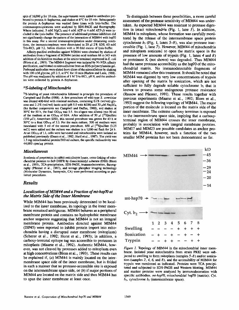
To distinguish between these possibilities, a more careful assessment of the protease sensitivity of MIM44 was under- taken. As expected MIM44 was resistant to protease diges- tion in intact mitochondria (Fig. 1, lane 3). In addition, MIM44 in mitoplasts, whose formation was carefully moni- tored by the release of the intermembrane space protein cytochrome b2 (Fig. 1, lanes 5-8), was also protease inac- cessible (Fig. 1, lane 7). However, MIM44 of mitochondria and mitoplasts sonicated to open the matrix space in the presence of low amounts of trypsin (Fig. 1, lanes 4 and 8) or proteinase K (not shown) was degraded. Thus MIM44 had the same protease accessibility as the hsp70 of the mito- chondrial matrix. No immunodetectable fragments of MIM44 remained after this treatment. It should be noted that MIM44 was digested by very low concentrations of trypsin after opening of the matrix by sonication, which are not sufficient to fully degrade soluble cytochrome b: that is known to possess some endogenous protease resistance (Rassow and Pfanner, 1991). These results together with previous experiments (Maarse et al., 1992; Blom et al., 1993) suggest the following topology of MIM44. The major portion of the molecule is located on the matrix side of the inner membrane. The extreme carboxy terminus is exposed to the intermembrane space side, implying that a carboxy- terminal region of MIM44 crosses the inner membrane, probably in association with integral membrane proteins. MIM17 and MIM23 are possible candidates as anchor pro- teins for MIM44, however, such a function of the two smaller MIM proteins has not been demonstrated so far.
Figure L Topology of MIM44 in the mitoehondriai inner mem- brane. Isolated yeast mitochondria from strain PK$2 were sub- jected to swelling to form mitoplasts (samples 5-8) and/or sonica- tion (samples 2, 4, 6, and 8), and the accessibility of MIM44 for trypsin was monitored as indicated. Proteins were TCA precipi- tated and subjected to SDS-PAGE and Western blotting. MIM44 and marker proteins were analyzed by immunodecoration with specific antibodies, mt-hspTO, mitochondrial hsp70 (matrix); Cyt. b2, cytochrome b2 (intermembrane space).
Rassow et al. Cooperation of Mitochondrial hsp TO and MIM44 1549
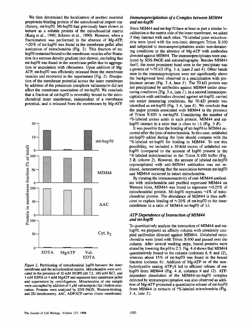
We then determined the localization of another essential preprotein-binding protein of the mitochondrial import ma- chinery, mt-hsp70. Mt-hsp70 had previously been shown to behave as a soluble protein of the mitochondrial matrix (Kang et al., 1990; Scherer et al., 1990). However, when a fractionation was performed in the absence of Mg-ATP, ",,20% of mt-hsp70 was found in the membrane pellet after sonication of mitochondria (Fig. 2). This fraction of mt- hspT0 remained bound to the membrane vesicles after floata- tion in a sucrose density gradient (not shown), excluding that mt-hsp70 was found in the membrane pellet due to aggrega- tion or association with ribosomes. Upon addition of Mg- ATE mt-hsp70 was efficiently released from the membrane vesicles and recovered in the supernatant (Fig. 2). Dissipa- tion of the membrane potential across the inner membrane by addition of the potassium ionophore valinomycin did not affect the membrane association of mt-hsp70. We conclude that a fraction of mt-hsp70 is reversibly bound to the mito- chondrial inner membrane, independent of a membrane potential, and is released from the membranes by Mg-ATP.
Figure 2. Partitioning of mitochondrial hsp70 between the inner membrane and the mitochondrial matrix. Mitochondria were soni- cated in the presence of 10 mM MOPS pH 7.2, 100 mM KC1, and 1 mM EDTA or 4 mM MgATP and separated into membrane pellet and supernatant by centrifugation. Mitochondria of one sample were uncoupled by addition of 1/zM valinomycin (Val.) before soni- cation. Proteins were analyzed by SDS-PAGE, Western-blotting, and 2D-densitometry. AAC, ADP/ATP carrier (inner membrane).
Immunoprecipitation of a Complex between MIM44 and mt-hsp70 Since MIM44 and mt-hsp70 have at least in part a similar lo- ealization at the matrix side of the inner membrane, we asked if they interact with each other. 3sS-labeled yeast mitochon- dria were lysed with the non-ionic detergent Triton X-100 and subjected to immunoprecipitations under non-denatur- ing conditions in the absence of Mg-ATP with antibodies directed against MIM44. The immunoprecipitates were ana- lyzed by SDS-PAGE and autoradiography. Besides MIM44 itself, the most prominent band seen in the precipitate was a protein of '~70 kD (Fig. 3 A, lanes 2 and 4). Other bands seen in the immunoprecipitate were not significantly above the background level observed in a precipitation with pre- immune serum (Fig. 3 A, lane 3). The 70-kD protein was not precipitated by antibodies against MIM44 under dena- turing conditions (Fig. 3 A, lane 1 ). In a second immunopre- cipitation with antibodies directed against mt-hspT0, carried out under denaturing conditions, the 70-kD protein was identified as mt-hsp70 (Fig. 3 A, lane 6). We conclude that the major protein associated with MIM44 in the presence of Triton X-100 is mt-hsp70. Considering the number of 35S-labeled amino acids in each protein, MIM44 and mt- hsp70 interact in a ratio that is close to 1:1 (Fig. 3 B).
It was possible that the binding of mt-hsp70 to MIM44 oc- curred after the lysis of mitochondria. In this case, unlabeled mt-hspT0 added during the lysis should compete with the 35S-labeled mt-hspT0 for binding to MIM44. To test this possibility, we included a 10-fold excess of unlabeled mt- hspT0 (compared to the amount of hsp70 present in the 35S-labeled mitochondria) in the Triton X-100 buffer (Fig. 3 B, column 2). However, the amount of labeled mt-hsp70 coprecipitated with anti-MIM44 antibodies was not re- duced, demonstrating that the association between mt-hsp70 and MIM44 occurred in intact mitochondria.
By titrating the immunoreactivity of anti-MIM44 antibod- ies with mitochondria and purified expressed MIM44 on Western blots, MIM44 was found to represent '~0.25% of mitochondrial protein. Mt-hspT0 represents ,~1% of mito- chondrial protein. The abundance of MIM44 is thus suffi- cient to explain binding of "~ 20% of mt-hspT0 to the inner membrane in a ratio of MIM44:mt-hsp70 of 1:1.
ATP Dependence of Interaction of MIM44 and mt-hspTO To quantitatively analyze the interaction of MIM44 and mt- hspT0, we prepared an affinity column with covalently cou- pled antibodies directed against MIM44. Unlabeled mito- chondria were lysed with Triton X-100 and passed over the column. After several washing steps, bound proteins were eluted by lowering the pH to 2.5. Fig. 4 A shows that MIM44 quantitatively bound to the column (columns 6, 9, and 12), whereas about 15% of mt-hsp70 was found in the bound fraction (column 6). Addition of Mg-ATP or of the non- hydrolyzable analog ATP'yS led to efficient release of mt- hspT0 from MIM44 (Fig. 4 A, columns 9 and 12). ATP- dependent dissolution of the MIM44:mt-hspT0 complex detected by immunoprecipitation was also observed as addi- tion of Mg-ATP promoted a quantitative release of mt-hsp70 from MIM44 in extracts of 35S-labeled mitochondria (Fig. 3 A, lane 5).
The Journal of Cell Biology, Volume 127, 1994 1550
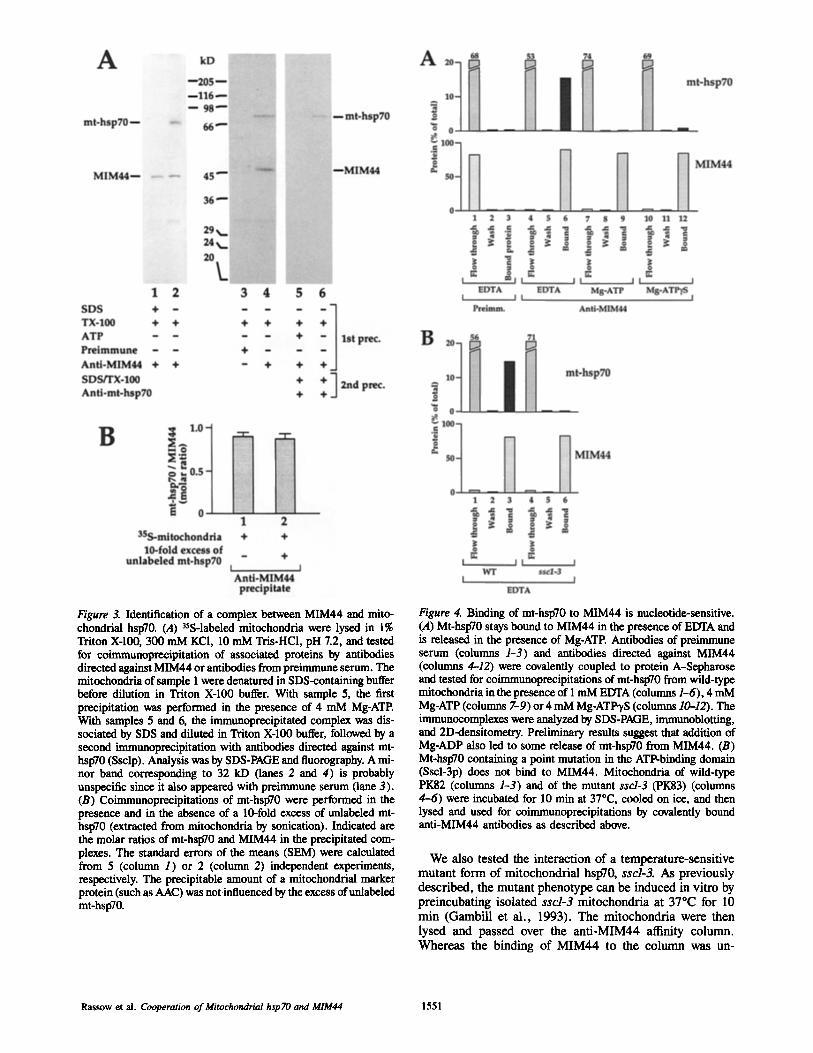
Figure 3. Identification of a complex between MIM44 and mito- chondrial hspT0. (A) 35S-labeled mitochondria were lysed in 1% Triton X-100, 300 mM KCI, 10 mM Tris-HC1, pH 7.2, and tested for coimmunoprecipitation of associated proteins by antibodies directed against MIM44 or antibodies from preimmune serum. The mitoehondria of sample 1 were denatured in SDS-containing buffer before dilution in Triton X-100 buffer. With sample 5, the first precipitation was performed in the presence of 4 mM Mg-ATP. With samples 5 and 6, the immunoprecipitated complex was dis- sociated by SDS and diluted in Triton X-100 buffer, followed by a second immunopreeipitation with antibodies directed against mt- hsp70 (Ssclp). Analysis was by SDS-PAGE and fluorography. A mi- nor band corresponding to 32 kD (lanes 2 and 4) is probably unspecific since it also appeared with preimmuna serum (lane 3). (B) Coimmunoprecipitations of mt-hsp70 were performed in the presence and in the absence of a 10-fold excess of unlabeled mt- hsp70 (extracted from mitochondria by sonication). Indicated are the molar ratios of mt-hsp70 and M1M44 in the precipitated com- plexes. The standard errors of the means (SEM) were calculated from 5 (colunm 1) or 2 (column 2) independent experiments, respectively. The precipitable amount of a mitochondrial marker protein (such as AAC) was not influenced by the excess of unlabeled mt-hsp70.
Figure 4. Binding of mt-hspT0 to MIM44 is nucleotide-sensitive. (A) Mt-hspT0 stays bound to MIM44 in the presence of EDTA and is released in the presence of Mg-ATP. Antibodies of preimmune serum (columns 1-3) and antibodies directed against MIM44 (eoltmms 4-12) were covalently coupled to protein A-Sepharose and tested for coimmunoprecipitations of mt-hspT0 from wild-type mitoebondria in the presence of 1 mM EDTA (cohmms 1-6), 4 mM Mg-ATP (columns 7-9) or 4 mM Mg-ATP'yS (columns 10--12). The immunocomplexes were analyzed by SDS-PAGE, immunoblotting, and 2D-densitometry. Preliminary results suggest that addition of Mg-ADP also led to some release of mt-hspT0 from MIM44. (B) Mt-hspT0 containing a point mutation in the ATP-binding domain (Sscl-3p) does not bind to MIM44. Mitocbondria of wild-type PK82 (colunms 1-3) and of the mutant sscl-3 (PK83) (columns 4-6) were incubated for 10 min at 37"C, cooled on ice, and then lysed and used for coimmunoprecipitations by covalently bound anti-MIM44 antibodies as described above.
We also tested the interaction of a temperature-sensitive mutant form of mitochondrial hspT0, sscl-3. As previously described, the mutant phenotype can be induced in vitro by preincubating isolated sscl-3 mitochondria at 37°C for 10 min (Gambill et al., 1993). The mitochondria were then lysed and passed over the anti-MIM44 affinity column. Whereas the binding of MIM44 to the column was un-
Rassow et al. Cooperation of Mitochondrial hsp 70 and MIM44 1551
changed, the mutant Sscl-3p did not bind to the column (Fig. 4 B, column 6). A preincubation of wild-type mitochondria at 37°C did not change the binding of mt-hsp70 (Ssclp) to MIM44 (Fig. 4 B, column 3). When the sscl-3 mitochondria were not preincubated at the non-permissive temperature, binding of Sscl-3p to MIM44 was not inhibited (data not shown). Since Sscl-3p has a mutation in the amino terminal ATPase domain, this result indicates that a functional ATP- ase domain of mt-hsp70 is needed for binding to MIM44.
Mitochondria with a Defective mt-hsp70 Accumulate Preproteins at MIM44
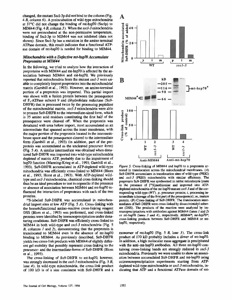
In the following, we tried to analyze how the interaction of preproteins with MIM44 and mt-hspT0 is affected by the as- sociation between MIM44 and mt-hsp70. We previously reported that mitochondria from the mutant sscl-3 were un- able to completely import preproteins into the mitochondrial matrix (Gambill et al., 1993). However, an amino-terminal portion of a preprotein was imported. This partial import was shown with a fusion protein between the presequence of Fo-ATPase subunit 9 and dihydrofolate reductase (Su9- DHFR) that is processed twice by the processing peptidase of the mitochondrial matrix, sscl-3 mitochondria were able to process Su9-DHFR to the intermediate-sized form, which is 35 amino acid residues constituting the first half of the presequence were cleaved off. When the preprotein was denatured with urea before import, most accumulated as an intermediate that spanned across the inner membrane, with the major portion of the preprotein located in the intermem- brane space and the presequence cleaved to the intermediate form (Gambill et al., 1993) (in addition, part of the pre- protein was accumulated as the uncleaved precursor form) (Fig. 5 A). A similar intermediate was obtained when dena- tured Su9-DHFR was imported into wild-type mitochondria depleted of matrix ATE probably due to the impairment of hsp70 function (Manning-Krieg et al., 1991; Gambill et al., 1993). Su9-DHFR accumulated in ATP-depleted wild-type mitochondria was efficiently cross-linked to MIM44 (Blom et al., 1993; Horst et al., 1993). With ATP-depleted wild- type and sscl-3 mitochondria, chemical cross-linking should thus be an ideal procedure to test in organello if the presence or absence of association between MIM44 and mt-hsp70 in- fluenced the interaction of preproteins with each of the two proteins.
35S-labeled Su9-DHFR was accumulated in mitochon- drial import sites at low ATP (Fig. 5 A). Cross-linking with the homobifunctional amino-reactive cross-linking reagent DSS (Blom et al., 1993) was performed, and cross-linked proteins were identified by immunoprecipitation under dena- turing conditions. Su9-DHFR was efficiently cross-linked to MIM44 in both wild-type and sscl-3 mitochondria (Fig. 5 B, columns 1 and 2), demonstrating that the preprotein is translocated to MIM44 even in the absence of mt-hsp70 binding to MIM44. As previously described, Su9-DHFR yields two cross-link products with MIM44 of slightly differ- ent gel mobility that possibly represent cross-linking to the precursor- and the intermediate-forms of Su9-DHFR (Blom et al., 1993).
The cross-linking of Su9-DHFR to mt-hsp70, however, was strongly decreased in the sscl-3 mitochondria (Fig. 5 B, lane 4). In wild-type mitochondria, the cross-link product of 100 kD is of a size consistent with Su9-DHFR and a
Figure 5. Cross-linking of MIM44 and hsp70 to a preprotein ar- rested in translocation across the mitochondrial membranes. (A) Su9-DHFR accumulates in translocation sites of wild-type (PK82) and sscl-3 (PK83) mitochondria with similar efficiency. The preprotein Su9-DHFR was synthesized in rabbit reticulocyte lysate in the presence of ['S]methionine and imported into ATP- depleted mitochondria of the mt-hsp70 mutant sscl-3 and of the cor- responding wild-type (WT). p, precursor protein; i, processing in- termediate (cleavage of the first part of the presequence); m, mature protein. (B) Cross-linking of Su9-DHFR. The translocation inter- mediates of Su9-DHFR were cross-linked by disuccinimidyl suber- ate (DSS). The products of the reaction were analyzed by im- munoprecipitations with antibodies against MIM44 (lanes I and 2) or mt-hspT0 (lanes 3 and 4), respectively. MIM44*, mt-hsp70*, cross-linking products between Su9-DHFR and MIM44 or mt- hsp70, respectively.
monomer of mt-hsp70 (Fig. 5 B, lane 3). The cross-link product of 170 kD probably includes a dimer of mt-hsp70. In addition, a high molecular mass aggregate is precipitated with the anti-mt-hsp70.antibodies. All three mt-hsp70 con- taining cross-linking bands are strongly reduced in sscl-3 mitochondria. Previously we were unable to show an associ- ation between accumulated Su9-DHFR and mt-hsp70 using coimmunoprecipitation experiments starting from ATP- depleted wild-type mitochondria or sscl-3 mitochondria, in- dicating that ATP and a functional ATPase domain of mt-
The Journal of Cell Biology, Volume 127, 1994 1552
hsp70 were needed to obtain a binding of the preprotein to mt-hsp70 that was stable enough to survive the coim- munoprecipitation procedure (Gambill et al., 1993). How- ever, by cross-linking we now demonstrate that mt-hsp70 in ATP-depleted wild-type mitochondria is in close proximity to the preprotein in transit. Since only a short portion of the presequence of accumulated Su9-DHFR is located on the matrix side of the inner membrane, this cross-linking pro- vides independent evidence that mt-hsp70 is in very close proximity to the protein import site, that is to MIM44.
Genetic Evidence for Interaction of MIM44 and mt-hsp70
We applied two genetic approaches to obtain independent evidence for an interaction of MIM44 and mt-hsp70, mul- ticopy suppression and synthetic lethal analysis.
We previously constructed a test plasmid encoding the URA3 gene product orotidine 5tphosphate decarboxylase (OMP decarboxylase) with the amino-terminal mitochon- drial-targeting sequence of superoxide dismutase (SOD). When this test plasmid was introduced into the yeast strain MB3, carrying a deletion of the chromosomal URA3 coding region, the fusion protein was efficiently imported into mito- chondria and the cells remained inviable in uracil-free me- dium due to lack of OMP decarboxylase activity in the cyto- sol (Maarse et al., 1992). The mim44 mutants MB3-4, MB3-42, MB3-52, MB3-68, and MB3-75 are at least par- tially blocked in mitochondrial import of the SOD-OMP de- carboxylase fusion protein, which allows them to grow in the absence of exogenously added uracil (Maarse et al., 1992; Dekker et al., 1993). The dependence of growth on the addi- tion of uracil can thus be taken as an indication of the effi- ciency of the import of the test protein into mitochondria in vivo. To test the effects of overexpression of mt-hsp70 on the growth ofmim44 mutants in the absence of uracil, the mim44 mutants carrying the test plasmid were transformed with the mt-hsp70 gene SSC1, cloned on either the centromeric vector YCplaclll or the multi-copy vector YEplacl81. Double transformants in which mt-hsp70 was expressed from the multi-copy plasmid showed a greatly diminished growth on uracil-free medium (Table II). This decrease in growth dem-
onstrates that overexpression of mt-hsp70 can at least par- tially suppress the import defect of all five mim44 mutants. Overexpression of mt-hspT0, however, did not rescue the le- thal phenotype of a mim44 deletion mutant (not shown), in- dicating that mt-hsp70 cannot fully replace the function of MIM44.
In a similar way we tested whether overexpression of MIM44 will suppress the ura-positive mutant phenotype of the sscl mutants MB3-27 and MB3-43 isolated in the manner described for the mira44 mutants (Dekker et al., 1993). Ex- pression of MIM44 from the multi-copy vector YEplacl81, but not from the centromeric vector YCplaclll, reduced growth of double transformed strains on medium lacking uracil (Table II). Thus, overexpression of MIM44 can par- tially relieve the import defect of the SOD-OMP decarboxy- lase fusion protein in both mt-hsp70 mutant strains, again in- dicating a genetic interaction of MIM44 and mt-hsp70.
The phenomenon of mutations in different genes produc- ing a severe growth defect or lethality can be indicative of functional interaction between gene products (Huffaker et al., 1987; Kaiser and Schekman, 1990; Scidmore et al., 1993). To determine if strains carrying both sscl-3 and mim44 temperature-sensitive alleles are viable, a strain con- taining sscl-3 and a deletion of the MIM44, rescued by a wild-type MIM44 gene on a low copy number plasmid also containing the URA3 gene was constructed. The strain was then transformed with plasmids containing the TRP1 gene and either the wild-type MIM44 gene, the mim44-6 allele, or the mira44-7 allele. All three strains were able to lose the URA3-containing plasmid, as evidenced by growth on media containing 5-fluoroorotic acid. However, while patches of strains containing the wild-type plasmid grew well, those with the temperature-sensitive alleles grew very poorly. At 23°C the strains containing the temperature-sensitive MIM44 alleles could only form very small colonies which were extremely difficult to propagate. At 30°C, very little growth was observed, whereas the individual sscl-3 and mim44 ts mutants grew reasonably well compared to wild- type (Fig. 6). The strongly pronounced temperature sensitiv- ity of the sscl and mira44 double mutants indicates a syn- thetic effect which argues in favor of a genetic interaction of MIM44 and mt-hsp70.
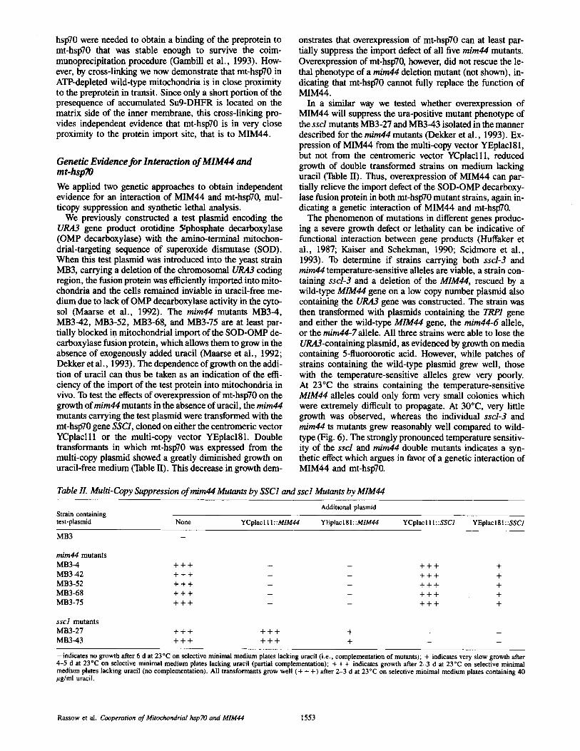
Table IL Multi-Copy Suppression of mim44 Mutants by SSC1 and sscl Mutants by MIM44
Additional plasmid Strain containing test-plasmid None YCplac l l I : :MIM44 YEplac181::MIM44 YCplaclII::SSC1 YEplacI81::SSC1
MB3
mira44 mutants
MB3-4 + + + - - + + + +
MB3-42 + + + - - + + + +
MB3-52 + + + - - + + + +
MB3-68 + + + - - + + + +
MB3-75 + + + - - + + + +
sscl mutants
M B 3 - 2 7 + + + + + + +
MB3-43 + + + + + + +
-indicates no growth after 6 d at 23°C on selective minimal medium plates lacking uracil (i.e., complementation of mutants); + indicates very slow growth after 4-5 d at 23"C on selective minimal medium plates lacking uracil (partial complementation); + + + indicates growth after 2-3 d at 23°C on selective minimal medium plates lacking uracil (no complementation). All transformants grow well (+ + +) after 2-3 d at 23°C on selective minimal medium plates containing 40 ttg/ml uracil.
Rassow et al. Cooperation of Mitochondrial hsp 70 and MIM44 1553
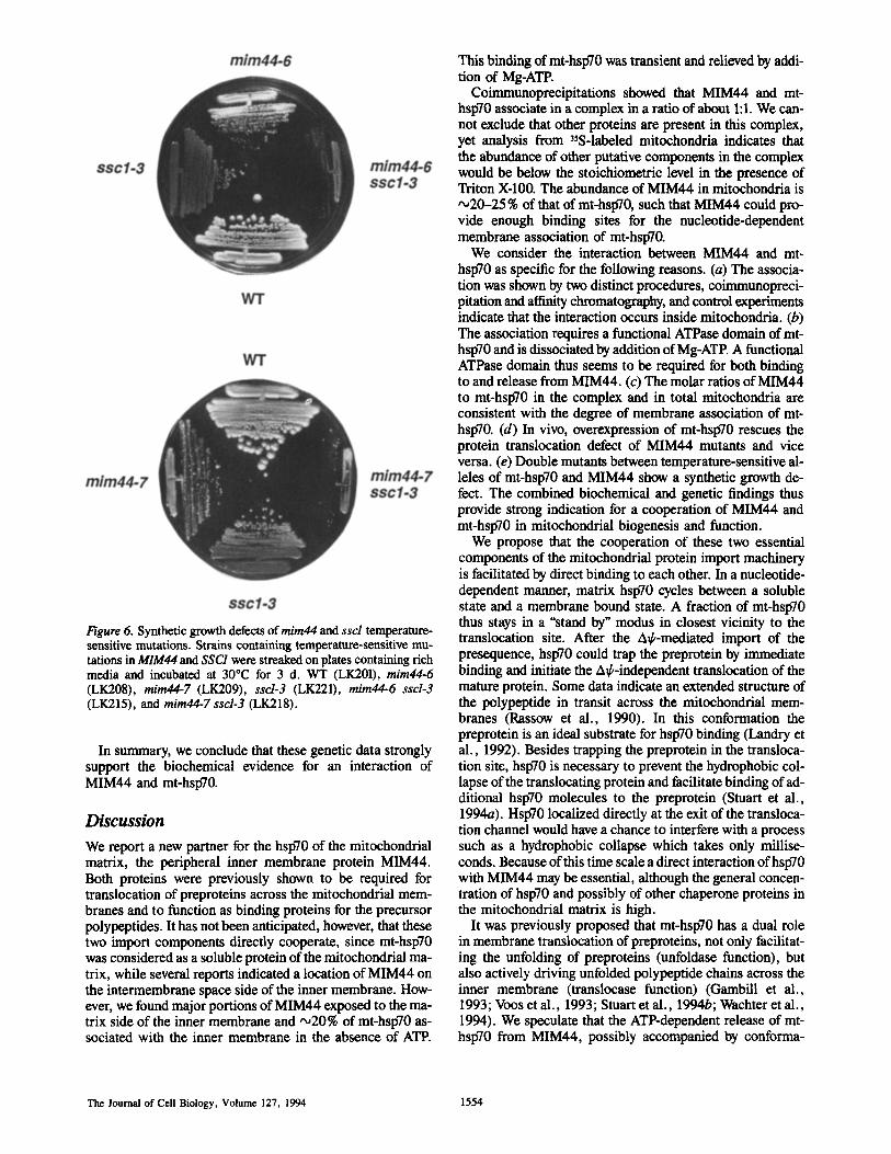
Figure 6. Synthetic growth defects of mim44 and ssd temperature- sensitive mutations. Strains containing temperature-sensitive mu- tations in MIM44 and SSC1 were streaked on plates containing rich media and incubated at 30°C for 3 d. WT (LK201), mira44-6 (LK208), mim44-7 (LK209), sscl-3 (LK221), mim44-6 ssd-3 (LK215), and mira44-7 sscl-3 (LK218).
In summary, we conclude that these genetic data strongly support the biochemical evidence for an interaction of MIM44 and mt-hsp70.
Discussion
We report a new partner for the hsp70 of the mitochondrial matrix, the peripheral inner membrane protein MIM44. Both proteins were previously shown to be required for translocation of preproteins across the mitochondrial mem- branes and to function as binding proteins for the precursor polypeptides. It has not been anticipated, however, that these two import components directly cooperate, since mt-hsp70 was considered as a soluble protein of the mitochondrial ma- trix, while several reports indicated a location of MIM44 on the intermembrane space side of the inner membrane. How- ever, we found major portions of MIM44 exposed to the ma- trix side of the inner membrane and ,020% of mt-hsp70 as- sociated with the inner membrane in the absence of ATE
This binding of mt-hsp70 was transient and relieved by addi- tion of Mg-ATP.
Coimmunopreeipitations showed that MIM44 and mt- hsp70 associate in a complex in a ratio of about 1:1. We can- not exclude that other proteins are present in this complex, yet analysis from 35S-labeled mitochondria indicates that the abundance of other putative components in the complex would be below the stoichiometric level in the presence of Triton X-100. The abundance of MIM44 in mitochondria is ,020-25 % of that of mt-hsp70, such that MIM44 could pro- vide enough binding sites for the nucleotide-dependent membrane association of mt-hsp70.
We consider the interaction between MIM44 and mt- hsp70 as specific for the following reasons. (a) The associa- tion was shown by two distinct procedures, coimmunopreci- pitation and affinity chromatography, and control experiments indicate that the interaction occurs inside mitochondria. (b) The association requires a functional ATPase domain of mt- hsp70 and is dissociated by addition of Mg-ATP. A functional ATPase domain thus seems to be required for both binding to and release from MIM44. (c) The molar ratios of MIM44 to mt-hsp70 in the complex and in total mitochondria are consistent with the degree of membrane association of mt- hsp70. (d) In vivo, overexpression of mt-hsp70 rescues the protein translocation defect of MIM44 mutants and vice versa. (e) Double mutants between temperature-sensitive al- leles of mt-hsp70 and MIM44 show a synthetic growth de- feet. The combined biochemical and genetic findings thus provide strong indication for a cooperation of MIM44 and mt-hsp70 in mitochondrial biogenesis and function.
We propose that the cooperation of these two essential components of the mitochondrial protein import machinery is facilitated by direct binding to each other. In a nucleotide- dependent manner, matrix hsp70 cycles between a soluble state and a membrane bound state. A fraction of mt-hsp70 thus stays in a "stand by" modus in closest vicinity to the translocation site. After the AC/-mediated import of the presequence, hsp70 could trap the preprotein by immediate binding and initiate the A~/,-independent translocation of the mature protein. Some data indicate an extended structure of the polypeptide in transit across the mitochondrial mem- branes (Rassow et al., 1990). In this conformation the preprotein is an ideal substrate for hsp70 binding (Landry et al., 1992). Besides trapping the preprotein in the transloca- tion site, hsp70 is necessary to prevent the hydrophobic col- lapse of the translocating protein and facilitate binding of ad- ditional hsp70 molecules to the preprotein (Stuart et al., 1994a). Hsp70 localized directly at the exit of the transloca- tion channel would have a chance to interfere with a process such as a hydrophobic collapse which takes only millise- conds. Because of this time scale a direct interaction of hsp70 with MIM44 may be essential, although the general concen- tration of hsp70 and possibly of other chaperone proteins in the mitochondrial matrix is high.
It was previously proposed that mt-hsp70 has a dual role in membrane translocation of preproteins, not only facilitat- ing the unfolding of preproteins (unfoldase function), but also actively driving unfolded polypeptide chains across the inner membrane (translocase function) (Gambill et al., 1993; Voos et al., 1993; Stuart et al., 1994b; Wachter et al., 1994). We speculate that the ATP-dependent release of mt- hsp70 from MIM44, possibly accompanied by conforma-
The Journal of Cell Biology, Volume 127, 1994 1554
tional changes of MIM44 and mt-hsp70, adds to the force driving polypeptide chains across the mitochondrial inner membrane. In support of this view, a stable association be- tween mt-hsp70 and a preprotein in transit (stable enough to survive a coimmunoprecipitation) is only obtained in the presence of ATP (Manning-Krieg et al., 1991; Gambill et al., 1993), while cross-linking under ATP-depleted condi- tions in organello indicates that mt-hsp70 is already in close proximity to the preprotein when it is bound to MIM44. We suggest that an ATP-dependent reaction cycle of mt-hsp70 (Hartl et al., 1994), which includes interaction with MIM44 and preproteins, is an essential step in protein import into the mitochondrial matrix.
Our results do not exclude a function of MIM44 in initial steps of translocation across the inner membrane in addition to its function at the matrix side of the membrane. MIM44 seems to funnel preproteins by a A~/-dependent step into the mitochondrial hsp70 system. Cross-linking studies suggest that MIM44 is a major component of the translocation site (Blom et al., 1993), and antibodies bound to MIM44 at the outer surface of the inner membrane apparently shield the translocation sites from preproteins (Scherer et al., 1992). We assume that a small carboxy-terminal portion of MIM44 is sufficient to mediate the effect of the antibodies.
Interestingly, Brodsky and Schekman (1993) reported that a fraction ofBiP (Kar2p), the hsp70 of the endoplasmic retic- ulum (ER), interacts with the membrane protein Sec63p in yeast in an ATP-dependent manner. Sec63p is part of the pro- tein import machinery of the ER and contains a domain of about 70 amino acid residues in its lumenal part that is ho- mologous to DnaJ, termed J-domain (Sadler et al., 1989; Feldheim et al., 1992; Cyr et al., 1994). Prokaryotic DnaJ and eukaryotic homologs have been shown to function as binding partners of hsp70 (Wickner et al., 1991, 1992; Liberek et al., 1991; Langer et al., 1992; Cyr et al., 1992, 1994). The transient interaction of BiP and Sec63p was pro- posed to occur via the J-domain and to be part of a reaction cycle required for protein translocation into the ER (Sanders et al., 1992; Brodsky and Schekman, 1993; Scidmore et al., 1993). MIM44 does not reveal a significant overall homol- ogy to known chaperones or other proteins, indicating that MIM44 is a new partner of hsp70. However, a comparison of the sequences of Sec63p and MIM44 revealed a motif of 18 amino acid residues with similarity between Sec63p (residues 138-155) and MIM44 (residues 185-202) (7 iden- tical and 5 isofunctional residues). The motif is located in the J-domain of Sec63p. When MIM44, Sec63p, and E. coli DnaJ were aligned together (Fig. 7), the similarity of the 18- residue motif was indicated to be of high significance accord- ing to the MACAW program (Schuler et al., 1991). Feldheim
~[I]VI44 185 E R D L A S G K R H R A V K S N E D 202
5ec63p ~3~ D R D I K S A Y R K L S V K F H P D 155
DnaJ 18E R E I R K A Y K R L A M K Y H P D35
: I : : " : : : : : : I I
Figure 7. A short motif of MIM44 with sequence similarity to Sec63p and DnaJ in a triple alignment. Vertical lines indicate iden- tical residues and double dots indicate isofunctional residues in all three proteins. Sequences were adopted from Ohki et al. (1986) (E. coli DnaJ), Sadler et al. (1989) (S. cerevisiae Sec63p), and Maarse et al. (1992) (S. cerevisiae MIM44).
et al. (1992) demonstrated that the highly conserved aspar- tate (the last residue of the motif found here) is required for the function of Sec63p. However, in MIM44 only this aspar- tate is present from the conserved HPD motif (histidine, pro- line, aspartate) that is found in all DnaJ-like proteins ana- lyzed so far, and the sequence similarity of MIM44 to the DnaJ-like proteins is over a much more limited region than was described between Sec63p and DnaJ (Sadler et al., 1989). Thus MIM44 cannot be seen as a DnaJ-like protein, yet future studies will have to address the possibility if this short conserved motif is involved in the interaction of MIM44 and mt-hsp70. It should be noted that, in contrast to MIM44, a direct interaction between Sec63p and prepro- teins in transit has not been found so far. The available evidence thus suggests an interesting analogy of association of the luminal hsp70 of both mitochondria and ER with a membrane-bound component of the translocation complex, although mechanistic details may be distinct for each or- ganelle. Very recently, a mitochondrial homolog of DnaJ (MDJ1) was identified that is located on the matrix side of the inner membrane (Rowley et al., 1994). While MDJ1 is involved in folding of imported proteins, it has not been pos- sible so far to demonstrate an interaction of MDJ1 and mt- hsp70 (Cyr et al., 1994). More importantly in the context of this study, a deletion of MDJ1 does not affect the transloca- tion of preproteins into mitochondria, making it very un- likely that MDJ1 plays a critical role in polypeptide translo- cation and the translocase function of mt-hsp70. Instead, we propose that MIM44 is responsible for accumulation of mt- hsp70 at the import sites, thus linking the A~k-dependent translocation machinery of the inner membrane to the ATP- driven motor of mitochondrial protein translocation.
We wish to thank Drs. L. Grivell (Amsterdam), P. Keil, and W. Voos (Freiburg) for helpful discussions and technical advice and Dr. B. Guiard (Gif-sur-Yvette) for providing an antiserum against yeast cytochrome b2.
The work was supported by the Deutsche Forschungsgemeinschaft, the Fonds tier Chemischen Industrie and Pub]ic Health Services grant R01 GM27870.
Received for publication 25 July 1994 and in revised form 5 September 1994.
RefereNces
Blom, J., M. Ktibrich, J. Rassow, W. Voos, P. J. T. Dekker, A. C. Maarse, M. Meijer, and N. Pfanner. 1993. The essential yeast protein MIM44 (en- coded by MPI1) is involved in an early step of preprotein translocation across the mitochondrial inner membrane. Mol. Cell. Biol. 13:7364-7371.
Boeke, J. D., J. Trueheart, G. Natsoulis, and G. R. Fink. 1987.5-Fluoroorotic acid as a selective agent in yeast molecular genetics. Methods Enzymol. 154:164-175.
Brodsky, J. L., and R. Schekman. 1993. A Sec63p-BiP complex from yeast is required for protein translocation in a reconstituted proteoliposome. J. Cell Biol. 123:1355-1363.
Campbell, J., and J. H. Duff-us. 1988. Yeast, a practical approach. IRL Press Limited, Oxford. 289 pp.
Chirico, W. J., M. G. Waters, and G. Blobel. 1988. 70K heat shock related proteins stimulate protein translocation into microsomes. Nature (Lond.). 332:805-810.
Cyr, D. M., X. Lu, and M. Douglas. 1992. Regulation of hspT0 function by a eukaryotic DnaJ homolog. J. Biol. Chem. 267:20927-20931.
Cyr, D. M., T. Langer, and M. G. Douglas. 1994. DnaJ-like proteins: molecu- lar chaperones and specific regulators of hspT0. Trends Biochem. Sci. 19: 176-181.
Daum, G., P. C. BOhni, and G. Schatz. 1982. Import of proteins into mitochon- dria: cytochrome b2 and cytochrome c peroxidase are located in the inter- membrane space of yeast mitochondria. J. Biol. Chem. 257:13028-13033.
Dekker, P. J. T., P. Keil, J. Rassow, A. C. Maarse, and M. Meijer. 1993. Identification of MIM23, a putative component of the protein import ma- chinery of the mitochondrial inner membrane. FEBS (Fed. Eur. Biochem.
Rassow et al. Cooperation of Mitochondrial hsp TO and MIM44 1555
Soc.) Lett. 330:66-70. Deshaies, R. J., B. D. Koch, M. Werner-Washburne, E. A. Craig, and R.
Schekrnan. 1988. A subfamily of stress proteins facilitates translocation of secretory and mitochondrial precursor polypeptides. Nature (Lond.). 332: 800-805.
Emtage, J. L. T., and R. E. Jensen. 1993. MAS6 encodes an essential inner membrane component of the yeast mitochondrial protein import pathway. J. Cell BioL 122:1003-1012.
Feldheim, D., J. Rothblatt, and R. Schekman. 1992. Topology and functional domains of Sec63p, an ER membrane protein required for secretory protein translocation. Mol. Cell. Biol. 12:3288-3296.
Gambill, D., W. Voos, P. J. Kang, B. Miao, T. I.,anger, E. A. Craig, and N. Planner. 1993. A dual role for mitochondrial heat shock protein 70 in mem- brane translocation of preproteins. J. Cell Biol. 123:109-117.
Gietz, R. D., and A. Sugino. 1988. New yeast-Escherichia coli shuttle vectors constructed with in vitro mutagenized yeast lacking six-base pair restriction sites. Gene (Amst.). 74:527-534.
Hachiya N., R. Alum, Y. Sakasegawa, M. Sakaguchi, K. Mihara, and T. Omura. 1993. A mitochondrial import factor purified from rat liver cytosol is an ATP-dependent conformational modulator for precursor proteins. EMBO (Eur. Mol. BioL Organ.) J. 12:1579-1586.
Harlow, E., and D. Lane. 1988. Antibodies-A laboratory manual. Cold Spring Harbor Laboratory, New York. 511-553.
Hartl, F.-U., J. Ostermann, B. Guiard, and W. Neupert. 1987. Successive translocation into and out of the mitochondrial matrix: targeting of proteins to the intermembrane space by a bipartite signal peptide. Cell. 51:1027- 1037.
Hartl, F.-U., R. Hiodan, and T. Langer. 1994. Molecular chaperones in protein folding: the art of avoiding sticky situations. Trends Biochem. Sci. 19:20--25.
Horst, M., P. Jen6, N. G. Krnnidou, L. Bolliger, W. Oppliger, P. Scherer, U. Manning-Krieg, T. Jascur, and G. Schatz. 1993. Protein import into yeast mitochondria: the inner membrane import site protein ISP45 is the MPII gene product. EMBO (Eur. Mol. Biol. Organ.) J. 12:3035-3041.
Huffaker, T. C., M. A. Hoyt, and D. Botstein. 1987. Genetic analysis of the yeast cytoskeleton. Annu. Rev. Genet. 21:259-284.
Kaiser, C., and R. Schekman. 1990. Distinct sets of SEC genes govern trans- port vesicle formation and fusion early in the secretory pathway. Cell. 61:723-733.
Kang, P. J., J. Ostermann, J. Shilling, W. Neupert, E. A. Craig, and N. Pfan- ner. 1990. Requirement for hsp70 in the mitocbondrial matrix for transloca- tion and folding of precursor proteins. Nature(Lond.). 348:137-142.
Kassenbrock, C. K., W. Cao, and M. G. Douglas. 1993. Genetic and biochemi- cal characterization of ISP6, a small mitocbondrial outer membrane protein associated with the protein translocation complex. EMBO (Eur. Mol. Biol. Organ.) J. 12:3023-3034.
Ktibrich, M., P. Keil, J. Rassow, P. J. T. Dekker, J. Blom, M. Meijer, and N. Pfanner. 1994. The polytopic mitochondrial inner membrane proteins MIMI7 and MIM23 operate at the same preprotein import site. FEBS (Fed. Eur. Biochem. Soc.) Len. 349:222-228.
Landry, S. J., R. Jordan, R. McMacken, and L. M. Gierasch. 1992. Different conformations for the same polypeptide bound to chaperones DnaK and GruEL. Nature (Lond.). 355:455-457.
Langer, T., C. Lu, H. Echols, J. Flanagan, M. K. Hayer, and F.-U. Hartl. 1992. Successive action of DnaK, DnaJ and GroEL along the pathway of chaperone-mediated protein folding. Nature (Lond.). 356:683-689.
Liberek, K., J. Marszalek, D. Ang, and C. Georgopoulos. 1991. Escherichia coli DnaJ and GrpE heat shock proteins jointly stimulate ATPase activity of DnaK. Proc. Natl. Acad. Sci. USA~ 88:2847-2878.
Maarse, A. C., J. Blom, L. A. Grivell, and M. Meijer. 1992. MPI1, an essen- tial gene encoding a mitochondrial membrane protein, is possibly involved in protein import into yeast mitochondria. EMBO (Eur. Mol. Biol. Organ.) J. 11:3619-3628.
Maarse, A. C., J. Blom, P. Keil, N. Pfanner, and M. Meijer. 1994. Identifi- cation of the essential yeast protein MIM 17, an integral mitochondrial inner membrane protein involved in protein import. FEBS (Fed. Eur. Biochem. Soc.) Len. 349:215-221.
Manning-Krieg, U. C., P. E. Scherer, and G. Schatz. 1991. Sequencial action of mitochondrial chaperones in protein import into the matrix. EMBO (Eur. Mol. Biol. Organ.)J. 10:3273-3280.
Murakami, K., and M. Mori. 1990. Purified presequence binding factor (PBF) forms an import-competent complex with a purified mitochondrial precursor protein. EMBO (Eur. Mol. BioL Organ.) J. 9:3201-3208.
Murakami, H., D. Pain, and G. Blobel. 1988.70 kD heat shock-related protein
is one of at least two cytosolic factors stimulating protein import into mito- chondria. J. Cell Biol. 107:2051-2057.
Ohki, M., F. Tamura, S. Nishimura, and H. Uchida. 1986. Nucleotide se- quence of the Escherichia coli dnaJ gene and purification of the gene prod- uct. J. Biol. Chem. 26h1778-1781.
Ostermann, J., A. L. Horwich, W. Neupert, and F.-U. Hartl. 1989. Protein folding in mitochondria requires complex formation with hsp60 and ATP hy- drolysis. Nature (Lond.). 34h125-130.
Pfanner, N., M. Tropschug, and W. Neupert. 1987. Mitochondrial protein im- port: nucleotide triphosphates are involved in conferring import-competence to precursors. Cell. 49:815-823.
Ramage, L., T. Junne, K. Hahne, T. Lithgow, and G. Schatz. 1993. Functional cooperation of mitochondrial protein import receptors in yeast. EMBO (Eur. Mol. Biol. Organ.)J. 12:4115-4123.
Rassow, J., and N. Pfanner. 1991. Mitochondrial preproteins en route from the outer membrane to the inner membrane are exposed to the intermembrane space. FEBS (Fed. Eur. Biochem. Soc.) Lett. 293:85-88.
Rassow, J., F.-U. Hartl, B. Guiard, N. Pfanner, and W. Neupert. 1990. Poly- peptides traverse the mitochondrial envelope in an extended state. FEBS (Fed. Fur. Biochem. Soc.)Lett. 275:190-194.
Rowley, N., C. Prip-Buus, B. Westermann, C. Brown, E. Schwarz, B. Barrell, and W. Neupert. 1994. Mdjlp, a novel chaperone of the Dna.l family, is in- volved in mitochondrial biogenesis and protein folding. Cell. 77:249-259.
Ryan, K. R., M. M. Menold, S. Garrett, and R. E. Jensen. 1994. SMSI, a high- copy suppressor of the yeast mas6 mutant, encodes an essential inner mem- brane protein required for mitochondriai protein import. Mol. Biol. Cell. 5:529-538.
Sadler, I., A. Chiang, T. Kurihara, J. Rothblatt, J~ Way, and P. Silver. 1989. A yeast gene important for protein assembly into the endoplasmic reticulum and the nucleus has homology to DnaJ, an Escherichia coli heat shock pro- tein. J. Cell Biol. 109:2665-2675.
Sanders, S. L., K. M. Whitfield, J. P. Voger, M. D. Rose, and R. Schekman. 1992. Sec61p and BiP directly facilitate polypeptide translocation into the endoplasmic reticulum. Cell. 69:353-365.
Scherer, P. E., U. C. Krieg, S. T. Hwang, D. Vestweher, and G. Schatz. 1990. A precursor protein partly translocated into yeast mitochondria is bound to a 70 kD mitochondrial stress protein. EMBO (Fur. Mol. Biol. Organ.) J. 9:4315--4322.
Scherer, P. E., U. C. Manning-Krieg, P. Jen6, G. Schatz, and M. Horst. 1992. Identification of a 45-kDa protein at the protein import site of the yeast mito- chondrial inner membrane. Proc. Natl. Acad. Sci. USA. 89:11931-11934.
Schuler, G. D., S. F. Altschul, and D. J. Lipman. 1991. A workbench for multi- ple alignment construction and analysis. Proteins Struct. Funct. Genet. 9:180-190.
Scidmore, M. A., H. H. Okamura, and M. D. Rose. 1993. Genetic interactions between KAR2 and SEC63, encoding eukaryotic homologues of DnaK and DnaJ in the endoplasmic reticulum. Mol. Biol. Cell. 4:1145-1159.
S611ner, T., J. Rassow, M. Wiedmann, J. Schlossmann, P. Keil, W. Neupert, and N. Pfanner. 1992. Mapping of the protein import machinery in the mito- chondrial outer membrane by crosslinking of translocation intermediates. Nature (Lond.). 355:84-87.
Stuart, R. A., D. M. Cyr, E. A. Craig, and W. Neupert. 1994a. Mitochondrial molecular chaperones: their role in protein translocation. Trends Biochem. Sci. 19:87-92.
Stuart, R. A., A. Gruhler, I. van der Klei, B. Guiard, H. Koll, and W. Neupert. 1994b. The requirement of matrix ATP for the import of precursor proteins into the mitochondrial matrix and intermembrane space. Eur. J. Biochem. 220:9-18.
Voos, W., D. Gambill, B. Guiard, N. Pfanner, and E. A. Craig. 1993. Prese- quence and mature part of preproteins strongly influence the dependence of mitochondrial protein import in heat shock protein 70 in the matrix. J. Cell Biol. 123:119-126.
Wachter, C., G. Schatz, and B. S. Glick. 1994. Protein import into mitochon- dria: the requirement for external ATP is precursor-specific whereas in- tramitocbondrial ATP is universally needed for translocation into the matrix. Mol. Biol. Cell. 5:465--474.
Wickner, S., J. Hoskins, and K. McKenny. 1991. Function of DnaJ and DnaK as chaperones in origin-specific DNA binding by Rep A. Nature (Lond.). 350:165-167.
Wickner, S., D. Skowyra, J. Hoskins, and K. McKenny. 1992. DnaJ, DnaK, and GrpE heat shock proteins are required in oriPl DNA replication solely at the RepA monomerization step. Proc. Natl. Acad. Sci. USA. 89:10345- 10349.
The Journal of Cell Biology, Volume 127, 1994 1556


![Two mitochondrial group I introns a metazoan, Metridium ... · labeledusing[y-32P]ATP,andT4polynucleotidekinase(Unit-ed States Biochemical) was annealed with M. senile mtRNA (10,tg)](https://img.pdfslide.us/doc/110x75/5e19d953f7b2e93a05043a56/two-mitochondrial-group-i-introns-a-metazoan-metridium-labeledusingy-32patpandt4polynucleotidekinaseunit-ed.jpg)


![Cronicon OPEN ACCESS EC PULMONOLOGY AND RESPIRATORY ... · Mia40, which interacts with a mitochondrial protein Mrp10 that renders import intermediates accessible to Mia40 [9]. Components](https://img.pdfslide.us/doc/110x75/5e2f7670f5e5772d22391027/cronicon-open-access-ec-pulmonology-and-respiratory-mia40-which-interacts-with.jpg)