Embed Size (px)
Citation preview
Mitochondria are semi-autonomous organelles that con-tain their own genome and protein synthesis machiner y. Although most mitochondrial proteins are encoded by genes in the nucleus and are post-translationally imported into the organelle, the mitochondrial genome contains some genes that are required for respiratory activity. The most prominent role for mitochondria is to supply the cell with metabolic energy in the form of ATP generated by oxidative phosphorylation. Remarkably, mitochondria are indispensable for life even in organ-isms that do not depend on respiration because they are strictly required for the assembly of iron–sulphur cluster s, which are essential cofactors of many mitochon-drial and extra-mitochondrial enzymes. Furthermore, mitochondria are involved in many catabolic and ana-bolic reactions, including the citric acid cycle, β-oxidation of fatty acids and the biosynthesis of haem, certain phospho lipids and other metabolites. In addition to their central role in various biochemical pathways, mitochon-dria are key regulators of apoptosis, and they participate in developmenta l processes and ageing.
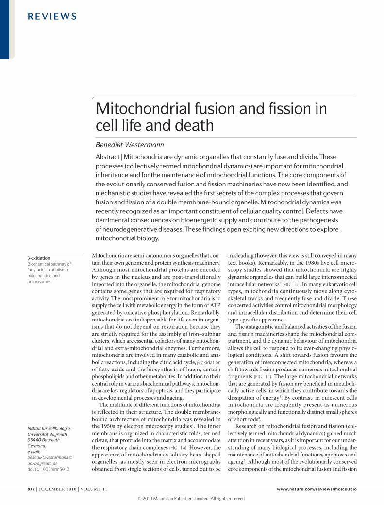
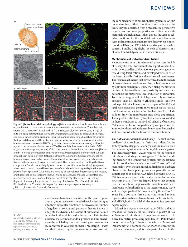
The multitude of different functions of mitochondria is reflected in their structure. The double membrane-bound architecture of mitochondria was revealed in the 1950s by electron microscopy studies1. The inner membrane is organized in characteristic folds, termed cristae, that protrude into the matrix and accommodate the respiratory chain complexes (FIG. 1a). However, the appearance of mitochondria as solitary bean-shaped organelles, as mostly seen in electron micrographs obtained from single sections of cells, turned out to be
misleading (however, this view is still conveyed in many text books). Remarkably, in the 1980s live cell micro-scopy studies showed that mitochondria are highly dynamic organelles that can build large interconnected intracellular networks2 (FIG. 1b). In many eukaryotic cell types, mitochondria continuously move along cyto-skeletal tracks and frequently fuse and divide. These concerted activities control mitochondrial morphology and intracellular distribution and determine their cell type-specific appearance.
The antagonistic and balanced activities of the fusion and fission machineries shape the mitochondrial com-partment, and the dynamic behaviour of mitochondria allows the cell to respond to its ever-changing physio-logical conditions. A shift towards fusion favours the generation of interconnected mitochondria, whereas a shift towards fission produces numerous mitochondrial fragments (FIG. 1c). The large mitochondrial networks that are generated by fusion are beneficial in metaboli-cally active cells, in which they contribute towards the dissipation of energy3. By contrast, in quiescent cells mitochondria are frequently present as numerous morpho logically and functionally distinct small spheres or short rods4.
Research on mitochondrial fusion and fission (col-lectively termed mitochondrial dynamics) gained much attention in recent years, as it is important for our under-standing of many biological processes, including the maintenance of mitochondrial functions, apoptosis and ageing5. Although most of the evolutionarily conserved core components of the mitochondrial fusion and fission
Institut für Zellbiologie, Universität Bayreuth, 95440 Bayreuth, Germany.e-mail: benedikt.westermann@ uni-bayreuth.dedoi:10.1038/nrm3013
β-oxidationBiochemical pathway of fatty acid catabolism in mitochondria and peroxisomes.
Mitochondrial fusion and fission in cell life and deathBenedikt Westermann
Abstract | Mitochondria are dynamic organelles that constantly fuse and divide. These processes (collectively termed mitochondrial dynamics) are important for mitochondrial inheritance and for the maintenance of mitochondrial functions. The core components of the evolutionarily conserved fusion and fission machineries have now been identified, and mechanistic studies have revealed the first secrets of the complex processes that govern fusion and fission of a double membrane-bound organelle. Mitochondrial dynamics was recently recognized as an important constituent of cellular quality control. Defects have detrimental consequences on bioenergetic supply and contribute to the pathogenesis of neurodegenerative diseases. These findings open exciting new directions to explore mitochondrial biology.
R E V I E W S
872 | deceMBeR 2010 | VoluMe 11 www.nature.com/reviews/molcellbio
© 20 Macmillan Publishers Limited. All rights reserved10
Nature Reviews | Molecular Cell Biology
a
c
b
Wild type No fusion No fission
Outer membraneInner membrane
CristaeIntermembrane
space
Matrix
µ
µ µµ µ µ µ
SNAREA protein belonging to a family of membrane proteins that mediate fusion of vesicles and organelles in the secretory pathway of eukaryotic cells.
machineries have been described in the past 15 years (TABLE 1), more recent work revealed mechanistic insights into their molecular function6,7. Moreover, the number of regulatory proteins and pathways that are known to integrate the dynamic behaviour of mitochondria into activities in the cell is steadily increasing. This Review describes the key mitochondrial proteins and the mecha-nisms by which they function, focusing on proteins that are conserved in yeast and animals. Three large GTPases and their interacting factors were found to constitute
the core machinery of mitochondrial dynamics. As our understanding of their function is most advanced in yeast, they are described from a mechanistic perspective in yeast, and common properties and differences with mammals are highlighted. I then discuss the various cel-lular functions of mitochondrial fusion and fission in yeast and mammals, including recent discoveries on mito-chondrial dNA (mtdNA) stability and organellar quality control. Finally, I highlight the role of dysfunctions of mitochondrial dynamics in human diseases.
Mechanisms of mitochondrial fusionMembrane fusion is a fundamental process in the life of eukaryotic cells. For example, transport vesicles fuse with the organelles of the secretory pathway, gametes fuse during fertilization, and enveloped viruses enter the host cytosol by fusion with endosomal membranes. The fusion machineries that have evolved to fit the needs of these different reactions are diverse, but they operate by common principles8. First, they bring membranes destined to be fused into close proximity and then they destabilize the bilayers by local induction of curvature. To achieve merging of lipid bilayers, membrane fusion proteins, such as soluble N-ethylmaleimide-sensitive fusion protein attachment protein receptors (SNAREs) and viral haemagglutinins, commonly contain heptad repeats that form inter- or intramolecular hairpin-like coiled-coils to draw the membranes into close apposition. These proteins also have hydrophobic domains inserted in these membranes to induce lipid bilayer deformation. Mitochondrial fusion is a particularly complex process, as mitochondria are double membrane-bound organelles and must coordinate the fusion of four membranes.
The mitochondrial fusion machinery. The first known mediator of mitochondrial fusion was identified in 1997 by molecular genetic analysis of the male sterile fuzzy onions (fzo) mutant in Drosophila melanogaster. The identified protein, FZo, is required for the fusion of mitochondria during spermatogenesis9. It is the found-ing member of a conserved protein family, termed mitofusins, that has members in yeast10,11, worms12 and mammals13. even though mitochondrial fusion has been observed in plants, plant genomes apparently do not contain genes encoding FZo-related proteins (BOX 1). Mitofusins in yeast and metazoa share a similar domain structure (FIG. 2). They are large GTPases that contain two transmembrane regions in the mitochondrial outer membrane, with a short loop in the intermembrane space and the major parts of the protein facing the cytosol14,15. Yeast Fzo1 contains three predicted hepta d repeat regions. Mammals have two mitofusin isoforms, MFN1 and MFN2, both of which lack the most amino-terminal heptad repeat.
Mgm1 is a dynamin-related large GTPase that is essential for inner membrane fusion in yeast16. It has an N-terminal mitochondrial targeting sequence that is cleaved by matrix-processing peptidase (MPP) following import. A large Mgm1 isoform contains an N-terminal transmembrane domain that anchors the protein in the inner membrane, and its main part is located in the
Figure 1 | Mitochondrial morphology. a | Mitochondria are double membrane-bound organelles with characteristic inner membrane folds, termed cristae. The schematic shows the structure of mitochondria. A transmission electron microscopy image of mitochondria in ultrathin sections of human fibroblast cells is also shown. b | In many cell types, mitochondria appear as long, tubular and sometimes branched structures that spread throughout the entire cytoplasm. Mitochondria (green) were stained in human osteosarcoma cells (U2OS) by indirect immunofluorescence using antibodies against the outer membrane protein TOM20. Nuclei (blue) were stained with DAPI (4′,6-diamidino-2-phenylindole). Cells were analysed by confocal microscopy. c | Fusion and fission regulate mitochondrial morphology. Mitochondria are branched, tubular organelles in budding yeast (left panel). Mutants lacking the fusion component Fzo1 have numerous small mitochondrial fragments that are produced by mitochondrial fission in the absence of fusion (central panel). By contrast, mutants lacking the fission component Dnm1 contain highly interconnected net-like mitochondria (right panel). Mitochondria were stained by expression of mitochondrion-targeted green fluorescent protein from a plasmid. Cells were analysed by deconvolution fluorescence microscopy, and fluorescence micrographs (shown in false colour) were merged with differential interference contrast images. Image in part a courtesy of S. Geimer, Universität Bayreuth, Germany. Image in part b courtesy of S. Jakobs, Max-Planck Institut für Biophysikalische Chemie, Göttingen, Germany. Images in part c courtesy of J. Förtsch, Universität Bayreuth, Germany.
R E V I E W S
NATuRe ReVIews | Molecular cell Biology VoluMe 11 | deceMBeR 2010 | 873
© 20 Macmillan Publishers Limited. All rights reserved10
HaemagglutininA glycoprotein of enveloped viruses, such as influenza virus. Haemagglutinins mediate fusion of the viral envelope with endosomal membranes to release the viral particle into the host cytosol.
Heptad repeatA structural protein motif that is commonly found in coiled coils that contain two or more α-helices coiled together forming a rope-like structure.
intermembrane space. A fraction of Mgm1 molecules is processed further during import by the rhomboid-related membrane protease Pcp1, generating a short isoform that lacks the transmembrane anchor17,18. Both isoforms contain a GTPase domain, a GTPase effector domain and several heptad repeats (FIG. 2). The mammalian Mgm1 orthologue, optic atrophy protein 1 (oPA1), and related proteins in worms and flies have also been shown to be required for mitochondrial fusion12,19,20. oPA1 is present in eight isoforms that are generated by alterna-tive splicing and alternative processing at two cleavage sites that are located between the N-terminal transmem-brane domain and the first heptad repeat (FIG. 2). several proteases have been implicated in alternative processing of mammalian oPA1, including the rhomboid-related protease presenilins-associated rhomboid-like (PARl)21, AAA proteases in the matrix and the intermembrane
space22–25, and the inner membrane peptidase oMA1 (also known as MPRP1)24,26.
several lines of evidence suggest that Fzo1 together with Mgm1, or mitofusins together with oPA1, are the core components of the mitochondrial fusion machiner y. First, mutant cells contain fragmented mitochondria in yeast10,11,27 and mammals28,29. second, mitochondria in fused cells that lack Fzo1, mitofusins, Mgm1 or oPA1 cannot mix their matrix contents, indicating a lack of fusion11,19,27,28. Third, mitochondria isolated from yeast mutants lacking Fzo1 or Mgm1 are unable to fuse in vitro16,30. And fourth, mitofusins, Mgm1 and oPA1 possess the domains that are predicted to be present in a fusogen: these are transmembrane domains and hepta d repeats, plus a GTPase domain that could provide bio-mechanical energy to overcome the energy barrier of lipid bilayer mixing.
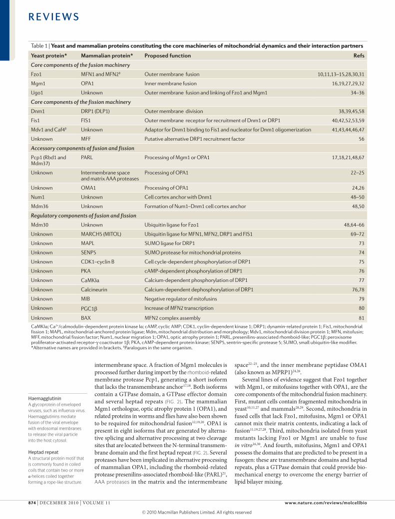
Table 1 | Yeast and mammalian proteins constituting the core machineries of mitochondrial dynamics and their interaction partners
yeast protein* Mammalian protein* Proposed function refs
Core components of the fusion machinery
Fzo1 MFN1 and MFN2‡ Outer membrane fusion 10,11,13–15,28,30,31
Mgm1 OPA1 Inner membrane fusion 16,19,27,29,32
Ugo1 Unknown Outer membrane fusion and linking of Fzo1 and Mgm1 34–36
Core components of the fission machinery
Dnm1 DRP1 (DLP1) Outer membrane division 38,39,45,58
Fis1 FIS1 Outer membrane receptor for recruitment of Dnm1 or DRP1 40,42,52,53,59
Mdv1 and Caf4‡ Unknown Adaptor for Dnm1 binding to Fis1 and nucleator for Dnm1 oligomerization 41,43,44,46,47
Unknown MFF Putative alternative DRP1 recruitment factor 56
Accessory components of fusion and fission
Pcp1 (Rbd1 and Mdm37)
PARL Processing of Mgm1 or OPA1 17,18,21,48,67
Unknown Intermembrane space and matrix AAA proteases
Processing of OPA1 22–25
Unknown OMA1 Processing of OPA1 24,26
Num1 Unknown Cell cortex anchor with Dnm1 48–50
Mdm36 Unknown Formation of Num1–Dnm1 cell cortex anchor 48,50
Regulatory components of fusion and fission
Mdm30 Unknown Ubiquitin ligase for Fzo1 48,64–66
Unknown MARCH5 (MITOL) Ubiquitin ligase for MFN1, MFN2, DRP1 and FIS1 69–72
Unknown MAPL SUMO ligase for DRP1 73
Unknown SENP5 SUMO protease for mitochondrial proteins 74
Unknown CDK1–cyclin B Cell cycle-dependent phosphorylation of DRP1 75
Unknown PKA cAMP-dependent phosphorylation of DRP1 76
Unknown CaMKIα Calcium-dependent phosphorylation of DRP1 77
Unknown Calcineurin Calcium-dependent dephosphorylation of DRP1 76,78
Unknown MIB Negative regulator of mitofusins 79
Unknown PGC1β Increase of MFN2 transcription 80
Unknown BAX MFN2 complex assembly 81
CaMKIα; Ca2+/calmodulin-dependent protein kinase Iα; cAMP, cyclic AMP; CDK1, cyclin-dependent kinase 1; DRP1; dynamin-related protein 1; Fis1, mitochondrial fission 1; MAPL, mitochondrial-anchored protein ligase; Mdm, mitochondrial distribution and morphology; Mdv1, mitochondrial division protein 1; MFN, mitofusin; MFF, mitochondrial fission factor; Num1, nuclear migration 1; OPA1, optic atrophy protein 1; PARL, presenilins-associated rhomboid-like; PGC1β; peroxisome proliferator-activated receptor-γ coactivator 1β; PKA, cAMP-dependent protein kinase; SENP5, sentrin-specific protease 5; SUMO, small ubiquitin-like modifier. *Alternative names are provided in brackets. ‡Paralogues in the same organism.
R E V I E W S
874 | deceMBeR 2010 | VoluMe 11 www.nature.com/reviews/molcellbio
© 20 Macmillan Publishers Limited. All rights reserved10
Nature Reviews | Molecular Cell Biology
0 6 9 12 15 18 21 24 30 3327 51 sec
0 6 12 18 24 30 39 42 6036 63 66 69 sec
2 µm
a
b
DynaminA large GTPase involved in membrane scission events.
RhomboidThe founding member of a conserved family of intramembrane Ser proteases that have their active sites buried in the plane of the membrane.
AAA proteasesLarge multisubunit proteases that belong to the superfamily of ATPases that are associated with diverse cellular activities.
LiposomeArtificial lipid bilayer vesicle composed of membrane lipids.
Mitochondrial carrier family signatureA protein sequence motif that is commonly found in mitochondrial carrier proteins that function in the transport of metabolites and other biomolecules across the mitochondrial inner membrane.
These observations can be integrated into a hypo-thetical model of mitochondrial fusion. It is conceivable that early during fusion two mitochondria approaching each other are tethered in a docking step. consistently, the carboxy-terminal heptad repeats of mammalian MFN1 have been shown to form an intermolecular antiparallel coiled coil that may tether adjacent mitochondria prior to fusion31. coiled coil formation by mitofusins might then draw the membranes close together and initiat e lipid bilayer mixing, and the GTPase could provide biomechanical energy for outer membrane fusion. The first mechanistic insights into the role of Mgm1 in inner membrane fusion came from the analysis of yeast mutant mitochondria in vitro. After the completion of outer mem-brane fusion, Mgm1 is required in trans on both inner membranes of the fusion partners. certain mgm1 mutant alleles show a specific defect in inner membrane tethering, whereas others are defective in inner membrane fusion, suggesting that Mgm1 participates in both pro cesses16. Interestingly, studies using purified Mgm1 variants reconstituted with liposomes showed that only the short Mgm1 isoform, which lacks the transmembrane region, has GTPase activity, and that its GTPase is activated in heterotypic complexes containing the membrane-bound long isoform. Thus, the long Mgm1 isoform is proposed to tether opposing inner membranes and harness GTPase-dependent conformational changes of the short isoform to initiate lipid bilayer mixing of the inner membrane32.
Coordination of outer and inner membrane fusion. dissipation of the membrane potential or mutations in Mgm1 or oPA1 selectively block inner membrane fusion. under these conditions, outer membrane fusion
proceeds in the absence of inner membrane fusion16,30,33. Thus, the fusion machineries in the mitochondrial outer and inner membranes can operate independently of each other. However, it is conceivable that under nor-mal conditions outer membrane fusion is coordinated with inner membrane fusion. Yeast mutants carrying insertions between the transmembrane regions of Fzo1 show a specific loss of Fzo1 interactions with the inner membrane and severe defects in mitochondrial fusion. Thus, the intermembrane space loop of Fzo1 plays an important part in the coordination of double-membrane fusion14. Furthermore, ugo1, an outer membrane protein that is essential for mitochondrial fusion in yeast34, also parti cipates in connecting the outer and inner membrane fusion machineries. It contains three transmembrane domains and two mitochondrial carrier family signatures of unknown function35 (FIG. 2). ugo1 physically inter-acts with both Fzo1 and Mgm1 (REF. 36). Intriguingly, temperature-sensitive ugo1 alleles revealed specific defects in outer or inner membrane fusion at a post-tethering step35. This phenotype is compatible with a role for ugo1 as a linker that connects Fzo1 with Mgm1 and coordi-nates double-membrane fusion. structurally or function-ally related ugo1 equivalents in higher eukaryote s have not been identified.
Mechanisms of mitochondrial fissiondynamin superfamily members are versatile large GTPases that mediate various membrane remodelling processes in eukaryotic cells. Mgm1 and oPA1, and in particular Fzo1 and mitofusins, are distantly related dynamin superfamily members. Although these pro-teins function in membrane fusion, classical dynamins
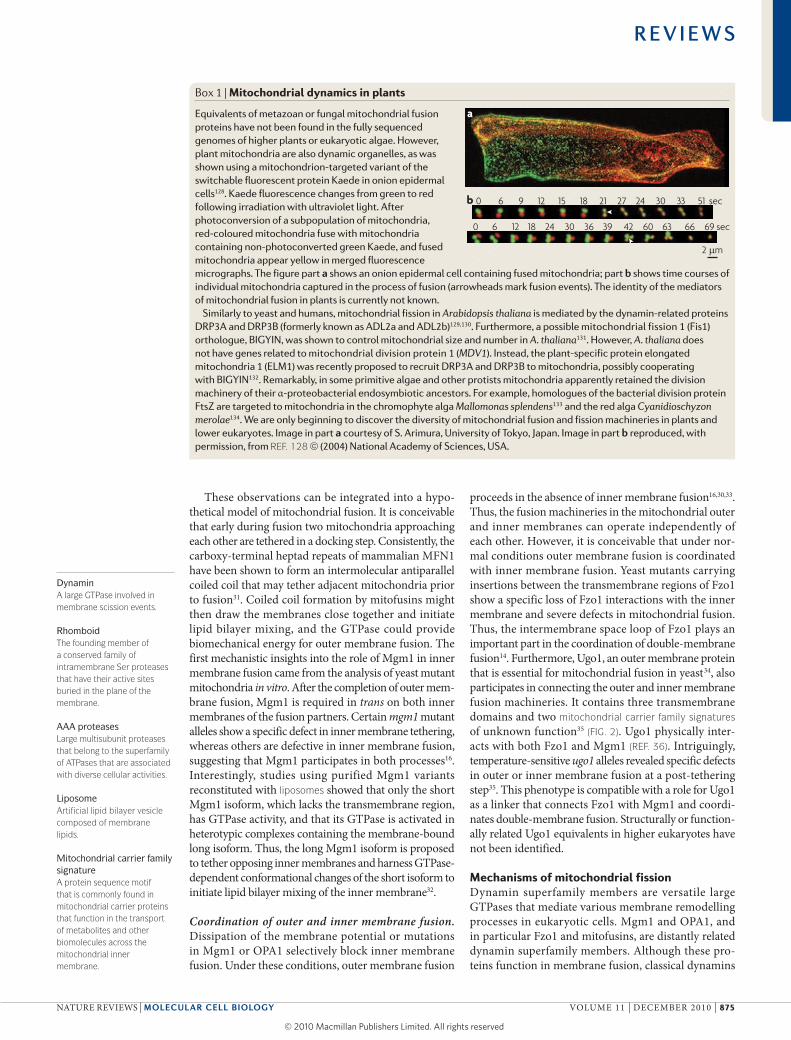
Box 1 | Mitochondrial dynamics in plants
Equivalents of metazoan or fungal mitochondrial fusion proteins have not been found in the fully sequenced genomes of higher plants or eukaryotic algae. However, plant mitochondria are also dynamic organelles, as was shown using a mitochondrion-targeted variant of the switchable fluorescent protein Kaede in onion epidermal cells128. Kaede fluorescence changes from green to red following irradiation with ultraviolet light. After photoconversion of a subpopulation of mitochondria, red-coloured mitochondria fuse with mitochondria containing non-photoconverted green Kaede, and fused mitochondria appear yellow in merged fluorescence micrographs. The figure part a shows an onion epidermal cell containing fused mitochondria; part b shows time courses of individual mitochondria captured in the process of fusion (arrowheads mark fusion events). The identity of the mediators of mitochondrial fusion in plants is currently not known.
Similarly to yeast and humans, mitochondrial fission in Arabidopsis thaliana is mediated by the dynamin-related proteins DRP3A and DRP3B (formerly known as ADL2a and ADL2b)129,130. Furthermore, a possible mitochondrial fission 1 (Fis1) orthologue, BIGYIN, was shown to control mitochondrial size and number in A. thaliana131. However, A. thaliana does not have genes related to mitochondrial division protein 1 (MDV1). Instead, the plant-specific protein elongated mitochondria 1 (ELM1) was recently proposed to recruit DRP3A and DRP3B to mitochondria, possibly cooperating with BIGYIN132. Remarkably, in some primitive algae and other protists mitochondria apparently retained the division machinery of their α-proteobacterial endosymbiotic ancestors. For example, homologues of the bacterial division protein FtsZ are targeted to mitochondria in the chromophyte alga Mallomonas splendens133 and the red alga Cyanidioschyzon merolae134. We are only beginning to discover the diversity of mitochondrial fusion and fission machineries in plants and lower eukaryotes. Image in part a courtesy of S. Arimura, University of Tokyo, Japan. Image in part b reproduced, with permission, from REF. 128 © (2004) National Academy of Sciences, USA.
R E V I E W S
NATuRe ReVIews | Molecular cell Biology VoluMe 11 | deceMBeR 2010 | 875
© 20 Macmillan Publishers Limited. All rights reserved10
Nature Reviews | Molecular Cell Biology
Fzo1 855 aa
Mgm1 881 aa
Ugo1 502 aa
Dnm1 757 aa
Mdv1 715 aa
Caf4 643 aa
Fis1 155 aa 152 aaFIS1
699–736 aaDRP1
960–1,015 aa
757 aa741 aa
MFN2MFN1
OPA1
Fusion machinery
Fission machinery
Yeast Human
MPP processingPcp1 processing
MPP processingS1 and S2 processing sites
Mitochondrial carrier signatures Alternative splice region
Alternative splice region
GTPase domainGTPase effector domainHeptad repeats
Middle domainWD40 repeatsTransmembrane domain
Mitochondrial pre-sequenceHydrophobic region
TPR domain
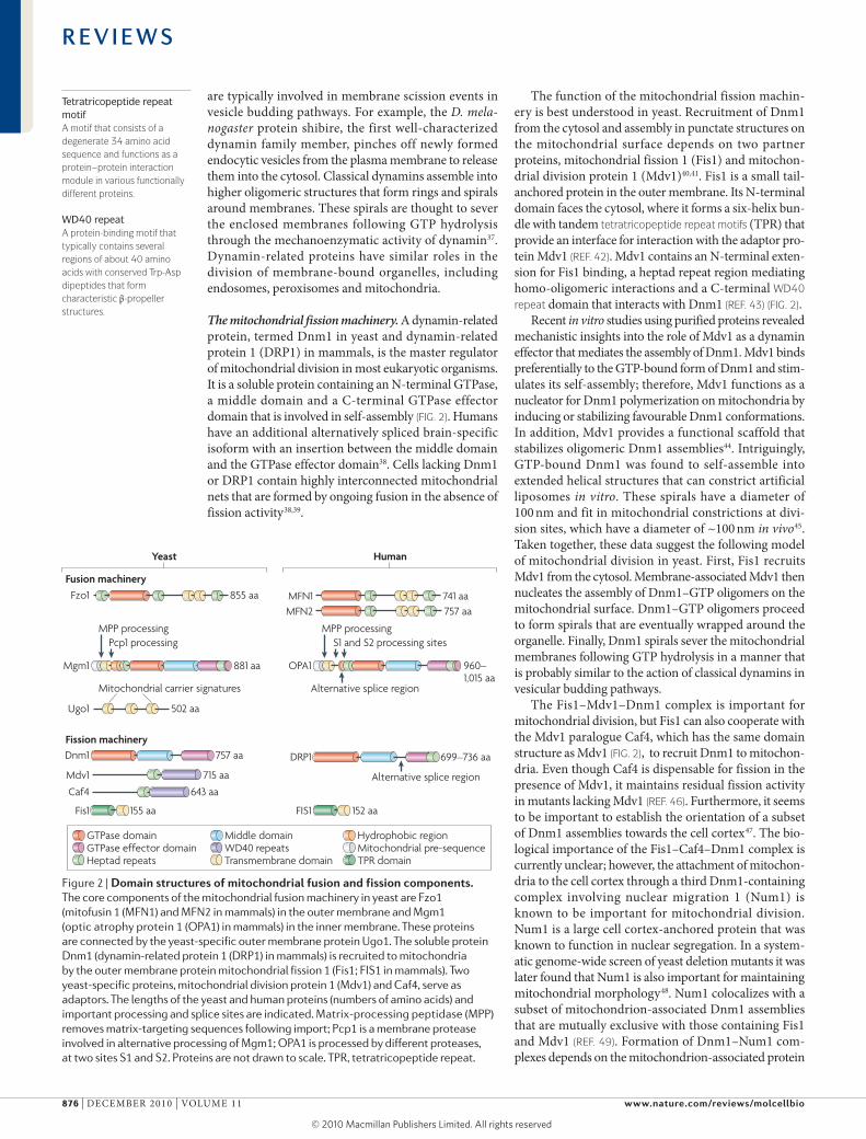
Tetratricopeptide repeat motifA motif that consists of a degenerate 34 amino acid sequence and functions as a protein–protein interaction module in various functionally different proteins.
WD40 repeatA protein-binding motif that typically contains several regions of about 40 amino acids with conserved Trp-Asp dipeptides that form characteristic β-propeller structures.
are typically involved in membrane scission events in vesicle budding pathways. For example, the D. mela-nogaster protein shibire, the first well-characterized dynamin family member, pinches off newly formed endocytic vesicles from the plasma membrane to release them into the cytosol. classical dynamins assemble into higher oligomeric structures that form rings and spirals around membranes. These spirals are thought to sever the enclosed membranes following GTP hydrolysis through the mechanoenzymatic activity of dynamin37. dynamin-related proteins have similar roles in the divisio n of membrane-bound organelles, includin g endosomes, peroxisomes and mitochondria.
The mitochondrial fission machinery. A dynamin-related protein, termed dnm1 in yeast and dynamin-related protein 1 (dRP1) in mammals, is the master regulator of mitochondrial division in most eukaryotic organisms. It is a soluble protein containing an N-terminal GTPase, a middle domain and a c-terminal GTPase effector domain that is involved in self-assembly (FIG. 2). Humans have an additional alternatively spliced brain-specific isoform with an insertion between the middle domain and the GTPase effector domain38. cells lacking dnm1 or dRP1 contain highly interconnected mitochondrial nets that are formed by ongoing fusion in the absence of fission activity38,39.
The function of the mitochondrial fission machin-ery is best understood in yeast. Recruitment of dnm1 from the cytosol and assembly in punctate structures on the mitochondrial surface depends on two partner proteins, mitochondrial fission 1 (Fis1) and mitochon-drial division protein 1 (Mdv1)40,41. Fis1 is a small tail-anchored protein in the outer membrane. Its N-terminal domain faces the cytosol, where it forms a six-helix bun-dle with tandem tetratricopeptide repeat motifs (TPR) that provide an interface for interaction with the adaptor pro-tein Mdv1 (REF. 42). Mdv1 contains an N-terminal exten-sion for Fis1 binding, a heptad repeat region mediating homo-oligomeric interactions and a c-terminal WD40 repeat domain that interacts with dnm1 (REF. 43) (FIG. 2).
Recent in vitro studies using purified proteins revealed mechanistic insights into the role of Mdv1 as a dynamin effector that mediates the assembly of dnm1. Mdv1 binds preferentially to the GTP-bound form of dnm1 and stim-ulates its self-assembly; therefore, Mdv1 functions as a nucleator for dnm1 polymerization on mitochondria by inducing or stabilizing favourable dnm1 conformations. In addition, Mdv1 provides a functional scaffold that stabi lizes oligomeric dnm1 assemblies44. Intriguingly, GTP-bound dnm1 was found to self-assemble into extended helical structures that can constrict artificial liposomes in vitro. These spirals have a diameter of 100 nm and fit in mitochondrial constrictions at divi-sion sites, which have a diameter of ~100 nm in vivo45. Taken together, these data suggest the following model of mitochondrial division in yeast. First, Fis1 recruits Mdv1 from the cytosol. Membrane-associated Mdv1 then nucleates the assembly of dnm1–GTP oligomers on the mitochondrial surface. dnm1–GTP oligomers proceed to form spirals that are eventually wrapped around the organelle. Finally, dnm1 spirals sever the mitochondrial membranes following GTP hydrolysis in a manner that is probably similar to the action of classical dynamins in vesicular budding pathways.
The Fis1–Mdv1–dnm1 complex is important for mitochondrial division, but Fis1 can also cooperate with the Mdv1 paralogue caf4, which has the same domain structure as Mdv1 (FIG. 2), to recruit dnm1 to mitochon-dria. even though caf4 is dispensable for fission in the presence of Mdv1, it maintains residual fission activity in mutants lacking Mdv1 (REF. 46). Furthermore, it seems to be important to establish the orientation of a subset of dnm1 assemblies towards the cell cortex47. The bio-logical importance of the Fis1–caf4–dnm1 complex is currently unclear; however, the attachment of mitochon-dria to the cell cortex through a third dnm1-containing complex involving nuclear migration 1 (Num1) is known to be important for mitochondrial division. Num1 is a large cell cortex-anchored protein that was known to function in nuclear segregation. In a system-atic genome-wide screen of yeast deletion mutants it was later found that Num1 is also important for maintaining mitochondrial morphology48. Num1 colocalizes with a subset of mitochondrion-associated dnm1 assemblies that are mutually exclusive with those containing Fis1 and Mdv1 (REF. 49). Formation of dnm1–Num1 com-plexes depends on the mitochondrion-associated protein
Figure 2 | Domain structures of mitochondrial fusion and fission components. The core components of the mitochondrial fusion machinery in yeast are Fzo1 (mitofusin 1 (MFN1) and MFN2 in mammals) in the outer membrane and Mgm1 (optic atrophy protein 1 (OPA1) in mammals) in the inner membrane. These proteins are connected by the yeast-specific outer membrane protein Ugo1. The soluble protein Dnm1 (dynamin-related protein 1 (DRP1) in mammals) is recruited to mitochondria by the outer membrane protein mitochondrial fission 1 (Fis1; FIS1 in mammals). Two yeast-specific proteins, mitochondrial division protein 1 (Mdv1) and Caf4, serve as adaptors. The lengths of the yeast and human proteins (numbers of amino acids) and important processing and splice sites are indicated. Matrix-processing peptidase (MPP) removes matrix-targeting sequences following import; Pcp1 is a membrane protease involved in alternative processing of Mgm1; OPA1 is processed by different proteases, at two sites S1 and S2. Proteins are not drawn to scale. TPR, tetratricopeptide repeat.
R E V I E W S
876 | deceMBeR 2010 | VoluMe 11 www.nature.com/reviews/molcellbio
© 20 Macmillan Publishers Limited. All rights reserved10
F-box proteinA protein that contains an F-box as a protein interaction domain. Most F-box proteins are substrate recognition subunits of SCF ubiquitin ligases and have roles in ubiquitin-dependent protein degradation.
Ubiquitin ligaseAn enzyme that mediates the covalent attachment of ubiquitin to substrate proteins to label them for degradation by the proteasome or other regulatory pathways. This process is termed ubiquitylation.
Mdm36 (mitochondrial distribution and morpholo gy 36). As mitochondria are highly motile and lose their locali-zation to the cell periphery in cells lacking Num1 or Mdm36, it is thought that these proteins together with dnm1 form cell cortex anchors that immobilize parts of the mitochondrial network50. Interestingly, it was shown that membrane scission by classical dynamins is facili-tated by the generation of tension on the membrane51. Thus, it is conceivable that mitochondrial cortex anchors together with cytoskeleton-dependent forces generate tension on mitochondrial membranes to promote mito-chondrial fission by dnm1 (REF. 50). It will be interest-ing to see whether mitochondria l division is linked to motilit y in other cell types.
Mammalian FIs1 interacts with dRP1 and appar-ently has a similar role in mitochondrial fission to its yeast counterpart, as FIs1 overexpression promotes mitochondrial fragmentation and FIs1 depletion pro-duces interconnected mitochondrial nets52,53. However, Mdv1 homologues have not been identified in meta-zoans which indicates significant differences between the metazoan and yeast mitochondrial division machin-eries. Furthermore, knockdown of human FIs1 does not affect the distribution of dRP1 in mitochondria54, and deletion of the two FIS1 homologous genes in Caenorhabditis elegans does not produce a strong mito-chondrial fission phenotype55, suggesting that additional pathways of dRP1 recruitment exist in metazoans. one possible candidate for an alternative fission factor is mitochondrial fission factor (MFF), a tail-anchored pro-tein that is conserved in metazoans but does not exist in yeast. MFF contains heptad repeats and a c-terminal transmembrane domain that is embedded in the outer membrane. depletion of MFF attenuates mitochondrial division, both in mammalian and D. melanogaster cells. Interestingly, MFF and FIs1 exist in separate complexes, suggesting that they have different roles in mitochondrial division56.
A separate division machinery in the inner membrane? only little is known about division of the mito-chondrial inner membrane. It is conceivable that Mdv1-coordinated assembly of dnm1 produces spirals that constrict the organelle from its normal diameter of ~300 nm to ~ 100 nm, as has been observed with puri-fied dnm1 on liposomes44,45. Following GTP hydrol ysis, constriction of dnm1 spirals might then sever both mitochondrial membranes at the same time. However, live cell micro scopy of yeast and C. elegans showed that mitochondrial constriction and separation of matrix compartments can proceed independently of dnm1 or dRP1 (REFS 57–59). Thus, it is possible that there is a separate machinery for constriction and/or division of the inner membrane. one possible candidate in yeast is Mdm33, a heptad repeat-containing inner membrane protein. Mutants lacking Mdm33 have extremely elon-gated and stretched mitochondria, indicating a lack of division activity. overexpression of Mdm33 leads to mitochondrial fragmentation and induces the formation of inner membrane septae, possibly as a consequence of increased inner membrane division activity60. Although
Mdm33 does not have homologues in higher eukaryotes, the inner membrane protein MTP18 has been postulated to have a role in fission in mammals. overexpression of MTP18 induces fragmentation of mitochondria, whereas depletion produces highly fused mitochondria, a pheno-type consistent with a role in inner membrane fission61. clearly, more work is required to define the roles of Mdm33 and MTP18 in mitochondrial dynamics.
Regulators of mitochondrial dynamicsMitochondrial fusion and fission are antagonistic activ-ities62,63. Thus, the rates of fusion and fission must be tightly controlled to keep the right balance required for the maintenance of mitochondrial morphology or to shift this balance to adapt the degree of mitochondrial interconnectivity to changing physiological conditions. owing to their central role in mitochondrial fusion and fission, Fzo1, Mgm1 and dnm1 in yeast and mito-fusins, oPA1 and dRP1 in mammals are prime targets for regulatory pathways. Although the core machiner-ies of mitochondrial dynamics are highly conserved, the mechanisms to control their activities seem to be more diverse in different organisms and cell types (FIG. 3).
In yeast, Fzo1 levels are controlled by the F-box protein Mdm30. Mutants lacking Mdm30 accumulate excess Fzo1 and contain fusion-incompetent mitochondria64. Mdm30 is a subunit of a skp1–cullin–F-box (scF) ubiquiti n ligase, which promotes ubiquitylation and degradation of Fzo1 by the proteasome65. This activity is required to con-trol steady-state levels of Fzo1 and to maintain fusion- competent mitochondria in vegetative cells. Intriguingly, Mdm30-dependent ubiquitylation and degradation is reduced in GTPase-deficient fzo1 mutants. This obser-vation suggests that the Fzo1 GTPase cycle is intimately linked to ubiquitylation and that Fzo1 degradation is an important new aspect in the fusion pathway66.
An attractive hypothesis, termed ‘alternative topo-genesis’, proposes that processing of Mgm1 by Pcp1 regulates mitochondrial fusion in response to matrix ATP levels67. Newly synthesized Mgm1 is imported into mitochondria through protein translocases that reside in the mitochondrial outer and inner membranes. during import, the mitochondrial pre-sequence is removed by MPP. Two pathways are then possible. The first hydro-phobic region might insert into the inner membrane, generating the membrane-anchored long isoform. Alternatively, Mgm1 might be translocated further into the matrix, thereby exposing the second hydrophobic region to the membrane protease Pcp1. This enables cleavage by Pcp1, generating the short Mgm1 isoform, which is released into the intermembrane space. As import of Mgm1 is ATP dependent, matrix ATP levels could directly determine the ratio of Mgm1 isoforms. At low ATP levels import would be slow, allowing the long isoform enough time to insert into the inner membrane before the Pcp1 processing site becomes accessible. By contrast, high ATP levels would favour fast import and thus promote further translocation to allow Pcp1 process-ing. Because both isoforms are required for mitochon-drial fusion, this mechanism could link mitochondrial fusion activity to mitochondrial energy levels.
R E V I E W S
NATuRe ReVIews | Molecular cell Biology VoluMe 11 | deceMBeR 2010 | 877
© 20 Macmillan Publishers Limited. All rights reserved10
Nature Reviews | Molecular Cell Biology
Fusion machinery
Fission machinery
Fzo1Ugo1
Ub
Mgm1
MPPInnermembrane
Outermembrane
MFN1 or MFN2
Ub
OPA1
MPP
?
?Fis1
Mdv1 or Caf4
Dnm1
FIS1
DRP1
?
Ub
PUb
?
?
SUMO
MARCH5
MARCH5
MAPL;SENP5
PKA,CaMKIα, Cdk1–cyclin B;calcineurin
Yeast Mammals
MARCH5
Mdm30
Pcp1
PARL, mAAA,iAAA andOMA1
Small ubiquitin-like modifierA small protein that is covalently attached to and removed from other proteins to modify their activity.
NucleoidA compact structure that contains DNA and proteins. Mitochondrial nucleoids contain several copies of the mitochondrial genome and are attached to the inner membrane.
Processing of oPA1 regulates mitochondrial fusion activity in mammalian cells. For example, generation of the short oPA1 isoform along with fragmentation of mitochondria is favoured at low membrane potential, low ATP levels or during apoptosis22,23,25,68. Although the alternative topogenesis model is useful to interpret observations in yeast, processing of oPA1 in higher organisms seems to be more complex. Many proteases are involved, including PARl, several AAA proteases and oMA1, and certain splice variants are differentially processed22–26. It is possible that tissue-specific expres-sion of oPA1-cleaving proteases and/or splice variants contributes to the complex pattern of oPA1 processing. However, recent studies suggest that at least some spe-cific processing steps fulfil a distinct function. cleavage
by the inner membrane protease oMA1 is induced in impaired mitochondria with low membrane poten-tial24,26. As the long and short oPA1 isoforms are both required for fusion25, complete conversion of oPA1 to the short isoform by oMA1 shuts off the fusion machinery of dysfunctional mitochondria. This activit y could contribute to quality control by preventing fusion of defective mitochondria with the mitochondrial net-work24,26. Importantly, processing by oMA1 is not restricted to newly imported oPA1, but allows the cell a more rapid stress response through complete cleavage of all long oPA1 molecules that are present.
Many additional pathways have been discovered that regulate the activities of the fusion and fission machineries in mammalian cells (TABLE 1). MARcH5 (also known as MITol), a ubiquitin ligase in the outer membrane, associates with and ubiquitylates dRP1, FIs1 and mitofusins69–72. loss of MARcH5 is associated with mitochon drial elongation and triggers cell senes-cence72. Mitochondrial-anchored protein ligase (MAPl) is a mitochondrion-anchored small ubiquitin-like modifier (suMo) ligase that attaches suMo to dRP1 to stimu-late mitochondrial fission73. Removal of suMo from dRP1 involves sentrin-specific protease 5 (seNP5), a suMo protease that recognizes several mitochondrial targets74. Furthermore, the activity of dRP1 can be reversibly modified by phosphorylation. Three protein kinases have been identified that phosphorylate dif-ferent dRP1 ser residues: cyclin-dependent kinase 1 (cdk1)–cyclin B75, cAMP-dependent protein kinase (PkA)76 and ca2+/calmodulin-dependent protein kinase Iα (caMkIα)77. The phosphatase calcineurin reg-ulates mitochondrial morphology by dephosphoryl ating dRP1 in response to ca2+signals76,78. Moreover, MIB was identified as a MFN1-interacting protein that attenuates mitochondrial fusion by an unknown mechanism79. In addition, mitochondrial dynamics can be modulated at the trans criptional level80 or by the regulated assembly of mitofusin complexes81. Thus, mammalian cells pos-sess a large repertoire of different regulatory factors to modulate mitochondrial dynamics in response to various signals.
Functions of mitochondrial dynamicsMitochondria cannot be generated de novo; instead, they proliferate by growth and division of pre-existing organelles. They contain their own genome and pro-tein translation machinery. mtdNA is present in multi-ple copies and packed into compact particles, termed nucleoids. It encodes ribosomal RNAs, tRNAs and some proteins required for respiration. However, most mito-chondrial proteins are encoded by nuclear genes and synthesized by cytosolic ribosomes. Mitochondrial bio-genesis involves the import of nucleus-encoded proteins from the cytosol, the incorporation of mitochondrion-synthesized and imported membrane lipids, the amplifi-cation of the mitochondrial genome and the translation of mitochondrion-encoded proteins82–84. damaged and surplus organelles are removed by autophagy. during their life cycle, mitochondria fuse with each other and split apart again (FIG. 4a); fusion serves to mix and
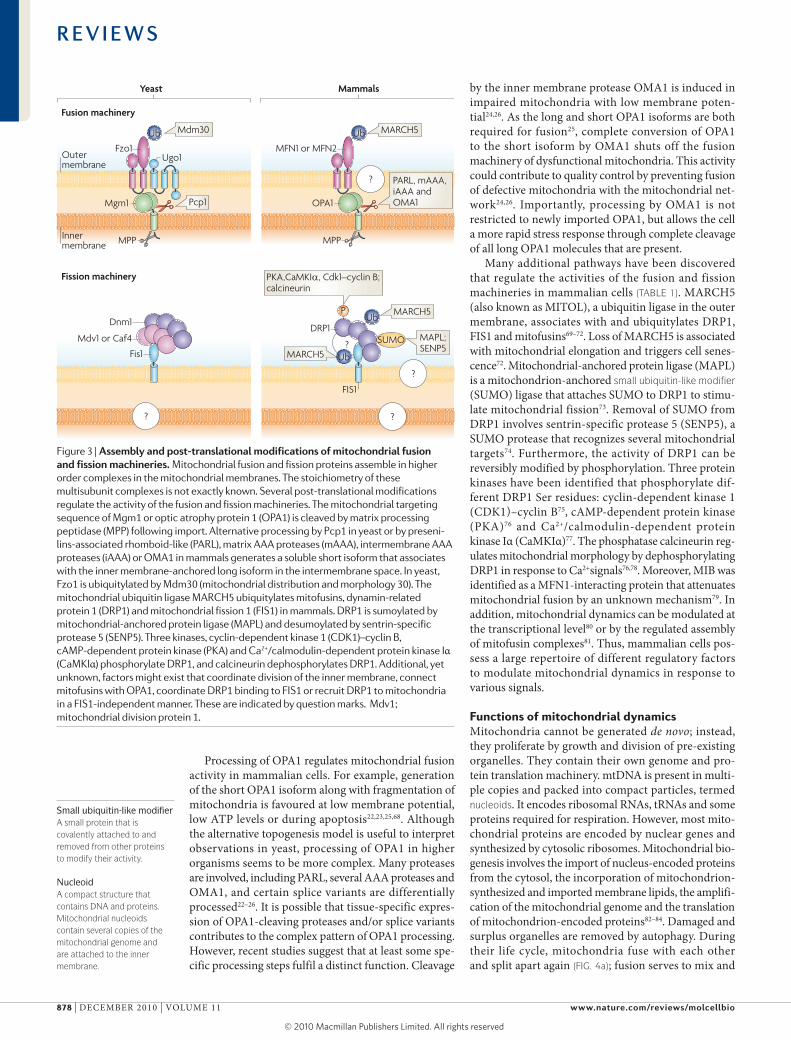
Figure 3 | assembly and post-translational modifications of mitochondrial fusion and fission machineries. Mitochondrial fusion and fission proteins assemble in higher order complexes in the mitochondrial membranes. The stoichiometry of these multisubunit complexes is not exactly known. Several post-translational modifications regulate the activity of the fusion and fission machineries. The mitochondrial targeting sequence of Mgm1 or optic atrophy protein 1 (OPA1) is cleaved by matrix processing peptidase (MPP) following import. Alternative processing by Pcp1 in yeast or by preseni-lins-associated rhomboid-like (PARL), matrix AAA proteases (mAAA), intermembrane AAA proteases (iAAA) or OMA1 in mammals generates a soluble short isoform that associates with the inner membrane-anchored long isoform in the intermembrane space. In yeast, Fzo1 is ubiquitylated by Mdm30 (mitochondrial distribution and morphology 30). The mitochondrial ubiquitin ligase MARCH5 ubiquitylates mitofusins, dynamin-related protein 1 (DRP1) and mitochondrial fission 1 (FIS1) in mammals. DRP1 is sumoylated by mitochondrial-anchored protein ligase (MAPL) and desumoylated by sentrin-specific protease 5 (SENP5). Three kinases, cyclin-dependent kinase 1 (CDK1)–cyclin B, cAMP-dependent protein kinase (PKA) and Ca2+/calmodulin-dependent protein kinase Iα (CaMKIα) phosphorylate DRP1, and calcineurin dephosphorylates DRP1. Additional, yet unknown, factors might exist that coordinate division of the inner membrane, connect mitofusins with OPA1, coordinate DRP1 binding to FIS1 or recruit DRP1 to mitochondria in a FIS1-independent manner. These are indicated by question marks. Mdv1; mitochondrial division protein 1.
R E V I E W S
878 | deceMBeR 2010 | VoluMe 11 www.nature.com/reviews/molcellbio
© 20 Macmillan Publishers Limited. All rights reserved10
Nature Reviews | Molecular Cell Biology
Biogenesis
Interconnectedmitochondrial network
Multipleheterogeneousmitochondria
Fissiona
bFusion
Turnover
Metaboliccapacity
High
Low
∆Ψ
Fission
Fusion
Dissipation ofmetabolic energy
Defense against ageing
Apoptosis
Distribution
Mitophagy
Inheritance
Transmission ofmembrane potential
Complementation of mtDNAproducts in heteroplasmic cells
Growth and division
Cytochrome c release
Cytoskeleton-mediatedtransport
Turnover ofdamaged organelles
YX
unify the mitochondrial compartment, whereas fission generates morphologically and functionally distinct organelles. These processes have important conse-quences for mitochondrial functions in cell life and death (FIG. 4b).
Mitochondrial inheritance. Mitochondria are essential organelles that must be inherited during cell division. It is obvious that the number of mitochondria must increase during proliferation of cells. Accordingly, the mitochondrial network of mammalian interphase cells in culture was observed to fragment before the cells
enter mitosis and undergo cytokinesis75,85. This allows partitioning of organelles to the daughter cells in a sto-chastic manner. cell cycle-dependent mitochondrial morphology changes are much less pronounced in yeast. Instead, mitochondria are inherited in an ordered manner by cytoskeleton-dependent bud-directed trans-port86. surprisingly, yeast mutants lacking dnm1 do not show a significant growth defect, even though their interconnected mitochondrial network must split in two parts during cell division. similarly, knockout of Drp1 does not affect the progression of cytokinesis in mouse embryonic fibroblasts85. It is not known whether mitochondrial division in mutant cells is mediated by mechanical forces imposed on the organelle during cytokinesis or by an as-yet-unidentified dnm1- or dRP1-independent division machinery. However, mice that lack mitofusins28 or dRP1 (REFS 85,87) die before birth, which indicates that mitochondrial dynamics is essential in mammalian development.
Mitochondrial fusion is important for inheritance and maintenance of mtdNA. In yeast, fusion-defective mutants rapidly lose their mitochondrial genome and consequently show defects in respiration88. This is pro bably because fragmentation of mitochondria pro-duces multiple small organelles, most of which lack mtdNA, so partitioning of these organelles to daughter cells produces a significant number of progeny lacking mtdNA. As a result, mitochondrial genomes are lost from the population after several generations.
disruption of fusion in mammalian cells also leads to mitochondrial heterogeneity and dysfunction, possibly as a consequence of nucleoid loss in individual mitochondria89,90. Thus, it seems that fusion serves as a fundamental mechanism to maintain a mitochondrial population with a full complement of nucleus- and mitochondrion-encoded gene products. Although mitochondrial fission inevitably generates organelles lacking nucleoids, fusion ensures that the mitochon-drial genome and gene products are replenished before functionality is lost.
Mitochondrial distribution and morphology. cells defective in mitochondrial division contain highly interconnected net-like mitochondria that typically accumulate in restricted areas, leaving large parts of the cell devoid of mitochondria. Proper mitochondrial distribution depends on division to split the mitochon-drial network into transportable units. obviously, this is particularly important in large and extended cells, such as neurons. Accordingly, dRP1 and oPA1 are crucial to establish proper mitochondrial content in dendrites. This, in turn, is essential for the maintenance of dendritic spines and synapses, which are neuronal structures with a particularly high energy demand91. Moreover, Drp1-knockout mice have severe develop-mental abnormalities, particularly in the forebrain and the cerebellum85,87. Intriguingly, mutant neurons in primary cell culture show an accumulation of mito-chondria in the cell body, with only a few clumped and irregularly distributed mitochondria in neurites; this indicates that dRP1 deficiency impedes proper
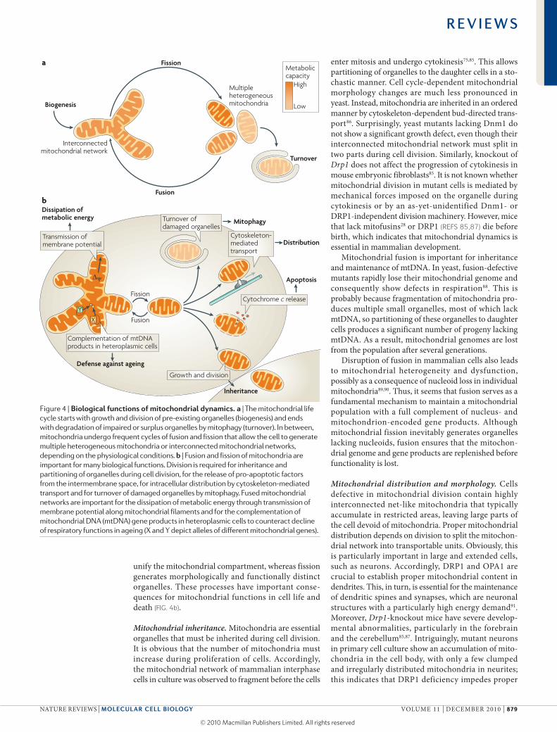
Figure 4 | Biological functions of mitochondrial dynamics. a | The mitochondrial life cycle starts with growth and division of pre-existing organelles (biogenesis) and ends with degradation of impaired or surplus organelles by mitophagy (turnover). In between, mitochondria undergo frequent cycles of fusion and fission that allow the cell to generate multiple heterogeneous mitochondria or interconnected mitochondrial networks, depending on the physiological conditions. b | Fusion and fission of mitochondria are important for many biological functions. Division is required for inheritance and partitioning of organelles during cell division, for the release of pro-apoptotic factors from the intermembrane space, for intracellular distribution by cytoskeleton-mediated transport and for turnover of damaged organelles by mitophagy. Fused mitochondrial networks are important for the dissipation of metabolic energy through transmission of membrane potential along mitochondrial filaments and for the complementation of mitochondrial DNA (mtDNA) gene products in heteroplasmic cells to counteract decline of respiratory functions in ageing (X and Y depict alleles of different mitochondrial genes).
R E V I E W S
NATuRe ReVIews | Molecular cell Biology VoluMe 11 | deceMBeR 2010 | 879
© 20 Macmillan Publishers Limited. All rights reserved10
Purkinje neuronA large neuron in the cerebellar cortex with a single long axon and many branched dendritic extensions.
Reactive oxygen speciesReactive oxygen species (ROS) are chemically reactive molecules containing oxygen, such as superoxide anions, peroxides and hydroxyl radicals. Mitochondrial ROS are generated by incomplete reduction of molecular oxygen in the respiratory chain.
mitochondrial distribution85. Mice carrying conditional Mfn2-knockout alleles show highly specific degenera-tion of Purkinje neurons. Mitochondria in these mutant cells are fragmented and fail to distribute to the long and branched neurite processes, indicating that fusion may also influence mitochondrial distribution90. A pos-sible explanation is that most mutant mitochondria have defects in respiration because they lack mtdNA due to an inheritance defect. This may be sensed by the neuronal transport system, which may then trans-port inactive mitochondria preferentially towards the cell body90.
In other cell types, fused mitochondrial networks act as electrically united systems that transmit the membrane potential generated by the proton pumps of the respira-tory chain92. This mechanism was proposed to play an important part in the dissipation of metabolic energy in muscle cells. In these unusually large cells, mitochon-drial filaments connect a dense layer of mitochondria in the oxygen-rich cell periphery with mitochondria in the oxygen-poor core of the muscle fibre, thereby form-ing a continuous network. Thus, a membrane potential generated in the cell periphery can be dissipated over a large area and used to produce ATP in remote parts of the cell to fuel molecular motors during muscle con-traction3. Moreover, mitochondrial fusion is mark-edly increased under stress conditions that involve mitochondrial ATP production93 and during the G1 to s transition, when mitochondrial ATP synthesis is enhanced94. The appearance of highly interconnected fused mitochondrial networks in these cells is consistent with a role for mitochondrial fusion in the dissipation of energy. Hence, it seems that concerted activities of the mitochondrial fusion and fission machineries shape the mitochondrial compartment and adapt it to the specific requirements of the cells.
Mitochondrial quality control and ageing. Research over the past decades leaves no doubt that mitochon-dria have a crucial role in ageing. The mitochondrial theory of ageing postulates that the respiratory chain produces reactive oxygen species (Ros) as byproducts of oxidative phosphorylation. Because mitochondria are a major source for Ros, mtdNA is particularly vulner-able to Ros-induced mutations and lesions. As a result, gradual and progressive accumulation of mtdNA muta-tions leads to a loss of functional respiratory chain com-plexes, resulting in a decline of bioenergetic capacity and eventually age-associated pathologies and death95. Mitochondrial dynamics was proposed to counteract this detrimental process through two activities: rescue of non-functional organelles by fusion and elimination of damaged organelles after fission.
somatic mitochondrial mutations result in the co existence of wild-type and mutant mtdNAs in the same cell, a state termed heteroplasmy. when two soli-tary mitochondria carry mutations in two different genes, both will have defects in respiration. However, when these mitochondria fuse, each fusion partner will contribute an intact allele and synthesize a func-tional gene product that is shared with the other
fusion partner, thereby restoring respiratory activity. In a proof-of-principle experiment, two respiration-deficien t Hela cell lines were established, each carry-ing a pathogenic mutation in a different mitochondrial tRNA gene. Remarkably, hybrids of these cell lines showed restoration of normal respiratory activity after a few days owing to mitochondrial fusion96. support for inter-mitochondrial complementation in the living organism was obtained from a mouse model that contained a mixture of intact and mutant mtdNAs. Irrespective of the level of heteroplasmy, mitochondria showed a homogenous pattern of respiratory activity at the cellular level as a result of fusion and inter mixing of mitochondrial contents97. These findings suggest that fusion of mitochondria and complementation of mitochondrial gene products are a defence mechanism against cellular ageing.
A recent study showed that mitochondrial fusion is important for the maintenance of a healthy mitochon-drial population in muscle cells98. Transgenic mice car-rying specific disruptions in Mfn1 and Mfn2 in skeletal muscle display several hallmarks of muscular mitochon-drial dysfunction, including loss of respiratory complex activity, mitochondrial swelling and compensatory proliferation, and muscle atrophy. These physiological abnormalities are preceded by severe loss of mtdNA and rapid accumulation of mtdNA mutations. several factors were proposed to contribute to the decline of mitochondrial functions: inefficient mtdNA replica-tion combined with inheritance defects, altered protein stoichiometries hampering maintenance and repair functions in solitary mitochondria, and lack of exchange of mitochondrial contents in heteroplasmic cells98.
Autophagy is a process of self-degradation of cellu-lar components that are harmful or no longer required. damaged or surplus organelles or portions of cytosol are sequestered by double-membrane autophagosomes that fuse with lysosomes or vacuoles and are broken down by hydrolytic enzymes99. The autophagic breakdown of mitochondria is termed mitophagy. It is tempting to speculate that mitophagy constitutes a mechanism to remove dysfunctional mitochondria from the cell and thereby prevent proliferation of mutated mtdNA. support for this hypothesis came recently from a semi-nal study that described the behaviour of fluorescently labelled mitochondria in cultured mammalian cells100. Mitochondrial division was found to frequently pro-duce two uneven daughter organelles, one with high membrane potential and one with decreased mem-brane potential and reduced oPA1 levels. Intriguingly, mitochondria with decreased membrane potential and reduced oPA1 levels are less likely to be engaged in subsequent fusion events and, instead, are prone to removal by mitophagy. Remarkably, inhibition of fission decreases mitophagy and results in decline of respiratory capacity, whereas arrest of autophagy leads to the accu-mulation of mitochondria with low membrane poten-tial and low oPA1. on the basis of these observations a hypothesis was proposed that integrates mitochondrial dynamics and turnover in the mitochondrial life cycle. Mitochondrial fission frequently generates solitary
R E V I E W S
880 | deceMBeR 2010 | VoluMe 11 www.nature.com/reviews/molcellbio
© 20 Macmillan Publishers Limited. All rights reserved10
Cytochrome cA small protein in the mitochondrial intermembrane space that transports electrons in the respiratory chain. It activates cell death pathways when released into the cytosol.
CaspaseAn enzyme belonging to a family of proteases that execute cell death events late in the apoptotic pathway.
mitochondria that might either maintain an intact membrane potential and re-fuse with the mitochon-drial network, or might be depolarized and depleted of oPA1, thereby preventing further rounds of fusion. This enables subsequent elimination by mitophagy100. Therefore, mitochondrial division may contribute to a quality control mechanism that facilitates removal of damaged mitochondria from the cell.
Mitochondrial fission is important for apoptosis. A key event in apoptosis is mitochondrial outer mem-brane permeabilization, which releases cytochrome c and other pro-apoptotic factors from the intermem-brane space into the cytosol to trigger downstream cell death pathways101,102. Regulation of apoptosis involves dRP1-dependent mitochondrial fragmentation in a wide range of organisms, including yeast103, worms104, flies105 and mammals106. Furthermore, the observa-tion that the pro-apoptotic proteins BAX and Bcl-2 antagonist/killer (BAk) interact with dRP1 and mito-fusins points to an intense crosstalk of the machinerie s of mitochondrial dynamics and apoptosis81,107–109. Although many issues remain controversial, it seems that mitochondrial fragmentation occurs early in the apoptotic pathway, just prior to or simultaneously with outer membrane permeabilization and before effector caspase activation. Further work is required to deter-mine how the components of mitochondrial fission and fusion actively participate in programmed cell death.
Mitochondrial dynamics in human diseasedefects in mitochondrial fusion and fission primarily affect neuronal functions, as nerve cells have a high energy demand and strictly depend on mitochon-drial functions, and neurons are particularly sensi-tive to perturbations of mitochondrial distribution110. dysfunctions of mitochondrial dynamics are implicated in inherited and age-associated neurodegenerative diseases (TABLE 2).
Mutations in the mitochondrial fusion genes OPA1 and MFN2 cause degeneration of specific nerves. OPA1 is the causative gene for autosomal dominant optic atrophy (AdoA), the most common form of inherited childhood blindness111,112. Patients with AdoA experi-ence progressive loss of vision owing to degeneration of retinal ganglion cells, the axons of which form the optic nerve. Mutations in MFN2 cause charcot–Marie–Tooth
disease type 2a113, which is characterized by progres-sive distal sensory and motor neuron impairments that result from degeneration of long peripheral nerves. Inherited diseases that are directly linked to essential fission components have not been described. However, a recent study reported the case of a newborn girl that carried a dominant-negative mutation in DRP1. The girl died 37 days after birth and exhibited, among other symptoms, severe neurological defects, including microencephaly, abnormal brain development and optic atrophy114.
Mitochondrial dynamics also have a role in the pathogenesis of age-associated progressive neuro-degenerative disorders110, including Parkinson’s disease. loss-of-function mutations in PINK1 and PARK2 (which encodes Parkin) are the major cause of autosomal-recessive early-onset Parkinson’s disease. PINk1 is a protein kinase that is selectively stabilized in mitochondria with low membrane potential, where it recruits Parkin to mark impaired mitochondria and promote mitophagy115,116. studies in D. melanogaster have revealed many genetic interactions of PINk1 and Parkin with fusion and fission components117,118. Moreover, D. melanogaster mitofusin was identified as a target for ubiquitylation by Parkin119, suggesting that mitochondrial dynamics is an important factor in Parkinson’s disease. However, loss of PINk1 and Parkin has opposite effects on mitochondrial morph-ology in different systems. PINk1 and Parkin promote mitochondrial fission in D. melanogaster 117–119, whereas they act as pro-fusion factors in human cells120. Thus, it remains to be shown how PINk1, Parkin and mito-chondrial dynamics can be integrated in mitochondrial quality control pathways in human cells.
Links between mitochondria and other organellesseveral recent studies revealed intriguing links between the machinery of mitochondrial dynamics and other organelles. dnm1 and Fis1 in yeast, and dRP1, FIs1 and MFF in mammalian cells, mediate the division of both mitochondria and peroxisomes56,121–123. Moreover, MAPl was found in vesicles that bud off from the mitochon-drial outer membrane and fuse with a subset of peroxi-somes124. It remains to be determined why mitochondria and peroxisomes share a division machinery and why they communicate by mitochondrion-derived vesicles. However, it is possible that these processes serve to
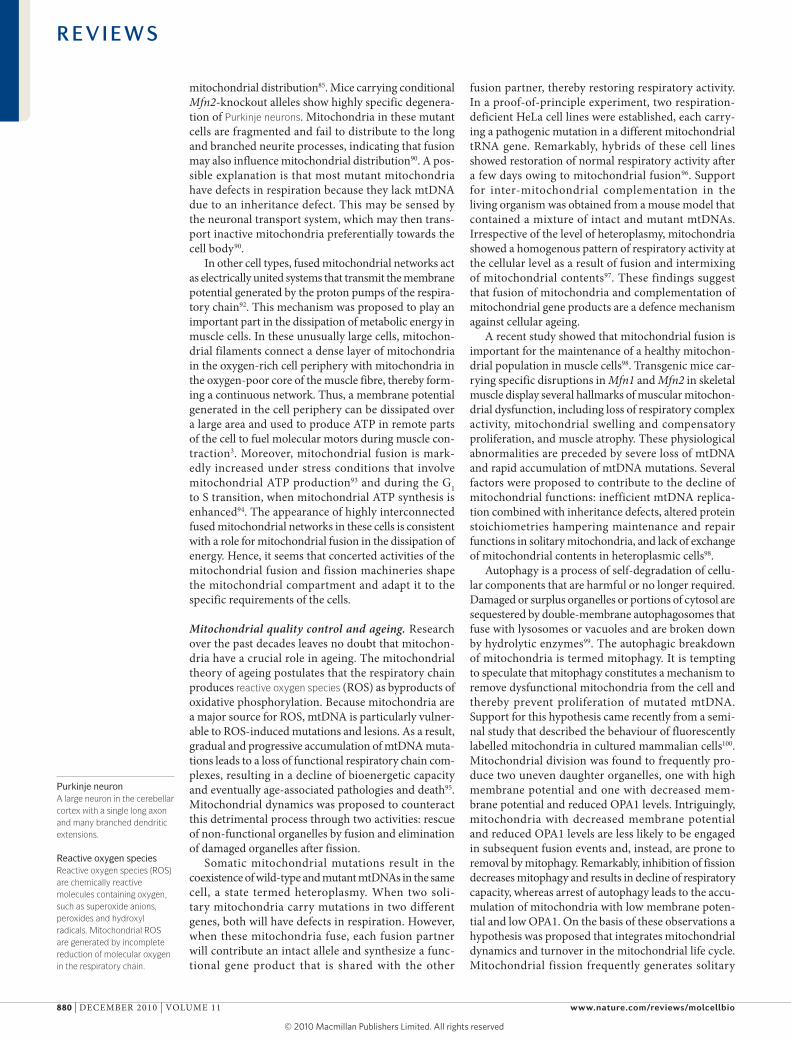
Table 2 | Human diseases associated with perturbations of mitochondrial dynamics
Molecular defect Disease Neuronal defect refs
Mutations in OPA1 that impede mitochondrial fusion
Autosomal dominant optic atrophy
Degeneration of retinal ganglion cells and optic nerve
111,112
Mutations in MFN2 that impede mitochondrial fusion
Charcot–Marie–Tooth disease type 2a
Degeneration of long peripheral sensory and motor neurons
113
Mutations in DRP1 that impede mitochondrial fission
Unnamed Abnormal brain development, optic atrophy and neonatal lethality
114
Mutations in PINK1 or PARK2 that disturb mitochondrial dynamics
Hereditary early onset Parkinson’s disease
Degeneration of dopaminergic neurons in the substantia nigra
117–120
DRP1, dynamin-related protein 1; MFN2, mitofusin 2; OPA1, optic atrophy protein 1; PARK2, Parkin.
R E V I E W S
NATuRe ReVIews | Molecular cell Biology VoluMe 11 | deceMBeR 2010 | 881
© 20 Macmillan Publishers Limited. All rights reserved10
1. Palade, G. E. An electron microscope study of the mitochondrial structure. J. Histochem. Cytochem. 1, 188–211 (1953).
2. Bereiter-Hahn, J. Behavior of mitochondria in the living cell. Int. Rev. Cytol. 122, 1–63 (1990).
3. Skulachev, V. P. Mitochondrial filaments and clusters as intracellular power-transmitting cables. Trends Biochem. Sci. 26, 23–29 (2001).
4. Collins, T. J., Berridge, M. J., Lipp, P. & Bootman, M. D. Mitochondria are morphologically and functionally heterogeneous within cells. EMBO J. 21, 1616–1627 (2002).
5. Detmer, S. A. & Chan, D. C. Functions and dysfunctions of mitochondrial dynamics. Nature Rev. Mol. Cell Biol. 8, 870–879 (2007).
6. Hoppins, S., Lackner, L. & Nunnari, J. The machines that divide and fuse mitochondria. Annu. Rev. Biochem. 76, 751–780 (2007).
7. Okamoto, K. & Shaw, J. M. Mitochondrial morphology and dynamics in yeast and multicellular eukaryotes. Annu. Rev. Genet. 39, 503–536 (2005).
8. Martens, S. & McMahon, H. T. Mechanisms of membrane fusion: disparate players and common principles. Nature Rev. Mol. Cell Biol. 9, 543–556 (2008).
9. Hales, K. G. & Fuller, M. T. Developmentally regulated mitochondrial fusion mediated by a conserved, novel, predicted GTPase. Cell 90, 121–129 (1997).Reports the identification of the first known mediator of mitochondrial fusion and, at the same time, describes the role of fusion in remodelling of mitochondria during spermatogenesis.
10. Rapaport, D., Brunner, M., Neupert, W. & Westermann, B. Fzo1p is a mitochondrial outer membrane protein essential for the biogenesis of functional mitochondria in Saccharomyces cerevisiae. J. Biol. Chem. 273, 20150–20155 (1998).
11. Hermann, G. J. et al. Mitochondrial fusion in yeast requires the transmembrane GTPase Fzo1p. J. Cell Biol. 143, 359–373 (1998).
12. Kanazawa, T. et al. The C. elegans Opa1 homologue EAT-3 is essential for resistance to free radicals. PLoS Genet. 4, e1000022 (2008).
13. Santel, A. & Fuller, M. T. Control of mitochondrial morphology by a human mitofusin. J. Cell Sci. 114, 867–874 (2001).
14. Fritz, S., Rapaport, D., Klanner, E., Neupert, W. & Westermann, B. Connection of the mitochondrial outer and inner membranes by Fzo1 is critical for organellar fusion. J. Cell Biol. 152, 683–692 (2001).
15. Rojo, M., Legros, F., Chateau, D. & Lombes, A. Membrane topology and mitochondrial targeting of mitofusins, ubiquitous mammalian homologs of the transmembrane GTPase Fzo. J. Cell Sci. 115, 1663–1674 (2002).
16. Meeusen, S. et al. Mitochondrial inner-membrane fusion and crista maintenance requires the dynamin-related GTPase Mgm1. Cell 127, 383–395 (2006).
17. Herlan, M., Vogel, F., Bornhövd, C., Neupert, W. & Reichert, A. S. Processing of Mgm1 by the rhomboid-type protease Pcp1 is required for maintenance of mitochondrial morphology and of mitochondrial DNA. J. Biol. Chem. 278, 27781–27788 (2003).
18. McQuibban, G. A., Saurya, S. & Freeman, M. Mitochondrial membrane remodelling regulated by a conserved rhomboid protease. Nature 423, 537–541 (2003).
19. Cipolat, S., Martins de Brito, O., Dal Zilio, B. & Scorrano, L. OPA1 requires mitofusin 1 to promote mitochondrial fusion. Proc. Natl Acad. Sci. USA 101, 15927–15932 (2004).
20. Yarosh, W. et al. The molecular mechanisms of OPA1-mediated optic atrophy in Drosophila model and prospects for antioxidant treatment. PLoS Genet. 4, e6 (2008).
21. Cipolat, S. et al. Mitochondrial rhomboid PARL regulates cytochrome c release during apoptosis via OPA1-dependent cristae remodeling. Cell 126, 163–175 (2006).
22. Griparic, L., Kanazawa, T. & van der Bliek, A. M. Regulation of the mitochondrial dynamin-like protein Opa1 by proteolytic cleavage. J. Cell Biol. 178, 757–764 (2007).
23. Ishihara, N., Fujita, Y., Oka, T. & Mihara, K. Regulation of mitochondrial morphology through proteolytic cleavage of OPA1. EMBO J. 25, 2966–2977 (2006).
24. Ehses, S. et al. Regulation of OPA1 processing and mitochondrial fusion by m-AAA protease isoenzymes and OMA1. J. Cell Biol. 187, 1023–1036 (2009).
25. Song, Z., Chen, H., Fiket, M., Alexander, C. & Chan, D. C. OPA1 processing controls mitochondrial fusion and is regulated by mRNA splicing, membrane potential, and Yme1L. J. Cell Biol. 178, 749–755 (2007).
26. Head, B., Griparic, L., Amiri, M., Gandre-Babbe, S. & van der Bliek, A. M. Inducible proteolytic inactivation of OPA1 mediated by the OMA1 protease in mammalian cells. J. Cell Biol. 187, 959–966 (2009).
27. Wong, E. D. et al. The dynamin-related GTPase, Mgm1p, is an intermembrane space protein required for maintenance of fusion competent mitochondria. J. Cell Biol. 151, 341–352 (2000).
28. Chen, H. et al. Mitofusins Mfn1 and Mfn2 coordinately regulate mitochondrial fusion and are essential for embryonic development. J. Cell Biol. 160, 189–200 (2003).Demonstrates that both mammalian mitofusin isoforms are required to promote mitochondrial fusion and play essential parts in development.
29. Olichon, A. et al. Loss of OPA1 perturbates the mitochondrial inner membrane structure and integrity, leading to cytochrome c release and apoptosis. J. Biol. Chem. 278, 7743–7746 (2003).
30. Meeusen, S., McCaffery, J. M. & Nunnari, J. Mitochondrial fusion intermediates revealed in vitro. Science 305, 1747–1752 (2004).Describes an in vitro assay that was used to dissect distinct steps of mitochondrial double-membrane fusion.
31. Koshiba, T. et al. Structural basis of mitochondrial tethering by mitofusin complexes. Science 305, 858–862 (2004).
32. DeVay, R. M. et al. Coassembly of Mgm1 isoforms requires cardiolipin and mediates mitochondrial inner membrane fusion. J. Cell Biol. 186, 793–803 (2009).
33. Malka, F. et al. Separate fusion of outer and inner mitochondrial membranes. EMBO Rep. 6, 853–859 (2005).
34. Sesaki, H. & Jensen, R. E. UGO1 encodes an outer membrane protein required for mitochondrial fusion. J. Cell Biol. 152, 1123–1134 (2001).
35. Hoppins, S., Horner, J., Song, C., McCaffery, J. M. & Nunnari, J. Mitochondrial outer and inner membrane fusion requires a modified carrier protein. J. Cell Biol. 184, 569–581 (2009).
36. Sesaki, H. & Jensen, R. E. Ugo1p links the Fzo1p and Mgm1p GTPases for mitochondrial fusion. J. Biol. Chem. 279, 28298–28303 (2004).
37. Praefcke, G. J. & McMahon, H. T. The dynamin superfamily: universal membrane tubulation and fission molecules? Nature Rev. Mol. Cell Biol. 5, 133–147 (2004).
38. Smirnowa, E., Shurland, D. L., Ryazantsev, S. N. & van der Bliek, A. M. A human dynamin-related protein controls the distribution of mitochondria. J. Cell Biol. 143, 351–358 (1998).
coordinate their activities in common biochemical path-ways, such as β-oxidation of fatty acids or scavenging of peroxides.
It has been known for a long time from electron micrographs that mitochondria are often closely associ-ated with the endoplasmic reticulum (eR). surprisingly, one of the two mammalian mitofusin isoforms, MFN2, was found to form a tether between the eR and mito-chondria. eR-located MFN2 is engaged in complexes with mitochondrial MFN1 or MFN2, thereby bridging the two organelles and bringing them close together to allow communication by ca2+ signalling125. The MFN2-dependent eR tether also has a role in the for-mation of autophagosomes. Mitochondria contribute membrane lipids to newly formed autophagosomes during starvation. As the mitochondrial mass remains constant during this process, the mitochondrion–eR tether is thought to ensure that mitochondrial lipids are immediately replenished from the eR. consistent with this, loss of MFN2 markedly impairs starvation-induced autophagy126. It therefore seems that mito-chondrial behaviour is interwoven with many cellular pathways, so studies have to consider the possibility that other organelles may also be affected by changes in mitochondrial dynamics.
PerspectivesResearch in the past 15 years led to the identification of the key proteins of mitochondrial fusion and fission in yeast and animals. In particular, comprehensive screens of yeast mutant collections have now systematically ana-lysed almost 90% of the yeast genes for a role in mito-chondrial distribution and morphology48,127. Thus, we can be confident that most of the conserved core com-ponents are known. The next challenge is to mechanisti-cally dissect these molecular machineries to learn how they achieve fusion and fission of a double membrane-bound organelle. Assembly and regulation of mitofusins and dRP1 are apparently much more complex in mam-malian cells than in yeast. Future studies will yield fur-ther insights into the cellular pathways that adapt the shape of the mitochondrial compartment to the needs of differentiated cells. A picture emerges that the main function of mitochondrial dynamics is to ensure proper inheritance and distribution of mitochondria and to maintain them in a healthy state. we now have to deter-mine the roles of the mediators of mitochondrial fusion and fission, and how dysfunctions contribute to disease mechanisms. Thus, the investigation of mitochondrial dynamics will remain an exciting field of research in the coming years.
R E V I E W S
882 | deceMBeR 2010 | VoluMe 11 www.nature.com/reviews/molcellbio
© 20 Macmillan Publishers Limited. All rights reserved10
39. Otsuga, D. et al. The dynamin-related GTPase, Dnm1p, controls mitochondrial morphology in yeast. J. Cell Biol. 143, 333–349 (1998).
40. Mozdy, A., McCaffery, J. M. & Shaw, J. M. Dnm1p GTPase-mediated mitochondrial fusion is a multi-step process requiring the novel integral membrane component Fis1p. J. Cell Biol. 151, 367–379 (2000).
41. Tieu, Q. & Nunnari, J. Mdv1p is a WD repeat protein that interacts with the dynamin-related GTPase, Dnm1p, to trigger mitochondrial division. J. Cell Biol. 151, 353–365 (2000).
42. Zhang, Y. & Chan, D. C. Structural basis for recruitment of mitochondrial fission complexes by Fis1. Proc. Natl Acad. Sci. USA 104, 18526–18530 (2007).
43. Tieu, Q., Okreglak, V., Naylor, K. & Nunnari, J. The WD repeat protein, Mdv1p, functions as a molecular adaptor by interacting with Dnm1p and Fis1p during mitochondrial fission. J. Cell Biol. 158, 445–452 (2002).
44. Lackner, L. L., Horner, J. S. & Nunnari, J. Mechanistic analysis of a dynamin effector. Science 325, 874–877 (2009).
45. Ingerman, E. et al. Dnm1 forms spirals that are structurally tailored to fit mitochondria. J. Cell Biol. 170, 1021–1027 (2005).References 44 and 45 report in vitro studies providing mechanistic insights into the self-assembly of Dnm1 into helical structures that are thought to drive membrane scission.
46. Griffin, E. E., Graumann, J. & Chan, D. C. The WD40 protein Caf4p is a component of the mitochondrial fission machinery and recruits Dnm1p to mitochondria. J. Cell Biol. 170, 237–248 (2005).
47. Schauss, A. C., Bewersdorf, J. & Jakobs, S. Fis1p and Caf4p, but not Mdv1p, determine the polar localization of Dnm1p clusters on the mitochondrial surface. J. Cell Sci. 119, 3098–3106 (2006).
48. Dimmer, K. S. et al. Genetic basis of mitochondrial function and morphology in Saccharomyces cerevisiae. Mol. Biol. Cell 13, 847–853 (2002).Describes a systematic, genome-wide screen in yeast that led to the discovery of several genes affecting mitochondrial dynamics, including MDM30, MDM33, MDM36, NUM1 and PCP1.
49. Cerveny, K. L., Studer, S. L., Jensen, R. E. & Sesaki, H. Yeast mitochondrial division and distribution require the cortical Num1 protein. Dev. Cell 12, 363–375 (2007).
50. Hammermeister, M., Schödel, K. & Westermann, B. Mdm36 is a mitochondrial fission-promoting protein in Saccharomyces cerevisiae. Mol. Biol. Cell 21, 2443–2452 (2010).
51. Roux, A., Uyhazi, K., Frost, A. & De Camilli, P. GTP-dependent twisting of dynamin implicates constriction and tension in membrane fission. Nature 441, 528–531 (2006).
52. Yoon, Y., Krueger, E. W., Oswald, B. J. & McNiven, M. A. The mitochondrial protein hFis1 regulates mitochondrial fission in mammalian cells through an interaction with the dynamin-like protein DLP1. Mol. Cell. Biol. 23, 5409–5420 (2003).
53. James, D. I., Parone, P. A., Mattenberger, Y. & Martinou, J. C. hFis1, a novel component of the mammalian mitochondrial fission machinery. J. Biol. Chem. 278, 36373–36379 (2003).
54. Lee, Y. J., Jeong, S. Y., Karbowski, M., Smith, C. L. & Youle, R. J. Roles of the mammalian mitochondrial fission and fusion mediators Fis1, Drp1, and Opa1 in apoptosis. Mol. Biol. Cell 15, 5001–5011 (2004).
55. Breckenridge, D. G. et al. Caenorhabditis elegans drp-1 and fis-2 regulate distinct cell-death execution pathways downstream of ced-3 and independent of ced-9. Mol. Cell 31, 586–597 (2008).
56. Gandre-Babbe, S. & van der Bliek, A. M. The novel tail-anchored membrane protein Mff controls mitochondrial and peroxisomal fission in mammalian cells. Mol. Biol. Cell 19, 2402–2412 (2008).
57. Labrousse, A. M., Zappaterra, M. D., Rube, D. A. & van der Bliek, A. M. C. elegans dynamin-related protein DRP-1 controls severing of the mitochondrial outer membrane. Mol. Cell 4, 815–826 (1999).
58. Legesse-Miller, A., Massol, R. H. & Kirchhausen, T. Constriction and Dnm1p recruitment are distinct processes in mitochondrial fission. Mol. Biol. Cell 14, 1953–1963 (2003).
59. Jakobs, S. et al. Spatial and temporal dynamics of budding yeast mitochondria lacking the division component Fis1p. J. Cell Sci. 116, 2005–2014 (2003).
60. Messerschmitt, M. et al. The inner membrane protein Mdm33 controls mitochondrial morphology in yeast. J. Cell Biol. 160, 553–564 (2003).
61. Tondera, D. et al. The mitochondrial protein MTP18 contributes to mitochondrial fission in mammalian cells. J. Cell Sci. 118, 3049–3059 (2005).
62. Sesaki, H. & Jensen, R. E. Division versus fusion: Dnm1p and Fzo1p antagonistically regulate mitochondrial shape. J. Cell Biol. 147, 699–706 (1999).
63. Bleazard, W. et al. The dynamin-related GTPase Dnm1 regulates mitochondrial fission in yeast. Nature Cell Biol. 1, 298–304 (1999).References 62 and 63 show in yeast that fusion and fission are antagonistic and balanced activities that are both required for the maintenance of mitochondrial morphology.
64. Fritz, S., Weinbach, N. & Westermann, B. Mdm30 is an F-box protein required for maintenance of fusion-competent mitochondria in yeast. Mol. Biol. Cell 14, 2303–2313 (2003).
65. Cohen, M. M., Leboucher, G. P., Livnat-Levanon, N., Glickman, M. H. & Weissman, A. M. Ubiquitin-proteasome-dependent degradation of a mitofusin, a critical regulator of mitochondrial fusion. Mol. Biol. Cell 19, 2457–2464 (2008).
66. Amiott, E. A., Cohen, M. M., Saint-Georges, Y., Weissman, A. M. & Shaw, J. M. A mutation associated with CMT2A neuropathy causes defects in Fzo1 GTP hydrolysis, ubiquitylation, and protein turnover. Mol. Biol. Cell 20, 5026–5035 (2009).
67. Herlan, M., Bornhövd, C., Hell, K., Neupert, W. & Reichert, A. S. Alternative topogenesis of Mgm1 and mitochondrial morphology depend on ATP and a functional import motor. J. Cell Biol. 165, 167–173 (2004).Formulation of an attractive hypothesis that suggests that alternative processing of Mgm1 could adapt mitochondrial fusion rates to matrix ATP levels.
68. Baricault, L. et al. OPA1 cleavage depends on decreased mitochondrial ATP level and bivalent metals. Exp. Cell Res. 313, 3800–3808 (2007).
69. Nakamura, N., Kimura, Y., Tokuda, M., Honda, S. & Hirose, S. MARCH-V is a novel mitofusin 2- and Drp1-binding protein able to change mitochondrial morphology. EMBO Rep. 7, 1019–1022 (2006).
70. Karbowski, M., Neutzner, A. & Youle, R. J. The mitochondrial E3 ubiquitin ligase MARCH5 is required for Drp1 dependent mitochondrial division. J. Cell Biol. 178, 71–84 (2007).
71. Yonashiro, R. et al. A novel mitochondrial ubiquitin ligase plays a critical role in mitochondrial dynamics. EMBO J. 25, 3618–3626 (2006).
72. Park, Y. Y. et al. Loss of MARCH5 mitochondrial E3 ubiquitin ligase induces cellular senescence through dynamin-related protein 1 and mitofusin 1. J. Cell Sci. 123, 619–626 (2010).
73. Braschi, E., Zunino, R. & McBride, H. M. MAPL is a new mitochondrial SUMO E3 ligase that regulates mitochondrial fission. EMBO Rep. 10, 748–754 (2009).
74. Zunino, R., Schauss, A., Rippstein, P., Andrade-Navarro, M. & McBride, H. M. The SUMO protease SENP5 is required to maintain mitochondrial morphology and function. J. Cell Sci. 120, 1178–1188 (2007).
75. Taguchi, N., Ishihara, N., Jofuku, A., Oka, T. & Mihara, K. Mitotic phosphorylation of dynamin-related GTPase Drp1 participates in mitochondrial fission. J. Biol. Chem. 282, 11521–11529 (2007).
76. Cribbs, J. T. & Strack, S. Reversible phosphorylation of Drp1 by cyclic AMP-dependent protein kinase and calcineurin regulates mitochondrial fission and cell death. EMBO Rep. 8, 939–944 (2007).
77. Han, X. J. et al. CaM kinase I α-induced phosphorylation of Drp1 regulates mitochondrial morphology. J. Cell Biol. 182, 573–585 (2008).
78. Cereghetti, G. M. et al. Dephosphorylation by calcineurin regulates translocation of Drp1 to mitochondria. Proc. Natl Acad. Sci. USA 105, 15803–15808 (2008).
79. Eura, Y., Ishihara, N., Oka, T. & Mihara, K. Identification of a novel protein that regulates mitochondrial fusion by modulating mitofusin (Mfn) protein function. J. Cell Sci. 119, 4913–4925 (2006).
80. Liesa, M. et al. Mitochondrial fusion is increased by the nuclear coactivator PGC-1β. PLoS One 3, e3613 (2008).
81. Karbowski, M., Norris, K. L., Cleland, M. M., Jeong, S. Y. & Youle, R. J. Role of Bax and Bak in mitochondrial morphogenesis. Nature 443, 658–662 (2006).
82. Chen, X. J. & Butow, R. A. The organization and inheritance of the mitochondrial genome. Nature Rev. Genet. 6, 815–825 (2005).
83. Pfanner, N. & Geissler, A. Versatility of the mitochondrial protein import machinery. Nature Rev. Mol. Cell. Biol. 2, 339–349 (2001).
84. Neupert, W. & Herrmann, J. M. Translocation of proteins into mitochondria. Annu. Rev. Biochem. 76, 723–749 (2007).
85. Ishihara, N. et al. Mitochondrial fission factor Drp1 is essential for embryonic development and synapse formation in mice. Nature Cell Biol. 11, 958–966 (2009).
86. Altmann, K., Frank, M., Neumann, D., Jakobs, S. & Westermann, B. The class V myosin motor protein, Myo2, plays a major role in mitochondrial motility in Saccharomyces cerevisiae. J. Cell Biol. 181, 119–130 (2008).
87. Wakabayashi, J. et al. The dynamin-related GTPase Drp1 is required for embryonic and brain development in mice. J. Cell Biol. 186, 805–816 (2009).References 85 and 87 show that DRP1-dependent fission and distribution of mitochondria is essential in embryonic and brain development in mice.
88. Merz, S. & Westermann, B. Genome-wide deletion mutant analysis reveals genes required for respiratory growth, mitochondrial genome maintenance and mitochondrial protein synthesis in Saccharomyces cerevisiae. Genome Biol. 10, R95 (2009).
89. Chen, H., Chomyn, A. & Chan, D. C. Disruption of fusion results in mitochondrial heterogeneity and dysfunction. J. Biol. Chem. 280, 26185–26192 (2005).
90. Chen, H., McCaffery, J. M. & Chan, D. C. Mitochondrial fusion protects against neurodegeneration in the cerebellum. Cell 130, 548–562 (2007).
91. Li, Z., Okamoto, K., Hayashi, Y. & Sheng, M. The importance of dendritic mitochondria in the morphogenesis and plasticity of spines and synapses. Cell 119, 873–887 (2004).References 90 and 91 show that mitochondrial dynamics is crucial for neuronal development and function.
92. Amchenkova, A. A., Bakeeva, L. E., Chentsov, Y. S., Skulachev, V. P. & Zorov, D. B. Coupling membranes as energy-transmitting cables. I. Filamentous mitochondria in fibroblasts and mitochondrial clusters in cardiomyocytes. J. Cell Biol. 107, 481–495 (1988).
93. Tondera, D. et al. SLP-2 is required for stress-induced mitochondrial hyperfusion. EMBO J. 28, 1589–1600 (2009).
94. Mitra, K., Wunder, C., Roysam, B., Lin, G. & Lippincott-Schwartz, J. A hyperfused mitochondrial state achieved at G1-S regulates cyclin E buildup and entry into S phase. Proc. Natl Acad. Sci. USA 106, 11960–11965 (2009).
95. Balaban, R. S., Nemoto, S. & Finkel, T. Mitochondria, oxidants, and aging. Cell 120, 483–495 (2005).
96. Ono, T., Isobe, K., Nakada, K. & Hayashi, J. I. Human cells are protected from mitochondrial dysfunction by complementation of DNA products in fused mitochondria. Nature Genet. 28, 272–275 (2001).Shows that complementation of gene products in fused mitochondria restores respiratory activity in heteroplasmic cells.
97. Nakada, K. et al. Inter-mitochondrial complementation: mitochondria-specific system preventing mice from expression of disease phenotypes by mutant mtDNA. Nature Med. 7, 934–940 (2001).
98. Chen, H. et al. Mitochondrial fusion is required for mtDNA stability in skeletal muscle and tolerance of mtDNA mutations. Cell 141, 280–289 (2010).Shows that mitochondrial fusion is required for the maintenance of functional mitochondria in muscle cells.
99. He, C. & Klionsky, D. J. Regulation mechanisms and signaling pathways of autophagy. Annu. Rev. Genet. 43, 67–93 (2009).
100. Twig, G. et al. Fission and selective fusion govern mitochondrial segregation and elimination by autophagy. EMBO J. 27, 433–446 (2008).Suggests a functional connection between mitochondrial dynamics and the removal of impaired mitochondria by mitophagy.
R E V I E W S
NATuRe ReVIews | Molecular cell Biology VoluMe 11 | deceMBeR 2010 | 883
© 20 Macmillan Publishers Limited. All rights reserved10
101. Youle, R. J. & Karbowski, M. Mitochondrial fission in apoptosis. Nature Rev. Mol. Cell Biol. 6, 657–663 (2005).
102. Suen, D. F., Norris, K. L. & Youle, R. J. Mitochondrial dynamics and apoptosis. Genes Dev. 22, 1577–1590 (2008).
103. Fannjiang, Y. et al. Mitochondrial fission proteins regulate programmed cell death in yeast. Genes Dev. 18, 2785–2797 (2004).
104. Jagasia, R., Grote, P., Westermann, B. & Conradt, B. DRP-1-mediated mitochondrial fragmentation during EGL-1-induced cell death in C. elegans. Nature 433, 754–760 (2005).
105. Goyal, G., Fell, B., Sarin, A., Youle, R. J. & Sriram, V. Role of mitochondrial remodeling in programmed cell death in Drosophila melanogaster. Dev. Cell 12, 807–816 (2007).
106. Frank, S. et al. The role of dynamin-related protein 1, a mediator of mitochondrial fission, in apoptosis. Dev. Cell 1, 515–525 (2001).First report demonstrating a role for DRP1 in apoptosis.
107. Brooks, C. et al. Bak regulates mitochondrial morphology and pathology during apoptosis by interacting with mitofusins. Proc. Natl Acad. Sci. USA 104, 11649–11654 (2007).
108. Karbowski, M. et al. Spatial and temporal association of Bax with mitochondrial fission sites, Drp1, and Mfn2 during apoptosis. J. Cell Biol. 159, 931–938 (2002).
109. Wasiak, S., Zunino, R. & McBride, H. M. Bax/Bak promote sumoylation of DRP1 and its stable association with mitochondria during apoptotic cell death. J. Cell Biol. 177, 439–450 (2007).
110. Knott, A. B., Perkins, G., Schwarzenbacher, R. & Bossy-Wetzel, E. Mitochondrial fragmentation in neurodegeneration. Nature Rev. Neurosci. 9, 505–518 (2008).
111. Alexander, C. et al. OPA1, encoding a dynamin-related GTPase, is mutated in autosomal dominant atrophy linked to chromosome 3q28. Nature Genet. 26, 211–215 (2000).
112. Delettre, C. et al. Nuclear gene OPA1, encoding a mitochondrial dynamin-related protein, is mutated in dominant optic atrophy. Nature Genet. 26, 207–210 (2000).
113. Züchner, S. et al. Mutations in the mitochondrial GTPase mitofusin 2 cause Charcot-Marie-Tooth neuropathy type 2A. Nature Genet. 36, 449–451 (2004).
114. Waterham, H. R. et al. A lethal defect of mitochondrial and peroxisomal fission. N. Engl. J. Med. 356, 1736–1741 (2007).
115. Narendra, D. P. et al. PINK1 is selectively stabilized on impaired mitochondria to activate Parkin. PLoS Biol. 8, e1000298 (2010).
116. Matsuda, N. et al. PINK1 stabilized by mitochondrial depolarization recruits Parkin to damaged mitochondria and activates latent Parkin for mitophagy. J. Cell Biol. 189, 211–221 (2010).
117. Deng, H., Dodson, M. W., Huang, H. & Guo, M. The Parkinson’s disease genes pink1 and parkin promote mitochondrial fission and/or inhibit fusion in Drosophila. Proc. Natl Acad. Sci. USA 105, 14503–14508 (2008).
118. Yang, Y. et al. Pink1 regulates mitochondrial dynamics through interaction with the fission/fusion machinery. Proc. Natl Acad. Sci. USA 105, 7070–7075 (2008).
119. Ziviani, E., Tao, R. N. & Whitworth, A. J. Drosophila parkin requires PINK1 for mitochondrial translocation and ubiquitinates mitofusin. Proc. Natl Acad. Sci. USA 107, 5018–5023 (2010).
120. Lutz, A. K. et al. Loss of parkin or PINK1 function increases Drp1-dependent mitochondrial fragmentation. J. Biol. Chem. 284, 22938–22951 (2009).
121. Kuravi, K. et al. Dynamin-related proteins Vps1p and Dnm1p control peroxisome abundance in Saccharomyces cerevisiae. J. Cell Sci. 119, 3994–4001 (2006).
122. Koch, A. et al. Dynamin-like protein 1 is involved in peroxisomal fission. J. Biol. Chem. 278, 8597–8605 (2003).
123. Koch, A., Yoon, Y., Bonekamp, N. A., McNiven, M. A. & Schrader, M. A role for Fis1 in both mitochondrial and peroxisomal fission in mammalian cells. Mol. Biol. Cell 16, 5077–5086 (2005).
124. Neuspiel, M. et al. Cargo-selected transport from the mitochondria to peroxisomes is mediated by vesicular carriers. Curr. Biol. 18, 102–108 (2008).
125. Martins de Brito, O. & Scorrano, L. Mitofusin 2 tethers endoplasmic reticulum to mitochondria. Nature 456, 605–610 (2008).
126. Hailey, D. W. et al. Mitochondria supply membranes for autophagosome biogenesis during starvation. Cell 141, 656–667 (2010).
127. Altmann, K. & Westermann, B. Role of essential genes in mitochondrial morphogenesis in
Saccharomyces cerevisiae. Mol. Biol. Cell 16, 5410–5417 (2005).
128. Arimura, S., Yamamoto, J., Aida, G. P., Nakazono, M. & Tsutsumi, N. Frequent fusion and fission of plant mitochondria with unequal nucleoid distribution. Proc. Natl Acad. Sci. USA 101, 7805–7808 (2004).
129. Arimura, S. & Tsutsumi, N. A dynamin-like protein (ADL2b), rather than FtsZ, is involved in Arabidopsis mitochondrial division. Proc. Natl Acad. Sci. USA 99, 5727–5731 (2002).
130. Logan, D. C., Scott, I. & Tobin, A. K. ADL2a, like ADL2b, is involved in the control of higher plant mitochondrial morphology. J. Exp. Bot. 55, 783–785 (2004).
131. Scott, I., Tobin, A. K. & Logan, D. C. BIGYIN, an orthologue of human and yeast FIS1 genes functions in the control of mitochondrial size and number in Arabidopsis thaliana. J. Exp. Bot. 57, 1275–1280 (2006).
132. Arimura, S. et al. Arabidopsis elongated mitochondria1 is required for localization of dynamin-related protein 3a to mitochondrial fission sites. Plant Cell 20, 1555–1566 (2008).
133. Beech, P. L. et al. Mitochondrial FtsZ in a chromophyte alga. Science 287, 1276–1279 (2000).
134. Takahara, M. et al. A putative mitochondrial ftsZ gene is present in the unicellular primitive red alga Cyanidioschyzon merolae. Mol. Gen. Genet. 264, 452–460 (2000).
AcknowledgementsThe author thanks S. Arimura, J. Förtsch, S. Geimer and S. Jakobs for the contribution of images, and J. Förtsch, J. Herrmann, T. Klecker and S. Geimer for comments on the manuscript. Research in the author’s laboratory is supported by grants from the Deutsche Forschungsgemeinschaft.
Competing interests statement The authors declare no competing financial interests.
FURTHER INFORMATIONBenedikt Westermann’s homepage: http://www.zellbiologie.uni-bayreuth.de
all liNks are active iN the oNliNe PDf
R E V I E W S
884 | deceMBeR 2010 | VoluMe 11 www.nature.com/reviews/molcellbio
© 20 Macmillan Publishers Limited. All rights reserved10