Embed Size (px)
Citation preview

German Edition: DOI: 10.1002/ange.201900723Molecular RecognitionInternational Edition: DOI: 10.1002/anie.201900723
Minimizing the Entropy Penalty for Ligand Binding: Lessons from theMolecular Recognition of the Histo Blood-Group Antigens by HumanGalectin-3Ana Gimeno, Sandra Delgado, Pablo Valverde, Sara Bertuzzi, Manuel Alvaro Berb&s,Javier Echavarren, Alessandra Lacetera, Sonsoles Mart&n-Santamar&a, Avadhesha Surolia,Francisco Javier CaÇada, Jesus Jim8nez-Barbero,* and Ana Ard#*
Abstract: Ligand conformational entropy plays an importantrole in carbohydrate recognition events. Glycans are charac-terized by intrinsic flexibility around the glycosidic linkages,thus in most cases, loss of conformational entropy of the sugarupon complex formation strongly affects the entropy of thebinding process. By employing a multidisciplinary approachcombining structural, conformational, binding energy, andkinetic information, we investigated the role of conformationalentropy in the recognition of the histo blood-group antigens Aand B by human galectin-3, a lectin of biomedical interest. Weshow that these rigid natural antigens are pre-organized ligandsfor hGal-3, and that restriction of the conformational flexibilityby the branched fucose (Fuc) residue modulates the thermo-dynamics and kinetics of the binding process. These resultshighlight the importance of glycan flexibility and provideinspiration for the design of high-affinity ligands as antagonistsfor lectins.
Molecular recognition of glycans by lectins is essentialbiological processes with major implications for health anddisease.[1] From the chemical perspective, these interactionsare generally weak, and except for isolated cases,[2] they arecharacterized by a thermodynamic profile in which thefavorable binding enthalpy compensates for an unfavorableentropic contribution. Indeed, despite the exo-anomericeffect,[3] which strongly influences the conformational prefer-ences around the glycosidic linkages, the chemical nature ofthese glycosidic linkages endows glycans with high flexibility.The binding event usually takes place with a loss of conforma-tional entropy, especially for the ligand,[4] and as a conse-quence, large entropic penalties are generally observed.[5,6]
Attempts to manipulate this thermodynamic balance havefocused on the design of pre-organized ligands. However,chemically induced modifications have rather unpredictablethermodynamic consequences and in fact, most of theseefforts have failed[7] by following the paradigm of theenthalpy/entropy compensation mechanism.[8]
Herein, we report the study of the molecular recognitionof the A and B blood-group tetrasaccharide antigens (A- andB-BGA) by the carbohydrate recognition domain of humangalectin-3 (hGal-3 CRD). This lectin has been related toseveral diseases, and it is actually a target for the developmentof high-affinity ligands as antagonists for the treatment ofcertain cancers and fibrotic diseases.[9] A- and B-BGAs havebeen reported to be Gal-3 binders,[10] and to bind withincreased affinity compared to lactose.[11] However, the basisfor this stronger interaction has not been clarified. In thiswork, we investigated this binding interaction by usinga combination of NMR, ITC experiments, and MD simula-tions and the results demonstrate that sugar flexibility acts asa modulator of the kinetics and thermodynamics of thebinding event. Specifically, the fucose (Fuc) moiety does notdirectly interact with the lectin, but preorganizes the ligand inthe bound conformation. Interestingly, the presence of theFuc moiety has been previously shown to restrict theconformational mobility of branched oligosaccharides,including BGA and Lex antigens.[12] The preorganization ofthe ligand in the bound geometry eliminates the conforma-tional selection event for complex formation, lowers theenergy barrier for the association, increases the on-rate andimpacts the binding entropy. As a consequence, the fucosy-lated tetrasaccharides bind more strongly than their trisac-
[*] Dr. A. Gimeno, S. Delgado, P. Valverde, S. Bertuzzi,Prof. Dr. J. Jim8nez-Barbero, Dr. A. Ard#CIC bioGUNE, Bizkaia Technology ParkBuilding 800, 48160 Derio, Bizkaia (Spain)E-mail: [email protected]
Dr. M. A. Berb&s, J. Echavarren, Dr. A. Lacetera,Dr. S. Mart&n-Santamar&a, Dr. F. J. CaÇadaCentro de Investigaciones Biolkgicas-CSICRamiro de Maeztu 9, 28040 Madrid (Spain)
Prof. Dr. A. SuroliaIndian Institute of Science, Bangalore-560012 (India)
Prof. Dr. J. Jim8nez-BarberoIkerbasque, Basque Foundation for ScienceMaria Diaz de Haro 3, 48013 Bilbao, Bizkaia (Spain)
andDepartment of Organic ChemistryII Faculty of Science and TechnologyUniversity of the Basque Country, EHU-UPVLeioa (Spain)
Supporting information and the ORCID identification number(s) forthe author(s) of this article can be found under:https://doi.org/10.1002/anie.201900723.
T 2019 The Authors. Published by Wiley-VCH Verlag GmbH & Co.KGaA. This is an open access article under the terms of the CreativeCommons Attribution-NonCommercial-NoDerivs License, whichpermits use and distribution in any medium, provided the originalwork is properly cited, the use is non-commercial and no modifica-tions or adaptations are made.
AngewandteChemieCommunications
7268 T 2019 The Authors. Published by Wiley-VCH Verlag GmbH & Co. KGaA, Weinheim Angew. Chem. Int. Ed. 2019, 58, 7268 –7272

charide parent analogues, thus making BGA/hGal-3 an idealsystem to unmask the commonly hidden contribution ofligand conformational entropy in binding events.
The structural details of the interaction between N-acetyllactosamine (LacNAc, 1; Scheme 1) and LacNAc-derivedglycomimetics with hGal-3 are well documented.[13] The b-Galresidue provides the key hydrogen-bonding and p-stackinginteractions, while the other moieties provide additionalstabilization to the complex. The type II A- and B-BGAs (4and 5) are LacNAc derivatives with Gala1-3 and Fuca1-2terminal glycosylations. Thus, the binding of trisaccharides 2and 3 compared to that of tetrasaccharides 4 and 5 wasanalyzed to dissect the effect of the different structuralelements.
1H-STD-NMR experiments[14] showed strong saturation-transfer difference (STD) intensities for H4, H5, and H6 ofthe b-Gal residue for the four ligands (2–5), as expected. For3–5, STDs were also observed for H1 and H2 of aGal oraGalNAc residues. On the contrary, no STD was observed forthe protons of the Fuc moiety in ligands 2, 4, and 5 (Figure 1and Figures S1–S4).
These data indicate that the four ligands share a commonbinding mode that is fully consistent with that described forLacNAc, where the bGal residue is the key binding ele-ment.[13a] The aGal/aGalNAc residues in 3, 4, and 5 provideadditional contacts with the lectin. By contrast, the Fuc is fullyexposed to the solvent, without establishing any direct contactwith the lectin.
Additional structural information was obtained throughthe chemical-shift perturbations induced by ligands 1–5 in the1H-15N heteronuclear single quantum coherence (HSQC)spectra of 15N-hGal-3. Interestingly, the addition of 10 equiv-alents of the Fuc-containing 2 to hGal-3 produced basicallythe same backbone chemical-shift perturbations as LacNAc(Figure 2), involving residues in the His158-Arg186 region.Thus, the presence of the a1-2 Fuc in 2 does not affect anyadditional lectin site. In contrast, for 3–5, which containterminal Gala and GalNAca, additional cross-peaks wereperturbed, corresponding to amino acids located in b-strandsS3 and S4 (Figure 2 and Figures S7–S9). Furthermore, theaddition of sub-stoichiometric amounts of 3–5 to hGal-3evidenced two sets of 1H-15N signals for specific amino acidsof the lectin, corresponding to its free and bound forms(Figure S10). The rate of the chemical exchange between thefree and bound forms of hGal-3 is now slow in the NMRchemical-shift timescale. This behavior differs from thetitrations with ligands 1 and 2, for which progressive cross-peak signal displacement indicated a binding event in the fast-exchange regime.[15]
Thus, both STD and HSQC experiments agree on showingthat all ligands (2–5) share the same binding mode asLacNAc. The additional aGal/aGalNAc units are located atthe so-called subsite B,[16] participating in direct contacts withresidues in the b-strand S3, while the Fuc moiety in 2, 4, and 5is exposed to the solvent. Molecular dynamics (MD) simu-lations for each complex built according to the NMR datawere stable along the 100 ns run (Figure 2C and theSupporting Information).
The thermodynamic parameters for the binding of 1–5 tohGal-3 were obtained by isothermal titration calorimetry(ITC; Table 1, Figure 3). The dissociation constants (KD) forlactose and LacNAc were similar to those reported,[17] withthe typical negative enthalpy counteracted by a positiveentropy.[18] The addition of a terminal aGal residue incompound 3 increased the affinity, essentially due to theenthalpic contribution, which is in agreement with theproposed 3D model that showed additional contacts for thisresidue. The introduction of the a1-2 linked Fuc moiety in 2,4, and 5 also improved the binding affinity, as previouslyreported.[10] However, in this case, the enthalpy was lower incomparison with the non-fucosylated counterparts, and theimproved affinity actually arises from the entropic term, withthe unfavorable entropy contribution being significantlysmaller. The drop in the TDS term is especially remarkablefor tetrasaccharides 4 and 5. These results contrast withpreviously reported thermodynamic studies, which proposedthe existence of enhanced enthalpy contributions to thebinding.[11]
Finally, the binding kinetics for the interaction of glycans3–5 with hGal-3 were characterized by taking advantage of
Figure 1. A) 1H-STD-NMR spectra and epitope map for the interactionof 4 with hGal-3. B) STD epitope mapping of sugars 2, 3, and 5.
Scheme 1. Glycan structures used in this study. Yellow circle: galactose(Gal), blue square: glucosamine (GlcNAc), red triangle: fucose (Fuc),yellow square: galactosamine (GalNAc).
AngewandteChemieCommunications
7269Angew. Chem. Int. Ed. 2019, 58, 7268 –7272 T 2019 The Authors. Published by Wiley-VCH Verlag GmbH & Co. KGaA, Weinheim www.angewandte.org
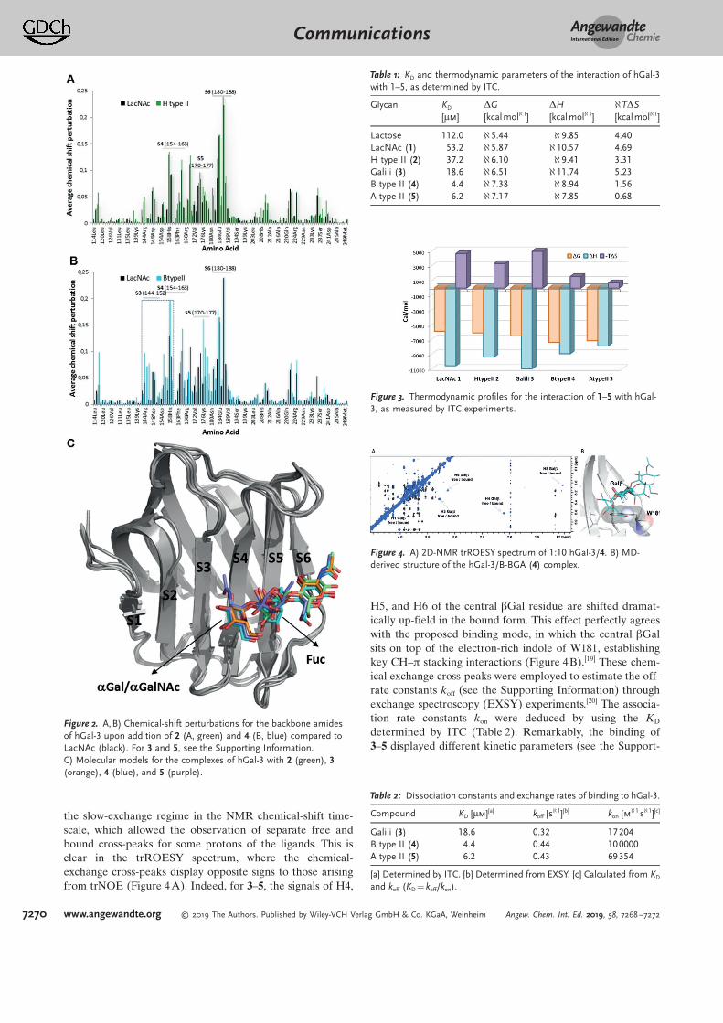
the slow-exchange regime in the NMR chemical-shift time-scale, which allowed the observation of separate free andbound cross-peaks for some protons of the ligands. This isclear in the trROESY spectrum, where the chemical-exchange cross-peaks display opposite signs to those arisingfrom trNOE (Figure 4A). Indeed, for 3–5, the signals of H4,
H5, and H6 of the central bGal residue are shifted dramat-ically up-field in the bound form. This effect perfectly agreeswith the proposed binding mode, in which the central bGalsits on top of the electron-rich indole of W181, establishingkey CH–p stacking interactions (Figure 4B).[19] These chem-ical exchange cross-peaks were employed to estimate the off-rate constants koff (see the Supporting Information) throughexchange spectroscopy (EXSY) experiments.[20] The associa-tion rate constants kon were deduced by using the KD
determined by ITC (Table 2). Remarkably, the binding of3–5 displayed different kinetic parameters (see the Support-
Figure 2. A,B) Chemical-shift perturbations for the backbone amidesof hGal-3 upon addition of 2 (A, green) and 4 (B, blue) compared toLacNAc (black). For 3 and 5, see the Supporting Information.C) Molecular models for the complexes of hGal-3 with 2 (green), 3(orange), 4 (blue), and 5 (purple).
Figure 3. Thermodynamic profiles for the interaction of 1–5 with hGal-3, as measured by ITC experiments.
Figure 4. A) 2D-NMR trROESY spectrum of 1:10 hGal-3/4. B) MD-derived structure of the hGal-3/B-BGA (4) complex.
Table 1: KD and thermodynamic parameters of the interaction of hGal-3with 1–5, as determined by ITC.
Glycan KD
[mm]DG[kcalmol@1]
DH[kcalmol@1]
@TDS[kcal mol@1]
Lactose 112.0 @5.44 @9.85 4.40LacNAc (1) 53.2 @5.87 @10.57 4.69H type II (2) 37.2 @6.10 @9.41 3.31Galili (3) 18.6 @6.51 @11.74 5.23B type II (4) 4.4 @7.38 @8.94 1.56A type II (5) 6.2 @7.17 @7.85 0.68
Table 2: Dissociation constants and exchange rates of binding to hGal-3.
Compound KD [mm][a] koff [s@1][b] kon [m@1 s@1][c]
Galili (3) 18.6 0.32 17204B type II (4) 4.4 0.44 100000A type II (5) 6.2 0.43 69354
[a] Determined by ITC. [b] Determined from EXSY. [c] Calculated from KD
and koff (KD = koff/kon).
AngewandteChemieCommunications
7270 www.angewandte.org T 2019 The Authors. Published by Wiley-VCH Verlag GmbH & Co. KGaA, Weinheim Angew. Chem. Int. Ed. 2019, 58, 7268 –7272

ing Information). Glycans 4 and 5 show similar off-rates to 3,but bind with a faster kon, thus reducing the KD.
Hence, in comparison with 3, the presence of the Fuc unitin 4 and 5 strongly reduces the kinetic barrier for ligandassociation. This is in fact the origin of the higher bindingaffinity, which is, in turn, directly related to the reducedbinding entropy.
It is well known that the presence of a1-2-Fuc reduces theinternal motion in ABO blood-group oligosaccharides.[12,21]
Our NMR and MD results (see the Supporting Informationfor details) agree with the existence of a single conforma-tional family for 2, 4 and 5. The situation is rather different forthe non-fucosylated trisaccharide 3, for which MD simula-tions predict that the Gala1-3Galb linkage is rather flexible,with continuous transitions along the entire trajectorybetween the syn-F/syn(@)-Y and syn-F/syn(++)-Y geometries(Figure 5B). In contrast, detailed conformational analysis forthe bound state showed single narrow minima for allglycosidic linkages. Thus, according to our results, trisacchar-ide 3 would acquire conformational stiffness upon complex-ation, which does not occur for the already preorganizedfucosylated ligands, which display the same conformation inthe free and bound states. In this way, the conformationalrestriction imposed by the Fuc in the branched sugarsprovides the proper presentation of the ligand epitope,thereby increasing the probability of successful collisionswhen they encounter the protein, and thus increasing the on-
rates. The energy barrier of theassociation event is then re-duced. Likewise, the minimalrigidification taking place uponbinding explains the minimalentropy penalty observed forthe fucosylated tetrasaccharides.
Therefore, this study high-lights the key contribution ofligand flexibility to both equilib-rium thermodynamics and bind-ing kinetics. This contribution isprobably always present, but itsclear participation is difficult toaddress. In this particular case,the chemical nature of the part-ners (the architecture of thebinding site for hGal-3 and therigidity of the histo blood anti-gens) has allowed us to bring tolight an effect that might beusually hidden.
In conclusion, conformation-ally restricted fucosylated A-and B-BGAs show faster associ-ation rates and less unfavorablebinding entrop than non-fucosy-lated analogues. As a key factor,the Fuc residue does not directlyinteract with the protein, butprovides the required preorgani-zation of the ligand to improve
the binding affinity. These features provide inspiration for thechemical design of high affinity ligands as antagonists forhGal-3 or for other lectins or receptors of biomedicalrelevance.
Acknowledgements
Avadhesha Surolia is a Science and Engineering ResearchBoard (SERB—India) Distinguished Fellow. We thankAgencia Estatal de Investigacion and ISCIIIof Spain and the European Research Council for financialsupport. The plasmid for hGal-3 was kindly provided by Dr.Filipa Marcelo (Universidade Nova Lisboa). We thank Dr.Sonia Huecas (CIB-CSIC) for discussions on ITC and Prof.H.-J. Gabius for discussions in early stages of this research.
Conflict of interest
The authors declare no conflict of interest.
Keywords: blood-group antigen · conformational entropy ·glycans · lectins · molecular recognition
How to cite: Angew. Chem. Int. Ed. 2019, 58, 7268–7272Angew. Chem. 2019, 131, 7346–7350
Figure 5. MD-based conformational analysis of 3–5 in the free and bound states.
AngewandteChemieCommunications
7271Angew. Chem. Int. Ed. 2019, 58, 7268 –7272 T 2019 The Authors. Published by Wiley-VCH Verlag GmbH & Co. KGaA, Weinheim www.angewandte.org

[1] R. D. Cummings, R. L. Schnaar, J. D. Esko, K. Drickamer, M. E.Taylor, Essentials of Glycobiology, 3rd ed. (Eds.: A. Varki, R. D.Cummings, J. D. Esko, P. Stanley, G. W. Hart, M. Aebi, A. G.Darvill, T. Kinoshita, N. H. Packer, J. H. Prestegard, R. L.Schnaar, P. H. Seeberger), Cold Spring Harbor LaboratoryPress, Cold Spring Harbor, 2015 – 2017.
[2] a) C. P. Sager, B. Fiege, P. Zihlmann, R. Vannam, S. Rabbani,R. P. Jakob, R. C. Preston, A. Zalewski, T. Maier, M. W. Peczuh,B. Ernst, Chem. Sci. 2018, 9, 646 – 654; b) F. P. C. Binder, K.Lemme, R. C. Preston, B. Ernst, Angew. Chem. Int. Ed. 2012, 51,7327 – 7331; Angew. Chem. 2012, 124, 7440 – 7444, and referencestherein; c) J. Topin, M. Lelimousin, J. Arnaud, A. Audfray, S.P8rez, A. Varrot, A. Imberty, ACS Chem. Biol. 2016, 11, 2011 –2020.
[3] a) I. Tvaroska, T. Bleha, Adv. Carbohydr. Chem. Biochem. 1989,47, 45 – 123; b) R. U. Lemieux, A. R. Morgan, Can. J. Chem.1965, 43, 2205 – 2213.
[4] M. L. Verteramo, O. Stenstrçm, M. M. Ignjatovic, O. Caldararu,M. A. Olsson, F. Manzoni, H. Leffler, E. Oksanen, D. T. Logan,U. J. Nilsson, U. Ryde, M. Akke, J. Am. Chem. Soc. 2019, 141,2012 – 2026.
[5] C. Diehl, O. Engstrçm, T. Delaine, M. H,kansson, S. Genheden,K. Modig, H. Leffler, U. Ryde, U. J. Nilsson, M. Akke, J. Am.Chem. Soc. 2010, 132, 14577 – 14589.
[6] J. P. Carver, Pure Appl. Chem. 1993, 65, 763 – 770.[7] N. Navarre, N. Amiot, A. Van Oijen, A. Imberty, A. Poveda, J.
Jim8nez-Barbero, A. Cooper, M. A. Nutley, G.-J. Boons, Chem.Eur. J. 1999, 5, 2281 – 2294.
[8] J. M. Fox, M. Zhao, M. J. Fink, K. Kang, G. M. Whitesides, Annu.Rev. Biophys. 2018, 47, 223 – 250.
[9] A. Girard, J. L. Magnani, Trends Glycosci. Glycotechnol. 2018,30, SE211 – SE220.
[10] S. R. Stowell, C. M. Arthur, P. Mehta, K. A. Slanina, O. Blixt, H.Leffler, D. F. Smith, R. D. Cummings, J. Biol. Chem. 2008, 283,10109 – 10123.
[11] K. Bachhawat-Sikdera, C. J. Thomasa, A. Surolia, FEBS Lett.2001, 500, 75 – 79.
[12] a) A. Imberty, S. P8rez, Chem. Rev. 2000, 100, 4567 – 4588; b) M.Zierke, M. Smiesko, S. Rabbani, T. Aeschbacher, B. Cutting,F. H.-T. Allain, M. Schubert, B. Ernst, J. Am. Chem. Soc. 2013,135, 13464 – 13472; c) M. D. Battistel, H. F. Azurmendi, M.Frank, D. I. Freedberg, J. Am. Chem. Soc. 2015, 137, 13444 –13447; d) T. Aeschbacher, M. Zierke, M. Smiesko, M. Collot, J.-
M. Mallet, B. Ernst, F. H.-T. Allain, M. Schubert, Chem. Eur. J.2017, 23, 11598 – 11610.
[13] a) P. Sçrme, P. Arnoux, B. Kahl-Knutsson, H. Leffler, J. M. Rini,U. J. Nilsson, J. Am. Chem. Soc. 2005, 127, 1737 – 1743; b) T.-J.Hsieh, H.-Y. Lin, Z. Tu, B.-S. Huang, S.-C. Wu, C.-H. Lin, PlosOne 2015, 10, e0125946; c) C. Atmanene, C. Ronin, S. Teletchea,F. M. Gautier, F. Djedaini-Pilard, F. Ciesielski, V. Vivat, C.Grandjean, Biochem. Biophys. Res. Commun. 2017, 489, 281 –286; d) J. Seetharaman, A. Kanigsberg, R. Slaaby, H. Leffler,S. H. Barondes, J. M. Rini, J. Biol. Chem. 1998, 273, 13047 –13052.
[14] a) M. Mayer, B. Meyer, J. Am. Chem. Soc. 2001, 123, 6108 – 6117;b) R. Marchetti, S. Perez, A. Arda, A. Imberty, J. Jimenez-Barbero, A. Silipo, A. Molinaro, ChemistryOpen 2016, 5, 274 –296; c) B. S. Blaum, U. Neu, T. Peters, T. Stehle, Acta Crystallogr.Sect. F 2018, 74, 451 – 462.
[15] M. P. Williamson, Prog. Nucl. Magn. Reson. Spectrosc. 2013, 73,1 – 16.
[16] H. Leffler, S. Carlsson, M. Hedlund, Y. Qian, F. Poirier,Glycoconjugate J. 2002, 19, 433 – 440.
[17] I. Cumpstey, E. Salomonsson, A. Sundin, H. Leffler, U. Nilsson,ChemBioChem 2007, 8, 1389 – 1398.
[18] The difference in binding affinity between lactose and LacNacwas due mainly to the enthalpic term. This is in agreement withthe structural differences found in the X-Ray crystallographicstructures of the complexes hGal-3/lactose and hGal-3/lacNAcdescribed in Ref. [13d].
[19] J. L. Asensio, A. Ard#, F. J. CaÇada, J. Jim8nez-Barbero, Acc.Chem. Res. 2013, 46, 946 – 954.
[20] M. P. Latham, G. R. Zimmermann, A. Pardi, J. Am. Chem. Soc.2009, 131, 5052 – 5053.
[21] a) R. U. Lemieux, K. Bock, L. T. J. Delbaere, S. Koto, V. S. R.Rao, Can. J. Chem. 1980, 58, 631 – 653; b) B. N. N. Rao, V. K.Dua, C. A. Bush, Biopolymers 1985, 24, 2207 – 2229; c) C. A.Bush, Z.-Y. Yan, B. N. N. Rao, J. Am. Chem. Soc. 1986, 108,6168 – 6173; d) L.-Y. Yan, C. A. Bush, Biopolymers 1990, 29,799 – 811; e) P. Cagas, C. A. Bush, Biopolymers 1992, 32, 277 –292; f) H. F. Azurmendi, C. A. Bush, Carbohydr. Res. 2002, 337,905 – 915.
Manuscript received: January 18, 2019Accepted manuscript online: April 3, 2019Version of record online: April 17, 2019
AngewandteChemieCommunications
7272 www.angewandte.org T 2019 The Authors. Published by Wiley-VCH Verlag GmbH & Co. KGaA, Weinheim Angew. Chem. Int. Ed. 2019, 58, 7268 –7272