Embed Size (px)
Citation preview

Mini-Symposium
Advances in Color Science: From Retina to Behavior
Bevil R. Conway,1,2 Soumya Chatterjee,2 Greg D. Field,3 Gregory D. Horwitz,4 Elizabeth N. Johnson,6 Kowa Koida,7
and Katherine Mancuso5
1Neuroscience Program, Wellesley College, Wellesley, Massachusetts 02481, 2Department of Neurobiology, Harvard Medical School, Boston, Massachusetts02115, 3Systems Neurobiology Laboratories, Salk Institute for Biological Studies, La Jolla, California 98037, Departments of 4Physiology & Biophysics, and5Ophthalmology, University of Washington, Seattle, Washington 98195, 6Duke Institute for Brain Sciences, Duke University, Durham, North Carolina27708, and 7Toyohashi University of Technology & National Institute for Physiological Sciences, Okazaki, Aichi 444-8585, Japan
Color has become a premier model system for understanding how information is processed by neural circuits, and for investigating therelationships among genes, neural circuits, and perception. Both the physical stimulus for color and the perceptual output experienced ascolor are quite well characterized, but the neural mechanisms that underlie the transformation from stimulus to perception are incom-pletely understood. The past several years have seen important scientific and technical advances that are changing our understanding ofthese mechanisms. Here, and in the accompanying minisymposium, we review the latest findings and hypotheses regarding colorcomputations in the retina, primary visual cortex, and higher-order visual areas, focusing on non-human primates, a model of humancolor vision.
In trichromatic primates, including humans and Old Worldmonkeys, there are three types of cone photoreceptors that areresponsible for color vision (Fig. 1A,B). The cone classes arecalled L, M, and S because of their spectral-sensitivity peaks,which lie in the long-, middle-, and short-wavelength regions ofthe visible spectrum. These labels replace the misleading terms“red,” “green,” and “blue.” Two physically distinct stimuli appearas different colors only if they produce different relative activa-tions in at least two cone types; conversely, any pair of physicallydistinct stimuli that activate the cone types in the same relativeamount appear the same, like the two yellows shown in Figure 1C.While photoreceptor responses are easily computed from thespectral distribution of the stimulus, there is no straightforwardrelationship between photoreceptor response and color (Hofer etal., 2005a; Shevell and Kingdom, 2008). The multitude of colorphenomena, including color afterimages, color assimilation,neon-color spreading, color constancy, and colored shadows, iscompelling because in many cases two physically identical stimuliare made to appear different colors, or two physically differentstimuli are made to appear the same simply by changing thespatial or temporal context (Fig. 2). A full description of theneural machinery for color should account for these observa-tions, as well as more cognitive phenomena involving the rela-
tionship between experience, language, memory, emotion andcolor. The neural basis of color has been reviewed previouslyfrom a range of perspectives (Gegenfurtner, 2003; Gegenfurtnerand Kiper, 2003; Lennie and Movshon, 2005; Sincich and Horton,2005; Solomon and Lennie, 2007; Conway, 2009; Dobkins, 2009;Jacobs and Nathans, 2009; Stockman and Brainard, 2010). Herewe focus on advances and pressing questions regarding the mech-anisms of color in retina, striate cortex, and extrastriate cortex ofnon-human primates, although we note that other species areemerging as excellent model systems of color processing (Lottoand Chittka, 2005; Van Hooser and Nelson, 2006; Osorio andVorobyev, 2008; Borst, 2009; Johnson et al., 2010; Srinivasan,2010).
Retinal mechanismsA single cone by itself is color blind because its activation dependson both the wavelength(s) and intensity of the stimulus. A com-parison of the signals from different classes of photoreceptors istherefore the most basic computational requirement of a color-vision system. The existence of cone-opponent retinal ganglioncells that perform such comparisons is well established in primate(Dacey and Packer, 2003; Jacobs, 2008). Cone-opponent retinalganglion cells respond with increased firing to an increment inactivation of one cone type (on-response) and to a decrement inactivation of a different cone type (off-response) (De Monasterioet al., 1975; Dacey and Lee, 1994). Cone-opponent retinal gan-glion cells come in four varieties: L-on/M-off, M-on/L-off, S-on/(L�M)-off, and (L�M)-on/S-off, although the receptive fields ofON cells often have markedly different sizes and temporal dy-namics compared with OFF cells, especially for cells receivingstrong S-cone input (Chichilnisky and Kalmar, 2002; Chatterjeeand Callaway, 2003; Conway and Livingstone, 2006; Tailby et al.,2008; Field et al., 2010). The four varieties of retinal ganglion cellswere originally thought to underlie the psychological finding offour unique hues (red, green, blue, and yellow) that are yoked by
Received Aug. 18, 2010; revised Sept. 22, 2010; accepted Sept. 23, 2010.This work was funded by National Science Foundation Grant 0918064, a Whitehall Foundation Award (B.R.C.),
and a Radcliffe Institute of Advanced Study Fellowship (B.R.C.); a McKnight Foundation Scholar’s Award and Na-tional Institutes of Health (NIH)-National Eye Institute (NEI) Grant EY018849 (G.D.H); the Helen Hay Whitney Foun-dation, NIH Grant EY13150, and NSF Grant PHY-0750525 (G.D.F.), NIH Grants R01EY016861, P30EY01730, andRR00166 (K.M.); Research to Prevent Blindness (K.M.); NIH Grants P30 EY12196 and F32 EY016287 (S.C.); andJapanese Grant-in-Aid for Scientific Research 16300103, Grant for Scientific Research on Priority Areas 17022040,and Japanese MEXT Grant-in-Aid for Young Scientists 20700299 (K.K.).
All authors contributed equally to this work.Correspondence should be addressed to Bevil R. Conway, Neuroscience Program, Wellesley College, 106 Central Street,
Wellesley, MA 02481. E-mail: [email protected]:10.1523/JNEUROSCI.4348-10.2010
Copyright © 2010 the authors 0270-6474/10/3014955-09$15.00/0
The Journal of Neuroscience, November 10, 2010 • 30(45):14955–14963 • 14955
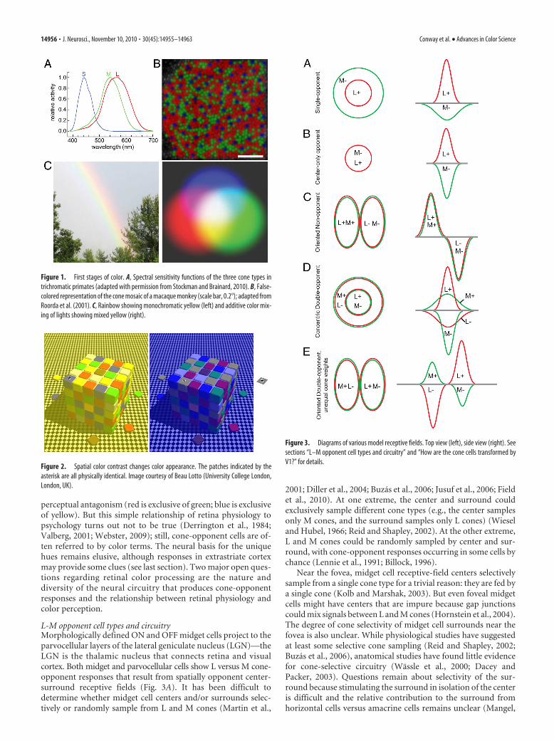
perceptual antagonism (red is exclusive of green; blue is exclusiveof yellow). But this simple relationship of retina physiology topsychology turns out not to be true (Derrington et al., 1984;Valberg, 2001; Webster, 2009); still, cone-opponent cells are of-ten referred to by color terms. The neural basis for the uniquehues remains elusive, although responses in extrastriate cortexmay provide some clues (see last section). Two major open ques-tions regarding retinal color processing are the nature anddiversity of the neural circuitry that produces cone-opponentresponses and the relationship between retinal physiology andcolor perception.
L-M opponent cell types and circuitryMorphologically defined ON and OFF midget cells project to theparvocellular layers of the lateral geniculate nucleus (LGN)—theLGN is the thalamic nucleus that connects retina and visualcortex. Both midget and parvocellular cells show L versus M cone-opponent responses that result from spatially opponent center-surround receptive fields (Fig. 3A). It has been difficult todetermine whether midget cell centers and/or surrounds selec-tively or randomly sample from L and M cones (Martin et al.,
2001; Diller et al., 2004; Buzas et al., 2006; Jusuf et al., 2006; Fieldet al., 2010). At one extreme, the center and surround couldexclusively sample different cone types (e.g., the center samplesonly M cones, and the surround samples only L cones) (Wieseland Hubel, 1966; Reid and Shapley, 2002). At the other extreme,L and M cones could be randomly sampled by center and sur-round, with cone-opponent responses occurring in some cells bychance (Lennie et al., 1991; Billock, 1996).
Near the fovea, midget cell receptive-field centers selectivelysample from a single cone type for a trivial reason: they are fed bya single cone (Kolb and Marshak, 2003). But even foveal midgetcells might have centers that are impure because gap junctionscould mix signals between L and M cones (Hornstein et al., 2004).The degree of cone selectivity of midget cell surrounds near thefovea is also unclear. While physiological studies have suggestedat least some selective cone sampling (Reid and Shapley, 2002;Buzas et al., 2006), anatomical studies have found little evidencefor cone-selective circuitry (Wassle et al., 2000; Dacey andPacker, 2003). Questions remain about selectivity of the sur-round because stimulating the surround in isolation of the centeris difficult and the relative contribution to the surround fromhorizontal cells versus amacrine cells remains unclear (Mangel,
Figure 1. First stages of color. A, Spectral sensitivity functions of the three cone types intrichromatic primates (adapted with permission from Stockman and Brainard, 2010). B, False-colored representation of the cone mosaic of a macaque monkey (scale bar, 0.2°); adapted fromRoorda et al. (2001). C, Rainbow showing monochromatic yellow (left) and additive color mix-ing of lights showing mixed yellow (right).
Figure 2. Spatial color contrast changes color appearance. The patches indicated by theasterisk are all physically identical. Image courtesy of Beau Lotto (University College London,London, UK).
Figure 3. Diagrams of various model receptive fields. Top view (left), side view (right). Seesections “L–M opponent cell types and circuitry” and “How are the cone cells transformed byV1?” for details.
14956 • J. Neurosci., November 10, 2010 • 30(45):14955–14963 Conway et al. • Advances in Color Science

1991; Cook and McReynolds, 1998; Ichinose and Lukasiewicz,2005; Davenport et al., 2008).
In the peripheral retina, where both the center and surroundof midget cells sample from multiple cones, the cone purity hasbeen similarly controversial (Martin et al., 2001; Jusuf et al.,2006). A recent study using receptive-field mapping at the reso-lution of single cones found small but consistent deviations fromrandom toward selective sampling in the receptive-field center,but not in the surround (Field et al., 2010). The mechanism andanatomical basis for this selectivity is uncertain. One might won-der whether spatial clumping of L and M cones by type couldcontribute (Roorda et al., 2001; Hofer et al., 2005b) (Fig. 1B), butrecent evidence suggests the contribution of cone clumping isminor (Field et al., 2010).
Do midget cells form the basic building blocks for the “red-green” color-vision circuit? Many investigators assume so, al-though the mismatch between the high density of midget cellsand the relatively low spatial acuity for color has raised somedoubts (Hubel and Livingstone, 1990; Calkins and Sterling,1999). Parasol cells can show L-M cone-opponent responses(Derrington et al., 1984; Lee and Sun, 2009), so they could playsome role in color vision, though this is controversial. Addition-ally, one of the many morphologically defined ”wide-field” reti-nal ganglion cells with low density may encode L-M opponentresponses that contribute to color vision (Dacey and Packer,2003); these cells may correspond to the still elusive red-greentype II cells alluded to by Wiesel and Hubel (1966) in their re-cordings of neurons located in the intercalated, or koniocellular(“K”), layers that separate the main layers of the LGN (Fig. 3B).
S versus L�M opponent cell types and circuitryThere are several types of retinal ganglion cells that oppose Ssignals against L�M. The small bistratified cell was the first mor-phologically identified “blue-yellow” cell (Dacey and Lee, 1994;Dacey and Packer, 2003), but there are others: the large bistrati-fied and giant monostratified cells (Dacey et al., 2003, 2005).These cell types project to the koniocellular layers of the LGN(Szmajda et al., 2008; Roy et al., 2009). In addition, OFF (but notON) midget cells in the central and peripheral retina also appearto carry S cone signals to the brain (Klug et al., 2003; Field et al.,2010; but see Lee et al., 2005; Sun et al., 2006). Unfortunately, therole of S versus L�M opponent retinal ganglion cells in blue-yellow color vision is all but clear (Derrington et al., 1984).
The circuitry producing cone-opponent responses in smallbistratified cells differs from that of midget cells (Dacey and Lee,1994; Crook et al., 2009). Small bistratified cells have dendrites intwo distinct strata of the inner plexiform layer. The inner den-drites receive input from ON-type bipolar cells that synapse ex-clusively with S cones. The outer dendrites receive input fromoff-type bipolar cells (Calkins et al., 1998; Grunert and Ghosh,1999; Percival et al., 2009) that carry L and M cone signals. In farperipheral retina and/or under low photopic conditions, the H2horizontal cell may also contribute to the (L�M)-off response(Field et al., 2007; Packer et al., 2010). Large bistratified cellsprobably have a circuitry similar to that of small bistratified cells(Dacey et al., 2003), and the circuitry of giant monostratified cells isyet to be determined.
Our understanding of the retinal mechanisms of color is far fromcomplete. The response properties of all �20 retinal ganglion celltypes need to be characterized to identify those with cone-opponentresponses. Also, the roles of excitation and inhibition in the innerand outer synaptic layers need to be clarified to understand howretinal circuitry produces cone opponency. Finally, more studies are
neededthatquantitativelyprobetherelationshipbetweenretinal signalsand color perception (Derrington et al., 1984; Kremers et al., 1992).
Retinal gene therapyUnlike Old World monkeys, most mammals are dichromats.New World monkeys, such as squirrel monkeys, have color visionthat is an evolutionary intermediate between dichromats andtrichromats (Jacobs, 2008). Instead of having L and M cone opsingenes, like humans and Old World monkeys, each New Worldmonkey has only a single X-chromosome opsin locus containingone of multiple alleles (Neitz et al., 1991). Males have only oneX-chromosome, so all New World male monkeys are dichromats.Heterozygous females who inherit different M/L alleles on eachX-chromosome express the pigments in different cone popula-tions due to random X-inactivation, producing trichromacy intwo thirds of females. Normal dichromatic and normal trichro-matic color vision coexist in these species, and the brain circuitryfor both is specified by the same genetic instructions. The ances-tors to humans and other Old World primates must have passedthrough an evolutionary stage in which the distribution of di-chromats and trichromats was similar to the New World pri-mates. Thus a single set of genetic instructions serves bothdichromacy and trichromacy, raising the possibility that no ad-ditional changes were required besides the addition of a thirdcone opsin. This would account for the observation that trans-genic mice, engineered to express three cone pigments, can showbehavioral trichromacy (Jacobs et al., 2007).
But does the red-green color-vision circuit require early visualexperience to become functional? This question was addressedusing gene therapy to add a third photopigment to the retinas ofadult dichromatic squirrel monkeys (Mancuso et al., 2009). Re-markably, this simple intervention was sufficient to confer trichro-matic color vision (Fig. 4). Because new color vision behaviorcorresponded in time to the appearance of robust transgene expres-sion levels, it seems that significant “rewiring” in the adult is notrequired. This result suggests that red-green color vision can beadded by taking advantage of preexisting neural circuitry (Mancusoet al., 2009; Shapley, 2009), that an early developmental process isnot required, and that trichromacy could have evolved in the ab-sence of any other change in the visual system except the addition ofa third cone type (Mancuso et al., 2010a).
The treated animals made all the usual discriminations madeby animals born with the genetic potential for trichromacy. Theydiscriminate reds from greens, and these colors from gray; and
Figure 4. Squirrel monkey performing a trichromatic color discrimination following genetherapy. Image courtesy of the Neitz laboratory.
Conway et al. • Advances in Color Science J. Neurosci., November 10, 2010 • 30(45):14955–14963 • 14957

they do not confuse blue and yellow with either red or green(Mancuso et al., 2010b). Thus, while the treated animals’ internalexperiences associated with stimulation of their new cone typeremain unknown, it is clear that reds, greens, blues, and yellowsare recognized as distinct sensations. This discovery opens inter-esting questions concerning the role of development and experi-ence in sculpting circuitry that mediates perception, includingproperties of the visual system that enable a new capacity to beadded after traditional critical periods have ended.
Striate cortex mechanismsThe cortical area that receives the largest projection from theLGN is primary visual cortex, also called V1 or striate cortex.Three outstanding questions continue to drive research intocolor processing in V1: Which neurons code color? How are thecone signals transformed by V1? And, is there a functional archi-tecture for color in V1?
Which V1 cells code color?Early investigations found that many V1 neurons are tuned forthe orientation and spatial frequency of black-and-white patterns(Fig. 3C), with few strongly selective for color (Hubel and Wiesel,1968; Schiller et al., 1976). The smaller population of stronglycolor-selective neurons, studied using broadband (white) lightpassed through colored filters, could be classified like LGN cellsin red/green or blue/yellow categories (Dow and Gouras, 1973;Gouras, 1974; Michael, 1978; Livingstone and Hubel, 1984; Vau-tin and Dow, 1985; Ts’o and Gilbert, 1988). These observationsmotivated the influential idea that color is processed by a special-ized subsystem that can be traced from retina to V1 (Livingstoneand Hubel, 1984).
But could the observation of distinct neuronal “types” (red/green and blue/yellow; color-coding and non-color-coding) inthese early studies be an artifact of the small set of stimuli used?Neurons that responded similarly to the colors tested might havebeen distinguished if tested with an alternate set, or a larger bat-tery of colors. In subsequent experiments, cathode ray tube mon-itors made it easy to present millions of colors, although only asmall subset of these could be included in an experiment of real-istic duration. Investigators needed a way to summarize colortuning succinctly and in a way that did not make assumptionsabout the number and types of chromatically distinct subpopu-lations present.
An important step was made by Derrington et al. (1984), whofound that the response of an LGN neuron to any stimulus couldbe predicted from a weighted sum of activity modulations in thethree cone types. This critical finding—that the LGN responsesare “linear”—meant that color tuning could be described con-cisely with a set of three numbers, which are the weights appliedto input from the three cone classes. On the basis of these coneweights, each LGN neuron could be assigned unambiguously toone of three clusters, providing justification for the classificationoriginally described by De Valois et al. (1966).
The cone weights of V1 neurons, however, appear to form acontinuum (Lennie et al., 1990; De Valois et al., 2000; Johnson etal., 2004; Solomon et al., 2004; Solomon and Lennie, 2005; Hor-witz et al., 2007). Chromatic signals from the LGN thus appear tobe mixed in V1 in myriad, perhaps arbitrary ways. But the anal-ysis of V1 cone weights is problematic in two key respects. First,cone weights do not indicate the degree to which a neuron is colorselective; it remains possible that only the most color-selectiveneurons relay color signals in V1 (Conway, 2001; Johnson et al.,2001; Wachtler et al., 2003; Horwitz et al., 2007). Second, the
concept of a “cone weight” is only meaningful if the response of aV1 neuron can be adequately described as a weighted sum of coneinputs. This assumption has come under scrutiny. Hanazawa etal. (2000) described V1 neurons whose color tuning was toosharp to result from a linear combination of cone signals. So-lomon et al. (2005) found that a divisive signal from the conescontributed significantly to the responses of some V1 neurons,and Horwitz et al. (2005) found that the responses of some cone-opponent V1 neurons were facilitated by luminance signals,which indicates a violation of linearity.
There is now consensus that many V1 neurons do not com-bine cone signals linearly, so we should not be surprised thatanalyses of cone weights give results that are difficult to interpret.We are now forced to reevaluate the original questions: whichcells in V1 code color, and might there be a specialized pathwayfor color processing in V1? Much of the evidence against this ideawas based on the fit of a model that describes the color tuning ofV1 neurons incompletely (Hanazawa et al., 2000; Solomon andLennie, 2005; Horwitz et al., 2007). An important future direc-tion is to revisit this question in a way that that makes fewerassumptions about how V1 neurons combine cone inputs.
How are the cone signals transformed by V1?Color contrast makes red appear redder when surrounded bygreen, while color constancy enables an object’s color to remainlargely constant as the illuminant changes (see Fig. 2). A flurry ofattention has been paid to mechanisms in V1 that may be respon-sible for these, and other, color–form interactions (Zhou et al.,2000; Gegenfurtner, 2001; Shapley and Hawken, 2002; Friedmanet al., 2003; Hurlbert, 2003, 2007; Kiper, 2003; Wachtler et al., 2003).This work has both reinvigorated and challenged the idea that color,or some aspect of it, may be handled by a specialized pathway.
The early discoveries of Hubel and Wiesel (1968) left a para-dox: if “form” is encoded by a discrete population of color-blindneurons, and “color” information by a separate population ofform-blind neurons, how does the brain encode color–form in-teractions required to account for color contrast and color con-stancy? Single-opponent cells (Fig. 3A) would be incapable ofcolor– contrast calculations because they are not spatially selec-tive for color: an “L-ON/M-OFF” single-opponent cell respondswell to spots, bars, or a full field of red, but not to a red spot on agreen background. One possible mechanism for encoding color–form interactions is through specialized “double-opponent” cells(Fig. 3D), first described in goldfish retina (Daw, 1967). Pressingquestions have centered on the existence, characterization, andwiring of such cells in primate V1.
To carry information expressly about color boundaries, it washypothesized that double-opponent cells in V1 would have per-fectly balanced cone inputs and concentrically organized centersand surrounds (Michael, 1978; Livingstone and Hubel, 1984).Such a cell would respond to a red spot on a green background,and not to luminance spots of any size. The concentric organiza-tion of their receptive fields would also mean that they would bespatially tuned, but not orientation selective. Double-opponentneurons fitting this description were reported in a few early studies(Hubel and Wiesel, 1968; Michael, 1978; Livingstone and Hubel,1984), but other studies with large samples of V1 cells found littleevidence of them (Thorell et al., 1984; Lennie et al., 1990).
New neurophysiological evidence shows that there is a signif-icant population of double-opponent cells in V1, although thesecells do not have all the originally hypothesized receptive-fieldproperties. The inputs to these cells are spatially segregated bycone type into excitatory and inhibitory receptive-field subunits
14958 • J. Neurosci., November 10, 2010 • 30(45):14955–14963 Conway et al. • Advances in Color Science

(Conway, 2001; Johnson et al., 2001, 2004, 2008; Conway et al.,2002; Conway and Livingstone, 2006), verifying that these neu-rons are both color-opponent and spatially opponent, but thecells frequently lack a concentric center–surround organization.Many double-opponent cells also respond to both color and lu-minance (Thorell et al., 1984; Conway, 2001; Johnson et al., 2001,2004, 2008; Conway and Livingstone, 2006; Horwitz et al., 2007).
A growing body of evidence shows that many double-opponent neurons are orientation-selective for both color andachromatic patterns, regardless of the constitution of cone input(Thorell et al., 1984; Conway, 2001; Johnson et al., 2001, 2008,2010; Conway et al., 2002; Heimel et al., 2005; Conway and Liv-ingstone, 2006; Horwitz et al., 2007). This transformation meansthat these cells respond to cues for form, such as boundaries andedges, and may take signals from color or black and white asneeded. Neurophysiological evidence that V1 neurons can beboth color-selective and orientation-selective is consistent withrecent functional magnetic resonance imaging (fMRI) and psy-chophysical studies in humans showing orientation-selective re-sponses to color stimuli (Beaudot and Mullen, 2005, 2006; Engel,2005; Huang et al., 2007; Sumner et al., 2008). The differencesfrom the originally proposed double-opponent cell necessitate arevision of the model (Fig. 3E).
Color likely depends on both single-opponent and double-opponent neurons, and on the further processing of their signals(Vladusich, 2007). Single-opponent cells would seem ideal forsignaling the color of a region covering the receptive field, whiledouble-opponent cells would be capable of signaling colorcontrast, color boundaries and edges, and contributing tocolor constancy. Future directions will involve developing rig-orous methods to test these predictions, building physiologi-cally relevant computational models, and establishing theextent to which these populations of neurons represent a dis-tinct specialized pathway.
Is there a functional organization for color in V1?Columns of cells with similar orientation tuning extend downthrough the vertical thickness of V1, and the orientation tuningof adjacent columns shifts gradually across the horizontal surface(Hubel and Wiesel, 1977), an architecture that has become moreprecisely understood as technology has advanced (Blasdel andSalama, 1986; Grinvald et al., 1986; Ohki et al., 2006). Pinningdown the functional organization of color in V1 has been morechallenging. The laminar projection of color-opponent signals
from LGN to V1 seems clear: L-M signals target the middle inputlayer (4C�), S-(L�M) (“Blue ON”) signals target the upper inputlayers (2/3 and 4A), and (L�M)-S (“Blue OFF”) signals are nar-rowly stratified in 4A (Lachica and Casagrande, 1992; Martin etal., 1997; Hendry and Reid, 2000; Chatterjee and Callaway, 2003;Casagrande et al., 2007). But there is no consensus on the hori-zontal organization of chromatic signals within V1. Livingstoneand Hubel (1984) showed that color cells tend to coincide withregions, called blobs, that stain for the enzyme cytochrome oxi-dase (Fig. 5A); this view was extended by Ts’o and Gilbert (1988),who suggested that some blobs were entirely red/green and othersentirely blue/yellow (Fig. 5B). But subsequent electrophysiologi-cal studies (Lennie et al., 1990; Leventhal et al., 1995; Friedman etal., 2003) called this architecture into question, suggesting noobvious clustering of color and no functional architecture relatedto blobs (Fig. 5C).
These contradictory results may not be surprising given thesampling limitations of extracellular recording and the challengeof aligning physiological recording sites with postmortem histo-chemical analysis. Intrinsic signal imaging, with its bird’s-eyeview of the cortical surface, rekindled the notion of color mapsorganized according to blobs. Landisman and Ts’o (2002) con-cluded that color-responsive regions are loosely associated withblobs, and that these regions are joined by color “bridges” span-ning adjacent blobs. Using a similar approach, Lu and Roe (2008)concluded that there is a tighter association between color-responsive regions and blobs. Finally, a recent optical-imagingstudy, which unfortunately did not provide histology, concludedthat color is functionally organized as an array of hue maps, witheach map containing a representation for multiple colors at over-lapping but slightly different spatial locations (Xiao et al., 2007).The low resolution of intrinsic optical imaging, however, makes itimpossible to determine how the signals map onto individualcells within neuronal ensembles.
The question of color architecture has been approached usingtwo-photon calcium imaging, which enables the activity of thou-sands of individual cells to be measured simultaneously. Prelim-inary results suggest that cone-opponent cells form clusters, andthat these clusters are in register with blobs (Chatterjee et al.,2008). The clusters appear to extend through upper layer 2/3,forming columns of chromatic selectivity. Moreover, these colorcolumns appear to be subdivided into regions of different chro-matic signatures, suggesting a “micromap” model (Fig. 5D) that
Figure 5. Alternative models for the organization of color-tuned neurons in V1. The gray patches depict cytochrome-oxidase blobs. Dots show color-tuned neurons that are clustered in blobs, butmixed randomly within each blob (A); clustered in blobs, and segregated according to color tuning within pure-color blobs (B); randomly arranged with respect to blobs (C); and clustered withinblobs and according to a micromap in which cells of similar tuning are adjacent within a given blob (D).
Conway et al. • Advances in Color Science J. Neurosci., November 10, 2010 • 30(45):14955–14963 • 14959

helps to explain discrepancies in previousobservations of color architecture. Thesetwo-photon imaging results reveal a pre-cise functional architecture for color andsupport the notion that V1 color signalsare processed by an anatomically segre-gated pathway, which may provide the ba-sis for a segregated output to extrastriateareas of the ventral stream.
Two-photon imaging allows the re-sponses of hundreds of neurons to bemeasured simultaneously; but, as dis-cussed in the previous sections, V1 neu-rons show variety in spatial and temporalreceptive-field organization, and many donot combine cone signals linearly. In theface of this heterogeneity, it is not trivialto determine stimulus parameters thatwould be suitable to adequately assesscolor tuning using two-photon imaging.It will be important, in future work, toestablish whether two-photon imagingcan provide an accurate measure of color tuning, perhaps bydirectly comparing measurements with those obtained usingsingle-unit recording.
Extrastriate cortex mechanismsOne heuristic of color processing (Fig. 6) holds that color signalsare transmitted along the ventral visual stream, from V1 throughsubcompartments of V2 referred to as the thin stripes (because oftheir pattern of staining with cytochrome oxidase), on to islandsof cortex dubbed “globs” located in V4 and posterior inferiortemporal cortex (PIT), and ultimately on to inferior temporalcortex (IT) (Desimone et al., 1985; Komatsu, 1998; Zeki andMarini, 1998; Conway et al., 2007; Matsumora et al., 2008;Harada et al., 2009; Yasuda et al., 2010). This simple hierarchicalmodel suggests that many color computations take place down-stream of V1. But compared with our understanding of retinaand V1, we know little about how extrastriate cortex contributesto color. Recent work using microelectrodes and fMRI-guidedmicroelectrode recordings has opened up new questions at theinterface of neurobiology and behavior (Conway et al., 2007;Koida and Komatsu, 2007; Matsumora et al., 2008; Stoughtonand Conway, 2008; Conway and Tsao, 2009).
IT is located in the temporal lobe of the monkey (anterior tothe ears), and can be divided into two parts (Iwai and Mishkin,1969): area TE, which includes anterior and central IT, and areaTEO, which includes PIT (Van Essen et al., 1990). The posteriorboundary of PIT adjoins area V4, although the boundary is im-precise—indeed the boundaries of all visual areas in the temporallobe are provisional (Brewer et al., 2002; Fize et al., 2003; Tootellet al., 2004; Stepniewska et al., 2005). Several studies have shownthat lesions or cooling of area TE impairs color discrimination(Dean, 1979; Horel, 1994; Heywood et al., 1995; Buckley et al.,1997), whereas lesions of other color-related areas (PIT and V4)cause little disruption in color discrimination (Heywood et al.,1998; Huxlin et al., 2000). In humans, rare lesions of ventraloccipital cortex can produce colorblindness, while sparing othervisual function (Bouvier and Engel, 2006), but the relationship ofthese regions, whose activity can be used to decode color (Brouwerand Heeger, 2009), to extrastriate regions in macaque monkeyremains controversial (Hadjikhani et al., 1998; Tootell and Had-jikhani, 1998; Zeki et al., 1998). Brain imaging, which has a higher
resolution in monkeys than in humans owing to the use of con-trast agents and the monkey’s smaller head, suggests that color ishandled by area TE along with a distributed network of globswithin PIT/V4, rather than a single entire extrastriate visual area(Conway and Tsao, 2006; Conway et al., 2007; Harada et al.,2009); this is consistent with anatomical and optical-imagingdata (Zeki and Shipp, 1989; DeYoe et al., 1994; Felleman et al.,1997; Ghose and Ts’o, 1997).
Neural recording studies have shown that many neurons in ITrespond selectively to color (Zeki, 1980; Desimone et al., 1984;Komatsu et al., 1992; Komatsu and Ideura, 1993; Kobatake andTanaka, 1994; Koida and Komatsu, 2007; Matsumora et al.,2008). These neurons are narrowly tuned to color and color sat-uration, and are concentrated in several subregions of IT, onelocated in TE and another in TEO (Conway et al., 2007; Yasuda etal., 2010). Neurons in TE show relatively weak shape selectivity(Matsumora et al., 2008), while neurons in the more posteriorregion show stronger shape selectivity (Conway et al., 2007;Yasuda et al., 2010).
The relationship between color perception and neuronal ac-tivity of anterior IT has recently been examined by Matsumora etal. (2008), who showed a significant correlation between the trial-to-trial fluctuations in neuronal responses of color-tuned neu-rons and the monkeys’ color judgment. They also found that thevariation in neural threshold across the color space correspondedwell with that of the behavioral threshold. Electrical stimulationof this region induces a large shift in the monkey’s color judg-ment, suggesting that neural activities are causally related to colorjudgment (Koida and Komatsu, 2008). Such a causal link is alsosuggested by recent microstimulation experiments in humansubjects (Murphey et al., 2008).
The ability to categorize stimuli by color is a fundamentalcognitive process. The population of neurons recorded in fMRI-identified globs of posterior IT shows a bias for the most satu-rated colors in a stimulus set, which also correspond to theelementary, or “unique” hue categories, suggesting a neural basisfor color categories (Stoughton and Conway, 2008; Conway andStoughton, 2009; Conway and Tsao, 2009; but see Mollon, 2009).Koida and Komatsu (2007) argue that IT neurons are involved incolor cognition by showing that IT neurons change firing ratewhen the monkey switches from a categorization to a discrimi-
Figure 6. Simple hierarchical model of color processing in the macaque cerebral cortex. Regions of cortex shown in gray, whichincrease in scale along the visual-processing hierarchy from V1 to TE, are implicated in color processing. Adapted from Conway(2009).
14960 • J. Neurosci., November 10, 2010 • 30(45):14955–14963 Conway et al. • Advances in Color Science

nation task; moreover, they found that a majority of neurons gavestronger responses when the animals performed a color-categorization task versus when they performed a color-discrimination task. Their results imply that IT neurons play animportant role in color categorization.
The color-selective neurons in extrastriate regions along theventral pathway must play an important role in color processing.With the refinement of physiological recording techniques, cou-pled with more sophisticated psychophysical stimulus para-digms, the specific computations performed by these neurons,the relationship of their activity to perception, and the neuralcircuitry that connects these cells to color cells in V1 and retina,should become clear.
ConclusionResearch using color vision as a model system has seen terrificadvances, offering the hope of a complete understanding ofthe transformation of color signals from retina to behavior. Theminisymposium that accompanies this review will showcase resultsthat aim to understand this transformation and the circuits thatbring it about. During the minisymposium, we will describe resultsfrom large retinal array recordings, in which the activity of hundredsof retinal ganglion cells is assessed simultaneously, affording the abil-ity to measure receptive fields of color-opponent cells at the resolu-tion of individual cones. We will go on to describe experimentsexploring gene therapy for red-green colorblindness, and the impli-cations of this research for theories of color vision. We will thendescribe results obtained using two-photon imaging, showing thefine-scale spatial organization of cone-opponent cells in V1. In ad-dition, using single-unit electrophysiological recordings and two-photon imaging, we will address how V1 neurons achieve colortuning. We will also discuss the spatial transformation of color sig-nals in V1 and the degree to which color is processed independentlyof other stimulus attributes like orientation. Finally, we will turn ourattention to extrastriate regions that likely mediate the consciousexperience of color, and address potential limits of animal models ofhuman color vision. Using this knowledge, we aim to enrich ourunderstanding of the relationship between genetics, neural circuits,perception, and behavior.
ReferencesBeaudot WH, Mullen KT (2005) Orientation selectivity in luminance and
color vision assessed using 2-d band-pass filtered spatial noise. Vision Res45:687– 696.
Beaudot WH, Mullen KT (2006) Orientation discrimination in human vi-sion: psychophysics and modeling. Vision Res 46:26 – 46.
Billock VA (1996) Consequences of retinal color coding for cortical colordecoding. Science 274:2118 –2119.
Blasdel GG, Salama G (1986) Voltage-sensitive dyes reveal a modular orga-nization in monkey striate cortex. Nature 321:579 –585.
Borst A (2009) Drosophila’s view on insect vision. Curr Biol 19:R36 –R47.Bouvier SE, Engel SA (2006) Behavioral deficits and cortical damage loci in
cerebral achromatopsia. Cereb Cortex 16:183–191.Brewer AA, Press WA, Logothetis NK, Wandell BA (2002) Visual areas in
macaque cortex measured using functional magnetic resonance imaging.J Neurosci 22:10416 –10426.
Brouwer GJ, Heeger DJ (2009) Decoding and reconstructing color fromresponses in human visual cortex. J Neurosci 29:13992–14003.
Buckley MJ, Gaffan D, Murray EA (1997) Functional double dissociationbetween two inferior temporal cortical areas: perirhinal cortex versusmiddle temporal gyrus. J Neurophysiol 77:587–598.
Buzas P, Blessing EM, Szmajda BA, Martin PR (2006) Specificity of M and Lcone inputs to receptive fields in the parvocellular pathway: random wir-ing with functional bias. J Neurosci 26:11148 –11161.
Calkins DJ, Sterling P (1999) Evidence that circuits for spatial and colorvision segregate at the first retinal synapse. Neuron 24:313–321.
Calkins DJ, Tsukamoto Y, Sterling P (1998) Microcircuitry and mosaic of ablue-yellow ganglion cell in the primate retina. J Neurosci 18:3373–3385.
Casagrande VA, Yazar F, Jones KD, Ding Y (2007) The morphology of thekoniocellular axon pathway in the macaque monkey. Cereb Cortex17:2334 –2345.
Chatterjee S, Callaway EM (2003) Parallel colour-opponent pathways toprimary visual cortex. Nature 426:668 – 671.
Chatterjee S, Ohki K, Reid RC (2008) Functional micro-architecture ofcolor selectivity in macaque primary visual cortex. Soc Neurosci Abstr34:666.15.
Chichilnisky EJ, Kalmar RS (2002) Functional asymmetries in ON and OFFganglion cells of primate retina. J Neurosci 22:2737–2747.
Conway BR (2001) Spatial structure of cone inputs to color cells in alertmacaque primary visual cortex (V-1). J Neurosci 21:2768 –2783.
Conway BR (2009) Color vision, cones, and color-coding in the cortex.Neuroscientist 15:274 –290.
Conway BR, Livingstone MS (2006) Spatial and temporal properties of conesignals in alert macaque primary visual cortex. J Neurosci 26:10826–10846.
Conway BR, Stoughton CM (2009) Towards a neural representation forunique hues. Curr Biol 19:R442–R443.
Conway BR, Tsao DY (2006) Color architecture in alert macaque cortexrevealed by FMRI. Cereb Cortex 16:1604 –1613.
Conway BR, Tsao DY (2009) Color-tuned neurons are spatially clusteredaccording to color preference within alert macaque posterior inferiortemporal cortex. Proc Natl Acad Sci U S A 106:18034 –18039.
Conway BR, Hubel DH, Livingstone MS (2002) Color contrast in macaqueV1. Cereb Cortex 12:915–925.
Conway BR, Moeller S, Tsao DY (2007) Specialized color modules in ma-caque extrastriate cortex. Neuron 56:560 –573.
Cook PB, McReynolds JS (1998) Lateral inhibition in the inner retina isimportant for spatial tuning of ganglion cells. Nat Neurosci 1:714 –719.
Crook JD, Davenport CM, Peterson BB, Packer OS, Detwiler PB, Dacey DM(2009) Parallel ON and OFF cone bipolar inputs establish spatially coex-tensive receptive field structure of blue-yellow ganglion cells in primateretina. J Neurosci 29:8372– 8387.
Dacey DM, Lee BB (1994) The ‘blue-on’ opponent pathway in primate ret-ina originates from a distinct bistratified ganglion cell type. Nature367:731–735.
Dacey DM, Packer OS (2003) Colour coding in the primate retina: diversecell types and cone-specific circuitry. Curr Opin Neurobiol 13:421– 427.
Dacey DM, Peterson BB, Robinson FR, Gamlin PD (2003) Fireworks in theprimate retina: in vitro photodynamics reveals diverse LGN-projectingganglion cell types. Neuron 37:15–27.
Dacey DM, Liao HW, Peterson BB, Robinson FR, Smith VC, Pokorny J, YauKW, Gamlin PD (2005) Melanopsin-expressing ganglion cells in pri-mate retina signal colour and irradiance and project to the LGN. Nature433:749 –754.
Davenport CM, Detwiler PB, Dacey DM (2008) Effects of pH buffering onhorizontal and ganglion cell light responses in primate retina: evidence forthe proton hypothesis of surround formation. J Neurosci 28:456 – 464.
Daw NW (1967) Goldfish retina: organization for simultaneous color con-trast. Science 158:942–944.
Dean P (1979) Visual cortex ablation and thresholds for successively pre-sented stimuli in rhesus monkeys: II. Hue. Exp Brain Res 35:69 – 83.
De Monasterio FM, Gouras P, Tolhurst DJ (1975) Concealed colour oppo-nency in ganglion cells of the rhesus monkey retina. J Physiol251:217–229.
Derrington AM, Krauskopf J, Lennie P (1984) Chromatic mechanisms inlateral geniculate nucleus of macaque. J Physiol 357:241–265.
Desimone R, Albright TD, Gross CG, Bruce C (1984) Stimulus-selectiveproperties of inferior temporal neurons in the macaque. J Neurosci4:2051–2062.
Desimone R, Schein SJ, Moran J, Ungerleider LG (1985) Contour, color andshape analysis beyond the striate cortex. Vision Res 25:441– 452.
De Valois RL, Abramov I, Jacobs GH (1966) Analysis of response patterns ofLGN cells. J Opt Soc Am 56:966 –977.
De Valois RL, Cottaris NP, Elfar SD, Mahon LE, Wilson JA (2000) Sometransformations of color information from lateral geniculate nucleus tostriate cortex. Proc Natl Acad Sci U S A 97:4997–5002.
DeYoe EA, Felleman DJ, Van Essen DC, McClendon E (1994) Multiple pro-cessing streams in occipitotemporal visual cortex. Nature 371:151–154.
Diller L, Packer OS, Verweij J, McMahon MJ, Williams DR, Dacey DM
Conway et al. • Advances in Color Science J. Neurosci., November 10, 2010 • 30(45):14955–14963 • 14961
(2004) L and M cone contributions to the midget and parasol ganglioncell receptive fields of macaque monkey retina. J Neurosci 24:1079 –1088.
Dobkins KR (2009) Does visual modularity increase over the course of de-velopment? Optom Vis Sci 86:E583–E588.
Dow BM, Gouras P (1973) Color and spatial specificity of single units inRhesus monkey foveal striate cortex. J Neurophysiol 36:79 –100.
Engel SA (2005) Adaptation of oriented and unoriented color-selective neu-rons in human visual areas. Neuron 45:613– 623.
Felleman DJ, Xiao Y, McClendon E (1997) Modular organization ofoccipito-temporal pathways: cortical connections between visual area 4and visual area 2 and posterior inferotemporal ventral area in macaquemonkeys. J Neurosci 17:3185–3200.
Field GD, Sher A, Gauthier JL, Greschner M, Shlens J, Litke AM, ChichilniskyEJ (2007) Spatial properties and functional organization of smallbistratified ganglion cells in primate retina. J Neurosci 27:13261–13272.
Field GD, Gauthier JL, Sher A, Greschner M, Machado TA, Jepson LH, ShlensJ, Gunning DE, Mathieson K, Dabrowski W, Paninski L, Litke AM, Chich-ilnisky EJ (2010) Functional connectivity in the retina at the resolutionof photoreceptors. Nature 467:673– 677.
Fize D, Vanduffel W, Nelissen K, Denys K, Chef d’Hotel C, Faugeras O, OrbanGA (2003) The retinotopic organization of primate dorsal V4 and sur-rounding areas: a functional magnetic resonance imaging study in awakemonkeys. J Neurosci 23:7395–7406.
Friedman HS, Zhou H, von der Heydt R (2003) The coding of uniformcolour figures in monkey visual cortex. J Physiol 548:593– 613.
Gegenfurtner K (2001) Color in the cortex revisited. Nat Neurosci4:339 –340.
Gegenfurtner KR (2003) Cortical mechanisms of colour vision. Nat RevNeurosci 4:563–572.
Gegenfurtner KR, Kiper DC (2003) Color vision. Annu Rev Neurosci26:181–206.
Ghose GM, Ts’o DY (1997) Form processing modules in primate area V4.J Neurophysiol 77:2191–2196.
Gouras P (1974) Opponent-colour cells in different layers of foveal striatecortex. J Physiol 238:583– 602.
Grinvald A, Lieke E, Frostig RD, Gilbert CD, Wiesel TN (1986) Functionalarchitecture of cortex revealed by optical imaging of intrinsic signals.Nature 324:361–364.
Grunert U, Ghosh KK (1999) Midget and parasol ganglion cells of the pri-mate retina express the alpha1 subunit of the glycine receptor. Vis Neu-rosci 16:957–966.
Hadjikhani N, Liu AK, Dale AM, Cavanagh P, Tootell RB (1998) Retinotopyand color sensitivity in human visual cortical area V8 [see comments].Nat Neurosci 1:235–241.
Hanazawa A, Komatsu H, Murakami I (2000) Neural selectivity for hue andsaturation of colour in the primary visual cortex of the monkey. EurJ Neurosci 12:1753–1763.
Harada T, Goda N, Ogawa T, Ito M, Toyoda H, Sadato N, Komatsu H (2009)Distribution of colour-selective activity in the monkey inferior temporalcortex revealed by functional magnetic resonance imaging. Eur J Neurosci30:1960 –1970.
Heimel JA, Van Hooser SD, Nelson SB (2005) Laminar organization of re-sponse properties in primary visual cortex of the gray squirrel (Sciuruscarolinensis). J Neurophysiol 94:3538 –3554.
Hendry SH, Reid RC (2000) The koniocellular pathway in primate vision.Annu Rev Neurosci 23:127–153.
Heywood CA, Gaffan D, Cowey A (1995) Cerebral achromatopsia in mon-keys. Eur J Neurosci 7:1064 –1073.
Heywood CA, Nicholas JJ, LeMare C, Cowey A (1998) The effect of lesionsto cortical areas V4 or AIT on pupillary responses to chromatic and ach-romatic stimuli in monkeys. Exp Brain Res 122:475– 480.
Hofer H, Singer B, Williams DR (2005a) Different sensations from coneswith the same photopigment. J Vis 5:444 – 454.
Hofer H, Carroll J, Neitz J, Neitz M, Williams DR (2005b) Organization ofthe human trichromatic cone mosaic. J Neurosci 25:9669 –9679.
Horel JA (1994) Retrieval of color and form during suppression of temporalcortex with cold. Behav Brain Res 65:165–172.
Hornstein EP, Verweij J, Schnapf JL (2004) Electrical coupling between redand green cones in primate retina. Nat Neurosci 7:745–750.
Horwitz GD, Chichilnisky EJ, Albright TD (2005) Blue-yellow signals are en-hanced by spatiotemporal luminance contrast in macaque V1. J Neuro-physiol 93:2263–2278.
Horwitz GD, Chichilnisky EJ, Albright TD (2007) Cone inputs to simple andcomplex cells in V1 of awake macaque. J Neurophysiol 97:3070–3081.
Huang PC, Mullen KT, Hess RF (2007) Collinear facilitation in color vision.J Vis 7:6 1–14.
Hubel D, Livingstone M (1990) Color puzzles. Cold Spring Harb SympQuant Biol 55:643– 649.
Hubel DH, Wiesel TN (1968) Receptive fields and functional architecture ofmonkey striate cortex. J Physiol 195:215–243.
Hubel DH, Wiesel TN (1977) Ferrier lecture. Functional architecture of ma-caque monkey visual cortex. Proc R Soc Lond B Biol Sci 198:1–59.
Hurlbert A (2003) Colour vision: primary visual cortex shows its influence.Curr Biol 13:R270 –R272.
Hurlbert A (2007) Colour constancy. Curr Biol 17:R906 –R907.Huxlin KR, Saunders RC, Marchionini D, Pham HA, Merigan WH (2000)
Perceptual deficits after lesions of inferotemporal cortex in macaques.Cereb Cortex 10:671– 683.
Ichinose T, Lukasiewicz PD (2005) Inner and outer retinal pathways bothcontribute to surround inhibition of salamander ganglion cells. J Physiol565:517–535.
Iwai E, Mishkin M (1969) Further evidence on the locus of the visual area inthe temporal lobe of the monkey. Exp Neurol 25:585–594.
Jacobs GH (2008) Primate color vision: a comparative perspective. Vis Neu-rosci 25:619 – 633.
Jacobs GH, Nathans J (2009) The evolution of Primate color vision. Sci Am300:56 – 63.
Jacobs GH, Williams GA, Cahill H, Nathans J (2007) Emergence of novelcolor vision in mice engineered to express a human cone photopigment.Science 315:1723–1725.
Johnson EN, Hawken MJ, Shapley R (2001) The spatial transformation ofcolor in the primary visual cortex of the macaque monkey. Nat Neurosci4:409 – 416.
Johnson EN, Hawken MJ, Shapley R (2004) Cone inputs in macaque pri-mary visual cortex. J Neurophysiol 91:2501–2514.
Johnson EN, Hawken MJ, Shapley R (2008) The orientation selectivity ofcolor-responsive neurons in macaque V1. J Neurosci 28:8096 – 8106.
Johnson EN, Van Hooser SD, Fitzpatrick D (2010) The representation ofseconds-cone signals in primary visual cortex. J Neurosci 30:10337–10350.
Jusuf PR, Martin PR, Grunert U (2006) Random wiring in the midget path-way of primate retina. J Neurosci 26:3908 –3917.
Kiper DC (2003) Colour and form in the early stages of cortical processing.J Physiol 548:335.
Klug K, Herr S, Ngo IT, Sterling P, Schein S (2003) Macaque retina containsan S-cone OFF midget pathway. J Neurosci 23:9881–9887.
Kobatake E, Tanaka K (1994) Neuronal selectivities to complex object fea-tures in the ventral visual pathway of the macaque cerebral cortex. J Neu-rophysiol 71:856 – 867.
Koida K, Komatsu H (2007) Effects of task demands on the responses of color-selective neurons in the inferior temporal cortex. Nat Neurosci 10:108–116.
Koida K, Komatsu H (2008) Impact on perceptual color judgments by mi-crostimulation of area TE. Soc Neurosci Abstr 34:853.2.
Kolb H, Marshak D (2003) The midget pathways of the primate retina. DocOphthalmol 106:67– 81.
Komatsu H (1998) Mechanisms of central color vision. Curr Opin Neuro-biol 8:503–508.
Komatsu H, Ideura Y (1993) Relationships between color, shape, and pat-tern selectivities of neurons in the inferior temporal cortex of the monkey.J Neurophysiol 70:677– 694.
Komatsu H, Ideura Y, Kaji S, Yamane S (1992) Color selectivity of neuronsin the inferior temporal cortex of the awake macaque monkey. J Neurosci12:408 – 424.
Kremers J, Lee BB, Kaiser PK (1992) Sensitivity of macaque retinal ganglioncells and human observers to combined luminance and chromatic tem-poral modulation. J Opt Soc Am A 9:1477–1485.
Lachica EA, Casagrande VA (1992) Direct W-like geniculate projections tothe cytochrome oxidase (CO) blobs in primate visual cortex: axon mor-phology. J Comp Neurol 319:141–158.
Landisman CE, Ts’o DY (2002) Color processing in macaque striate cortex:relationships to ocular dominance, cytochrome oxidase, and orientation.J Neurophysiol 87:3126 –3137.
Lee BB, Sun H (2009) The chromatic input to cells of the magnocellularpathway of primates. J Vis 9:15 11–18.
Lee SC, Telkes I, Grunert U (2005) S-cones do not contribute to the OFF-
14962 • J. Neurosci., November 10, 2010 • 30(45):14955–14963 Conway et al. • Advances in Color Science

midget pathway in the retina of the marmoset, Callithrix jacchus. EurJ Neurosci 22:437– 447.
Lennie P, Movshon JA (2005) Coding of color and form in the geniculostri-ate visual pathway (invited review). J Opt Soc Am A Opt Image Sci Vis22:2013–2033.
Lennie P, Krauskopf J, Sclar G (1990) Chromatic mechanisms in striatecortex of macaque. J Neurosci 10:649 – 669.
Lennie P, Haake, Williams DR (1991) The design of chromatically oppo-nent recetive fields. In: Computational models of visual processing(Landy MS, Movshon JA, eds) pp 71– 82. Cambridge, MA: MIT.
Leventhal AG, Thompson KG, Liu D, Zhou Y, Ault SJ (1995) Concomitantsensitivity to orientation, direction, and color of cells in layers 2, 3, and 4of monkey striate cortex. J Neurosci 15:1808 –1818.
Livingstone MS, Hubel DH (1984) Anatomy and physiology of a color sys-tem in the primate visual cortex. J Neurosci 4:309 –356.
Lotto RB, Chittka L (2005) Seeing the light: illumination as a contextual cue tocolor choice behavior in bumblebees. Proc Natl Acad Sci U S A 102:3852–3856.
Lu HD, Roe AW (2008) Functional organization of color domains in V1 andV2 of Macaque monkey revealed by optical imaging. Cereb Cortex18:516 –533.
Mancuso K, Hauswirth WW, Li Q, Connor TB, Kuchenbecker JA, MauckMC, Neitz J, Neitz M (2009) Gene therapy for red-green colour blind-ness in adult primates. Nature 461:784 –787.
Mancuso K, Mauck MC, Kuchenbecker JA, Neitz M, Neitz J (2010a) Amulti-stage color model revisited: implications for a gene therapy cure forred-green colorblindness. Adv Exp Med Biol 664:631– 638.
Mancuso K, Neitz M, Hauswirth WW, Li Q, Connor TB, Kuchenbecker JA,Mauck MC, Neitz J (2010b) Long-term results of gene therapy for red-green colorblindness in monkeys. Invest Ophthalmol Vis Sci 51:E-Abstract 6292.
Mangel SC (1991) Analysis of the horizontal cell contribution to the receptivefield surround of ganglion cells in the rabbit retina. J Physiol 442:211–234.
Martin PR, White AJ, Goodchild AK, Wilder HD, Sefton AE (1997) Evi-dence that blue-on cells are part of the third geniculocortical pathway inprimates. Eur J Neurosci 9:1536 –1541.
Martin PR, Lee BB, White AJ, Solomon SG, Ruttiger L (2001) Chromaticsensitivity of ganglion cells in the peripheral primate retina. Nature410:933–936.
Matsumora T, Koida K, Komatsu H (2008) Relationship between color dis-crimination and neural responses in the inferior temporal cortex of themonkey. J Neurophysiol 100:3361–3374.
Michael CR (1978) Color vision mechanisms in monkey striate cortex:dual-opponent cells with concentric receptive fields. J Neurophysiol41:572–588.
Mollon JD (2009) A neural basis for unique hues? Curr Biol 19:R441–R442;author reply R442–R443.
Murphey DK, Yoshor D, Beauchamp MS (2008) Perception matches selec-tivity in the human anterior color center. Curr Biol 18:216 –220.
Neitz M, Neitz J, Jacobs GH (1991) Spectral tuning of pigments underlyingred-green color vision. Science 252:971–974.
Ohki K, Chung S, Kara P, Hubener M, Bonhoeffer T, Reid RC (2006) Highlyordered arrangement of single neurons in orientation pinwheels. Nature442:925–928.
Osorio D, Vorobyev M (2008) A review of the evolution of animal colourvision and visual communication signals. Vision Res 48:2042–2051.
Packer OS, Verweij J, Li PH, Schnapf JL, Dacey DM (2010) Blue-yellowopponency in primate S cone photoreceptors. J Neurosci 30:568 –572.
Percival KA, Jusuf PR, Martin PR, Grunert U (2009) Synaptic inputs ontosmall bistratified (blue-ON/yellow-OFF) ganglion cells in marmoset ret-ina. J Comp Neurol 517:655– 669.
Reid RC, Shapley RM (2002) Space and time maps of cone photoreceptorsignals in macaque lateral geniculate nucleus. J Neurosci 22:6158 – 6175.
Roorda A, Metha AB, Lennie P, Williams DR (2001) Packing arrangementof the three cone classes in primate retina. Vision Res 41:1291–1306.
Roy S, Jayakumar J, Martin PR, Dreher B, Saalmann YB, Hu D, Vidyasagar TR(2009) Segregation of short-wavelength-sensitive (S) cone signals in themacaque dorsal lateral geniculate nucleus. Eur J Neurosci 30:1517–1526.
Schiller PH, Finlay BL, Volman SF (1976) Quantitative studies of single-cellproperties in monkey striate cortex. I. Spatiotemporal organization ofreceptive fields. J Neurophysiol 39:1288 –1319.
Shapley R (2009) Vision: gene therapy in colour. Nature 461:737–739.
Shapley R, Hawken M (2002) Neural mechanisms for color perception inthe primary visual cortex. Curr Opin Neurobiol 12:426 – 432.
Shevell SK, Kingdom FA (2008) Color in complex scenes. Annu Rev Psychol59:143–166.
Sincich LC, Horton JC (2005) The circuitry of V1 and V2: integration ofcolor, form, and motion. Annu Rev Neurosci 28:303–326.
Solomon SG, Lennie P (2005) Chromatic gain controls in visual corticalneurons. J Neurosci 25:4779 – 4792.
Solomon SG, Lennie P (2007) The machinery of colour vision. Nat RevNeurosci 8:276 –286.
Solomon SG, Peirce JW, Lennie P (2004) The impact of suppressive surroundson chromatic properties of cortical neurons. J Neurosci 24:148–160.
Srinivasan MV (2010) Honey bees as a model for vision, perception, andcognition. Annu Rev Entomol 55:267–284.
Stepniewska I, Collins CE, Kaas JH (2005) Reappraisal of DL/V4 boundariesbased on connectivity patterns of dorsolateral visual cortex in macaques.Cereb Cortex 15:809 – 822.
Stockman A, Brainard DH (2010) Color vision mechanisms. In: The OSA hand-book of optics, Ed 3 (Bass M, ed) pp 11.11–11.104. New York: McGraw-Hill.
Stoughton CM, Conway BR (2008) Neural basis for unique hues. Curr Biol18:R698 –R699.
Sumner P, Anderson EJ, Sylvester R, Haynes JD, Rees G (2008) Combinedorientation and colour information in human V1 for both L-M andS-cone chromatic axes. Neuroimage 39:814 – 824.
Sun H, Smithson HE, Zaidi Q, Lee BB (2006) Do magnocellular and parvocellularganglion cells avoid short-wavelength cone input? Vis Neurosci 23:441–446.
Szmajda BA, Grunert U, Martin PR (2008) Retinal ganglion cell inputs tothe koniocellular pathway. J Comp Neurol 510:251–268.
Tailby C, Solomon SG, Lennie P (2008) Functional asymmetries in visualpathways carrying S-cone signals in macaque. J Neurosci 28:4078 – 4087.
Thorell LG, De Valois RL, Albrecht DG (1984) Spatial mapping of monkeyV1 cells with pure color and luminance stimuli. Vision Res 24:751–769.
Tootell RB, Hadjikhani N (1998) Reply to “Has a new color area been dis-covered” [Letter to Editor]. Nat Neurosci 1:335–336.
Tootell RB, Nelissen K, Vanduffel W, Orban GA (2004) Search for color‘center(s)’ in macaque visual cortex. Cereb Cortex 14:353–363.
Ts’o DY, Gilbert CD (1988) The organization of chromatic and spatial in-teractions in the primate striate cortex. J Neurosci 8:1712–1727.
Valberg A (2001) Unique hues: an old problem for a new generation. VisionRes 41:1645–1657.
Van Essen DC, Felleman DJ, DeYoe EA, Olavarria J, Knierim J (1990) Mod-ular and hierarchical organization of extrastriate visual cortex in the ma-caque monkey. Cold Spring Harb Symp Quant Biol 55:679 – 696.
Van Hooser SD, Nelson SB (2006) The squirrel as a rodent model of thehuman visual system. Vis Neurosci 23:765–778.
Vautin RG, Dow BM (1985) Color cell groups in foveal striate cortex of thebehaving macaque. J Neurophysiol 54:273–292.
Vladusich T (2007) Chromatic aberration and the roles of double-opponentand color-luminance neurons in color vision. Neural Netw 20:153–155.
Wachtler T, Sejnowski TJ, Albright TD (2003) Representation of color stim-uli in awake macaque primary visual cortex. Neuron 37:681– 691.
Wassle H, Dacey DM, Haun T, Haverkamp S, Grunert U, Boycott BB (2000)The mosaic of horizontal cells in the macaque monkey retina: with acomment on biplexiform ganglion cells. Vis Neurosci 17:591– 608.
Webster MA (2009) Calibrating color vision. Curr Biol 19:R150 –R152.Wiesel TN, Hubel DH (1966) Spatial and chromatic interactions in the lat-
eral geniculate body of the rhesus monkey. J Neurophysiol 29:1115–1156.Xiao Y, Casti A, Xiao J, Kaplan E (2007) Hue maps in primate striate cortex.
Neuroimage 35:771–786.Yasuda M, Banno T, Komatsu H (2010) Color selectivity of neurons in the
posterior inferior temporal cortex of the macaque monkey. Cereb Cortex20:1630 –1646.
Zeki S (1980) The representation of colours in the cerebral cortex. Nature284:412– 418.
Zeki S, Marini L (1998) Three cortical stages of colour processing in thehuman brain. Brain 121:1669 –1685.
Zeki S, Shipp S (1989) Modular Connections between areas V2 and V4 ofmacaque monkey visual cortex. Eur J Neurosci 1:494 –506.
Zeki S, McKeefry DJ, Bartels A, Frackowiak RS (1998) Has a new color areabeen discovered? [Letter to Editor]. Nat Neurosci 1:335.
Zhou H, Friedman HS, von der Heydt R (2000) Coding of border owner-ship in monkey visual cortex. J Neurosci 20:6594 – 6611.
Conway et al. • Advances in Color Science J. Neurosci., November 10, 2010 • 30(45):14955–14963 • 14963