Embed Size (px)
Citation preview

Neurotology for dummiesMikael KarlbergÖNH Lund -Trelleborg

Aims:
1. Karlberg´s basic clinical 5 minutes exam (+ video-Frenzel and a tuning fork 256 Hz)
2. Some tests for specific questions(fistula test, vestibulo-spinal tests, cerebellar tests…)
3. Acute vestibular syndrome
4. Some cases
5. Ask questions!!! All the time (>30 years experience!!)
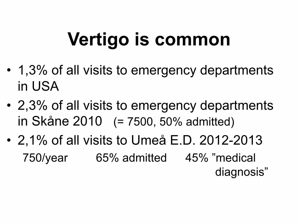
Vertigo is common• 1,3% of all visits to emergency departments
in USA• 2,3% of all visits to emergency departments
in Skåne 2010 (= 7500, 50% admitted)• 2,1% of all visits to Umeå E.D. 2012-2013
750/year 65% admitted 45% ”medicaldiagnosis”

Acute vertigo/dizziness i northern SwedenNUS Umeå 2012-2013 (population 150.000)
1437 pat > 18 ys dizziness = 2.1%65% admitted39% CT / MR
54% symptom diagnosis only R42.914% oto-vestibular (BPPV 6.5%)9% cardio-vascular9% neurologic
Ljunggren & Salzer pers rep 2015

5.4% stroke / TIA
CT brain: 4.8% pathologyMR brain 13.5% pathology
Acute vestibular syndrome (AVS): 11% stroke
Focal neurological findings / or ataxia:53% stroke!!!
0.56% will have a stroke within 3 months
Ljunggren & Salzer pers rep 2015
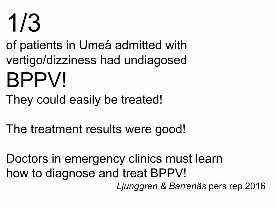
1/3of patients in Umeå admitted withvertigo/dizziness had undiagosed
BPPV!They could easily be treated!
The treatment results were good!
Doctors in emergency clinics must learnhow to diagnose and treat BPPV!
Ljunggren & Barrenäs pers rep 2016
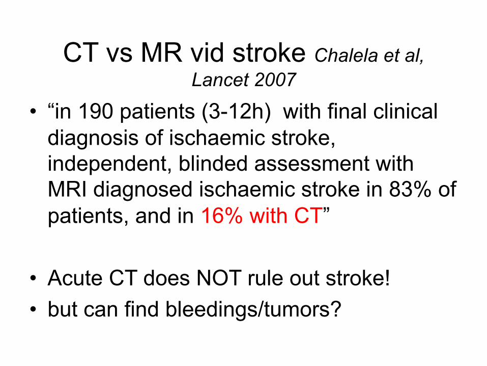
CT vs MR vid stroke Chalela et al, Lancet 2007
• “in 190 patients (3-12h) with final clinical diagnosis of ischaemic stroke, independent, blinded assessment with MRI diagnosed ischaemic stroke in 83% of patients, and in 16% with CT”
• Acute CT does NOT rule out stroke! • but can find bleedings/tumors?

How common is ”vertigo” in intracranial bleeding?
595 patients with intracerebral bleedings (8 år)2.2% (13 of 595): vertigo + NIHSS < 2Only 1 patient had “only vertigo” but obviousdysmetria in statusThe rest had headache / confusion / hemiparesis /dysartria mmDo we have to rule out bleedings in ”only vertigo”?
Kerber et al. Emerg Med J. Publ on line Jan 18, 2011

What is our mission as ENTs for acute vertigo?
• Identify the most common disorders!!posterior and lateral canal BPPV 30% of all!vestibular neuritis 90 of acute vestibular syndrome
• Identify otitis / labyrinthitis!!• Identify ”what it´s not!” (any of the above)• Use the clinical tests that best differ between
peripheral and central vertigo (and there are recent studies on this!)

The organs of balance(vestibular organs)
Semicircular canals= ”gyros”
Head rotations in all planes
stabilises the eyes in space(vestibulo-ocular reflex)
Otolithic organs = ”accelerometers”
gravitionhead position relative the ground
linear acceleration
stabilises body (and eyes) muscletension in neck, trunk and legs
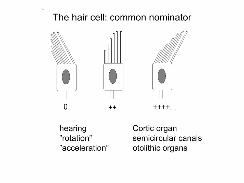
The hair cell: common nominator
hearing Cortic organ”rotation” semicircular canals”acceleration” otolithic organs
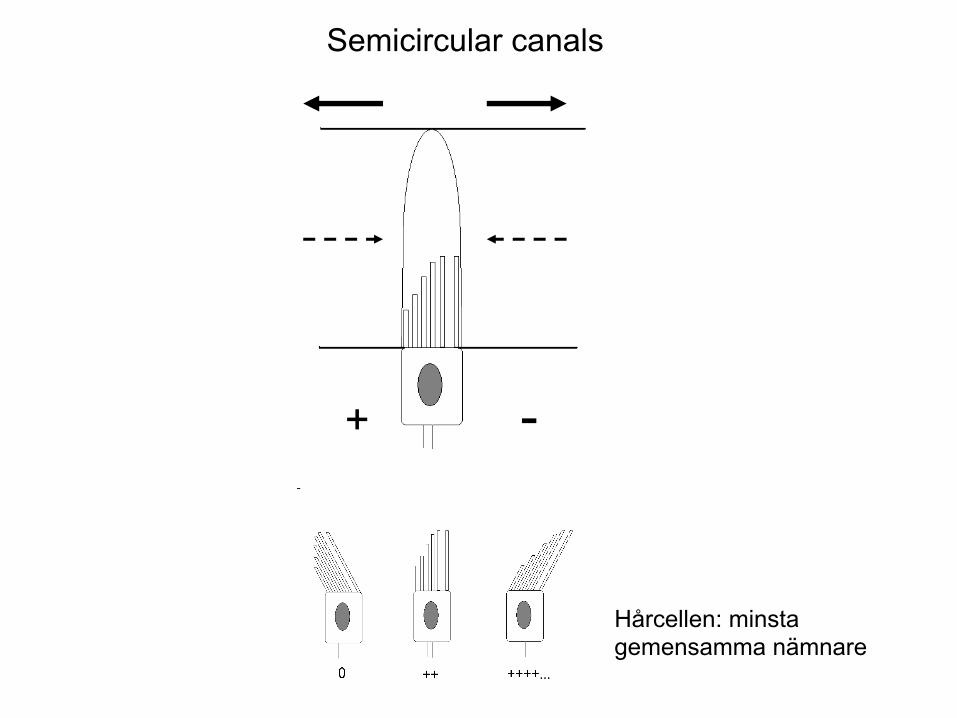
-+
Semicircular canals
Hårcellen: minstagemensamma nämnare

lesion=”peripheral”
Semicircular canalsconnected to extra-ocularmuscles
3-4 neurons
Fast reflex 9 ms latency

LARP = Left AnteriorRight Posterior
RALP = Right AnteriorLeft Posterior
= Right LateralLeft Lateral
A differences in signallingbetween canal pairs gives eyemovements as if a head movementin that plane takes place!
The anatomy of the canal planescan vary between individuals!!
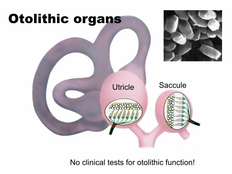
SacculeUtricle
Otolithic organs
No clinical tests for otolithic function!

Neuroanatomyafter Balaban & co-workers
oculo-motorpostural muscles
autonomic reflexes:cardio-vascular/respiratory/gastro-intestinal
anxiety / conditioningvestibulo-parabrachial nc network
coeruleo-vestibular networkRaphe nc – vestibular network
Limbic system (hippocampus)
adrenergic systemarousal
serotoninergic system?SSRI discontinuation syndrome
cortex:orientation

1. Spontaneous nystagmus
Direction? Describe with words!!!
Quick phase = direction
Grade 1, 2, 3?Grade 1 = gaze evoked nystagmus in one directionGrade 2 = nystagmus in primary position + in one
gaze directionGrade 3 = nystagmus in all horisontal gaze directions
Vestibular spontaneous nystagmus stronger with gaze in quick phaseVisual suppression = ”vestibular” nystagmus only visible with ”tools”Always use Video-Nystagmoscopy or Frenzel glasses!

2. Gaze-evoked nystagmus
CerebellumToxic: alcohol, litium, anti-epileptics…….
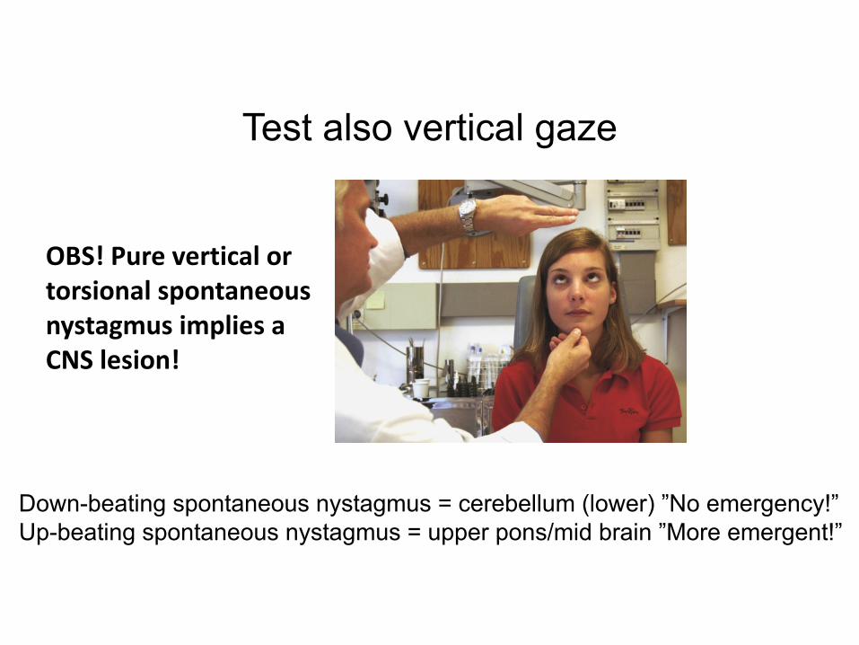
Test also vertical gaze
OBS!PureverticalortorsionalspontaneousnystagmusimpliesaCNSlesion!
Down-beating spontaneous nystagmus = cerebellum (lower) ”No emergency!”Up-beating spontaneous nystagmus = upper pons/mid brain ”More emergent!”

Lots of different strange & rare nystagmus!!
Congenital nystagmus: can look as anything!
Rebound nystagmus: changes direction with gaze direction = cerebellum
Ocular flutter: eyes jump with horisontal sackades
Opsoclonus: eyes jump with saccades in all directions
Ocular flutter and opsoclonus = brain stem
If you see something you don´t understand,search the net (YouTube / Neurocular)!!!
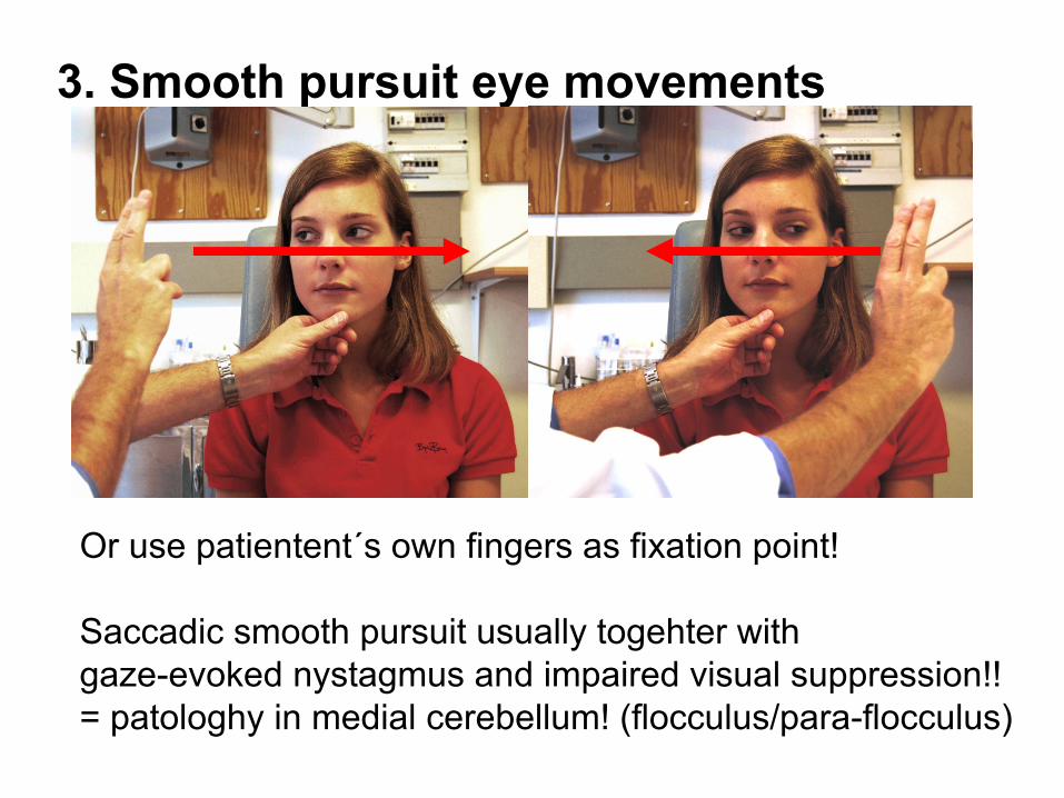
3. Smooth pursuit eye movements
Or use patientent´s own fingers as fixation point!
Saccadic smooth pursuit usually togehter withgaze-evoked nystagmus and impaired visual suppression!!= patologhy in medial cerebellum! (flocculus/para-flocculus)

Skewdeviation=”verticalsquint”
Coveroneeye5-6secondsCovertheothereyeVerticalsaccade?
5-6s
4. Test skew = alternernating cover test

Ocular tilt reaction
1. Ocular torsion2. Head tilt3. Skew deviation4. Tilt of subjective visual vertical
Lee et al. JNNP, 2005;76:1742-1743
tonic vestibular imbalanceIn the frontal plane
unilteral brain stem lesion(Wallenberg syndrome)
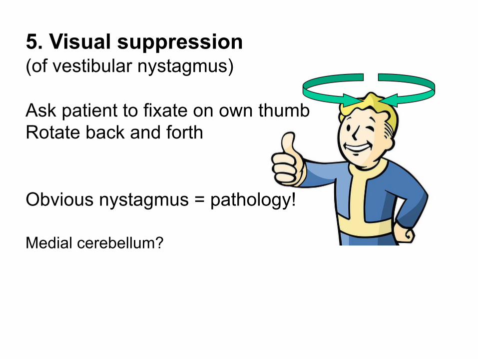
5. Visual suppression(of vestibular nystagmus)
Ask patient to fixate on own thumbRotate back and forth
Obvious nystagmus = pathology!
Medial cerebellum?
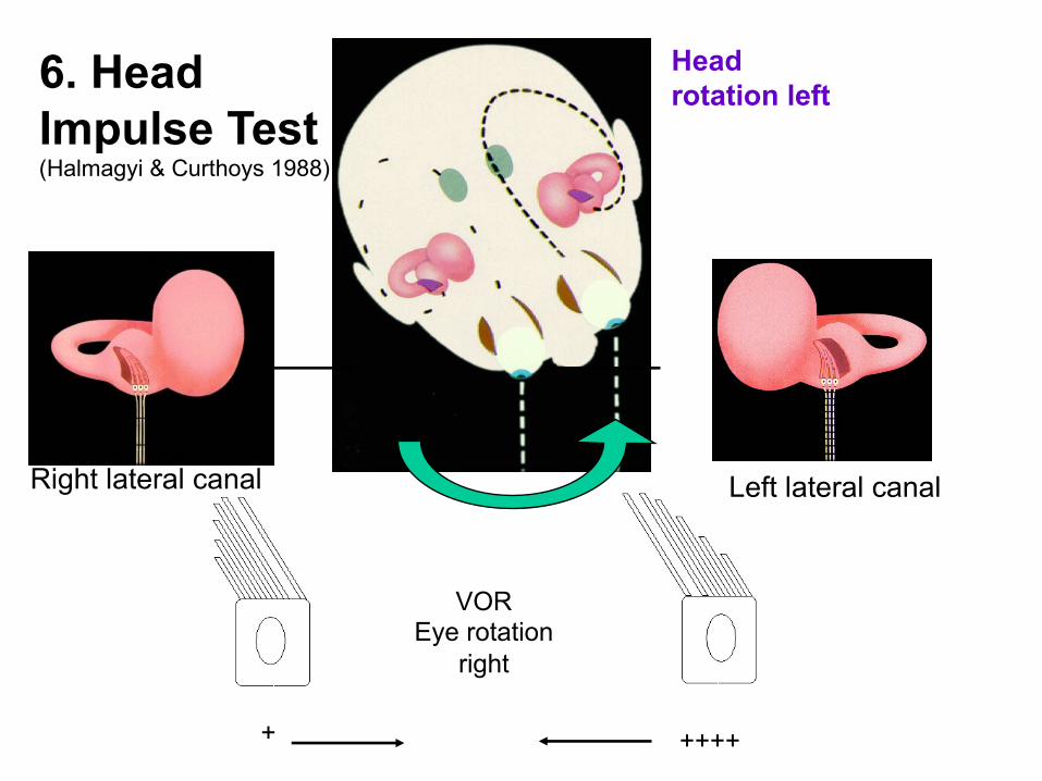
Head rotation left
Right lateral canal Left lateral canal
+ ++++
VOREye rotation
right
6. HeadImpulse Test(Halmagyi & Curthoys 1988)
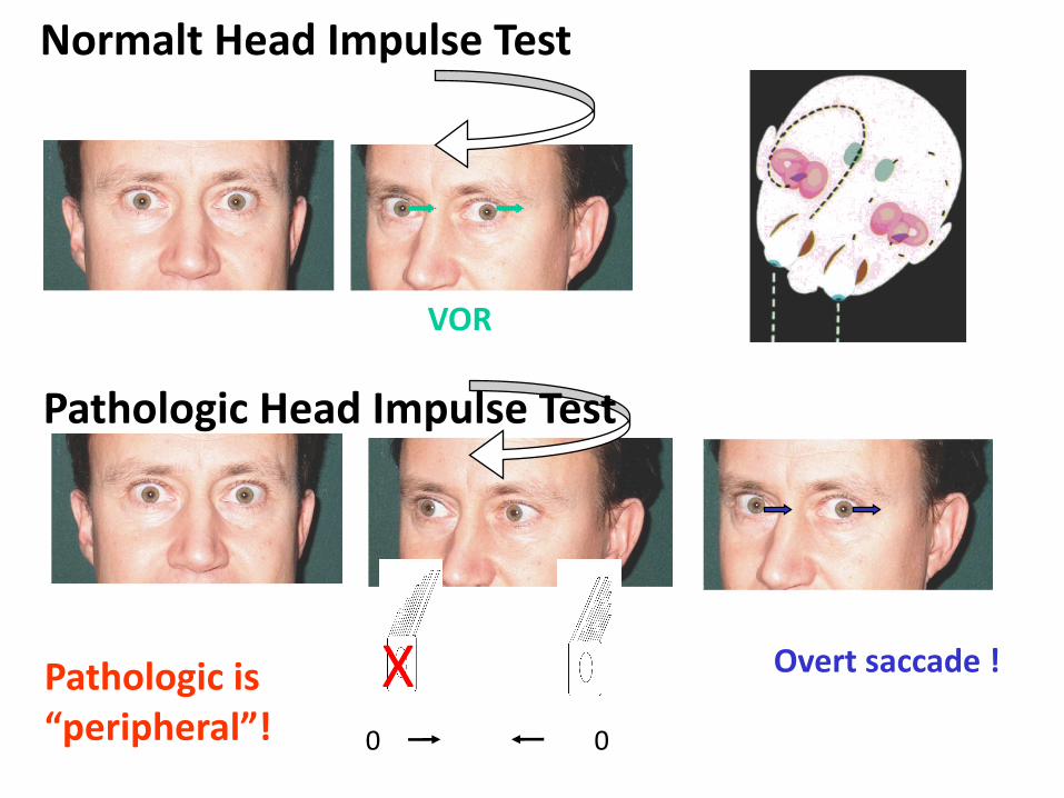
PathologicHeadImpulseTest
NormaltHeadImpulseTest
X0 0
Overtsaccade!
VOR
Pathologicis“peripheral”!

Lesionsgivesamesymptoms/findings
”Positive” Head Impulse Test
Only with lesion in ”1:st neuron”
= semicircular canal / vestibular nerveand (Sorry!) brainstem!

Video-nystagmoscopy:
1. Spontaneous nystagmus2. (ev.blickriktning)3. Lean forward (lateral
canal-BPPV?)4. Lateral BPPV-test
(pillow– head 30º nose up – left / right)5. Dix-Hallpike test (head hanging over pillow)6. Head-shake test (sitting 30 head rotations 2 Hz)
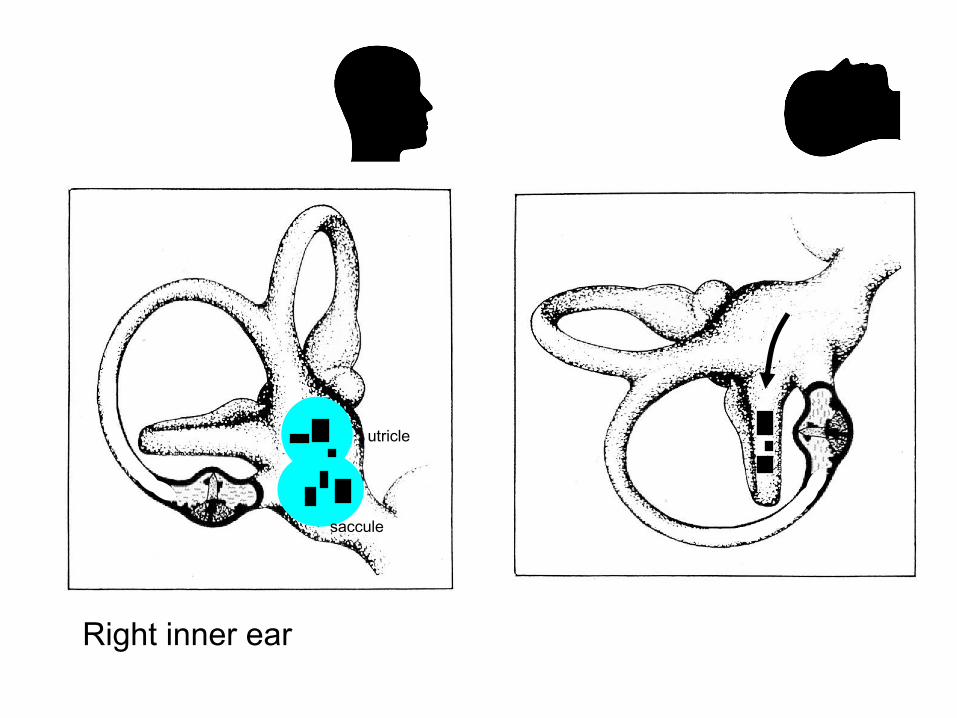
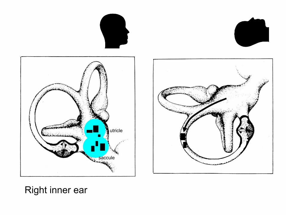
Right inner ear
utricle
saccule

Lateral canal-BPPV test
”Sicker” than posteriorcanal BPPV!

BPPV - lateral canal(20-40%) canalolithiasis
.
.
.
Loose otoliths in lateral canalintense vertigo (Ambulance! Emergency!)
nystagmus towards the floor (canal) inboth right and left side!Long duration but decreasingIntense vertigo/nystagmus=sick side
Treatment:Lie on ”best” side 10 minutes, thenprone 10 minutesGufoni/Appiani-manouvreForced prolonged positioning = lieon best side 24 hours

BPPV - lateral canalcupulolithiasis
otoliths stuck on lateral canal cupulaLess intense vertigo
nystagmus towards heaven (cupola) inboth right and left side!Not decreasing!!Intense vertigo/nystagmus=healthy side
Treatment:Turn into canalolithiasisHead-shaking/vibration?Shake head several times an hourLie on ”best” sideBrandt-Daroff excersises
.
.
.

BPPV - lateral canal”canalith jam”
. loose otoconia in lateral canal ”get stuck”continous vertigo /nystagmus
spontaneous nystagmus left / rightnot affected by position changes
Treatment:Turn into canalolithiasis with ”loose”otoconiavibration / head shakingthen positional treatment
.
.
Right inner ear
utricle
saccule

BPPV – posterior canal(60-80%)
Dix-Hallpike test
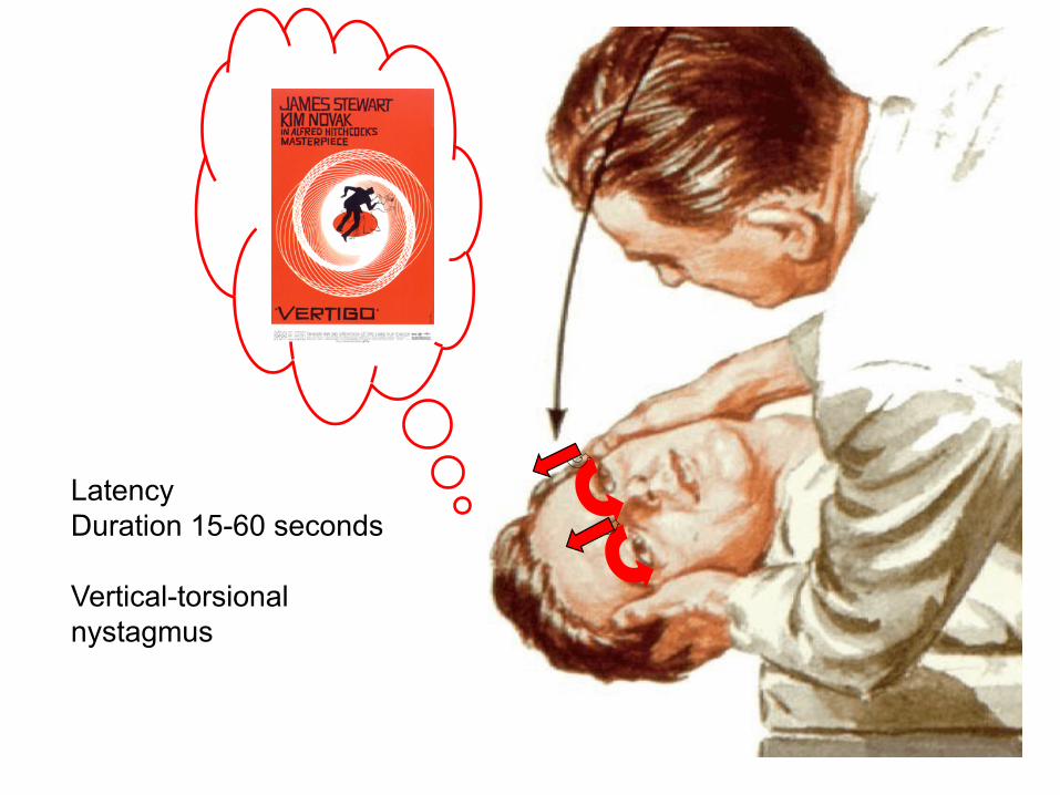
LatencyDuration 15-60 seconds
Vertical-torsionalnystagmus

Alternative, ”kinder”, way to test (older, overweight..)

BPPV - superior canal(0-10%)
RARE!
Dix-Hallpike: torsional-vertical nystagmusDown-beating vertical nystagmus!!
Disappears = canalolithiasisContinous = cupulolithiasis
Treatment:Epley manouvreBrandt-Daroff excersises”Mecka excersises”
utrikulus
sacculus
Head-shaking nystagmussensitive but unspecific30 shakes ca 2 Hz
>3 nystagmus beats = ”not normal”Beats towards ”most functioning side”Might change direction
Can be peripheral or central
Vertical nystagmus after horisontal head-shakningis always central!

Neurologic tests
1. Vibratory sensation ankle (128 Hz)2. Romberg´s test3. ”Balance walk”

”Ankle-Weber”
In patients that lateralises Weber
Put tuning fork (128 Hz) to lateral malleolus
Hears it clearly in one ear = ”3rd window”
sSCC patient
10 normal-hearing young subjects
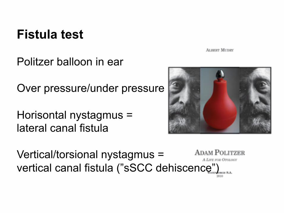
Fistula test
Politzer balloon in ear
Over pressure/under pressure
Horisontal nystagmus =lateral canal fistula
Vertical/torsional nystagmus =vertical canal fistula (”sSCC dehiscence”)
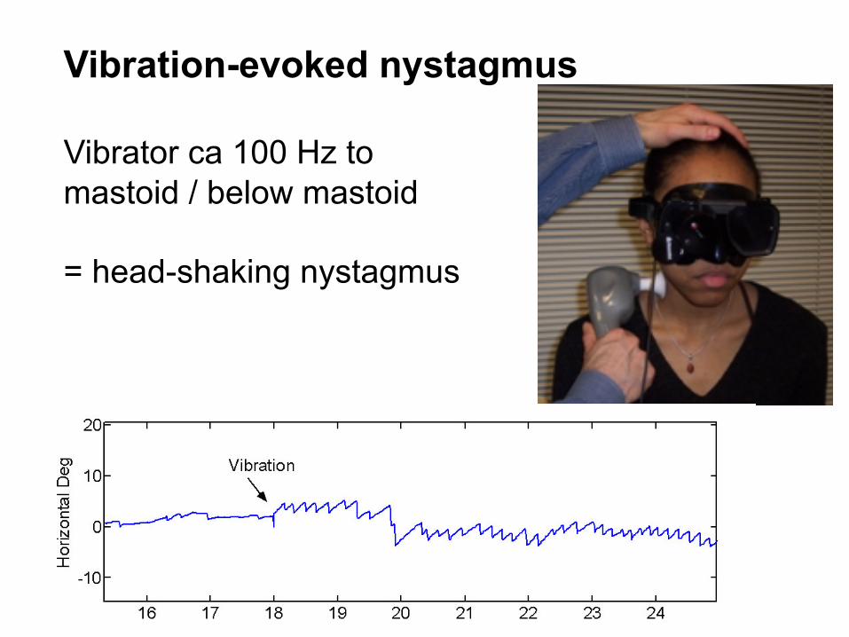
Vibration-evoked nystagmus
Vibrator ca 100 Hz tomastoid / below mastoid
= head-shaking nystagmus
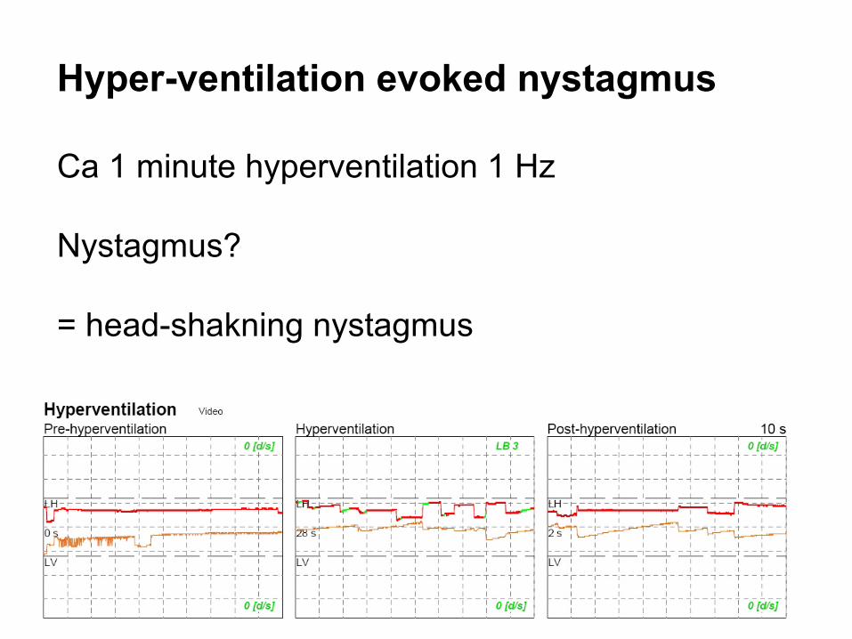
Hyper-ventilation evoked nystagmus
Ca 1 minute hyperventilation 1 Hz
Nystagmus?
= head-shakning nystagmus
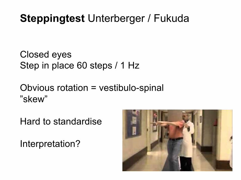
Steppingtest Unterberger / Fukuda
Closed eyesStep in place 60 steps / 1 Hz
Obvious rotation = vestibulo-spinal ”skew”
Hard to standardise
Interpretation?

”Acute vestibular syndrome”vestibular neuritis or stroke?
• Sudden onset of continous vertigo (>24 h)• Nausea and/or vomiting (usually)• Spontaneous nystagmus• Worsening by head movements• Unsteadiness / ”lateropulsion”
• No other obvious neurological symptoms• No hearing loss

Acute vestibular neuritis”sudden peripheral idiopathic unilateral vestibular loss”
probably an inflammatory neuropathy(as Bell´s palsy?)
90 % of acute vestibular syndrome
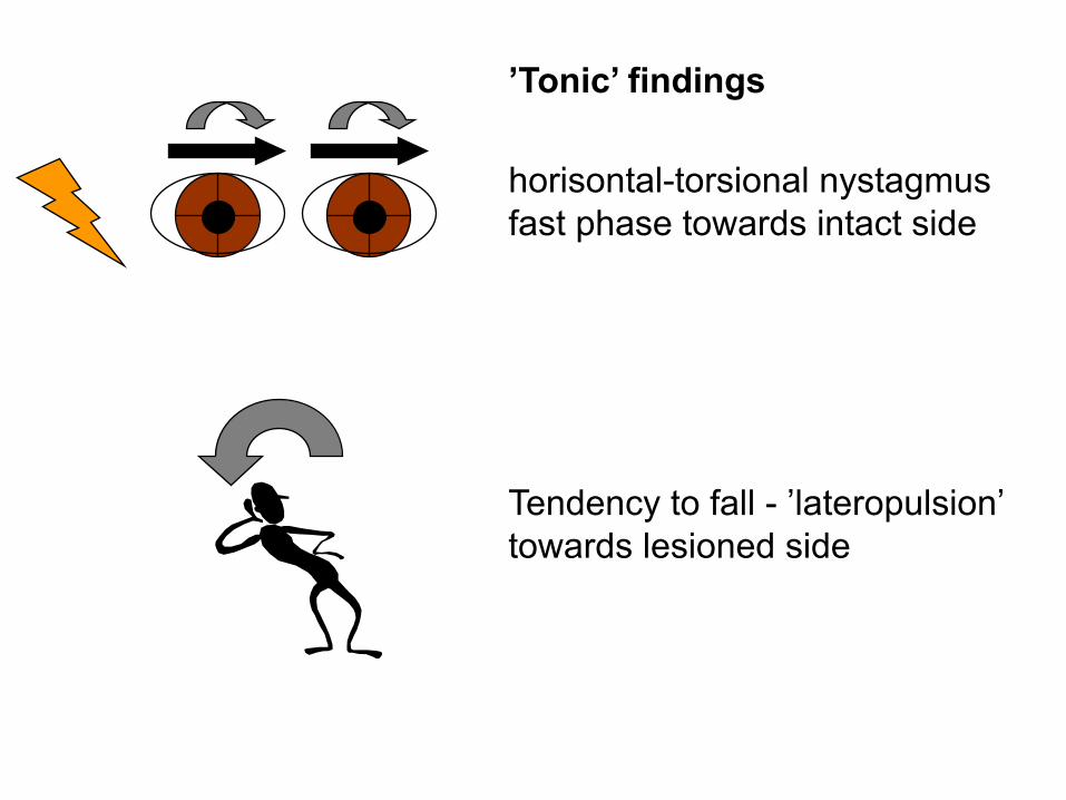
horisontal-torsional nystagmusfast phase towards intact side
Tendency to fall - ’lateropulsion’towards lesioned side
’Tonic’ findings

Pathologic head-impulse test to lesioned side
X0 0
Sackade !
”Dynamic” finding

Subjective Visual Horisontaltilts towards lesioned side5-15°
VEMPsusually intact (2/3)
44°H2O
Caloric testcanal paresis

Why is the inferior vestibular nerve spared?
Temporal bone studies
inferior vestibular nerve : double innervation2 nerves in separate channels Arbusow et al -99
superior vestibular nervenarrower nerve channellonger channelnarrower arterioli Goebel & Gianoli -00
Inflammatory entrapment of the superior vestibular nerve???

Since 2004 we have offered steroidsto patients with acute vestibular neuritis
Betapred (betamethasone) 8 mg i.v.followed by:
T. Prednisolone 60mg x 1 x V50mg x I, 40mg x I, 30mg x I, 20mg x I, 10mg x I
Caloric test after 12 months
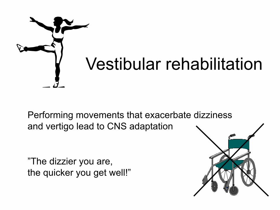
Vestibular rehabilitation
Performing movements that exacerbate dizzinessand vertigo lead to CNS adaptation
”The dizzier you are,the quicker you get well!”
Stroke (”pseudoneuritis”) in acutevestibular syndrome (AVS)Epidemiology• In USA 250,000 - 500,000 patients with
AVS at emergency departments yearly (Newman-Toker)
• Most have vestibular neuritis (VN), but some have an ischemic stroke i brain stem or cerebellum ??“~25% ±15%”??
• In Umeå 2012-13: 11% of AVS = stroke

Clinical examination
• When?• (How does it feel?)• Pain?? Head? Chest? Ear? Neck?• Hearing? Changed?/ Hearing loss?
Side difference? • Other? Vision? Nausea?• Loss of consciousness?• Other neurological symptom?
Double vision, swallowing, dysphonia, sensibility/ motorOBS! Also transient symptoms?!
Acutevertigo
Acuteexam/’vitalsigns’
Acutehistory
Acutehistory:

Acute exam• Level of consciousness• BP/pulse/Heart/Lungs Arrythmia?, chock?• Neck stiffness?? Meningitis, SAH • Motor-/sensory: extremiteter/face/ finger-nose
CNS-lesion, brain stem/cerebellum• Eye movements – nystagmus? spontaneous
nystagmus =peripheral vestibular lesion or CNS lesion
HINTS: Head Impulse test – Nystagmus – Test Skew• Ear otitis - (cholesteatoma) –labyrinthitis – zoster oticus• Positional vertigo tests: Dix-Hallpike, test of lateral
sSCC BPPV
Akutstatus/’vitalsigns’
Akutanamnes
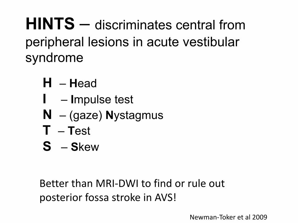
HINTS – discriminates central from peripheral lesions in acute vestibular syndrome
H – HeadI – Impulse testN – (gaze) NystagmusT – TestS – Skew
Newman-Tokeretal2009
BetterthanMRI-DWItofindorruleoutposteriorfossastrokeinAVS!

PathologicHeadImpulsetest
NormalHeadImpulsetest
X0 0
Sackade!
VOR
“Pathologicisperipheral!” Film!

Head impulse test differs PICA-infarction from ”vestibular neuritis”
PICA-infarction:AcuteVestibularSyndromewithnormalheadimpulsetestTHEMOSTIMPORTANTofHINTS!!
Newman-Toker2010

lesionsgiveidenticalsymptoms/findingsperipheralorcentral?
N=Nystagmus
spontaneousnystagmus=eitherCNSor”ear”
”ear”=unilateralvestibularlesionCNS=cerebellumORbrainstem
Horisontal-(torsional) nystagmusfast phase towards intact sideOnly ”benign” nystagmus!!!

Vertigoduetolesionintheearorinthebrainstem?peripheralorcentral? lesionsgiveidenticalsymptoms/findings

Skewdeviation=”verticalsquint”
Coveroneeye5-6secondsCovertheothereyeVerticalsaccade?
5-6s
Test Skew Alternernating cover test

HINTS3testsinHINTS- HeadImpulse- Nystagmus- TestofSkew=Covertest
DangerousH.I.N.T.S.(inpatientwithacutevestibularsyndrome)
- Normalheadimpulsetest- Gazenystagmus/purelyverticalortorsionalspontaneousnystagmus
- Verticalrefixationsaccadeincovertest
AnyoftheseinpatientwithAVS-suspectposteriorfossastroke!

• Acute vestibular syndrome is caused either by vestibular neuritis (90%) or by stroke (10%)
• High dose steroids + vestibular rehab improves outcome after vestibular neuritis
• HINTS – differ between stroke and VN• Pain + vertigo is ”dangerous”!• Acute unilat hearing loss + vertigo?
Think stroke!!!!• Other neurological symptoms? Think
stroke!• Impossible to stand up? Think stroke!• Stop ordering acute CT brain for vertigo!