Embed Size (px)
Citation preview
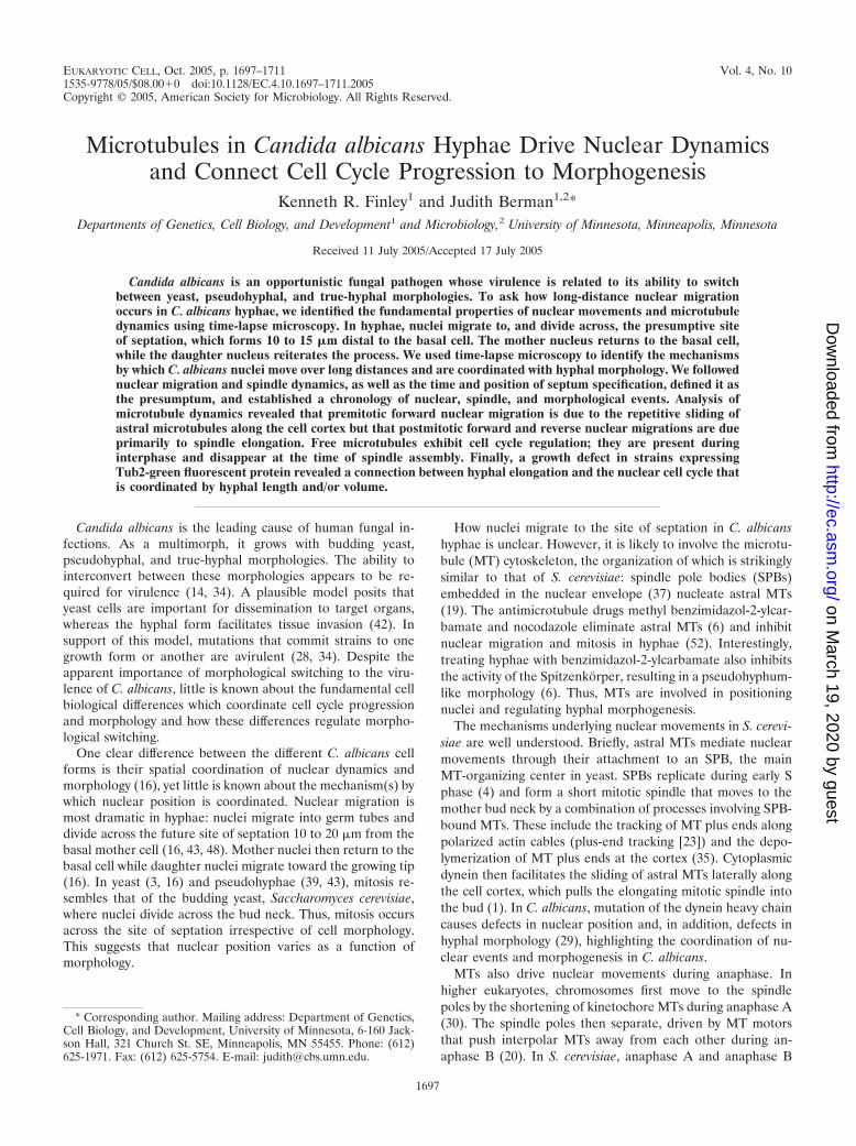
EUKARYOTIC CELL, Oct. 2005, p. 1697–1711 Vol. 4, No. 101535-9778/05/$08.00�0 doi:10.1128/EC.4.10.1697–1711.2005Copyright © 2005, American Society for Microbiology. All Rights Reserved.
Microtubules in Candida albicans Hyphae Drive Nuclear Dynamicsand Connect Cell Cycle Progression to Morphogenesis
Kenneth R. Finley1 and Judith Berman1,2*Departments of Genetics, Cell Biology, and Development1 and Microbiology,2 University of Minnesota, Minneapolis, Minnesota
Received 11 July 2005/Accepted 17 July 2005
Candida albicans is an opportunistic fungal pathogen whose virulence is related to its ability to switchbetween yeast, pseudohyphal, and true-hyphal morphologies. To ask how long-distance nuclear migrationoccurs in C. albicans hyphae, we identified the fundamental properties of nuclear movements and microtubuledynamics using time-lapse microscopy. In hyphae, nuclei migrate to, and divide across, the presumptive siteof septation, which forms 10 to 15 �m distal to the basal cell. The mother nucleus returns to the basal cell,while the daughter nucleus reiterates the process. We used time-lapse microscopy to identify the mechanismsby which C. albicans nuclei move over long distances and are coordinated with hyphal morphology. We followednuclear migration and spindle dynamics, as well as the time and position of septum specification, defined it asthe presumptum, and established a chronology of nuclear, spindle, and morphological events. Analysis ofmicrotubule dynamics revealed that premitotic forward nuclear migration is due to the repetitive sliding ofastral microtubules along the cell cortex but that postmitotic forward and reverse nuclear migrations are dueprimarily to spindle elongation. Free microtubules exhibit cell cycle regulation; they are present duringinterphase and disappear at the time of spindle assembly. Finally, a growth defect in strains expressingTub2-green fluorescent protein revealed a connection between hyphal elongation and the nuclear cell cycle thatis coordinated by hyphal length and/or volume.
Candida albicans is the leading cause of human fungal in-fections. As a multimorph, it grows with budding yeast,pseudohyphal, and true-hyphal morphologies. The ability tointerconvert between these morphologies appears to be re-quired for virulence (14, 34). A plausible model posits thatyeast cells are important for dissemination to target organs,whereas the hyphal form facilitates tissue invasion (42). Insupport of this model, mutations that commit strains to onegrowth form or another are avirulent (28, 34). Despite theapparent importance of morphological switching to the viru-lence of C. albicans, little is known about the fundamental cellbiological differences which coordinate cell cycle progressionand morphology and how these differences regulate morpho-logical switching.
One clear difference between the different C. albicans cellforms is their spatial coordination of nuclear dynamics andmorphology (16), yet little is known about the mechanism(s) bywhich nuclear position is coordinated. Nuclear migration ismost dramatic in hyphae: nuclei migrate into germ tubes anddivide across the future site of septation 10 to 20 �m from thebasal mother cell (16, 43, 48). Mother nuclei then return to thebasal cell while daughter nuclei migrate toward the growing tip(16). In yeast (3, 16) and pseudohyphae (39, 43), mitosis re-sembles that of the budding yeast, Saccharomyces cerevisiae,where nuclei divide across the bud neck. Thus, mitosis occursacross the site of septation irrespective of cell morphology.This suggests that nuclear position varies as a function ofmorphology.
How nuclei migrate to the site of septation in C. albicanshyphae is unclear. However, it is likely to involve the microtu-bule (MT) cytoskeleton, the organization of which is strikinglysimilar to that of S. cerevisiae: spindle pole bodies (SPBs)embedded in the nuclear envelope (37) nucleate astral MTs(19). The antimicrotubule drugs methyl benzimidazol-2-ylcar-bamate and nocodazole eliminate astral MTs (6) and inhibitnuclear migration and mitosis in hyphae (52). Interestingly,treating hyphae with benzimidazol-2-ylcarbamate also inhibitsthe activity of the Spitzenkorper, resulting in a pseudohyphum-like morphology (6). Thus, MTs are involved in positioningnuclei and regulating hyphal morphogenesis.
The mechanisms underlying nuclear movements in S. cerevi-siae are well understood. Briefly, astral MTs mediate nuclearmovements through their attachment to an SPB, the mainMT-organizing center in yeast. SPBs replicate during early Sphase (4) and form a short mitotic spindle that moves to themother bud neck by a combination of processes involving SPB-bound MTs. These include the tracking of MT plus ends alongpolarized actin cables (plus-end tracking [23]) and the depo-lymerization of MT plus ends at the cortex (35). Cytoplasmicdynein then facilitates the sliding of astral MTs laterally alongthe cell cortex, which pulls the elongating mitotic spindle intothe bud (1). In C. albicans, mutation of the dynein heavy chaincauses defects in nuclear position and, in addition, defects inhyphal morphology (29), highlighting the coordination of nu-clear events and morphogenesis in C. albicans.
MTs also drive nuclear movements during anaphase. Inhigher eukaryotes, chromosomes first move to the spindlepoles by the shortening of kinetochore MTs during anaphase A(30). The spindle poles then separate, driven by MT motorsthat push interpolar MTs away from each other during an-aphase B (20). In S. cerevisiae, anaphase A and anaphase B
* Corresponding author. Mailing address: Department of Genetics,Cell Biology, and Development, University of Minnesota, 6-160 Jack-son Hall, 321 Church St. SE, Minneapolis, MN 55455. Phone: (612)625-1971. Fax: (612) 625-5754. E-mail: [email protected].
1697
on March 19, 2020 by guest
http://ec.asm.org/
Dow
nloaded from
start at roughly the same time and anaphase B accounts for themajority of nuclear movement during anaphase (31). Thus,these four MT-based mechanisms (plus-end tracking, depoly-merization, sliding, and anaphase spindle elongation) combineto faithfully segregate daughter nuclei to daughter cells.
A dynamic, cell cycle-regulated MT cytoskeleton is a hall-mark of eukaryotic cells. In higher eukaryotes, when the cellenters mitosis, extensive arrays of interphase MTs break downand the components are reincorporated into the mitotic spin-dle (33). During this process, bulk MT polymer decreases asinterphase MTs disassemble and returns to interphase levelsonce metaphase spindles form (reviewed in reference [25]). Asimilar phenomenon has not been observed in S. cerevisiae butis evident in several fungi, including the fission yeast Schizo-saccharomyces pombe and the dimorphic phytopathogen Usti-lago maydis, where SPB-independent interphase MTs disas-semble prior to the assembly of the mitotic spindle (18, 41).
To begin to understand the cell biological mechanisms thatconnect cell cycle regulation and morphogenesis and to deter-mine the mechanisms underlying nuclear migration with re-spect to hyphal morphology in C. albicans, we used time-lapsefluorescence microscopy to capture complete cell cycles. Byfollowing nuclear, spindle and septin dynamics, we establisheda clear chronology of cell cycle events during the growth of thefirst hyphal cell from a yeast mother cell. Short-interval time-lapse microscopy revealed that forward nuclear migration re-sults from the repetitive sliding of astral MTs along the cellcortex, while reverse migration is due primarily to spindleelongation. A pool of SPB-independent MTs is evident duringinterphase and disappears coincident with spindle assembly,suggesting that the components are reincorporated into themitotic spindle. Finally, the growth defect in strains expressingTub2-green fluorescent protein (GFP) revealed that the nu-clear cell cycle is coordinated with hyphal length and/or vol-ume and that when the rate of hyphal-tip growth is slowed, thecell cycle delays at two events: the formation of the septin ringand the onset of anaphase.
MATERIALS AND METHODS
Strain construction. Strains containing C-terminal fluorescent fusions weregenerated in strain BWP17 (50) as described previously (13) and are listed inTable 1. Transformation cassettes were generated by PCR using the appropriategene-specific primer pair (reverse primers are designated R1, R2, or R3 forURA3, HIS1, or ARG4, respectively [Table 1]) and a plasmid template containingspecific fluorescent-protein- and nutritional-marker-encoding sequences (13).Positive transformants were screened by fluorescence microscopy and confirmedby PCR. Strains were made prototrophic at the ARG4 locus by the transforma-tion of either NotI-linearized pRS-ARG-URA-BN (9) or EcoRI-linearized pRS-ARG4�Spe1 (50).
Preculture and hyphal induction. All media contained uridine at 80 �g/ml,except those used for selecting URA3 transformants. Bovine serum (Sigma, St.Louis, MO) was always used at 10% (vol/vol). To enrich cultures for unbuddedsinglet cells, strains were cultured overnight in YPAD (36) at 30°C. Ten to 20 �lof the culture was pelleted, resuspended in 100 to 200 �l of fresh syntheticcomplete medium (SDC [36]), and spread with glass beads on an SDC-serum-agar (2% agar) plate, preheated to 37°C. A round, 25-mm no. 1 coverslip (Fisher,Pittsburgh, PA) was placed over the cells, and the plate was returned to 37°C for10 to 15 min when complete cell cycle time-lapse microscopy was performed orfor longer periods to capture specific cell cycle stages.
Microscopy. Plates were placed on the stage of a Nikon E600 epifluorescencemicroscope under a 60� Plan-Apo (numerical aperture, 1.4) objective preheatedto 37°C with an FCS2 objective heater (Bioptechs, Butler, PA). Heat transferthrough type 37 immersion oil optimized for GFP and yellow fluorescent protein(YFP) microscopy at 37°C (Cargille, Cedar Grove, NJ) was sufficient to maintain
hyphal growth in and around the field of view for more than 5 h. Microscopy ofcyan fluorescent protein (CFP) required type DF immersion oil (Cargille) withunheated objectives. Filter sets were from Chroma Technology Corp (Rocking-ham, VT). GFP images were captured with an Endow bandpass filter set (no.41017) (11). For CFP/YFP imaging, filter set no. 86004 JP5v2, which utilizes asingle dichroic mirror, was used. Images were captured with a CoolSnap HQcooled charge-coupled-device camera (Photometrics, Tucson, AZ). Time-lapseautomation, image processing, and data collection from 16-bit images wereaccomplished with Metamorph imaging software, v6.2 (Universal Imaging Corp.,Westchester, PA). Data were exported to Microsoft Excel (Microsoft Corp.,Redmond, WA), and statistical analyses were performed with the StatisticalAnalysis Package in Excel. Standard errors of the means (SEM) are included forvalues determined by calculating the arithmetic mean of the population. Forfigure assembly, images were converted to 8 bits and exported to Adobe Photo-shop, v7.0 (Adobe Systems, Mountain View, CA).
Many acquisition details are provided in the text and figure legends. Generally,an ND4 neutral density filter was used with 200-ms exposures for Nop1-YFP orTub2-GFP alone or in combination with Cdc3-YFP/GFP. The exposure time forNop1-CFP was 500 ms. Two-by-two camera binning was always used. Differentialinterference contrast (DIC) images were taken in parallel by manually adjustingthe focus prior to image capture. Where Z-stacks were acquired, four images ateach time point were acquired with a step size of 1 �m. Two-dimensional imageswere generated using the Maximum function in Metamorph, and the Arithmeticdialogue was used to generate merged fluorescent/DIC images. Supplementalmovies available at http://www.cbs.umn.edu/labs/berman/kensmovies.htm arenumbered to reflect the corresponding figure numbers herein.
Analysis of nuclear movements. The “neck” of the basal cell/germ tube (Fig.1C) was arbitrarily defined as the zero point of a hyphal cell (position, 0 �m),where negative values represented positions within the basal cell while positivevalues were distal to the basal cell.
Individual hyphae were cropped from the time-lapse stacks and arranged in amontage into tif image files, where individual measurements were made frame byframe. Hyphal growth rates were calculated by linear regression analysis on plotsof hyphal length versus time generated with the Track Points feature of Meta-morph. We defined the time of presumptive septum (presumptum) appearanceas the time when a hypha grew past the point where the septum eventuallyappeared in the DIC channel. This was done based on the observations thatseptin rings (i) develop just after the tip passes the site where the septin ring willform, (ii) are static with respect to the position of the hyphal tip (Fig. 1C) (CherylGale, personal communication), and (iii) mark the future site of septation (12,43, 48). Due to the asynchrony of the populations, we used either the mitoticframe (Nop1) or spindle disassembly frame (Tub2) to align data graphically. Thisapproach minimized the variance of events temporally proximal to mitosis. Tocalculate the mean values for times between nuclear and morphogenetic events,we determined the time between any two given events within a single hypha andthen found the mean for that event among all hyphae. As many hyphae grew outof the field of view and others grew out of the plane of focus, it was not possibleto quantify every parameter for every hypha.
Analysis of microtubule dynamics. For analyses of MT dynamics, hyphae wereinduced for 90 min to enrich for short spindles. Images were captured at 3-sec-ond intervals. Rates of MT growth, shrinkage, and sliding were determined bylinear regression analysis (R2 � 0.9) of MT length versus time, only for MTs thatremained in focus for 15 seconds or longer. For fluorescent-speckle microscopy(FSM) and the quantification of free MTs during the cell cycle, hyphae wereinduced at 37°C for various periods of time to capture the relevant cell cyclestages and images were captured for �5 min at 1- or 3-second intervals with a100� Plan-Apo objective (numerical aperture, 1.4).
We performed MT life history analysis (5) to quantify the frequency of MTtransitions from growth to shrinkage (catastrophe) and from shrinkage to growth(rescue). A single MT life history was defined as the de novo growth of an MTfrom an SPB and termination by one of three events: complete MT depolymer-ization, MT release from the SPB (where no MT remained attached to the SPB),or MT “breakage,” which resulted in a free MT and a shorter MT remainingattached to the SPB. In the case of MT breakage, the life history terminatedwhen the remaining SPB-bound MT had depolymerized completely; the free MTwas not considered in the analysis. This analysis did not require that MTs remainin focus, only that we could discern relevant periods (growing, shrinking, andtransitions).
We used FSM (49) to assess the polarity of MTs bound to and released bySPBs. We identified MTs with varied degrees of Tub2-GFP incorporation, re-sulting in bright and dark regions that served as reference points with respect toeach other, the free MT end, and the SPB.
1698 FINLEY AND BERMAN EUKARYOT. CELL
on March 19, 2020 by guest
http://ec.asm.org/
Dow
nloaded from
RESULTS
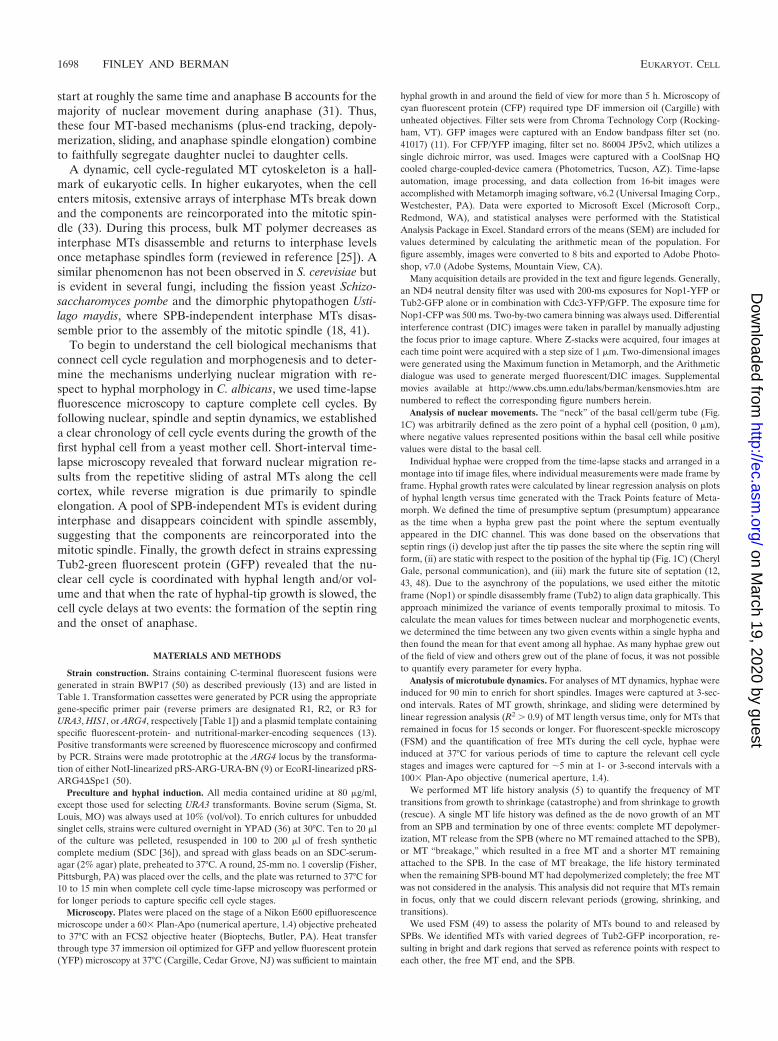
Nuclear organization in Candida albicans. To follow thedynamics of nuclear migration in hyphae, we generated in vivofluorescent protein fusions to the C termini (13) of puta-tive nuclear markers (http://www-sequence.stanford.edu/group/candida), including a nuclear pore protein, Nup49 (orf19.4987),a histone H4 gene, (Hhf1; orf19.1059), and a small nucleolarribonucleoprotein, Nop1 (orf19.3138). Nop1 localized consti-tutively to a bright, round structure in yeast and hyphal cellsthat fluoresced significantly more than the other nuclear tags(Hhf1 or Nup49), enabling the use of short exposure times thatresulted in less phototoxicity. We also visualized the nucleoluswith respect to the nuclear membrane with a strain coexpress-ing Nop1-CFP and Nup49-GFP (YJB8552). The continuity ofthe Nup49 signal throughout the cell cycle indicated that, as inyeast cells (37), the nuclear membrane remains intact during
mitosis in hyphae. Importantly, Nop1-CFP appeared within theNup49-GFP signal at all stages of the cell cycle (Fig. 1A).
To follow C. albicans nucleolar movement with respect tothe chromosomes, we coexpressed Nop1-CFP and Hhf1-GFP(histone H4) in strain YJB8179. The nucleolus remainedclosely associated with Hhf1-GFP and divided with the last ofthe chromosomal DNA (Fig. 1B). This resembles nucleolarbehavior in S. cerevisiae, where nucleolar antigens (includingNop1 [10]) are closely associated with chromosomal DNA (17,51), even though nucleolar separation is delayed with respectto the separation of the rest of the genome (8, 44, 46, 47).Thus, Nop1 is a useful nuclear marker that resides within thenuclear membrane and is closely associated with genomic DNA.
Cdc3-YFP reveals the site of septation. Previous studiesshowed that the true septin ring specifies the site of septation(12, 43, 48) but did not establish the timing of septin ring
TABLE 1. C. albicans strains and PCR primers used in this study
Strain or primer Genotype or sequence (5�–3�)a Source
StrainsBWP17 ura3�::imm434/ura3�::imm434 his1::hisG/hisI::hisG arg4::hisG/arg4::hisG 50YMG5628 BWP17 CDC3/CDC3-YFP:URA3 13YMG6449 BWP17 NOP1/NOP1-YFP:HISI This studyYJB7062 BWP17 NOP1/NOP1-YFP:URA3 This studyYJB7150 BWP17 HHF1/HHF1-GFP::URA3 This studyYJB7468 BWP17 NOP1/NOP1-CFP::ARG4 This studyYJB8179 YJB7150 NOP1/NOP1-CFP::ARG4 This studyYJB8548 YJB7468 TUB2/TUB2-GFP::HIS1 This studyYJB8860 YJB7062 CDC3/CDC3-GFP::HIS1 This studyYJB8552 YJB7468 NUP49/NUP49-GFP::URA3 This studyYJB8895 BWP17 TUB2/TUB2-GFP::HIS1 This studyYJB8897 YMG5628 TUB2/TUB2-GFP::HIS1 This studyYJB8952 YJB8895 ARG4::URA3::arg4::hisG/arg4::hisG This studyYJB8959 YJB8860 ARG4::arg4::hisG/arg4::hisG This studyYJB8961 YJB8897 ARG4::arg4::hisG/arg4::hisG This study
PrimersNOP1F ACCTTGGAACCTTATGAAAGAGACCATTGTATTGTTGTTGGTAGATACA 6
TGAGAAGCGGAATAAAGAAAggtggtggttctaaaggtgaagaattattNOP1R1 TTTTTAGTTTTCAATAATCAAATGTATTAATCCTATTGTACAAAATATTT This study
TTATTTAAAATTTAGAGTATCCCtctagaaggaccacctttgattgNOP1R2 TTTTTAGTTTTCAATAATCAAATGTATTAATCCTATTGTACAAAATATTT 6
TTATTTAAAATTTAGAGTATCCCgaattccggaatatttatgagaaacNOP1R3 TTTTTAGTTTTCAATAATCAAATGTATTAATCCTATTGTACAAAATATTT This study
TTATTTAAAATTTAGAGTATCCCactagtattgtagtacaaggtatcNUP49F TATAGTTTATTTATGGAATTGACTGAAACAATGGCTCAATTACATAATG This study
AAGTGAATAGATTAACTAAAggtggtggttctaaaggtgaagaattattNUP49R1 CAAAAAAAGAAAAATAAGAAAAACTATTACTATTATACATTATAATAT This study
ATAATACAATTGTTGCACTTTCtctagaaggaccacctttgattgHHF1F GTCACTTCATTGGATGTTGTTTACGCTTTGAAGAGACAAGGTAGAACCT This study
TGTATGGTTTCGGTGGTggtggtggttctaaaggtgaagaattattHHF1R1 TTATACAAAAAACTTCTTAAATTAATACTATACAATAAAGAAAACGAAC This study
TAAAAAGACAATTAGAAtctagaaggaccacctttgattgTUB2F CAAGAAGCTAGTATTGATGAAGAAGAATTAGAATATGCCGATGAAATC This study
CCATTAGAAGATGCCGCCATGGAAggtggtggttctaaaggtgaagaattattTUB2R2 CATTAACTAAACCAAAAAAAACCATAATTATATTAGAAGTGAACAAAA This study
AAAAAATTATAATTAATTATATTGgaattccggaatatttatgagaaacCDC3F ACAAAAATTATTACCACAAGACCCACCAGCACAACCAGCTCCACAAAA 13
GAGTCGTAAAGGATTTTTACGTggtggtggttctaaaggtgaagaattattCDC3R1 AATTAAACAAACAGATTAACAAACAAATAAACTAAATTAAGTTACATA 13
CTATTTAGCTATACCTCGGCCCtctagaaggaccacctttgattgCDC3R2 AATTAAACAAACAGATTAACAAACAAATAAACTAAATTAAGTTACATA 13
CTATTTAGCTATACCTCGGCCCgaattccggaatatttatgagaaac
a Uppercase indicates homology to genomic DNA for integrative purposes. Lowercase indicates homology to plasmids described in the work of Gerami-Nejad et al. (13).
VOL. 4, 2005 NUCLEAR MIGRATION IN C. ALBICANS HYPHAE 1699
on March 19, 2020 by guest
http://ec.asm.org/
Dow
nloaded from
appearance with respect to the position of the hyphal tip.Time-lapse microscopy of germ tubes expressing Cdc3-YFP(YJB5628) revealed that, like Cdc10 (48), Cdc3 appears ingrowing hyphal tips (Fig. 1C). More importantly, in all hyphaeexamined (n � 13), Cdc3 localized to the future site of septa-tion coincident with the time that the hyphal tip passed thisposition in the germ tube. This gave the impression that Cdc3was left behind the extending tip as it passed this position,rather than that the ring appeared within the tube at a latertime. Consistent with previous reports (43, 48), the septin ringhad divided into two rings when the septum appeared in theDIC channel (Fig. 1C and inset). We conclude that the trueseptin ring appears when the eventual site of septation is spec-ified, and this occurs at the time that tip growth passes this site.Similar to what occurs with S. cerevisiae, where septins bearingepitope tags (GFP or hemagglutinin) at their C termini are notfully functional (M. Longtine, personal communication), fluo-rescently tagged C. albicans septins affect cell cycle dynamics(K.R.F., unpublished observations). Thus, most of our studieswere performed in strains that do not carry fluorescent-pro-tein-tagged septins and we retrospectively designate the posi-tion where the true septin ring appears (at the time when thehyphal tip passes that position) by noting the eventual positionof the septum by DIC microscopy. We term this presumptivesite of septation the presumptum (presumptive septum).
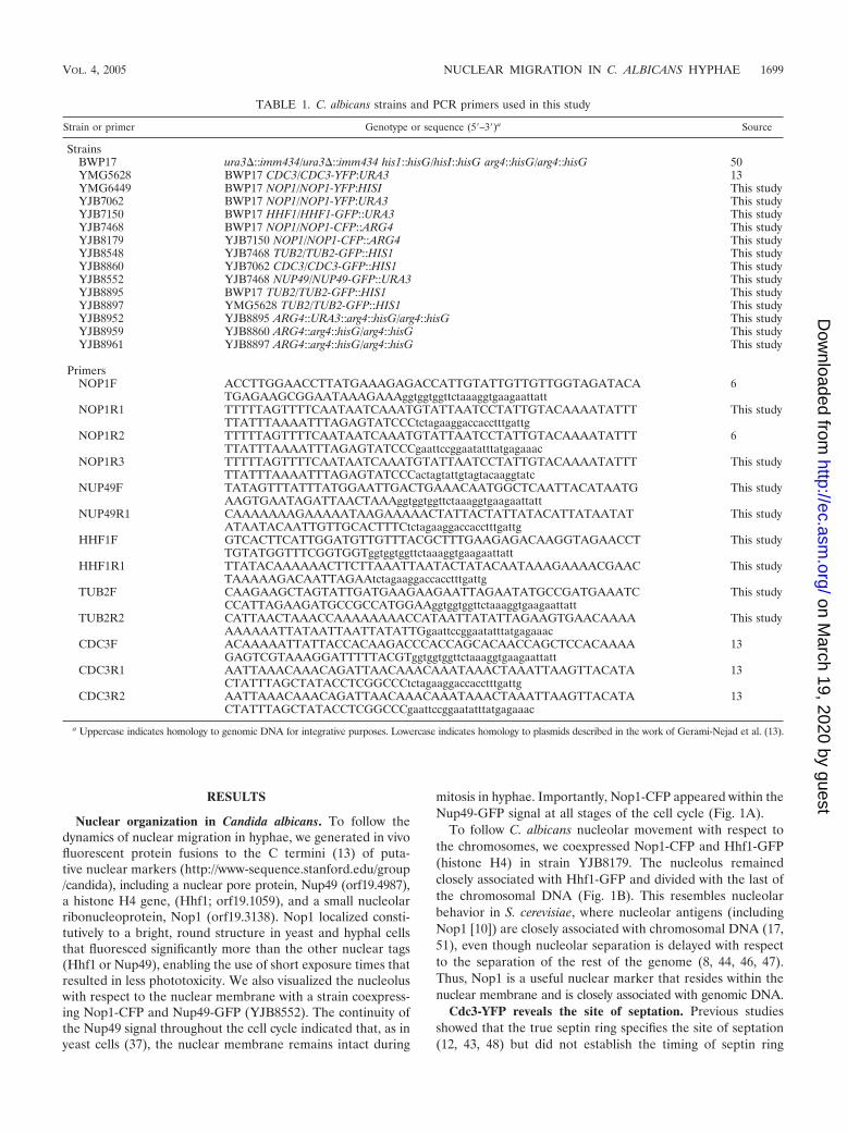
The dynamics of nuclear migration in hyphae. We nextquantified the real-time dynamics and properties of nuclearmigration (detected with Nop1-YFP in strain YJB6449) withrespect to the basal cell and the presumptum. We defined thehyphal neck position (y) as 0.0 �m, such that positions withinthe basal cell were negative values and positions within thegerm tube were positive values. Figure 2A shows representa-tive Nop1 behavior through the first hyphal cell cycle thatstarted from an unbudded cell and ended with the appearanceof the septum in the DIC channel (Fig. 2.mov at the above-named website). When Nop1 first entered the germ tube, itfrequently stalled and stretched at the neck, sometimes resolv-ing into separate Nop1 signals (e.g., Fig. 1B, 01:00 [where 01:00indicates 1 min, 0 s). These nucleoli, which likely reflect tran-sient separation of the two rRNA gene-containing chromo-somes, reassociated after they had passed the neck, and sub-sequent mitoses were unambiguous (Fig. 2A, 01:23, and2.mov).
Inspection of premitotic nuclear movement in individualhyphae revealed that forward nuclear migration to the pre-sumptum was nonlinear but that postmitotic movements wereless erratic (Fig. 2.mov and 2B). Postmitotically, the motherNop1 signal (mNop1) moved toward the basal cell while thedaughter Nop1 signal (dNop1) moved forward in the hypha.Interestingly, the movement of both nuclei stalled before the
FIG. 1. Nuclear organization and Cdc3 localization in C. albicans hyphae. (A, B) Hyphae were induced under coverslips, and images were takenat the times indicated (minutes:seconds) after hyphal induction. Hhf1-GFP and Nup49-GFP were imaged in the YFP channel to minimize CFPdetection. (A) Nop1-CFP (red) occupies a smaller area than the putative nuclear membrane (Nup49-GFP) (green) during all stages of the cellcycle. (B) The separation of Nop1-CFP occurs late in mitosis (01:15), and it remains in close proximity to Hhf1-GFP throughout the cell cycle(merge). Note that when Nop1 first migrates into the germ tube, the signal stretches and sometimes separates briefly (01:00; caret). (C) Cdc3localizes to the true septin ring at the time that the site of septation is specified. Images from a time-lapse movie of hyphae expressing Cdc3-YFP.Cdc3-YFP is detected at the hyphal tip (00:46; carat) before it localizes to the presumptum (arrowhead; 01:26). Subsequently, the septum appearsas a dark band by DIC microscopy (asterisk and inset; 02:52). Bars, 10 �m. Times are expressed as (hours:minutes).
1700 FINLEY AND BERMAN EUKARYOT. CELL
on March 19, 2020 by guest
http://ec.asm.org/
Dow
nloaded from
septum became visible (Fig. 2B, 01:45), implicating a commonmechanism of movement.
To ask about the general nature of nuclear movements, weaveraged the Nop1 data using the time of mitotic Nop1 sepa-ration as the common reference point (Fig. 2C) (n � 29). Wefound four rules of nuclear movement in these hyphae. First,nuclei never moved into germ tubes until after presumptumformation. Second, on average, Nop1 first divided when it waswithin 2.4 0.4 �m of the presumptum. Third, mitotic sepa-ration of the nuclei was biased toward the mother cell: themidpoint between mNop1 and dNop1 (Fig. 2C) did not per-fectly coincide with the presumptum (Fig. 2C). Surprisingly, in22/24 instances, mNop1 movement toward the basal cell stalledbefore septation, and nuclei remained within the germ tube,3.5 0.3 �m away from the basal cell. Only after septation didmNop1 migrate slowly into the basal cell (Fig. 2.mov and 2C).Together, these results show that premitotic forward nuclearmigration is nonlinear and initiates only after the site of sep-tation is specified. These results also show that nuclear move-ment away from the presumptum is rapid before septation andslow after septation and that mother nuclei tend to reenter thebasal cell slowly during this postseptation phase.
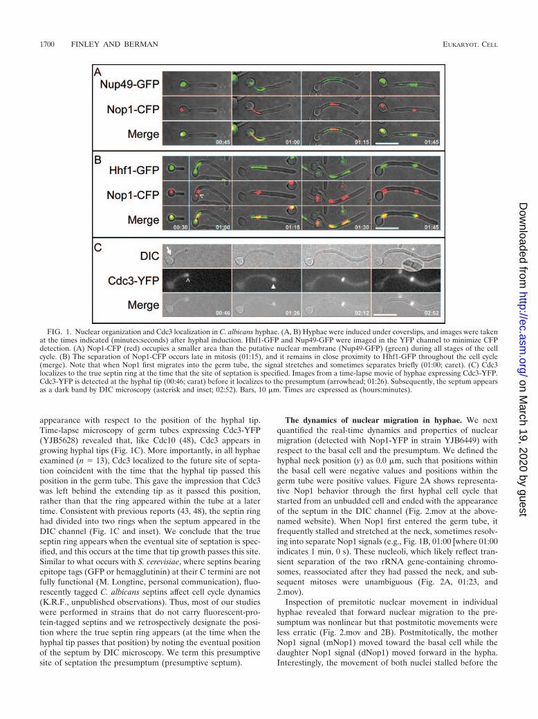
Nuclear migration is due to spindle dynamics. Microtubulesare likely to be responsible for the dynamics of C. albicansnuclei. Based on studies of S. cerevisiae, forward movement ofNop1 could be the result of MT plus-end tracking and/orplus-end depolymerization or it could result from MT slidingalong the cortex. The postmitotic movement of Nop1 signalscould be the result of similar processes or could be the resultof chromosome dynamics during spindle elongation. To iden-tify relevant periods of the cell cycle where MT and/or spindledynamics may mediate C. albicans nuclear migration and tocompare and contrast spindle dynamics with Nop1 dynamics,we performed time-lapse microscopy on hyphae expressingGFP fused to the -subunit of tubulin (Tub2; strain YJB8952),which labels SPBs, mitotic spindles, and MTs (19).
During early hyphal growth, an unduplicated SPB with orwithout astral MTs was sometimes evident (Fig. 3A, 01:01).During early hyphal emergence, however, individual MTs andunduplicated SPBs did not image well and were not alwaysdiscernible because of abundant short MTs that were not as-sociated with the SPB (see Fig. 9). Nonetheless, shortly afterpresumptum formation, SPBs suddenly became prominent(Fig. 3A, 01:31), which implies that SPB duplication occurs
FIG. 2. Dynamics of Nop1 nuclear migration relative to the presumptum. Hyphae expressing Nop1-YFP were analyzed at 2.5-min intervals bycapturing a single DIC image and a Z-stack of YFP images at each time point. (A) Selected frames from Fig. 2.mov show a representative exampleof Nop1 dynamics in a typical hypha. Nop1 remains in the basal cell until after the presumptum forms (asterisk; 00:45). Subsequently, Nop1migrates to the presumptum (position indicated by asterisks) and divides (01:23). Both mNop1 and dNop1 move away from the presumptum (01:23to 01:48) and mNop1 remains within the germ tube when septation occurs (asterisk; 01:48). Time is given as hours:minutes (hh:mm) from the timeof hyphal emergence. Bar, 10 �m. (B, C) The hyphal neck is defined as 0 �m. Negative values represent positions within the basal cell, whilepositive values represent positions within the germ tube. The dashed line represents the position of the presumptum, where the left end representsthe time that the hyphal tip extends beyond the eventual site of septation. The black arrowheads indicate the time that septation occurs. mNop1,circles; dNop1, triangles; midpoint of mNop1 and dNop1, plus signs. See the text for details. (B) Graphical representation of Nop1 position in Fig.2.mov. (C) Mean Nop1 positions were plotted against time by aligning the data at the time of mitotic Nop1 separation. Values are the averagesfor 29 cells. Dist., distance.
VOL. 4, 2005 NUCLEAR MIGRATION IN C. ALBICANS HYPHAE 1701
on March 19, 2020 by guest
http://ec.asm.org/
Dow
nloaded from

after the site of septation is specified. After entering the germtube, the duplicated SPBs adopted a short spindle configura-tion and migrated toward the presumptum in a series of non-linear steps (Fig. 3B). In this example, the short spindle mi-grated past the presumptum, into the daughter compartment,and back to the mother side prior to the onset of spindleelongation. Similarly, as spindles elongated, they were some-times displaced forward and backward without significantchanges in spindle length (Fig. 3B, arrows).
To compare spindle dynamics with nuclear dynamics, weaveraged the data from 15 cells and aligned the data at the timeof spindle disassembly, the final reference point common to allspindles (Fig. 3C). We found that the four observations madewith Nop1-YFP were almost identical for the studies withTub2-GFP. First, the SPB did not enter the germ tube untilafter presumptum formation. Second, short spindles beganelongating when they were within 1.7 0.7 �m (n � 15) of thepresumptum. Third, separation of the mitotic spindle was bi-ased toward the basal cell. The midpoint of the mitotic spindle(Fig. 3C) was closer to the basal cell during the early stages ofanaphase. Later in mitosis the bias was less evident. Finally, the
mother SPB (mSPB) approaches but does not enter the basalcell prior to septation. Thus, the data are consistent with thehypothesis that SPB-bound microtubules mediate the forwardspindle migration into the germ tube and that mitotic spindleelongation accounts for most of the movement that returns themother nucleus toward the basal cell prior to septation. How-ever, the reentry of the mother nucleus into the basal cellusually occurs postmitotically and is due to a mechanism thatmediates slower migration rates.
Cell cycle events occur at constant hyphal lengths. Strainscarrying fluorescent proteins fused to Tub2 have been reportedto grow more slowly than wild-type strains (19). Consistentwith this, the rate of hyphal-tip growth was lower in the Tub2-GFP strain (YJB8952; 0.23 0.02 �m/minute) than in theNop1-YFP strain (YJB6449; 0.37 0.01 �m/minute). Thisresulted in a significant increase (P value � 0.001) in mean cellcycle length (time from hyphal emergence to septation) inhyphae expressing Tub2-GFP (156.8 7.8 min, n � 15) com-pared to that of hyphae expressing Nop1-YFP (108.1 2.3min, n � 27). We hypothesized that the GFP moiety on -tu-bulin interfered with nuclear cell cycle events, universally pro-
FIG. 3. Spindle dynamics relative to the presumptum. Hyphae were induced and analyzed at 2.5-min intervals as for Fig. 2. (A) Selected framesfrom Fig. 3.mov show a representative example of spindle dynamics in a typical hypha. The SPB becomes prominent after the presumptum forms(01:31) and forms a short spindle (01:59) as it migrates forward in the germ tube. As the short spindle approaches the presumptum, spindleelongation ensues (02:22). The septum appears shortly after spindle disassembly (02:46). Asterisks indicate presumptum/septum position, andwhite arrowheads indicate SPBs. Time is measured in hours:minutes (hh:mm). Bar, 10 �m. (B, C) Chart values, dashed lines, and the blackarrowhead are as defined in the legend for Fig. 2. mSPB, circles; dSPB, triangles; spindle midpoint, plus signs; spindle length, multiplication signs.See the text for details. (B) Graphical representation of spindle position and spindle length from Fig. 3.mov. During mitosis, elongating mitoticspindles remain subject to external forces that move them asymmetrically about the presumptum (arrows). (C) Mean SPB positions were plottedagainst time by aligning the data at the time of spindle disassembly. Septation occurred 3.7 1.2 min after spindle disassembly (not shown).Valuesare the averages for 15 cells.
1702 FINLEY AND BERMAN EUKARYOT. CELL
on March 19, 2020 by guest
http://ec.asm.org/
Dow
nloaded from
tracting all cell cycle events and resulting in decreased produc-tion and/or delivery of hyphal growth stimuli to the hyphal tip.
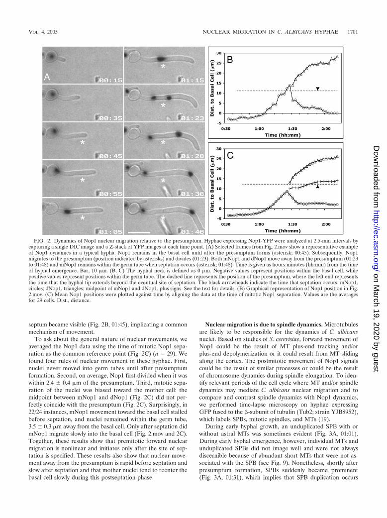
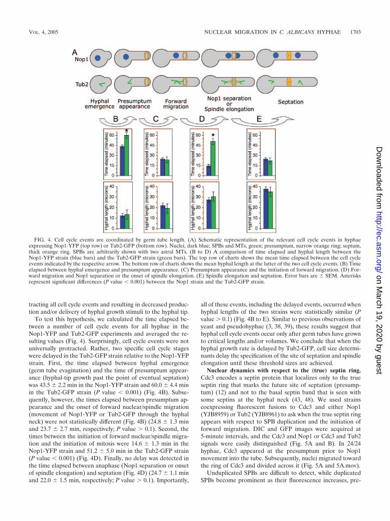
To test this hypothesis, we calculated the time elapsed be-tween a number of cell cycle events for all hyphae in theNop1-YFP and Tub2-GFP experiments and averaged the re-sulting values (Fig. 4). Surprisingly, cell cycle events were notuniversally protracted. Rather, two specific cell cycle stageswere delayed in the Tub2-GFP strain relative to the Nop1-YFPstrain. First, the time elapsed between hyphal emergence(germ tube evagination) and the time of presumptum appear-ance (hyphal-tip growth past the point of eventual septation)was 43.5 2.2 min in the Nop1-YFP strain and 60.0 4.4 minin the Tub2-GFP strain (P value � 0.001) (Fig. 4B). Subse-quently, however, the times elapsed between presumptum ap-pearance and the onset of forward nuclear/spindle migration(movement of Nop1-YFP or Tub2-GFP through the hyphalneck) were not statistically different (Fig. 4B) (24.8 1.3 minand 23.7 2.7 min, respectively; P value � 0.1). Second, thetimes between the initiation of forward nuclear/spindle migra-tion and the initiation of mitosis were 14.6 1.3 min in theNop1-YFP strain and 51.2 5.0 min in the Tub2-GFP strain(P value � 0.001) (Fig. 4D). Finally, no delay was detected inthe time elapsed between anaphase (Nop1 separation or onsetof spindle elongation) and septation (Fig. 4D) (24.7 1.1 minand 22.0 1.5 min, respectively; P value � 0.1). Importantly,
all of these events, including the delayed events, occurred whenhyphal lengths of the two strains were statistically similar (Pvalue � 0.1) (Fig. 4B to E). Similar to previous observations ofyeast and pseudohyphae (3, 38, 39), these results suggest thathyphal cell cycle events occur only after germ tubes have grownto critical lengths and/or volumes. We conclude that when thehyphal growth rate is delayed by Tub2-GFP, cell size determi-nants delay the specification of the site of septation and spindleelongation until these threshold sizes are achieved.
Nuclear dynamics with respect to the (true) septin ring.Cdc3 encodes a septin protein that localizes only to the trueseptin ring that marks the future site of septation (presump-tum) (12) and not to the basal septin band that is seen withsome septins at the hyphal neck (43, 48). We used strainscoexpressing fluorescent fusions to Cdc3 and either Nop1(YJB8959) or Tub2 (YJB8961) to ask when the true septin ringappears with respect to SPB duplication and the initiation offorward migration. DIC and GFP images were acquired at5-minute intervals, and the Cdc3 and Nop1 or Cdc3 and Tub2signals were easily distinguished (Fig. 5A and B). In 24/24hyphae, Cdc3 appeared at the presumptum prior to Nop1movement into the tube. Subsequently, nuclei migrated towardthe ring of Cdc3 and divided across it (Fig. 5A and 5A.mov).
Unduplicated SPBs are difficult to detect, while duplicatedSPBs become prominent as their fluorescence increases, pre-
FIG. 4. Cell cycle events are coordinated by germ tube length. (A) Schematic representation of the relevant cell cycle events in hyphaeexpressing Nop1-YFP (top row) or Tub2-GFP (bottom row). Nuclei, dark blue; SPBs and MTs, green; presumptum, narrow orange ring; septum,thick orange ring. SPBs are arbitrarily shown with two astral MTs. (B to D) A comparison of time elapsed and hyphal length between theNop1-YFP strain (blue bars) and the Tub2-GFP strain (green bars). The top row of charts shows the mean time elapsed between the cell cycleevents indicated by the respective arrow. The bottom row of charts shows the mean hyphal length at the latter of the two cell cycle events. (B) Timeelapsed between hyphal emergence and presumptum appearance. (C) Presumptum appearance and the initiation of forward migration. (D) For-ward migration and Nop1 separation or the onset of spindle elongation. (E) Spindle elongation and septation. Error bars are SEM. Asterisksrepresent significant differences (P value � 0.001) between the Nop1 strain and the Tub2-GFP strain.
VOL. 4, 2005 NUCLEAR MIGRATION IN C. ALBICANS HYPHAE 1703
on March 19, 2020 by guest
http://ec.asm.org/
Dow
nloaded from

sumably due to the incorporation of GFP-labeled tubulin intothe new SPB. We used this property to determine when SPBsduplicated with respect to the appearance of Cdc3 at the pre-sumptum. In 12/12 hyphae, the fluorescence of the SPB beganincreasing shortly after Cdc3 appeared at the presumptum(Fig. 5B, 00:50 to 01:11, and 5B.mov), suggesting that SPBduplication occurs after the appearance of the septin ring,although we cannot rule out the possibility that the appearanceof Cdc3 and the initiation of SPB duplication occur at the sametime. In either case, the two events are likely to be linked, andwe conclude that the site of septation in hyphae is specifiedeither at the same time as or shortly before SPB duplicationand prior to forward nuclear migration.
Forward nuclear migration is the result of repetitive MTsliding events. In S. cerevisiae, spindle alignment is facilitatedby the combination of three iterative mechanisms (reviewed inreference [22]): the tracking of MT plus ends along the cortex(23, 27), the depolymerization of MT plus ends at the cortex(35), and the sliding of MTs laterally associated with the cortex(1). The nonlinear nature of forward spindle migration sug-gests that C. albicans short spindles move forward into thegerm tube by iterative displacements. Short-interval (3-second)time-lapse microscopy of strain YJB8952, which carries Tub2-GFP, allowed us to distinguish between different types of MTdynamics associated with spindle movements. Selected framesfrom a movie (Fig. 6_1.mov at the above-named website) dem-onstrating the dynamics and quantification of the dynamics areshown in Fig. 6A and B, respectively. A second example ofspindle displacements resulting from repetitive MT slidingevents is also shown in Fig. 6_2.mov.
The major type of MT dynamics seen in germ tubes duringforward migration of the spindle was MT sliding. At 00:00, a shortspindle (SS) (19) was positioned at the hyphal neck, with a singleMT or MT bundle (referred to as MT) emanating from each pole.The MT bound to the daughter SPB (dSPB) associated laterallyalong the contour of the cortex and was oriented toward thehyphal tip, while the mSPB-bound MT remained oriented awayfrom the germ tube within the basal cell. The mSPB-bound MTbegan to depolymerize (cf. images at 00:00 and 00:08) while theSS moved forward into the germ tube (Fig. 6A, left column, and6_1.mov, 00:00 to 00:45). Interestingly, the dSPB-bound MT be-came longer during the time that the SS moved toward the hyphaltip (Fig. 6A). Thus, spindle displacements occur while MTs grow,and therefore forward movements cannot be due to MT depoly-merization. Furthermore, it is unlikely that tracking of MT plusends along the cortex is a major contributor to forward migrationas this process generally occurs without significant changes in MTlength (23, 27).
Detachment of this MT from the SPB (00:54) was accom-panied by the loss of forward movement (Fig. 6A, middlecolumn). Furthermore, the SS moved back toward the basalcell, most likely due to the mSPB-bound MT, which appearedat 00:36 and persisted until 1:38, when it began to depolymer-ize, finally disappearing at 02:05. Forward movement resumedwhen a new dSPB-bound MT appeared and grew (Fig. 6A,right column, 02:14). This repetitive behavior of MT growthand simultaneous displacement of the short spindle was ob-served in all (n � 13) hyphae that had short, migratory spin-dles. Although our analysis does not exclude minor contribu-tions from either plus-end tracking or MT depolymerization,
FIG. 5. Nuclear dynamics with respect to the true septin ring. (A) Hyphae expressing Nop1-YFP and Cdc3-GFP were imaged at 5-minuteintervals. Nop1 moved into the germ tube after Cdc3-GFP localized to the presumptum. Arrows indicate Nop1 just before (00:40) and after (00:45)mitosis. (B) Hyphae expressing Tub2-GFP and Cdc3-YFP were imaged at 5-minute intervals. SPB duplication (carats) was detected after Cdc3revealed the presumptum. Arrowheads indicate spindle poles. Time is indicated in hours:minutes. Bar, 10 �m.
1704 FINLEY AND BERMAN EUKARYOT. CELL
on March 19, 2020 by guest
http://ec.asm.org/
Dow
nloaded from

FIG. 6. Hyphal spindles align by repetitive MT sliding events. Hyphae were induced under coverslips for �90 min to enrich for hyphae withshort spindles. Using a preheated (37°C) 60� objective, a single DIC image was taken at the start of imaging and single-plane GFP images weretaken at 3-second intervals. (A) Selected frames from Fig. 6.mov. The distal end of a dSPB-bound MT (arrowheads; 00:00 to 00:45) moved towardthe hyphal tip faster than the short spindle that anchored it (slanted line; 00:00 to 00:45). At 00:54, the MT detached from the SPB, slid towardthe hyphal tip, and depolymerized (carats). Upon MT detachment, the short spindle moved back toward the basal cell (slanted line; 01:02 to 01:29),presumably due to the MT that grew from the mSPB (00:36 to 01:02). Time is indicated in minutes:seconds (mm:ss). Bar, 10 �m. (B) Quantificationof MT length and spindle displacement in panel A. The spindle moved toward the hyphal tip (squares) as MT length increased (circles). Whenthe SPB-bound MT detached from the SPB (asterisk), the dSPB moved back toward the basal cell, presumably due to the mSPB-bound MT tothat was mostly out of focus in the basal cell. Forward movement resumed when this MT depolymerized (01:56 to 02:05 in panel A), and a newMT grew from dSPB. Dist., distance.
VOL. 4, 2005 NUCLEAR MIGRATION IN C. ALBICANS HYPHAE 1705
on March 19, 2020 by guest
http://ec.asm.org/
Dow
nloaded from

we conclude that forward spindle migration is predominantlythe result of repetitive MT growth and sliding events.
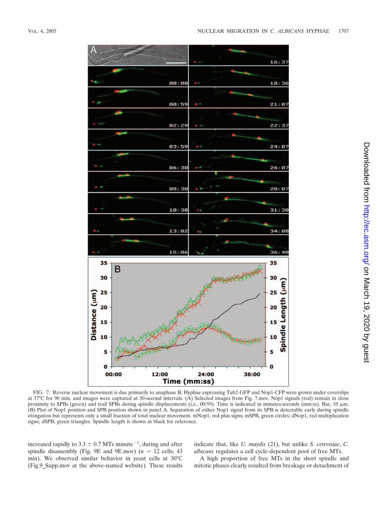
After chromosome separation, anaphase B (spindle elonga-tion) drives most nuclear movements. A remaining questionregarding the return of mSPB toward the basal cell is whetherthe movement is due largely to anaphase A (the shortening ofkinetochore MTs which moves chromosomes to the spindlepoles) or to anaphase B (the movement of the chromosomes asa function of SPB position during spindle elongation). If an-aphase A predominates, then spindle length would be longwhen the Nop1 signal divides. Furthermore, the separationbetween a Nop1 signal and its spindle pole would be relativelylarge. Alternatively, if anaphase B predominates, then spindlelength would be short when Nop1 divides and the Nop1 signalwould be relatively close to the SPB. We followed the dynamicsof SPBs and nucleoli in a strain coexpressing Tub2-GFP andNop1-CFP (YJB8548) and acquired DIC, CFP, and YFP im-ages of hyphae with migratory short spindles at 30-secondintervals.
A representative example of Nop1 behavior with respect tothe spindle is shown in Fig. 7. In all of the hyphae examined (n� 12), the Nop1 signal separated when spindles were short (3.1 0.31 �m). When short spindles were displaced within hy-phae, Nop1 exhibited similar movements with a slight delay(Fig. 7.mov), consistent with the idea that Nop1 position is afunction of SPB position. Nop1 signals maintain close, yetvariable, proximity to the short spindle prior to mitotic Nop1separation and to the respective SPBs after mitosis (Fig. 7B).We conclude that the movement of mNop1 toward the basalcell (reverse migration), as well as the forward movement ofdNop1 after Nop1 separation until spindle disassembly, is dueprimarily to forces that push the poles apart during anaphaseB. Additionally, repetitive forces acting upon astral MTs con-tribute to the overall position of the elongating spindle (Fig.7.mov), although that contribution is much more evident dur-ing premitotic forward migration than during postmitotic re-verse migration.
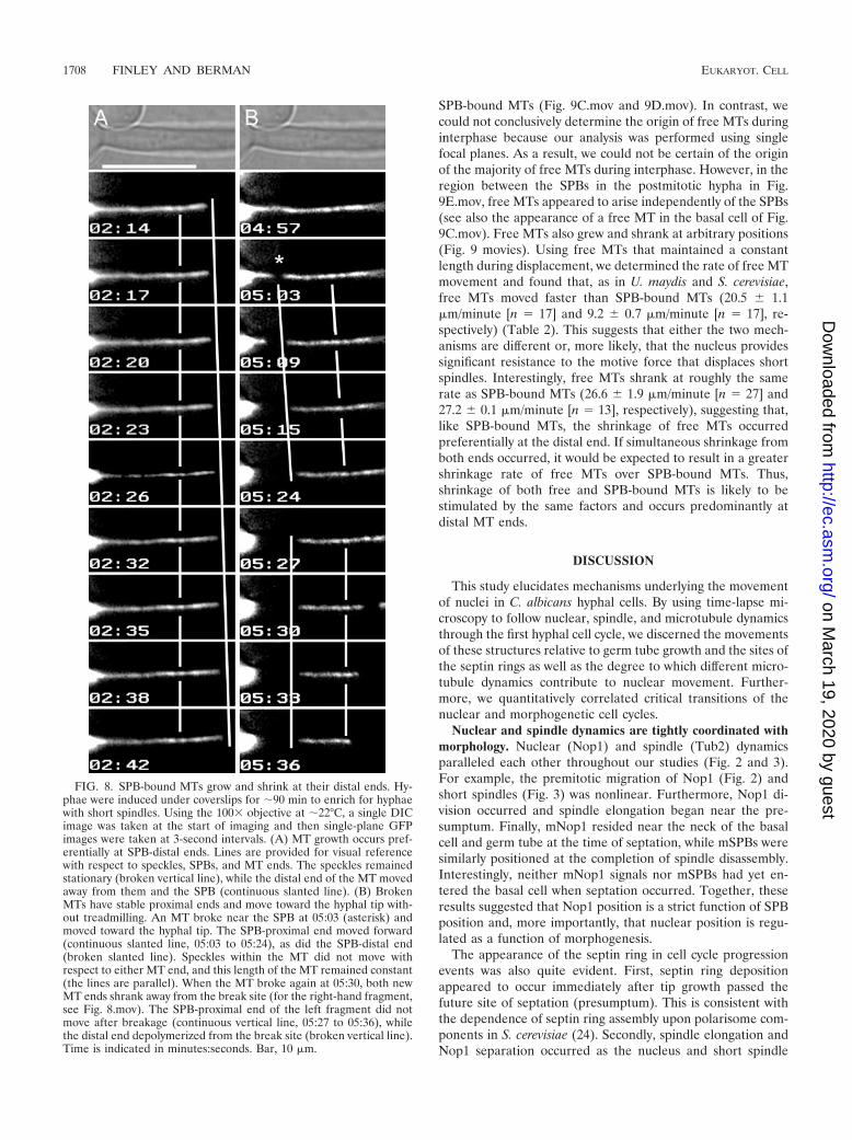
Polarity and stability of MT ends. The organization of the C.albicans MT cytoskeleton suggests that SPB-anchored MTsgrow and shrink preferentially at the ends distal to the SPB,while MT ends proximal to the SPB are stably associated withthe SPB. To test this hypothesis, we performed fluorescent-speckle microscopy (49) by analyzing MTs that had differentiallevels of incorporation of Tub2-GFP (speckles) and measuringthe movement of these speckles with respect to other MTlandmarks, such as SPBs or MT distal ends, to determinewhere subunit addition and/or removal were occurring (49). Aspeckled MT is shown in Fig. 8, which is an enlarged regionfrom movie Fig. 8.mov that also includes the distal end of thehypha. As the MT grew from 02:14 to 02:42, the specklesremained a constant distance from the SPB and from eachother, indicating that tubulin subunits were not being addedwithin the MT or at the SPB. Rather, the distal end movedfurther away from the speckles, indicating that MT growthoccurs at the distal ends of SPB-bound MTs.
Using FSM to analyze the fates of broken MTs also revealedimportant features regarding the relative stability of MT endsin C. albicans hyphae. In Fig. 8B (and Fig. 8.mov), the MTbroke (05:03). The remaining SPB-bound MT fragment shrankrapidly (05:03 to 05:24), while the free MT fragment moved
toward the hyphal tip (05:03 to 05:24). Importantly, there wasno change in either the interspeckle distances or the distancebetween a given speckle and the SPB-proximal end, indicatingthat the new SPB-proximal end of the free MT was stable afterbreakage. The subsequent movement of the free MT towardthe hyphal tip further indicated that free MTs are displaced bytranslocation toward their proximal ends, as opposed to tread-milling (the simultaneous removal of subunits from the prox-imal end and subunit addition at the distal end). SPB proximal-end stability upon breakage is not absolute, however. Whenthe free MT broke again (Fig. 8B, 05:27), it generated left andright fragments, both of which shrank from the break site.Importantly, however, the loss of speckles at the distal end ofthe left fragment occurred exclusively from the break site. Weconclude that SPB-bound MTs grow and shrink preferentiallyfrom their distal ends and that broken or released MTs gen-erally have stable ends and are transported, largely intact,toward the hyphal tips.
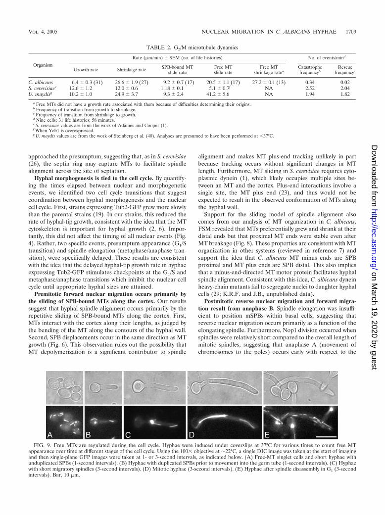
C. albicans microtubule dynamics differ from those of S.cerevisiae and U. maydis. The dynamics of the C. albicans MTcytoskeleton associated with the short spindle stage in hyphaeat 37°C are summarized in Table 2. We compared our resultswith data published for the S. cerevisiae MT cytoskeleton at37°C (1) and with data published for the yeast-like cells of U.maydis (40). MT dynamics were similar among these fungi inthat C. albicans MTs were in a perpetual state of flux termeddynamic instability, frequently switching between growing andshrinking states. However, in contrast to S. cerevisiae, whereMT growth and shrinkage occurred at approximately the samerates (1, 45), but like U. maydis, C. albicans MTs grew consid-erably more slowly (6.4 0.3 �m/minute; n � 31) than theyshrank (26.6 1.9 �m/minute; n � 27), spending 4.2-foldmore time in growth than in shrinkage.
While MT growth and shrinkage rates were comparableamong these organisms, C. albicans transition frequencies werestrikingly lower (Table 2). Transitions from growth to shrink-age (catastrophe) and from shrinkage to growth (rescue) oc-curred at 0.34 events minute�1 and 0.02 events minute�1,respectively, in C. albicans and at �2 events minute�1 and at�2 events minute�1, respectively, in the other two fungi. Thus,the rates of C. albicans MT growth and shrinkage are compa-rable to those of S. cerevisiae and U. maydis, while the transi-tion frequencies are very different.
The presence of free MTs is regulated during the cell cycle.During our analysis of MT dynamics, it became apparent thatsome cells had an abundance of free MTs but that others hadrelatively few. We asked if the behavior of free MTs was cellcycle dependent by quantifying the number of free MTs ob-served per minute throughout the cell cycle. Free MTs ap-peared at a rate of 2.8 0.2 MTs minute�1 prior to hyphalemergence and in short hyphae with unduplicated SPBs (Fig.9A and 9A.mov) (n � 14 cells; 56 min). Free MTs appeared atthe comparable rate of 3.4 0.3 MTs minute�1 in hyphae withduplicated SPBs that had not yet entered the germ tube (Fig.9B and 9B.mov) (n � 13 cells; 54 min). However, once shortspindles entered the germ tubes, the number of free MTsmin�1 dropped dramatically to 1.0 0.2 MTs minute�1 (Fig.9C and 9C.mov) (n � 13 cells; 61 min) and remained low at 0.9 0.1 MTs minute�1 through mitosis (Fig. 9D and 9D.mov) (n� 8 cells, 35 min). The frequency of free MT appearance
1706 FINLEY AND BERMAN EUKARYOT. CELL
on March 19, 2020 by guest
http://ec.asm.org/
Dow
nloaded from
increased rapidly to 3.3 0.7 MTs minute�1, during and afterspindle disassembly (Fig. 9E and 9E.mov) (n � 12 cells; 43min). We observed similar behavior in yeast cells at 30°C(Fig.9_Supp.mov at the above-named website). These results
indicate that, like U. maydis (21), but unlike S. cerevisiae, C.albicans regulates a cell cycle-dependent pool of free MTs.
A high proportion of free MTs in the short spindle andmitotic phases clearly resulted from breakage or detachment of
FIG. 7. Reverse nuclear movement is due primarily to anaphase B. Hyphae expressing Tub2-GFP and Nop1-CFP were grown under coverslipsat 37°C for 90 min, and images were captured at 30-second intervals. (A) Selected images from Fig. 7.mov. Nop1 signals (red) remain in closeproximity to SPBs (green) and trail SPBs during spindle displacements (i.e., 00:59). Time is indicated in minutes:seconds (mm:ss). Bar, 10 �m.(B) Plot of Nop1 position and SPB position shown in panel A. Separation of either Nop1 signal from its SPB is detectable early during spindleelongation but represents only a small fraction of total nuclear movement. mNop1, red plus signs; mSPB, green circles; dNop1, red multiplicationsigns; dSPB, green triangles. Spindle length is shown in black for reference.
VOL. 4, 2005 NUCLEAR MIGRATION IN C. ALBICANS HYPHAE 1707
on March 19, 2020 by guest
http://ec.asm.org/
Dow
nloaded from
SPB-bound MTs (Fig. 9C.mov and 9D.mov). In contrast, wecould not conclusively determine the origin of free MTs duringinterphase because our analysis was performed using singlefocal planes. As a result, we could not be certain of the originof the majority of free MTs during interphase. However, in theregion between the SPBs in the postmitotic hypha in Fig.9E.mov, free MTs appeared to arise independently of the SPBs(see also the appearance of a free MT in the basal cell of Fig.9C.mov). Free MTs also grew and shrank at arbitrary positions(Fig. 9 movies). Using free MTs that maintained a constantlength during displacement, we determined the rate of free MTmovement and found that, as in U. maydis and S. cerevisiae,free MTs moved faster than SPB-bound MTs (20.5 1.1�m/minute [n � 17] and 9.2 0.7 �m/minute [n � 17], re-spectively) (Table 2). This suggests that either the two mech-anisms are different or, more likely, that the nucleus providessignificant resistance to the motive force that displaces shortspindles. Interestingly, free MTs shrank at roughly the samerate as SPB-bound MTs (26.6 1.9 �m/minute [n � 27] and27.2 0.1 �m/minute [n � 13], respectively), suggesting that,like SPB-bound MTs, the shrinkage of free MTs occurredpreferentially at the distal end. If simultaneous shrinkage fromboth ends occurred, it would be expected to result in a greatershrinkage rate of free MTs over SPB-bound MTs. Thus,shrinkage of both free and SPB-bound MTs is likely to bestimulated by the same factors and occurs predominantly atdistal MT ends.
DISCUSSION
This study elucidates mechanisms underlying the movementof nuclei in C. albicans hyphal cells. By using time-lapse mi-croscopy to follow nuclear, spindle, and microtubule dynamicsthrough the first hyphal cell cycle, we discerned the movementsof these structures relative to germ tube growth and the sites ofthe septin rings as well as the degree to which different micro-tubule dynamics contribute to nuclear movement. Further-more, we quantitatively correlated critical transitions of thenuclear and morphogenetic cell cycles.
Nuclear and spindle dynamics are tightly coordinated withmorphology. Nuclear (Nop1) and spindle (Tub2) dynamicsparalleled each other throughout our studies (Fig. 2 and 3).For example, the premitotic migration of Nop1 (Fig. 2) andshort spindles (Fig. 3) was nonlinear. Furthermore, Nop1 di-vision occurred and spindle elongation began near the pre-sumptum. Finally, mNop1 resided near the neck of the basalcell and germ tube at the time of septation, while mSPBs weresimilarly positioned at the completion of spindle disassembly.Interestingly, neither mNop1 signals nor mSPBs had yet en-tered the basal cell when septation occurred. Together, theseresults suggested that Nop1 position is a strict function of SPBposition and, more importantly, that nuclear position is regu-lated as a function of morphogenesis.
The appearance of the septin ring in cell cycle progressionevents was also quite evident. First, septin ring depositionappeared to occur immediately after tip growth passed thefuture site of septation (presumptum). This is consistent withthe dependence of septin ring assembly upon polarisome com-ponents in S. cerevisiae (24). Secondly, spindle elongation andNop1 separation occurred as the nucleus and short spindle
FIG. 8. SPB-bound MTs grow and shrink at their distal ends. Hy-phae were induced under coverslips for �90 min to enrich for hyphaewith short spindles. Using the 100� objective at �22°C, a single DICimage was taken at the start of imaging and then single-plane GFPimages were taken at 3-second intervals. (A) MT growth occurs pref-erentially at SPB-distal ends. Lines are provided for visual referencewith respect to speckles, SPBs, and MT ends. The speckles remainedstationary (broken vertical line), while the distal end of the MT movedaway from them and the SPB (continuous slanted line). (B) BrokenMTs have stable proximal ends and move toward the hyphal tip with-out treadmilling. An MT broke near the SPB at 05:03 (asterisk) andmoved toward the hyphal tip. The SPB-proximal end moved forward(continuous slanted line, 05:03 to 05:24), as did the SPB-distal end(broken slanted line). Speckles within the MT did not move withrespect to either MT end, and this length of the MT remained constant(the lines are parallel). When the MT broke again at 05:30, both newMT ends shrank away from the break site (for the right-hand fragment,see Fig. 8.mov). The SPB-proximal end of the left fragment did notmove after breakage (continuous vertical line, 05:27 to 05:36), whilethe distal end depolymerized from the break site (broken vertical line).Time is indicated in minutes:seconds. Bar, 10 �m.
1708 FINLEY AND BERMAN EUKARYOT. CELL
on March 19, 2020 by guest
http://ec.asm.org/
Dow
nloaded from
approached the presumptum, suggesting that, as in S. cerevisiae(26), the septin ring may capture MTs to facilitate spindlealignment across the site of septation.
Hyphal morphogenesis is tied to the cell cycle. By quantify-ing the times elapsed between nuclear and morphogeneticevents, we identified two cell cycle transitions that suggestcoordination between hyphal morphogenesis and the nuclearcell cycle. First, strains expressing Tub2-GFP grew more slowlythan the parental strains (19). In our strains, this reduced therate of hyphal-tip growth, consistent with the idea that the MTcytoskeleton is important for hyphal growth (2, 6). Impor-tantly, this did not affect the timing of all nuclear events (Fig.4). Rather, two specific events, presumptum appearance (G1/Stransition) and spindle elongation (metaphase/anaphase tran-sition), were specifically delayed. These results are consistentwith the idea that the delayed hyphal-tip growth rate in hyphaeexpressing Tub2-GFP stimulates checkpoints at the G1/S andmetaphase/anaphase transitions which inhibit the nuclear cellcycle until appropriate hyphal sizes are attained.
Premitotic forward nuclear migration occurs primarily bythe sliding of SPB-bound MTs along the cortex. Our resultssuggest that hyphal spindle alignment occurs primarily by therepetitive sliding of SPB-bound MTs along the cortex. First,MTs interact with the cortex along their lengths, as judged bythe bending of the MT along the contours of the hyphal wall.Second, SPB displacements occur in the same direction as MTgrowth (Fig. 6). This observation rules out the possibility thatMT depolymerization is a significant contributor to spindle
alignment and makes MT plus-end tracking unlikely in partbecause tracking occurs without significant changes in MTlength. Furthermore, MT sliding in S. cerevisiae requires cyto-plasmic dynein (1), which likely occupies multiple sites be-tween an MT and the cortex. Plus-end interactions involve asingle site, the MT plus end (23), and thus would not beexpected to result in the observed conformation of MTs alongthe hyphal wall.
Support for the sliding model of spindle alignment alsocomes from our analysis of MT organization in C. albicans.FSM revealed that MTs preferentially grew and shrank at theirdistal ends but that proximal MT ends were stable even afterMT breakage (Fig. 8). These properties are consistent with MTorganization in other systems (reviewed in reference 7) andsupport the idea that C. albicans MT minus ends are SPBproximal and MT plus ends are SPB distal. This also impliesthat a minus-end-directed MT motor protein facilitates hyphalspindle alignment. Consistent with this idea, C. albicans dyneinheavy-chain mutants fail to segregate nuclei to daughter hyphalcells (29; K.R.F. and J.B., unpublished data).
Postmitotic reverse nuclear migration and forward migra-tion result from anaphase B. Spindle elongation was insuffi-cient to position mSPBs within basal cells, suggesting thatreverse nuclear migration occurs primarily as a function of theelongating spindle. Furthermore, Nop1 division occurred whenspindles were relatively short compared to the overall length ofmitotic spindles, suggesting that anaphase A (movement ofchromosomes to the poles) occurs early with respect to the
FIG. 9. Free MTs are regulated during the cell cycle. Hyphae were induced under coverslips at 37°C for various times to count free MTappearance over time at different stages of the cell cycle. Using the 100� objective at �22°C, a single DIC image was taken at the start of imagingand then single-plane GFP images were taken at 1- or 3-second intervals, as indicated below. (A) Free-MT singlet cells and short hyphae withunduplicated SPBs (1-second intervals). (B) Hyphae with duplicated SPBs prior to movement into the germ tube (1-second intervals). (C) Hyphaewith short migratory spindles (3-second intervals). (D) Mitotic hyphae (3-second intervals). (E) Hyphae after spindle disassembly in G1 (3-secondintervals). Bar, 10 �m.
TABLE 2. G2/M microtubule dynamics
Organism
Rate (�m/min) SEM (no. of life histories) No. of events/mind
Growth rate Shrinkage rate SPB-bound MTslide rate
Free MTslide rate
Free MTshrinkage ratea
Catastrophefrequencyb
Rescuefrequencyc
C. albicans 6.4 0.3 (31) 26.6 1.9 (27) 9.2 0.7 (17) 20.5 1.1 (17) 27.2 0.1 (13) 0.34 0.02S. cerevisiaee 12.6 1.2 12.0 0.6 1.18 0.1 5.1 0.7f NA 2.52 2.04U. maydisg 10.2 1.0 24.9 3.7 9.3 2.4 41.2 5.6 NA 1.94 1.82
a Free MTs did not have a growth rate associated with them because of difficulties determining their origins.b Frequency of transition from growth to shrinkage.c Frequency of transition from shrinkage to growth.d Nine cells; 31 life histories; 58 minutes.e S. cerevisiae values are from the work of Adames and Cooper (1).f When Yeb1 is overexpressed.g U. maydis values are from the work of Steinberg et al. (40). Analyses are presumed to have been performed at �37°C.
VOL. 4, 2005 NUCLEAR MIGRATION IN C. ALBICANS HYPHAE 1709
on March 19, 2020 by guest
http://ec.asm.org/
Dow
nloaded from

onset of anaphase B (movement of chromosomes due to spin-dle elongation). As a result, anaphase A makes little contribu-tion to total nuclear movement in hyphae and reverse nuclearmigration (as well as forward nuclear migration after chromo-some separation) results from spindle elongation.
Free MTs are cell cycle regulated. Most fungi exhibit spin-dle-independent MT organization. For example, in the fissionyeast S. pombe, two microtubule-organizing centers at the cellequator organize SPB-independent MT arrays during inter-phase (18). In U. maydis, polar microtubule-organizing centersnucleate SPB-independent MTs during interphase (41). Inboth of these fungi, the interphase organization of MTs disap-pears during mitosis. We found a similar phenomenon in C.albicans yeast and hyphal cells: free MTs were abundant duringinterphase, much less prevalent during G2 and mitosis, andrapidly reappeared during spindle disassembly.
Microtubules are required for the maintenance of the Spit-zenkorper in C. albicans (6). We considered the possibility thatfree MTs might support hyphal growth but considered thisunlikely because hyphal growth is linear throughout the cellcycle (15). Similarly, in Aspergilllus nidulans, cytoplasmic MTsare cell cycle regulated, yet hyphal growth rates do not de-crease (21, 32). Thus, we propose that free MTs constitute acytoplasmic pool of tubulin that facilitates the assembly of thelong mitotic spindles observed in C. albicans hyphae.
Model of nuclear migration. We propose the followingmodel for nuclear and MT dynamics in C. albicans hyphae(Fig. 10). During G0/G1, tubulin saturates the cytosol of un-budded cells and short hyphae. Once a hypha reaches a criticalsize, it transitions into S phase: the septin ring appears, SPBsduplicate, and the MT sliding machinery is activated. As the
hypha proceeds into G2, tubulin is incorporated into the de-veloping spindle and the number of free MTs is reduced. Alsoin G2, repetitive sliding events from SPB-bound MTs facilitatenuclear migration to the septin ring. In early M phase, theshort spindle approaches the ring and the hypha transitionsinto anaphase. This transition also is dependent upon the hy-pha reaching a critical size. During anaphase A, kinetochoreMTs shorten and pull the chromosomes the short distance tothe spindle poles. Subsequently, during anaphase B, interpolarMTs push the spindle poles apart, preferentially pushing themother nucleus toward the hyphal neck. As the spindle disas-sembles, the concentration of cytosolic tubulin reaches satura-tion and free MTs reappear. Following septation, a slower,unknown mechanism moves the G0 mother nucleus back intothe basal cell. Thus, in C. albicans hyphae, nuclei and MTsexhibit behaviors that, in some ways, resemble those of S.cerevisiae (appearance of the septin ring prior to nuclear move-ment and nuclear movement to and division across the septinring) and in other ways are more like those of other organisms(cell cycle-regulated MT cytoskeleton).
ACKNOWLEDGMENTS
We thank Maryam Gerami-Nejad for strain construction and CherylGale and Maryam Gerami-Nejad for providing plasmids and data priorto publication. We thank Mark Longtine for communication of un-published data. We are grateful to Mark McClellan for his technicalexpertise. We thank Pete Sudbery, Tom Hays, Duncan Clarke, andCheryl Gale for critical reading of the manuscript.
This work was supported by NIH grant AI/DE 14666 to J.B. K.R.F.was supported, in part, by NIH Biotechnology Training GrantGM08347.
FIG. 10. Model of nuclear and microtubule dynamics during a hyphal cell cycle in C. albicans. See the text for details. Cellular structures areas defined for Fig. 4, except that SPBs are yellow. Arrows (G2) reflect premitotic nuclear migration.
1710 FINLEY AND BERMAN EUKARYOT. CELL
on March 19, 2020 by guest
http://ec.asm.org/
Dow
nloaded from
REFERENCES
1. Adames, N. R., and J. A. Cooper. 2000. Microtubule interactions with the cellcortex causing nuclear movements in Saccharomyces cerevisiae. J. Cell Biol.149:863–874.
2. Akashi, T., T. Kanbe, and K. Tanaka. 1994. The role of the cytoskeleton inthe polarized growth of the germ tube in Candida albicans. Microbiology140:271–280.
3. Bedell, G. W., A. Werth, and D. R. Soll. 1980. The regulation of nuclearmigration and division during synchronous bud formation in released sta-tionary phase cultures of the yeast Candida albicans. Exp. Cell Res. 127:103–113.
4. Byers, B., and L. Goetsch. 1975. Behavior of spindles and spindle plaques inthe cell cycle and conjugation of Saccharomyces cerevisiae. J. Bacteriol. 124:511–523.
5. Carminati, J. L., and T. Stearns. 1997. Microtubules orient the mitoticspindle in yeast through dynein-dependent interactions with the cell cortex.J. Cell Biol. 138:629–641.
6. Crampin, H., K. Finley, M. Gerami-Nejad, H. Court, C. Gale, J. Berman,and P. Sudbery. 2005. Candida albicans hyphae have a Spitzenkorper that isdistinct from the polarisome found in yeast and pseudohyphae. J. Cell Sci.118:2935–2947.
7. Dammermann, A., A. Desai, and K. Oegema. 2003. The minus end in sight.Curr. Biol. 13:R614–R624.
8. D’Amours, D., F. Stegmeier, and A. Amon. 2004. Cdc14 and condensincontrol the dissolution of cohesin-independent chromosome linkages at re-peated DNA. Cell 117:455–469.
9. Davis, D., J. E. Edwards, Jr., A. P. Mitchell, and A. S. Ibrahim. 2000.Candida albicans RIM101 pH response pathway is required for host-patho-gen interactions. Infect. Immun. 68:5953–5959.
10. Dvorkin, N., M. W. Clark, and B. A. Hamkalo. 1991. Ultrastructural local-ization of nucleic acid sequences in Saccharomyces cerevisiae nucleoli. Chro-mosoma 100:519–523.
11. Endow, S. A., and D. J. Komma. 1997. Spindle dynamics during meiosis inDrosophila oocytes. J. Cell Biol. 137:1321–1336.
12. Gale, C., M. Gerami-Nejad, M. McClellan, S. Vandoninck, M. S. Longtine,and J. Berman. 2001. Candida albicans Int1p interacts with the septin ring inyeast and hyphal cells. Mol. Biol. Cell. 12:3538–3549.
13. Gerami-Nejad, M., J. Berman, and C. A. Gale. 2001. Cassettes for PCR-mediated construction of green, yellow, and cyan fluorescent protein fusionsin Candida albicans. Yeast 18:859–864.
14. Gow, N. A., A. J. Brown, and F. C. Odds. 2002. Fungal morphogenesis andhost invasion. Curr. Opin. Microbiol. 5:366–371.
15. Gow, N. A., and G. W. Gooday. 1982. Vacuolation, branch production andlinear growth of germ tubes in Candida albicans. J. Gen. Microbiol. 128:2195–2198.
16. Gow, N. A., G. Henderson, and G. W. Gooday. 1986. Cytological interrela-tionships between the cell cycle and duplication cycle of Candida albicans.Microbios 47:97–105.
17. Granot, D., and M. Snyder. 1991. Segregation of the nucleolus during mitosisin budding and fission yeast. Cell Motil. Cytoskel. 20:47–54.
18. Hagan, I. M., and J. S. Hyams. 1988. The use of cell division cycle mutantsto investigate the control of microtubule distribution in the fission yeastSchizosaccharomyces pombe. J. Cell Sci. 89:343–357.
19. Hazan, I., M. Sepulveda-Becerra, and H. Liu. 2002. Hyphal elongation isregulated independently of cell cycle in Candida albicans. Mol. Biol. Cell.13:134–145.
20. Hogan, C. J., H. Wein, L. Wordeman, J. M. Scholey, K. E. Sawin, and W. Z.Cande. 1993. Inhibition of anaphase spindle elongation in vitro by a peptideantibody that recognizes kinesin motor domain. Proc. Natl. Acad. Sci. USA90:6611–6615.
21. Horio, T., and B. R. Oakley. 2005. The role of microtubules in rapid hyphaltip growth of Aspergillus nidulans. Mol. Biol. Cell. 16:918–926.
22. Huisman, S. M., and M. Segal. 2005. Cortical capture of microtubules andspindle polarity in budding yeast—where’s the catch? J. Cell Sci. 118:463–471.
23. Hwang, E., J. Kusch, Y. Barral, and T. C. Huffaker. 2003. Spindle orientationin Saccharomyces cerevisiae depends on the transport of microtubule endsalong polarized actin cables. J. Cell Biol. 161:483–488.
24. Kadota, J., T. Yamamoto, S. Yoshiuchi, E. Bi, and K. Tanaka. 2004. Septinring assembly requires concerted action of polarisome components, a PAKkinase Cla4p, and the actin cytoskeleton in Saccharomyces cerevisiae. Mol.Biol. Cell. 15:5329–5345.
25. Kline-Smith, S. L., and C. E. Walczak. 2004. Mitotic spindle assembly andchromosome segregation: refocusing on microtubule dynamics. Mol. Cell.15:317–327.
26. Kusch, J., A. Meyer, M. P. Snyder, and Y. Barral. 2002. Microtubule captureby the cleavage apparatus is required for proper spindle positioning in yeast.Genes Dev. 16:1627–1639.
27. Liakopoulos, D., J. Kusch, S. Grava, J. Vogel, and Y. Barral. 2003. Asym-metric loading of Kar9 onto spindle poles and microtubules ensures properspindle alignment. Cell 112:561–574.
28. Lo, H. J., J. R. Kohler, B. DiDomenico, D. Loebenberg, A. Cacciapuoti, andG. R. Fink. 1997. Nonfilamentous C. albicans mutants are avirulent. Cell90:939–949.
29. Martin, R., A. Walther, and J. Wendland. 2004. Deletion of the Dyneinheavy-chain gene DYN1 leads to aberrant nuclear positioning and defectivehyphal development in Candida albicans. Eukaryot. Cell 3:1574–1588.
30. Mitchison, T. J., and E. D. Salmon. 1992. Poleward kinetochore fiber move-ment occurs during both metaphase and anaphase-A in newt lung cell mi-tosis. J. Cell Biol. 119:569–582.
31. Pearson, C. G., P. S. Maddox, E. D. Salmon, and K. Bloom. 2001. Buddingyeast chromosome structure and dynamics during mitosis. J. Cell Biol. 152:1255–1266.
32. Riquelme, M., R. Fischer, and S. Bartnicki-Garcia. 2003. Apical growth andmitosis are independent processes in Aspergillus nidulans. Protoplasma 222:211–215.
33. Rusan, N. M., C. J. Fagerstrom, A. M. Yvon, and P. Wadsworth. 2001. Cellcycle-dependent changes in microtubule dynamics in living cells expressinggreen fluorescent protein-alpha tubulin. Mol. Biol. Cell. 12:971–980.
34. Saville, S. P., A. L. Lazzell, C. Monteagudo, and J. L. Lopez-Ribot. 2003.Engineered control of cell morphology in vivo reveals distinct roles for yeastand filamentous forms of Candida albicans during infection. Eukaryot. Cell2:1053–1060.
35. Shaw, S. L., E. Yeh, P. Maddox, E. D. Salmon, and K. Bloom. 1997. Astralmicrotubule dynamics in yeast: a microtubule-based searching mechanismfor spindle orientation and nuclear migration into the bud. J. Cell Biol.139:985–994.
36. Sherman, F. 1991. Getting started with yeast. Methods Enzymol. 194:3–21.37. Soll, D. R. 1985. Candida albicans, p. 167–195. In P. J. Szaniszlo (ed.), Fungal
dimorphism: with emphasis on fungi pathogenic to humans. Plenum Pub-lishing Co., New York, N.Y.
38. Soll, D. R., G. Bedell, J. Thiel, and M. Brummel. 1981. The dependency ofnuclear division on volume in the dimorphic yeast Candida albicans. Exp.Cell Res. 133:55–62.
39. Soll, D. R., M. Stasi, and G. Bedell. 1978. The regulation of nuclear migra-tion and division during pseudo-mycelium outgrowth in the dimorphic yeastCandida albicans. Exp. Cell Res. 116:207–215.
40. Steinberg, G., R. Wedlich-Soldner, M. Brill, and I. Schulz. 2001. Microtu-bules in the fungal pathogen Ustilago maydis are highly dynamic and deter-mine cell polarity. J. Cell Sci. 114:609–622.
41. Straube, A., M. Brill, B. R. Oakley, T. Horio, and G. Steinberg. 2003.Microtubule organization requires cell cycle-dependent nucleation at dis-persed cytoplasmic sites: polar and perinuclear microtubule organizing cen-ters in the plant pathogen Ustilago maydis. Mol. Biol. Cell. 14:642–657.
42. Sudbery, P., N. Gow, and J. Berman. The distinct morphogenic states ofCandida albicans. Trends Microbiol. 12:317–324, 2004.
43. Sudbery, P. E. 2001. The germ tubes of Candida albicans hyphae andpseudohyphae show different patterns of septin ring localization. Mol. Mi-crobiol. 41:19–31.
44. Sullivan, M., T. Higuchi, V. L. Katis, and F. Uhlmann. 2004. Cdc14 phos-phatase induces rDNA condensation and resolves cohesin-independent co-hesion during budding yeast anaphase. Cell 117:471–482.
45. Tirnauer, J. S., E. O’Toole, L. Berrueta, B. E. Bierer, and D. Pellman. 1999.Yeast Bim1p promotes the G1-specific dynamics of microtubules. J. CellBiol. 145:993–1007.
46. Torres-Rosell, J., F. Machin, A. Jarmuz, and L. Aragon. 2004. Nucleolarsegregation lags behind the rest of the genome and requires Cdc14p activa-tion by the FEAR network. Cell Cycle. 3:496–502.
47. Wang, B. D., V. Yong-Gonzalez, and A. V. Strunnikov. 2004. Cdc14p/FEARpathway controls segregation of nucleolus in S. cerevisiae by facilitatingcondensin targeting to rDNA chromatin in anaphase. Cell Cycle. 3:960–967.
48. Warenda, A. J., and J. B. Konopka. 2002. Septin function in Candida albicansmorphogenesis. Mol. Biol. Cell 13:2732Ð2746.
49. Waterman-Storer, C. M., A. Desai, J. C. Bulinski, and E. D. Salmon. 1998.Fluorescent speckle microscopy, a method to visualize the dynamics of pro-tein assemblies in living cells. Curr. Biol. 8:1227–1230.
50. Wilson, R. B., D. Davis, and A. P. Mitchell. 1999. Rapid hypothesis testingwith Candida albicans through gene disruption with short homology regions.J. Bacteriol. 181:1868–1874.
51. Yang, C. H., E. J. Lambie, J. Hardin, J. Craft, and M. Snyder. 1989. Higherorder structure is present in the yeast nucleus: autoantibody probes demon-strate that the nucleolus lies opposite the spindle pole body. Chromosoma98:123–128.
52. Yokoyama, K., H. Kaji, K. Nishimura, and M. Miyaji. 1990. The role ofmicrofilaments and microtubules in apical growth and dimorphism of Can-dida albicans. J. Gen. Microbiol. 136:1067–1075.
VOL. 4, 2005 NUCLEAR MIGRATION IN C. ALBICANS HYPHAE 1711
on March 19, 2020 by guest
http://ec.asm.org/
Dow
nloaded from