Embed Size (px)
Citation preview
Department of Plastic and Maxillofacial Surgery
The Royal Children’s Hospital, Melbourne, Australia
microtia & ear anomaliesin children

ear anomalies in children 1
microtia & ear anomaliesin children
The Department of Plastic and Maxillofacial Surgery provides
comprehensive care to children with a wide range of birth defects
and deformities after accidents. Since 1970, our group of highly
trained and specialized surgeons has cared for thousands of patients
and their families. Our microtia team aim to provide world leading care
for patients with congenital or accident related deformities of the ear
and is one of a handful of clinics worldwide to provide such a service.
This booklet was prepared by Mr Nicholas Lotz FRACS, craniofacial
fellow, in conjunction with plastic surgeons Mr Chris Coombs FRACS and
Mr Andrew Greensmith FRACS, and ENT surgeons Mr Rob Briggs FRACS
and Mr Markus Dahm FRACS.
ear anomalies in children 2 ear anomalies in children 3
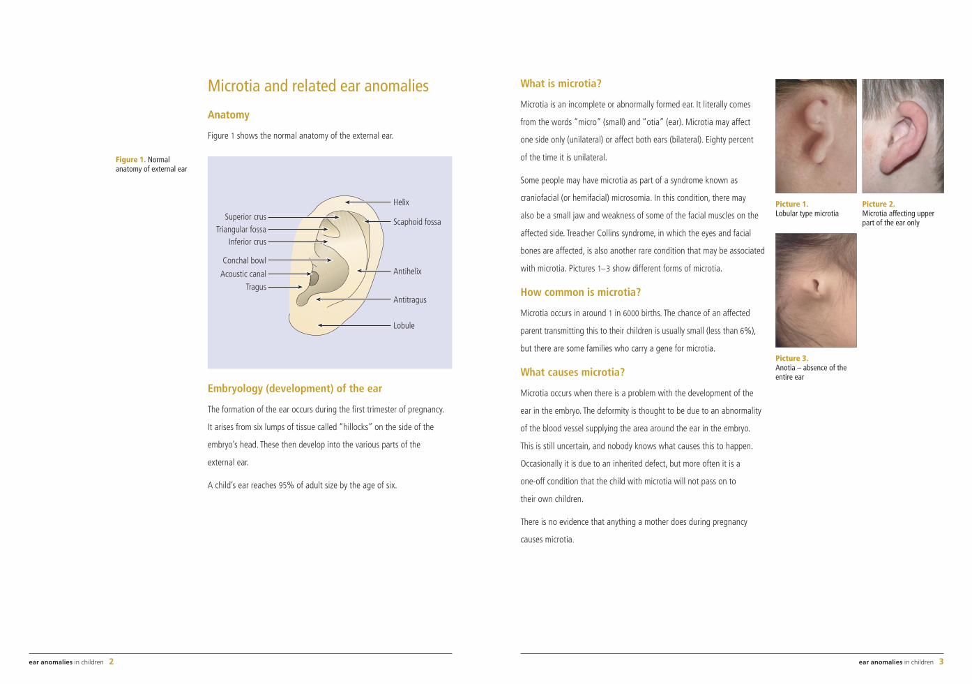
Microtia and related ear anomalies
Anatomy
Figure 1 shows the normal anatomy of the external ear.
Embryology (development) of the ear
The formation of the ear occurs during the first trimester of pregnancy.
It arises from six lumps of tissue called “hillocks” on the side of the
embryo’s head. These then develop into the various parts of the
external ear.
A child’s ear reaches 95% of adult size by the age of six.
What is microtia?
Microtia is an incomplete or abnormally formed ear. It literally comes
from the words “micro” (small) and “otia” (ear). Microtia may affect
one side only (unilateral) or affect both ears (bilateral). Eighty percent
of the time it is unilateral.
Some people may have microtia as part of a syndrome known as
craniofacial (or hemifacial) microsomia. In this condition, there may
also be a small jaw and weakness of some of the facial muscles on the
affected side. Treacher Collins syndrome, in which the eyes and facial
bones are affected, is also another rare condition that may be associated
with microtia. Pictures 1–3 show different forms of microtia.
How common is microtia?
Microtia occurs in around 1 in 6000 births. The chance of an affected
parent transmitting this to their children is usually small (less than 6%),
but there are some families who carry a gene for microtia.
What causes microtia?
Microtia occurs when there is a problem with the development of the
ear in the embryo. The deformity is thought to be due to an abnormality
of the blood vessel supplying the area around the ear in the embryo.
This is still uncertain, and nobody knows what causes this to happen.
Occasionally it is due to an inherited defect, but more often it is a
one-off condition that the child with microtia will not pass on to
their own children.
There is no evidence that anything a mother does during pregnancy
causes microtia.
Figure 1. Normal anatomy of external ear
Antihelix
Lobule
Antitragus
Helix
Scaphoid fossaSuperior crusTriangular fossa
Conchal bowl
Acoustic canal
Tragus
Inferior crus
Picture 1. Lobular type microtia
Picture 2. Microtia affecting upper part of the ear only
Picture 3. Anotia – absence of the entire ear
ear anomalies in children 4 ear anomalies in children 5
Is the hearing affected?
In some cases of microtia the hearing may be normal or partly reduced
and not require any treatment. Patients with microtia may not have an
external ear canal, but this does not necessarily mean that they cannot
hear on that side, as the inner ear may be normal. In others, there may be
problems with the formation of the middle ear which can affect hearing.
Children with unilateral microtia with absence of the ear canal on that
side will often have problems identifying which direction a sound is
coming from.
Your child will need to have audiograms to test their hearing, and also
a CT scan to look at the small bones in the middle ear to determine
whether or not hearing can be improved in the ear.
The CT scan is also used to determine the position of the facial nerve as
it passes through the middle ear. The position of the facial nerve in the
middle ear can also determine if it is possible to reconstruct an ear canal
on the affected side. As any surgery to restore hearing is usually done
after reconstruction of the external ear, a CT scan is often not done until
after seven years of age at which time they may not require a general
anaesthetic for the scan.
Who treats microtia?
It is important that microtia and other ear deformities are treated by
surgeons specializing in these conditions to ensure the best chance
of a good result. The treatment of microtia needs to address the
functional problems (hearing loss) as well as the aesthetic concerns
(how the ear looks).
Not all children with microtia will have problems with their hearing.
Your child will be seen in a special clinic by doctors and health
professionals from the following areas:
• Plastic surgeons, who are involved with making a new external ear.
• ENT (ear, nose and throat) surgeons, who are involved with restoring
or improving hearing to the affected ear (if needed).
• Craniofacial surgeons if the jaw or other bones of the face are
involved (in cases of craniofacial microsomia).
• Geneticists who can advise you further on the possibility of future
children being affected.
• Other services that your child at The Royal Children’s Hospital may
require will also be organized through the microtia clinic.
What surgery is involved?
To create a new ear, the choices are either to use tissue from the
patient’s own body (autogenous reconstruction), to use synthetic
materials (alloplastic reconstruction), or a combination of the two.
Autogenous reconstruction involves making a new ear from the patient’s
rib cartilage which is then placed under the skin on the side of the scalp
where the ear should have been. The ear lobe that is usually present is
repositioned into its normal position. A second operation is required to
lift the ear out from the side of the head and create the groove behind
the ear with a skin graft.
ear anomalies in children 6 ear anomalies in children 7
An alloplastic reconstruction can involve either a plastic clip-on ear
(osseointegrated prosthesis), which attaches to surgically placed studs
in the bone on the side of the head, or the use of a material called
Medpor (porous polyethylene), which is placed under the skin in a
similar manner to the rib cartilage in an autogenous reconstruction.
Sometimes your child may require surgery to pin back the other ear to
more closely match the reconstructed one.
Is an autogenous reconstruction or an alloplastic one better?
The answer is probably an autogenous reconstruction using the patient’s
own rib, but in some cases an alloplastic reconstruction may be preferred.
The advantage of an autogenous ear reconstruction is that it is more
resistant to trauma, and hence can be treated more like a normal ear
after the completion of surgery. It also has less risks of future infection
or exposure than a Medpor ear. A clip-on osseointegrated prosthesis
requires much more long-term maintenance in terms of keeping the
studs clean, and there can also be problems getting the ear prosthesis
to have solid fixation to the head. It is important to remember that if an
osseointegrated prosthetic reconstruction is performed it is not possible
to have an autogenous reconstruction later. On the other hand a prosthetic
reconstruction is possible after a failed autogenous reconstruction.
When is the surgery best performed?
Ear reconstruction can be performed any time from around the age
of six years, but is often deferred until the child requests surgery
(8–10 years of age). At this time the rib cartilage is also large and
strong enough to be carved properly into an ear. Some children are not
concerned about their ear deformity and never request a reconstruction,
so it is important that your child is involved in any decision for surgery.
Surgery for microtia
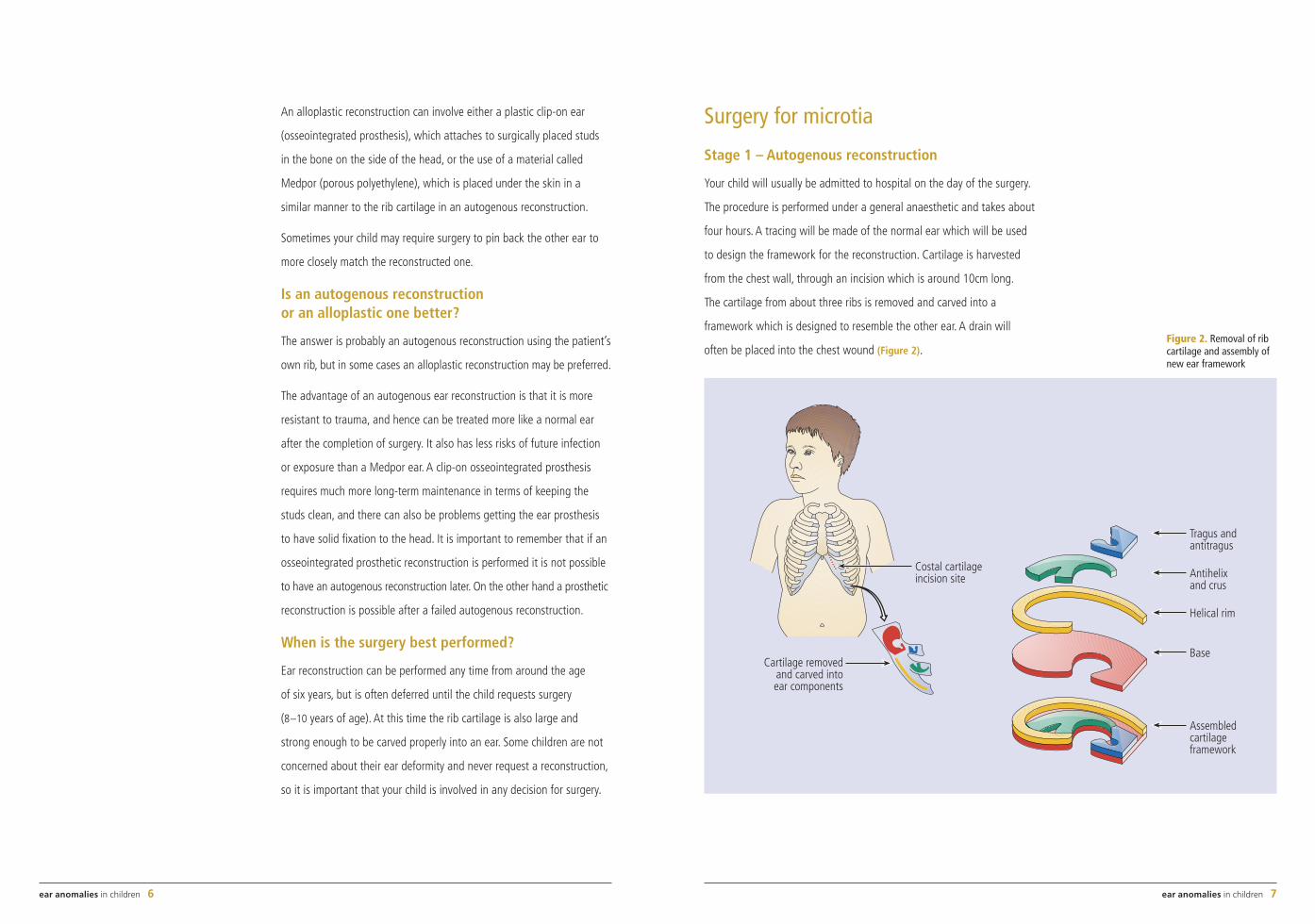
Stage 1 – Autogenous reconstruction
Your child will usually be admitted to hospital on the day of the surgery.
The procedure is performed under a general anaesthetic and takes about
four hours. A tracing will be made of the normal ear which will be used
to design the framework for the reconstruction. Cartilage is harvested
from the chest wall, through an incision which is around 10cm long.
The cartilage from about three ribs is removed and carved into a
framework which is designed to resemble the other ear. A drain will
often be placed into the chest wound (Figure 2).Figure 2. Removal of rib cartilage and assembly of new ear framework
Costal cartilageincision site
Cartilage removedand carved intoear components
Tragus andantitragus
Antihelixand crus
Helical rim
Base
Assembledcartilageframework
ear anomalies in children 8 ear anomalies in children 9
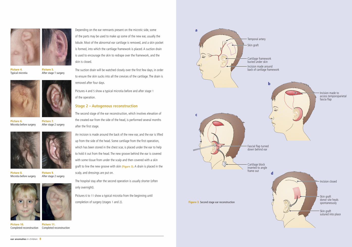
Depending on the ear remnants present on the microtic side, some
of the parts may be used to make up some of the new ear, usually the
lobule. Most of the abnormal ear cartilage is removed, and a skin pocket
is formed, into which the cartilage framework is placed. A suction drain
is used to encourage the skin to redrape over the framework, and the
skin is closed.
The suction drain will be watched closely over the first few days, in order
to ensure the skin sucks into all the crevices of the cartilage. The drain is
removed after four days.
Pictures 4 and 5 show a typical microtia before and after stage 1
of the operation.
Stage 2 – Autogenous reconstruction
The second stage of the ear reconstruction, which involves elevation of
the created ear from the side of the head, is performed several months
after the first stage.
An incision is made around the back of the new ear, and the ear is lifted
up from the side of the head. Some cartilage from the first operation,
which has been stored in the chest scar, is placed under the ear to help
to hold it out from the head. The new groove behind the ear is covered
with some tissue from under the scalp and then covered with a skin
graft to line the new groove with skin (Figure 3). A drain is placed in the
scalp, and dressings are put on.
The hospital stay after the second operation is usually shorter (often
only overnight).
Pictures 6 to 11 show a typical microtia from the beginning until
completion of surgery (stages 1 and 2). Figure 3. Second stage ear reconstruction
Cartilage frameworkburied under skin
Incision made aroundback of cartilage framework
Temporal artery
Skin graft
a
Incision made toaccess temporoparietalfascia flap
b
Fascial flap turneddown behind ear
Cartilage blockinserted to angleframe out
c
Skin graftdonor site healsspontaneously
Incision closed
Skin graftsutured into place
d
Picture 4. Typical microtia
Picture 5. After stage 1 surgery
Picture 8. Microtia before surgery
Picture 9. After stage 2 surgery
Picture 10. Completed reconstruction
Picture 11. Completed reconstruction
Picture 6. Microtia before surgery
Picture 7. After stage 2 surgery
ear anomalies in children 10 ear anomalies in children 11
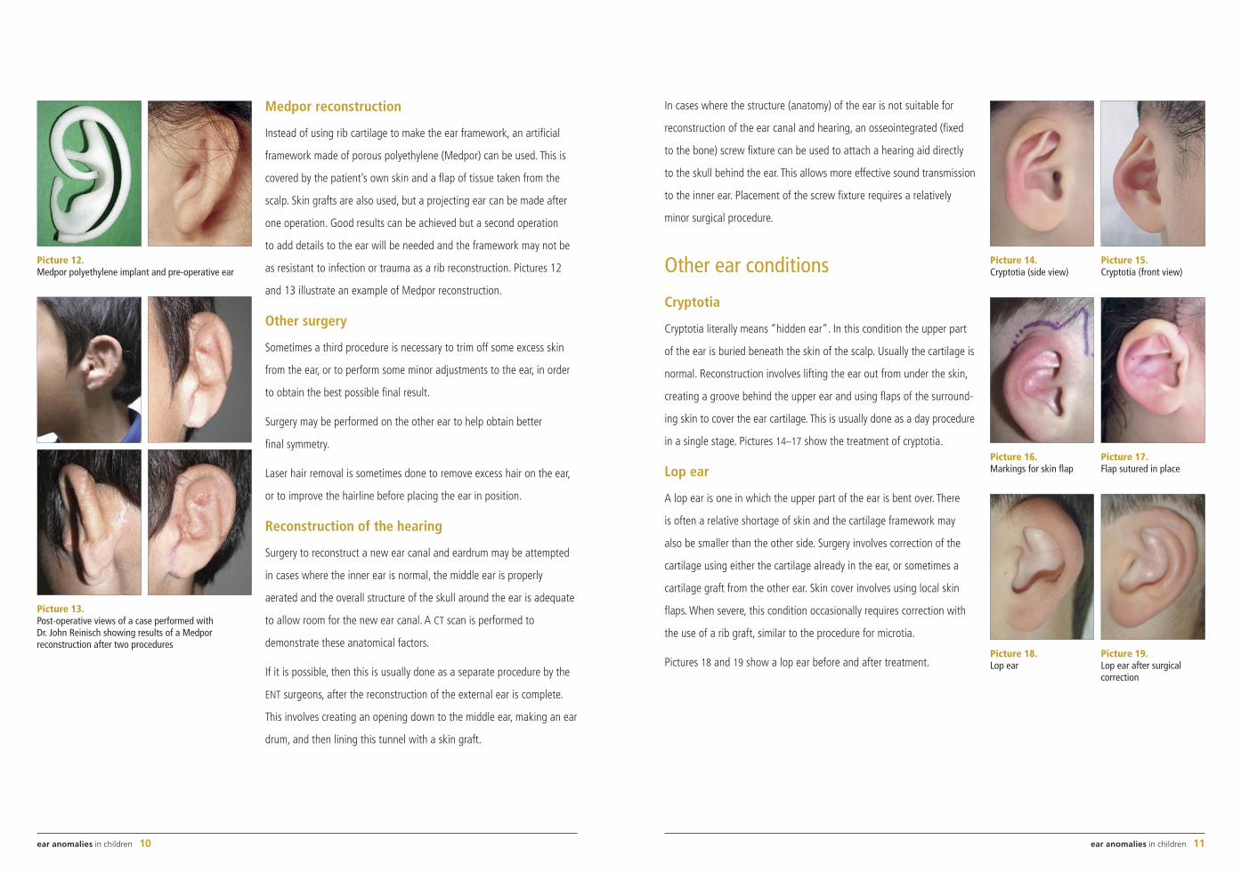
Medpor reconstruction
Instead of using rib cartilage to make the ear framework, an artificial
framework made of porous polyethylene (Medpor) can be used. This is
covered by the patient’s own skin and a flap of tissue taken from the
scalp. Skin grafts are also used, but a projecting ear can be made after
one operation. Good results can be achieved but a second operation
to add details to the ear will be needed and the framework may not be
as resistant to infection or trauma as a rib reconstruction. Pictures 12
and 13 illustrate an example of Medpor reconstruction.
Other surgery
Sometimes a third procedure is necessary to trim off some excess skin
from the ear, or to perform some minor adjustments to the ear, in order
to obtain the best possible final result.
Surgery may be performed on the other ear to help obtain better
final symmetry.
Laser hair removal is sometimes done to remove excess hair on the ear,
or to improve the hairline before placing the ear in position.
Reconstruction of the hearing
Surgery to reconstruct a new ear canal and eardrum may be attempted
in cases where the inner ear is normal, the middle ear is properly
aerated and the overall structure of the skull around the ear is adequate
to allow room for the new ear canal. A CT scan is performed to
demonstrate these anatomical factors.
If it is possible, then this is usually done as a separate procedure by the
ENT surgeons, after the reconstruction of the external ear is complete.
This involves creating an opening down to the middle ear, making an ear
drum, and then lining this tunnel with a skin graft.
In cases where the structure (anatomy) of the ear is not suitable for
reconstruction of the ear canal and hearing, an osseointegrated (fixed
to the bone) screw fixture can be used to attach a hearing aid directly
to the skull behind the ear. This allows more effective sound transmission
to the inner ear. Placement of the screw fixture requires a relatively
minor surgical procedure.
Other ear conditions
Cryptotia
Cryptotia literally means “hidden ear”. In this condition the upper part
of the ear is buried beneath the skin of the scalp. Usually the cartilage is
normal. Reconstruction involves lifting the ear out from under the skin,
creating a groove behind the upper ear and using flaps of the surround-
ing skin to cover the ear cartilage. This is usually done as a day procedure
in a single stage. Pictures 14–17 show the treatment of cryptotia.
Lop ear
A lop ear is one in which the upper part of the ear is bent over. There
is often a relative shortage of skin and the cartilage framework may
also be smaller than the other side. Surgery involves correction of the
cartilage using either the cartilage already in the ear, or sometimes a
cartilage graft from the other ear. Skin cover involves using local skin
flaps. When severe, this condition occasionally requires correction with
the use of a rib graft, similar to the procedure for microtia.
Pictures 18 and 19 show a lop ear before and after treatment.
Picture 14. Cryptotia (side view)
Picture 15. Cryptotia (front view)
Picture 16. Markings for skin flap
Picture 17. Flap sutured in place
Picture 18. Lop ear
Picture 19. Lop ear after surgical correction
Picture 12. Medpor polyethylene implant and pre-operative ear
Picture 13. Post-operative views of a case performed with Dr. John Reinisch showing results of a Medpor reconstruction after two procedures
This publication is provided
with the compliments of the
Mark and Chapter
Freemasons of Victoria
Freemasonry is an ancient and respectable
institution, embracing individuals of every
nation, of every faith and every condition
of life. It can be defined as a benevolent,
charitable, educational and ethical society.
It strives to teach every moral and social
virtue and exhorts its membership to practice
the universal principles of brotherly love,
relief and truth.
Masonic Centre of Victoria
300 Albert Street
East Melbourne Victoria
Australia 3002
Telephone + 61 3 9419 8687
0439
39 D
esig
n an
d co
ver p
hoto
grap
hy: E
RC, T
he R
oyal
Chi
ldre
n’s
Hosp
ital,
2006


![HEAR MAPS a New Classification for Congenital Microtia ... · (Hearing, Ear [microtia], Atresia grade, Remnant earlobe, Mandible development, Asymmetry of soft tissue, Paralysis](https://img.pdfslide.us/doc/110x75/60e4c2c1d26f8d5c325501dd/hear-maps-a-new-classiication-for-congenital-microtia-hearing-ear-microtia.jpg)