Embed Size (px)
Citation preview

www.PRSJournal.com652
Since the first attempt at auricular recon-struction by Sushruta in 600 bc, multiple techniques have been described. By the
1890s, more than 40 different frameworks had been described.1 The multitude of techniques described throughout history is evidence of the complex nature of auricular reconstruction as new techniques develop from shortfalls of previously described methods. Use of a costal cartilaginous framework has become the standard for autolo-gous auricular reconstruction, and many surgeons
such as Tanzer, Brent, Firmin, and Nagata have continued to modify this technique,2–15 with the goal of better replicating the human ear by means of a streamlined and reliable method.
Satoru Nagata first published his two-stage technique for microtia reconstruction in 1987.6 This technique provides a three-dimensional
Disclosure: The authors have no financial interest to declare in relation to the content of this article.
Copyright © 2014 by the American Society of Plastic Surgeons
DOI: 10.1097/PRS.0000000000000063
Leila Kasrai, F.R.C.S.C., M.P.H.
Alison K. Snyder-Warwick, M.D.
David M. Fisher, F.R.C.S.C., F.A.C.S.
Toronto, Ontario, Canada; and St. Louis, Mo.
Background: The authors have been using the Nagata technique since 2002. In this review of 100 consecutive ear reconstructions, the authors present tech-nique modifications that have evolved over this period that have contributed to improved auricular contour and that now allow for auricular reconstruction in a single stage.Methods: This study is a retrospective review of a prospectively acquired database. The series is restricted to primary reconstructions performed for congenital microtia. Photographs of 10 consecutive patients are presented to demonstrate the results of the technique. Surgical complication rates are discussed.Results: One hundred ear reconstructions were performed in 96 patients. There were 75 primary cases of congenital microtia. Twenty-four ears under-went a two-stage reconstruction, and 51 ears were reconstructed with a Nagata stage I procedure or a single-stage reconstruction. There was a gradual shift in technique, with a trend to perform fewer Nagata stage II outsetting proce-dures and more single-stage reconstructions. In patients who underwent an ear reconstruction in two stages, the surgical complication rate was 22 percent. In the last 40 consecutive ear reconstructions since abandoning the two-stage approach, the surgical complication rate is now 15 percent.Conclusions: A modification of Nagata’s technique of autologous ear recon-struction for microtia is described. Modifications of the three-dimensional framework address the contour of the inferior crus and control tragal projec-tion and position. Inclusion of a projection block and recruitment of retroau-ricular skin allow for symmetric projection of the ear in a single stage. (Plast. Reconstr. Surg. 133: 652, 2014.)CLINICAL QUESTION/LEVEL OF EVIDENCE: Therapeutic, IV.
From the Department of Surgery, University of Toronto, the Division of Plastic Surgery, St. Joseph’s Health Centre; and Washington University School of Medicine, St. Louis Chil-dren’s Hospital.Received for publication August 29, 2013; accepted Novem-ber 10, 2013.Presented at the Fifth International Congress on Auricular Reconstruction, in Sydney, Australia, March 12, 2012.
Single-Stage Autologous Ear Reconstruction for Microtia
Supplemental digital content is available for this article. Direct URL citations appear in the text; simply type the URL address into any Web browser to access this content. Clickable links to the material are provided in the HTML text of this article on the Journal’s Web site (www.PRSJournal.com).
cpt
PEDIATRIC/CRANIOFACIAL

Volume 133, Number 3 • Ear Reconstruction for Microtia
653
reconstruction in two stages, fewer than previ-ously described. Since his original description, Nagata has modified his technique.8–11,14,15 We have been using the Nagata technique, or a modification of it, since 2002. In this review of our series of 100 consecutive ear reconstruc-tions, we present technique modifications that have evolved over this time. These modifications have contributed to improved auricular contour and allow for auricular reconstruction in a single stage. Photographs of the results of 10 consecu-tive ear reconstructions using this modified tech-nique are presented.
PATIENTS AND METHODSThis study is a review of a consecutive series
of cases performed by the senior authors (L.K. and D.M.F.) over an 11-year period at St. Joseph’s Health Centre and The Hospital for Sick Chil-dren. It is a retrospective review of a prospectively acquired database. The series is restricted to pri-mary reconstructions performed for congenital microtia, excluding secondary ear reconstruc-tions and reconstructions for acquired ear defor-mities. Early surgical complications, defined as those requiring revision during the early healing phase, are presented.
Operative TechniqueSurgical PlanningReconstruction is performed after age 10
years, provided the chest circumference at the level of the xiphoid process is at least 60 cm. At this
age, (1) the contralateral ear has almost reached its adult size and can serve as an accurate template for the reconstruction, (2) the costal cartilage is of sufficient volume to create an appropriate sized three-dimensional framework, and (3) the patient can be an active participant in the decision-mak-ing process.
With unilateral involvement, the unaffected ear is used as a model for the planned reconstruc-tion. Auricular templates (Fig. 1) of standard sizes on transparent film are used to size and position the auricular reconstruction symmetric with the contralateral ear. The template is then used as a guide for carving the three-dimensional frame-work. Symmetric positioning of the ear is not compromised. If the position of the ear extends into the hair-bearing scalp, a temporoparietal fascial flap and scalp split-thickness skin graft are required.
Auricular Incisions and Pocket PreparationCoverage of a three-dimensional frame-
work requires more skin than can be provided by the retroauricular non–hair-bearing skin. Nagata has expanded the retroauricular mastoid skin flap by including the skin on the posterior aspect of the lobule.7 For lobule type microtia, a rounded W-shaped flap is marked on the mas-toid skin and extends onto the posterior surface of the lobule (Fig. 2). The posterior limb of the W begins 5 mm posterior to the planned posi-tion of the framework, allowing for recruitment of mastoid skin. The anterior limb is positioned to maximize the surface area of the flap but not
Fig. 1. Preprinted auricular templates of standard sizes on transparent film are used to size and position the auricular reconstruction symmetric with the contralateral ear. The size of the cartilage framework is 3 mm shorter in vertical height, accounting for 1.5 mm of skin thickness. The two-dimensional template is then cut out from the film and used as a guide for carving of the three-dimensional framework (base frame, blue; helix, antihelix, and tra-gus, green; projection block is not shown).

654
Plastic and Reconstructive Surgery • March 2014
to involve the curved free border of the lobule. The two central limbs of the W-shaped flap will be sutured edge-to-edge, producing an inverted cone that will line the depth of the conchal bowl deep to the intertragal incisura. The transverse incision that defines the distal tip of the anterior lobule flap is placed transversely just below the point where the lobule begins to curve inward to meet the remainder of the anlage. This incision will meet a curvilinear incision defining the pro-posed posterior limit of the tragus. Just below the caudal limit of this curvilinear incision, a 2-mm diameter circle of skin is excised to define the intertragal incisura. The incisions produce three flaps: (1) bilobed W-flap, continuous with the
retroauricular mastoid skin pocket; (2) anterior lobule flap; and (3) tragal flap. The W-flap and anterior lobule flap will transpose in a reciprocal Z-plasty fashion.
Skin incisions are made. The lobule is split, ensuring equal thickness of the anterior and posterior lobule skin flaps. Skin of the W-flap and mastoid skin pocket are undermined at a thickness of 1.5 to 2 mm, taking care to preserve the subdermal plexus. The dissection extends at least 10 mm beyond the size of the planned reconstructed auricle. A subcutaneous pedicle is maintained at the central portion of the W-flap in the future site of the conchal bowl. The entire remnant auricular cartilage is dissected
Fig. 2. The incisions produce three flaps: (1) bilobed W-flap, continuous with the retroauricular mastoid skin pocket, and com-posed of a posterior mastoid (M) flap and a posterior lobule (PL) flap; (2) anterior lobule (AL) flap; and (3) tragal (T) flap. The W-flap and anterior lobule flap once elevated will transpose in a reciprocal Z-plasty fashion.

Volume 133, Number 3 • Ear Reconstruction for Microtia
655
free from soft-tissue attachments and removed. The region of the external auditory canal is excavated.
For small conchal type microtia, the curvi-linear incision that defines the posterior margin of the tragus is made behind the depression of the small conchal bowl. The skin of the depres-sion is then everted and used to drape over the tragus.
For large conchal type microtia (Fig. 3), the W-shaped incision is more flat and is placed more cephalad, above the lobule. The incision traverses the helical rim above the antitragus and extends with varying extent into the conchal bowl. Native
auricular cartilage of the lower half of the ear ( tragus and antitragus) is preserved.
Costal Cartilage HarvestWe prefer to harvest from the right side to
preserve the protection overlying the heart. A transverse incision (5 to 7 cm) is made between the seventh and eighth ribs, overlying the carti-laginous portion of the ribs. Dissection proceeds through the skin and subcutaneous tissues and a suprafascial pocket is created overlying the sixth through ninth costal cartilages. A vertical incision is made just medial to the lateral bor-der of the anterior rectus sheath, and the rectus
Fig. 3. For large conchal type microtia, the W-shaped incision is more flat and is placed more cephalad, above the lobule. The incision traverses the helical rim above the anti-tragus and extends with varying extent into the conchal bowl. Native auricular carti-lage of the lower half of the ear (tragus and antitragus) is preserved.

656
Plastic and Reconstructive Surgery • March 2014
muscle is elevated off of the perichondrium. The perichondrium is incised on the anterior surface of the ribs and dissected circumfer-entially from the underlying cartilage. Care is taken to avoid damage to the cartilage and to leave the perichondrium intact. The sixth and seventh cartilages are harvested en bloc. The eighth and ninth cartilages are harvested separately. Requirement of the ninth cartilage depends on the type of microtia and the size of the patient. After the cartilage harvest, the chest wound is instilled with saline and the integrity of the pleura confirmed.
After framework fabrication, excess carti-lage is diced into small pieces. The perichon-drium is repaired, creating perichondrial sleeves into which the excess costal cartilage pieces are placed. Kawanabe and Nagata14 have shown that this reconstruction avoids postoperative chest wall deformities. Intercostal blocks facilitate postop-erative pain relief.
Framework Preparation and PlacementThe framework should be constructed to rec-
reate the anatomy of the normal auricle. Consid-ering the 1.5-mm thickness of the skin, convexities of the projection components need to be made narrow, and the concavities of the depressions need to be exaggerated. Cartilage components are articulated with 40-gauge wire sutures. The
twisted “knots” are placed on the deep medial sur-face. External loops of the wires are countersunk into the cartilage surface to avoid contour irregu-larity and risk of exposure.
The base frame is typically created from the sixth and seventh costal cartilages. If the syn-chondrosis is stable, it is kept. If unstable, it is excised and the adjacent sixth and seventh ribs are sutured together. For a complete three-dimensional framework, the base frame sup-ports the helix, antihelix, and lobule; forms the scaphoid fossa; and contributes to the posterior conchal wall. A short projection strut is made to contour and support the attachment of the crus helicis.
The eighth costal cartilage is used to fashion the helix. Wire sutures are placed at 3-mm inter-vals, evenly distributing the tension and increas-ing the stability of the framework.
The antihelix is typically made from the ninth costal cartilage. Nagata uses the inferior margin of the cartilage and splits it at one end to create the superior and inferior crura and the triangu-lar fossa. This time-efficient method fails to cre-ate correct contour of the inferior crus, which has a sharp lower margin and projects over the cymba like a shelf. When the ninth rib is suffi-ciently broad, it is placed on its side and the entire antitragus is carved out of the rib (Fig. 4). The triangular fossa is excavated from between
Fig. 4. When the ninth rib is sufficiently broad, it is used to create the entire antitragus (center). The triangular fossa is excavated from between the upper and inferior crura, each of which is carved to create its distinct anatomical contour (a smooth, broad, low-profile upper crus and a pronounced inferior crus with a sharp shelf-like lower border). A “closing strut” (right) extends from the deep cephalic surface of the tragus and is attached to the deep surface of the base frame under the helical root. The strut provides additional stability to the framework, controls the width of the sulcus between the helical root and the tragus, and maintains the projection of the tragus.

Volume 133, Number 3 • Ear Reconstruction for Microtia
657
the upper and inferior crura, each of which is carved to create its distinct anatomical contour (a smooth, broad, low-profile upper crus and a pronounced inferior crus with a sharp shelf-like lower border). When the ninth costal cartilage is not broad enough to make the entire antihelix, the upper and lower crura are made indepen-dently. Generally, we fashion the antitragus and inferior crus in one piece and add the upper crus as a second piece (Fig. 5). In this way, an uninter-rupted posterior wall of the cavum and cymba is formed. The projecting block and tragus are fash-ioned from the remaining portions of the sixth and seventh costal cartilages.
We have further modified the tragus with a “closing strut” (Fig. 4, right). This extends from the deep cephalic surface of the tragus and will be attached to the deep surface of the base frame under the helical root. The strut provides addi-tional stability to the framework, controls the width of the sulcus between the helical root and the tragus, and maintains the projection of the tragus. It will convert the C-shaped framework into a closed ring. The lower end of the tragus is initially secured to the lobule portion of the base frame. The upper end of the strut will not be secured to the base frame until the framework has been placed within the skin pocket and has encircled the subcutaneous pedicle.
In the classic Nagata two-stage proce-dure, projection of the ear and creation of the
retroauricular sulcus is accomplished at the sec-ond stage. A second cartilage harvest provides a projection block, and the posterior surface of the ear is covered with a temporoparietal fascial flap and scalp split-thickness skin flap. We now include the projection block in our three-dimen-sional framework (Fig. 6) to provide sufficient projection in a single operation. The crescentic projection block measures 6 to 10 mm in height, adjusting for the desired degree of projection. Because Nagata has expanded the retroauricu-lar mastoid skin flap by including the skin on the
Fig. 5. When the ninth rib is not sufficiently broad to create a one-piece antitragus, the antitragus is made in two pieces. Generally, we fashion the antitragus and inferior crus in one piece and add the upper crus as a second piece. In this way, an uninter-rupted posterior wall of the cavum and cymba is formed.
Fig. 6. The three-dimensional framework and two-dimen-sional template. A projection block is added to our three-dimensional framework, allowing for sufficient projection of the construct.

658
Plastic and Reconstructive Surgery • March 2014
posterior aspect of the lobule, there is greater skin coverage than is afforded by previously described techniques. However, there is always a balance between the degree of projection that can be achieved and the availability of skin cover-age without undue tension. In each case, we are literally “pushing the envelope.” Wide undermin-ing of retroauricular skin provides some addi-tional recruitment of skin.
The framework is introduced into the skin pocket, rotating the framework around the pedicle. The closing strut is then secured to the undersurface of the base frame beneath the heli-cal root with wire suture. The breadth of the sul-cus between the tragus and helical root is defined by this latter maneuver. The framework is then anchored to the mastoid fascia with 3-0 clear nylon sutures, superiorly at the anterior convex-ity of the helix and inferiorly at the lobule. Two suction drains are placed through separate stab incisions above the hairline. Continuous suction allows the skin flaps to drape and contour over the framework while the flaps are positioned and inset with 6-0 nylon suture. Invariably, there is some tailoring of the flaps required at this stage (Fig. 7). Auricular pits and tags are excised at this stage.
Surgical DressingsAntibiotic-impregnated gauze dressings,
rolled into bolsters, are positioned on either side of the helical rim and sutured in placed with 3-0 Prolene (Ethicon, Inc., Somerville, N.J.). Drains are placed to suction. Antibiotic ointment is applied. A foam ring is placed around the ear to
prevent direct pressure on the ear, and a net head dressing is applied.
RESULTSDuring the past 11 years, we have performed
100 ear reconstructions in 96 patients. Exclud-ing 15 acquired ear deformities and 10 second-ary reconstructions, there were 75 primary cases of congenital microtia (18 large conchal type, two small conchal type, 54 lobule type, and one case of anotia). Twenty-four ears underwent a two-stage reconstruction, and 51 ears were reconstructed with a Nagata stage I procedure or a single-stage reconstruction. There was a gradual shift in technique, and over time the trend was to perform fewer Nagata stage II out-setting procedures and more single-stage recon-structions. Presently, our intent is to provide a fully reconstructed, well-projected ear in a single operation.
For this review, we have defined a surgical complication as a case that required any reop-eration in the first 3 postoperative weeks for partial flap necrosis, suture line dehiscence, or overt or impending cartilage exposure. In the 75 ear reconstructions for congenital microtia, the overall complication rate was 18 percent. In patients that underwent an ear reconstruction in two stages, the surgical complication rate was 22 percent. Although the surgical complication rate for the second-stage procedures was only 12.5 per-cent, there were other cases in which hypertrophic scarring, skin graft contracture, and late distor-tion of the framework compromised the final aes-thetic appearance. In the last 40 consecutive ear reconstructions since abandoning the two-stage approach, the surgical complication rate is now 15 percent.
Two example cases are provided (Figs. 8 through 11) to demonstrate the potential results for this technique. Preoperative and postop-erative photographs of 10 consecutive cases of single-stage ear reconstruction are provided as supplemental digital content. (See Figure, Sup-plemental Digital Content 1, which shows pre-operative and postoperative photographs of 10 consecutive cases of single-stage ear reconstruc-tion, http://links.lww.com/PRS/A950.) Note that in none of these patients has a contralateral setback been performed.
DISCUSSIONAuricular reconstruction for microtia remains
a great challenge. Historically, multiple techniques
Fig. 7. Once complete, a moist gauze is draped over the frame-work to simulate the appearance after inset and skin coverage. The overall contour of the framework is assessed and any refine-ments can then be performed.
Volume 133, Number 3 • Ear Reconstruction for Microtia
659
have been attempted in pursuit of a more ideal auricular reconstruction. Since its introduction in 1959 by Tanzer,16 the use of a costal cartilagi-nous framework for autologous reconstruction has become the standard. Autogenous costal cartilage frameworks can produce aesthetically pleasing results with low complication rates, even following trauma.4,7–13,17 Within this technique, modifications have been made with efforts for better defined anatomical landmarks, improved cutaneous vascular reliability, and fewer recon-structive stages. One theme in auricular recon-struction is continued refinements, inspired by
the weaknesses of previous techniques, in pursuit of an optimal result.
Nagata has refined the technique of autolo-gous ear reconstruction and has reduced it to a two-stage procedure. Nagata has described the planes of the normal ear and emphasizes a three-dimensional reconstruction of this normal anatomy. His modifications of Tanzer’s incisions have allowed for increased skin surface area and improved flap vascularity. He has demonstrated outstanding results with few complications.7–11 Nagata’s methods of cartilage harvest and costal cartilage reconstruction have eliminated the chest
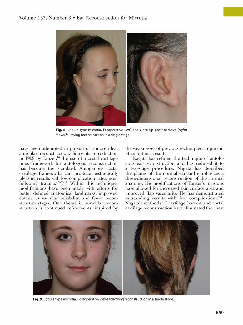
Fig. 9. Lobule type microtia. Postoperative views following reconstruction in a single stage.
Fig. 8. Lobule type microtia. Preoperative (left) and close-up postoperative (right) views following reconstruction in a single stage.

660
Plastic and Reconstructive Surgery • March 2014
wall deformity associated with previous techniques of costal cartilage harvest.14,15 In our opinion, his results are unrivaled.
Many surgeons worldwide have adopted the Nagata technique. A survey of attendees of the 2007 International Ear Reconstruction Congress demon-strated that 93 percent of respondents construct a multiple-layer framework and 57 percent preserve a subcutaneous pedicle in microtia reconstruction,18 both techniques championed by Nagata. Although this subspecialist survey shows a large Nagata influ-ence, other surgeons have not accepted this tech-nique because of its complexity, associated learning curve, and concerns about complications.
Concerns regarding complication rates associ-ated with the Nagata technique have been raised. Because the Nagata technique involves soft-tissue transposition at the time of framework insertion, soft-tissue necrosis is a potential complication.17 Firmin12 described partial necrosis of the poste-rior lobular flap as a specific complication of the Nagata technique. She did not feel that mainte-nance of a subcutaneous pedicle augmented the vascularity of the posterior flap and noted partial necrosis of this flap in 13.9 percent of her 144 patients. Others, however, have noted resolution of partial flap necrosis with preservation of the subcutaneous pedicle.19
Fig. 10. Lobule type microtia. Preoperative (left) and postoperative (right) views following reconstruction in a single stage.
Fig. 11. Lobule type microtia. Postoperative views following reconstruction in a single stage.

Volume 133, Number 3 • Ear Reconstruction for Microtia
661
The Nagata method is a reliable method for microtia reconstruction and is capable of pro-ducing remarkable results. We have found his technique to be reproducible and safe. How-ever, we too, have made technical refinements over time to improve results in our patients. In some of our cases, the aesthetic quality of the reconstruction decreased after the second stage of reconstruction. Soft-tissue swelling, scar contracture, and framework distortion detracted from the overall auricular form, which after the first stage had been quite sat-isfactory. Furthermore, the degree of projec-tion was unpredictable and often unimpressive. Accordingly, we have gradually modified our technique to achieve ear reconstruction in a single stage, thus eliminating the second stage, which in our experience has been problematic and unpredictable. In addition to minor modi-fications of the three-dimensional framework, we use an additional cartilage block posterior to the framework to project the ear from the side of the head in the first and only planned stage. Admittedly, this reconstruction does not produce a deep retroauricular sulcus. It is, how-ever, capable of producing symmetric auricular projection if the contralateral ear is not overly protruded. A shallow sulcus does develop as the postoperative swelling resolves, generally achiev-ing enough of a depression to create a shadow and the impression of a sulcus. If the contralat-eral ear is outstanding, symmetrical projection of the ears is more reliably created in our expe-rience by simple setback otoplasty of the contra-lateral ear. The technique avoids any morbidity of a second operation. The temporoparietal fascial flap is reserved for low hairline cases and is otherwise preserved for use in salvaging complications.
CONCLUSIONSA modification of Nagata’s technique of autol-
ogous ear reconstruction for microtia is described. Modifications of the three-dimensional framework address the contour of the inferior crus and con-trol tragal projection and position. Inclusion of a projection block and recruitment of retroauricu-lar skin allow for increased projection of the ear. Symmetric ear projection can be accomplished in a single procedure, provided the contralateral ear is not overly protruded. For our patients, this method produces improved aesthetic results with a lower complication rate.
CODING PERSPECTIVE
cpt This information prepared by Dr. Raymund Janevicius is intended to provide coding guidance.
69339 Sculpting of cartilaginous framework
21230-51 Cartilage graft harvest and transfer
14061-51 Creation of cutaneous pocket14060-59 W-flap14060-59 Anterior lobule flap14060-59 Tragal flap
• No single code exists for ear reconstruction for microtia. The different methods of re-construction use combinations of flaps and tissue mobilization that must be accurately described.
• The harvest and placement of the cartilage graft is reported with code 21230.
• No code accurately describes the intricacies of sculpting and detailing the cartilaginous framework. This is one of the few instances where an unlisted procedure code must be used, 69399.
• When a single soft-tissue defect is recon-structed with multiple flaps, Current Proce-dural Terminology guidelines instruct the surgeon to report one adjacent tissue trans-fer code based on the total defect surface area (primary plus secondary defect). Ear reconstruction presents an entirely differ-ent scenario, because each flap has a sep-arate function and is used to reconstruct a separate structure. One is not closing a single defect with multiple flaps. Each flap is therefore reported separately (14061, 14060).
• Because each flap is considered a “separate procedure,” modifier 59 must be appended to the adjacent tissue transfer codes.
• It is imperative that these procedures be preauthorized in writing with the insurance company before performing surgery. The preauthorization must clearly articulate why multiple adjacent tissue transfer codes are used together and why an unlisted pro-cedure code must be used. Photographs and diagrams of the procedure are very in-structive in the preauthorization process, as are preoperative and postoperative photo-graphs of a typical reconstruction.

662
Plastic and Reconstructive Surgery • March 2014
Leila Kasrai, F.R.C.S.C., M.P.H.Department of Surgery
University of TorontoDivision of Plastic SurgerySt. Joseph’s Health Centre
30 The QueenswayM6R 1B5 Toronto, Ontario, Canada
David M. Fisher, F.R.C.S.C., F.A.C.S.Division of Plastic Surgery
The Hospital for Sick Children555 University Avenue
Toronto, Ontario M5G 1X8, [email protected]
ACKNOWLEDGMENTThe authors thank Dr. Satoru Nagata for his gener-
ous mentorship and kind support.
REFERENCES 1. Berghaus A, Stelter K, Naumann A, Hempel JM. Ear
reconstruction with porous polyethylene implants. Adv Otorhinolaryngol. 2010;68:53–64.
2. Tanzer RC. Total reconstruction of the auricle: The evolution of a plan of treatment. Plast Reconstr Surg. 1971;47:523–533.
3. Brent B. Ear reconstruction with an expansile framework of autogenous rib cartilage. Plast Reconstr Surg. 1974;53:619–628.
4. Tanzer RC. Microtia: A long-term follow-up of 44 recon-structed auricles. Plast Reconstr Surg. 1978;61:161–166.
5. Brent B. The correction of microtia with autogenous cartilage grafts: I. The classic deformity? Plast Reconstr Surg. 1980;66:1–12.
6. Nagata S, Fukuda O. A new reconstruction for the lobule type microtia. Jpn J Plast Reconstr Surg. 1987;7:689.
7. Nagata S. A new method of total reconstruction of the auri-cle for microtia. Plast Reconstr Surg. 1993;92:187–201.
8. Nagata S. Modification of the stages in total reconstruction of the auricle: Part I. Grafting of the three-dimensional cos-tal cartilage framework for lobule-type microtia. Plast Reconstr Surg. 1994;93:221–230.
9. Nagata S. Modification of the stages in total reconstruction of the auricle: Part II. Grafting the three-dimensional costal cartilage framework for concha-type microtia. Plast Reconstr Surg. 1994;93:231–242; discussion 267–268.
10. Nagata S. Modification of the stages in total reconstruction of the auricle: Part III. Grafting the three-dimensional cos-tal cartilage framework for small concha-type microtia. Plast Reconstr Surg. 1994;93:243–253; discussion 267–268.
11. Nagata S. Modification of the stages in total reconstruction of the auricle: Part IV. Ear elevation for the constructed auri-cle. Plast Reconstr Surg. 1994;93:254–266; discussion 267–268.
12. Firmin F. Ear reconstruction in cases of typical microtia: Personal experience based on 352 microtic ear corrections. Scand J Plast Reconstr Surg Hand Surg. 1998;32:35–47.
13. Brent B. Technical advances in ear reconstruction with autogenous rib cartilage grafts: Personal experience with 1200 cases. Plast Reconstr Surg. 1999;104:319–334; discussion 335–338.
14. Kawanabe Y, Nagata S. A new method of costal cartilage har-vest for total auricular reconstruction: Part I. Avoidance and prevention of intraoperative and postoperative complica-tions and problems. Plast Reconstr Surg. 2006;117:2011–2018.
15. Kawanabe Y, Nagata S. A new method of costal cartilage har-vest for total auricular reconstruction: Part II. Evaluation and analysis of the regenerated costal cartilage. Plast Reconstr Surg. 2007;119:308–315.
16. Tanzer RC. Total reconstruction of the external ear. Plast Reconstr Surg Transplant Bull. 1959;23:1–15.
17. Brent B. Microtia repair with rib cartilage grafts: A review of personal experience with 1000 cases. Clin Plast Surg. 2002;29:257–271, vii.
18. Breugem CC, Stewart KJ, Kon M. International trends in the treatment of microtia. J Craniofac Surg. 2011;22:1367–1369.
19. Suutarla S, Rautio J, Klockars T. The learning curve in micro-tia surgery. Facial Plast Surg. 2009;25:164–168.