Embed Size (px)
Citation preview
G. Iacob et al Microsurgical removal of posterior petrosal and petroclival meningiomas
Microsurgical removal of posterior petrosal and petroclival meningiomas: a report of 8 cases
G. Iacob, M. Craciun, Silvia Tene
Clinic of Neurosurgery, Universitary Hospital Bucharest România “Meningiomas involve us all in a never – ending quest for technical improvement
and less disruptive surgical approaches” Gazi Yasargil (28)
Abstract Background: Posterior petrosal and
petroclival meningiomas are rare, benign tumors, representing difficult tumours to be treated by microsurgery: for often significant space-occupying effect, brain stem compression, frequent tight brain stem adherence, as well as encasement of the basilar artery, its perforators and cranial nerves.
Methods: We report our experience referring on 6 cases of posterior petrosal and 2 cases of petroclival meningiomas operated in the last 5 years; retrospectively analyzed, including: the history, clinical data, imaging studies, surgical & histological records, follow-up records. All patients were woman, The main age was 56 years (range 34-72 years). Bony changes at the petrous apex was seen in one case with petroclival meningioma.
Results: Gross total resection was achieved in 7 of 8 patients Simson gr.1-2., using a conventional retromastoid, retrosigmoid approach and in one case a petrosal approach. Retromastoid - retrosigmoid approach was preferred approach for removing posterior petrous meningiomas, used also in one case of petroclival meningioma; especially if preoperative hearing is intact. In all cases
the establishment of an arachnoid plane was critical for separating the tumor from the cerebellum and brainstem as well as microdissection of the neurovascular structures. Once the tumor was excised, its dural attachment was removed or coagulated and hyperostotic bone was drilled away. Tumor histology was fibrous, meningothelial and psammomatous meningioma. After surgery two patients had a transitory palsy of the third (extrinsec) and the seventh nerve, installed immediately after operation, but one patient with a petroclival meningioma operated using the posterior petrosal approach died after a hemorrhagic infarct in the midbrain. No recurrence or progression of disease occurred one year after.
Conclusions: A variety of techniques have been advocated for complete resection of posterior petrosal and petroclival meningiomas, with minimal brain retraction. For each case planning the safest approach should be sustaind on: detailed radiologic studies to define tumor size, location and extensions, the attachment of the tumor to the dura overlying the posterior face of the petrous apex, tumor encasement of the basilar and pontine perforators, venous anatomy delineation (especially the anatomy of the vein of
Romanian Neurosurgery (2011) XVIII 2
Labbé). the consistency of the tumor, the operative distance to the tumor and neurovascular structures, minimal brain retraction, good visualization and lighting. The posterior petrosal approach is safe for total removal of clival and petroclival tumor, also for selected cases of petroclival meningioma even a retromastoid-retrosigmoid approach can be suitable.
Keywords: skull base tumor, posterior fossa tumor, posterior petrosal and petroclival meningioma
Introduction Taking account on the point of dural
attachment, concerning to the internal auditory meatus and the direction of cranial nerve displacement, which can significantly determine surgery induced morbidity there are several distinct meningiomas (1) (3) (12) (17) (19): petrosal meningioma - a hetrogenous group of meningioma in regards to tumor histology, surgical considerations, operative risks and outcome that originate anterior, superior to the internal auditory meatus and posterior to it; petroclival meningioma refer to meningioma that originate in the upper two-thirds of the clivus, at the petroclival junction, near the spheno-occipital synchondrosis, disposed partly supratentorial and partly infratentorial; arise medial to the trigeminal nerve, displace often the brain stem and the basilary artery to the opposite side; clival meningioma originate from the midclivus at the upper two-thirds, encasing typically the basilar artery and its perforators. If these tumors are fibrous and nonsuckable, it’s the most formidable to expose and disect; sphenopetroclival meningioma are the most extensive, invading the cavernous sinus
(unilateral or bilateral), sella turcica, sphenoid sinus, involving the bony clivus and the petrous apex.
Material and methods We report our experience referring on 6
cases of posterior petrosal and 2 cases of petroclival meningiomas operated in the last 5 years; retrospectively analyzed, including: the history, clinical data, imaging studies, surgical & histological records, follow-up records. The main age was 56 years (range 34-72 years), our patients were 6 female, 2 male. The onset of clinical symptoms of our series was insidious: mean 3 month - 2 years before admission, with: headaches, dizziness, facial pain and numbness, hearing loss, tinnitus, in one case cerebellar dysfunction and gait ataxia. In one case the patient was admitted for severe ataxia, diplopia, papilledema, with moderate hydrocephalus. Imaging studies were based on:
- native and contrast cerebral CT scan demonstrate: tumor topography, brain stem compression, hyperostosis at the petrous apex in one cases with petroclival meningioma. In one case of a petroclival meningioma a moderate hydrocephalus was recorded.
- arteriography was performed in all cases to proof the blood supply coming from dural attachment, tumoral blush and the venous stage, especially to delineate to each patient the basal temporal veins, the vein of Labbé, their relationship to the superior petrosal sinus, sigmoid sinus with jugular bulb location.
- cerebral MRI using T1, T2, – weighted sequences, FSE/STIR, TOF and resonance venography (MRV) were made to define: tumor (hypointense on T1 – weighted
G. Iacob et al Microsurgical removal of posterior petrosal and petroclival meningiomas
sequences, isointense on T2 – weighted sequences, with homogeneous enhancement after iv Gd administration), tumor size (between 35 and 6o mm, mean 43,8 mm), tumor origin (6 posterior petrous and 2 petroclival areas). According to the site of tumor origin in reference to the internal auditory meatus we have had: 4 posterior petrous meningiomas, 1 superior petrous meningiomas and 1 ventral petrous meningiomas; involvement of the internal auditory canal by the tumor was detected in 2 cases of posterior petrous meningioma; tumor compression on brain stem and ipsilateral cerebellum in all cases, arachnoid brain stem invasion in 1 case, no tumor extension on temporal bone, sella turcica, sphenoid and cavernous sinus, tumor consistency on T2 details (7 cases of soft tumor and one case of firm petroclival tumor), peculiar venous drainage of the temporal lobe (the vein of Labbé and basal temporal veins, bilateral demonstration of the superior petrosal, transverse and sigmoid sinuses), ventricular size and topography (in one case a moderate hydrocephalus was recorded).
Gross total resection, Simson gr. 1-2, using microdissection of tumor and neurovascular structures was achieved in 7 of 8 patients, using a conventional retromastoid, retrosigmoid approach and in one case a petrosal approach. Retrosigmoid approach was preferred approach for removing posterior petrous meningiomas, used also in one case of petroclival meningioma of a patient with intact preoperative hearing. This approach has the following advantages: hearing preservation, early drainage of cerebrospinal fluid cisterns allowing cerebellar relaxation. In all cases the establishment of an arachnoid plane was essential for separating the tumor from the
cerebellum, compressed brainstem and nerves; suited by intracapsular removal; as well as microdissection of the neurovascular structures. Tumor histology in petrous meningioma was: 3 fibrous, 1 meningothelial and 1 psammomatous meningioma. After tumor excision, its dural attachment is removed or coagulated and hyperostotic bone is drilled away. In one case of a petroclival tumor, a posterior petrosal approach (presigmoidian) was used; cutting the superior petrosal sinus respecting the Labbé vein, splitting the tentorium. Tumor histology in petroclival meningioma was fibrous and meningothelial meningioma. After surgery two patients had a transitory palsy of the third (extrinsec) and the seventh nerve, installed immediately after operation, but one patient died after a hemorrhagic infarct in the midbrain. No recurrence or progression of disease occurred after operation. Illustrative cases
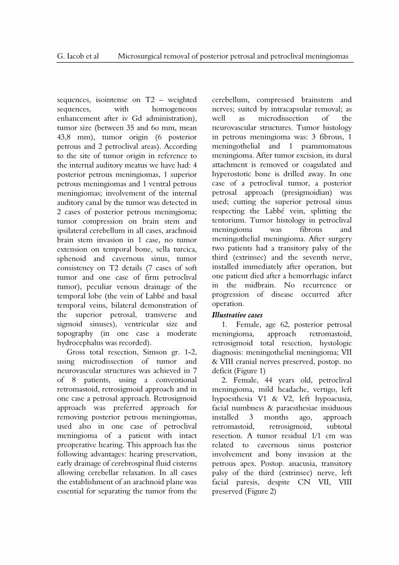
1. Female, age 62, posterior petrosal meningioma, approach retromastoid, retrosigmoid total resection, hystologic diagnosis: meningothelial meningioma; VII & VIII cranial nerves preserved, postop. no deficit (Figure 1)
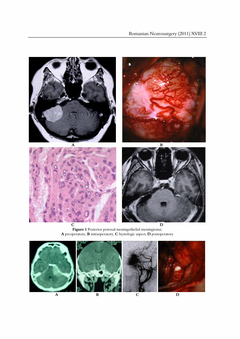
2. Female, 44 years old, petroclival meningioma, mild headache, vertigo, left hypoesthesia V1 & V2, left hypoacusia, facial numbness & paraesthesiae insiduous installed 3 months ago, approach retromastoid, retrosigmoid, subtotal resection. A tumor residual 1/1 cm was related to cavernous sinus posterior involvement and bony invasion at the petrous apex. Postop. anacusia, transitory palsy of the third (extrinsec) nerve, left facial paresis, despite CN VII, VIII preserved (Figure 2)
Romanian Neurosurgery (2011) XVIII 2
A B
C D Figure 1 Posterior petrosal meningothelial meningioma:
A preoperatory, B intraoperatory, C hystologic aspect, D postoperatory
A B C D
G. Iacob et al Microsurgical removal of posterior petrosal and petroclival meningiomas
E F G
Figure 2 Petroclival meningioma: A-B CT preop, C Arteriography, D Intraoperatory, E-G CT postoperatory
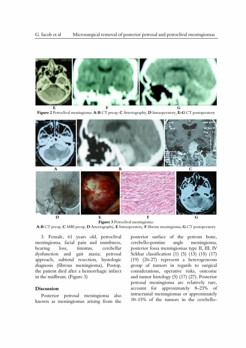
A B C
D E F G
Figure 3 Petroclival meningioma: A-B CT preop, C MRI preop, D Arteriography, E Intraoperatory, F fibrous meningioma, G CT postoperatory
3. Female, 61 years old, petroclival
meningioma, facial pain and numbness, hearing loss, tinnitus, cerebellar dysfunction and gait ataxia; petrosal approach, subtotal resection, hystologic diagnosis (fibrous meningioma), Postop. the patient died after a hemorrhagic infarct in the midbrain. (Figure 3)
Discussion Posterior petrosal meningioma also
known as meningiomas arising from the
posterior surface of the petrous bone, cerebello-pontine angle meningioma, posterior fossa meningiomas type II, III, IV Sekhar classification (1) (5) (13) (15) (17) (19) (26-27) represent a heterogeneous group of tumors in regards to surgical considerations, operative risks, outcome and tumor histology (5) (17) (27). Posterior petrosal meningioma are relatively rare, account for approximately 8–23% of intracranial meningiomas or approximately 10–15% of the tumors in the cerebello-
Romanian Neurosurgery (2011) XVIII 2
pontine angle versus 75% vestibular schwanoma, arise in more varied locations, usually larger than the average VS at presentation (17). According to the site of tumor origin in reference to the internal auditory meatus such tumors should be classified as: posterior petrous meningiomas, superior petrous meningiomas (the most common - seventh-eighth cranial nerve complex is usually displaced anteriorly, cranially or caudally, the fifth nerve is frequently displaced cranially or anteriorly) and ventral petrous meningiomas.(5) (15) (17) Posterior petrosal meningioma can be asymptomatic for a long period of time or be discovered during cranial imaging for other reasons. In general, clinical symptoms are insidious, lasted from 1 to 4 years. Most frequent: facial pain, even on the contralateral side, due to mass effect, hemifacial spasm, facial numbness develop before eighth cranial nerve symptoms (17). If tumor arise near the internal auditory meatus hearing loss, tinnitus, disequilibrium are seen. Otherwise if tumor is attached below the internal auditory meatus extending inferiorly near the jugular foramen swallowing difficulty, hoarseness could be seen. In late stages of the disease, depending on their size, symptoms of compression of the pons such as ataxia, gait imbalance, diplopia, papilledema, even obstructive hydrocephalus in 10–20% of these patients were observed (5) (13) (15) (26-27).
High-resolution, thin-slice CT scan with bone window may show rarely focal or extended hyperostosis (spicula) in the petrous bone, suggesting the possible origin of the meningioma. The overall tumor size was 11–60 mm (17) (26-27). Tumor involvement of the internal auditory canal
could be detected in 25% of cases, even osseous hypertrophy ventral to the internal auditory canal at the site of tumor origin. On MRI: tumor are well circumscribed, hypointense to isointense on T1 – weighted sequences and isointense to hyperintense on T2 – weighted sequences, with homogeneous enhancement after contrast (Gadolinium) administration. T2 sequences are utile to define tumor contact to the brain stem with edema. Since information of any possible contact with nerve and vascular structures is mandatory prior to surgical planning, (3D) MP-RAGE and 3D CISS sequences may be used. Dural tail is frequently seen with meningiomas, both on the dura of the petrous bone and the tentorium, but it’s not specific: also some schwannomas and metastases may have dural tail caused by tumor infiltration and/or reactive changes (17). Arteriography may show tumoral blush in huge tumors, blood supply comes from dural attachment, usually through branches of the artery of Bernasconi-Cassinari. Embolization should be considered when this artery is particularly large (15). In the case of large tumors it’s possible to see intra- or immediate postembolization swelling of the meningioma with cranial nerves ischemia risk. MRA is used to define vascular anatomy of the venous system, especially the patency, the size and the ”dominance” of the sigmoid sinus. CT data can be fused with previous MRI data for surgical neuronavigation. Neuroophtalmologic evaluation, gait analysis, somatosensory and motor evoked potentials, neuropsychological testing, otologic examination and auditory brain stem evoked potentials should complete preoperative evaluation (17).
Petroclival meningioma are in general,
G. Iacob et al Microsurgical removal of posterior petrosal and petroclival meningiomas
rare, benign, deep located tumors of the skull base (3) (11) (14) (19) (22). Disposed medial to cranial nerves V,VII, VIII, IX, X. XI petroclival meningioma frequently extend to the cavernous sinus, middle cranial fossa, down to the magnum foramen; compress the brainstem, may infiltrate or involve the skull base bone, the dura mater. This tumor is accounting for 3 to 10 per cent of posterior fossa meningiomas, only 1,7 per cent of Cushing and Eisenhardt’s 205 meningiomas (3). Most patient are woman, the average age is in the middle 40’s, with a very wide range. The typical presentation of the petroclival meningioma is that of an insidious onset, with slow, relentless growth, can become enormous before they become apparent clinically (14). The clinical presentation present: - headache the most frequent complaint; - cranial neuropathies with the fifth (even trigeminal neuralgia associated with contralateral posterior fossa tumors), eighth cranial nerves and facial nerve being the most frequently involved. The lower cranial nerves are involved in nearly one third, more rare cranial nerves III, IV and VI, despite their proximity and ultimate involvement of tumor; - mass effect on the cerebellum with cerebellar signs, which occur in nearly 70% of cases; - brain stem compression expressed by long tract signs - spastic paresis is reported in 15–57% of patients and somatosensory deficits reported in 15–20% of patients; - increased intracranial pressure, either from the tumor mass or obstructive hydrocephalus (14). Larger, symptomatic petroclival meningioma or those that show evidence of growth or aggressive features should be treated with surgical resection (still the gold standard of treatment), radiosurgery or conventional fractionated radiation therapy.
If in pre microscopic era morbidity & mortality were estimated to ≥ 50%, the petrosal approach and total petrosectomy are the major advancement in total removal of petroclival meningioma, shortening the operative distance to 3 cm, intercepting early the vascular supply to the tumor, exposing the anterior and lateral aspects of the brain stem (11) (22). CT scan examination may reveal (6) bone erosion, hyperostosis, calcifications, a large tumor implantation, slightly hyperintense in comparison with the brain tissue; shows strong contrast enhancement, usually infiltrate the dura mater. High definition MRI studies are essential for surgical planning, shows precisely the extension, dura mater infiltration - “dura tail sign”, arachnoid invasion – edema in T2 - weighted images, displacement and compression of normal structures, relationship of the lesion with vessels (MRA), cranial nerves, brain stem (6) (23). By proton MR spectroscopy, meningiomas show marked elevation of the choline peak, very low or no NAA, and presence of alanine (at 1.5ppm). DSA is performed when the tumor is seen to be highly vascularized on MRI, also do define venous system anatomy (7). An ICA balloon test occlusion is performed when the ICA is involved. Embolization of tumor feeders of the external carotid artery and the meningohypophyseal trunk (Bernasconi-Cassinari artery) is performed in selected cases of very highly vascularized tumors (14).
For each case planning the safest approach should be sustained on (2) (4-5) (8-11) (14) (16-18) (21-22) (24-25) (29):
A. patients age & general condition: preoperative hearing evaluation and deficits
B. detailed radiologic studies CT,
Romanian Neurosurgery (2011) XVIII 2
high definition MRI(FSE/STIR), CT – based neuronavigation fused images with the MRI data, angiography, especially venous stage or magnetic resonance venography (MRV) to define: delination of patient’s anatomy concerning venous system anatomy: venous drainage of the temporal lobe (the vein of Labbé, other basal temporal veins and their relationship to the superior petrosal sinus, tentorium, sigmoid sinus), bilateral demonstration of the transverse and sigmoid sinuses, jugular bulb location; tumor origin - petrous and/or multiple clival areas attachment, also reference to the internal auditory meatus; tumor extension, cavernous sinus invasion may be a cause of subtotal removal, brain stem compression and arachnoid invasion, extension of dural attachments at the skull base, tumor encasement of the basilar and pontine perforators (in such cases is better to leave a small tumor piece to avoid brainstem infarction, tumor size – smaller the tumor, better the outcome ! - significant factor influencing surgical morbidity and mortality, tumor consistency (see T2 details): soft or firm tumor (fibrous and nonsuckable), the operative distance to the tumor and neurovascular structures
C. surgeon preferance approach, neuronavigation to create virtual surgical vectors to select a single corridor or a combined approach !; technique, skill and expertise, minimal brain retraction, a judicious application of cytoreductive surgery at the first operation, facilitated by the presence of an intact arachnoidal membrane separating the meningioma from the brain stem, the consistency of the tumor or whether tumor encasement of the basilar and pontine perforators exist; management of the vessels, especially venous structures, cranial nerves,
brainstem; mentioning that the first operation provides the best and frequently the only opportunity for total removal, remaining a daunting task! According to Sami experience (14) (19), surgery should be the treatment of choice for small petroclival meningiomas (up to 3 cm in diameter); the only chance of cure from this benign tumor. These small lesions can be totally removed (Simpson’s grade 1) with preservation of the involved structures.
D. technological: microscope, good visualization and lighting, microinstruments, high speed drill, CUSA, electrophysiology (cranial nerve monitoring evoked potentials)
A variety of techniques have been advocated for complete resection of posterior petrosal and petroclival meningiomas, with minimal brain retraction: - suboccipital retrosigmoid (13) (17-18) (25), used mainly for tumors located in the posterior fossa with small extensions into the middle fossa and posterior cavernous sinus (the exposure could be enlarged removing the suprameatal tubercule and the petrous apex; - petrosal approach (2) (4) (8) (10) (14) (22) (24) requiring only minimal retraction of the cerebellum, shortening the operative distance to the clivus by 3 cm, offering to the surgeon a direct line of sight to the lesion, also the anterior and lateral aspects of the brain stem, intercepting early in the operation the vascular tumor supply; - presigmoid “minipetrosal” approach with variants of transpetrosal or combined approaches (16); - combined suboccipital-retrosigmoid with the petrosal approach; - subtemporal and retrosigmoid keyhole approach for extensive petroclival meningioma surgery (29).
G. Iacob et al Microsurgical removal of posterior petrosal and petroclival meningiomas
Complete tumor removal should include: - finding an arachnoid plane to separate the tumor from the cerebellum and brainstem; - delicate microdissection of the meningioma at its origin from the nerves or their vasculature, internal decompression. Once the tumor is excised, its dural attachment is removed or coagulated, the spiculae and hyperostotic bone is drilled away, to remove tumor nodules extending into this bone (14) (22). Tumor resection should be made in the sense of „Simpson grade 1” as defined by Al-Mefty et al. (1), especially concerning petrous meningiomas originating posterior to the internal auditory meatus, more fibrous, ingrowing into the internal acoustic canal, other foramina or its origin in the dural lining of these structures; - good hemostasis on the origin of the meningioma. In the case of petroclival meningioma for small tumours, if tumor growth is detected on serial magnetic resonance imaging or treatment is desired by the patient, surgery should be the first choice. To patients with advanced age or significant co-morbidities tumor should be observed, in case of tumor growth radiosurgery is a solution. For medium sized tumours to symptomatic patients, surgery is mandatory. In large and giant petroclival meningiomas, tumour resection as radical as possible judged intraoperatively, with decompression of neural structures should be performed and in the case of growing tumour remnants, radiosurgery (20).
Reported rates of total removal (Simpson I) of 86.1% in meningiomas around the posterior petrous bone with dural involvement around the IAC – Sami et al. and 55% gross total tumor removal (Simpson’s grade I and II) could be
achieved in petroclival meningiomas; with a mortality rates between 0%–5% (14). Generally better results are seen for the retromeatal tumors as compared to the premeatal tumors, although retromeatal tumors tend to be bigger than premeatal meningiomas (14) (17) (27). Conventional radiotherapy, focused radiosurgical techniques, radiosurgery have been used as adjuvant therapy for tumor rests or primary treatment for small (up to 3 cm in diameter) petroclival meningiomas. Adjuvant treatment with chemotherapy (hydroxyurea) or hormonal therapy (mifepristone) has been described, but the results were not satisfactory (14).
Surgical limitations especially in petroclival meningiomas, in spite of all advances in skull-base surgery for total excisions are: age factor (above 70 yrs !), large tumor, encasement of VB system, its perforators, cavernous sinus extension, cranial nerves involvement, brain stem edema (FSE /STIR MRI) & arachnoid invasion. Residual tumor growth should be treated by surgery or radiosurgery (4) (24).
Conclusions Good results as measured by tumour
control and preservation of quality of life can be achieved in posterior petrosal and petroclival meningiomas using a variety or combined techniques. For each case a judicious intraoperative judgement is mandatory. Tumor size, consistency, tumor involvement and invasion of the cavernous sinus, cranial nerves, vessels, pia mater may limit surgical approach. In younger patients surgery should not be delayed because surgical morbidity relates positively with tumour size.
In our series the posterior petrosal approach is a major advancement in the safe
Romanian Neurosurgery (2011) XVIII 2
and total removal of clival and petroclival tumor; but also the conventional suboccipital retromastoid-retrosigmoid approach can be suitable for a select group of petroclival meningioma providing a direct and early exposure of the lateral and inferior tumor extensions in relationship to the clivus, also the attachment of the tumor to the dura overlying the posterior face of the petrous apex.
References 1. Al-Mefty O. – Meningiomas of the Posterior Cranial Base, in Operative Atlas of Meningiomas, Lippincott-Raven Publishers 1998; part 3, 209-348 2. Al-Mefty O., Fox J.L., Smith R.R. - Petrosal approach for petroclival meningiomas, Neurosurgery 1988; 22, 510-517 3. Al-Mefty O., Smith R.R. - Clival and petroclival meningiomas, in Al-Mefty 0. - Meningiomas, Raven Press, New York, 1991; 517-537 4. Bambakidis NC, Kakarla UK, Kim LJ, Nakaji P, Porter RW, Daspit CP, Spetzler RF - Evolution of surgical approaches in the treatment of petroclival meningiomas: a retrospective review, Neurosurgery 2008; 62:1182–1191 5. Bassiouni H, Hunold A, Asgari S, Stolke D. Meningiomas of the posterior petrous bone: functional outcome after microsurgery, J Neurosurg 2004; 100:1014–24 6. Carvalho GA, Matthies C, Tatagiba M, Eghbal R, Samii M - Impact of computed tomographic and magnetic resonance imaging findings on surgical outcome in petroclival meningiomas, Neurosurgery 2000; 47:1287–1294 7. Deda H, Erden I, Yagmurlu B - Evaluation of petrosal sinus patency with 3-dimensional contrast-enhanced magnetic resonance venography in petroclival meningiomas for surgical strategy. Surg Neurol 2005; 64(Suppl 2):S67–S71 8. Erkmen K., Pravdenkova S., Al-Mefty O. - Surgical Management of Petroclival Meningiomas: Factors Determining the Choice of Approach, Neurosurg Focus 2005; 19(2) 9. Goel A, Muzumdar D. - Conventional posterior fossa approach for surgery on petroclival meningiomas: a report on an experience with 28 cases, Surg Neurol 2004; 62:3323–40 10.Liu JK, Gottfried ON, Couldwell WT. - Surgical management of posterior petrous meningiomas, Neurosurg Focus 2003; 14(6):Article 7
11.Mayberg M., Symon, L. - Meningiomas of the clivus and apical petrous bone. J. Neurosurg., 1986; 65, 160-167 12.Mayberg MR, Symon LD - Meningiomas of the clivus and apical petrous bone: report of 35 cases. J Neurosurg, 1986; 65:160–167 13.Mehdorn H.M., Buhl R.M. - Petrous Meningiomas I: An Overview, in Lee J.H. – Meningiomas, Diagnosis, Treatment and Outcome, Springer 2008; 47, 433-441 14.Ramina R., Fernandes B. Y., Neto M.C. – Petroclival Meningiomas: Diagnosis, Treatment and Results, in Ramina R., Aguiar P., Tatagiba M. - Samii’s Essentials in Neurosurgery, 2008; 13, 121-134 15.Roberti F, Sekhar LN, Kalavakonda C, Wright DC. - Posterior fossa meningiomas: surgical experience in 161 cases. Surg Neurol. 2001; 56:8–21 16.Roche PH, Fournier HD, Sameshima T, Fukushima T - The combined petrosal approach. Anatomical principles, surgical technique and indications, Neurochirurgie 2008; 54:1–10 17.Sade B. and Joung H. Lee J.H. – Petrous Meningiomas II: Ventral, Posterior and Superior Subtypes, in Lee J.H. – Meningiomas, Diagnosis, Treatment and Outcome, Springer 2008; 48, 442-447 18.Samii M, Tatagiba M, Carvalho GA. - Resection of large petroclival meningiomas by the simple retrosigmoid route, J Clin Neurosci 1999; 6:27–30 19.Samii M., Ammirati M. - Petroclival meningiomas, in: Schmidek H.H. - Meningiomas and their surgical management, W. B. Saunders Co., 1991; 396-403 20.Seifert V. - Clinical management of petroclival meningiomas and the eternal quest for preservation of quality of life, Acta Neurochir 2010; 152:1099–1116 21.Sekhar L.N. et al. - Surgical excision of meningiomas involving the clivus: preoperative and intraoperative features as predictors of postoperative functional deterioration, J Neurosurg. 1994; 81(6), 860-8 22.Sekhar, L. N., Jannetta, P. J.- Petroclival and medial tentorial meningiomas. in: Sekhar L.N., Schramm V.S.Jr.- Tumors of the Cranial Base: Diagnosis and Treatment, Futura, Mount Kisco, New York 1987; 623-640 23.Simis A, Pires de Aguiar PH, Leite CC, Santana PA Jr, Rosemberg S, Teixeira MJ - Peritumoural brain edema in benign meningiomas: correlation with clinical, radiologic and surgical factors and possible role on recurrence, Surg Neurol 2008; 70:471–477, discussion 477 24.Tahara A, de Santana PA Jr, Calfat Maldaun MV, PanagopoulosAT, da Silva AN, Zicarelli CA, Pires de Aguiar PH - Petroclival meningiomas: surgical management and common complications, J Clin Neurosci 2009, 16:655–65

G. Iacob et al Microsurgical removal of posterior petrosal and petroclival meningiomas
25.Tatagiba M., Acioly M.A. - Retrosigmoid Approach to the Posterior and Middle Fossae in Ramina R., Aguiar P., Tatagiba M. - Samii’s Essentials in Neurosurgery, 2008; 14, 146 26.Voss NF, Vrionis FD, Heilman CB, Robertson JH. Meningiomas of the cerebellopontine angle. Surg Neurol 2000; 53: 439–47 27.Wu ZB, Yu CJ, Guan SS. Posterior petrous meningiomas: 82 cases, J Neurosurg 2005; 102:284–9
28.Yasargil G - Meningiomas of the basal posterior cranial fossa. Adv Tech Stand Neurosurg 1980; 7:1–115 29.Zhu W, Mao Y, Zhou LF, Zhang R, Chen L - Combined subtemporal and retrosigmoid keyhole approach for extensive petroclival meningioma surgery: report of experience with 7 cases, Minim Invasive Neurosurg 2007; 50:106–110