Embed Size (px)
Citation preview
© Turkish Society of Radiology 2012
132
C ushing syndrome is an uncommon condition with an estimated incidence of 1 per 500 000 persons (1) that results in increased mortality and impaired health (2). The vast majority of cases
are due to adrenocorticotropic hormone (ACTH)-secreting pituitary adenomas, which may be successfully treated with surgical resection. Noninvasive biochemical and imaging assays, including the high-dose dexamethasone suppression test and pituitary MRI, are limited because of their relatively low sensitivity. Bilateral inferior petrosal sinus sam-pling (BIPSS) is highly sensitive and specific for accurately diagnosing pituitary Cushing syndrome and may be helpful in lateralizing the loca-tion of the adenoma. BIPSS is therefore considered the gold standard for identifying the pituitary gland as the source of ACTH secretion in Cushing syndrome (3).
Clinical considerationsCushing syndrome is an endocrine disorder that results from hyper-
cortisolism that involves the hypothalamus-pituitary-adrenal axis (Fig. 1). Classic symptoms are nonspecific and include rapid central weight gain, moon facies, thinning of the skin with purple striae, hirsutism and baldness, hyperglycemia, hypertension, menstrual irregularities or im-potence, and proximal muscle weakness (4). The diagnosis of Cushing syndrome may be complicated, particularly in cases of ambiguous clini-cal findings in patients with isolated symptoms or with atypical presen-tations, such as episodic hypercortisolism (5–7). Initial laboratory tests include urinary free cortisol, late-night salivary cortisol, and the low-dose dexamethasone suppression test. These tests are used to diagnose hypercortisolemia (8). Once Cushing syndrome has been demonstrated, further evaluations are targeted at identifying the cause.
Cushing syndrome is most frequently due to exogenous administration of glucocorticoid drugs. Endogenous Cushing syndrome may be caused by cortisol- or ACTH-secreting tumors (Table) (9, 10), and the majority of cases are due to Cushing syndrome, which refers specifically to an ACTH-secreting pituitary adenoma (11). First-line testing includes measuring plasma ACTH levels (12). Low ACTH levels indicate ACTH-independent Cushing syndrome, which is further evaluated with abdominal cross-sec-tional imaging to identify an adrenal cause of hypercortisolemia.
High plasma ACTH indicates ACTH-dependent Cushing syndrome, which may be pituitary or ectopic in origin. Pituitary sources include hormone-secreting adenomas. Ectopic sources include carcinoid and neuroendocrine tumors, gastrinomas, medullary thyroid carcinomas, pheochromocytomas, bronchoalveolar carcinomas, and pancreatic car-cinomas (13).
To distinguish pituitary from ectopic ACTH secretion, evaluation begins with noninvasive tests—the high-dose dexamethasone suppression test,
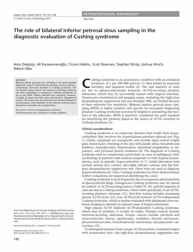
INTERVENTIONAL RADIOLOGYREVIEW
The role of bilateral inferior petrosal sinus sampling in the diagnostic evaluation of Cushing syndrome
Amy Deipolyi, Ali Karaosmanoğlu, Cicero Habito, Scott Brannan, Stephan Wicky, Joshua Hirsch,Rahmi Oklu
From the Departments of Vascular Imaging and Interventions(A.D., A.K., C.H., S.B., S.W., R.O. [email protected]), andNeurointerventional Radiology (J.H.), Massachusetts GeneralHospital and Harvard Medical School, Boston, Massachusetts,USA.
Received 8 February 2011; accepted 11 February 2011.
Published online 23 February 2011DOI 10.4261/1305-3825.DIR.4279-11.0
ABSTRACTBilateral inferior petrosal sinus sampling is the gold standard diagnostic assay in identifying the pituitary source of adreno-corticotropic hormone secretion in Cushing syndrome. The noninvasive assays used in the workup of Cushing syndrome are often misleading or ambiguous, yielding sensitivities of up to only 80%. Inferior petrosal sinus sampling, however, is highly accurate and safe when performed in experienced centers. We review here the historical and technical details of the procedure, with emphasis on the relevant anatomy and a discussion of possible rare complications.
Key words: • ACTH-secreting pituitary adenoma • inferior petrosal sinus sampling • Cushing syndrome
Diagn Interv Radiol 2012; 18:132–138
Bilateral inferior petrosal sinus sampling in Cushing syndrome • 133Volume 18 • Issue 1
to 60%, although spoiled gradient-recalled acquisition sequences can in-crease this sensitivity to approximately 80% (20). Dynamic contrast-enhanced MRI may also improve imaging accu-racy, given that adenomas tend to be slowly enhancing (21). False-negative MR results are due to microadenomas that are too small to detect by imag-ing, which is unfortunate given that most ACTH-secreting adenomas are subcentimeter (i.e., microadenomas) (19). Given that MRI can be equivocal in half of the patients tested, only rela-tively large lesions (>6 mm) detected on MRI with supporting biochemical confirmation and expected clinical symptoms reliably confirm the diagno-sis of Cushing syndrome (12).
If these noninvasive tests fail to dis-tinguish Cushing syndrome from ec-topic ACTH secretion or if they provide equivocal or ambiguous results, BIPSS with CRH stimulation is indicated for further evaluation (22).
Anatomic considerationsKnowledge of the pituitary venous
drainage is essential for the BIPSS technique (Fig. 2). The cavernous si-
nuses are just lateral to the pituitary fossa and contain the carotid artery and cranial nerves (23). They are in-terconnected by four intercavernous pathways. The anterior and posterior intercavernous sinuses are present in all people and run in front of and be-hind the pituitary (24). The inferior in-tercavernous sinus, coursing along the sellar floor between the anterior and posterior pituitary lobes, is absent in a small number of people; however, it is usually present in three forms (plexus-like, venous lake, and mixed). Finally, the basilar plexus is present in all peo-ple, is located along the dorsum sellae, and is the largest interconnection in most cases (24).
Hypophyseal veins exit the anterior pituitary lobe and drain into a plexi-form venous network overlying the pituitary surface, which in turn drains laterally into the intercavernous and cavernous sinuses. In spite of the broad communication between the cavern-ous sinuses, venous drainage from the pituitary is unilateral under normal physiologic conditions (23). This fact theoretically enables BIPSS to lateralize ACTH-secreting pituitary adenomas and necessitates bilateral sampling, as unilateral sampling could provide false-positive results (23). The inferior petrosal sinus (IPS) drains the cavern-ous sinus posteriorly, passes through the anterior jugular foramen, and drains into the internal jugular vein. As the IPS courses through the dura, it receives tributaries from the dura, pons, medulla, internal auditory mea-tus, and anterior condylar vein, which communicates with the plexus sur-rounding the twelfth cranial nerve in the hypoglossal canal (25).
There is interperson variability in the position of the junction of the IPS into
the corticotropin-releasing hormone (CRH) stimulation test, and eventually, cross-sectional imaging (12). The pri-mary noninvasive diagnostic assay, the high-dose dexamethasone suppression test, is based on the fact that the secre-tion of ACTH by a pituitary adenoma is inhibited by high doses of dexametha-sone, whereas ectopic sources of ACTH are not. However, this test offers only 60% to 80% sensitivity and specificity (8, 14). The CRH stimulation test re-lies upon the fact that most pituitary tumors respond to CRH administra-tion by increasing ACTH secretion and, thus, cortisol levels. Unfortunately, many ectopic ACTH-secreting tumors also respond in this way, limiting inter-pretation of the results (15).
Pituitary gadolinium-enhanced MRI with fine cuts through the sella turcica is indicated in patients with ACTH-dependent Cushing syndrome (8) and is far superior to CT imaging (16). However, interpretation of noninvasive cross-sectional imaging is complicated by the high prevalence (10% to 20%) of nonfunctioning pituitary incidenta-lomas (17–19). Conventional spin echo imaging yields a sensitivity of only 50%
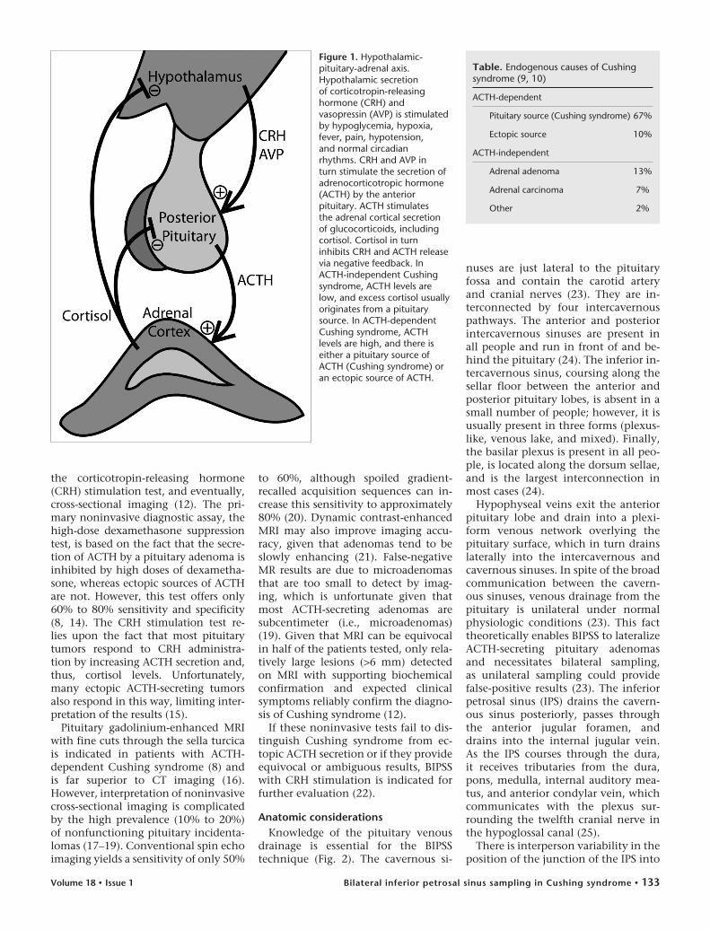
Figure 1. Hypothalamic-pituitary-adrenal axis. Hypothalamic secretion of corticotropin-releasing hormone (CRH) and vasopressin (AVP) is stimulated by hypoglycemia, hypoxia, fever, pain, hypotension, and normal circadian rhythms. CRH and AVP in turn stimulate the secretion of adrenocorticotropic hormone (ACTH) by the anterior pituitary. ACTH stimulates the adrenal cortical secretion of glucocorticoids, including cortisol. Cortisol in turn inhibits CRH and ACTH release via negative feedback. In ACTH-independent Cushing syndrome, ACTH levels are low, and excess cortisol usually originates from a pituitary source. In ACTH-dependent Cushing syndrome, ACTH levels are high, and there is either a pituitary source of ACTH (Cushing syndrome) or an ectopic source of ACTH.
Table. Endogenous causes of Cushing syndrome (9, 10)
ACTH-dependent
Pituitary source (Cushing syndrome) 67%
Ectopic source 10%
ACTH-independent
Adrenal adenoma 13%
Adrenal carcinoma 7%
Other 2%
Song et al.134 • January 2012 • Diagnostic and Interventional Radiology
the internal jugular vein. The IPS typi-cally joins the internal jugular vein at the level of the inferior margin of the jugular foramen, roughly 6 mm below its entry into the foramen, although in some patients, the junction may be extracranial or within the foramen. Rarely, the junction is intracranial or the IPS drains into the sigmoid sinus rather than into the internal jugular vein. Within the jugular foramen, the diameter of the IPS is 2 to 4 mm (25).
In addition to variability in the posi-tion of the junction, there is also vari-ation in its anatomic form (26). Four types of variant anatomy have been described (27). Most commonly (Type I; 45%), the IPS drains directly into the internal jugular bulb, with absent or nearly absent communication with the anterior condylar vein (ACV). In Type II anatomy (24%), the IPS anastomoses with the ACV before draining into the internal jugular vein. The ACV drains into the vertebral venous plexus. In Type III anatomy (24%), the IPS drains
into the internal jugular vein as a plex-us of veins rather than as a single vein. This plexus also drains partly into the vertebral venous plexus. In the uncom-mon Type IV anatomy, the IPS drains solely or predominantly into the ver-tebral venous plexus via the ACV. The connection between the IPS and jugu-lar vein in this case is hypoplastic or does not exist. This configuration is reported to be present in up to 7% of patients (27), though another study re-ported that it existed in less than 1% (25).
Bonelli and colleagues (28) suggested redefining this classification system, such that Type I and II anatomy differ based on the distance between the IPS-ACV anastomosis and the IPS–internal jugular vein junction. In our experi-ence, the key anatomical consideration is to define the junction of the IPS and ACV if there is one and to advance the catheter past it so that IPS samples are not diluted by drainage from the ACV. We therefore suggest three relevant types of anatomy, essentially combin-ing types I and II into a single Type A (Fig. 3).
Anatomic variations can obscure the interpretation of BIPSS results. For example, when the IPS is hypoplastic (Type IV or C), BIPSS yields false-nega-tive results (29). Furthermore, the IPSs are also asymmetric in as many as 40% of patients (13, 30), leading to errors in lateralization. For this reason, venous angiography is routinely done during BIPSS prior to sampling to avoid such interpretative errors.
Historical considerationsDr. Harvey Cushing was an American
neurosurgeon who first described Cushing syndrome in the 1930s (31). Trans-sphenoidal surgery for pituitary tumors had been developed by Dr. Hermann Schloffer in the early 1900s, but it was abandoned by the 1920s be-cause of its high mortality. From 1910 to 1925, Dr. Cushing worked to refine the trans-sphenoidal approach, but he instead transitioned to the transcranial approach, which had lower complica-tion rates. Building on the teaching of his predecessors Drs. Normal Dott and Gerard Guiot, Dr. Jules Hardy began using intraoperative fluoroscopy and introduced the operative microscope, which significantly decreased morbidi-ty and increased the success of the pro-cedure (32, 33). The use of transsphe-
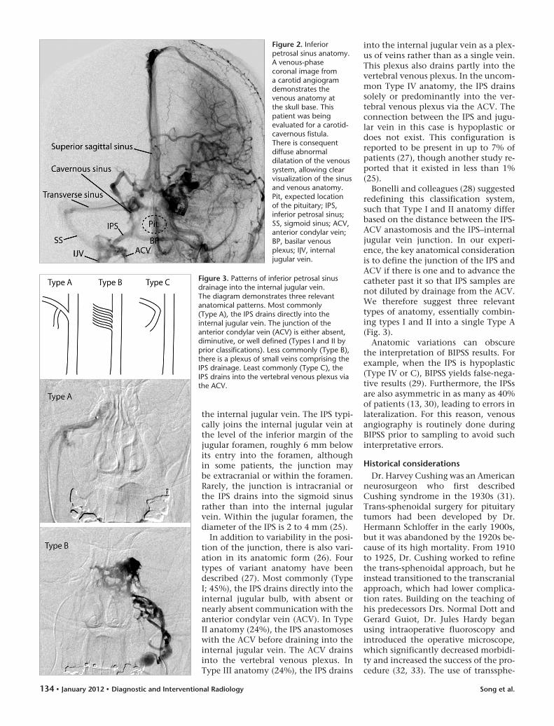
Figure 2. Inferior petrosal sinus anatomy. A venous-phase coronal image from a carotid angiogram demonstrates the venous anatomy at the skull base. This patient was being evaluated for a carotid-cavernous fistula. There is consequent diffuse abnormal dilatation of the venous system, allowing clear visualization of the sinus and venous anatomy. Pit, expected location of the pituitary; IPS, inferior petrosal sinus; SS, sigmoid sinus; ACV, anterior condylar vein; BP, basilar venous plexus; IJV, internal jugular vein.
Figure 3. Patterns of inferior petrosal sinus drainage into the internal jugular vein. The diagram demonstrates three relevant anatomical patterns. Most commonly (Type A), the IPS drains directly into the internal jugular vein. The junction of the anterior condylar vein (ACV) is either absent, diminutive, or well defined (Types I and II by prior classifications). Less commonly (Type B), there is a plexus of small veins comprising the IPS drainage. Least commonly (Type C), the IPS drains into the vertebral venous plexus via the ACV.
Bilateral inferior petrosal sinus sampling in Cushing syndrome • 135Volume 18 • Issue 1
noidal surgery for Cushing syndrome spread during the 1970s, and now it is the treatment of choice, resulting in posttreatment survival rates similar to the general population (34).
Improvements in the surgical tech-niques motivated advances in the diag-nostic tools. BIPSS as a diagnostic mo-dality for Cushing syndrome was es-tablished in the 1970s. A case report in 1977 was the first to describe catheteri-zation of and sampling of ACTH from the IPS to diagnose Cushing syndrome in a patient with perplexing clinical and laboratory features (35). This was followed by a case series in 1981 dem-onstrating the safety and efficacy of BIPSS in distinguishing Cushing syn-drome from ectopic ACTH production, highlighting the importance of select-ing the IPS, because measurements from the jugular veins were nondiag-nostic (36).
The most important development in the BIPSS technique since that time has been the introduction of CRH ad-ministration during the procedure. CRH stimulates ACTH secretion in normal patients and in those with pi-tuitary adenomas (37). Landolt et al. (38) were the first to administer CRH during BIPSS, and they demonstrated that CRH enhances the distinction between normal tissue and adenoma, therefore improving the sensitivity of BIPSS considerably.
Technical details and considerationsThe standard protocol at our institu-
tion is to sample ACTH peripherally and from both IPSs before and after CRH administration. The procedure is performed under conscious sedation to enable monitoring of symptoms in-dicating complications. For example, because of the high sensitivity of the jugular fossa periosteum, catheteriza-tion that is too high will result in pa-tient otalgia (23), which can only be assessed if the patient is conscious.
After the patient is prepped, 21 gauge micropuncture needles are used to access both common femoral veins in order to place a 6 F sheath into the right femoral vein and a 5 F sheath into the left femoral vein. The larger sheath is used on the right for pe-ripheral sampling while the 5 F Davis catheter is in place. We then admin-ister 3 000–5 000 units of heparin. In addition to refraining from cannulat-ing the IPS too centrally, heparin ad-
ministration is important to avoid IPS and cavernous sinus thrombosis (22).
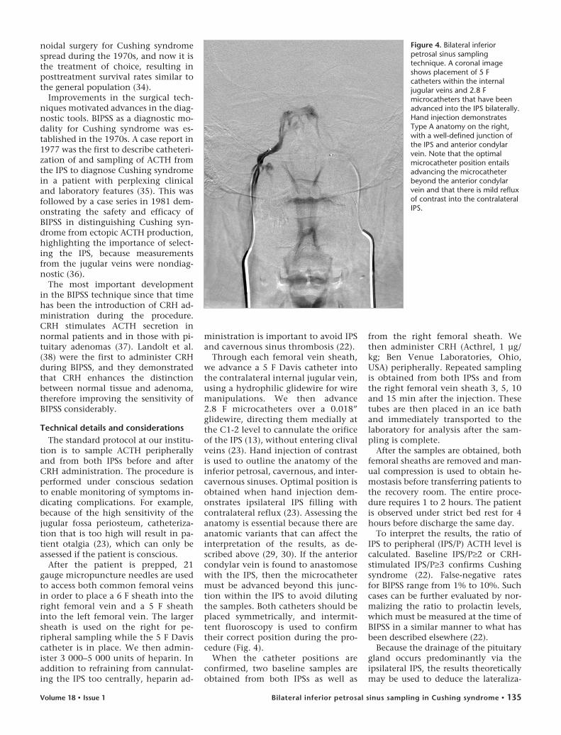
Through each femoral vein sheath, we advance a 5 F Davis catheter into the contralateral internal jugular vein, using a hydrophilic glidewire for wire manipulations. We then advance 2.8 F microcatheters over a 0.018” glidewire, directing them medially at the C1-2 level to cannulate the orifice of the IPS (13), without entering clival veins (23). Hand injection of contrast is used to outline the anatomy of the inferior petrosal, cavernous, and inter-cavernous sinuses. Optimal position is obtained when hand injection dem-onstrates ipsilateral IPS filling with contralateral reflux (23). Assessing the anatomy is essential because there are anatomic variants that can affect the interpretation of the results, as de-scribed above (29, 30). If the anterior condylar vein is found to anastomose with the IPS, then the microcatheter must be advanced beyond this junc-tion within the IPS to avoid diluting the samples. Both catheters should be placed symmetrically, and intermit-tent fluoroscopy is used to confirm their correct position during the pro-cedure (Fig. 4).
When the catheter positions are confirmed, two baseline samples are obtained from both IPSs as well as
from the right femoral sheath. We then administer CRH (Acthrel, 1 μg/kg; Ben Venue Laboratories, Ohio, USA) peripherally. Repeated sampling is obtained from both IPSs and from the right femoral vein sheath 3, 5, 10 and 15 min after the injection. These tubes are then placed in an ice bath and immediately transported to the laboratory for analysis after the sam-pling is complete.
After the samples are obtained, both femoral sheaths are removed and man-ual compression is used to obtain he-mostasis before transferring patients to the recovery room. The entire proce-dure requires 1 to 2 hours. The patient is observed under strict bed rest for 4 hours before discharge the same day.
To interpret the results, the ratio of IPS to peripheral (IPS/P) ACTH level is calculated. Baseline IPS/P≥2 or CRH-stimulated IPS/P≥3 confirms Cushing syndrome (22). False-negative rates for BIPSS range from 1% to 10%. Such cases can be further evaluated by nor-malizing the ratio to prolactin levels, which must be measured at the time of BIPSS in a similar manner to what has been described elsewhere (22).
Because the drainage of the pituitary gland occurs predominantly via the ipsilateral IPS, the results theoretically may be used to deduce the lateraliza-
Figure 4. Bilateral inferior petrosal sinus sampling technique. A coronal image shows placement of 5 F catheters within the internal jugular veins and 2.8 F microcatheters that have been advanced into the IPS bilaterally. Hand injection demonstrates Type A anatomy on the right, with a well-defined junction of the IPS and anterior condylar vein. Note that the optimal microcatheter position entails advancing the microcatheter beyond the anterior condylar vein and that there is mild reflux of contrast into the contralateral IPS.
Song et al.136 • January 2012 • Diagnostic and Interventional Radiology
tion of the adenoma, although such results are controversial. An intersinus ratio of at least 1.4 has been considered as evidence of ipsilateral localization of an adenoma. Studies have demonstrat-ed an accuracy of 50% to 100% (78% overall) using surgical findings as the gold standard, and accuracy is not im-proved with CRH administration (39). Therefore, irrespective of lateralization suggested by the BIPSS findings, sur-geons routinely perform a full explora-tion of the entire pituitary gland rather than a hemihypophysectomy based solely on BIPSS results.
ComplicationsBIPSS is a safe procedure when per-
formed by experienced interventional radiologists. The most common com-plication is groin hematoma from femoral access, occurring in at most 3%–4% of patients (25). Serious com-plications are very rare. In a series of 508 procedures at the NIH, there was one serious neurologic complication (pontine hemorrhage) and one patient with a severe vasovagal reaction (25). Three other patients in this series had transient complaints such as vertigo or paresthesias without abnormalities on brain CT or MRI. The authors described an additional case report at another in-stitution of a patient with nausea and symptoms of a medial medullary syn-drome who was found to have a non-hemorrhagic right medullary infarc-tion. In a more recent study of 86 BIPSS procedures, two patients had transient CN VI palsies (40). Another group re-ported a venous subarachnoid hemor-rhage causing acute obstructive hydro-cephalus in one patient out of the 94 procedures performed at their institu-tion (41). In another series of 44 BIPSS, there was one case of pontomedullary dysfunction, with MRI demonstrat-ing a brainstem infarction (42). Given how rare such complications are, it is difficult to deduce what factors may increase risk, although catheter choice and variant venous anatomy may contribute.
Thromboembolic events have also occasionally been reported. Patients with Cushing syndrome are already at risk given their hypercoagulable state. In one series published at Vanderbilt University, 2 of 34 patients with Cushing syndrome developed deep ve-nous thrombosis after BIPSS, with one of these patients expiring from a pul-
monary embolism (43). However, this center did not routinely heparinize pa-tients periprocedurally. At another site, out of 94 procedures, there was one lower extremity deep venous thrombo-sis (41). Another group reported deep venous thrombosis in two patients un-dergoing BIPSS and discussed the im-portance of prophylactic anticoagula-tion in this patient group (44).
Accuracy and utilityWhen performed in experienced
centers, BIPSS is highly accurate in diagnosing Cushing syndrome. In a meta-analysis review of 21 studies, the overall sensitivity and specificity of BIPSS were found to be 96% and 100%, respectively (39). CRH administration increases sensitivity such that it ap-proaches 100% (45). At our institution, in a review of 185 BIPSS procedures for 179 patients from 1986 to 2002, there was 90% sensitivity and 67% specifi-city, with a 5% false-negative rate (46), in line with data reported by other centers. Given the low negative predic-tive value in that study (20%), the au-thors suggested that trans-sphenoidal exploration is indicated in patients with negative studies and no identifi-able ectopic ACTH sources.
When compared with other diag-nostic modalities, BIPSS is consistent-ly more accurate. For instance, using stringent diagnostic criteria, Wiggam et al. (47) reported sensitivities of 48% for high-dose dexamethasone test-ing, 70% for CRH testing, and 82% for BIPSS. Compared with cross-sec-tional imaging, BIPSS is also superior. Kaskarelis et al. (48) reported an accu-racy of 50% for MRI and 88% for BIPSS in 54 patients. Colao et al. (49), in a study of 84 patients, reported sensitivi-ties of 40% for CT, 50% for MRI, and 90% for BIPSS, although noninvasive imaging was superior in lateralizing adenomas (75%–80% for CT and MRI versus 65% for BIPSS).
Even though BIPSS is more accurate than any other diagnostic modalities in routine use, because it is more in-vasive and costly, its application varies among institutions. There is general consensus that BIPSS is indicated only once ACTH-dependent hypercortiso-lemia has been firmly established (13, 22). In other words, BIPSS is not meant to diagnose Cushing syndrome, but to diagnose a pituitary source of ACTH hypersecretion.
There is also general agreement that BIPSS is always indicated when non-invasive testing yields conflicting or equivocal results (9, 12, 22). In some institutions, BIPSS is routinely per-formed in all patients evaluated for Cushing syndrome, even those with definitive noninvasive workups (50), given the high prevalence of pituitary incidentalomas and the excellent accu-racy and safety of BIPSS in experienced hands (50). One retrospective study of 193 patients with ACTH-dependent Cushing syndrome compared clinical outcomes between a group with clear-cut noninvasive testing and no BIPSS versus a group with equivocal nonin-vasive testing who did undergo BIPSS. All patients received trans-sphenoidal surgery. The authors reported no dif-ference in remission rate or recurrence between the two groups after surgery and concluded that selective appli-cation of BIPSS in the evaluation of Cushing syndrome does not lead to misdiagnosis (51). However, prospec-tive studies comparing broad as op-posed to selective application have not been done, and currently there are no formal societal guidelines (22).
Alternative proceduresBecause of the technically demand-
ing nature of BIPSS, some have studied a less demanding alternative, internal jugular venous sampling. Doppman et al. (52) collected jugular and inferior petrosal samples from 20 patients with Cushing syndrome and reported a sen-sitivity of 80% for jugular venous sam-pling compared with a 95% sensitivity for BIPSS. Another group reported a sensitivity of 94% for BIPSS and 83% for jugular venous sampling at specifi-cities of 100%, a difference that was not statistically significant (53). The authors concluded that jugular venous sampling may be used in centers lack-ing technical expertise, although nega-tive findings should be reevaluated with BIPSS.
Because BIPSS has not consistently provided accurate lateralization of ad-enomas, cavernous sinus sampling has been studied as an alternative proce-dure, with the idea that the cavernous sinus, which is in closer proximity to the pituitary, would provide more ac-curate ACTH gradients (54). Results have been mixed, and conclusions regarding its safety and accuracy are awaiting a larger case series (55).
Bilateral inferior petrosal sinus sampling in Cushing syndrome • 137Volume 18 • Issue 1
Because CRH is not always available in all institutions, some have used desmopressin (a synthetic analog of vasopressin) instead to stimulate the pituitary during BIPSS. Several small series have suggested that this alterna-tive may be safe and effective, although larger confirmatory studies have not yet been done (22).
At present, BIPSS with CRH stimula-tion remains the gold standard in the evaluation of Cushing syndrome.
As a conclusion, Cushing syndrome is an uncommon endocrine disorder caused by excess pituitary secretion of ACTH that leads to hypercortisolemia. Frequently, exclusion of extrapitui-tary sources of ACTH is difficult, given the limited sensitivity of noninvasive tests. BIPSS offers a sensitivity and specificity approaching 100%. Careful delineation of the patient’s venous anatomy and attention to technical considerations, including proper cath-eter placement and periprocedural heparinization, are essential to pro-vide a safe procedure with interpret-able results. While some have begun to study alternative techniques, such as cavernous sinus sampling or pitui-tary stimulation with desmopressin, BIPSS with CRH administration is the current gold standard for diagnosing Cushing syndrome.
AcknowledgementsWe thank Dr. Ronil V. Chandra and Dr.
James Rabinov for providing Figure 2.Dr. Oklu is the recipient of the Junior Faculty
Investigator Award from the American College of Phlebology.
Conflict of interest disclosureThe authors declared no conflicts of interest.
References 1. Ross NS. Epidemiology of Cushing’s syn-
drome and subclinical disease. Endocrinol Metab Clin North Am 1994; 23:539–546.
2. Lindholm J, Juul S, Jorgensen JO, et al. Incidence and late prognosis of cushing’s syndrome: a population-based study. J Clin Endocrinol Metab 2001; 86:117–123.
3. Lad SP, Patil CG, Laws ER Jr, Katznelson L. The role of inferior petrosal sinus sampling in the diagnostic localization of Cushing’s disease. Neurosurg Focus 2007; 23:E2.
4. Cotran RS, Kumar V, Collins T. Robbins pathologic basis of disease. 6 ed. 1999, Philadelphia: WB Saunders.
5. Atkinson AB, Kennedy AL, Carson DJ, Hadden DR, Weaver JA, Sheridan B. Five cases of cyclical Cushing’s syndrome. Br Med J (Clin Res Ed) 1985; 291:1453–1457.
6. Boscaro M, Barzon L, Sonino N. The di-agnosis of Cushing’s syndrome: atypical presentations and laboratory shortcom-ings. Arch Intern Med 2000; 160:3045–3053.
7. Velez DA, Mayberg MR, Ludlam WH. Cyclic Cushing syndrome: definitions and treatment implications. Neurosurg Focus 2007; 23:E4; Discussion E4a.
8. Arnaldi G, Angeli A, Atkinson AB, et al. Diagnosis and complications of Cushing’s syndrome: a consensus statement. J Clin Endocrinol Metab 2003; 88:5593–5602.
9. Boscaro M, Arnaldi G. Approach to the pa-tient with possible Cushing’s syndrome. J Clin Endocrinol Metab 2009; 94:3121–3131.
10. Nieman L. Causes and pathophysiology of Cushing’s syndrome. In: DS Basow, ed. UpToDate. Waltham, MA; 2011.
11. Nieman L, Lacroix A, Martin K. Establishing the case of Cushing’s syndrome. In: DS Basow, ed. UpToDate. Waltham, MA; 2011.
12. Gross BA, Mindea SA, Pick AJ, Chandler JP, Batjer HH. Diagnostic approach to Cushing disease. Neurosurg Focus 2007; 23:E1.
13. Tomycz ND, Horowitz MB. Inferior petro-sal sinus sampling in the diagnosis of sellar neuropathology. Neurosurg Clin N Am 2009; 20:361–367.
14. Aron DC, Raff H, Findling JW. Effectiveness versus efficacy: the limited value in clini-cal practice of high dose dexamethasone suppression testing in the differential di-agnosis of adrenocorticotropin-dependent Cushing’s syndrome. J Clin Endocrinol Metab 1997; 82:1780–1785.
15. Nieman L, Lacroix A, Martin K. Establishing the cause of Cushing’s syndrome. In: DS Basow, ed. Waltham, MA: UpToDate, 2011.
16. Escourolle H, Abecassis JP, Bertagna X, et al. Comparison of computerized tomog-raphy and magnetic resonance imaging for the examination of the pituitary gland in patients with Cushing’s disease. Clin Endocrinol (Oxf) 1993; 39:307–313.
17. Ezzat S, Asa SL, Couldwell WT, et al. The prevalence of pituitary adenomas: a sys-tematic review. Cancer 2004; 101:613–619.
18. Hall WA, Luciano MG, Doppman JL, Patronas NJ, Oldfield EH. Pituitary mag-netic resonance imaging in normal hu-man volunteers: occult adenomas in the general population. Ann Intern Med 1994; 120:817–820.
19. Molitch ME, Russell EJ. The pituitary “in-cidentaloma”. Ann Intern Med 1990; 112:925–931.
20. Patronas N, Bulakbasi N, Stratakis CA, et al. Spoiled gradient recalled acquisition in the steady state technique is superior to conventional postcontrast spin echo technique for magnetic resonance imaging detection of adrenocorticotropin-secreting pituitary tumors. J Clin Endocrinol Metab 2003; 88:1565–1569.
21. Jagannathan J, Sheehan JP, Jane JA Jr. Evaluation and management of Cushing syndrome in cases of negative sellar mag-netic resonance imaging. Neurosurg Focus 2007; 23:E3.
22. Javorsky BR, Findling JW. Inferior petrosal sampling for the differential diagnosis of ACTH-dependent Cushing’s syndrome. In: Bronstein MD, ed. Cushing’s syndrome: pathophysiology, diagnosis and treat-ment. New York: Humana Press, 2010.
23. Doppman JL, Oldfield E, Krudy AG, et al. Petrosal sinus sampling for Cushing syn-drome: anatomical and technical consider-ations. Work in progress. Radiology 1984; 150:99–103.
24. Aquini MG, Marrone AC, Schneider FL. Intercavernous venous communications in the human skull base. Skull Base Surg 1994; 4:145–150.
25. Miller DL, Doppman JL. Petrosal sinus sam-pling: technique and rationale. Radiology 1991; 178:37–47.
26. Braun JP, Tournade A. Venous drainage in the craniocervical region. Neuroradiology 1977; 13:155–158.
27. Shiu PC, Hanafee WN, Wilson GH, Rand RW. Cavernous sinus venography. Am J Roentgenol Radium Ther Nucl Med 1968; 104:57–62.
28. Bonelli FS, Huston J 3rd, Carpenter PC, Erickson D, Young WF Jr, Meyer FB. Adrenocorticotropic hormone-dependent Cushing’s syndrome: sensitivity and spe-cificity of inferior petrosal sinus sampling. AJNR Am J Neuroradiol 2000; 21:690–696.
29. Doppman JL, Chang R, Oldfield EH, Chrousos G, Stratakis CA, Nieman LK. The hypoplastic inferior petrosal sinus: a potential source of false-negative results in petrosal sampling for Cushing’s disease. J Clin Endocrinol Metab 1999; 84:533–540.
30. Mamelak AN, Dowd CF, Tyrrell JB, McDonald JF, Wilson CB. Venous angiog-raphy is needed to interpret inferior pet-rosal sinus and cavernous sinus sampling data for lateralizing adrenocorticotropin-secreting adenomas. J Clin Endocrinol Metab 1996; 81:475–481.
31. Cushing H. The basophil adenomas of the pituitary body and their clinical manifes-tations (pituitary basophilism). Bull Johns Hopkins Hosp 1932; 50:137–195.
32. Hardy J. Transsphenoidal hypophysecto-my. J Neurosurg 1971; 34:582–594.
33. Liu JK, Das K, Weiss MH, Laws ER Jr, Couldwell WT. The history and evolution of transsphenoidal surgery. J Neurosurg 2001; 95:1083–1096.
34. Kelly DF. Transsphenoidal surgery for Cushing’s disease: a review of success rates, remission predictors, management of failed surgery, and Nelson’s syndrome. Neurosurg Focus 2007; 23:E5.
35. Corrigan DF, Schaaf M, Whaley RA, Czerwinski CL, Earll JM. Selective venous sampling to differentiate ectopic ACTH secretion from pituitary Cushing’s syn-drome. N Engl J Med 1977; 296:861–862.
36. Findling JW, Aron DC, Tyrrell JB, et al. Selective venous sampling for ACTH in Cushing’s syndrome: differentiation be-tween Cushing disease and the ectopic ACTH syndrome. Ann Intern Med 1981; 94:647–652.
37. Orth DN, DeBold CR, DeCherney GS, et al. Pituitary microadenomas causing Cushing’s disease respond to corticotro-pin-releasing factor. J Clin Endocrinol Metab 1982; 55:1017–1019.
Song et al.138 • January 2012 • Diagnostic and Interventional Radiology
38. Landolt AM, Valavanis A, Girard J, Eberle AN. Corticotrophin-releasing factor-test used with bilateral, simultaneous inferior petrosal sinus blood-sampling for the di-agnosis of pituitary-dependent Cushing’s disease. Clin Endocrinol (Oxf) 1986; 25:687–696.
39. Newell-Price J, Trainer P, Besser M, Grossman A. The diagnosis and differen-tial diagnosis of Cushing’s syndrome and pseudo-Cushing’s states. Endocr Rev 1998; 19:647–672.
40. Lefournier V, Martinie M, Vasdev A, et al. Accuracy of bilateral inferior petrosal or cavernous sinuses sampling in predict-ing the lateralization of Cushing’s disease pituitary microadenoma: influence of catheter position and anatomy of venous drainage. J Clin Endocrinol Metab 2003; 88:196–203.
41. Bonelli FS, Huston J 3rd, Meyer FB, Carpenter PC. Venous subarachnoid he-morrhage after inferior petrosal sinus sam-pling for adrenocorticotropic hormone. AJNR Am J Neuroradiol 1999; 20:306–307.
42. Gandhi CD, Meyer SA, Patel AB, Johnson DM, Post KD. Neurologic complications of inferior petrosal sinus sampling. AJNR Am J Neuroradiol 2008; 29:760–765.
43. Blevins LS Jr, Clark RV, Owens DS. Thromboembolic complications after in-ferior petrosal sinus sampling in patients with Cushing’s syndrome. Endocr Pract 1998; 4:365–367.
44. Obuobie K, Davies JS, Ogunko A, Scanlon MF. Venous thrombo-embolism follow-ing inferior petrosal sinus sampling in Cushing’s disease. J Endocrinol Invest 2000; 23:542-544.
45. Oldfield EH, Doppman JL, Nieman LK, et al. Petrosal sinus sampling with and with-out corticotropin-releasing hormone for the differential diagnosis of Cushing’s syn-drome. N Engl J Med 1991; 325:897–905.
46. Swearingen B, Katznelson L, Miller K, et al. Diagnostic errors after inferior petrosal sinus sampling. J Clin Endocrinol Metab 2004; 89:3752–3763.
47. Wiggam MI, Heaney AP, McIlrath EM, et al. Bilateral inferior petrosal sinus sampling in the differential diagnosis of adrenocorti-cotropin-dependent Cushing’s syndrome: a comparison with other diagnostic tests. J Clin Endocrinol Metab 2000; 85:1525–1532.
48. Kaskarelis IS, Tsatalou EG, Benakis SV, et al. Bilateral inferior petrosal sinuses sampling in the routine investigation of Cushing’s syndrome: a comparison with MRI. AJR Am J Roentgenol 2006; 187:562–570.
49. Colao A, Faggiano A, Pivonello R, Pecori Giraldi F, Cavagnini F, Lombardi G. Inferior petrosal sinus sampling in the dif-ferential diagnosis of Cushing’s syndrome: results of an Italian multicenter study. Eur J Endocrinol 2001; 144:499–507.
50. Kaltsas GA, Giannulis MG, Newell-Price JD, et al. A critical analysis of the value of simultaneous inferior petrosal sinus sam-pling in Cushing’s disease and the occult ectopic adrenocorticotropin syndrome. J Clin Endocrinol Metab 1999; 84:487–492.
51. Jehle S, Walsh JE, Freda PU, Post KD. Selective use of bilateral inferior petrosal sinus sampling in patients with adrenocor-ticotropin-dependent Cushing’s syndrome prior to transsphenoidal surgery. J Clin Endocrinol Metab 2008; 93:4624–4632.
52. Doppman JL, Oldfield EH, Nieman LK. Bilateral sampling of the internal jugular vein to distinguish between mechanisms of adrenocorticotropic hormone-depend-ent Cushing syndrome. Ann Intern Med 1998; 128:33–36.
53. Ilias I, Chang R, Pacak K, et al. Jugular ve-nous sampling: an alternative to petrosal sinus sampling for the diagnostic evalu-ation of adrenocorticotropic hormone-dependent Cushing’s syndrome. J Clin Endocrinol Metab 2004; 89:3795–3800.
54. Hayashi N, Kurimoto M, Kubo M, et al. The impact of cavernous sinus drainage pattern on the results of venous sampling in pa-tients with suspected cushing syndrome. AJNR Am J Neuroradiol 2008; 29:69–72.
55. Oldfield EH, Doppman JL. Petrosal versus cavernous sinus sampling. J Neurosurg 1998; 89:890–893.