Embed Size (px)
Citation preview
MICROSCOPY AND STAININGCHAPTER 3
2
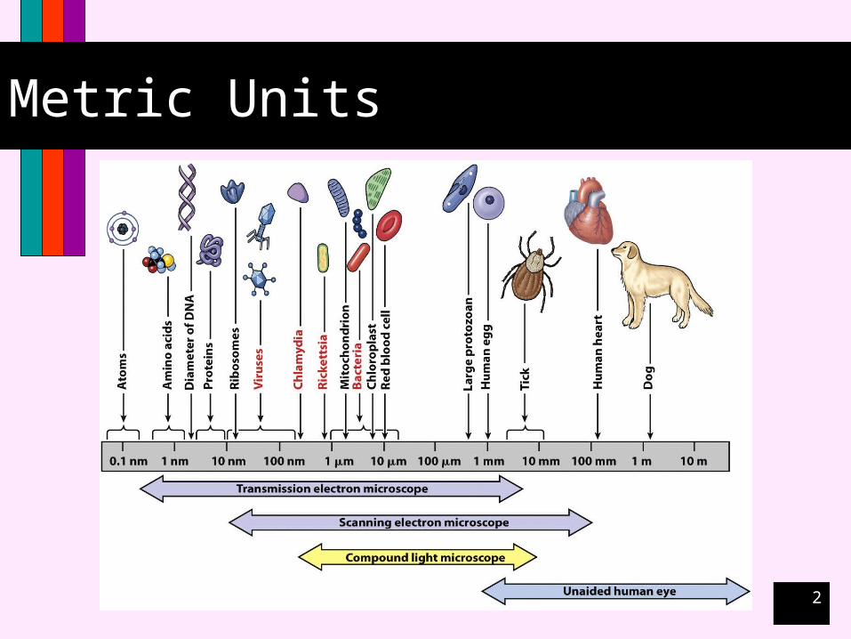
Metric Units

3
Light Properties
Wavelength
4
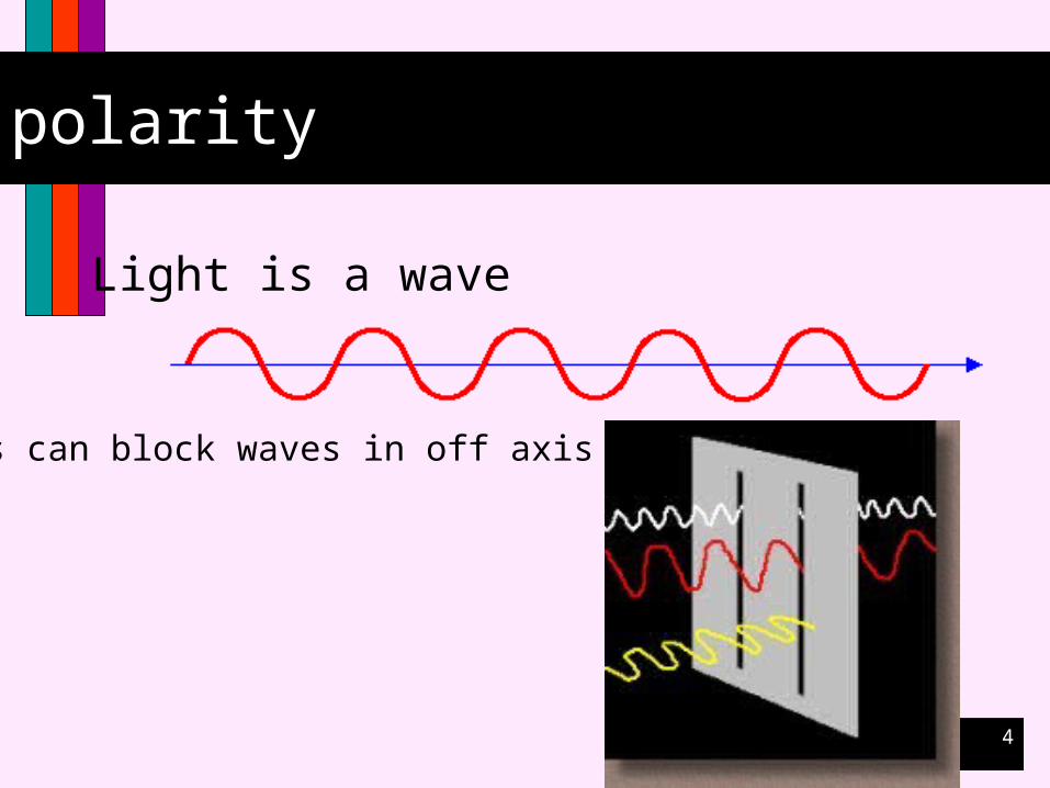
polarity
Light is a wave
Filters can block waves in off axis planes

5
Waves can be added
+ +
= =

6
Light Properties
Resolution
7
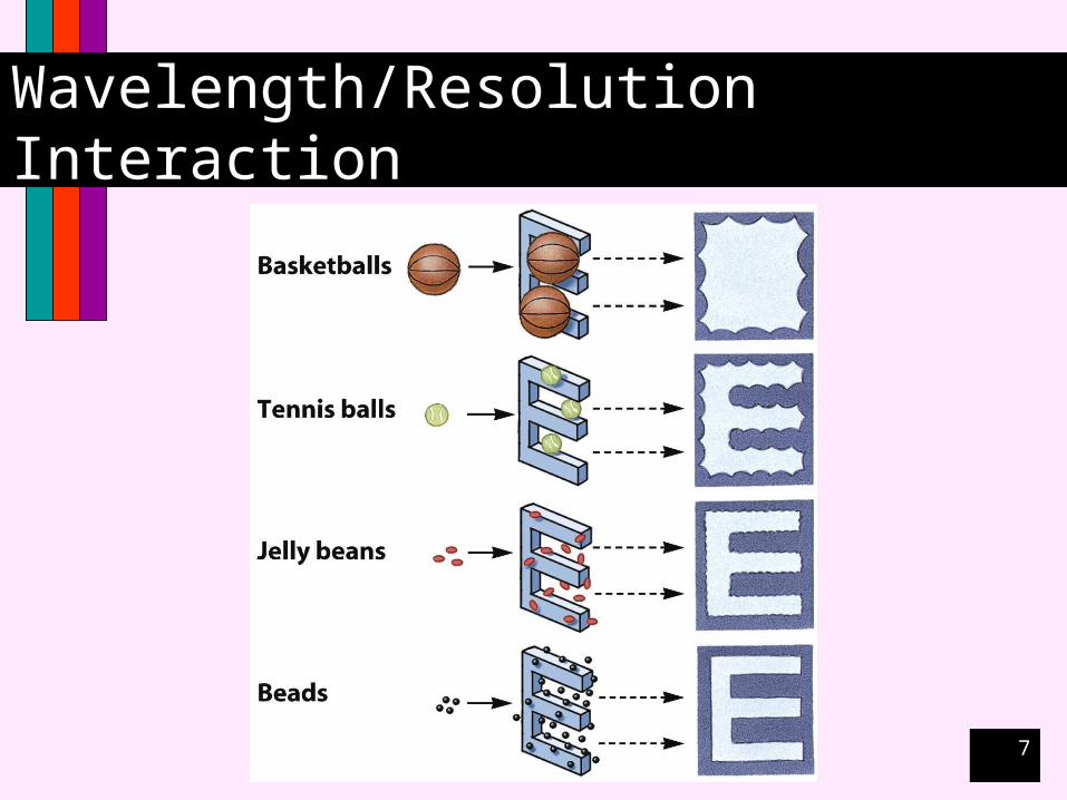
Wavelength/Resolution Interaction
8
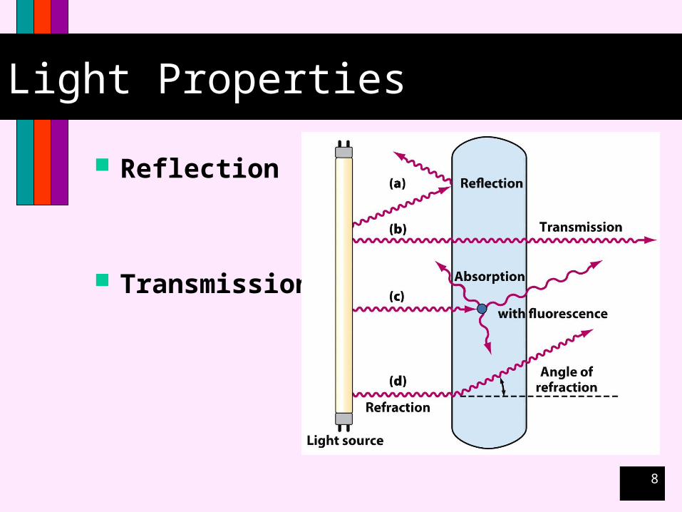
Light Properties
Reflection
Transmission
9
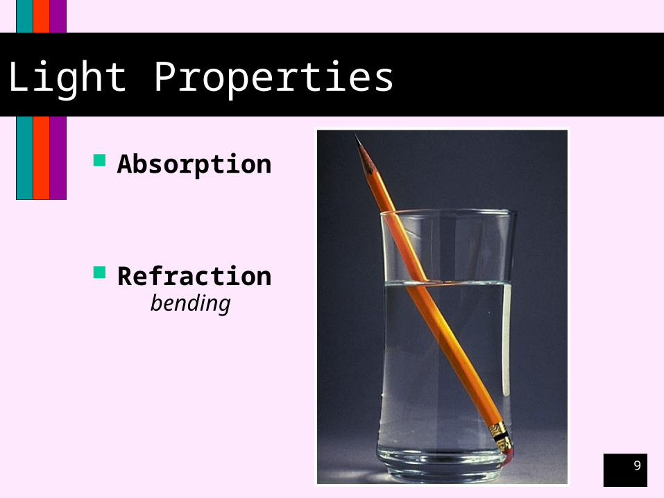
Light Properties
Absorption
Refractionbending
10
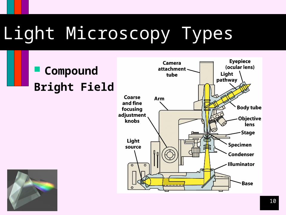
Light Microscopy Types
Compound
Bright Field
11
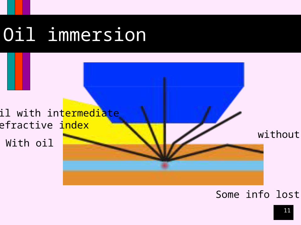
Oil immersion
With oilwithout
Some info lost
Oil with intermediaterefractive index
12
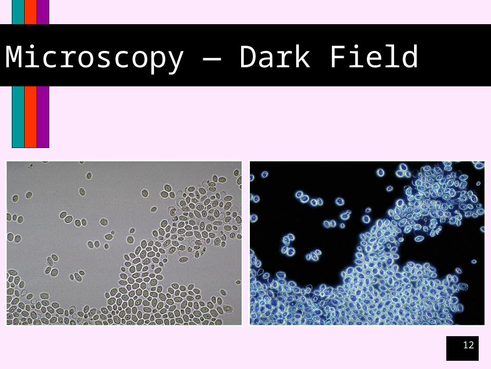
Microscopy — Dark Field

13
Microscopy — Phase Contrast
Dual Beam
14
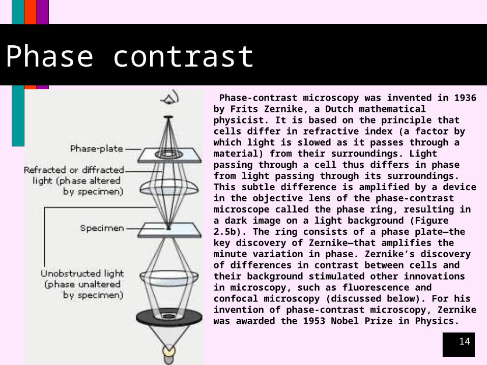
Phase contrast Phase-contrast microscopy was invented in 1936 by Frits
Zernike, a Dutch mathematical physicist. It is based on the principle that cells differ in refractive index (a factor by which light is slowed as it passes through a material) from their surroundings. Light passing through a cell thus differs in phase from light passing through its surroundings. This subtle difference is amplified by a device in the objective lens of the phase-contrast microscope called the phase ring, resulting in a dark image on a light background (Figure 2.5b). The ring consists of a phase plate—the key discovery of Zernike—that amplifies the minute variation in phase. Zernike’s discovery of differences in contrast between cells and their background stimulated other innovations in microscopy, such as fluorescence and confocal microscopy (discussed below). For his invention of phase-contrast microscopy, Zernike was awarded the 1953 Nobel Prize in Physics.

15
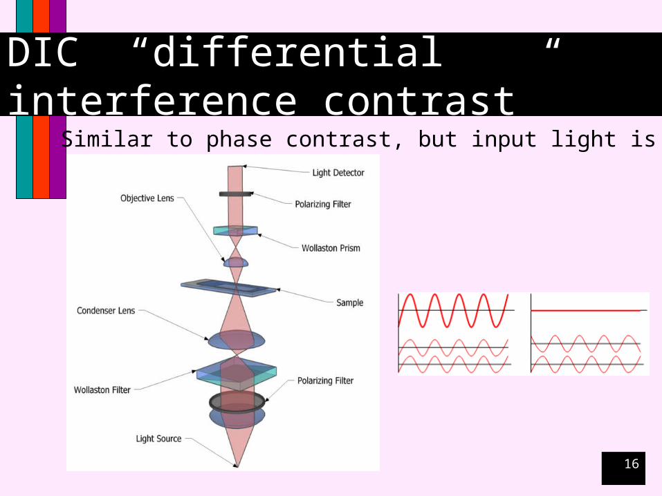
Microscopy — DIC
Differential Interference Contrast
16
DIC “differential interference contrast”
Similar to phase contrast, but input light is polarized
17
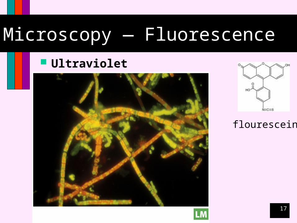
Microscopy — Fluorescence Ultraviolet light
flourescein
18
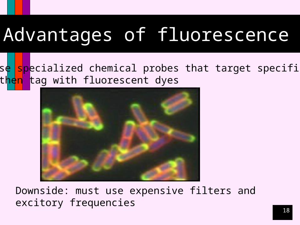
Advantages of fluorescence
Can use specialized chemical probes that target specific features and then tag with fluorescent dyes
Downside: must use expensive filters and excitory frequencies

19
Microscopy — Confocal
ConfocalAllows 3 dimensional viewing
Allows multiple dyes to be overlaid

20
Confocal microscopy
Allows 3 dimensions
21
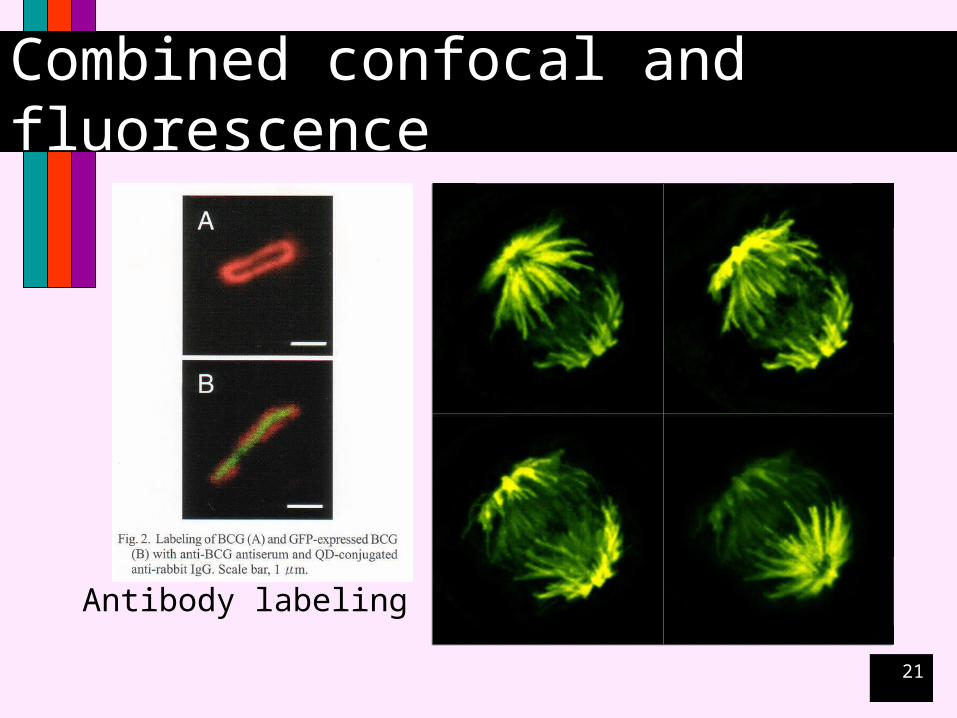
Combined confocal and fluorescence
Antibody labeling

22
Microscopy Imaging
Digital

23
Fig. 2-15

24
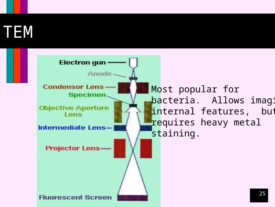
Electron Microscopy Transmission
(TEM)
Scanning (SEM)
Scanning Tunneling (STM)
25
TEM
Most popular for bacteria. Allows imaging internal features, but requires heavy metalstaining.
26
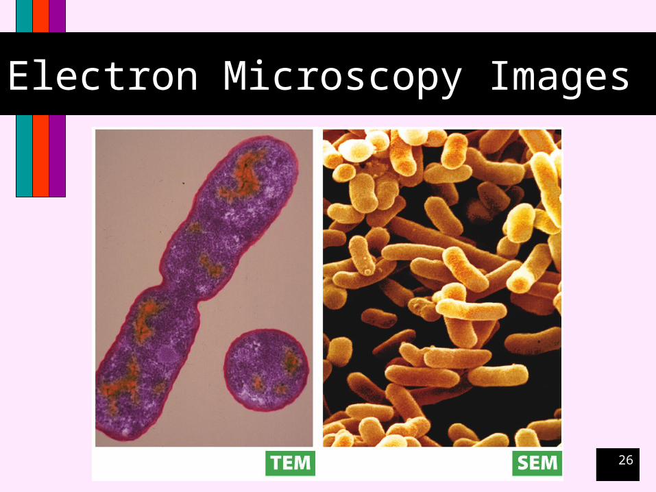
Electron Microscopy Images
27
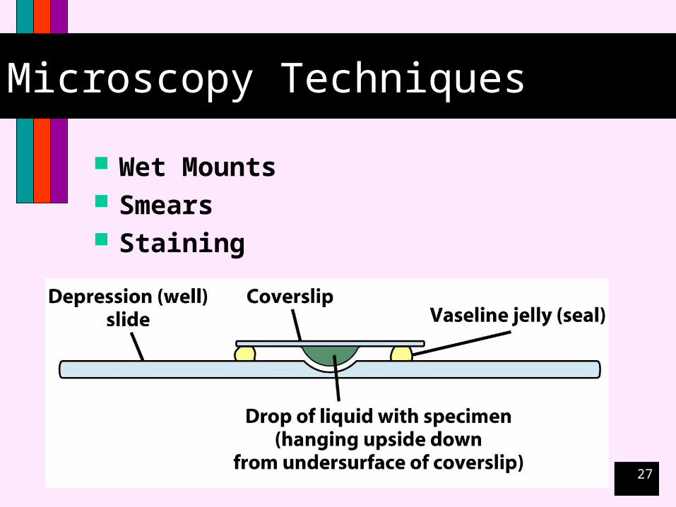
Microscopy Techniques
Wet Mounts Smears Staining
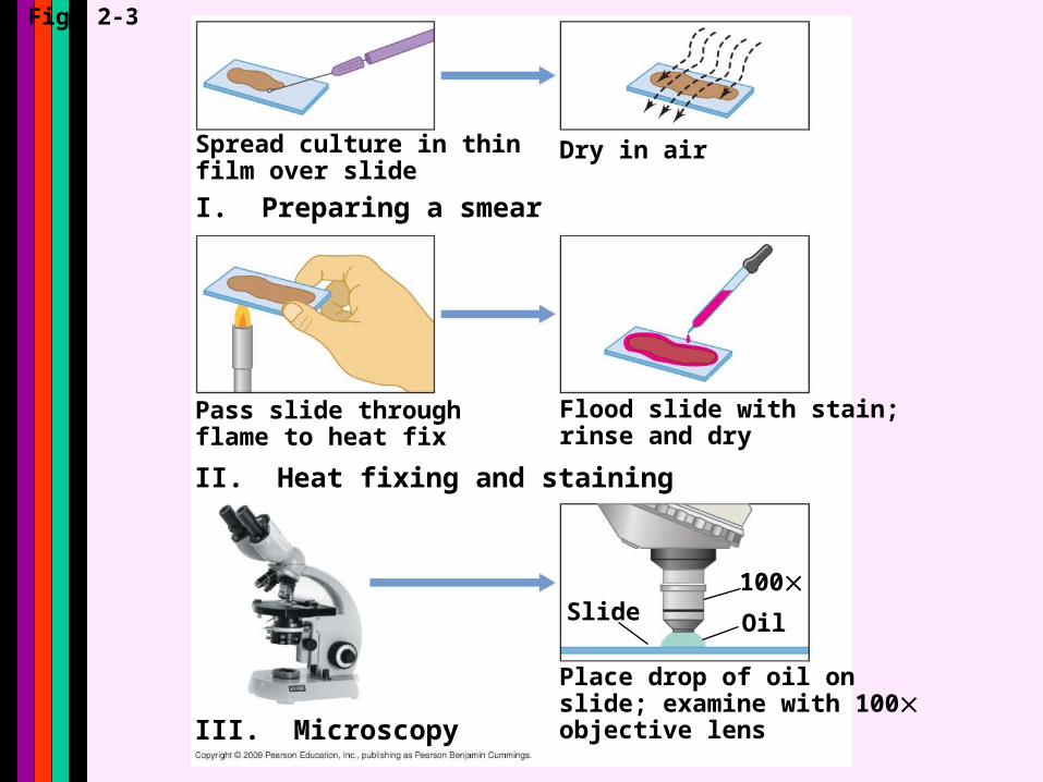
Fig. 2-3
100Slide
Dry in air
Flood slide with stain;rinse and dry
Place drop of oil onslide; examine with 100objective lens
Spread culture in thinfilm over slide
Pass slide throughflame to heat fix
Oil
I. Preparing a smear
III. Microscopy
II. Heat fixing and staining
29
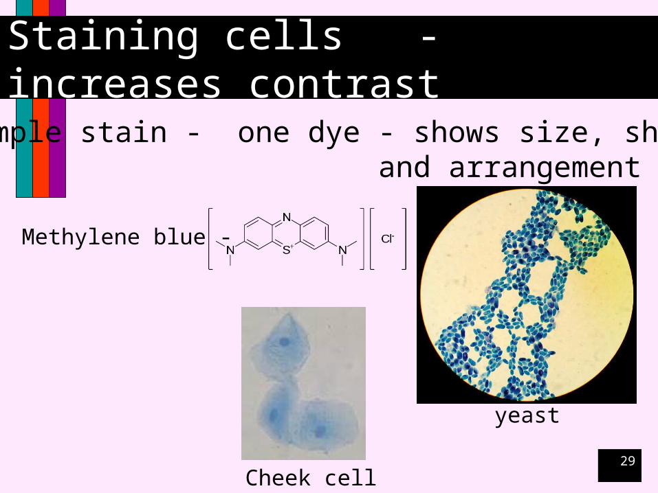
Staining cells - increases contrast
Simple stain - one dye - shows size, shape, and arrangement
Methylene blue -
yeast
Cheek cell
30
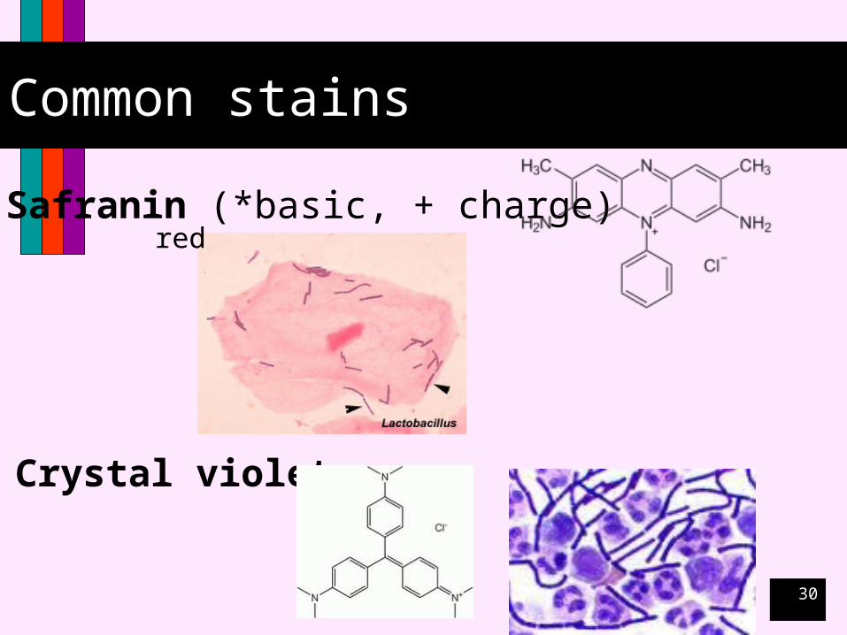
Common stains
Safranin (*basic, + charge)
Crystal violet
red
31
Differential stains
Use multiple dyes or dyes that interact with organisms differently.
Primary stain / counterstain
32
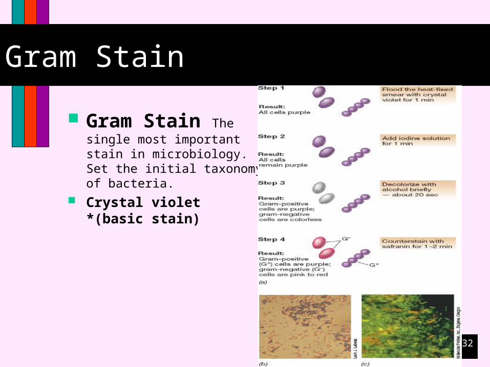
Gram Stain
Gram Stain The single most important stain in microbiology. Set the initial taxonomy of bacteria.
Crystal violet *(basic stain)

33
Gram Stain
34
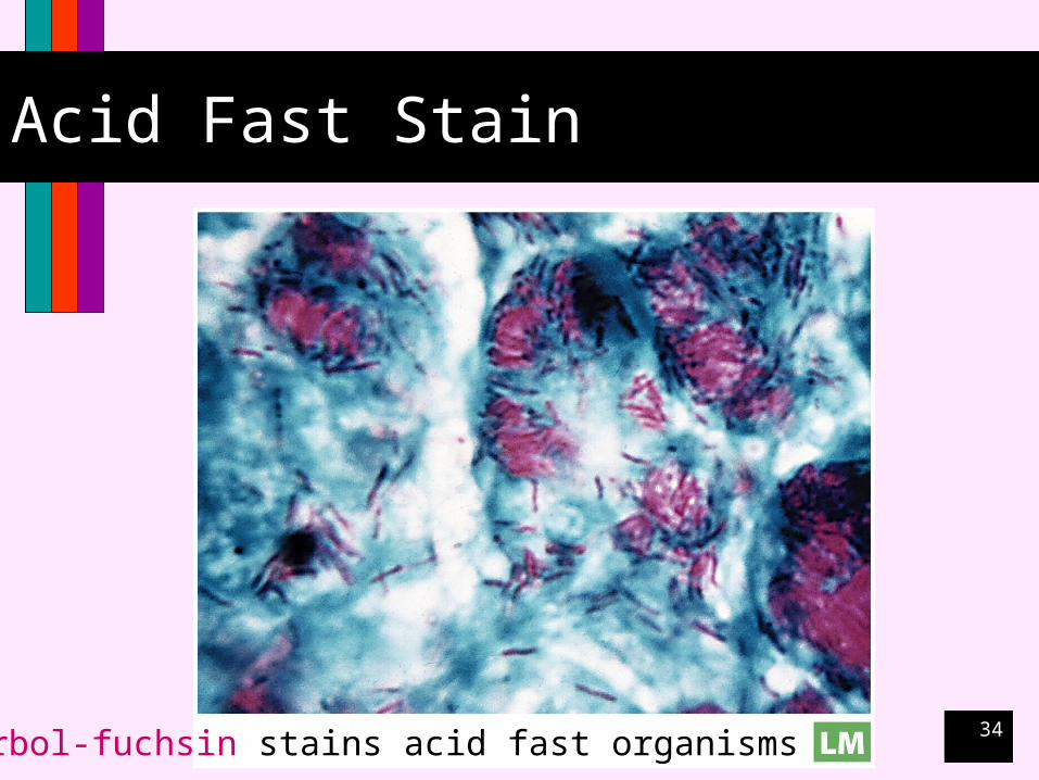
Acid Fast Stain
Carbol-fuchsin stains acid fast organisms
35
Acid Fast
The Ziehl-Neelsen stain, also known as the acid-fast stain, was first described by two German doctors; Franz Ziehl (1859 to 1926), a bacteriologist and Friedrich Neelsen (1854 to 1894), a pathologist. It is a special bacteriological stain used to identify acid-fast organisms, mainly Mycobacteria. Mycobacterium tuberculosis is the most important of this group, as it is responsible for the disease called tuberculosis (TB). It is helpful in diagnosing Mycobacterium tuberculosis since its lipid rich cell wall makes it resistant to Gram stain. It can also be used to stain few other bacteria like Nocardia. The reagents used are Ziehl-Neelsen carbolfuchsin, acid alcohol and methylene blue.
36
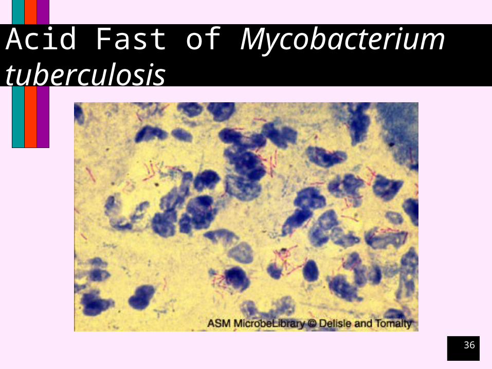
Acid Fast of Mycobacterium tuberculosis

37
Negative Stain
Sometimes referred to as capsular stainIndia ink or nigrosin
38
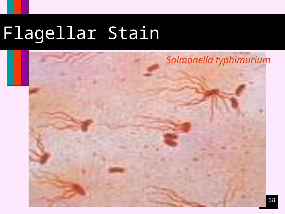
Flagellar StainSalmonella typhimurium
39
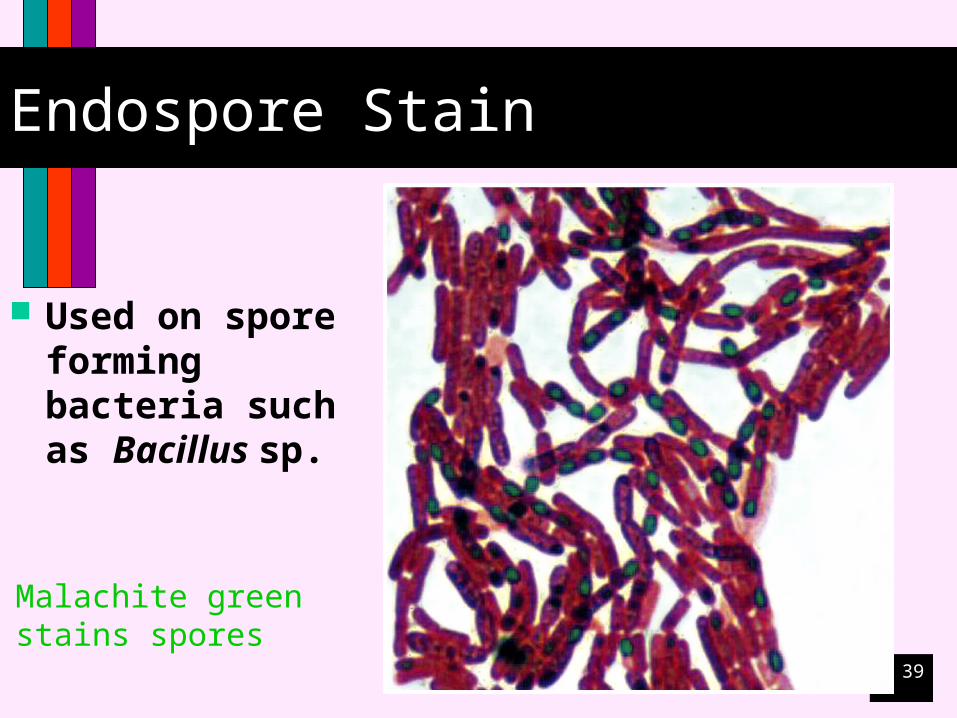
Endospore Stain
Used on spore forming bacteria such as Bacillus sp.
Malachite greenstains spores