Embed Size (px)
Citation preview

microRNA target identification by RNA pull down with biotinylated microRNA mimicsNon-canonical microRNA binding is frequent and contributes significantly to regulation of gene expression
AbstractNew exciting experimental approaches to transcriptome-wide identification of microRNA binding sites are revealing surprising details about microRNA interaction with RNA targets. Approximately one out of five microRNA interactions occur without canonical perfect base pairing to the seed sequence. Instead they are often governed by imperfect binding to the center of the microRNA. One primate specific microRNA appears to follow completely different rules for target recognition that are not foreseen at all by current prediction tools.
Read about how improved RNA pull down techniques with biotinylated microRNA mimics are transforming our understanding of microRNA biology.

IntroductionCentral to the understanding of microRNA function is knowledge about the genes they regulate. Bioinformatic microRNA target predictions are complicated by the fact that microRNAs interact with their mRNA targets with only partial base complementarity and in addition that these interactions are influenced by local mRNA secondary structure. Most microRNA prediction tools work on the assumption that perfect Watson-Crick base pairing between residues 2-8 of the microRNA (also known as the seed) and a sequence in the 3’UTR of the target mRNA is required for RISC mediated silencing. These tools incorporate factors such as free energy release by binding and phylogenetic conservation of binding sites to improve confidence of predictions. Although such tools are very useful and accurately predict true microRNA targets, they are also known for generating predictions with a high false positive rate. Perhaps less appreciated, but potentially more important, is that such tools also have a significant false negative rate.
It has been shown that microRNAs also regulate gene expression through non-canonical interactions with mRNA targets which are generally ignored by prediction tools (1). There are known examples of microRNAs regulating translation through base pairing of a central region of the microRNA with the mRNA target (2). Several recent papers provide a wealth of data showing that non-canonical microRNA interactions are frequent, diverse, functional and much more prevalent than previously appreciated (3-6). These important discoveries were achieved by ingenious experimental approaches
to global detection of microRNA targets and these technologies are certain to have a great impact on our understanding of microRNA function.
“Bioinformatic microRNA target predictions are associated with both high false positive and false negative rates
Strategies for experimental microRNA target identificationmicroRNA target identification often involves experiments in which changes to transcript abundancies in response to manipulation of microRNA activity is measured. This can be achieved by different types of genetic manipulation or by use of oligonucleotide based microRNA inhibitors and microRNA mimics. These are powerful and important methods, but the results are often confounded by side effects of transfection and secondary effects of the change in microRNA activity. microRNAs may directly or indirectly affect the activity of multiple transcription factors which in turn can have profound effects on transcription that are not the direct result of microRNA interaction with mRNA targets.
In addition, microRNA regulation of gene expression is often quite subtle at the transcriptional level and is best appreciated by measuring the abundance of the corresponding gene products.
RISCmRNA
microRNA
Mimic withbiotin label
Cell transfection with microRNA mimic
Pull down with streptavidin coated magnetic beads
RNA binding biotinylated protein
Lyse cell 24hr post transfection
RNA extraction qPCRMicroarrayNGS
Mimic boundindependentlyof RISC
Potential sourcesof background noise
Figure 1a. microRNA target identification with biotinylated mimics. Biotinylated microRNA mimics are delivered to cells by transfection. Cells are harvested and lysed 24hr later. Complexes of biotinylated RISC and mRNA are pulled down with strepatividine coated magnetic beads. After wash steps the captured RNA is purified and analyzed with microarrays, RT-qPCR or by next generation sequencing. Background noise can arise from microRNA mimic binding to RNA independently of RISC, naturally occuring biotinylated proteins that bind to RNA and non-specific binding of RNA to the magnetic beads and plastic surface of microwells.

However proteomics is still beyond the economical reach of most research groups. Other strategies are based on identifying sites of RISC binding on mRNAs rather than the effect of these interactions on gene expression. Such methods are typically based on reversible cross linking of associated RNA and protein followed by immunoprecipitation (CLIP). The virtue of cross linking is to “freeze” interactions between RNA binding proteins and RNA. Subsequent pull down with antibodies against protein components of RISC (typically Argonaute) and shearing or enzymatic degradation of the RNA into small fragments allows identification of RISC binding sites by microarray profiling, RT-qPCR or next generation sequencing (NGS) (7,8).
In addition, cross linking induced mutagenesis can be exploited to identify such sites of RISC interaction with single nucleotide resolution (9). Although state-of-the-art technologies, these approaches do not reveal which microRNAs are involved in the RISC binding. Instead, this is determined bioinformatically by analysis of the identified RNA sequences. Such analysis is often based on identification of sequences complementary to known microRNA seed sequences and ignores non-canonical microRNA binding sites.
Introduction of biotinylated microRNA mimics into cells has been exploited to obtain experimental evidence of physical interaction between a specific microRNA of interest and its mRNA targets (10,
11). The biotin is placed at the 3’terminal end of the microRNA strand. Streptavidine affinity purified RNA associated with biotinylated microRNA can be analyzed on microarrays or by NGS – see Figure 1a. Evidently, the interaction between RISC and target RNA is so strong that stabilization of the binding by cross linking is not necessary. The data that this method initially generated was associated with high levels of background noise which hampered widespread use of this approach. Nevertheless, significant improvements can be achieved by a two step procedure in which RISC/mRNA complexes are first immunoprecipitated with antibodies against Argonaute and then purified with streptavidin coated magnetic beads (12) – see Figure 1b. However, as is so often the case, the devil is in the detail and further important improvements of protocols include blocking of the magnetic beads and micro-wells with protein and exogenous RNA, and optimization of buffers and wash steps (3, 4).
Non-canonical interactions between microRNA and mRNA are both frequent and functionalPerfect base complementarity between the seed region of microRNAs and sequences in the 3’UTR is known to be critical to microRNA target site recognition, with possible additional contribution from limited base pairing of the microRNA 3'-end. In contrast, the central sequence of microRNAs appears not to be involved in base pairing in the majority of known microRNA/mRNA interactions. However, central microRNA sequences are in general phylogenetically conserved suggesting that they, just like seed sequences, are
RISCmRNA
microRNA
Mimic withbiotin label
Cell transfection with microRNA mimic
AG02 IP
RNA binding biotinylated protein
Lyse cell 24hr post transfection
Pull down withstreptavidin coatedmagnetic beads qPCR
MicroarrayNGS
RNA extraction
Mimic boundindependentlyof RISC
Figure 1b. Reduction of background noise by Argonaute immunoprecipitation. An initial immmunoprecipitation with antibodies towards protein components of RISC can eliminate certain sources of background noise. Blocking of magnetic beads and microwell plastic surfaces with exogenous protein and RNA is another effective measure.
under very strong selectional pressure - although this may have explanations other than a consequence of direct base pairing with RNA targets.
“With fine tuned protocols, biotin pull down identifies genuine microRNA targets with great sensitivity and a very low false positive rate.
Examples of microRNAs exerting repression of mRNA translation through base paring of the central part of their sequences have been reported (2), but it was not clear how prevalent such non-seed interactions were. This question was elegantly addressed in work recently published in Genome Biology from the group of Nicole Cloonan in Australia (3).The paper describes results of RNA pull down experiments with 10 different biotinylated microRNA mimics. The affinity purified RNA was analyzed on microarrays coupled with in-depth bioinformatics analysis and downstream experimental validation of novel microRNA targets with Luciferase reporter assays. The results comprehensively validate this experimental approach. With fine-tuned protocols, biotin pull down identifies genuine microRNA targets with great sensitivity and a very low false positive rate. However no experimental approach is perfect and, unsurprisingly, a negative bias was observed for RNA targets that are strongly destabilized by interaction with the microRNA. By targeted bioinformatic analysis of the data which specifically searched for binding sites that base pair with the central part of microRNAs (including only a part of the seed), it was discovered that this class of non-canonical binding sites is much more frequent than previously appreciated and often appears together with classical seed binding sites and even inside CDS – see Figure 2.
The functional relevance of this type of interaction was demonstrated by the cloning of 12 different centered microRNA binding sites in the 3’UTR of a Luciferase reporter gene. Ten of these sites had a significant effect on Luciferase activity, in most cases without affecting stability of the corresponding mRNA. It therefore appears that this new class of microRNA centered binding sites contributes significantly to gene expression, but primarily at the translational level.
“Non-canonical microRNA binding sites are mostly ignored by current bioinformatic analyses and filtered out as false positive signals.
Another recent paper reported results of similar pull down experiments with a biotinylated hsa-miR-522 mimic (4). In this study, a RNase protection approach was used to narrow down the analysis of microRNA binding sites to short RNA sequences. Streptavidine bead captured RNA was treated with RNase T1 that cleaves single stranded RNA at G residues. By sequencing short RNA strands protected by RISC, microRNA recognition sites were identified with great accuracy – see Figure 3. The results are very surprising. Apparently this primate specific microRNA primarily regulates gene expression through interaction with unusual binding sites that feature partial complementarity to either residues 2-9 or residues 13-20 of the microRNA. Only a small fraction of the binding sites were predicted by commonly used prediction tools.
Additional strong evidence of prevalent non-canonical microRNA binding has been obtained with an exciting novel approach in which binding sites are detected by ligation of microRNA to associated RNA in cross linked samples. Close to 20% of all detected interactions were attributed to microRNA centered binding site. The discovery of prevalent non-canonical microRNA binding adds yet another layer to the complexity of microRNA regulation and will have profound consequences on how microRNA target identification is approached in the future. Global analysis of microRNA regulation of gene expression produces vast amounts of data that requires bioinformatic handling. Since microRNA centered binding sites and other examples of non-canonical interactions are mostly ignored by current bioinformatic analysis, evidence of such interactions have largely gone undetected and filtered out as false positive signals.
RNA pull-down with biotinylated microRNA mimics and other novel experimental approaches to global analysis of microRNA interactions will no doubt reveal other interesting aspects of microRNA regulation in the near future. For example, we have everything to learn about microRNA interactions with longer ncRNA species!
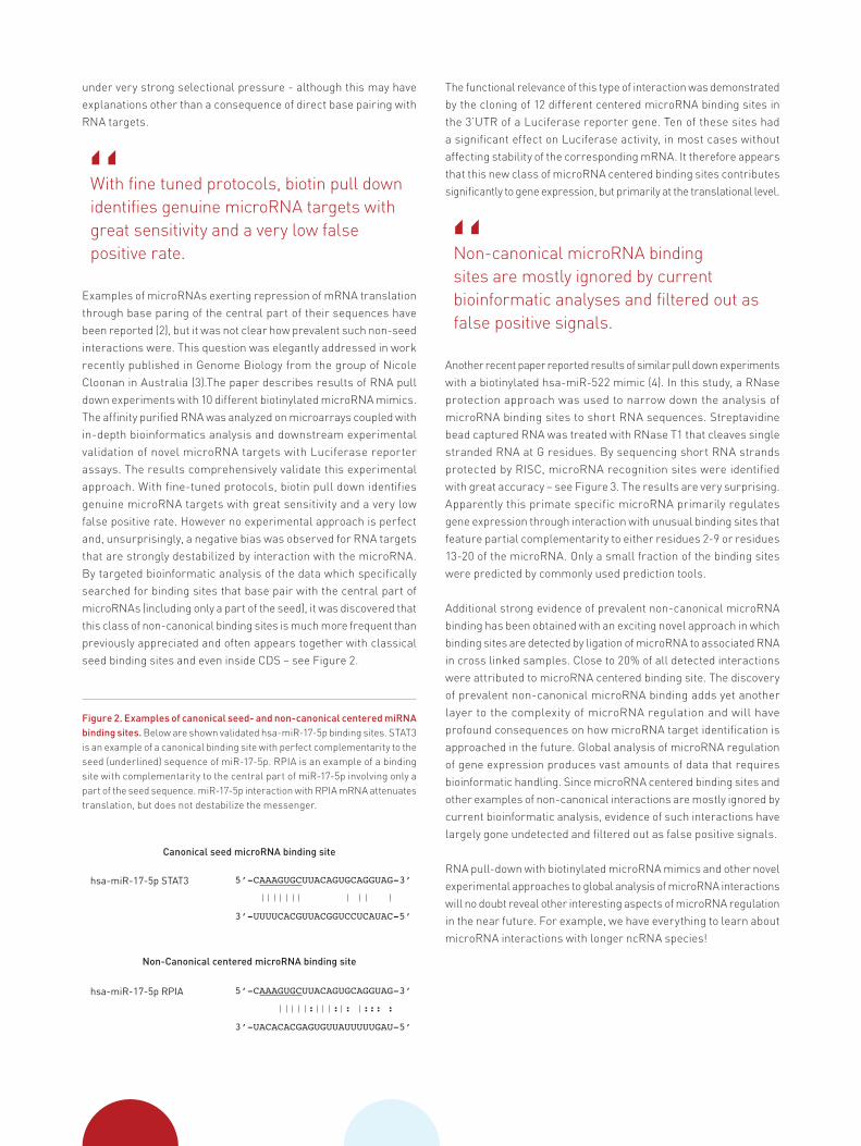
Figure 2. Examples of canonical seed- and non-canonical centered miRNA binding sites. Below are shown validated hsa-miR-17-5p binding sites. STAT3 is an example of a canonical binding site with perfect complementarity to the seed (underlined) sequence of miR-17-5p. RPIA is an example of a binding site with complementarity to the central part of miR-17-5p involving only a part of the seed sequence. miR-17-5p interaction with RPIA mRNA attenuates translation, but does not destabilize the messenger.
5’-CAAAGUGCUUACAGUGCAGGUAG-3’
3’-UUUUCACGUUACGGUCCUCAUAC-5’
Canonical seed microRNA binding site
Non-Canonical centered microRNA binding site
hsa-miR-17-5p STAT3
5’-CAAAGUGCUUACAGUGCAGGUAG-3’
3’-UACACACGAGUGUUAUUUUUGAU-5’
: : : ::: :
hsa-miR-17-5p RPIA

Guidelines for working with biotinylated microRNA mimicsUse of microRNA mimics carries the risk of introducing artefacts due to saturation of RISC. Care must be taken to ensure that mimics are used at concentrations that don’t cause gross pertubations of endogenous microRNA regulation. This can be done by studying the effects of transfecting increasing amounts of mimic with luciferase assays that report the activity of unrelated microRNAs endogenous to the cell line under study.
“Special triple RNA strand designof miRCURY LNATM microRNA mimicsensures complete microRNA strandspecificity
Modification of 5’- and 3’ ends of the microRNA strand in mimics are generally not well tolerated at all. However, regardless of a report to the contrary (13), we and others have observed that potent microRNA activity is achievable with biotinylated mimics, although they are significantly less potent than non-modified mimics, presumably due to less efficient loading into RISC (see Figure 4). Various linkers between the microRNA and biotin have been investigated, but these appear to have little effect. Nevertheless, the possibility that biotin modification of the guidestrand creates a bias for incorporation of the passenger strand cannot be excluded.
It is therefore important to use modified mimics that ensure that only the microRNA strand is chosen by RISC. The special triple RNA strand design of miRCURY LNA™ microRNA mimics ensures complete microRNA strand specificity since the passenger strand is split into two LNA™ modified RNA strands – see Figures 5 and 6.
Background noise can be reduced significantly by:• Blocking of magnetic beads and microwells with protein (bovine serum albumin) and exogenous RNA (M2 bacteriophage RNA or yeast tRNA) (3,4)• Optimization of buffers and washing steps (3,4)• Depletion of contaminating ribosomal RNA with rRNA removal kits• Introduction of an initial Argonaute immunoprecipitation step and working with the subsequent Argonaute enriched fraction
Data analysisIn order to capture potential non-canonical microRNA interactions it is crucial that the bioinformatic analysis scans for binding sites with complementarity to all parts of the microRNA sequence rather than just the seed.
Figure 3. Determination of exact sites of microRNA binding by on-bead RNase protection assay. RNase T1 digests single strand RNA at G residues. Bead captured RNA is exposed to a mild RNase T1 treatment. The beads are washed after treatment to get rid of RNA released by the digestion. RNA protected by RISC is then extracted and characterized by sequencing.
Figure 4. Silencing of microRNA reporter assays with biotinylated miRCURY LNA™ microRNA Mimics. HeLa cells were transfected with a plasmid harboring a Renilla luciferase gene (transfection efficiency control) and a Firefly luciferase reporter gene with a sequence complementary to hsa-miR-1-3p in the 3' UTR (pmiR-1). The day after the cell cultures were transfected with different concentrations of hsa-miR-1-3p microRNA mimic or cel-miR-39-3p negative control. Reporter gene expression was measured 24 h after transfection with a Dual Luciferase assay. Ratios of Firefly and Renilla luciferase activity were calculated and normalized to values obtained with a Firefly Luciferase reporter with no miR target sequence (pLuc). The results illustrate that potent microRNA activity can be achieved with biotinylated mimics even if they are ~50x less active than unlabeled mimics.
Target MRE detection by sequencing
WashRNA extraction
RNase T1 treatment
NNNGNNN NNNGNNN
NNN NNNG
8
7
6
5
4
3
2
1
0
Nor
mal
ized
Lum
ines
cenc
e (R
LU)
pLuc pmiR-1
0.05nM 0.5nM 5nM 0.05nM 0.5nM 5nM 0.05nM 0.5nM 5nM
cel-miR-39-3phsa-miR-1-3p hsa-miR-1-3p-biotin
miRCURY LNA™ microRNA Mimics in HeLa cells3’-Biotin on microRNA guide strand

Complete the functional analysis of your microRNA with Exiqon products:
• miRCURY LNA™ microRNA Mimics• miRCURY LNA™ microRNA Inhibitors• miRCURY LNA™ microRNA Target Site Blockers
Read more at www.exiqon.com/mirna-inhibitors
Experimental validationExperimental validation of novel microRNA targets should ideally be performed with both microRNA mimics and inhibitors. The advantage of inhibitors is that they will reveal if the observed microRNA-mRNA interaction is relevant in the studied biological context and not only significant when the microRNA is artificially highly abundant.
Remember that there is a negative bias with this approach for RNA targets that are significantly degraded by interaction with the studied microRNA. Such targets are more likely to be identified by methods that measure RNA abundancies.
Figure 5. Biotinylated miRCURY LNA™ microRNA Mimics consist of three RNA strands. A 3’ biotinylated microRNA (guide) strand with sequence exactly according to miRBase annotation. The passenger strand is split in two LNA™ modified RNA strands complementary to the microRNA strand. Only the microRNA strand is incorporated by RISC. The two passenger strands are too short to act as microRNAs and are rapidly degraded after displacement from the microRNA strand.
Figure 6. Perfect microRNA strand specific activity with miRCURY LNA™ microRNA Mimics. Hela cells harboring hsa-miR-16-3p (top panel) and hsa-miR-16-5p (bottom panel) luciferase reporter plasmids respectively were transfected with hsa-miR-16-3p and hsa-miR-16-5p mimics and cel-miR-39-3p negative mimic control. The results demonstrate that suppression of luciferase actity is only achieved with the miRCURY LNA™ microRNA Mimic corresponding to the reporter assay.
Passenger strand made up of two LNATM-enhanced RNA strands
microRNA (guide strand)
3’ Biotin
microRNA strand-specific activity with miRCURY LNA™ microRNA Mimics
10
9
8
7
6
5
4
3
2
1
0N
orm
aliz
ed L
umin
esce
nce
(RLU
)
pLuc
p16*
hsa-miR-16-5p hsa-miR-16-3p cel-miR-39-3p
5nM
0.5nM
5nM
0.5nM
5nM
0.5nM
hsa-miR-16-3p reporter assay
miRCURY LNATM microRNA Mimics
14
12
10
8
6
4
2
0
Nor
mal
ized
Lum
ines
cenc
e (R
LU)
pLuc
p16
hsa-miR-16-5p hsa-miR-16-3p cel-miR-39-3p
5nM
0.5nM
5nM
0.5nM
5nM
0.5nM
hsa-miR-16-5p reporter assay
miRCURY LNATM microRNA Mimics
Go to www.exiqon.com/e-talk for more presentations and webinars on tips and interesting data concerning functional analysis of microRNA
AAA

References1. Chi SW, Hannon GJ, Darnell RB. An alternative mode of microRNA target recognition. Nat Struct Mol Biol. 2012;19:321–327. 2. Shin C, Nam JW, Farh KK, Chiang HR, Shkumatava A, Bartel DP. Expanding the microRNA targeting code: functional sites with centered pairing. Mol Cell. 2010, 38(6):789-802.3. Martin HC, Wani S, Steptoe AL, Krishnan K, Nones K, Nourbakhsh E, Vlassov A, Grimmond SM, Cloonan N. Imperfect centered miRNA binding sites are common and can mediate repression of target mRNAs. Genome Biol. 2014, 15(3):R51.4. Tan SM, Kirchner R, Jin J, Hofmann O, McReynolds L, Hide W, Lieberman J. Sequencing of captive target transcripts identifies the network of regulated genes and functions of primate-specific miR-522. Cell Rep. 2014; 8(4):1225-39.5. Helwak A, Kudla G, Dudnakova T, Tollervey D. Mapping the human miRNA interactome by CLASH reveals frequent noncanonical binding. Cell. 2013 153(3):654-65.6. Grosswendt S, Filipchyk A, Manzano M, Klironomos F, Schilling M, Herzog M, Gottwein E, Rajewsky N. Unambiguous identification of miRNA:target site interactions by different types of ligation reactions. Mol Cell. 2014, 54(6):1042-54.7. Chi SW, Zang JB, Mele A, Darnell RB. Argonaute HITS-CLIP decodes miRNA-mRNA interaction maps. Nature. 2009, 460(7254):479-86.8. Hafner M, Landthaler M, Burger L, Khorshid M, Hausser J, Berninger P, Rothballer A, Ascano M Jr, Jungkamp AC, Munschauer M, Ulrich A, Wardle GS, Dewell S, Zavolan M, Tuschl T. Transcriptome-wide identification of RNA- binding protein and microRNA target sites by PAR-CLIP. Cell. 2010, 141(1):129-41.9. Moore MJ, Zhang C, Gantman EC, Mele A, Darnell JC, Darnell RB. Mapping Argonaute and conventional RNA-binding protein interactions with RNA a single-nucleotide resolution using HITS-CLIP and CIMS analysis. Nat Protoc. 2014 9(2):263-93.10. Ørom UA, Lund AH. Isolation of microRNA targets using biotinylated synthetic microRNAs. Methods. 2007, 43(2):162-5.11. Ørom UA, Nielsen FC, Lund AH. MicroRNA-10a binds the 5'UTR of ribosomal protein mRNAs and enhances their translation. Mol Cell. 2008, 30(4):460-71.12. Nonne N, Ameyar-Zazoua M, Souidi M, Harel-Bellan A. Tandem affinity purification of miRNA target mRNAs (TAP-Tar). Nucleic Acids Res. 2010, 38(4):e20.13. Guo YE, Steitz JA. 3'-Biotin-tagged microRNA-27 does not associate with Argonaute proteins in cells. RNA. 2014, 20(7):985-8.
Transforming our understanding of microRNA biology

9234
90 -
v1.
0 -
12/2
014
Contact informationOutside North AmericaPhone: +45 45 65 09 29 · Fax: +45 45 66 18 88North AmericaPhone: +1 781 376 4150 · Fax: +1 781 376 4152
www.exiqon.com