Embed Size (px)
Citation preview
MicroRNA Expression Profile in Hyperoxia-Exposed Newborn MiceDuring the Development of Bronchopulmonary Dysplasia
Xiaoying Zhang MD, Wei Peng MM, Sheng Zhang MD, Chunzhi Wang MM, Xiyu He MM,Zhimei Zhang MM, Lina Zhu MM, Yan Wang MD, Zhichun Feng MM
BACKGROUND: Bronchopulmonary dysplasia (BPD) is a chronic lung disease of preterm neo-nates; the underlying pathogenesis is not fully understood. MicroRNAs (length 21–25 nucleotides)are ribonucleic acid (RNA) molecules that have important functions in development, cellular dif-ferentiation, apoptosis, proliferation, and migration; very little is known regarding their role indevelopmental lung diseases. METHODS: We exposed neonatal mice to either room air or 60%oxygen, beginning at birth, and we used microRNA microarray and real-time polymerase chainreaction on lung samples. RESULTS: The hyperoxia-exposed mice developed a lung injury thatmimicked human BPD. Fifty-one microRNAs shared similar profiles in the hyperoxia-exposed BPDlungs and the normal lungs, which indicates that those microRNAs might play a protective roleduring the septation process. In the BPD lungs, compared to the control lungs, 14 microRNAs wereup-regulated, and 7 microRNAs were down-regulated, which indicates that these microRNAs mightplay an important role in the development of BPD. Some of the candidate microRNAs can regulatecell proliferation. CONCLUSIONS: To our knowledge, this study is the first to identify microRNAsassociated with BPD development, which provides a clue for further investigation of their functionin BPD development. Key words: microRNA; bronchopulmonary dysplasia; hyperoxia; pathogenesis.[Respir Care 2011;56(7):1009–1015. © 2011 Daedalus Enterprises]
Introduction
Babies who are born prematurely or who experiencerespiratory problems shortly after birth are at risk for bron-chopulmonary dysplasia (BPD), which is a common chronic
lung disease in preterm neonates and can have a multifac-torial etiology, including hyperoxia exposure, prematurelung, and neonatal therapies.1,2 Although the incidenceand severity of respiratory distress syndrome have de-creased with antenatal corticosteroid therapy and postnatalsurfactant replacement, the overall incidence of BPD hasbeen increasing.3,4 BPD is predominantly characterized bypersistent abnormalities of lung structure and arrested lungdevelopment, including impaired alveolar and vasculargrowth.1,5 The pathogenesis of BPD is not fully under-stood.
MicroRNAs are 21–25 nucleotides long, non-codingRNAs that are involved in various biological processes,including cell proliferation, cell death, stress resistance,and tumorigenesis.6-8 Recent studies indicate that micro-RNAs are important in lung development9 and that abnor-mal microRNA levels cause aberrant expression of geneproducts that may contribute to lung disease, includinglung cancers and asthma,10,11 so microRNAs might be use-ful diagnositic and/or prognostic markers for some dis-eases. To date, however, to our knowledge there have beenno studies on microRNAs in the development of BPD.
The authors are affiliated with the Department of Pediatrics, BaYi Chil-dren’s Hospital of The General Military Hospital of Beijing People’sLiberation Army, Beijing, People’s Republic of China.
The authors have disclosed no conflicts of interest.
This study was partly supported by National Natural Science Foundationof China grant 81070524, 30973210.
Supplementary materials related to this paper are available at http://www.rcjournal.com.
Correspondence: Zhichun Feng MM, Department of Pediatrics, BaYiChildren’s Hospital of The General Military Hospital of Beijing People’sLiberation Army, 5 Nanmencang Road, Dongcheng District, Beijing100700, People’s Republic of China. E-mail: [email protected].
DOI: 10.4187/respcare.01032
RESPIRATORY CARE • JULY 2011 VOL 56 NO 7 1009
To assess whether microRNAs are involved in the de-velopment of BPD, we assayed microRNAs from 60%hyperoxia-exposed newborn mice—an ideal model becausethe hyperoxia-induced changes in lung structure closelymimic the histology of the altered lung architecture inhuman BPD.12-14 One critical determinant of normal post-natal lung architecture is septation, so we assayed the lung-tissue microRNAs before, during, and after septation, onpostnatal days 2, 7, and 21.
Methods
Animals
We purchased newborn Kunming mice from the Chi-nese Academy of Sciences (Beijing, China) and randomlyassigned 15 to room air and 15 to hyperoxia (FIO2
60% for21 days), beginning at birth. Nursing dams were alternatedevery 24 hours between the air and hyperoxia litters.14 Allthe mice were maintained in a pathogen-free facility andused in accordance with our institutional guidelines foranimal care. The animals were euthanized with intraperi-toneal sodium pentobarbital after exposure on day 2, day 7,or day 21.
Cells
We cultured A549 cells in 1640 (Gibco-BRL, Invitro-gen, Carlsbad, California), with 10% fetal bovine serum(Gibco-BRL). The cells were maintained in a humidified37°C incubator, with an atmosphere of 5% CO2.
Histology
To prepare the lungs for histology we opened the chestcavity and exposed the trachea, inserted a blunt cannulaand tied it to the trachea, and inflated the lungs with phos-phate buffered saline 4% formalin fixative at 25 cm H2O.After overnight fixation at 4°C, the tissue was dehydratedand paraffin embedded, cut into 5-�m sections, stainedwith hemotoxylin and eosin, and examined with light mi-croscopy.15,16 We performed radial alveolar counts, as pre-viously described.17 From the center of the respiratorybronchiole, a perpendicular line is drawn to the edge of theacinus, and the number of the septa intersected by that lineare counted. The radial alveolar count was made at mag-nification �100.
Lung RNA Assay
Total RNA was isolated from individual whole lungswith an microRNA isolation kit (miRVana, Ambion, Aus-tin, Texas) per the manufacturer’s instructions. RNA qual-ity was controlled with an optical density (OD) 260/280
ratio of 1.8 to 2 and an OD 260/230 of � 1.8, and 1 �gtotal RNA was used.
Microarray Methods
We pooled 3 individual total RNA samples from thesame time-point as one sample, to minimize biologicalvariability and the number of microarrays to generate thenecessary data. These RNA samples were treated withpoly(A) tailing, flash tag ligation, and hybridization (Gene-Chip miRNA Array, Affymetrix, Santa Clara, California)per the manufacturer’s protocol.
Microarray Data Analysis
We scanned the chips, computed the signal intensity(GeneChip Command Console 1.1 software, Affymetrix,Santa Clara, California), and summarized, normalized, con-ducted quality-control procedures on the data (microRNAQC Tool software, Affymetrix, Santa Clara, California),and created cluster and tree-view graphs, as previouslydescribed.18
Reverse Transcription Reaction and QuantitativeReal-Time Polymerase Chain Reaction
We purified the total RNAs (Absolutely RNA Nanoprepkit, Stratagene, Agilent Technologies, Santa Clara, Cali-fornia). We carried out reverse transcription reactions andreal-time-polymerase chain reaction as previously de-scribed.16 We calculated the relative expression of mi-croRNA compared to u6 with the 2-��Ct method.16 (Seethe supplementary materials related to this paper at http://www.rcjournal.com.)
Transient Transfection
Transfections were performed (Lipofectamine 2000, In-vitrogen, Carlsbad, California) per the manufacturer’s in-structions and our previous report.16 Cells (1–3 � 106)grown to a confluency of 50–60% in 10-cm petri disheswere transfected with different double-stranded microRNAmimics (600 pmol, GenePharma, Shanghai, People’s Re-public of China) or microRNA mock, and the cells wereharvested 48 hours post-transfection.
Measurement of Cell Proliferation
A549 cells transfected with microRNAs (1 � 104) wereplated in 96-well plates, and the cell proliferation wasmeasured with a proliferation kit (XTT II, Boehringer,Mannheim, Germany).16 Optical density was read with amicroplate reader (Bio-Rad, Hercules, California).
MICRORNA EXPRESSION PROFILE IN HYPEROXIA-EXPOSED NEWBORN MICE
1010 RESPIRATORY CARE • JULY 2011 VOL 56 NO 7

Statistical Analysis
Data are presented as mean � SD. We used analysis ofvariance to calculate differences between body weight,radial alveolar count, and proliferation, and if P was � .05,we performed the least significant difference test. We usedthe Pearson test to determine the correlation of the micro-RNA levels between the microRNA microarray assay andthe real-time polymerase chain reaction assay. Differenceswere deemed statistically significant at P � .05.
Results
Effects of 60% Oxygen Exposure on Newborn Mice
There was no mortality in either group, but the hyper-oxia group had significantly lower body weight on day 7(8.10 � 0.26 g vs 8.94 � 0.24 g, P � .01) and on day 21(21.04 � 0.26 g vs 26.7 � 0.31 g, P � .001), but not onday 2 (5.73 � 0.25 g vs 6.09 � 0.23 g, P � .14). Structuralchanges were evident in the hyperoxia and control micelungs on day 2, day 7, and day 21 (ie, before, during, andafter septation). At 2 days the hyperoxia mice had noobvious qualitative differences in the lung regions charac-terized by either large distal air spaces or absence of in-terstitial thickening. However, at 7 days the hyperoxiamice had heterogeneous lung structure changes, includingpatchy areas of parenchymal thickening, small air spacesinterspersed in enlarged air spaces. At 21 days the hypoxiamice had a lung injury pattern that has morphologic sim-ilarities to human BPD (Fig. 1A). Hyperoxia reduced ra-dial alveolar count by 34% on day 7 (P � .001) and by33% on day 21 (P � .001) (see Fig. 1B). The alveolarenlargement and decrease in surface area are associatedwith decreased oxygen transfer and arterial oxygen satu-ration, resulting in the characteristic BPD-related reduc-tion in lung function.
MicroRNA Expression Differences During Septation
To determine the microRNA quantities during the sep-tation process, we used a novel microRNA array-basedapproach. We report these data as fold-change values, andwe considered a � 2-fold change of expression importantand selected those changes for study. We randomly se-lected 6 microRNAs that exhibited � 2-fold change inexpression to validate the data with real-time polymerasechain reaction for mature microRNAs (see the supplemen-tary materials related to this paper at http://www.rcjournal.com). The correlation coefficient between microarray andreal-time polymerase chain reaction for the 6 microRNAswas 0.884 (P � .007), which means the microarray dataagreed with the real-time polymerase chain reaction dataand the 2 methods had very high internal consistency. We
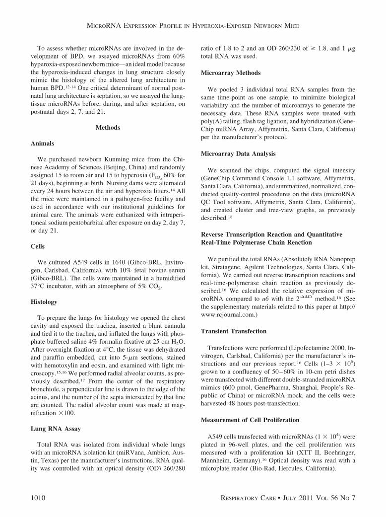
therefore used microarray for analysis of the microRNAsprofiles. We determined the expression changes of indi-vidual microRNAs via cluster analysis. During the septa-tion process, 72 microRNAs changed expression level inthe control group (Fig. 2A) and 87 microRNAs changed inthe hyperoxia group (see Fig. 2B). Among those micro-RNAs, 51 were shared by the control and hyperoxia groups,and the expression levels (fold-changes) were very similar(see the supplementary materials related to this paper athttp://www.rcjournal.com). Of note, miR-29a was the mosthighly up-regulated microRNA, and miR-100, miR-342–5p, miR-497 and the miR-30 family were also highly up-regulated in both groups. MicroRNAs greatly down-regu-lated in both groups were miR-122, miR-298, miR-411,miR-541, miR-382, miR-134, miR-668, miR-485,miR-409, miR-329, miR-665, miR-433, miR-127 andmiR-379. These data show similarities during septation inthe 2 groups, which indicates that these microRNAs playan important role during septation, whatever the hyperoxiainjury.
Fig. 1. Lung development at 2 days, 7 days, and 21 days after birthin mice raised in a hyperoxic environment (60% oxygen) versuscontrol mice (normal air environment). A: Lung development ismarkedly abnormal (enlarged air spaces and simplified structure)in the hyperoxia group on day 7 and day 21, compared to thecontrol group. The black bars represent 100 �m. B: The radialalveolar count is significantly different on day 7 and day 21. Thewhisker bars indicate standard deviations.
MICRORNA EXPRESSION PROFILE IN HYPEROXIA-EXPOSED NEWBORN MICE
RESPIRATORY CARE • JULY 2011 VOL 56 NO 7 1011
MicroRNA Expression Differences During BPDDevelopment
Detailed account of the microRNA expression profilingduring BPD development has not previously been reported,so we compared the groups’ expression profiles (Figs. 3and 4). Twenty-one microRNAs levels were greatlychanged in the control mice lungs, but there were no sub-stantial changes in the BPD mice lungs (see Fig. 3A),whereas 36 microRNAs levels showed aberrant regulationin the BPD mice lungs, but no substantial changes in thecontrol mice lungs (Fig. 3B), which implies these micro-RNAs might be important in BPD development. Further-more, these microRNA levels on day 7 were often inter-mediate between those on day 2 and day 21, which suggestsa gradual developmental shift in the expression pattern. In
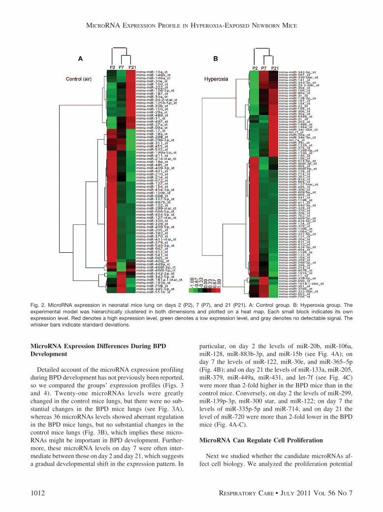
particular, on day 2 the levels of miR-20b, miR-106a,miR-128, miR-883b-3p, and miR-15b (see Fig. 4A); onday 7 the levels of miR-122, miR-30e, and miR-365–5p(Fig. 4B); and on day 21 the levels of miR-133a, miR-205,miR-379, miR-449a, miR-431, and let-7f (see Fig. 4C)were more than 2-fold higher in the BPD mice than in thecontrol mice. Conversely, on day 2 the levels of miR-299,miR-139p-3p, miR-300 star, and miR-122; on day 7 thelevels of miR-335p-5p and miR-714; and on day 21 thelevel of miR-720 were more than 2-fold lower in the BPDmice (Fig. 4A-C).
MicroRNA Can Regulate Cell Proliferation
Next we studied whether the candidate microRNAs af-fect cell biology. We analyzed the proliferation potential
Fig. 2. MicroRNA expression in neonatal mice lung on days 2 (P2), 7 (P7), and 21 (P21). A: Control group. B: Hyperoxia group. Theexperimental model was hierarchically clustered in both dimensions and plotted on a heat map. Each small block indicates its ownexpression level. Red denotes a high expression level, green denotes a low expression level, and gray denotes no detectable signal. Thewhisker bars indicate standard deviations.
MICRORNA EXPRESSION PROFILE IN HYPEROXIA-EXPOSED NEWBORN MICE
1012 RESPIRATORY CARE • JULY 2011 VOL 56 NO 7

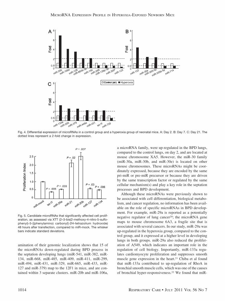
of cells (A549) transfected with microRNAs (hsa-let-7f,hsa-miR-29a, hsa-miR-21, hsa-miR-20b, hsa-miR-127, andhsa-miR-134) or miR-mock. The results revealed that over-expression of miR-21 or �20b induced cell proliferationsignificantly (P � .02 and P � .007, respectively), whereasmiR-29a, �127, �134, and let-7f significantly reducedcell proliferation (P � .003, P � .03, P � .006 and P � .006,respectively), compared to miR-mock (Fig. 5). Overex-pression of these microRNAs significantly affected cell pro-liferation in vitro, which indicates that these microRNAsare important in the biological and pathological processes.
Discussion
Little is known about the expression profiles and rolesof microRNAs in BPD development, so we compared mi-croRNA expression in control and hyperoxia-exposed miceand investigated their role during septation. The mouse isan ideal animal to study BPD development, because mouselung displays morphological and pathological BPD char-acteristics during the procedure of branching morphogen-esis, vascularization, and alveolarization.12-14 On day 21
the mice lungs displayed the characteristic BPD-relatedreduction in lung function. We predicted that some mi-croRNAs play an important protective role during septa-tion, as there were some similarities between the controland BPD lungs from day 2 to day 21. In both groups,miR-29a and the miR-30 family were the most highlyup-regulated microRNAs, which agrees with the report byWilliams et al.19 However, 14 microRNAs (miR-20b, miR-106a, miR-128, miR-883b-3p, miR-15b, miR-122,miR-30e, miR-365–5p, miR-133a, miR-205, miR-379,miR-449a, miR-431, and miR-431) were greatly up-regu-lated in the BPD lungs, compared to the control lungs,whereas 7 microRNAs (miR-299, miR-139p-3p, miR-300 star, miR-122, miR-335p-5p, miR-714, and miR-720)were significantly down-regulated in the BPD lungs, com-pared to the control lungs, some of which could regulatecell proliferation. All together, these results suggest thatthese microRNAs are coordinately expressed and centralto BPD development.
Multiple microRNAs were differentially expressed dur-ing BPD development. Although the functional role ofthese changes in microRNA expression is unknown, ex-
Fig. 3. Differential MicroRNA expression in neonatal mice lung on days 2 (P2), 7 (P7), and 21 (P21). A: Control group. B: Hyperoxia group.The experimental model was hierarchically clustered in both dimensions and plotted on a heat map. Each block indicates its own expressionlevel. Red denotes a high expression level, green denotes a low expression level, and gray denotes no detectable signal. Twenty-onemicroRNA expression levels were greatly dysregulated, compared to the air-exposed control mice lungs. B: Thirty-six microRNAs levelsshowed aberrant regulation in the hyperoxia-exposed BPD mice lungs.
MICRORNA EXPRESSION PROFILE IN HYPEROXIA-EXPOSED NEWBORN MICE
RESPIRATORY CARE • JULY 2011 VOL 56 NO 7 1013
amination of their genomic localization shows that 15 ofthe microRNAs down-regulated during BPD process inthe septation developing lungs (miR-541, miR-382, miR-134, miR-668, miR-485, miR-409, miR-411, miR-299,miR-494, miR-431, miR-329, miR-665, miR-433, miR-127 and miR-379) map to the 12F1 in mice, and are con-tained within 3 separate clusters. miR-20b and miR-106a,
a microRNA family, were up-regulated in the BPD lungs,compared to the control lungs, on day 2, and are located atmouse chromosome XA5. However, the miR-30 family(miR-30a, miR-30b, and miR-30e) is located on othermouse chromosomes. These microRNAs might be coor-dinately expressed, because they are encoded by the samepri-miR or pre-miR precursor or because they are drivenby the same transcription factor or regulated by the samecellular mechanism(s) and play a key role in the septationprocesses and BPD development.
Although these microRNAs were previously shown tobe associated with cell differentiation, biological metabo-lism, and cancer regulation, no information has been avail-able on the role of specific microRNAs in BPD develop-ment. For example, miR-29a is reported as a potentiallynegative regulator of lung cancer20; the microRNA genemaps to mouse chromosome 6A3, a fragile site that isassociated with several cancers. In our study, miR-29a wasup-regulated in the hyperoxia group, compared to the con-trol group, and it expressed at a higher level in developinglungs in both groups. miR-29a also reduced the prolifer-ation of A549, which indicates an important role in theregulation of cell biology. Importantly, miR-133a regu-lates cardiomyocyte proliferation and suppresses smoothmuscle gene expression in the heart.21 Chiba et al foundthat miR-133a contributed to up-regulation of RhoA inbronchial smooth muscle cells, which was one of the causesof bronchial hyper-responsiveness.22 We found that miR-
Fig. 4. Differential expression of microRNAs in a control group and a hyperoxia group of neonatal mice. A: Day 2. B: Day 7. C: Day 21. Thedotted lines represent a 2-fold change in expression.
Fig. 5. Candidate microRNAs that significantly affected cell prolif-eration, as assessed via XTT (2–3-bis(2-methoxy-4-nitro-5-sulfo-phenyl)-5-[(phenylamino) carbonyl]-2H-tetrazolium hydroxide)48 hours after transfection, compared to miR-mock. The whiskerbars indicate standard deviations.
MICRORNA EXPRESSION PROFILE IN HYPEROXIA-EXPOSED NEWBORN MICE
1014 RESPIRATORY CARE • JULY 2011 VOL 56 NO 7

133a was up-regulated in the BPD lungs from day 2 today 21, but not in the control lungs, and on day 21, miR-133a was greatly up-regulated in the BPD lungs, comparedto the control lungs, which suggests that miR-133a is im-portant in BPD development as the lung matures. Andepigenetic regulation of miR-370 by interleukin-6 contrib-utes to tumor growth.23 However, overexpression of let-7in the human lung epithelial A549 cell line inhibits cellgrowth,24 which is consistent with our results. Interest-ingly, miR-20b and let-7f were all up-regulated in the BPDlungs, compared to the control lungs, but they had differ-ent effects on cell proliferation. Overexpression of miR-20b significantly increased cell proliferation, whereas let-7fsignificantly reduced cell proliferation. However, we donot know whether the different effects are caused by thedifferent genera (Homo sapiens vs Mus) or the differentBPD stages (day 2 vs day 21), or what is the criticalmechanism that initiates and advances BPD mediated bymicroRNAs. This report provides a preliminary under-standing of the roles of microRNAs in BPD development.
Conclusions
Fifty-one microRNAs share similar profiles in the BPDlungs and control lungs. Fourteen microRNAs were up-reg-ulated and 7 were down-regulated in the BPD lungs, com-pared to the control lungs. Some of the candidate microRNAscan affect cell regulation. This is the first study to identify themicroRNAs associated with BPD development. The exactfunctions of these microRNA genes remains to be deter-mined, but we believe this study provides a basis for furtherinvestigation of their function in BPD development.
REFERENCES
1. Baker CD, Ryan SL, Ingram DA, Seedorf GJ, Abman SH, Balasu-bramaniam V. Endothelial colony-forming cells from preterm in-fants are increased and more susceptible to hyperoxia. Am J RespirCrit Care Med 2009;180(5):454-461.
2. Deakins KM. Bronchopulmonary dysplasia. Respir Care 2009;54(9):1252-1262.
3. Kwinta P, Grudzien A, Pawlik D, Olechowski W, Lauterbach R,Pietrzyk JJ. [Prevalence and risk factors of bronchopulmonary dys-plasia among extremely low birth weight newborns of regional birthcohort of south-east Poland]. Przegl Lek 2009;66(1-2):14-20. Articlein Polish.
4. Woynarowska M, Rutkowska M, Szamotulska K. [Risk factors, fre-quency and severity of bronchopulmonary dysplasia (BPD) diag-nosed according to the new disease definition in preterm neonates].Med Wieku Rozwoj 2008;12(4 Pt 1):933-941. Article in Polish.
5. Balasubramaniam V, Mervis CF, Maxey AM, Markham NE, AbmanSH. Hyperoxia reduces bone marrow, circulating, and lung endothe-lial progenitor cells in the developing lung: implications for thepathogenesis of bronchopulmonary dysplasia. Am J Physiol LungCell Mol Physiol 2007;292(5):L1073-1084.
6. Cheng J, Zhou L, Xie QF, Markham NE, Abman SH, Xing CY, et al.The impact of miR-34a on protein output in hepatocellular carci-noma HepG2 cells. Proteomics 2010;10(8):1557-1572.
7. Hu W, Chan CS, Wu R, Zhang C, Sun Y, Song JS, et al. Negativeregulation of tumor suppressor p53 by microRNA miR-504. MolCell 2010;38(5):689-699.
8. Gao P, Bai X, Yang L, Lv D, Li Y, Cai H, et al. Over-expression ofosa-MIR396c decreases salt and alkali stress tolerance. Planta 2010;231(5):991-1001.
9. Williams AE, Perry MM, Moschos SA, Lindsay MA. microRNAexpression in the aging mouse lung. BMC Genomics 2007;8(172.
10. Du L, Pertsemlidis A. microRNAs and lung cancer: tumors and22-mers. Cancer Metastasis Rev 2010;29(1):109-122.
11. Mohamed JS, Lopez MA, Boriek AM. Mechanical stretch upregu-lates microRNA-26a and induces human airway smooth muscle hy-pertrophy by suppressing glycogen synthase kinase-3{beta}. J BiolChem 2010;285(38):29336-29347.
12. Yi M, Jankov RP, Belcastro R, Humes D, Copland I, Shek S, et al.Opposing effects of 60% oxygen and neutrophil influx on alveolo-genesis in the neonatal rat. Am J Respir Crit Care Med 2004;170(11):1188-1196.
13. Jankov RP, Luo X, Belcastro R, Copland I, Frndova H, Lye SJ, et al.Gadolinium chloride inhibits pulmonary macrophage influx and pre-vents O(2)-induced pulmonary hypertension in the neonatal rat. Pe-diatr Res 2001;50(2):172-183.
14. Ahmed MN, Suliman HB, Folz RJ, Nozik-Grayck E, Golson ML,Mason SN, et al. Extracellular superoxide dismutase protects lungdevelopment in hyperoxia-exposed newborn mice. Am J Respir CritCare Med 2003;167(3):400-405.
15. Bry K, Whitsett JA, Lappalainen U. IL-1beta disrupts postnatal lungmorphogenesis in the mouse. Am J Respir Cell Mol Biol 2007;36(1):32-42.
16. Zhang X, Liu S, Hu T, Liu S, He Y, Sun S. Up-regulated microRNA-143 transcribed by nuclear factor kappa B enhances hepatocarci-noma metastasis by repressing fibronectin expression. Hepatology2009;50(2):490-499.
17. Balasubramaniam V, Tang JR, Maxey A, Plopper CG, Abman SH.Mild hypoxia impairs alveolarization in the endothelial nitric oxidesynthase-deficient mouse. Am J Physiol Lung Cell Mol Physiol 2003;284(6):L964-L971.
18. Takamizawa J, Konishi H, Yanagisawa K, Tomida S, Osada H,Endoh H, et al. Reduced expression of the let-7 microRNAs inhuman lung cancers in association with shortened postoperative sur-vival. Cancer Res 2004;64(11):3753-3756.
19. Williams AE, Moschos SA, Perry MM, Barnes PJ, Lindsay MA.Maternally imprinted microRNAs are differentially expressed duringmouse and human lung development. Dev Dyn 2007;236(2):572-580.
20. Fabbri M, Garzon R, Cimmino A, Liu Z, Zanesi N, Callegari E, et al.MicroRNA-29 family reverts aberrant methylation in lung cancer bytargeting DNA methyltransferases 3A and 3B. Proc Natl Acad SciUSA 2007;104(40):15805-15810.
21. Liu N, Bezprozvannaya S, Williams AH, Qi X, Richardson JA,Bassel-Duby R, et al. MicroRNA-133a regulates cardiomyocyte pro-liferation and suppresses smooth muscle gene expression in the heart.Genes Dev 2008;22(23):3242-3254.
22. Chiba Y, Tanabe M, Goto K, Sakai H, Misawa M. Down-regu-lation of miR-133a contributes to up-regulation of Rhoa in bron-chial smooth muscle cells. Am J Respir Crit Care Med 2009;180(8):713-719.
23. Meng F, Wehbe-Janek H, Henson R, Smith H, Patel T. Epigeneticregulation of microRNA-370 by interleukin-6 in malignant humancholangiocytes. Oncogene 2008;27(3):378-386.
24. Johnson CD, Esquela-Kerscher A, Stefani G, Byrom M, Kelnar K,Ovcharenko D, et al. The let-7 microRNA represses cell proliferationpathways in human cells. Cancer Res 2007;67(16):7713-7722.
MICRORNA EXPRESSION PROFILE IN HYPEROXIA-EXPOSED NEWBORN MICE
RESPIRATORY CARE • JULY 2011 VOL 56 NO 7 1015