Embed Size (px)
Citation preview
MINI REVIEW
MicroRNA and cancer: Current status and prospective
Wei Wu1*, Miao Sun1, Gang-Ming Zou2 and Jianjun Chen1*
1Department of Medicine, University of Chicago, Chicago, IL2Herman B Wells Center for Pediatrics Research, Indiana University School of Medicine, Indianapolis, IN
Gene expression in normal cells is highly regulated by complexgene regulatory networks. Disruption of these networks may leadto cancer. Recent studies have revealed the existence of an abun-dant class of small nonprotein-coding regulatory RNAs, known asmicroRNAs (miRNAs). MiRNAs may regulate diverse biologicalprocesses including development, cell proliferation, differentiationand apoptosis, through suppressing the expression of their targetgenes. Posttranscriptional silencing of target genes by miRNAsoccurs either by cleavage of homologous target messenger RNAs(mRNAs), or by inhibition of target protein synthesis. Computa-tional predictions indicate that 1 miRNA may target on hundredsof genes, and suggest that over 50% of human protein-codinggenes might be regulated by miRNAs. MiRNAs are receiving in-creased attention in cancer genomic research. We are beginningto understand that miRNAs may act as oncogenes and/or tumorsuppressor genes within the molecular architecture of gene regula-tory networks, thereby contributing to the development of cancer.MiRNAs may provide useful diagnostic and prognostic markersfor cancer diagnosis and treatment, as well as serving as potentialtherapeutic targets or tools.' 2006 Wiley-Liss, Inc.
Key words: microRNAs; targets; oncomir; tumor suppressor gene;apoptosis; antagomir; cancer
Cancer is a complex genetic disease in which oncogene amplica-tion and/or tumor suppressor gene mutation leads to step-wisederegulation of cell proliferation and apoptosis. Evidence is emerg-ing that particular microRNAs (miRNAs) may play a role in humancancer pathogenesis. MiRNAs are endogenous �22 nucleotide (nt)noncoding RNAs (ncRNAs), which can play important regulatoryroles in animals and plants by pairing to the messenger RNAs(mRNAs) of target genes and specifying mRNA cleavage orrepression of protein synthesis.1–4 Recent evidence indicates thatmiRNAs exhibit important regulatory roles in development, cellproliferation, cell survival, and apoptosis, and thus play a centralrole in gene regulation in health and disease.5–7 The miRNAs thatwere first discovered, lin-48,9 and let-7,10,11 were identified geneti-cally in Caenorhabditis elegans (C. elegans); it became clear thatmiRNAs represent novel means of regulating developmental tim-ing in C. elegans. Subsequently, hundreds of nonprotein-codingmiRNAs were identified across species, showing that they arehighly conserved.6,12–14
In practice, when a small RNA is discovered by cDNA cloning,typically, it can be classified as a miRNA if it meets 2 of the follow-ing 3 criteria: (i) its expression should be confirmed by Northernblotting, RT-PCR or RNase protection assay, and so on (i.e. expres-sion criterion); (ii) the sequence should be present in 1 arm of thehairpin precursor, which lacks large internal loops or bulges (i.e.structure criterion); (iii) the sequences should be phylogeneticallyconserved (i.e. conservation criterion).15 If a small RNA is found bymethods other than cDNA cloning, its expression must be demon-strated along with the precursor structure and conservation.15 Todate, the public miRNA database, MiRBase (http://microrna.san-ger.ac.uk; release 8.1), has collected 462 human and 340 mousemiRNA sequences. Both cloning and bioinformatic approacheshave shown that the human genome contains a much larger numberof miRNAs than previously appreciated.16 It has been suggestedthat the total number might be above 800 or even 1,000.5 MiRNA
genes can be located in the introns and/or exons of protein-codinggenes or in the intergenic regions between protein-coding genes.They can form polycistronic clusters or exist individually.1,13,17
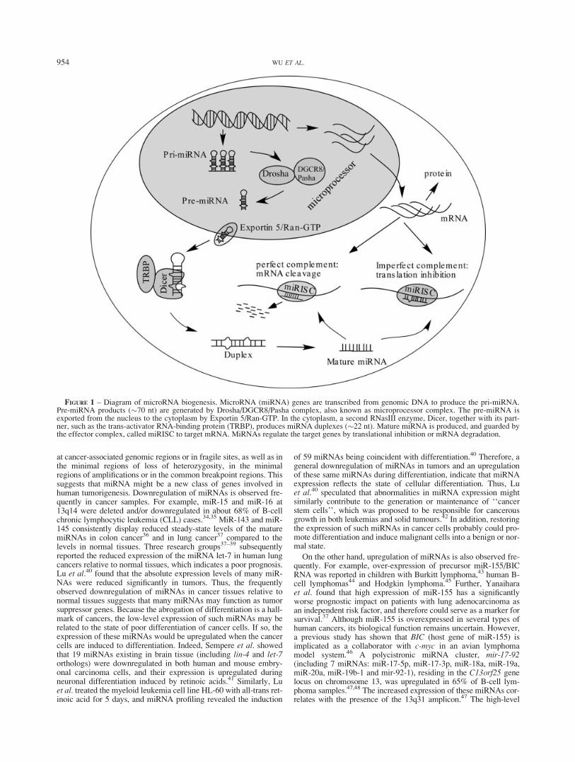
Biochemical approaches have provided a basic understandingof the molecular details of miRNA biogenesis.15 As shown inFigure 1, a primary transcript RNA (pri-miRNA) transcribed from amiRNA gene by RNA polymerase II is first processed into a stem-loop structure of about 70–100 nt (precursor miRNA; pre-miRNA)by a double-strand (ds)-RNA-specific ribonuclease, Drosha, with thehelp of its binding partner DGCR8.18 These pre-miRNAs are trans-ported into the cytoplasm via an Exportin-5-RanGTP dependentmechanism.19–21 In the cytoplasm, they are digested by a second,dsRNA-specific ribonuclease called Dicer with the help of TRBPand AGO2.22–24 The released 17–25 nt mature miRNA is bound bya complex called miRNA-associated RNA-induced silencing com-plex (miRISC). Two mechanisms for miRNA action are currentlyknown: mRNA cleavage and translational repression of mRNAwithout RNA cleavage. In animals, the complex-bound, single-stranded miRNAs binds specific target mRNAs through the sequencethat is significantly, though not completely complementary to the tar-get mRNAs.25 By a mechanism that is not fully characterized, thebound mRNA remains untranslated, resulting in reduced expressionof the corresponding genes. A single miRNA could regulate multiple tar-get genes, while a single gene could be targeted by multiple miRNAs,suggesting that the miRNAome and mRNAome interaction is acomplicated network. For more details, see reviews.1,6,15,17,26–28
Several excellent review papers focusing on the link betweenmiRNAs and cancer, e.g., Refs. 7, 26, 27 and 29–32 have beenpublished in recent years. Particularly, Esquela-Kerscher andSlack’s review26 is a very elegant and comprehensive one on thistopic, in which the connection of miRNAs and cancer has beendiscussed in depth. In the present review, we are trying to describea more general and up-to-date picture regarding the association ofmiRNAs with cancer, as well as miRNA regulation, signalingpathways and target prediction and validation. Some recentadvancements in this field that have not been included in the pre-vious reviews are also discussed here.
Deregulation of miRNA expression in cancer
Rapidly accumulating evidence has revealed that miRNAs areassociated with cancer because of deregulation.7,26,29,30,33 Genome-wide studies demonstrated that miRNA genes are frequently located
Wei Wu’s current address is: Division of Cancer Biology, Departmentof Medicine, Northwestern University, Chicago, IL 60201, USA.*Correspondence to: Division of Cancer Biology, Department of Med-
icine, Northwestern University, ENH research Institute, 1001 UniversityPlace, Evanston, IL 60201, USA. Fax:11-224-364-7402.E-mail: [email protected] or Department of Medicine, Univer-sity of Chicago, 5841 S. Maryland Avenue, MC2115, Chicago, IL 60637,USA. Fax:11-773-702-3002. E-mail: [email protected]
Grant sponsors: G. Harold and Leila Y. Mathers Charitable Foundation;Cancer Research Foundation Young Investigator Award.
Received 28 March 2006; Accepted after revision 29 September 2006DOI 10.1002/ijc.22454Published online 12 December 2006 inWiley InterScience (www.interscience.
wiley.com).
Int. J. Cancer: 120, 953–960 (2006)' 2006 Wiley-Liss, Inc.
Publication of the International Union Against Cancer
at cancer-associated genomic regions or in fragile sites, as well as inthe minimal regions of loss of heterozygosity, in the minimalregions of amplifications or in the common breakpoint regions. Thissuggests that miRNA might be a new class of genes involved inhuman tumorigenesis. Downregulation of miRNAs is observed fre-quently in cancer samples. For example, miR-15 and miR-16 at13q14 were deleted and/or downregulated in about 68% of B-cellchronic lymphocytic leukemia (CLL) cases.34,35 MiR-143 and miR-145 consistently display reduced steady-state levels of the maturemiRNAs in colon cancer36 and in lung cancer37 compared to thelevels in normal tissues. Three research groups37–39 subsequentlyreported the reduced expression of the miRNA let-7 in human lungcancers relative to normal tissues, which indicates a poor prognosis.Lu et al.40 found that the absolute expression levels of many miR-NAs were reduced significantly in tumors. Thus, the frequentlyobserved downregulation of miRNAs in cancer tissues relative tonormal tissues suggests that many miRNAs may function as tumorsuppressor genes. Because the abrogation of differentiation is a hall-mark of cancers, the low-level expression of such miRNAs may berelated to the state of poor differentiation of cancer cells. If so, theexpression of these miRNAs would be upregulated when the cancercells are induced to differentiation. Indeed, Sempere et al. showedthat 19 miRNAs existing in brain tissue (including lin-4 and let-7orthologs) were downregulated in both human and mouse embry-onal carcinoma cells, and their expression is upregulated duringneuronal differentiation induced by retinoic acids.41 Similarly, Luet al. treated the myeloid leukemia cell line HL-60 with all-trans ret-inoic acid for 5 days, and miRNA profiling revealed the induction
of 59 miRNAs being coincident with differentiation.40 Therefore, ageneral downregulation of miRNAs in tumors and an upregulationof these same miRNAs during differentiation, indicate that miRNAexpression reflects the state of cellular differentiation. Thus, Luet al.40 speculated that abnormalities in miRNA expression mightsimilarly contribute to the generation or maintenance of ‘‘cancerstem cells’’, which was proposed to be responsible for cancerousgrowth in both leukemias and solid tumours.42 In addition, restoringthe expression of such miRNAs in cancer cells probably could pro-mote differentiation and induce malignant cells into a benign or nor-mal state.
On the other hand, upregulation of miRNAs is also observed fre-quently. For example, over-expression of precursor miR-155/BICRNA was reported in children with Burkitt lymphoma,43 human B-cell lymphomas44 and Hodgkin lymphoma.45 Further, Yanaiharaet al. found that high expression of miR-155 has a significantlyworse prognostic impact on patients with lung adenocarcinoma asan independent risk factor, and therefore could serve as a marker forsurvival.37 Although miR-155 is overexpressed in several types ofhuman cancers, its biological function remains uncertain. However,a previous study has shown that BIC (host gene of miR-155) isimplicated as a collaborator with c-myc in an avian lymphomamodel system.46 A polycistronic miRNA cluster, mir-17-92(including 7 miRNAs: miR-17-5p, miR-17-3p, miR-18a, miR-19a,miR-20a, miR-19b-1 and mir-92-1), residing in the C13orf25 genelocus on chromosome 13, was upregulated in 65% of B-cell lym-phoma samples.47,48 The increased expression of these miRNAs cor-relates with the presence of the 13q31 amplicon.47 The high-level
FIGURE 1 – Diagram of microRNA biogenesis. MicroRNA (miRNA) genes are transcribed from genomic DNA to produce the pri-miRNA.Pre-miRNA products (�70 nt) are generated by Drosha/DGCR8/Pasha complex, also known as microprocessor complex. The pre-miRNA isexported from the nucleus to the cytoplasm by Exportin 5/Ran-GTP. In the cytoplasm, a second RNasIII enzyme, Dicer, together with its part-ner, such as the trans-activator RNA-binding protein (TRBP), produces miRNA duplexes (�22 nt). Mature miRNA is produced, and guarded bythe effector complex, called miRISC to target mRNA. MiRNAs regulate the target genes by translational inhibition or mRNA degradation.
954 WU ET AL.
amplification of 13q31 has been observed in hematological andother solid neoplasms,48,49 including diffuse large B-cell lym-phoma,50 primary cutaneous B-cell lymphoma,51 follicular lym-phoma,52 mantel cell lymphoma,53 nasal-type natural killer/T-celllymphoma,54 glioma,49 nonsmall cell lung cancer,49 bladder can-cer,49 squamous-cell carcinoma of the head and neck,49 peripheralnerve sheath tumor,55 malignant fibrous histiocytoma,56 alveolarrhabdomyosarcoma57 and liposarcoma.58 The oncogenic functionof the mir-17-92 cluster is further validated by a number of piecesof evidence as follows. MiR-19a and miR-92-1 are overexpressedin cells from patients with B-cell CLL.35 MiR-17-5p is overex-pressed in breast, colon, lung, pancreas and prostate tumors, whilemiR-20a is overexpressed in colon, pancreas and prostate tumors.59
MiR-17-3p is overexpressed in lung adenocarcinoma and is relatedto the patient’s survival.37 MiRNAs from the mir-17-92 cluster areoverexpressed in lung cancers and enhance cell proliferation.60
Moreover, introduction of the expression construct of the mir-17-92 cluster, but not the putative open reading frame of C13orf25,could enhance lung cancer cell growth.60 Most importantly, to testthe hypothesis directly that increased expression of this cluster con-tributes to cancer formation, He et al.47 overexpressed the mir-17-19b-1 cluster (the vertebrate-specific portion of the mir-17-92 clus-ter) using a mouse model of human B-cell lymphoma. Hematopoi-etic stem cells (HSCs) derived from fetal livers of c-myc transgenicmice generate B-cell lymphomas by 4–6 months of age when trans-planted into lethally irradiated recipients.61 Irradiated animals thatreceived HSCs overexpressing both Myc and the mir-17-19b-1cluster developed malignant lymphomas faster (�51 days) thanthose animals that received HSCs expressing Myc alone, or in com-bination with either 96 unrelated, single miRNAs or any individualmiRNA of the mir-17-19b-1 cluster (3–6 months).47 More recently,Volinia et al.59 identified a large number of overexpressed miR-NAs, including miR-17-5p, miR-20a, miR-21, miR-92, miR-106aand miR-155, in solid tumor samples. Therefore, overexpression ofmiRNAs appears to play an important role in tumorigenesis, withthe miRNAs functioning directly or indirectly as oncomiRNAs.The aberrant expression of miRNAs in tumor cells appears to bestrongly associated with cancer development.
A current challenge is to reveal the mechanism of regulation ofmiRNA gene expression itself, i.e. why and how the miRNAs arederegulated. It is very important for us to understand further, themechanism of the development of miRNA-associated cancer. Insome cases, the cause of miRNA deregulation is clear. For exam-ple, in the case of the mir-17-92 cluster, it is now very clear thatthe increase expression of these miRNAs correlates with the pres-ence of the 13q31 amplicon.47,48 Similarly, the downregulation ofmiR-15 and miR-16 at 13q14 observed in about 68% of B-cellCLL cases is largely due to a deletion at 13q14.34,35 However, inmost of cases, there is no clue as to why and how the miRNAs arederegulated. Given that miRNAs, like protein-coding genes, aretranscribed by RNA polymerase II,62 and that many miRNAs arelocated within the introns of protein coding genes,63 the regulationof miRNA gene expression is probably similar to the regulation ofprotein-coding gene expression. For example, CpG-island methyl-ation and/or histone deacetylation might be a possible mechanismfor downregulation of miRNAs,64 although it might not be a com-mon mechanism in the regulation of miRNAs.37,65 In addition,some miRNAs probably are regulated along with their host genes,e.g., the mir-17-92 cluster is upregulated along with the host geneC13orf25, because of locus amplification.47,48 Furthermore, theexpression of miRNAs could be regulated through regulatingmicroprocessors of the miRNAs, for example, by regulating theexpression of Dicer.27,66
Computational prediction of targets of miRNAs
Another challenge is to accurately identify targets that are regu-lated by miRNAs.26 In animals, miRNAs confer inhibitory regula-tion of the target-gene translation level through binding in the 30UTRs. This was first evidenced by lin-4 targeting of the lin-14/lin-
28 genes,8,9 and let-7 targeting of the lin-41 gene11 in C. elegans.Recently, 4 bioinformatics approaches have been developed forpredicting miRNA targets in humans.3,67–70 Most bioinformaticalgorithms use an ‘‘miRNA seed’’ that encompasses the first 2–8bases of the mature miRNA sequence to search for complementar-ity to sequences in the 30 UTRs of all expressed genes.71 Thesestudies have revealed that a single miRNA might bind to hundredsof gene targets which can be diverse in their function, and that 1target gene might be regulated by several different miRNAs.26
Briefly, Lewis et al.3,67 developed a sophisticated algorithm, calledTargetScan, and its improved version, TargetScanS. TargetScan3
searches the UTRs for segments of perfect Watson-Crick comple-mentarity to base 2–8 of the miRNA (numbered from the 50 end),calculates a folding free energy G to each miRNA-target site inter-action using RNAeval, and then assigns a Z score to each UTR.TargetScanS67 is an improved algorithm that requires a shorterseed (6 nt), which is preceded by an adenosine and is located in ashort ‘‘island’’ of conservation in at least 4 mammalian genomes(i.e., human, mouse, rat and dog). It simply relies on perfect Wat-son-Crick seed matches that are conserved in the UTR regions ofwhole genome alignments, and does not rely on free-energy calcu-lation.67 John et al.70 developed an algorithm named miRanda,which optimizes sequence complementarity using position-specificrules and relies on strict requirements of interspecies conservation.Kiriakidou et al.69 developed an algorithm called DIANA-MicroT,which is trained to identify miRNA targets having a single bindingsite. This algorithm takes an approach that is different from thoseof the other algorithms described earlier71: (i) It focuses on single-binding-site targets and (ii) it seeks binding sites that have a typicalcentral bulge and require 30-binding, beyond the obligatory 50-seed.Krek et al.68 developed an advanced algorithm called PicTar,which is trained to identify both binding sites targeted by a singlemiRNA, and those that are coregulated by several miRNAs in acoordinated manner. It employs pair-wise alignment for accuratefiltering for binding sites that are conserved across 8 vertebrates,and takes into account clustering and coexpression of miRNAs,ontologic information, as well as free energy of RNA:RNAduplexes by using RNA hybrid.72
Recently, we collected all the putative miRNA-target pairs pre-dicted by each algorithm to set up a complete list of all the pre-dicted human miRNA-target pairs, because there is no evidence asyet to show that one is much better than the others. Briefly, wecollected 21,293 unique miRNA-target pairs predicted by TargetS-canS,67 46,155 unique miRNA-target pairs predicted by PicTar,68
54,578 unique miRNA-target pairs predicted by miRanda70 and185 unique miRNA-target pairs predicted by DIANA-MicroT.69
After combination, we obtained 101,031 predicted unique miRNA-target pairs. Of the 101,031 pairs, 0.01%(12), 2.8% (2,817), 15.4%(15,510) and 81.8% (82,692) were predicted by 4, 3, 2 and a singlealgorithm, respectively (Sun M, Chen J. unpublished). Notably,most of the pairs (81.8%) were predicted by only a single algo-rithm. Although the false positive rate of each of the 4 algorithmswas estimated to be 20–30%,71 it is unlikely that most of the82,692 pairs are false-positive ones. Instead, it is possible that eachalgorithm may only predict a part of the whole miRNA-target pairs.For example, although the KIT gene was predicted only by miR-anda as a target of miR-221, it has been biologically validated to bea real target.73,74 Thus, the combination of these 4 predictions mightprovide a much more comprehensive list of putative miRNA-targetpairs than do any single prediction.
Current methodologies of miRNA target validation
Because of the potential high false-positive rate of computa-tional prediction of miRNA targets, validating target predictions isvery important. MiRNA target prediction algorithms may be infor-matically validated by evaluation of an algorithm’s success in cor-rectly identifying known miRNA targets (i.e. targets that have al-ready been validated biologically, and scoring them highly), orthey may be validated by comparing the number of postulated
955MICRORNA AND CANCER
binding sites that an algorithm finds for a real miRNA withthat found for a control group of artificially generated ‘‘fictitiousmiRNAs’’. Nonetheless, all of these strategies merely provide indi-rect validation, and they have several limitations.71 However,whereas the ultimate validation of predicted miRNA targets is bio-logical validation, at present there is no a high-throughput method forbiologically validating miRNA targets.71 The conventional biologicalvalidation methodologies are still extremely labor-intensive and donot allow high-throughput target validation. The commonly used val-idation methodologies include luciferase reporter assays,3,39,68,69,75
mutation studies,69,75 gene-silencing techniques,39,75,76 rescueassays77 and classic genetic studies.8,39,77 Overall, only about 30animal miRNA targets have been validated to date with use ofthese various techniques.71
A new mechanism of miRNA-mediated gene regulation inanimals and a potential high-throughput method foridentifying miRNA-target pairs
In a classic model of miRNA function in animals,1,2 miRNAsthat form imperfect duplexes with their targets inhibit proteinexpression without affecting mRNA levels (as illustrated in Fig. 1).However, recent findings indicate that miRNAs that share only par-tial complementarity with their targets can also induce mRNA deg-radation.74,78–83 For example, in C. elegans, regulation by the let-7miRNA results in degradation of its lin-41 target mRNA. Further-more, lin-14 and lin-28 mRNA levels significantly decrease inresponse to lin-4 miRNA expression.81 In addition, transfection ofexogenous miRNA duplexes into HeLa cells can cause moderatedownregulation of hundreds of mRNAs, many of which contain therecognition motif of the overexpressed miRNA in their 30 UTR.80Moreover, in vivo knockdown of a liver-specific miRNA (miR-122) has shown that hundreds of mRNAs, many of them seeminglyto be direct targets of this miRNA, were moderately upregulated.78
Taken together, these studies suggest that mRNAs containing par-tial miRNA complementary sites can be targeted for degradationin vivo, and that miRNA-dependent regulation of mRNA stabilitymay be more common than previously appreciated.82
Thus, as shown by some pilot studies,74,82,83 the predicted miRNA-target pairs could be roughly validated on a large scale by compari-son of expression patterns between miRNAs and the predicted tar-gets through conducting microarray analysis on both miRNAs andprotein-coding genes. However, the aims of the pilot studies arenot to set up a high-throughput method for biological validation ofthe predicted miRNA-target pairs, and the specificity, robustnessand reproducibility of the approaches have not been systematicallytested and evaluated. In fact, because only a few miRNA targetshave been biologically validated to be affected by miRNAs at themRNA level,78,80,81 it is unclear whether the majority of miRNAtargets can be significantly degraded at the mRNA level by miR-NAs in animals. In addition, although it was reported that the folddecrease of lin-14 mRNA is consistent with that in LIN-14 proteinlevels mediated by lin-4 miRNA,81 it was suggested that thechanges in target mRNAs are usually weak.82 It is unclear to whatextent the mRNA-level changes of the miRNA targets can reflectthe changes at their protein level. Thus, a large-scale protein-levelanalysis technique, such as a protein array, probably should also beintegrated in the potential high-throughput technique for miRNA-target-pair validation.
MiRNA signaling pathways in cancer development
As described earlier, thousands of target genes of human miR-NAs have been predicted using bioinformatic tools. Interestingly,predominant miRNA targets are transcription factor or kinases.3
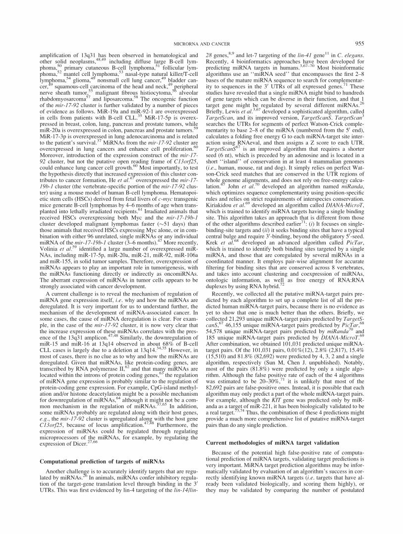
Researchers are currently attempting to dissect the target genes ofmiRNAs and their signaling pathways involved in cancer. SeveralmiRNA signaling pathways have been carefully studied (Fig. 2).As an example, Johnson et al. showed that the 30 UTR of thehuman RAS gene contains multiple let-7 complimentary binding
sites, allowing let-7 to regulate RAS expression. Furthermore, theoverexpression of let-7 in human cancer cell lines results in adecreased level of RAS compared with untreated cells. Conversely,knockdown of let-7 in human cancer cell lines, in which let-7 isnormally expressed at a high level, leads to a remarkable increasein RAS protein expression, providing a mechanism for let-7 nega-tively regulating RAS. Consistent with the observations in vitro,expression of let-7 is lower in lung tumors than in normal lung tis-sue, while that of RAS protein is significantly higher in lungtumors.39 Introduction of let-7 isoforms (let-7a and let-7f) into alung cancer cell line A549 resulted in a 78.6% reduction in thenumber of colonies,38 indicating that growth inhibition may be thebiological significance of let-7, presumably through negatively reg-ulating RAS signaling pathways. A few additional genes have alsobeen predicted to be a potential target for let-7, such as LIM kinase2,3 which belongs to a gene family having a role in the regulationof cell shape and motility and possibly in metastasis as well.
As aforementioned, the mir-17-92 cluster is overexpressed inmultiple malignancies. To reveal the mechanisms of the mir-17-92oncogenic effect directly, He et al.47 utilized a mouse B-cell lym-phoma model and demonstrated that enforced expression of themir-17-92 cluster gene acted with c-myc expression to acceleratetumor development in this model. Tumors derived from haemato-poietic stem cells expressing a subset of the mir-17-92 cluster and c-myc possesses less apoptosis than c-myc-induced lymphomas, whichunderwent extensive apoptosis. Because of its oncogenic function,the authors nominate this miRNA cluster as the first candidate non-coding oncogene, ‘‘oncomiR-1.’’47 Interestingly, O’Donnell et al.75
independently found that c-Myc binds directly to the miR-17-92locus and activates expression of a cluster of 6 miRNAs. One ofthe transcriptional targets of c-Myc, E2F1 is negatively regulatedby 2 miRNAs in this cluster, miR-17-5p and miR-20a. This studyexpands the known classes of transcripts within the MYC targetgene network, and uncovers a mechanism through which MYCsimultaneously activates E2F1 transcription and limits its transla-tion by the miR-17-92 cluster of miRNAs, allowing a tight controlof proliferative signal. Collectively, these findings suggest that themiR-17-92 cluster acts as an oncogene at one set of conditions,while as a tumor suppressor at another.
In most malignant tumors, the PI3K/Akt signaling pathway isactivated and results in an aggressive proliferation. The tumor sup-pressor gene, phosphatase and tensin homolog deleted on chromo-some ten (PTEN), inhibits PI3K function. Loss of PTEN expres-sion is associated with PI3K/Akt signaling activation in many
FIGURE 2 – Schematic representation of miRNA-mediated signalingpathways. MiRNAs functioning as either tumor suppressors (Red) oroncogenes (green) participate in the cell survival and apoptosis path-way, by which may contribute to cancer initiation and development.
956 WU ET AL.
types of tumors.84 MiR-19a, b has been demonstrated to bind the30 UTR of PTEN mRNA in vitro.3 In lymphomas, overexpressionof miR-19 is correlated with underexpression of PTEN protein,35
suggesting that overexpression of mir-19 in tumor cells could bean alternative mechanism by which the PI3K/Akt signaling path-way is activated.
Other studies have linked miRNAs to the regulation of apopto-sis. Chan et al.85 revealed a strong over-expression of miR-21 inhuman glioblastoma tumor tissues and cell lines. Knockdown ofmiR-21 in cultured glioblastoma cells triggers activation of cas-pases and leads to increased apoptotic cell death, suggesting thataberrantly expressed miR-21 may contribute to the malignant phe-notype by blocking expression of critical apoptosis-related genes.It appears that miR-21 has a broad function in tumor progression,because miR-21 is also upregulated in breast cancer86 and lungcancer.37 Cimmino et al.87 demonstrated that expression of miR-15a and miR-16-1 is inversely correlated to expression of BCL-2in CLL and that both miRNAs negatively regulate BCL-2 proteinat a posttranscriptional level; BCL-2 repression by these miRNAsinduces apoptopsis in a leukemic cell lines. Cheng et al.88
employed a library of miRNA inhibitors to screen for miRNAinvolved in cell growth and apoptosis, and observed that miRNA-mediated regulation has a complexity of cellular outcomes andthat miRNAs can be mediators of regulation of cell growth and ap-optosis pathways.
KIT is an important tyrosine kinase receptor in cell differentia-tion and growth. Inhibiting the KIT signaling via miRNAs maycontribute to uncontrolled cell growth in certain type of cells.73
He et al.74 reported that miR-221, -222 and -146 are upregulatedin papillary thyroid carcinoma and are associated with dramaticdecrease of expression of KIT at both transcript and protein levels.The authors further demonstrated that point mutations occurredfrequently in the KIT loci (5 out of 10 samples) within the compli-mentary binding regions for these miRNAs, and therefore dis-rupted the miRNA–mRNA target interactions in these tumors.
Volinia et al.89 observed that miR-106a overexpression in coloncarcinoma inversely correlates to RB1 protein expression, and thatdownregulation of miR-20a in breast cancer is associated to highexpression of TGFBR2 protein, while overexpression of miR-20ain some lung cancer samples is associated with downregulation ofTGFBR2 protein. These results suggest that miR-106a and miR-20a might negatively regulate RB1 and TGF b signaling, respec-tively, which was confirmed by a luciferase assay in vitro.
More recently, to perform genetic screens for novel functions ofmiRNAs, Voorhoeve et al.90 developed a library (miR-Lib) ofvectors expressing the majority of cloned human miRNAs andmade a corresponding microarray (miR-Array) containing allmiR-Lib inserts. In a screen for miRNAs that cooperate with onco-genes in cellular transformation, the authors identified miR-372and miR-373 as potential oncogenes in testicular germ cell tumors.Each of them permits proliferation and tumorigenesis of primaryhuman cells that harbor both oncogenic RAS and active wild-typep53. These miRNAs neutralize p53-mediated CDK inhibition, pos-sibly through direct inhibition of the expression of the large tumorsuppressor homolog 2 (LATS2), a serine-threonine kinase whosedeletion accelerates cellular proliferation and tumorigenic develop-ment. In all, the authors provided compelling evidence that thesemiRNAs are potential novel oncogenes participating in the devel-opment of human testicular germ cell tumors by numbing the p53pathway, thus allowing tumorigenic growth in the presence ofwild-type p53.
Taken together, the deregulation of miRNAs participates inactivation of cell proliferation or inactivation of apoptotic signal-ing pathway in conjunction with other genetic changes leading tocancer pathogenesis.91 Further studies would reveal that miRNAsfunction as key regulators of many more cancer-related genes.Besides the fact that miRNAs inhibit the target genes directly,Gregory and Shiekhattar27 provided evidence that defect of micro-processor of miRNA is one alterative mechanism through which
the expression of miRNA can be altered and miRNA perturbationis linked to cancer. Reduced expression of Dicer is associated withpoor prognosis in lung cancer patients.66
MicroRNA expression profiles for cancer diagnosis
Emerging data indicate that miRNAs play an important role inregulating gene activation in cancer initiation and progression.Researchers are now using the miRNA-expression signatures toclassify cancers and to define miRNA markers that might predictfavorable prognosis.26 Various methods have been employed forthis purpose. Northern blotting was used in early profiling studies,and is still widely used, because it can convincingly and simulta-neously detect both the precursor and mature miRNAs. It providesdata about the Dicer processing step, which may be regulated.92
However, Northern blotting is not a high-throughput and sensitivemethod. Thus, over the past years, real-time RT-PCR and microar-ray methods have been adapted to detect the expression of miR-NAs. High-throughput real-time RT-PCR methods have beendeveloped to screen the expression of precursor and/or maturemiRNAs using either SYBR Green dye or TaqMan chemistry(probe) (e.g., Refs. 93–95). To date, largely, there are 4 platformsof microRNA-arrays including dotted cDNA arrays,96 oligonu-cleotide arrays,97–99 bead-based flow cytometric miRNA expres-sion profiling40 and ‘‘locked nucleic acid’’ (LAN)-modified oligo-nucleotide arrays100,101 (for reviews, see Refs. 92 and 102). Thesetechnologies have been utilized in screening miRNA expressionpattern in hematopoietic malignancies and solid tumors.17 Forexample, using Northern blotting, Chen et al.103 demonstrated thatmiR-181, miR-223 and miR-142s were differentially or preferen-tially expressed in hematopoietic tissues. Using a high-throughputTaqman real-time RT-PCR method, Jiang et al.94 screened 222miRNA precursors in 32 commonly used cell lines of lung, breast,head and neck, colorectal, prostate, pancreatic and hematopoieticcancers. Using oligonucleotide microchips,99 Croce’s group hasrevealed distinct signatures of miRNAs in human breast cancer,86
in human megakaryocytopoiesis104 and in B cell CLL.35,105 Theyobserved that a unique miRNA signature is associated with prog-nosis factors and disease progression in B cell CLL.105 Using asimilar oligonucleotide microchip, Yanaihara et al.37 analyzed themiRNA expression in 104 pairs of primary lung cancers and corre-sponding noncancerous lung tissues, and found that the expressionof 43 miRNAs was statistically differentially displayed in cancertissues; their further analysis revealed that the high expression ofmiR-155 and the low expression of let-7a-2 are correlated withpoor survival. In addition to studies on individual type of tumors,a large-scale microRNAome analysis was performed on 540 sam-ples including lung, breast, stomach, prostate, colon and pancre-atic tumors with oligonucleotide microchips; cancer-associatedmiRNAs were identified as miRNA signatures, including somewell characterized ones such as miR-17-5p, miR-20a, miR-21,miR-92, miR-106a and miR-155 in solid tumors.59 Lu et al.40
used a new, bead-based flow cytometric miRNA expression profil-ing method to conduct a systematic expression analysis of 217mammalian miRNAs on 334 samples, including multiple humancancers. They observed a general downregulation of miRNAs intumors when compared with that in normal tissues. Furthermore,they successfully classified poorly differentiated tumors usingmiRNA expression profiles.
Taken together, miRNA expression profiles can be utilized todefine the expression pattern of diverse cancers, to predict theprognosis, and to monitor response to therapy or toxicity. It isnoteworthy that small RNAs can be measured easily from the for-malin-fixed tissue specimens used routinely in hospital pathologylaboratories. Thus, potential miRNA-based diagnostics could fitsimply into the standard hospital workflow.98 To improve thespecificity and sensitivity of miRNA microarrays, Neely et al.100
recently developed a new approach, LAN-modified oligonucleo-tide array, in which they designed 2 probes for each miRNA (eachprobe is half of the length of the mature miRNA) and labeled with
957MICRORNA AND CANCER
different fluorophores. With such an approach, the authors wereable to quantitate miRNA expression from as little as 50 ng totalRNA with a high specificity.100 In addition, Kloosterman et al.101
designed LNA oligonucleotides and applied them for in situhybridization to monitor the spatial and temporal expression ofmiRNAs in vivo. Such technique improvement and renovationwill make it more promising to use miRNA expression profiles forcancer diagnosis, for defining a treatment strategy for patients, forprediction of the prognosis, and for monitoring treatment re-sponse.
Targeting miRNA regulation with antisenseoligonucleotides: a potential therapeutic intervention
To investigate the potential causative roles of deregulatedmiRNA processes in human disease, synthetic, chemically-modi-fied antisense oligonucleotides targeting to miRNAs or protein-coding genes have been widely used. They have been proven to bea powerful research tool, and may eventually become a new thera-peutic tool.106–108 On the one hand, if a disease phenotype isrelated to the overexpression of miRNAs, antisense oligonucleo-tides that are complementary to either the mature miRNAs or theirprecursors can be designed to specifically inhibit the activity ofthe miRNAs. There are 3 types of modifications for anti-miRNAoligonucleotides (AMO)106: (i) 20-O-Methyl (OMe) AMOs, (ii) 20-O-Methoxyethyl (MOE) AMOs and (iii) Locked nucleic acid(LNA) AMOs. Various AMOs have been implemented in experi-mental studies. For example, Cheng et al.88 applied a library ofMe-AMOs to screen for miRNAs that are involved in cell growthand apoptosis. Esau et al.109 inhibited miR-122 in mice with aMOE-AMO to uncover the role of the liver-specific miR-122 inthe adult liver and demonstrated that miR-122 is a key regulator ofcholesterol and fatty-acid metabolism in the adult liver. Chanet al.85 successfully applied 20-O-methyl- and DNA/LNA-mixedoligonucleotides to specifically knockdown miR-21 to investigatethe potential contribution of this miRNA in the regulation of apo-ptosis-associated genes in glioblastoma cell lines. On the otherhand, to enhance the function of miRNAs due to a deletion or aloss of function mutation, a therapeutic approach could entail ex-ogenous delivery of corrective synthetic miRNAs in the form of(siRNA-like) double strand oligoRNAs.106 One example showsthat enforced expression of let-7 in the lung adenocarcinoma cellline A549 inhibited lung cancer cell growth in vitro.38 This holdsa promise that let-7 may be useful in treatment of lung cancer orenhancing the current treatments. Synthesized miRNA-14U wasused to inhibit the overexpressed HER-2 protein in ovarian cancercell line SKOV3.110
To develop a pharmacological approach for silencing miRNAsin vivo, Krutzfeldt et al.78 designed chemically modified, cholesterol-conjugated single-stranded RNA analogues (termed ‘‘antagomirs’’),
which are complementary to miRNAs. Their study shows that thetermed antagomirs are a powerful tool to silence specific miRNAsin vivo.78 Therefore, we may expect that in the future, activation ofmiRNAs with ‘‘agomiR’’ or silencing of miRNAs with ‘‘anta-gomirs’’ could become a therapeutic strategy for cancer treatment.Nevertheless, there are still several difficulties to overcome beforeapplying such strategies in clinic. For example, as miRNAs mayhave dual functions (oncogene or tumor suppressor) in differentorgans or conditions, is it possible to design ‘‘agomiR’’ or ‘‘anta-gomirs’’ that target specific tissues or disease sites?108 In addition,the regulation and stability of the injected ‘‘agomiR’’ or ‘‘anta-gomirs’’ in vivo, the proper dosages for the optimal effect, and thepotential side effects, should be carefully examined and addressed.Moreover, because a single miRNA may regulate hundreds of differ-ent targets while a single target gene may be regulated by several dif-ferent miRNAs, the regulation network may be very complex. Thus,more cautions should be taken in considering clinical applications.
Conclusion
The microRNA field is rapidly developing, as shown by thefacts that the number of experimentally identified miRNAs andtheir targets is increasing, and that the functions and the signalingpathways of more and more miRNAs have been carefully studied.The recent findings suggest that miRNAs play an important role inregulation of gene activation by binding to the mRNAs of targetgenes, by switching the genes on or off, or by fine-tuning thegenes, to control cell viability, cell cycle, proliferation or apopto-sis. It was predicted that more than half of human genes might beregulated by miRNAs. Deregulation of miRNA expression is oftenassociated with cancer. These studies may change the landscapeof cancer genetics and uncover new mechanisms that contribute tocancer development. The future challenges are to learn how miR-NAs are regulated in human genome, and to identify more biologi-cal targets of miRNAs and the relevant signaling pathways. None-theless, activation of miRNAs with ‘‘agomiR’’ or silencing ofmiRNAs with ‘‘antagomirs’’ could probably become a therapeuticstrategy for cancer treatment in the future, although we are farfrom that point.
Acknowledgements
We thank Dr. Janet D. Rowley and Dr. Suzanne D. Conzen fortheir critical comments and valuable suggestions, and 3 anony-mous referees for their constructive comments. We also appreciatethe help of Ms. Mary Beth Neilly in manuscript revision. We apol-ogize to our colleagues whose outstanding contributions to thegrowing miRNA field were not cited as primary references becauseof the space constraints.
References
1. Bartel DP. MicroRNAs: genomics, biogenesis, mechanism, and func-tion. Cell 2004;116:281–97.
2. Ambros V. The functions of animal microRNAs. Nature 2004;431:350–5.
3. Lewis BP, Shih IH, Jones-Rhoades MW, Bartel DP, Burge CB. Pre-diction of mammalian microRNA targets. Cell 2003;115:787–98.
4. Ambros V. MicroRNA pathways in flies and worms: growth, death,fat, stress, and timing. Cell 2003;113:673–6.
5. Bentwich I, Avniel A, Karov Y, Aharonov R, Gilad S, Barad O, Bar-zilai A, Einat P, Einav U, Meiri E, Sharon E, Spector Y, et al. Identi-fication of hundreds of conserved and nonconserved human micro-RNAs. Nat Genet 2005;37:766–70.
6. He L, Hannon GJ. MicroRNAs: small RNAs with a big role in generegulation. Nat Rev Genet 2004;5:522–31.
7. Meltzer PS. Cancer genomics: small RNAs with big impacts. Nature2005;435:745–746.
8. Lee RC, Feinbaum RL, Ambros V. The C. elegans heterochronicgene lin-4 encodes small RNAs with antisense complementarity tolin-14. Cell 1993;75:843–54.
9. Wightman B, Ha I, Ruvkun G. Posttranscriptional regulation of theheterochronic gene lin-14 by lin-4 mediates temporal pattern forma-tion in C. elegans. Cell 1993;75:855–62.
10. Pasquinelli AE, Reinhart BJ, Slack F, Martindale MQ, Kuroda MI,Maller B, Hayward DC, Ball EE, Degnan B, Muller P, Spring J,Srinivasan A, et al. Conservation of the sequence and temporal expres-sion of let-7 heterochronic regulatory RNA. Nature 2000;408:86–9.
11. Reinhart BJ, Slack FJ, Basson M, Pasquinelli AE, Bettinger JC,Rougvie AE, Horvitz HR, Ruvkun G. The 21-nucleotide let-7 RNAregulates developmental timing in Caenorhabditis elegans. Nature2000;403:901–6.
12. Lee RC, Ambros V. An extensive class of small RNAs in Caeno-rhabditis elegans. Science 2001;294:862–4.
13. Lagos-Quintana M, Rauhut R, Lendeckel W, Tuschl T. Identificationof novel genes coding for small expressed RNAs. Science 2001;294:853–8.
14. Lau NC, Lim LP, Weinstein EG, Bartel DP. An abundant class oftiny RNAs with probable regulatory roles in Caenorhabditis elegans.Science 2001;294:858–62.
958 WU ET AL.
15. Kim VN. MicroRNA biogenesis: coordinated cropping and dicing.Nat Rev Mol Cell Biol 2005;6:376–85.
16. Xie X, Lu J, Kulbokas EJ, Golub TR, Mootha V, Lindblad-Toh K,Lander ES, Kellis M. Systematic discovery of regulatory motifs inhuman promoters and 30 UTRs by comparison of several mammals.Nature 2005;434:338–45.
17. Kim VN, Nam JW. Genomics of microRNA. Trends Genet 2006;22:165–73.
18. Han J, Lee Y, Yeom KH, Nam JW, Heo I, Rhee JK, Sohn SY, Cho Y,Zhang BT, Kim VN. Molecular basis for the recognition of primarymicroRNAs by the Drosha-DGCR8 complex. Cell 2006;125:887–901.
19. Lund E, Guttinger S, Calado A, Dahlberg JE, Kutay U. Nuclear ex-port of microRNA precursors. Science 2004;303:95–8.
20. Yi R, Qin Y, Macara IG, Cullen BR. Exportin-5 mediates the nuclearexport of pre-microRNAs and short hairpin RNAs. Genes Dev 2003;17:3011–16.
21. Bohnsack MT, Czaplinski K, Gorlich D. Exportin 5 is a RanGTP-de-pendent dsRNA-binding protein that mediates nuclear export of pre-miRNAs. RNA 2004;10:185–91.
22. Chendrimada TP, Gregory RI, Kumaraswamy E, Norman J, Cooch N,Nishikura K, Shiekhattar R. TRBP recruits the Dicer complex toAgo2 for microRNA processing and gene silencing. Nature 2005;436:740–4.
23. Gregory RI, Chendrimada TP, Cooch N, Shiekhattar R. HumanRISC couples microRNA biogenesis and posttranscriptional genesilencing. Cell 2005;123:631–40.
24. Meister G, Landthaler M, Patkaniowska A, Dorsett Y, Teng G,Tuschl T. Human Argonaute2 mediates RNA cleavage targeted bymiRNAs and siRNAs. Mol Cell 2004;15:185–97.
25. Hutvagner G, Zamore PD. A microRNA in a multiple-turnover RNAienzyme complex. Science 2002;297:2056–60.
26. Esquela-Kerscher A, Slack FJ. Oncomirs—microRNAs with a role incancer. Nat Rev Cancer 2006;6:259–69.
27. Gregory RI, Shiekhattar R. MicroRNA biogenesis and cancer. Can-cer Res 2005;65:3509–12.
28. Preall JB, Sontheimer EJ. RNAi: RISC gets loaded. Cell 2005;123:543–5.
29. McManus MT. MicroRNAs and cancer. Semin Cancer Biol 2003;13:253–8.
30. Croce CM, Calin GA. miRNAs, cancer, and stem cell division. Cell2005;122:6–7.
31. Chen CZ. MicroRNAs as oncogenes and tumor suppressors. N EnglJ Med 2005;353:1768–71.
32. Calin GA, Garzon R, Cimmino A, Fabbri M, Croce CM. MicroRNAsand leukemias: how strong is the connection? Leuk Res 2006;30:653–5.
33. Sevignani C, Calin GA, Siracusa LD, Croce CM. Mammalian micro-RNAs: a small world for fine-tuning gene expression. Mamm Genome2006;17:189–202.
34. Calin GA, Sevignani C, Dumitru CD, Hyslop T, Noch E, Yendamuri S,Shimizu M, Rattan S, Bullrich F, Negrini M, Croce CM. HumanmicroRNA genes are frequently located at fragile sites and genomicregions involved in cancers. Proc Natl Acad Sci USA 2004;101:2999–3004.
35. Calin GA, Liu CG, Sevignani C, Ferracin M, Felli N, Dumitru CD,Shimizu M, Cimmino A, Zupo S, Dono M, Dell’Aquila ML, Alder Het al. MicroRNA profiling reveals distinct signatures in B cellchronic lymphocytic leukemias. Proc Natl Acad Sci USA 2004;101:11755–60.
36. Michael MZ, O’Connor SM, van Holst Pellekaan NG, Young GP,James RJ. Reduced accumulation of specific microRNAs in colorec-tal neoplasia. Mol Cancer Res 2003;1:882–91.
37. Yanaihara N, Caplen N, Bowman E, Seike M, Kumamoto K, Yi M,Stephens RM, Okamoto A, Yokota J, Tanaka T, Calin GA, Liu CGet al. Unique microRNA molecular profiles in lung cancer diagnosisand prognosis. Cancer Cell 2006;9:189–98.
38. Takamizawa J, Konishi H, Yanagisawa K, Tomida S, Osada H,Endoh H, Harano T, Yatabe Y, Nagino M, Nimura Y, Mitsudomi T,Takahashi T. Reduced expression of the let-7 microRNAs in humanlung cancers in association with shortened postoperative survival.Cancer Res 2004;64:3753–6.
39. Johnson SM, Grosshans H, Shingara J, Byrom M, Jarvis R, Cheng A,Labourier E, Reinert KL, Brown D, Slack FJ. RAS is regulated by thelet-7 microRNA family. Cell 2005;120:635–47.
40. Lu J, Getz G, Miska EA, Alvarez-Saavedra E, Lamb J, Peck D,Sweet-Cordero A, Ebert BL, Mak RH, Ferrando AA, Downing JR,Jacks T, et al. MicroRNA expression profiles classify human cancers.Nature 2005;435:834–8.
41. Sempere LF, Freemantle S, Pitha-Rowe I, Moss E, Dmitrovsky E,Ambros V. Expression profiling of mammalian microRNAs uncoversa subset of brain-expressed microRNAs with possible roles in murineand human neuronal differentiation. Genome Biol 2004;5:R13.
42. Reya T, Morrison SJ, Clarke MF, Weissman IL. Stem cells, cancer,and cancer stem cells. Nature 2001;414:105–11.
43. Metzler M, Wilda M, Busch K, Viehmann S, Borkhardt A.High expression of precursor microRNA-155/BIC RNA in chil-dren with Burkitt lymphoma. Genes Chromosomes Cancer 2004;39:167–9.
44. Eis PS, Tam W, Sun L, Chadburn A, Li Z, Gomez MF, Lund E,Dahlberg JE. Accumulation of miR-155 and BIC RNA in human Bcell lymphomas. Proc Natl Acad Sci USA 2005;102:3627–32.
45. Kluiver J, Poppema S, de Jong D, Blokzijl T, Harms G, Jacobs S,Kroesen BJ, van den Berg A. BIC and miR-155 are highly expressedin Hodgkin, primary mediastinal and diffuse large B cell lymphomas.J Pathol 2005;207:243–9.
46. Tam W, Hughes SH, Hayward WS, Besmer P. Avian bic, a gene iso-lated from a common retroviral site in avian leukosis virus-inducedlymphomas that encodes a noncoding RNA, cooperates with c-mycin lymphomagenesis and erythroleukemogenesis. J Virol 2002;76:4275–86.
47. He L, Thomson JM, Hemann MT, Hernando-Monge E, Mu D,Goodson S, Powers S, Cordon-Cardo C, Lowe SW, Hannon GJ,Hammond SM. A microRNA polycistron as a potential human onco-gene. Nature 2005;435:828–33.
48. Ota A, Tagawa H, Karnan S, Tsuzuki S, Karpas A, Kira S, Yoshida Y,Seto M. Identification and characterization of a novel gene, C13orf25,as a target for 13q31-q32 amplification in malignant lymphoma. CancerRes 2004;64:3087–95.
49. Knuutila S, Bjorkqvist AM, Autio K, Tarkkanen M, Wolf M, Monni O,Szymanska J, Larramendy ML, Tapper J, Pere H, El-Rifai W,Hemmer S, et al. DNA copy number amplifications in human neo-plasms: review of comparative genomic hybridization studies. AmJ Pathol 1998;152:1107–23.
50. Rao PH, Houldsworth J, Dyomina K, Parsa NZ, Cigudosa JC,Louie DC, Popplewell L, Offit K, Jhanwar SC, Chaganti RS. Chromo-somal and gene amplification in diffuse large B-cell lymphoma. Blood1998;92:234–40.
51. Mao X, Lillington D, Child F, Russell-Jones R, Young B, Whittaker S.Comparative genomic hybridization analysis of primary cutaneousB-cell lymphomas: identification of common genomic alterations indisease pathogenesis. Genes Chromosomes Cancer 2002;35:144–55.
52. Neat MJ, Foot N, Jenner M, Goff L, Ashcroft K, Burford D, DunhamA, Norton A, Lister TA, Fitzgibbon J. Localisation of a novel regionof recurrent amplification in follicular lymphoma to an approxi-mately 6.8 Mb region of 13q32-33. Genes Chromosomes Cancer2001;32:236–43.
53. Monni O, Oinonen R, Elonen E, Franssila K, Teerenhovi L, Joensuu H,Knuutila S. Gain of 3q and deletion of 11q22 are frequent aberrationsin mantle cell lymphoma. Genes Chromosomes Cancer 1998;21:298–307.
54. Ko YH, Choi KE, Han JH, Kim JM, Ree HJ. Comparative genomichybridization study of nasal-type NK/T-cell lymphoma. Cytometry2001;46:85–91.
55. Schmidt H, Wurl P, Taubert H, Meye A, Bache M, Holzhausen HJ,Hinze R. Genomic imbalances of 7p and 17q in malignant peripheralnerve sheath tumors are clinically relevant. Genes ChromosomesCancer 1999;25:205–11.
56. Larramendy ML, Tarkkanen M, Blomqvist C, Virolainen M,Wiklund T, Asko-Seljavaara S, Elomaa I, Knuutila S. Comparativegenomic hybridization of malignant fibrous histiocytoma reveals anovel prognostic marker. Am J Pathol 1997;151:1153–61.
57. Gordon AT, Brinkschmidt C, Anderson J, Coleman N, Dockhorn-Dworniczak B, Pritchard-Jones K, Shipley J. A novel and consistentamplicon at 13q31 associated with alveolar rhabdomyosarcoma.Genes Chromosomes Cancer 2000;28:220–6.
58. Schmidt H, Bartel F, Kappler M, Wurl P, Lange H, Bache M,Holzhausen HJ, Taubert H. Gains of 13q are correlated with a poorprognosis in liposarcoma. Mod Pathol 2005;18:638–44.
59. Volinia S, Calin GA, Liu CG, Ambs S, Cimmino A, Petrocca F,Visone R, Iorio M, Roldo C, Ferracin M, Prueitt RL, Yanaihara N,et al. A microRNA expression signature of human solid tumors de-fines cancer gene targets. Proc Natl Acad Sci USA 2006;103:2257–61.
60. Hayashita Y, Osada H, Tatematsu Y, Yamada H, Yanagisawa K,Tomida S, Yatabe Y, Kawahara K, Sekido Y, Takahashi T. A poly-cistronic microRNA cluster, miR-17-92, is overexpressed in humanlung cancers and enhances cell proliferation. Cancer Res 2005;65:9628–32.
61. Schmitt CA, Fridman JS, Yang M, Baranov E, Hoffman RM, Lowe SW.Dissecting p53 tumor suppressor functions in vivo. Cancer Cell 2002;1:289–98.
62. Lee Y, Kim M, Han J, Yeom KH, Lee S, Baek SH, Kim VN. Micro-RNA genes are transcribed by RNA polymerase II. EMBO J 2004;23:4051–60.
959MICRORNA AND CANCER
63. Rodriguez A, Griffiths-Jones S, Ashurst JL, Bradley A. Identificationof mammalian microRNA host genes and transcription units. Ge-nome Res 2004;14:1902–10.
64. Scott GK, Mattie MD, Berger CE, Benz SC, Benz CC. Rapid altera-tion of microRNA levels by histone deacetylase inhibition. CancerRes 2006;66:1277–81.
65. Diederichs S, Haber DA. Sequence variations of microRNAs inhuman cancer: alterations in predicted secondary structure do notaffect processing. Cancer Res 2006;66:6097–104.
66. Karube Y, Tanaka H, Osada H, Tomida S, Tatematsu Y, Yanagisawa K,Yatabe Y, Takamizawa J, Miyoshi S, Mitsudomi T, Takahashi T.Reduced expression of Dicer associated with poor prognosis in lungcancer patients. Cancer Sci 2005;96:111–15.
67. Lewis BP, Burge CB, Bartel DP. Conserved seed pairing, oftenflanked by adenosines, indicates that thousands of human genes aremicroRNA targets. Cell 2005;120:15–20.
68. Krek A, Grun D, Poy MN, Wolf R, Rosenberg L, Epstein EJ, Mac-Menamin P, da Piedade I, Gunsalus KC, Stoffel M, Rajewsky N.Combinatorial microRNA target predictions. Nat Genet 2005;37:495–500.
69. Kiriakidou M, Nelson PT, Kouranov A, Fitziev P, Bouyioukos C,Mourelatos Z, Hatzigeorgiou A. A combined computational-experi-mental approach predicts human microRNA targets. Genes Dev 2004;18:1165–78.
70. John B, Enright AJ, Aravin A, Tuschl T, Sander C, Marks DS.Human microRNA targets. PLoS Biol 2004;2:e363.
71. Bentwich I. Prediction and validation of microRNAs and their tar-gets. FEBS Lett 2005;579:5904–10.
72. Rehmsmeier M, Steffen P, Hochsmann M, Giegerich R. Fast andeffective prediction of microRNA/target duplexes. RNA 2004;10:1507–17.
73. Felli N, Fontana L, Pelosi E, Botta R, Bonci D, Facchiano F, Liuzzi F,Lulli V, Morsilli O, Santoro S, Valtieri M, Calin GA, et al. Micro-RNAs 221 and 222 inhibit normal erythropoiesis and erythroleukemiccell growth via kit receptor down-modulation. Proc Natl Acad SciUSA 2005;102:18081–6.
74. He H, Jazdzewski K, Li W, Liyanarachchi S, Nagy R, Volinia S,Calin GA, Liu CG, Franssila K, Suster S, Kloos RT, Croce CM, et al.The role of microRNA genes in papillary thyroid carcinoma. ProcNatl Acad Sci USA 2005;102:19075–80.
75. O’Donnell KA, Wentzel EA, Zeller KI, Dang CV, Mendell JT.c-Myc-regulated microRNAs modulate E2F1 expression. Nature 2005;435:839–43.
76. Poy MN, Eliasson L, Krutzfeldt J, Kuwajima S, Ma X, MacdonaldPE, Pfeffer S, Tuschl T, Rajewsky N, Rorsman P, Stoffel M. A pan-creatic islet-specific microRNA regulates insulin secretion. Nature2004;432:226–30.
77. Brennecke J, Hipfner DR, Stark A, Russell RB, Cohen SM. bantamencodes a developmentally regulated microRNA that controls cellproliferation and regulates the proapoptotic gene hid in Drosophila.Cell 2003;113:25–36.
78. Krutzfeldt J, Rajewsky N, Braich R, Rajeev KG, Tuschl T, Manoharan M,Stoffel M. Silencing of microRNAs in vivo with �antagomirs�. Nature2005;438:685–9.
79. Jackson AL, Bartz SR, Schelter J, Kobayashi SV, Burchard J, Mao M,Li B, Cavet G, Linsley PS. Expression profiling reveals off-target generegulation by RNAi. Nat Biotechnol 2003;21:635–7.
80. Lim LP, Lau NC, Garrett-Engele P, Grimson A, Schelter JM, Castle J,Bartel DP, Linsley PS, Johnson JM. Microarray analysis shows that somemicroRNAs downregulate large numbers of target mRNAs. Nature2005;433:769–73.
81. Bagga S, Bracht J, Hunter S, Massirer K, Holtz J, Eachus R,Pasquinelli AE. Regulation by let-7 and lin-4 miRNAs results in tar-get mRNA degradation. Cell 2005;122:553–63.
82. Sood P, Krek A, Zavolan M, Macino G, Rajewsky N. Cell-type-spe-cific signatures of microRNAs on target mRNA expression. ProcNatl Acad Sci USA 2006;103:2746–51.
83. Farh KK, Grimson A, Jan C, Lewis BP, Johnston WK, Lim LP,Burge CB, Bartel DP. The widespread impact of mammalian Micro-RNAs on mRNA repression and evolution. Science 2005;310:1817–21.
84. Altomare DA, Testa JR. Perturbations of the AKT signaling pathwayin human cancer. Oncogene 2005;24:7455–64.
85. Chan JA, Krichevsky AM, Kosik KS. MicroRNA-21 is an antiapop-totic factor in human glioblastoma cells. Cancer Res 2005;65:6029–33.
86. Iorio MV, Ferracin M, Liu CG, Veronese A, Spizzo R, Sabbioni S,Magri E, Pedriali M, Fabbri M, Campiglio M, Menard S, Palazzo JPet al. MicroRNA gene expression deregulation in human breast can-cer. Cancer Res 2005;65:7065–70.
87. Cimmino A, Calin GA, Fabbri M, Iorio MV, Ferracin M, Shimizu M,Wojcik SE, Aqeilan RI, Zupo S, Dono M, Rassenti L, Alder H, et al.miR-15 and miR-16 induce apoptosis by targeting BCL2. Proc NatlAcad Sci USA 2005;102:13944–9.
88. Cheng AM, Byrom MW, Shelton J, Ford LP. Antisense inhibition ofhuman miRNAs and indications for an involvement of miRNA incell growth and apoptosis. Nucleic Acids Res 2005;33:1290–7.
89. Volinia S, Calin GA, Liu CG, Ambs S, Cimmino A, Petrocca F,Visone R, Iorio M, Roldo C, Ferracin M, Prueitt RL, Yanaihara N,et al. A microRNA expression signature of human solid tumorsdefines cancer gene targets. Proc Natl Acad Sci USA 2006;103:2257–61.
90. Voorhoeve PM, le Sage C, Schrier M, Gillis AJ, Stoop H, Nagel R,Liu YP, van Duijse J, Drost J, Griekspoor A, Zlotorynski E, Yabuta N,et al. A genetic screen implicates miRNA-372 and miRNA-373 asoncogenes in testicular germ cell tumors. Cell 2006;124:1169–81.
91. Hammond SM. MicroRNAs as oncogenes. Curr Opin Genet Dev2006;16:4–9.
92. Hammond SM. microRNA detection comes of age. Nat Methods 2006;3:12–13.
93. Schmittgen TD, Jiang J, Liu Q, Yang L. A high-throughput methodto monitor the expression of microRNA precursors. Nucleic Acids Res2004;32:e43.
94. Jiang J, Lee EJ, Gusev Y, Schmittgen TD. Real-time expressionprofiling of microRNA precursors in human cancer cell lines. NucleicAcids Res 2005;33:5394–403.
95. Chen C, Ridzon DA, Broomer AJ, Zhou Z, Lee DH, Nguyen JT,Barbisin M, Xu NL, Mahuvakar VR, Andersen MR, Lao KQ, Livak KJ,et al. Real-time quantification of microRNAs by stem-loop RT-PCR.Nucleic Acids Res 2005;33:e179.
96. Sioud M, Rosok O. Profiling microRNA expression using sensitivecDNA probes and filter arrays. Biotechniques 2004;37:8–80,574–6.
97. Thomson JM, Parker J, Perou CM, Hammond SM. A custom micro-array platform for analysis of microRNA gene expression. Nat Meth-ods 2004;1:47–53.
98. Nelson PT, Baldwin DA, Scearce LM, Oberholtzer JC, Tobias JW,Mourelatos Z. Microarray-based, high-throughput gene expressionprofiling of microRNAs. Nat Methods 2004;1:155–61.
99. Liu CG, Calin GA, Meloon B, Gamliel N, Sevignani C, Ferracin M,Dumitru CD, Shimizu M, Zupo S, Dono M, Alder H, Bullrich F, et al.An oligonucleotide microchip for genome-wide microRNA profilingin human and mouse tissues. Proc Natl Acad Sci USA 2004;101:9740–4.
100. Neely LA, Patel S, Garver J, Gallo M, Hackett M, McLaughlin S,Nadel M, Harris J, Gullans S, Rooke J. A single-molecule methodfor the quantitation of microRNA gene expression. Nat Methods2006;3:41–6.
101. Kloosterman WP, Wienholds E, de Bruijn E, Kauppinen S, Plasterk RH.In situ detection of miRNAs in animal embryos using LNA-modifiedoligonucleotide probes. Nat Methods 2006;3:27–9.
102. Esquela-Kerscher A, Slack FJ. The age of high-throughput micro-RNA profiling. Nat Methods 2004;1:106–107.
103. Chen CZ, Li L, Lodish HF, Bartel DP. MicroRNAs modulate hema-topoietic lineage differentiation. Science 2004;303:83–6.
104. Garzon R, Pichiorri F, Palumbo T, Iuliano R, Cimmino A, Aqeilan R,Volinia S, Bhatt D, Alder H, Marcucci G, Calin GA, Liu CG, et al.MicroRNA fingerprints during human megakaryocytopoiesis. ProcNatl Acad Sci USA 2006;103:5078–83.
105. Calin GA, Ferracin M, Cimmino A, Di Leva G, Shimizu M,Wojcik SE, Iorio MV, Visone R, Sever NI, Fabbri M, Iuliano R,Palumbo T, et al. A MicroRNA signature associated with prognosisand progression in chronic lymphocytic leukemia. N Engl J Med 2005;353:1793–801.
106. Weiler J, Hunziker J, Hall J. Anti-miRNA oligonucleotides (AMOs):ammunition to target miRNAs implicated in human disease? GeneTher 2006;13:496–502.
107. Gleave ME, Monia BP. Antisense therapy for cancer. Nat Rev Cancer2005;5:468–79.
108. Hammond SM. MicroRNA therapeutics: a new niche for antisensenucleic acids. Trends Mol Med 2006;12:99–101.
109. Esau C, Davis S, Murray SF, Yu XX, Pandey SK, Pear M, Watts L,Booten SL, Graham M, McKay R, Subramaniam A, Propp S, et al.miR-122 regulation of lipid metabolism revealed by in vivo antisensetargeting. Cell Metab 2006;3:87–98.
110. Tsuda N, Kawano K, Efferson CL, Ioannides CG. Synthetic micro-RNA and double-stranded RNA targeting the 30-untranslated regionof HER-2/neu mRNA inhibit HER-2 protein expression in ovariancancer cells. Int J Oncol 2005;27:1299–306.
960 WU ET AL.