Embed Size (px)
Citation preview
REVIEW
Microfluidic Paper-Based Analytical Devices (lPADs)and Micro Total Analysis Systems (lTAS): Development,Applications and Future Trends
Piotr Lisowski • Paweł K. Zarzycki
Received: 18 October 2012 / Revised: 26 December 2012 / Accepted: 30 January 2013 / Published online: 22 February 2013
� The Author(s) 2013. This article is published with open access at Springerlink.com
Abstract Microfluidic paper-based analytical devices
and micro total analysis systems are relatively new group
of analytical tools, capable of analyzing complex bio-
chemical samples containing macromolecules, proteins,
nucleic acids, toxins, cells or pathogens. Within one ana-
lytical run, fluidic manipulations like transportation, sort-
ing, mixing or separation are available. Recently,
microfluidic devices are a subject of extensive research,
mostly for fast and non-expensive biochemical analysis but
also for screening of medical samples and forensic diag-
nostics. They are used for neurotransmitter detection,
cancer diagnosis and treatment, cell and tissue culture
growth and amplification, drug discovery and determina-
tion, detection and identification of microorganisms. This
review summarizes development history, basic fabrication
methods, applications and also future development trends
for production of such devices.
Keywords Microfluidic paper-based analytical devices
(lPADs) � Micro total analysis systems (lTAS) �Micro-chip chromatography � Micro-planar
chromatography (micro-TLC) � Detection systems �Biochemical analysis
Introduction
Over the past 20 years, there is a rapid development and
increasing interest of microfluidic devices also called a
micro total analysis system (lTAS), lab-on-chip (LOC) or
microfluidic paper-based analytical devices (lPADs) [1, 2].
The ability to perform laboratory operations on nano- or
pico-scale, using miniaturized equipment (laboratory glass,
laboratory reactors) has opened new ways in modern ana-
lytical chemistry, medicine, genetic, cell biology and many
other research areas. Manipulation of small volumes of
fluids using channels with dimension of tens to hundred of
micrometers is very appealing and has been regarded as the
most powerful advantage of lab-on-chip [3]. Recently,
chemists apply mini-laboratories to synthesize new mole-
cules or materials. Biologists use them to study complex
cellular processes in the extensive study of many areas of
cell biology. For analytical chemists, microfluidic devices
are convenient tools for detection and determination of
many organic and inorganic compounds. These simple
devices offer analytical and diagnostic abilities that could
revolutionize medicine and pharmaceutical industry. They
are small, light, portable, and have low manufacturing,
usage and disposal costs. Specific to the field of micro-
fluidics is the benefit of low consumption of reagents and
analytes [4, 5]. They have been used for wide range of
practical applications in many research fields: biomedical
science, genomics, forensics, toxicology, immunology,
environmental studies, chemistry or biochemistry. Up to
this date, microfluidics were successfully used in clinical
analysis of blood [6–9], to detect and identify pathogens,
proteins [10–13] and environmental contaminants [14–16],
in genetic research [17, 18] and drug industry [19–21]. In
developing countries, miniaturized portable medical diag-
nostic tools are especially important for the people having
Published in the topical collection Miniaturized and New Featured
Planar Chromatography and Related Techniques with guest editor
Paweł K. Zarzycki.
P. Lisowski (&) � P. K. Zarzycki
Section of Toxicology and Bioanalytics,
Koszalin University of Technology, Sniadeckich 2,
75-453 Koszalin, Poland
e-mail: [email protected]; [email protected]
URL: http://www.wbiis.tu.koszalin.pl/labtox
123
Chromatographia (2013) 76:1201–1214
DOI 10.1007/s10337-013-2413-y
no direct access to medical laboratories with basic diag-
nostic and analytical facilities [3].
History and Development of Microfluidic Devices
It is assumed that the first microfluidic device was devel-
oped in 1975 [22, 23]; however, some of them evolved
from separation techniques based on thin-layer and gas
chromatography. In 1938, Ukrainian scientists N.A.
lzmailov and his student M.S. Shraiber published the arti-
cle ‘‘Spot chromatographic method of analysis and its
applications in pharmacy’’ in the journal Farmatsiya
(Pharmacy) [24]. They were searching for appropriate
methods for the rapid analysis of plant extracts. They
coated microscope slides with a suspension of various
adsorbents (calcium, magnesium, and aluminum oxide),
deposited one drop of the sample solution on this layer and
added one drop of the solvent. The separated sample
components appeared as concentric rings that fluoresced in
various colors under a UV lamp, and from that reason one
of their conclusions was described as follows: ‘‘A spot
chromatographic method of analysis was developed; this
method consists in that the separation of substances into
zones is observed in thin layers of adsorbents using a drop
of the substance’’ [25]. In 1947 T.I. Williams described a
further improvement of the method of Izmailov and Shra-
iber [26]. He prepared the adsorbent-coated glass plates in
the form of a sandwich, where the adsorbent layer was
covered by a second glass plate with a small hole through
which the sample (and solvent) drops could be applied. For
years, simplified methodology called micro planar chro-
matography was frequently applied for efficient separation
and quantification of inorganic and organic substances
[27–31]. Most recently, thermostated micro-TLC protocols
were successfully applied for qualitative and quantitative
analysis, fractionation, screening and fingerprinting of
highly organic compounds loaded biological and environ-
mental samples [32–38]. First publication concerning
construction of microfluidic separation device based on
miniaturized gas chromatograph and involving integrated
circuit processing technology was published in 1975. It
consisted of a 5-cm-diameter silicon wafer with an open-
tubular capillary column, two sample injection valves and a
thermal conductivity detector. This separation device was
able to separate a simple mixture of compounds in a matter
of seconds [22, 23]. No further research on given minia-
turized gas chromatographs was initiated until the 1990s.
The response of the scientific community to this first sili-
con chip device was virtually none, presumably because of
the lack of technological experience and the research work
focusing on fabrication of the key components like micro-
pumps, microvalves and chemical sensors [1]. Progress in
molecular biology greatly stimulated development of cap-
illary electrophoresis for the separation and analysis of
DNA and proteins. Also, similar to advances with inte-
grated circuits in electronic and computer industry, bio-
logical and chemical analysis devices miniaturization
efforts have been made. In 1990, Manz and co-workers
presented a miniaturized open-tubular liquid chromato-
graph using silicon chip technology [39]. Manz is also
author of the concept of ‘‘miniaturized total chemical
analysis system’’ or lTAS [5]. In his article, he presented a
silicon chip analyzers incorporating sample pretreatment,
separation, and detection devices and disclosed the ideas of
integrating a capillary electrophoresis setting onto a chip.
In the last two decades, development of new microfabri-
cation techniques and materials, separation and detection
methods (see Table 1) used in lTAS devices was very fast
and it is widely described [1, 40, 41]. Currently, the con-
cept of paper-based analytical devices (lPADs) was orig-
inated. The first one was invented and described by
Whitesides Group of Harvard University in 2007 [42] but
its origins date back to the 1940s of the last century, to the
work of Muller and Clegg [43], excluding paper strips for
the determination of pH. They used a filter paper, made a
wax barrier on it and observed that the restricted channel
sped up the pigments sample diffusion process, amount of
sample consumption and micrograms separation order of
magnitude [43]. In recent times, lPADs are widely used in
health diagnostics, biochemical analysis, forensic and food
quality control [44].
Components of Microfluidic Devices (lTAS)
Generally, considering the publications dealing with the
development of microdevices, we can see them clearly
divided into two groups. The first one (lTAS) contains the
microfluidic devices manufactured by variety of methods
and materials that are connected to the external units such
as to a sampling unit, detector unit and electronic unit [3].
The second type of microfluidic devices include a cheap,
simple in production, paper-based laboratory chips, which
in themselves are fully equipped laboratory unit, designed
to perform specific tasks, mainly for the detection of var-
ious types of substances [44].
The main components of the microfluidic units from the
first group are injectors, channels, pumps, valves, storage
containers, mixers, electromagnets, microheaters, droplet
and bubble generators [1, 3] (see Table 1). Manufacturing
methods used for LOC devices were developed in the
semiconductor industry [45]; therefore, substrates such as
silicon, glass (first microfluidic devices were made in sil-
icon and glass [39]), quartz, soft or hard polymers are used
in their production (see Table 1). In addition, various
1202 P. Lisowski, P. K. Zarzycki
123
biomaterials including calcium alginate, cross-linked gel-
atin or hydrogels were applied. They have been useful to
provide a physiologically relevant cellular microenviron-
ments that are necessary to cell and tissue culture growth
[46, 47]. Silicon is chemically and thermally stable, and
channels can be fabricated in this material by etching or
photolithography [48]. However, silicon wafers are
expensive, brittle and opaque in the UV and visible regions
and are therefore not suitable for devices where detection is
based on optical sensors [49]. Glass was an early replace-
ment for silicon, being less expensive, transparent, nega-
tively charged, and a good support for electroosmotic flow
[50]. Polymers are inexpensive, which allow for the man-
ufacture of disposable devices. They also offer interesting
properties for microfluidic devices. Elastomeric materials
have good structural rigidity and strength and enable the
fabrication of small and rigid microfluidic devices. Chan-
nels can be formed in polymers by moulding and various
soft lithography processes and sealing of discrete parts can
be achieved thermally or with adhesives. However, their
surface chemistry is more complex than that of silicon or
glass. They are often incompatible with organic solvents
and cannot be used at high temperatures [48]. Etching in
quartz and glass is also too expensive and time-consuming
[48] in comparison to other materials like polymers,
photopatternable silicone elastomers, thermoset polysters,
poly(methylmethacrylate) (PMMA), poly(dimethylsilox-
ane) (PDMS), polyamide (PA), cyclic olefin copolymer
(COC) and SU-8 (negative photoresist). Applications of
above-mentioned microfluidics body materials, which are
currently most commonly used materials, enable to mini-
mize the cost of microfabrication [51].
Flow Control in lTAS
Fluid transport and flow in microfluidic devices is achieved
and controlled by passive (surface tension, capillary forces)
or active (generally pumping—with electrokinetic, elec-
trohydrodynamic, magnetohydrodynamic, electroosmotic
pumps; centrifugal force,) mechanisms [52, 53] and is
defined by Reynolds number (Re), which is the ratio of the
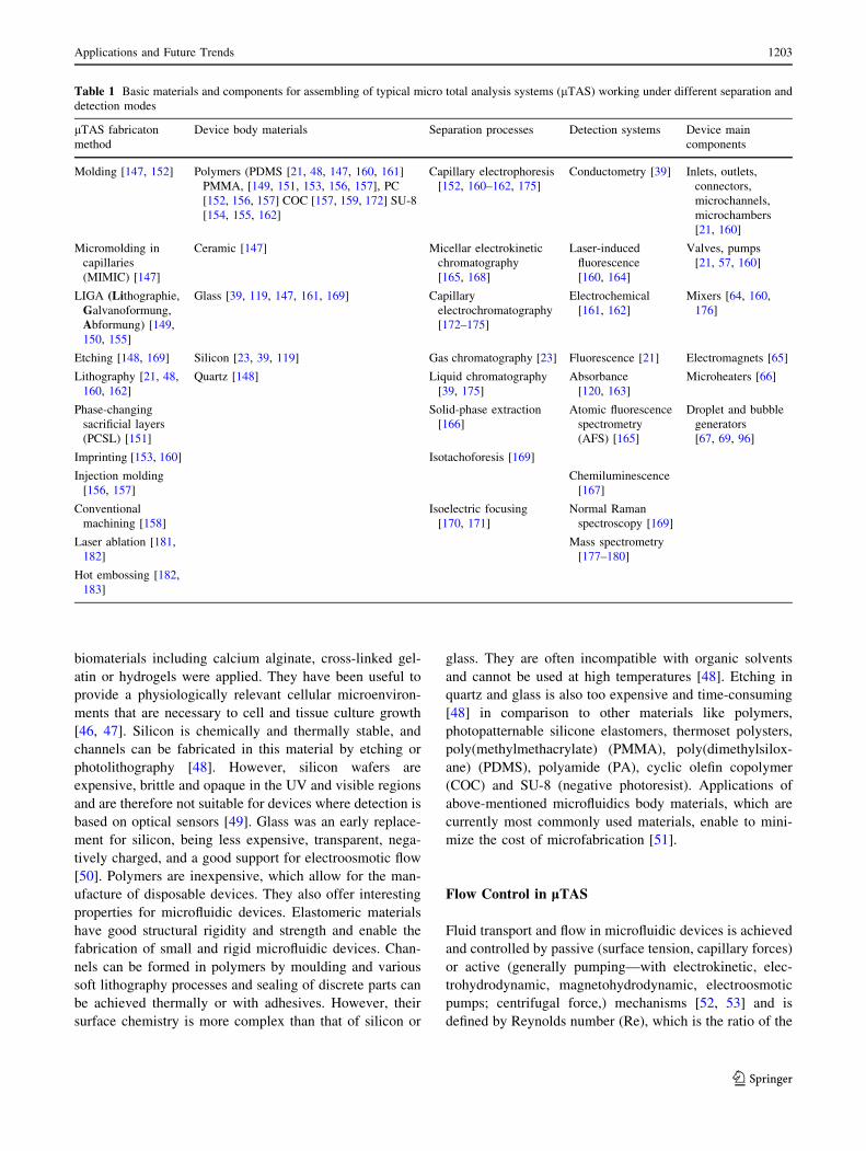
Table 1 Basic materials and components for assembling of typical micro total analysis systems (lTAS) working under different separation and
detection modes
lTAS fabricaton
method
Device body materials Separation processes Detection systems Device main
components
Molding [147, 152] Polymers (PDMS [21, 48, 147, 160, 161]
PMMA, [149, 151, 153, 156, 157], PC
[152, 156, 157] COC [157, 159, 172] SU-8
[154, 155, 162]
Capillary electrophoresis
[152, 160–162, 175]
Conductometry [39] Inlets, outlets,
connectors,
microchannels,
microchambers
[21, 160]
Micromolding in
capillaries
(MIMIC) [147]
Ceramic [147] Micellar electrokinetic
chromatography
[165, 168]
Laser-induced
fluorescence
[160, 164]
Valves, pumps
[21, 57, 160]
LIGA (Lithographie,
Galvanoformung,
Abformung) [149,
150, 155]
Glass [39, 119, 147, 161, 169] Capillary
electrochromatography
[172–175]
Electrochemical
[161, 162]
Mixers [64, 160,
176]
Etching [148, 169] Silicon [23, 39, 119] Gas chromatography [23] Fluorescence [21] Electromagnets [65]
Lithography [21, 48,
160, 162]
Quartz [148] Liquid chromatography
[39, 175]
Absorbance
[120, 163]
Microheaters [66]
Phase-changing
sacrificial layers
(PCSL) [151]
Solid-phase extraction
[166]
Atomic fluorescence
spectrometry
(AFS) [165]
Droplet and bubble
generators
[67, 69, 96]
Imprinting [153, 160] Isotachoforesis [169]
Injection molding
[156, 157]
Chemiluminescence
[167]
Conventional
machining [158]
Isoelectric focusing
[170, 171]
Normal Raman
spectroscopy [169]
Laser ablation [181,
182]
Mass spectrometry
[177–180]
Hot embossing [182,
183]
Applications and Future Trends 1203
123
active forces (inertial forces) to the passive forces of
internal friction in the fluid, appearing as a dynamic
viscosity:
Re ¼ qvd
l
where q is the density of the fluid (kg/m3), v is the mean
fluid velocity (m/s), d is the channel diameter and l the
dynamic viscosity of the channel (kg/m*s). When
Re \\ 1, the flow is laminar and very smooth. When
Re [ 103, the flow is turbulent and mainly characterized by
vortices [54]. Flow within microstructures typically has
Reynolds numbers of 10-3–10-5 and is characterized by a
laminar flow. Contrary to fluid dynamic in larger scales
devices, in microfluidic devices viscous forces dominate
and turbulences are non-existent. Surface tension can be an
important driving force and mixing is slow and occurs
through diffusion [55]. Microvalves allow controlling flow
and can be segregated in two categories: passive valves
(which do not require mechanical actuation) and active
valves (which do) (Fig. 1). Typical passive valves are
cantilever valves, diaphragm valves, diffuser/nozzle valves
[56]. Actuation of active valves is generally piezoelectric,
thermopneumatic, electrostatic and electromagnetic, but
pneumatically actuated, flexible membrane-based valves
(and pumps) are most popular forms of active elements in
microfluidic devices [56, 57].
For many biological and chemical applications, mixing
of transported fluids in microchannels is very important.
Mixers are therefore essential in enhancing mixing effi-
ciency and for rapid homogenization of the reagents. All
mixing ultimately occurs due to molecular diffusion and
therefore the basic idea is reducing the distance over which
mixing must occur [59]. They can be classified as active
(needed external energy) or passive (mixing in specific
geometry of the channel). Passive mixers are usually easier
to fabricate than active mixers and are more suitable for
applications [60]. Typical active mixers based on elec-
trowetting [61], nonlinear electrokinetic effects [62] and
acoustic streaming [63] are usually complicated to fabri-
cate. However, simple, portable, hand-powered mixer that
exploits movement of bubbles in microchannels was
developed by Garstecki and co-workers [64]. More com-
plicated procedures are needed to incorporate (especially
on polymer-based devices) metal building components into
microfluidic systems, for applications such as on-chip
heating and magnetic sorting. A microsolidics method has
been developed to fabricate complex metallic structures
(like microheaters or magnets) by injecting liquid solder
into microfluidic channels, and allowing the solder to cool
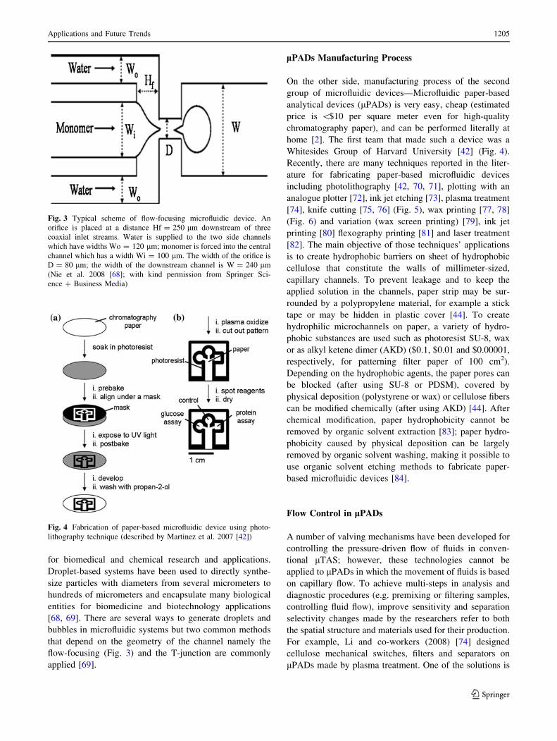
and solidify [65, 66] (Fig. 2).
Microsolidics simplifies the incorporation of metals into
microfluidic channels, but has several disadvantages. They
can only be used with metals (or alloys) characterized by
low-melting point (\300 �C) and affinity for the surface of
the channel wall. These solders are usually more expensive
than commonly used, and some are not biocompatible,
particularly those that contain heavy metals like Pb or Cd.
This method cannot be used to fill ‘‘dead-end’’ channel, and
it is currently difficult to use this process to fabricate wires
with cross-sectional dimensions \10 lm [65, 66].
The other components used in microfluidic devices are
droplet and bubble generators. It has been shown that
droplet and bubble-based microfluidics can perform Bool-
ean logic functions [67]. The use of immiscible fluids for
the formation of emulsions in microfluidic devices in
controlled, individual segments (droplets) enabled rapid
mixing of fluids and is a potent high-throughput platform
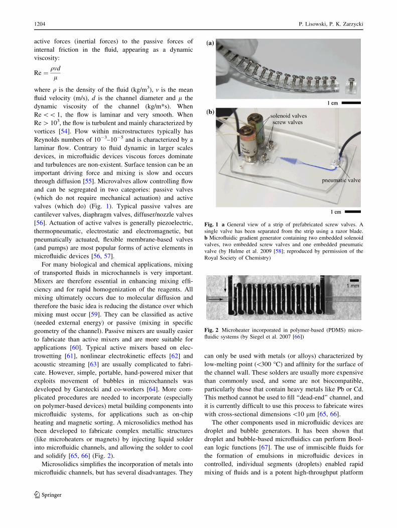
Fig. 1 a General view of a strip of prefabricated screw valves. A
single valve has been separated from the strip using a razor blade.
b Microfluidic gradient generator containing two embedded solenoid
valves, two embedded screw valves and one embedded pneumatic
valve (by Hulme et al. 2009 [58]; reproduced by permission of the
Royal Society of Chemistry)
Fig. 2 Microheater incorporated in polymer-based (PDMS) micro-
fluidic systems (by Siegel et al. 2007 [66])
1204 P. Lisowski, P. K. Zarzycki
123
for biomedical and chemical research and applications.
Droplet-based systems have been used to directly synthe-
size particles with diameters from several micrometers to
hundreds of micrometers and encapsulate many biological
entities for biomedicine and biotechnology applications
[68, 69]. There are several ways to generate droplets and
bubbles in microfluidic systems but two common methods
that depend on the geometry of the channel namely the
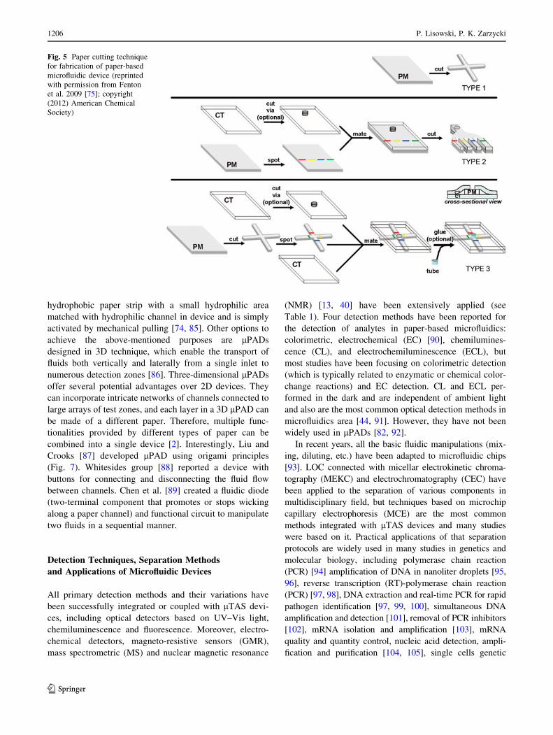
flow-focusing (Fig. 3) and the T-junction are commonly
applied [69].
lPADs Manufacturing Process
On the other side, manufacturing process of the second
group of microfluidic devices—Microfluidic paper-based
analytical devices (lPADs) is very easy, cheap (estimated
price is \$10 per square meter even for high-quality
chromatography paper), and can be performed literally at
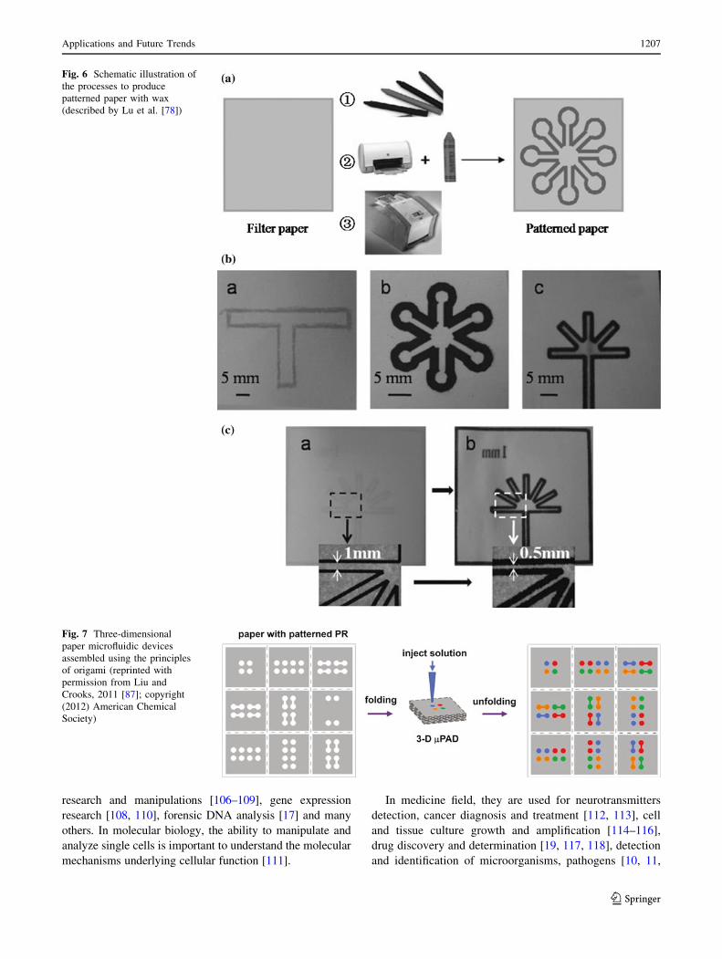
home [2]. The first team that made such a device was a
Whitesides Group of Harvard University [42] (Fig. 4).
Recently, there are many techniques reported in the liter-
ature for fabricating paper-based microfluidic devices
including photolithography [42, 70, 71], plotting with an
analogue plotter [72], ink jet etching [73], plasma treatment
[74], knife cutting [75, 76] (Fig. 5), wax printing [77, 78]
(Fig. 6) and variation (wax screen printing) [79], ink jet
printing [80] flexography printing [81] and laser treatment
[82]. The main objective of those techniques’ applications
is to create hydrophobic barriers on sheet of hydrophobic
cellulose that constitute the walls of millimeter-sized,
capillary channels. To prevent leakage and to keep the
applied solution in the channels, paper strip may be sur-
rounded by a polypropylene material, for example a stick
tape or may be hidden in plastic cover [44]. To create
hydrophilic microchannels on paper, a variety of hydro-
phobic substances are used such as photoresist SU-8, wax
or as alkyl ketene dimer (AKD) ($0.1, $0.01 and $0.00001,
respectively, for patterning filter paper of 100 cm2).
Depending on the hydrophobic agents, the paper pores can
be blocked (after using SU-8 or PDSM), covered by
physical deposition (polystyrene or wax) or cellulose fibers
can be modified chemically (after using AKD) [44]. After
chemical modification, paper hydrophobicity cannot be
removed by organic solvent extraction [83]; paper hydro-
phobicity caused by physical deposition can be largely
removed by organic solvent washing, making it possible to
use organic solvent etching methods to fabricate paper-
based microfluidic devices [84].
Flow Control in lPADs
A number of valving mechanisms have been developed for
controlling the pressure-driven flow of fluids in conven-
tional lTAS; however, these technologies cannot be
applied to lPADs in which the movement of fluids is based
on capillary flow. To achieve multi-steps in analysis and
diagnostic procedures (e.g. premixing or filtering samples,
controlling fluid flow), improve sensitivity and separation
selectivity changes made by the researchers refer to both
the spatial structure and materials used for their production.
For example, Li and co-workers (2008) [74] designed
cellulose mechanical switches, filters and separators on
lPADs made by plasma treatment. One of the solutions is
Fig. 3 Typical scheme of flow-focusing microfluidic device. An
orifice is placed at a distance Hf = 250 lm downstream of three
coaxial inlet streams. Water is supplied to the two side channels
which have widths Wo = 120 lm; monomer is forced into the central
channel which has a width Wi = 100 lm. The width of the orifice is
D = 80 lm; the width of the downstream channel is W = 240 lm
(Nie et al. 2008 [68]; with kind permission from Springer Sci-
ence ? Business Media)
Fig. 4 Fabrication of paper-based microfluidic device using photo-
lithography technique (described by Martinez et al. 2007 [42])
Applications and Future Trends 1205
123
hydrophobic paper strip with a small hydrophilic area
matched with hydrophilic channel in device and is simply
activated by mechanical pulling [74, 85]. Other options to
achieve the above-mentioned purposes are lPADs
designed in 3D technique, which enable the transport of
fluids both vertically and laterally from a single inlet to
numerous detection zones [86]. Three-dimensional lPADs
offer several potential advantages over 2D devices. They
can incorporate intricate networks of channels connected to
large arrays of test zones, and each layer in a 3D lPAD can
be made of a different paper. Therefore, multiple func-
tionalities provided by different types of paper can be
combined into a single device [2]. Interestingly, Liu and
Crooks [87] developed lPAD using origami principles
(Fig. 7). Whitesides group [88] reported a device with
buttons for connecting and disconnecting the fluid flow
between channels. Chen et al. [89] created a fluidic diode
(two-terminal component that promotes or stops wicking
along a paper channel) and functional circuit to manipulate
two fluids in a sequential manner.
Detection Techniques, Separation Methods
and Applications of Microfluidic Devices
All primary detection methods and their variations have
been successfully integrated or coupled with lTAS devi-
ces, including optical detectors based on UV–Vis light,
chemiluminescence and fluorescence. Moreover, electro-
chemical detectors, magneto-resistive sensors (GMR),
mass spectrometric (MS) and nuclear magnetic resonance
(NMR) [13, 40] have been extensively applied (see
Table 1). Four detection methods have been reported for
the detection of analytes in paper-based microfluidics:
colorimetric, electrochemical (EC) [90], chemilumines-
cence (CL), and electrochemiluminescence (ECL), but
most studies have been focusing on colorimetric detection
(which is typically related to enzymatic or chemical color-
change reactions) and EC detection. CL and ECL per-
formed in the dark and are independent of ambient light
and also are the most common optical detection methods in
microfluidics area [44, 91]. However, they have not been
widely used in lPADs [82, 92].
In recent years, all the basic fluidic manipulations (mix-
ing, diluting, etc.) have been adapted to microfluidic chips
[93]. LOC connected with micellar electrokinetic chroma-
tography (MEKC) and electrochromatography (CEC) have
been applied to the separation of various components in
multidisciplinary field, but techniques based on microchip
capillary electrophoresis (MCE) are the most common
methods integrated with lTAS devices and many studies
were based on it. Practical applications of that separation
protocols are widely used in many studies in genetics and
molecular biology, including polymerase chain reaction
(PCR) [94] amplification of DNA in nanoliter droplets [95,
96], reverse transcription (RT)-polymerase chain reaction
(PCR) [97, 98], DNA extraction and real-time PCR for rapid
pathogen identification [97, 99, 100], simultaneous DNA
amplification and detection [101], removal of PCR inhibitors
[102], mRNA isolation and amplification [103], mRNA
quality and quantity control, nucleic acid detection, ampli-
fication and purification [104, 105], single cells genetic
Fig. 5 Paper cutting technique
for fabrication of paper-based
microfluidic device (reprinted
with permission from Fenton
et al. 2009 [75]; copyright
(2012) American Chemical
Society)
1206 P. Lisowski, P. K. Zarzycki
123
research and manipulations [106–109], gene expression
research [108, 110], forensic DNA analysis [17] and many
others. In molecular biology, the ability to manipulate and
analyze single cells is important to understand the molecular
mechanisms underlying cellular function [111].
In medicine field, they are used for neurotransmitters
detection, cancer diagnosis and treatment [112, 113], cell
and tissue culture growth and amplification [114–116],
drug discovery and determination [19, 117, 118], detection
and identification of microorganisms, pathogens [10, 11,
Fig. 6 Schematic illustration of
the processes to produce
patterned paper with wax
(described by Lu et al. [78])
Fig. 7 Three-dimensional
paper microfluidic devices
assembled using the principles
of origami (reprinted with
permission from Liu and
Crooks, 2011 [87]; copyright
(2012) American Chemical
Society)
Applications and Future Trends 1207
123
97, 99, 119] and proteins [12]. LOC devices combined with
different detectors have been applied to detect environ-
mental pollutants in order to separate metal ions [120–122],
phenols [123, 124], explosives [125, 126] nitroaromatics
[127], organic peroxides [128], and other environmentally
relevant substances [15, 40, 93]. Most of these applications
were performed in aqueous solution, except for one non-
aqueous MCE example developed by the group of Collins
[120] for measuring six toxic metal cations including Cd2?,
Pb2?, Cu2?, Co2?, Ni2?, and Hg2?.
In recent years, the second group of microfluidic devi-
ces—lPADs are a subject of research activities, mostly for
biochemical analysis but also for medical and forensic
diagnostics. In the laboratory, paper filters are commonly
used for chromatography and filtration purposes. One of
the first paper-based diagnostic devices created was for
urinalysis [42]. These devices utilize colorimetric assays to
measure glucose and protein concentration in urine. Car-
rilho et al. [129] designed paper-based plates as a low-cost
alternative to the conventional plastic microliter plates.
This allowed mixing of different analytes for different
assays that were not possible in a plastic plate. Another
important application for paper-based devices is pathogen
and toxin detection. One of the first functioning paper-
based detection devices had been developed by Brennan’s
research group—the paper-based device was able to detect
neurotoxins paraoxon and aflatoxin B1 within five minutes
at low concentrations, *100 and *30 nM, respectively
[130]. Lateral flow paper chromatography and vertical flow
diagnostic sensors based on bioactive paper are recently
used to determine human blood type [8, 131–134]. Bio-
active paper strips are also applied in genetic and bio-
chemical analyses, like DNA detection [135] or ELISA
tests [136, 137].
Currently, most paper-based devices utilize colorimetric
assays, although there have been reports of electrochemical
sensing in paper-based devices for detection of glucose,
lactate, and uric acid in biological samples [90] and heavy
metal detection in water [138, 139]. In this work, a novel
method of electrokinetic sensing in a paper-based micro-
fluidic device was proposed. Chemiluminescence [82] and
electrochemiluminescence [92] detection techniques are
described; however, they have not been widely used in
lPADs. Several studies on bioactive paper [130, 135–137,
140] have proposed a few promising ideas to enhance the
sensitivity and selectivity of the colorimetric detection for
paper-based microfluidic devices.
LOC Development and Future Trends
Most important for our future is environmental protection
and ensuring the people health. Healthcare procedures
(monitoring, control, prevention) and diagnostic tests are
expensive. In poor and development countries infectious
diseases, that would be treatable in a developed nation, are
often deadly. Healthcare clinics, even if they have drugs to
treat a certain illness, often suffer from the lack of diag-
nostic equipment and qualified staff. This creates a need to
develop low-cost, simple to-use, point-of-care (POC)
diagnostic methods for diagnosis and monitoring the
treatment of patients [141]. The same problem—lack of
diagnostic tools—exists in case of environmental moni-
toring. Lab-on-chip technology can be important and vital
component of efforts to improve both a global health
through the development of point-of-care testing devices
[141] and environmental protection trough development
analytical devices for environmental samples. In genetic
research and molecular biology field, understanding the
cell biology will become accessible through high-
throughput single-cell data analysis. For example, multi-
plex quantitative polymerase chain reaction (qPCR) is
limited in the number of reactions. If we wish to measure
100 genes from 100 cells, we need 10,000 reactions.
Microfluidic chips can be used to overcome these limita-
tions by combinatorially mixing the samples and gene
detectors and by performing thousands of reactions in
parallel on a single chip and can provide high mRNA-to-
cDNA efficiency and decreased risk of contamination [110,
142]. It is also possible to isolate and amplify single
chromosomes from a single cell. Fan et al. [109] developed
a microfluidic device capable of separating and amplifying
homologous copies of each chromosome from a single
human metaphase cell in independent chambers. This
enabled them to study the two alleles (or haplotypes) of
each chromosome independently. This method can be used
to obtain accurate haplotype information in personal gen-
ome sequences, to understand meiotic recombination and
to directly study the human leukocyte antigen haplotypes
of an individual. Molecular biologist needs particular
devices for rapid isolation and characterization of single
cells from small biological specimens. Future challenges
include sensitive, high-throughput and simple-to-use mic-
rodevices for characterizing proteins, signaling, epigenetic,
and metabolic states in single cells, and correlating these
measurements with physiological characteristics. Existing
techniques for whole-genome/trancriptome amplification
prior to sequencing suffer from bias and non-specific
products that have to be characterized and eliminated. In
particular, there is a need to develop methods for multi-
plexing samples and precise unbiased counting of mole-
cules, which is possible with using microfluidic devices.
Depending on the chip platform being used, several thou-
sand to several hundred thousand distinct oligonucleotides
can be synthesized on a single chip. In principle, these
massive parallel microarrays can reduce the cost of
1208 P. Lisowski, P. K. Zarzycki
123
oligonucleotides by orders of magnitude [105]. In devel-
oped countries, there are many valued features of diag-
nostic tools, including speed, sensitivity and specificity. In
developing countries, with limited resources, where the
healthcare infrastructure is less well developed, many dif-
ficulties must be overcome to apply the LOC device;
therefore, ease of use must also be considered. In this
case, paper-based devices can be extremely useful.
New techniques for making paper devices and their use in
clinical and environmental laboratory will be evaluated
in accordance with the principles of economics—mainly in
terms of material costs and production in mass produc-
tion—the utility and ease of use. Their advantage is the
simplicity of design and ease of interpretation of test
results. In contrast to more complicated LOC devices, they
are self-contained and independent from the other devices.
These features also do not exclude the possibility of
cooperation lPADs with other units. Their compatibility to
telemedicine, particularly with mobile phone transmission
or interpretation of test results is study [70, 143]. White-
sides’ group in Harvard University showed that an image
of the device can be taken by a camera on a mobile phone
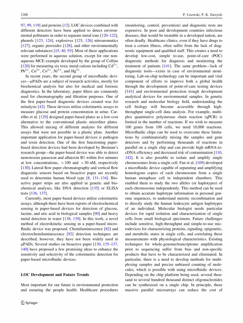
and then sent to a remote location for analysis [70] (Fig. 8).
Liu and Crooks [144] reported, for point-of-care diagnosis,
microelectrochemical biosensing platform that is based on
paper fluidics and powered by an integral metal/air battery.
Vella and co-workers [145] described micropatterned paper
device designed for blood from a fingerstick uses to mea-
suring markers of liver function. These studies are just
beginning to show the potential for paper-based diagnostic
devices for developing countries. However, paper-based
microfluidic devices that rely on complicated instrumen-
tation for result interpretation may only have value for
laboratory uses [44]. The possibility of using paper in
preconcentration is obvious, since it is already widely used
in chromatographic applications and is much cheaper than
conventional materials used in SPE. Furthermore, paper
spray ionization has been recently reported as a direct
sampling ionization method for mass spectrometric analy-
sis [146]. Thus, the use of paper as a platform for pre-
concentration and mass spectrometric analysis can be an
excellent low-cost alternative to the conventional analytical
methods for trace compounds. The combination of paper
spray with miniature mass spectrometers offers a powerful
impetus to wide application of mass spectrometry in non-
laboratory environments [146]. lPADs area research is still
at an early stage and significant efforts will be needed to
nurture it into a more matured platform technology in
diagnostic, point-of-care (POC), and environmental moni-
toring applications [44].
Open Access This article is distributed under the terms of the
Creative Commons Attribution License which permits any use, dis-
tribution, and reproduction in any medium, provided the original
author(s) and the source are credited.
References
1. Reyes DR, Dimitri I, Auroux PA, Manz A (2002) Micro total
analysis systems. 1. Introduction, theory, and technology. Anal
Chem 74:2623–2636
Fig. 8 General strategy for performing inexpensive bioassays in
remote locations and for exchanging the results of the tests with
offsite technicians (reprinted with permission from Martinez et al.
2008 [70]; copyright (2012) American Chemical Society)
Applications and Future Trends 1209
123
2. Martinez AW, Phillips ST, Whitesides GM (2010) Diagnostics
for the developing world: microfluidic paper-based analytical
devices. Anal Chem 82:3–10
3. Whitesides GM (2006) The origins and the future of micro-
fluidics. Nature 442:368–373
4. Craighead H (2006) Future lab-on-a-chip technologies for
interrogating individual molecules. Nature 442:387–393
5. Manz A, Graber N, Widmer HM (1990) Miniaturized total
chemical analysis systems—a novel concept for chemical
sensing. Sens Actuat B-Chem 1:244–248
6. Lauks IR (1998) Microfabricated biosensors and microanalytical
systems for blood analysis. Acc Chem Res 31:317–324
7. Floris J, Staal SS, Lenk SO, Staijen E, Kohlheyer D, Eijkel JCT,
van den Berg A (2010) A prefilled, ready-to-use electrophoresis
based lab-on-a-chip device for monitoring lithium in blood. Lab
Chip 10:1799–1806
8. Khan MS, Thouas G, Shen W, Whyte G, Garnier G (2010) Paper
diagnostic for instantaneous blood typing. Anal Chem 82:4158–
4164
9. Hou HW, Bhagat AAS, Lee WC, Huang S, Han J, Lim CT
(2011) Microfluidic devices for blood fractionation. Microma-
chines 2:319–343
10. Liu RH, Munro SB, Nguyen T, Siuda T, Suciu D, Bizak M,
Slota M, Fuji HS, Danley D, McShea A (2006) Integrated
microfluidic custom array device for bacterial genotyping and
identification. J Assoc Lab Autom 11:360–367
11. Bunyakul N, Edwards KA, Promptmas C, Baeumner AJ (2009)
Cholera toxin subunit B detection in microfluidic devices. Anal
Bioanal Chem 393:177–186
12. Diercks AH, Ozinsky A, Hansen CL, Spotts JM, Rodriguez DJ,
Aderem A (2009) A microfluidic device for multiplexed protein
detection in nano-liter volumes. Anal Biochem 386:30–35
13. Mairhofer J, Roppert K, Ertl P (2009) Microfluidic systems for
pathogen sensing: a review. Sensors 9:4804–4823
14. Marle L, Greenway GM (2005) Microfluidic devices for envi-
ronmental monitoring. Trends Anal Chem 24(9):795–802
15. Li HF, Lin JM (2009) Applications of microfluidic systems in
environmental analysis. Anal Bioanal Chem 393(2):555–567
16. Lafleur JP, Jensen TG, Kutter JP (2011) Gold nanoparticle-based
microfluidic sensor for mercury detection. 15th International
Conference on Miniaturized Systems for Chemistry and Life
Sciences ICMSCLS; October 2–6, Seattle, Washington, USA
17. Hopwood AJ, Hurth C, Yang J, Cai Z, Moran N, Lee-Edghill
JG, Nordquist A, Lenigk R, Estes MD, Haley JP, McAlister CR,
Chen X, Brooks C, Smith S, Elliott K, Koumi P, Zenhausern F,
Tully G (2010) Integrated microfluidic system for rapid forensic
DNA analysis: sample collection to DNA profile. Anal Chem
82(16):6991–6999
18. Shui L, Bomer JG, Jin M, Carlen ET, van den Berg A (2011)
Microfluidic DNA fragmentation for on-chip genomic analysis.
Nanotechnology 22(49):494013–494019
19. Kang L, Chung BG, Langer R, Khademhosseini A (2008)
Microfluidics for drug discovery and development. Drug Discov
Today 13:1–13
20. Sekhon BS, Kamboj S (2010) Microfluidics technology for drug
discovery and development—an overview. Int J Pharm Tech
Res 2(1):804–809
21. Liu C, Wang L, Xu Z, Li J, Ding X, Wang Q, Chunyu L (2012)
A multilayer microdevice for cell-based high-throughput drug
screening. J Micromech Microeng 22:1–7
22. Terry SC (1975) A gas chromatographic air analyser fabricated
on silicon wafer using integrated circuit technology. PhD Thesis.
Stanford CA
23. Terry SC, Jerman JH, Angell JB (1979) A gas chromatographic
air analyzer fabricated on a silicon wafer. IEEE Trans Electron
Devices 26(12):1880–1886
24. Izmailov NA, Shraiber MS (1938) A drop-chromatographic
method of analysis and its applications to pharmacy. Farmatsiya
3:1–7
25. Shostenko YV, Georgievskii VP, Levin MG (2000) History of
the discovery of thin-layer chromatography. J Anal Chem
55(9):904–905
26. Berezkin VG (2007) N.A. Izmailov i M.S. Shraiber: otkrytie
tonkosloinoi khromatografii (N.A. Izmailov and M.S. Shraiber:
the discovery of thin-layer chromatography). GEOS, Moscow
27. Brinkman UATh, de Vries G, van Dalen E (1966) Chromato-
graphic techniques using liquid anion exchangers* III. System-
atic thin-layer chromatography of the elements in HCl systems.
J Chromatogr 23:447–463
28. Merkus FWHM (1969) Inorganic micro-thin-layer chromatog-
raphy. J Chromatogr 41:497–499
29. Svetashev VI, Vaskovsky VE (1972) A simplified technique
for thin-layer microchromatography of lipids. J Chromatogr
67:376–378
30. Vaskovsky VE, Dembitzky VM (1975) Determination of plas-
malogen contents of phospholipid classes by reaction micro-
thin-layer chromatography. J Chromatogr 115:645–647
31. Dembitzky VM (1988) Quantification of plasmalogen, alkylacyl
and diacyl glycerophospholipids by micro-thin-layer chroma-
tography. J Chromatogr 436:467–473
32. Zarzycki PK (2008) Simple horizontal chamber for thermostated
micro-thin-layer chromatography. J Chromatogr A 1187:250–259
33. Zarzycki PK, Zarzycka MB (2008) Application of temperature-
controlled micro planar chromatography for separation and
quantification of testosterone and its derivatives. Anal Bioanal
Chem 391(6):2219–2225
34. Zarzycki PK, Zarzycka MB (2008) Evaluation of the water and
organic liquids extraction efficiency of the spirulina maxima
dyes using thermostated micro-thin-layer chromatography.
J AOAC Int 91(5):1196–1202
35. Zarzycki PK, Ohta H, Harasimiuk FB, Jinno K (2007) Fast
separation and quantification of C60 and C70 fullerenes using
thermostated micro thin-layer chromatography. Anal Sci
23:1391–1396
36. Zarzycki PK, Slaczka MM, Zarzycka MB, Bartoszuk MA,
Włodarczyk E, Baran MJ (2011) Temperature-controlled micro-
TLC: a versatile green chemistry and fast analytical tool for
separation and preliminary screening of steroids fraction from
biological and environmental samples. J Steroid Biochem Mol
Biol 127:418–427
37. Zarzycki PK, Zarzycka MB, Clifton VL, Adamski J, Głod BK
(2011) Low parachor solvents extraction and thermostated
micro-TLC separation for fast screening and classification of
spirulina from pharmaceutical formulations and food samples.
J Chromatogr A 1218:5694–5704
38. Zarzycki PK, Slaczka MM, Zarzycka MB, Włodarczyk E E,
Baran MJ (2011c) Application of micro-thin-layer chromatog-
raphy as a simple fractionation tool for fast screening of raw
extracts derived from complex biological, pharmaceutical and
environmental samples. Anal Chim Acta 688:168–174
39. Manz A, Miyahara Y, Miura J, Watanabe Y, Miyagi H, Sato K
(1990) Design of an open-tubular column liquid chromatograph
using silicon chip technology. Sensor Actuat B 1:249–255
40. Auroux PA, Iossifidis D, Reyes DR, Manz A (2002) Micro Total
Analysis Systems. 2. Analytical standard operations and appli-
cations. Anal Chem 74:2637–2652
41. Sharma H, Nguyen D, Chen A, Lew V, Khine M (2011)
Unconventional low-cost fabrication and patterning techniques
for point of care diagnostics. Ann Biomed Eng 39(4):1313–1327
42. Martinez AW, Phillips ST, Butte MJ, Whitesides GM (2007)
Patterned paper as a platform for inexpensive, low-volume,
portable bioassays. Angew Chem Int Ed 46:1318–1320
1210 P. Lisowski, P. K. Zarzycki
123
43. Muller RH, Clegg DL (1949) Automatic paper chromatography.
Anal Chem 21(9):1123–1125
44. Li X, Ballerini DR, Shen W (2012) A perspective on paper-
based microfluidics: current status and future trends. Biomi-
crofluidics 6:011301–011313
45. Vilkner T, Janasek D, Manz A (2004) Micro total analysis
systems recent developments. Anal Chem 76:3373–3385
46. Braschler T, Valero A, Colella L, Pataky K, Brugger J, Renaud
P (2010) Fluidic microstructuring of alginate hydrogels for the
single cell niche. Lab Chip 10:2771–2777
47. Chueh BH, Zheng Y, Torisawa YS, Hsiao AY, Ge C, Hsiong S,
Huebsch N, Franceschi R, Mooney DJ, Takayama S (2010)
Patterning alginate hydrogels using light-directed release of
caged calcium in a microfluidic device. Biomed Microdevices
12(1):145–151
48. McDonald JC, Duffy DC, Anderson JR, Chiu DT, Wu H,
Schueller OJ, Whitesides GM (2000) Fabrication of microfluidic
systems in poly(dimethylsiloxane). Electrophoresis 21:27–40
49. Shoji S, Esashi M (1994) Microflow devices and systems.
J Micromech Microeng 4(4):157–171
50. Yao SH, Hertzog DE, Zeng SL, Mikkelsen JC, Santiago JG
(2003) Porous glass electroosmotic pumps: design and experi-
ments. J Colloid Interface Sci 268:143–153
51. Whitesides GM (2003) The ‘right’ size in nanobiotechnology.
Nat Biotechnol 21:1161–1165
52. Laser DJ, Santiago JG (2004) A review of micropumps.
J Micromech Microeng 14:r35–r64
53. Fiorini GS, Chiu DT (2005) Disposable microfluidic devices:
fabrication, function, and application. Biotechniques 38:429–446
54. Kundu PK, Cohen IM (2008) Fluid Mechanics, 4th edn. Aca-
demic Press, Burlington
55. Purcell EM (1977) Life at low Reynolds numbers. Am J Phys
45:3–11
56. Koch M, Evans A, Brunnschweiler A (2000) Microfluidic
Technology and Applications. Research Studies Press, Baldock
57. Amirouche F, Zhou Y, Johnson T (2009) Current micropump
technologies and their biomedical applications. Microsyst
Technol 15:647–666
58. Hulme SE, Shevkoplyas SS, Whitesides GM (2009) Incorpora-
tion of prefabricated screw, pneumatic, and solenoid valves into
microfluidic devices. Lab Chip 9(1):79–86
59. Squires TM, Quake SR (2005) Microfluidics: fluid physics at the
nanoliter scale. Rev Mod Phys 77:977–1026
60. Sudarsan AP, Ugaz VM (2006) Multivortex micromixing. Proc
Natl Acad Sci U. S. A. 103:7228–7233
61. Paik P, Pamula VK, Pollack MG, Fair RB (2003) Electrowett-
ing-based droplet mixers for microfluidic systems. Lab Chip
3:28–33
62. Takhistov P, Duginova K, Chang HC (2003) Electrokinetic
mixing vortices due to electrolyte depletion at microchannel
junctions. J Colloid Interface Sci 263:133–143
63. Yang Z, Matsumoto S, Goto H, Matsumoto M, Maeda R (2001)
Ultrasonic micromixer for microfluid systems. Sens Actuators A
93:266–272
64. Garstecki P, Fuerstman MJ, Fischbach MA, Sia SK, Whitesides
GM (2006) Mixing with bubbles: a practical technology for use
with portable microfluidic devices. Lab Chip 6:207–212
65. Siegel AC, Shevkoplyas SS, Weibel DB, Bruzewicz DA, Mar-
tinez AW, Whitesides GM (2006) Cofabrication of electro-
magnets and microfluidic systems in poly(dimethylsiloxane).
Angew Chem Int Ed 45:6877–6882
66. Siegel AC, Bruzewicz DA, Weibel DB, Whitesides GM (2007)
Microsolidics: fabrication of three-dimensional metallic micro-
structures in poly(dimethylsiloxane). Adv Mater 19:727–733
67. Prakash M, Gershenfeld N (2007) Microfluidic bubble logic.
Science 315:832–835
68. Nie Z, Seo MS, Xu S, Lewis PC, Mok M, Kumacheva E,
Whitesides GM, Garstecki P, Stone HA (2008) Emulsification in
a microfluidic flow-focusing device: effect of the viscosities of
the liquids. Microfluid Nanofluid 5:585–594
69. Teh SY, Lin R, Hung LH, Lee AP (2008) Droplet microfluidics.
Lab Chip 2:198–220
70. Martinez AW, Scott TP, Carrilho E, Thomas SW III, Sindi H,
Whitesides GM (2008) Simple telemedicine for developing
regions: camera phones and paper-based microfluidic devices
for real-time, off-site diagnosis. Anal Chem 80(10):3699–3707
71. Klasner SA, Price AK, Hoeman KW, Wilson RS, Bell KJ,
Culbertson CT (2010) Paper-based microfluidic devices for
analysis of clinically relevant analytes present in urine and
saliva. Anal Bioanal Chem 397:1821–1829
72. Bruzewicz DA, Reches M, Whitesides GM (2008) Low-cost
printing of poly(dimethylsiloxane) barriers to define micro-
channels in paper. Anal Chem 80:3387–3392
73. Abe K, Kotera K, Suzuki K, Citterio D (2008) Inkjet-printed
microfluidic multianalyte chemical sensing paper. Anal Chem
80(18):6928–6934
74. Li X, Tian J, Nguyen TH, Shen W (2008) Paper-based microflu-
idic devices by plasma treatment. Anal Chem 80(23):9131–9134
75. Fenton EM, Mascarenas MR, Lopez GP, Sibbett SS (2009)
Multiplex lateral-flow test strips fabricated by two-dimensional
shaping. ACS Appl Mater Interfaces 1(1):124–129
76. Wang W, Wu WY, Zhu JJ (2010) Tree-shaped paper strip for
semiquantitative colorimetric detection of protein with self-
calibration. J Chromatogr A 1217(24):3896–3899
77. Carrilho E, Martinez AW, Whitesides GM (2009) Understand-
ing wax printing: a simple micropatterning process for paper-
based microfluidics. Anal Chem 81:7091–7095
78. Lu Y, Shi W, Jiang L, Qin J, Lin B (2009) Rapid prototyping of
paper-based microfluidics with wax for low-cost, portable bio-
assay. Electrophoresis 30:1497–1500
79. Dungchai W, Chailapakul O, Henry CS (2011) A low-cost,
simple, and rapid fabrication method for paper-based micro-
fluidics using wax screen-printing. Analyst 136(1):77–82
80. Li X, Tian J, Garnier G, Shen W (2010) Fabrication of paper-
based microfluidic sensors by printing. Colloids Surf B
76(2):564–570
81. Olkkonen J, Lehtinen K, Erho T (2010) Flexographically printed
fluidic structures in paper. Anal Chem 82(24):10246–10250
82. Chitnis G, Ding Z, Chang CL, Savran CA, Ziaie B (2011) Laser-
treated hydrophobic paper: an inexpensive microfluidic plat-
form. Lab Chip 11:1161–1165
83. Shen W, Filonanko Y, Truong Y. Parker IH, Brack N, Pigram P,
Liesegang J (2000) Contact angle measurement and surface
energetics of sized and unsized paper. Colloids Surf A 173(1-
3):117-126
84. Abe K, Kotera K, Suzuki K, Citterio D (2010) Inkjet-printed
paperfluidic immuno-chemical sensing device. Anal Bioanal
Chem 398:885–893
85. Shen W, Li X, Tian J, Nguyen TH, Gil G (2010) Switches for
microfluidic systems. Pat nr WO 2010(017578):A1
86. Martinez AW, Phillips ST, Whitesides GM (2008) Three-
dimensional microfluidic devices fabricated in layered paper and
tape. Proc Natl Acad Sci USA 105(50):19606–19611
87. Liu H, Crooks RM (2011) Three-dimensional paper microfluidic
devices assembled using the principles of origami. J Am Chem
Soc 133:17564–17566
88. Martinez AW, Phillips ST, Nie Z, Cheng CM, Carrilho E, Wiley
BJ, Whitesides GM (2010) Programmable diagnostic devices
made from paper and tape. Lab Chip 10(19):2499–2504
89. Chen H, Cogswell J, Anagnostopoulos, Faghri M (2012) A
fluidic diode, valves, and a sequential-loading circuit fabricated
on layered paper. Lab Chip 12:2909–2913
Applications and Future Trends 1211
123
90. Dungchai W, Chailapakul O, Henry CS (2009) Electrochemical
detection for paper-based microfluidics. Anal Chem 81:5821–5826
91. Baker CA, Duong CT, Grimley A, Roper MG (2009) Recent
advances in microfluidic detection systems. Bioanalysis
1(5):967–975
92. Delaney JL, Hogan CF, Tian J, Shen W (2011) Electrogenerated
chemiluminescence detection in paper-based microfluidic sen-
sors. Anal Chem 83(4):1300–1306
93. Verpoorte E (2002) Microfluidic chips for clinical and forensic
analysis. Electrophoresis 23:672–712
94. Kopp MU, de Mello AJ, Manz A (1998) Chemical amplifica-
tion: continuous-flow PCR on a chip. Science 280:1046–1048
95. Waters LC, Jacobso SC, Kroutchinina N, Khandurina J, Foote
RS, Ramsey JM (1998) Multiple sample PCR amplification and
electrophoretic analysis on a microchip. Anal Chem 70:5172–
5176
96. Kumaresan P, Yang CJ, Cronier SA, Blazej RG, Mathies RA
(2008) High-throughput single copy DNA amplification and cell
analysis in engineered nanoliter droplets. Anal Chem
80:3522–3529
97. Lee SH, Kim SW, Kang JY, Ahn CH (2008) A polymer lab-on-
a-chip for reverse transcription (RT)-PCR based point-of-care
clinical diagnostics. Lab Chip 8:2121–2127
98. Kim YT, Chen Y, Choi JY, Kim WJ, Dae HM, Jung J, Seo TS
(2012) Integrated microdevice of reverse transcription-poly-
merase chain reaction with colorimetric immunochromato-
graphic detection for rapid gene expression analysis of influenza
A H1N1 virus. Biosens Bioelectron 33(1):88–94
99. Lee JG, Cheong KH, Huh N, Kim S, Choi JW, Ko C (2006)
Microchip-based one step DNA extraction and real-time PCR in
one chamber for rapid pathogen identification. Lab Chip
6:886–895
100. Wang Z, Sekulovic A, Kutter JP, Bang DD, Wolff A (2006)
Real-time PCR using a PCR microchip with integrated thermal
system and polymer waveguides for the detection of Campylo-
bacter jejuni. MEMS, Istanbul, pp 542–545
101. Lee TMH, Carles MC, Hsing IM (2003) Microfabricated PCR-
electrochemical device for simultaneous DNA amplification and
detection. Lab Chip 3:100–105
102. Perch-Nielsen IR, Bang DD, Poulsen CR, El-Alia J, Wolff A
(2003) Removal of PCR inhibitors using dielectrophoresis as a
selective filter in a microsystem. Lab Chip 3:212–216
103. Marcus JS, Anderson WF, Quake SR (2006) Microfluidic single-
cell mRNA isolation and analysis. Anal Chem 78:3084–3089
104. Hong JW, Studer V, Hang G, Anderson WF, Quake SR (2004)
A nanoliter-scale nucleic acid processor with parallel architec-
ture. Nat Biotech 22:435–439
105. Lee CC, Snyder TM, Quake SR (2010) A microfluidic oligo-
nucleotide synthesizer. Nucleic Acids Res 38(8):2514–2521
106. Hong JW, Chen Y, Anderson WF, Quake SR (2006) Molecular
biology on a microfluidic chip. J Cond Matt Phys 18:691–701
107. Marcy Y, Ouverney C, Blik EM, Losekann T, Ivanova N,
Martin HG, Szeto E, Platt D, Hugenholtz P, Relman DA, Quake
SR (2007) Dissecting biological ‘‘dark matter’’ with single cell
genetic analysis of rare and uncultivated TM7 microbes from the
human mouth. PNAS 104(29):1889–11894
108. Diehn M, Cho RW, Lobo NA, Kalisky T, Dorie MJ et al (2009)
Association of reactive oxygen species levels and radioresis-
tance in cancer stem cells. Nature 458:780–783
109. Fan HC, Wang J, Potanina A, Quake SR (2011) Whole-genome
molecular haplotyping of single cells. Nat Biotechnol 29:51–57
110. Spurgeon SL, Jones RC, Ramakrishnan R (2008) High
throughput gene expression measurement with real time PCR in
a microfluidic dynamic array. PLoS One 3(2):e1662
111. Sims CE, Allbritton NL (2007) Analysis of single mammalian
cells on-chip. Lab Chip 7:423–440
112. Tan SJ, Yobas L, Lee GY, Ong CN, Lim CT (2009) Microde-
vice for the isolation and enumeration of cancer cells from
blood. Biomed Microdevices 11:883–892
113. Mohamed H, Murray M, Turner JN, Caggana M (2009) Isolation
of tumor cells using size and deformation. J Chromatogr A
1216(47):8289–8295
114. El-Ali J, Sorger PK, Jensen KF (2006) Cells on chips. Nature
442:403–411
115. Hufnagel H, Huebner A, Gulch C, Guse K, Abell C, Hollfelder F
(2009) An integrated cell culture lab on a chip: modular mic-
rodevices for cultivation of mammalian cells and delivery into
microfluidic microdroplets. Lab Chip 9:1576–1582
116. Ni M, Tong WH, Choudhury D, Rahim NAA, Iliescu C, Yu H
(2009) Cell culture on MEMS platforms: a review. Int J Mol Sci
10:5411–5544
117. Chiem NH, Harrison DJ (1998) Microchip systems for immu-
noassay: an integrated immunoreactor with electrophoretic
separation for serum theophylline determination. Clin Chem
44(3):591–598
118. Winkle RF, Nagy JM, Cass AEG, Sharma S (2008) Towards
microfluidic technology-based MALDI-MS platforms for drug
discovery: a review. Exp Opin Drug Discov 3:1281–1292
119. Pal R, Yang M, Lin R, Johnson BN et al (2005) An integrated
microfluidic device for influenza and other genetic analyses. Lab
Chip 5:1024–1032
120. Deng G, Collins GE (2003) Nonaqueous based microchip sep-
aration of toxic metal ions using 2-(5-bromo-2-pyridylazo)-
5-(N-propyl-N-sulfopropylamino)phenol. J Chromatogr A 989:
311–316
121. Qu S, Chen X, Chen D, Yang P, Chen G (2006) Poly(methyl
methacrylate) CE microchips replicated from poly(dimethylsi-
loxane) templates for the determination of cations. Electropho-
resis 27:4910–4918
122. Kou S, Nam SW, Shumi W, Lee MH, Bae SW, Du J, Kim JS,
Hong JI, Peng X, Yoon J, Park S (2009) Microfluidic detection
of multiple heavy metal ions using fluorescent chemosensors.
Bull Korean Chem Soc 30(5):1173–1176
123. Wang J, Chatrathi MP, Tian B (2000) Capillary electrophoresis
microchips with thick-film amperometric detectors: separation
and detection of phenolic compounds. Anal Chim Acta
416:9–14
124. Ding Y, Garcia CD (2006) Pulsed amperometric detection with
poly(dimethylsiloxane)-fabricated capillary electrophoresis
microchips for the determination of EPA priority pollutants.
Analyst 131:208–214
125. Wang J, Chen G, Chatrathi MP, Musameh M (2004) Capillary
electrophoresis microchip with a carbon nanotube-modified
electrochemical detector. Anal Chem 76:298–302
126. Pumera M (2008) Trends in analysis of explosives by micro-
chip electrophoresis and conventional CE. Electrophoresis
29:269–273
127. Yao X, Wang J, Zhang L, Yang P, Chen G (2006) A three-
dimensionally adjustable amperometric detector for microchip
electrophoretic measurement of nitroaromatic pollutants.
Talanta 69:1285–1291
128. Wang J, Escarpa A, Pumera M, Feldman J (2002) Capillary
electrophoresis-electrochemistry microfluidic system for the
determination of organic peroxides. J Chromatogr A 952:
249–254
129. Carrilho E, Phillips ST, Vella SJ, Martinez AW, Whitesides GM
(2009) Paper microzone plates. Anal Chem 81:5990–5998
130. Hossain SMZ, Luckham RE, Smith AM, Lebert JM, Davies LM,
Pelton RH, Filipe CDM, Brennan JD (2009a) Development of a
bioactive paper sensor for detection of neurotoxins using pie-
zoelectric inkjet printing of sol–gel-derived bioinks. Anal Chem
81:5474–5483
1212 P. Lisowski, P. K. Zarzycki
123
131. Al-Tamimi M, Shen W, Rania Z, Huy T, Garnier G (2012)
Validation of paper-based assay for rapid blood typing. Anal
Chem 84:1661–1668
132. Li M, Tian J, Al-Tamimi M, Shen W (2012) Paper-based blood-
typing device that reports patient’s blood type ‘‘in writing’’.
Angew Chem Int Ed 51:5497–5501
133. Jarujamrus P, Tian J, Li X, Siripinyanond A, Shiowatana J, Shen
W (2012) Mechanisms of antibody and red blood cell interactions
in paper-based blood typing devices. Analyst 137:2205–2210
134. Su J, Al-Tamimi M, Garnier G (2012) Engineering paper as a
substrate for blood typing bio-diagnostics. Cellulose 19:1749–
1758
135. Ali MM, Aguirre SD, Xu Y, Filipe CDM, Pelton R, Li Y (2009)
Detection of DNA using bioactive paper strips. Chem Commun
43:6640–6642
136. Cheng C-M, Martinez AW, Gong J, Mace CR, Phillips ST,
Carrilho E, Mirica KA, Whitesides GM (2010) Paper-based
ELISA. Angew Chem Int Ed 49:4771–4774
137. Curry PS, Elkin BT, Campbell M, Nielsen K, Hutchins W,
Ribble C, Kutz SJ (2011) Filter-paper blood samples for ELISA
detection of Brucella antibodies in caribou. J Wildl Dis
47(1):12–20
138. Nie ZH, Nijhuis CA, Gong J, Chen X, Kumachev A, Martinez
AW, Narovlyansky M, Whitesides GM (2010) Electrochemical
sensing in paper-based microfluidic devices. Lab Chip
10(4):477-4
139. Shi J, Tang F, Xing H, Zheng H, Bi L, Wang W (2012) Elec-
trochemical detection of Pb and Cd in paper-based microfluidic
devices. J Braz Chem Soc 23(6):1124–1130
140. Hossain SMZ, Luckham RE, McFadden MJ, Brennan JD (2009)
Reagentless bidirectional lateral flow bioactive paper sensors for
detection of pesticides in beverage and food samples. Anal
Chem 81:9055–9064
141. Yager P, Domingo GJ, Gerdes J (2008) Point-of-Care diagnos-
tics for global health. Annu Rev Biomed Eng 10:107–144
142. Liu J, Hansen C, Quake SR (2003) Solving the ‘‘world-to-chip’’
interface problem with a microfluidic matrix. Anal Chem
75:4718–4723
143. Li X, Tian J, Shen W (2010) Quantitative biomarker assay with
microfluidic paper-based analytical devices. Anal Bioanal Chem
396:495–501
144. Liu H, Crooks RM (2012) Paper-based electrochemical sensing
platform with integral battery and electrochromic read-out. Anal
Chem 84(5):2528–2532
145. Vella SJ, Beattie P, Cademartiri R, Laromaine A, Martinez AW,
Phillips ST, Mirica KA, Whitesides GM (2012) Measuring
markers of liver function using a micropatterned paper device
designed for blood from a fingerstick. Anal Chem 84:2883–2891
146. Liu J, Wang H, Manicke NE, Lin JM, Cooks RG, Ouyang Z
(2010) Development, characterization, and application of paper
spray ionization. Anal Chem 82(6):2463–2471
147. Kim E, Xia Y, Whitesides GM (1996) Micromolding in capil-
laries: applications in materials science. J Am Chem Soc
118:5722–5731
148. Danel JS, Delapierre G (1991) Quartz: a material for microde-
vices. J Micromech Microeng 1:187–198
149. Becker EW, Ehrfeld W, Hagmann P, Maner A, Munchmeyer D
(1986) Fabrication of microstructures with high aspect ratios and
great structural heights by synchrotron radiation lithography,
galvanoforming, and plastic moulding (LIGA process). Micro-
electron Eng 4(1):35–56
150. Ehrfeld W, Hessel V, Lowe H, Schulz C, Weber L (1999)
Materials of LIGA technology. Microsys Tech 5(3):105–112
151. Fuentes HV, Wolley AT (2008) Phase-changing sacrificial layer
fabrication of multilayer polymer microfluidic devices. Anal
Chem 80:333–339
152. Liu Y, Ganser D, Schneider A, Liu R, Grodzinski P, Krout-
chinina N (2001) Microfabricated polycarbonate CE devices for
DNA analysis. Anal Chem 73(17):4196–4201
153. Tseng WL, Lin YW, Chen KC, Chang HT (2002) DNA analysis
on microfabricated electrophoretic devices with bubble cells.
Electrophoresis 23:2477–2484
154. Agirregabiria M, Blanco FJ, Berganzo J, Arroyo MT, Fullaondo
A, Mayora K, Ruano-Lopez JM (2005) Fabrication of SU-8
multilayer microstructures based on successive CMOS com-
patible adhesive bonding and releasing steps. Lab Chip
5:545–552
155. Zhang J, Tan KL, Hong GD, Yang LJ, Gong HQ (2001) Poly-
merization optimization of SU-8 photoresist and its applications
in microfluidic systems and MEMS. J Micromech Microeng
11:20–26
156. Attia UM, Marson S, Alcock JR (2009) Micro-injection
moulding of polymer microfluidic devices. Microfluid Nanofluid
7:1–28
157. Piotter V, Mueller K, Plewa K, Ruprecht R, Hausselt J (2002)
Performance and simulation of thermoplastic micro injection
molding. Microsys Tech 8:387–390
158. Schaller Th, Bohn L, Mayer J, Schubert K (1999) Microstruc-
ture grooves with a width of less than 50 mm cut with ground
hard metal micro end mills. Prec Eng 23:229–235
159. Stachowiak TB, Mair DA, Holden TG, Lee LJ, Svec F, Frechet
JMJ (2007) Hydrophilic surface modification of cyclic olefin
copolymer microfluidic chip using sequential photografting.
J Sep Sci 30:1088–1093
160. Duffy DC, McDonald JC, Schueller OJA, Whitesides GM
(1998) Rapid prototyping of microfluidic systems in poly
(dimethylsiloxane). Anal Chem 70:4974–4984
161. Jang L, Lu Y, Dai Z, Xie M, Lin B (2005) Mini-electrochemical
detector for microchip electrophoresis. Lab Chip 5:930–934
162. Alvarez-Castano M, Fernandez-Abedul MT, Costa-Garcıa A,
Agirregabiria M, Fernandez LJ, Ruano-Lopez JM, Barredo-Presa
B (2009) Fabrication of SU-8 based microchip electrophoresis
with integrated electrochemical detection for neurotransmitters.
Talanta 80:24–30
163. O’Toole M, Diamond D (2008) Absorbance based light emitting
diode optical sensors and sensing devices. Sensors 8:2453–2479
164. Chang I-H, Tulock JJ, Liu J, Kim W-S, Cannon DM Jr, Lu Y,
Bohn PW, Sweedler JV, Croperk DM (2005) Miniaturized lead
sensor based on lead-specific DNAzyme in a nanocapillary
interconnected microfluidic device. Environ Sci Technol
39:3756–3761
165. Li F, Wang DD, Yan XP, Su RG, Lin JM (2005) Speciation
analysis of inorganic arsenic by microchip capillary electro-
phoresis coupled with hydride generation atomic fluorescence
spectrometry. J Chromatogr A 1081:232–237
166. Masaki H, Hoshi J, Amano S, Sasaki Y, Korenaga T (2006)
Integration of the cleanup process: pretreatment of polycyclic
aromatic hydrocarbons from diesel exhaust particles on silica
gel beads in a microchannel. Anal Sci 22:345–348
167. Som-aum W, Li H, Liu J, Lin JM (2008) Determination of
arsenate by sorption preconcentration on polystyrene beads
packed in a microfluidic device with chemiluminescence
detection. Analyst 133:1169–1175
168. Moore AW, Jacobson SC, Ramsey JM (1995) Microchip sepa-
rations of neutral species via micellar electrokinetic capillary
chromatography. Anal Chem 67:4184–4189
169. Walker PA, Morris MD, Burns MA, Johnson BN (1998) Iso-
tachophoretic separation on a microchip Normal Raman spec-
troscopy detection. Anal Chem 70:3766–3769
170. Hofmann O, Che D, Cruickshank KA, Muller UR (1999)
Adaptation of capillary isoelectric focusing to microchannels on
a glass chip. Anal Chem 71:678–686
Applications and Future Trends 1213
123
171. Huang T, Pawliszyn J (2002) Microfabrication of a tapered
channel for isoelectric focusing with thermally generated pH
gradient. Electrophoresis 20:3504–3510
172. Ladner Y, Cretier G, Faure K (2010) Electrochromatography in
cyclic olefin copolymer microchips: a step towards field portable
analysis. J Chromatogr A 51:8001–8008
173. Broyles BS, Jacobson SC, Ramsey JM (2003) Sample filtration,
concentration, and separation integrated on microfluidic devices.
Anal Chem 75(11):2761–2767
174. Li HF, Zeng H, Chen Z, Lin JM (2009) Chip-based enantiose-
lective open-tubular capillary electrochromatography using
bovine serum albumin-gold nanoparticle conjugates as the sta-
tionary phase. Electrophoresis 30(6):1022–1029
175. Regnier FE, He B, Lin S, Busse J (1999) Chromatography and
electrophoresis on chips: critical elements of future integrated,
microfluidic analytical systems for life science. Trends Bio-
technol 17(3):101–106
176. Stroock AD, Dertinger SKW, Ajdari A, Mezit I, Stone HA,
Whitesides GM (2002) Chaotic mixer for microchannels. Sci-
ence 295:647–651
177. Lee J, Soper SA, Murray KK (2009) Microfluidic chips for mass
spectrometry-based proteomics. J Mass Spectrom 44(5):579–593
178. Figeys D, Gygi SP, McKinnon G, Aebersold R (1998) An
integrated microfluidics tandem mass spectrometry system for
automated protein analysis. Anal Chem 70:3728–3734
179. Jiang Y, Wang PC, Locascio LE, Lee CS (2001) Integrated
plastic microfluidic devices with ESI-MS for drug screening and
residue analysis. Anal Chem 73:2048–2053
180. Gao J, Xu JD, Locascio LE, Lee CS (2001) Integrated micro-
fluidic system enabling protein digestion, peptide separation,
and protein identification. Anal Chem 73:2648–2655
181. Wang SH, Lee CH, Chen HP (2006) Thermoplastic micro-
channel fabrication using carbon dioxide laser ablation. J Chro-
matogr A 1111(2):252–257
182. Locascio LE, Ross DJ, Howell PB, Gaitan M (2006) Fabrication
of polymer microfluidic systems by hot embossing and laser
ablation. Methods Mol Biol 339:37–46
183. Becker H, Heim U (2000) Hot embossing as a method for the
fabrication of polymer high aspect ratio structures. Sens Actuat
A 83:130–135
1214 P. Lisowski, P. K. Zarzycki
123