Embed Size (px)
Citation preview
Microcirculation, lymphatic Microcirculation, lymphatic system, specific blood system, specific blood
circulatory systems (fetal circulatory systems (fetal circulation)circulation)
Romana Šlamberová, MD PhDRomana Šlamberová, MD PhDDepartment of Normal, Pathological and Department of Normal, Pathological and
Clinical PhysiologyClinical Physiology

IntroductionIntroduction
Slides from the lecture. Respecting the copyrights it was not
possible to publish pictures showed at the lecture at our website.
© 2006, Romana Slamberova, MD PhD
Microcirculation (1)Microcirculation (1)
The microcirculation is the blood flow through blood vessels smaller than 100 µm (i.e. arterioles, capillaries, and venules).
Function: Transport of cells, oxygen
and other substances to/from the tissues
Regulation of body temperature

Capillary Hydrostatic Capillary Hydrostatic PressurePressure
This pressure drives fluid out of the capillary (i.e., filtration), and is highest at the arteriolar end of the capillary and lowest at the venular end.
Depending upon the organ, the pressure may drop along the length of the capillary (axial pressure gradient) by 15-30 mmHg.
The axial gradient favors filtration at the arteriolar end (where PC is greatest) and reabsorption at the venular end of the capillary (where PC is the lowest).
The average capillary hydrostatic pressure is determined by arterial and venous pressures (PA and PV), and by the ratio of post-to-precapillary resistances (RV/RA).
PC is more sensitive to changes in PV than by changes in PA.
Capillary Osmotic Pressure Capillary Osmotic Pressure
Osmotic pressure is the hydrostatic pressure produced by a solution in a space divided by a differentially permeable membrane due to a differential in the concentrations of solute.
Because the capillary barrier is readily permeable to ions, the osmotic pressure within the capillary is principally determined by plasma proteins that are relatively impermeable.
Therefore, instead of speaking of "osmotic" pressure, this pressure is referred to as the "oncotic".
Albumin generates about 70% of the oncotic pressure. This pressure is typically 25-30 mmHg.
The oncotic pressure increases along the length of the capillary, particularly in capillaries having high net filtration (e.g., in renal glomerular capillaries), because the filtering fluid leaves behind proteins leading to an increase in protein concentration.
Microcirculation (2)Microcirculation (2)
The total length of capillaries in an average adult human is approximately 42 000 km (25,000 miles), this is approx. equator of Earth.
EndotheliumEndothelium (1) (1)
The endothelium (0.5 μm) is the layer of thin specialized epithelium, comprised of a single layer of flat cells that line the interior surface of blood vessels, forming an interface between circulating blood in the lumen and the rest of the vessel wall.
Space between cells 6-7 nm (little bit less than albumin)
Endothelial cells line the entire circulatory system, from the heart (endocardium) to the smallest capillary.
Both blood and lymphatic capillaries are composed of a single layer of endothelial cells.
EndotheliumEndothelium (2) (2)
Function vasoconstriction and vasodilation, and hence the
control of blood pressure blood clotting (thrombosis & fibrinolysis) formation of new blood vessels (angiogenesis) inflammation and swelling (oedema) transit of white blood cells
Pathology Atherosclerosis (patients with diabetes mellitus,
hypertension and hyperlipidemia)
ArteriolesArterioles
An arteriole is a small diameter (<20 μm, up to 5-9 μm) blood vessel that extends and branches out from an artery and leads to capillaries.
Arterioles have thin muscular walls (usually only one to two layers of smooth muscle) and are the primary site of vascular resistance.
In a healthy vascular system the endothelium, inner lining of arterioles and other blood vessels, is smooth and relaxed.
This healthy condition is promoted by the ample production of nitric oxide in the endothelium.
The mean blood pressure in the arteries supplying the body is a result of the interaction between the cardiac output (the volume of blood the heart is pumping per minute) and the vascular resistance, usually termed total peripheral resistance.
Any pathology which constricts blood flow, such as stenosis, will increase total peripheral resistance and lead to hypertension.
Total peripheral resistanceTotal peripheral resistance
Total peripheral resistance refers to the cumulative resistance of the thousands of arterioles in the body, or the lungs, respectively.
It is approximately equal to the resistance of the arterioles, since the arterioles are the chief resistance vessels in the body.
Total Peripheral Resistance = Mean Arterial Pressure / Cardiac Output.
The total peripheral resistance of healthy lung arterioles is typically about 0.15 to 0.20 that of the body, so pulmonary artery mean blood pressures are typically about 0.15 to 0.20 of aortic mean blood pressures.
CapillaryCapillary (1) (1)
Capillaries, are the smallest of a body's blood vessels, measuring 5-10 μm (erythrocytes?).
They connect arteries and veins, and most closely interact with tissues.
Capillaries have walls composed of a single layer of cells, the endothelium.
This layer is so thin that molecules such as oxygen, water and lipids can pass through them by diffusion and enter the tissues.
Waste products such as carbon dioxide and urea can diffuse back into the blood to be carried away for removal from the body.
Capillary permeability can be increased by the release of certain cytokines.
CapillaryCapillary (2) (2)
The "capillary bed" is the network of capillaries supplying an organ.
The more metabolically active the cells, the more capillaries it will require to supply nutrients.
The capillary bed usually carries no more than 25% of the amount of blood it could contain, although this amount can be increased through autoregulation (i.e. active muscle cells) by inducing relaxation of smooth muscle.
Any signalling molecules they release (such as endothelin for constriction and Nitric oxide for dilation) act on the smooth muscle cells in the walls of nearby, larger vessels, e.g. arterioles.
EndothelinEndothelin
Endothelin is a 21-amino acid vasoconstricting peptide that plays a key part in vascular homeostasis = one of the strongest vasoconstrictors.
In a healthy individual a delicate balance between vasoconstriction and vasodilation is maintained by endothelin, calcitonin (vasoconstrictors) and by nitric oxide, prostacyclin (vasodilators).
Overproduction of endothelin can cause pulmonary artery hypertension.

Nitric oxide Nitric oxide
The chemical compound nitric oxide is a gas with chemical formula NO.
In the body, nitric oxide (the 'endothelium-derived relaxing factor', or 'EDRF') is synthesized from arginine and oxygen by various nitric oxide synthase (NOS) enzymes and by sequential reduction of inorganic nitrate.
Function: The endothelium (inner lining) of blood vessels use nitric oxide to
signal the surrounding smooth muscle to relax, thus dilating the artery and increasing blood flow.
Nitric oxide is a key biological messenger, playing a role in a variety of biological processes (vessel dialatation, neurotransmission, penile erections, hair growth / loss).
"Nitro" vasodialators such as nitroglyceric are converted to nitric oxide.
Immune system: generated by macrophages, toxic to bacteria and other human pathogens.
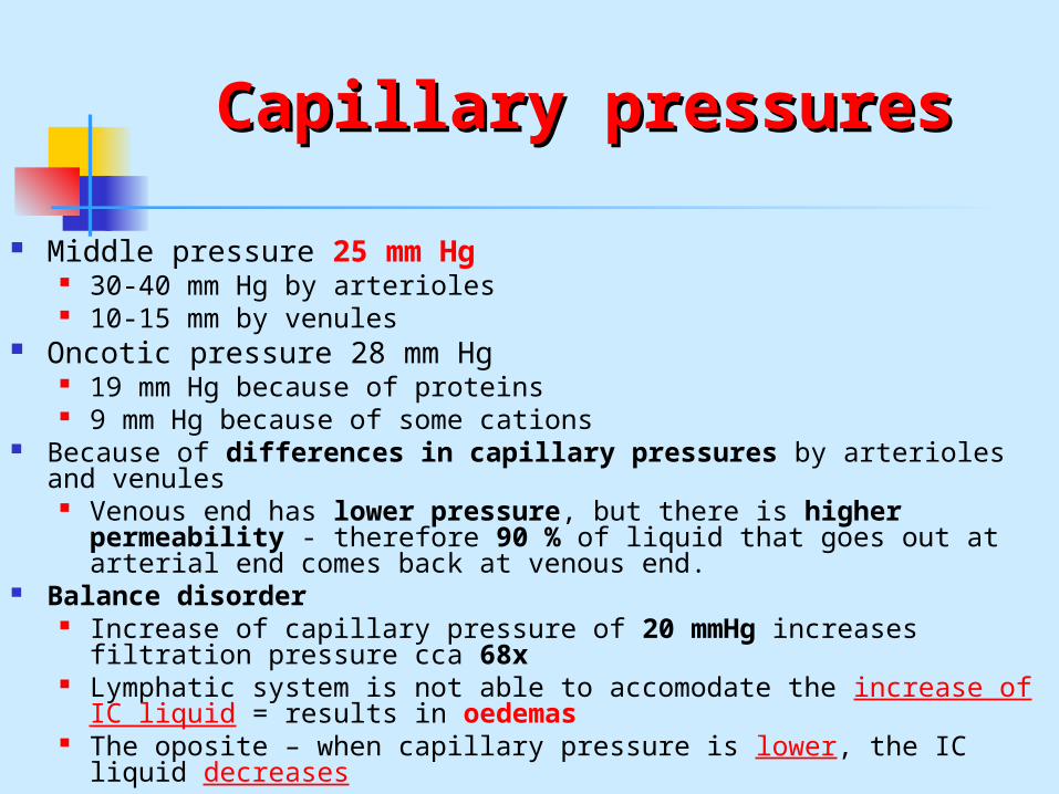
Capillary pressuresCapillary pressures
Middle pressure 25 mm Hg 30-40 mm Hg by arterioles 10-15 mm by venules
Oncotic pressure 28 mm Hg 19 mm Hg because of proteins 9 mm Hg because of some cations
Because of differences in capillary pressures by arterioles and venules
Venous end has lower pressure, but there is higher permeability - therefore 90 % of liquid that goes out at arterial end comes back at venous end.
Balance disorder Increase of capillary pressure of 20 mmHg increases filtration
pressure cca 68x Lymphatic system is not able to accomodate the increase of IC
liquid = results in oedemas The oposite – when capillary pressure is lower, the IC liquid
decreases
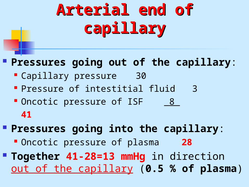
Arterial end of capillaryArterial end of capillary
Pressures going out of the capillary: Capillary pressure 30 Pressure of intestitial fluid 3 Oncotic pressure of ISF 8
41 Pressures going into the capillary:
Oncotic pressure of plasma 28 Together 41-28=13 mmHg in direction
out of the capillary (0.5 % of plasma)
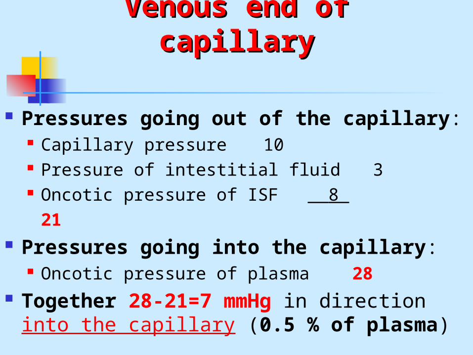
Venous end of capillaryVenous end of capillary
Pressures going out of the capillary: Capillary pressure 10 Pressure of intestitial fluid 3 Oncotic pressure of ISF 8
21 Pressures going into the capillary:
Oncotic pressure of plasma 28 Together 28-21=7 mmHg in direction
into the capillary (0.5 % of plasma)
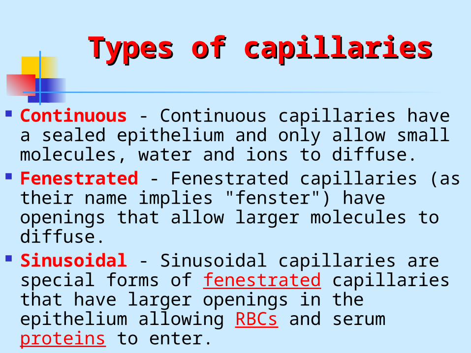
Types of capillariesTypes of capillaries
Continuous - Continuous capillaries have a sealed epithelium and only allow small molecules, water and ions to diffuse.
Fenestrated - Fenestrated capillaries (as their name implies "fenster") have openings that allow larger molecules to diffuse.
Sinusoidal - Sinusoidal capillaries are special forms of fenestrated capillaries that have larger openings in the epithelium allowing RBCs and serum proteins to enter.
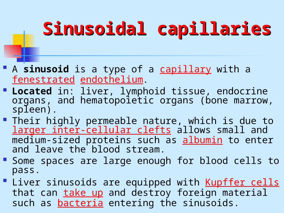
Sinusoidal capillariesSinusoidal capillaries
A sinusoid is a type of a capillary with a fenestrated endothelium.
Located in: liver, lymphoid tissue, endocrine organs, and hematopoietic organs (bone marrow, spleen).
Their highly permeable nature, which is due to larger inter-cellular clefts allows small and medium-sized proteins such as albumin to enter and leave the blood stream.
Some spaces are large enough for blood cells to pass.
Liver sinusoids are equipped with Kupffer cells that can take up and destroy foreign material such as bacteria entering the sinusoids.
VenuleVenuless
A venule is a small blood vessel that allows deoxygenated blood to return from the capillary beds to the larger blood vessels called veins.
Venules have three layers: An inner endothelium composed of squamous
epithelial cells that act as a membrane a middle layer of muscle and elastic tissue (poorly
developed so that venules have thinner walls than arterioles)
an outer layer of fibrous connective tissue.
Lymphatic systemLymphatic system
The lymphatic system is a complex network of lymphoid organs, lymph nodes, lymph ducts, and lymph vessels that produce and transport lymph fluid from tissues to the circulatory system.
The lymphatic system is a major component of the immune system.
Functions: removal of excess fluids from body tissues absorption of fatty acids and subsequent transport of fat
and chyle to the circulatory system production of immune cells (such as lymphocytes,
monocytes, and antibody producing cells called plasma cells)
LymphLymph
Lymph originates as blood plasma that leaks from the capillaries of the circulatory system, becoming interstitial fluid, and filling the space between individual cells of tissue.
Plasma is forced out of the capillaries by hydrostatic pressure, and as it mixes with the interstitial fluid, the volume of fluid accumulates slowly.
Most of the fluid is returned to the capillaries by osmosis (about 90% of the former plasma).
The excess interstitial fluid is collected by the lymphatic system by diffusion into lymph capillaries, and is processed by lymph nodes prior to being returned to the circulatory system.
Once within the lymphatic system the fluid is called lymph, and has almost the same composition as the original interstitial fluid.
Lymphatic circulationLymphatic circulation (1)(1)
The lymphatic system acts as a secondary circulatory system, except that it collaborates with white blood cells in lymph nodes to protect the body from being infected by cancer cells, fungi, viruses or bacteria.
Unlike the circulatory system, the lymphatic system is not closed and has no central pump; the lymph moves slowly and under low pressure due to peristalsis, the operation of semilunar valves in the lymph veins, and the milking action of skeletal muscles.
Like veins, lymph vessels have one-way, semilunar valves and depend mainly on the movement of skeletal muscles to squeeze fluid through them.
Rhythmic contraction of the vessel walls may also help draw fluid into the lymphatic capillaries.
This fluid is then transported to progressively larger lymphatic vessels culminating in the right lymphatic duct (for lymph from the right upper body) and the thoracic duct (for the rest of the body); these ducts drain into the circulatory system at the right and left subclavian veins.
Lymphatic circulationLymphatic circulation (2)(2)
The thoracic duct, is an important part of the lymphatic system—it is the largest lymphatic vessel in the body.
It collects most of the lymph in the body (except that from the right arm and the right side of the chest, neck and head, which is collected by the right lymphatic duct) and drains into the systemic (blood) circulation.
The thoracic duct drains into the left subclavian vein.
In an adult, the thoracic duct transports up to 4 L of lymph per day. When the thoracic duct is blocked or damaged a large amount of lymph can quickly accumulate in the pleural cavity, this situation is called chylothorax.
The first sign of a malignancy (intraabdominal) = enlarged Virchow's node (lymph node in the left supraclavicular area).
Fatty Acid Transport Fatty Acid Transport SystemSystem
Lymph vessels are present in the lining of the GIT.
While most other nutrients absorbed by the small intestine are passed on to the portal venous system to drain, via the portal vein, into the liver for processing, fats are passed on to the lymphatic system, to be transported to the blood circulation via the thoracic duct.
The enriched lymph originating in the lymphatics of the small intestine is called chyle.
The nutrients that are released to the circulatory system are processed by the liver.
Lymphoid organsLymphoid organs
The thymus, spleen, lymph nodes, peyer's patches, tonsils, vermiform appendix, and red bone marrow are accessory lymphoid tissues that comprise the lymphoid organs.
These organs contain a net that support circulating B- and T-lymphocytes and other immune cells like macrophages and dendritic cells.
Another sub-component of the lymphatic system is the reticuloendothelial system.
When micro-organisms invade the body or the body encounters other antigens, those are transported from the tissue to the lymph circulation. The lymph nodes filter the lymph fluid and remove foreign material, such as bacteria and cancer cells. Specialized cells called macrophages and dendritic cells phagocytose pathogens and present antigens to lymphocytes.
When these pathogens are recognized, the lymph nodes enlarge and additional immune cells are produced to help fight the infection.
ThymusThymus
The thymus is an organ located in the upper anterior portion of the chest cavity.
The thymus plays an important role in the development of the immune system in early life, and its cells form a part of the body's normal immune system.
It is most active before puberty, after which it shrinks in size and activity in most individuals and is replaced with fat.
Function: Production (maturation) of T cells.
SpleenSpleen
The spleen is located in the upper left part of the abdomen, behind the stomach and just below the diaphragm.
The spleen is the largest collection of lymphoid tissue in the body.
It is regarded as one of the centres of activity of the reticuloendothelial system.
Its absence leads to a predisposition to certain infections.
Function: Blood reservoir Destruction of old red blood cells Immune functions Blood cells production in embryogenesis
Fetal circulation (1)Fetal circulation (1)
The circulatory system of a human fetus works differently from that of born humans, mainly because the lungs are not in use: the fetus obtains oxygen and nutrients from mother through the placenta and the umbilical cord.
Blood from the placenta is carried by the umbilical vein.
About half of this enters the ductus venosus and is carried to the inferior vena cava,
while the other half enters the liver proper from the inferior border of the liver.
Fetal circulation (2)Fetal circulation (2)
The blood then moves to the right atrium of the heart. In the fetus, there is an opening between the right and left atrium (the foramen ovale), and most of the blood flows from the right into the left atrium, thus bypassing pulmonary circulation.
The majority of blood flow is into the left ventricle from where it is pumped through the aorta into the body.
Some of the blood moves from the aorta through the internal iliac arteries to the umbilical arteries, and re-enters the placenta, where carbon dioxide and other waste products from the fetus are taken up and enter the mother's circulation.
Some of the blood from the right atrium does not enter the left atrium, but enters the right ventricle and is pumped into the pulmonary artery.
In the fetus, there is a special connection between the pulmonary artery and the aorta, called the ductus arteriosus, which directs most of this blood away from the lungs (which aren't being used for respiration at this point as the fetus is suspended in amniotic fluid).
Postnatal development of Postnatal development of circulationcirculation
With the first breath after birth, the pulmonary resistance is dramatically reduced. More blood moves from the right atrium to the right ventricle and into the pulmonary arteries, and less flows through the foramen ovale to the left atrium.
The blood from the lungs travels through the pulmonary veins to the left atrium, increasing the pressure there.
The decreased right atrial pressure and the increased left atrial pressure pushes the septum primum against the septum secundum, closing the foramen ovale, which now becomes the fosse ovalis. This completes the separation of the circulatory system into the left and the right.
The ductus arteriosus normally closes off within one or two days of birth, leaving behind the ligamentum arteriosum.
The umbilical vein and the ductus venosus closes off within two to five days after birth, leaving behind the ligamentum teres and the ligamentum venosus of the liver respectively.
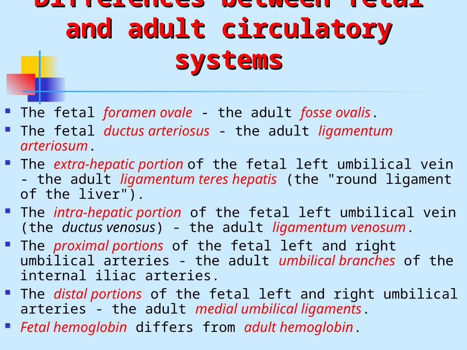
Differences between fetal and Differences between fetal and adult circulatory systemsadult circulatory systems
The fetal foramen ovale - the adult fosse ovalis. The fetal ductus arteriosus - the adult ligamentum
arteriosum. The extra-hepatic portion of the fetal left umbilical vein -
the adult ligamentum teres hepatis (the "round ligament of the liver").
The intra-hepatic portion of the fetal left umbilical vein (the ductus venosus) - the adult ligamentum venosum.
The proximal portions of the fetal left and right umbilical arteries - the adult umbilical branches of the internal iliac arteries.
The distal portions of the fetal left and right umbilical arteries - the adult medial umbilical ligaments.
Fetal hemoglobin differs from adult hemoglobin.

Fetal hemoglobin (1)Fetal hemoglobin (1)
Fetal hemoglobin differs most from adult hemoglobin in that it is able to bind oxygen with greater affinity than the adult form, giving the developing fetus better access to oxygen from the mother's bloodstream.
The P50 value for fetal hemoglobin (i.e., the partial pressure of oxygen at which the protein is 50% saturated; lower values indicate greater affinity) is roughly 19 mmHg, whereas adult hemoglobin has a value of approximately 26.8 mmHg.

Fetal hemoglobin (2)Fetal hemoglobin (2)
At birth, fetal hemoglobin comprises 50-95% of the child's hemoglobin.
These levels decline after six months as adult hemoglobin synthesis is activated, while fetal hemoglobin synthesis is deactivated.
Soon after, adult hemoglobin (hemoglobin A) takes over as the predominant form of hemoglobin in normal children.
Neonatal jaundice tends to develop because of two factors
the breakdown of fetal hemoglobin as it is replaced with adult hemoglobin
the relatively immature hepatic metabolic pathways, which are unable to conjugate bilirubin as fast as an adult.