Embed Size (px)
Citation preview
Page1
2016-2017 Microbe Mission Exam Answer Key
Princeton Science Olympiad Invitational Part 1: Microbial Organisms
Matching. Each statement will have ONLY one answer. Some answers will be used more than once; (1 pt. each) A. Prions B. Viruses C. Archaea D. Bacteria E. Fungi F. Protozoa G. Algae 1. Are considered obligatory intracellular parasites. B 2. Eukaryotic organisms that are important in food chain as they are mainly decomposers of plant
matter there by recycling vital elements. E 3. Cause diseases that are the result of an altered protein. A 4. Not known to cause disease in humans. C 5. Mostly aquatic species that are photo autotrophs. G 6. Causes chronic and long-lasting infections called mycoses. E 7. Causes paralytic shellfish poisoning. G 8. Contain cell walls that are composed of carbohydrate called cellulose. G 9. Causes Giardiasis. F 10. Can be recognized in their motile feeding stage called trophozoite. F 11. Are not cells; Do not independently fulfil the characteristics of life. B 12. Live in extreme salty environments such as the Dead Sea. C 13. Have cell walls made up of lipopolysaccharide membrane. D 14. Causes Creutzfeldt-Jakob disease. A 15. Contain cell walls made up of pseudomurein. C 16. Caused the inhibition of growth of bacteria that lead to a discovery by Alexander Fleming.
E 17. Have structures called pseudopods which surround the food materials and bring it in to the cells
as food vacuoles. F 18. Classified based on their modes of locomotion (ciliates, flagellates and amoeba) F
Page2
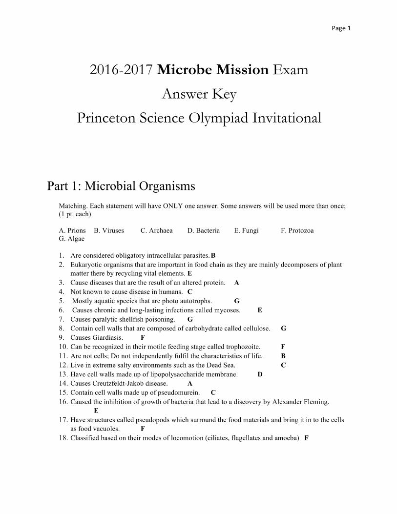
Part 2: Bacterial Growth
(2 pts each)
i. This area in the graph shown above is associated with susceptibility to radiation and antimicrobial drugs. __B___
ii. This area in the graph represents the stage when the metabolic activities of the surviving cells are slowed. __C____
iii. Many bacterial cells undergo involution at this stage. ___D___ iv. The process by which bacteria are maintained in the log phase by draining the spent medium
and adding fresh medium is called. _(continuous culture)____ v. This area in the graph represents the stage when the generation time is constant. ___B____ vi. Name the phase of bacterial growth that is represented by area D on the graph. _(death or
logarithmic decline phase)__
2. A student washes his hands and leaves 100 bacterial cells on a new bar of soap. After 24 hours he decides to do a plate count and dilutes 1 g of the soap 1:105 and plate it on a standard plate count agar. After 24 hours of incubation there are 249 colonies counted. How many bacteria were on the soap sample? (show your work)
Calculation: number of colonies on plate x reciprocal of dilution of the sample is equal to number of bacteria/mL.
249 colonies x 100,000 = 2.49 x 10⁷ bacteria/mL in the sample.
Page3
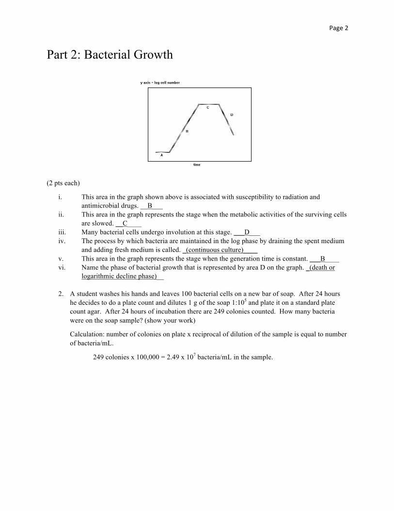
3. The following growth curve represents growth of an E.coli culture that is incubated with two carbon sources.
a. Explain what happened at the time marked X.
At X, the bacteria began a first log phase (Exponential growth phase)
b. Explain what happened at the time marked Y. At Y, the bacteria began a second lag phase during which they synthesized enzymes required to use the second carbon source.
c. Which substrate provided best growth conditions and what evidence from the growth curve supports your answer. The first substrate provided the better growth conditions. The slope of the line is steeper, indicating that the bacteria grew faster.
Time
Num
bero
fcells(lo
gscale)
X
Y
Page4
Part 3: Diseases and Microbes
Key: Disease Key: Microbe 1. Mumps A. Fungi 2. Schistosomiasis B. Virus 3. Pertussis C. Bacteria 4. Histoplasmosis D. Protozoan 5. Botulism E. Algae 6. Cryptosporidiosis F. Parasitic Worm 7. Dengue Fever G. Prion 8. Legionnaire's Disease 9. Scrapie 10. Dutch Elm Disease For each of the described diseases, choose the appropriate microbe that causes the disease from the Microbe Key and the name of the disease from the Disease Key. (2 pts each)
1. First described as a clinical disease in 1800s and known as sausage disease_____________. C (bacteria), 5 (botulism)
2. This disease is transmitted by a mosquito and is also called Breakbone fever____________. B (virus), 7 (Dengue fever)
3. This is debilitating disease of the cardiovascular system and the symptoms of the disease result from the eggs shed by the adult_________________. F (parasitic worm), 2 (Schistosomiasis)
4. A childhood disease and the violent coughing symptoms in children can actually result in broken ribs.___________. C (bacteria), 3 (pertussis)
5. Type of Pneumonia and the microbe that causes the disease grows in water, such as air-conditioning cooling towers and then can be disseminated to air____________. C (bacteria), 8 (Legionnaire’s disease)
6. This disease resemble Tuberculosis and is acquired by airborne conidia_____________________. A (fungi), 4 (histoplasmosis)
7. This disease destroyed the population of Elm trees_________. A (fungi), 10 (Dutch elm disease)
8. This disease causes swelling of the Parotid glands ______________. B (virus), 1 (mumps)
9. The disease is transmitted to humans through recreational and drinking water systems that causes diarrhea of 10 to 14 days duration________________. D (protozoan), 6 (cryptoridiosis)
10. The degenerative disease that affects the nervous system of sheep and goats_________. G (prion), 9 (scrapie)
Page5
Part 4: Microscopes:
1. State the use of following microscopes (4 pts each): a. Phase Contrast Microscope:
Detailed examination of internal structures of living specimens. No staining required. The responses for this and the following parts should essentially cover the points specified in the answer. Use individual discretion when awarding points based on the completeness of the answer and level of detail.
b. Confocal Microscope:
Facilitates 2D and 3D images of cells for biomedical application. Uses laser light to illuminate one plane of a specimen at a time.
c. Scanning Electron Microscope:
Used to study the surface features of cells and viruses. Gives 1000 to 10,000X magnification. Uses a beam of electrons. Image is 3D.
d. Transmission Electron Microscope:
Uses electrons to examine viruses and internal structure of cells. Magnified 10,000 to 100,000X. Gives 2D images.
2. For each of the following, determine the best type of microscope to observe the following samples. (1 pt each)
1. Stained bacterial smear - Compound light microscope
2. Unstained bacterial cells when cells are small and no detail is needed –
Darkfield microscope 3. Unstained live tissue when it is desirable to see some intracellular detail. –
Phase-contrast microscope 4. A sample that emits lights when illuminated with UV light - Fluorescence microscope
5. Intracellular detail of a cell that is about 1 micrometer long - Electron microscope
6. Unstained live cells where intracellular structures are shown in color –
Differential interference contrast microscope
Page6
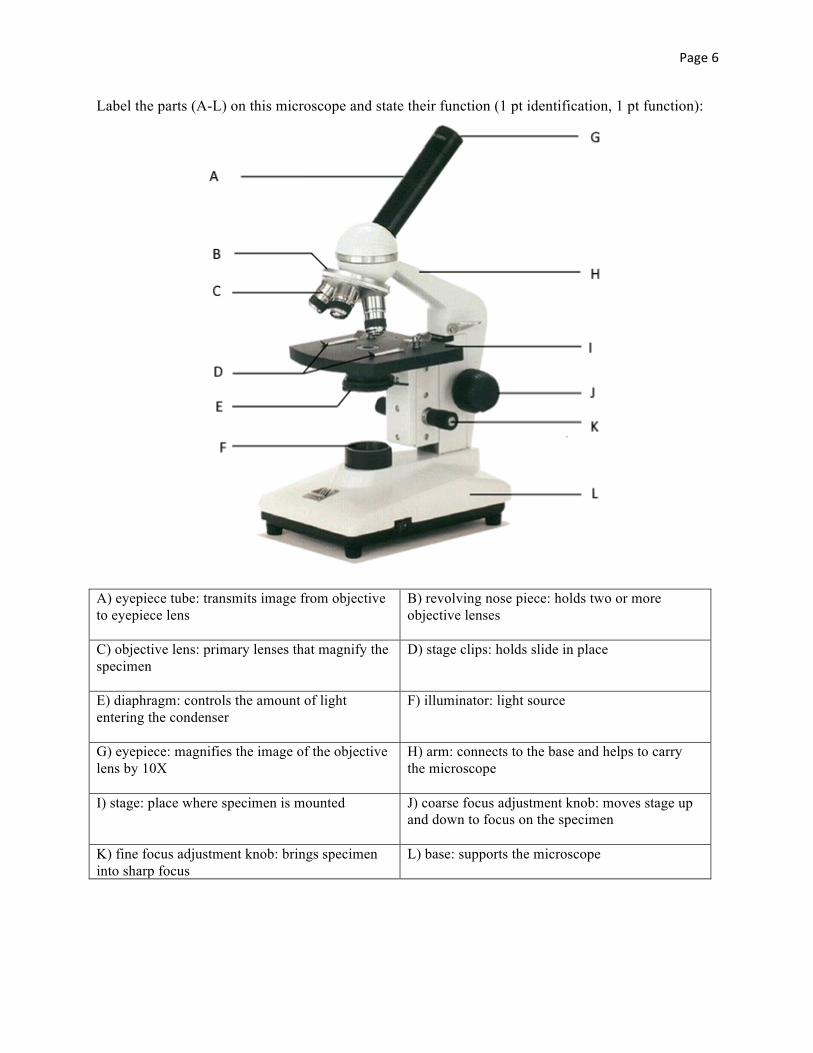
Label the parts (A-L) on this microscope and state their function (1 pt identification, 1 pt function): A. A) eyepiece tube: transmits image from objective to eyepiece lens
B) revolving nose piece: holds two or more objective lenses
C) objective lens: primary lenses that magnify the specimen
D) stage clips: holds slide in place
E) diaphragm: controls the amount of light entering the condenser
F) illuminator: light source
G) eyepiece: magnifies the image of the objective lens by 10X
H) arm: connects to the base and helps to carry the microscope
I) stage: place where specimen is mounted J) coarse focus adjustment knob: moves stage up and down to focus on the specimen
K) fine focus adjustment knob: brings specimen into sharp focus
L) base: supports the microscope
Page7
Part 5: Microbes and Food Industry: Answer True or False. (2 points each) i. Endospores can survive sterilization. ___F____
ii. Food Safety is Monitored by EPA. ___F____
iii. Most amino acids used in food and medicine are produced by bacteria. ___T____
iv. The sugars in fruits such as grapes are fermented by algae to produce wines. _F___
v. Canning works to preserve food because of anaerobic environment and heat. __T___
vi. Biotechnology is a science that uses living organisms to produce commercial products. __T___
vii. Fermentation works to preserve foods because of pH. __T___
viii. Microorganisms are themselves commercial products. __T___
Page8
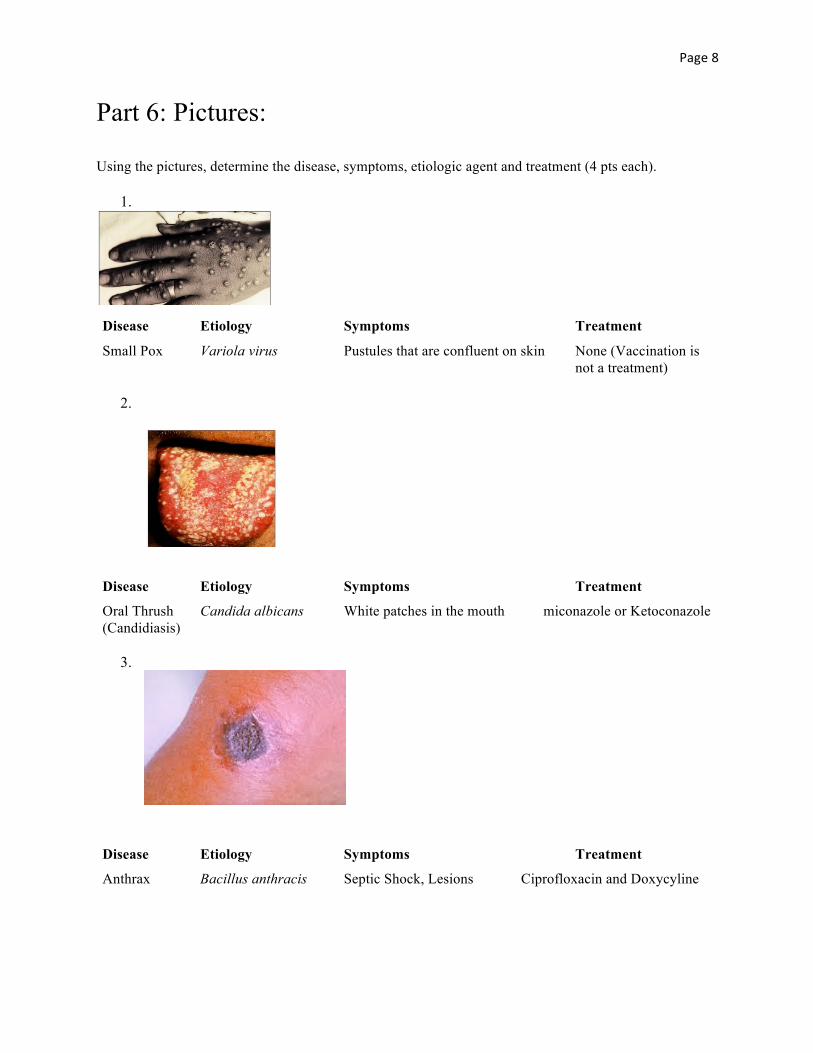
Part 6: Pictures: Using the pictures, determine the disease, symptoms, etiologic agent and treatment (4 pts each).
1.
Disease Etiology Symptoms Treatment
Small Pox Variola virus Pustules that are confluent on skin None (Vaccination is not a treatment)
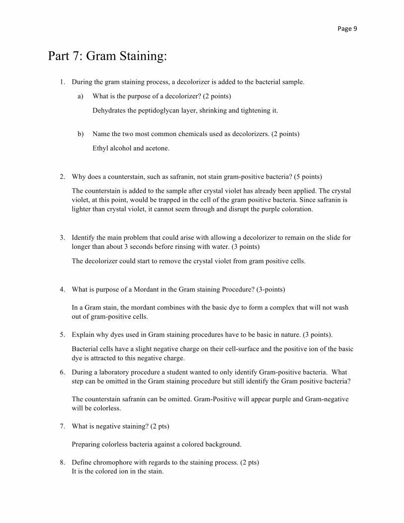
2.
Disease Etiology Symptoms Treatment
Oral Thrush Candida albicans White patches in the mouth miconazole or Ketoconazole (Candidiasis)
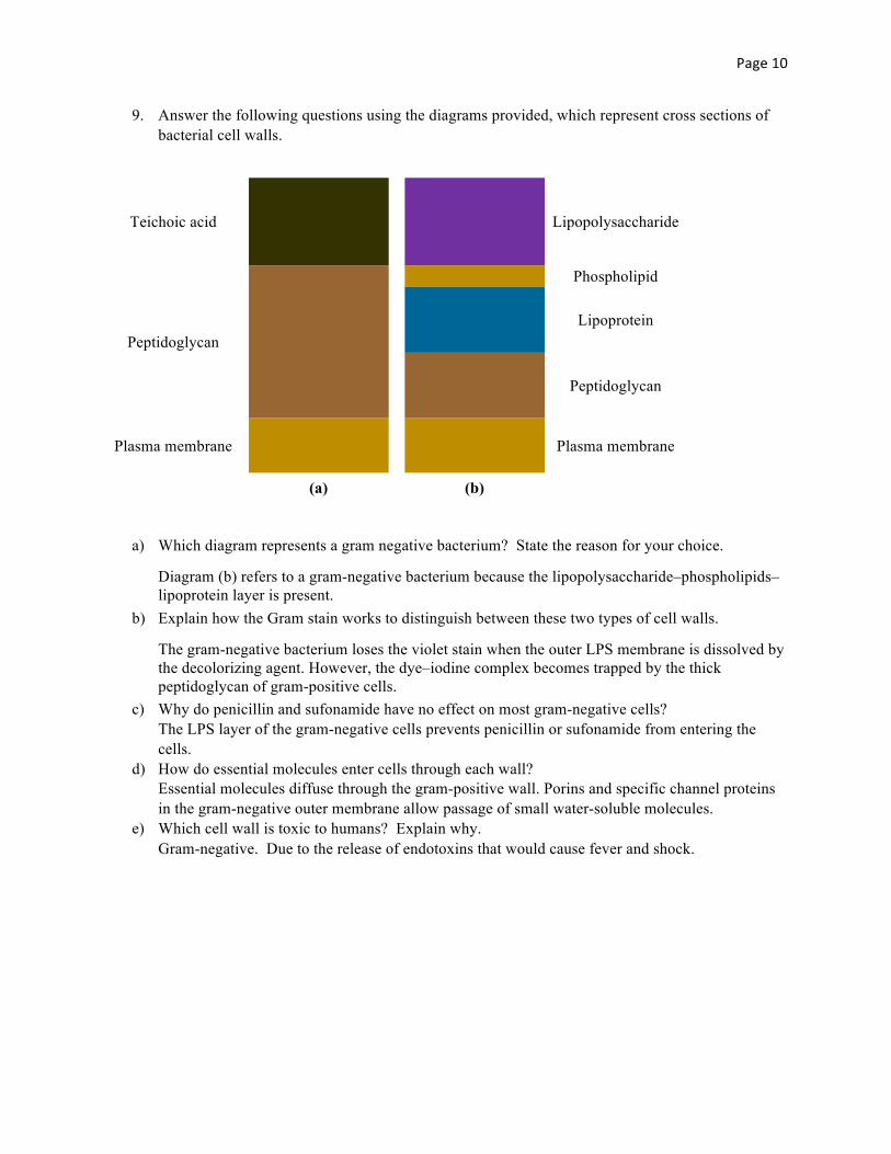
3.
Disease Etiology Symptoms Treatment
Anthrax Bacillus anthracis Septic Shock, Lesions Ciprofloxacin and Doxycyline
Page9
Part 7: Gram Staining:
1. During the gram staining process, a decolorizer is added to the bacterial sample.
a) What is the purpose of a decolorizer? (2 points)
Dehydrates the peptidoglycan layer, shrinking and tightening it.
b) Name the two most common chemicals used as decolorizers. (2 points)
Ethyl alcohol and acetone.
2. Why does a counterstain, such as safranin, not stain gram-positive bacteria? (5 points)
The counterstain is added to the sample after crystal violet has already been applied. The crystal violet, at this point, would be trapped in the cell of the gram positive bacteria. Since safranin is lighter than crystal violet, it cannot seem through and disrupt the purple coloration.
3. Identify the main problem that could arise with allowing a decolorizer to remain on the slide for longer than about 3 seconds before rinsing with water. (3 points)
The decolorizer could start to remove the crystal violet from gram positive cells.
4. What is purpose of a Mordant in the Gram staining Procedure? (3-points) In a Gram stain, the mordant combines with the basic dye to form a complex that will not wash out of gram-positive cells.
5. Explain why dyes used in Gram staining procedures have to be basic in nature. (3 points).
Bacterial cells have a slight negative charge on their cell-surface and the positive ion of the basic dye is attracted to this negative charge.
6. During a laboratory procedure a student wanted to only identify Gram-positive bacteria. What step can be omitted in the Gram staining procedure but still identify the Gram positive bacteria? The counterstain safranin can be omitted. Gram-Positive will appear purple and Gram-negative will be colorless.
7. What is negative staining? (2 pts) Preparing colorless bacteria against a colored background.
8. Define chromophore with regards to the staining process. (2 pts) It is the colored ion in the stain.
Page10
9. Answer the following questions using the diagrams provided, which represent cross sections of bacterial cell walls.
Teichoic acid
Lipopolysaccharide
Peptidoglycan
Phospholipid
Lipoprotein
Peptidoglycan
Plasma membrane
Plasma membrane
(a) (b)
a) Which diagram represents a gram negative bacterium? State the reason for your choice.
Diagram (b) refers to a gram-negative bacterium because the lipopolysaccharide–phospholipids–lipoprotein layer is present.
b) Explain how the Gram stain works to distinguish between these two types of cell walls.
The gram-negative bacterium loses the violet stain when the outer LPS membrane is dissolved by the decolorizing agent. However, the dye–iodine complex becomes trapped by the thick peptidoglycan of gram-positive cells.
c) Why do penicillin and sufonamide have no effect on most gram-negative cells? The LPS layer of the gram-negative cells prevents penicillin or sufonamide from entering the cells.
d) How do essential molecules enter cells through each wall? Essential molecules diffuse through the gram-positive wall. Porins and specific channel proteins in the gram-negative outer membrane allow passage of small water-soluble molecules.
e) Which cell wall is toxic to humans? Explain why. Gram-negative. Due to the release of endotoxins that would cause fever and shock.
Page11
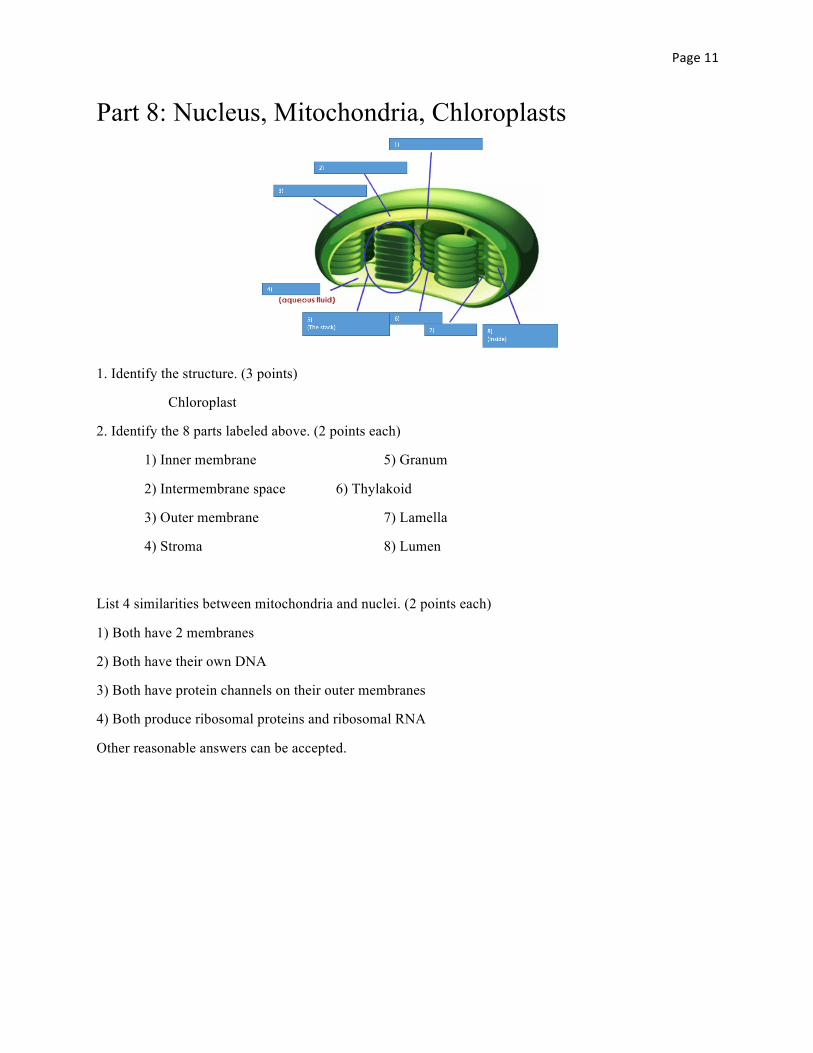
Part 8: Nucleus, Mitochondria, Chloroplasts
1. Identify the structure. (3 points)
Chloroplast
2. Identify the 8 parts labeled above. (2 points each)
1) Inner membrane 5) Granum
2) Intermembrane space 6) Thylakoid
3) Outer membrane 7) Lamella
4) Stroma 8) Lumen
List 4 similarities between mitochondria and nuclei. (2 points each)
1) Both have 2 membranes
2) Both have their own DNA
3) Both have protein channels on their outer membranes
4) Both produce ribosomal proteins and ribosomal RNA
Other reasonable answers can be accepted.
Page12
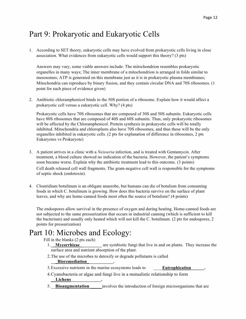
Part 9: Prokaryotic and Eukaryotic Cells
1. According to SET theory, eukaryotic cells may have evolved from prokaryotic cells living in close association. What evidences from eukaryotic cells would support this theory? (3 pts) Answers may vary, some viable answers include: The mitochondrion resembles prokaryotic organelles in many ways; The inner membrane of a mitochondrion is arranged in folds similar to mesosomes; ATP is generated on this membrane just as it is in prokaryotic plasma membranes; Mitochondria can reproduce by binary fission, and they contain circular DNA and 70S ribosomes. (1 point for each piece of evidence given)
2. Antibiotic chloramphenicol binds to the 50S portion of a ribosome. Explain how it would affect a prokaryotic cell versus a eukaryotic cell. Why? (4 pts)
Prokaryotic cells have 70S ribosomes that are composed of 30S and 50S subunits. Eukaryotic cells have 80S ribosomes that are composed of 40S and 60S subunits. Thus, only prokaryotic ribosomes will be affected by the Chloramphenicol. Protein synthesis in prokaryotic cells will be totally inhibited. Mitochondria and chloroplasts also have 70S ribosomes, and thus these will be the only organelles inhibited in eukaryotic cells. (2 pts for explanation of difference in ribosomes, 2 pts Eukaryotes vs Prokaryote)
3. A patient arrives in a clinic with a Neisseria infection, and is treated with Gentamycin. After treatment, a blood culture showed no indication of the bacteria. However, the patient’s symptoms soon became worse. Explain why the antibiotic treatment lead to this outcome. (3 points) Cell death released cell wall fragments. The gram-negative cell wall is responsible for the symptoms of septic shock (endotoxin).
4. Clostridium botulinum is an obligate anaerobe, but humans can die of botulism from consuming
foods in which C. botulinum is growing. How does this bacteria survive on the surface of plant leaves, and why are home-canned foods most often the source of botulism? (4 points)
The endospores allow survival in the presence of oxygen and during heating. Home-canned foods are
not subjected to the same pressurization that occurs in industrial canning (which is sufficient to kill the bacterium) and usually only heated which will not kill the C. botulinum. (2 pts for endospores, 2 points for pressurization)
Part 10: Microbes and Ecology: Fill in the blanks (2 pts each)
1. __Mycorrhizae__________ are symbiotic fungi that live in and on plants. They increase the surface area and nutrient absorption of the plant.
2. The use of the microbes to detoxify or degrade pollutants is called ___Bioremediation____________.
3. Excessive nutrients in the marine ecosystems leads to ____Eutrophication______. 4. Cyanobacteria or algae and fungi live in a mutualistic relationship to form
__Lichens_____________. 5. __Bioaugmentation _____involves the introduction of foreign microorganisms that are
Page13
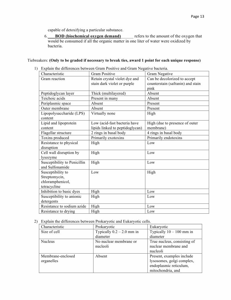
capable of detoxifying a particular substance. 6. ___ BOD (biochemical oxygen demand)______ refers to the amount of the oxygen that
would be consumed if all the organic matter in one liter of water were oxidized by bacteria.
Tiebreakers: (Only to be graded if necessary to break ties, award 1 point for each unique response)
1) Explain the differences between Gram Positive and Gram Negative bacteria. Characteristic Gram Positive Gram Negative Gram reaction Retain crystal violet dye and
stain dark violet or purple Can be decolorized to accept counterstain (safranin) and stain pink
Peptidoglycan layer Thick (multilayered) Absent Teichoic acids Present in many Absent Periplasmic space Absent Present Outer membrane Absent Present Lipopolysaccharide (LPS) content
Virtually none High
Lipid and lipoprotein content
Low (acid-fast bacteria have lipids linked to peptidoglycan)
High (due to presence of outer membrane)
Flagellar structure 2 rings in basal body 4 rings in basal body Toxins produced Primarily exotoxins Primarily endotoxins Resistance to physical disruption
High Low
Cell wall disruption by lysozyme
High Low
Susceptibility to Penicillin and Sulfonamide
High Low
Susceptibility to Streptomycin, chloramphenicol, tetracycline
Low High
Inhibition to basic dyes High Low Susceptibility to anionic detergents
High Low
Resistance to sodium azide High Low Resistance to drying High Low
2) Explain the differences between Prokaryotic and Eukaryotic cells. Characteristic Prokaryotic Eukaryotic Size of cell Typically 0.2 – 2.0 mm in
diameter Typically 10 – 100 mm in diameter
Nucleus No nuclear membrane or nucleoli
True nucleus, consisting of nuclear membrane and nucleoli
Membrane-enclosed organelles
Absent Present, examples include lysosomes, golgi complex, endoplasmic reticulum, mitochondria, and
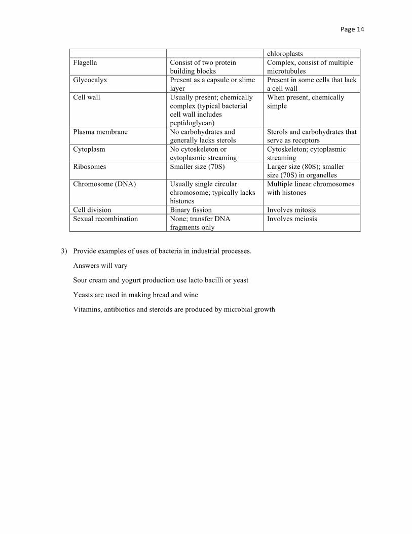
Page14
chloroplasts Flagella Consist of two protein
building blocks Complex, consist of multiple microtubules
Glycocalyx Present as a capsule or slime layer
Present in some cells that lack a cell wall
Cell wall Usually present; chemically complex (typical bacterial cell wall includes peptidoglycan)
When present, chemically simple
Plasma membrane No carbohydrates and generally lacks sterols
Sterols and carbohydrates that serve as receptors
Cytoplasm No cytoskeleton or cytoplasmic streaming
Cytoskeleton; cytoplasmic streaming
Ribosomes Smaller size (70S) Larger size (80S); smaller size (70S) in organelles
Chromosome (DNA) Usually single circular chromosome; typically lacks histones
Multiple linear chromosomes with histones
Cell division Binary fission Involves mitosis Sexual recombination None; transfer DNA
fragments only Involves meiosis
3) Provide examples of uses of bacteria in industrial processes.
Answers will vary
Sour cream and yogurt production use lacto bacilli or yeast
Yeasts are used in making bread and wine
Vitamins, antibiotics and steroids are produced by microbial growth