Embed Size (px)
Citation preview
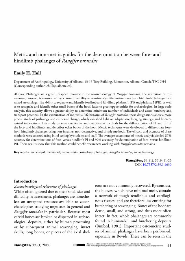
This journal is published under the terms of the Creative Commons Attribution 3.0 Unported LicenseEditor in Chief: Birgitta Åhman, Technical Editor: Eva Wiklund and Graphic Design: H-G Olofsson, www.rangiferjournal.com 11Rangifer, 39, (1) 2019
Metric and non-metric guides for the determination between fore- and hindlimb phalanges of Rangifer tarandus
Emily H. Hull
Department of Anthropology, University of Alberta, 13-15 Tory Building, Edmonton, Alberta, Canada T6G 2H4 (Corresponding author: [email protected]).
Abstract: Phalanges are a great untapped resource in the zooarchaeology of Rangifer tarandus. The utilization of this resource, however, is constrained by a current inability to consistently differentiate fore- from hindlimb phalanges in a mixed assemblage. The ability to separate and identify forelimb and hindlimb phalanx 1 (PI) and phalanx 2 (PII), as well as to recognize and identify other small bones of the hoof, leads to great opportunities for archaeologists. In large-scale analysis, this capacity allows a greater ability to determine minimum number of individuals and assess butchery and transport practices. In the examination of individual life histories of Rangifer tarandus, these designations allow a more precise study of pathology and entheseal change, which can shed light on adaptation, foraging strategy, and human-animal interactions. This study presents qualitative and quantitative methods for the differentiation of PI and PII of the fore- and hindlimbs and describes other bones of the hoof. Metric techniques were developed to differentiate fore- from hindlimb phalanges using non-invasive, non-destructive, and simple methods. The efficacy and accuracy of these methods were assessed using blind testing by students and staff. The average success rates of metric analysis yielded 87% accuracy for determinations of fore- versus hindlimb PI and 92% accuracy for determination of fore- versus hindlimb PII. These results show that this method could benefit researchers working with Rangifer tarandus remains.
Key words: metacarpal; metatarsal; osteometrics; osteology; phalanges; Rangifer tarandus; zooarchaeology.
Rangifer, 39, (1), 2019: 11-26 DOI 10.7557/2.39.1.4630
eton are not commonly recovered. By contrast, the hooves, which have minimal meat, contain a network of tough tendinous and cartilagi-nous tissues, and are therefore less enticing for butchering or scavenging. Bones of the hoof are dense, small, and strong, and thus more often intact. In fact, whole phalanges are commonly found in human-kill and butchering deposits (Binford, 1981). Important osteometric stud-ies of animal phalanges have been performed, especially in Bovids. These can be seen in the
IntroductionZooarchaeological relevance of phalangesWhile often ignored due to their small size and difficulty in assessment, phalanges are nonethe-less an untapped resource available to zooar-chaeologists studying ungulates in general and Rangifer tarandus in particular. Because most cervid bones are broken or dispersed in archae-ological deposits, either by human processing or by subsequent animal scavenging, intact skulls, long bones, or pieces of the axial skel-
Rangifer, 39, (1) 2019This journal is published under the terms of the Creative Commons Attribution 3.0 Unported LicenseEditor in Chief: Birgitta Åhman, Technical Editor: Eva Wiklund and Graphic Design: H-G Olofsson, www.rangiferjournal.com12
phalanges, as well as differences in entheseal changes at muscle attachment sites, may tell archaeologists much about the behavioral pat-terns of individual animals in life (Bartosie-wicz & Gál, 2013; Villotte & Knüsel, 2013; Niinimäki & Salmi, 2016; Salmi & Niinimäki, 2016).
BackgroundRangifer tarandus is a circumpolar and medium-sized cervid species with large hooves (Banfield, 1961). They are artiodactyls with cloven hooves and large dewclaws that often function as ad-ditional, rather than vestigial, toes. Their pat-tern of morphology follows that of the Telem-etacarpalia, a subgroup of Cervidae. In this morphological adaptation, metacarpal (MC) I is not present, and metacarpals III and IV are fused into the central metapodial. Metacarpals II and V are foreshortened to become the dew-claws, which each include a vestigial metacarpal bone, and small PI, PII, and PIII, as well as a small sesamoid bone. In the metatarsal (MT), an analogous development is present, in which the vestigial metatarsals II and V are absent, leaving only the small PI, PII, and PIII, and small sesamoid bone. Metatarsals III and IV are fused into a single metatarsal (Nieminen, 1980, Nieminen, 1994; Cap et al., 2002).
The unique morphology of Rangifer tarandus is epitomized by the size of the feet, much of which is due to the dish-shaped cartilage which covers PIII and appears, in living animals, as the hoof. Telfer & Kelsall (1984) found that Rangifer tarandus hoof-to-body-size ratio is more similar to the paw-size of North American predators than to the hoof-size of other cervids. This may be due to their cold-weather adapta-tion, and again indicates that their morphology must be studied separately and not determined from proxy studies of other artiodactyls (For-mozov, 1946; Nieminen, 1994; Geist, 1998).
designation between fore- and hind-limb cat-tle phalanges (Dottrens, 1946), the use of pha-langes in sex determination of bison (Duffield, 1973), and the subsequent study of metrics and paleopathology in the phalanges of cattle (Bartosiewicz, 1993; Bartosiewicz et al., 1993). Cervids, however, have not been the subject of such studies, perhaps because Rangifer tarandus is the only domesticated cervid. This study de-scribes both qualitative and quantitative meth-ods for the study and distinction of Rangifer tarandus phalanges.
In zooarchaeological quantification, pha-langes of Rangifer are often lumped together, with no attempts to divide fore- and hindlimb phalanges. The extreme difficulty in separating phalanges, due to similarities in morphology and size, may lead to the belief that phalanges of the fore- and hindlimb cannot be differenti-ated.
Separating both phalanx 1 (PI) and phalanx 2 (PII) of the fore- from hindlimb is significant to both assemblage-based analyses and indi-vidual life history studies in zooarchaeology. As phalanges are often among the most abun-dant complete bones in archaeological Rangi-fer tarandus assemblages, they offer a wealth of information. In assemblage-based analysis, more precise calculations of minimum number of individuals (MNI) are made possible by the specific identification of phalanges. For exam-ple, an assemblage with 400 first phalanges, as-sessed together without designations, must be initially considered to have an MNI of 50, as each individual Rangifer tarandus possesses 8 such elements. With more detailed assessment, MNI values can become much more precise. Further, the ratio of fore- to hindlimb pha-langes may also give information as to human utilization, butchery practices, and preferential meat procurement (Binford, 1961; Binford, 1978; Steele, 2015). Identifying phalanges is also useful for analyses of Rangifer life histories. Different pathologies of fore- and hindlimb
This journal is published under the terms of the Creative Commons Attribution 3.0 Unported LicenseEditor in Chief: Birgitta Åhman, Technical Editor: Eva Wiklund and Graphic Design: H-G Olofsson, www.rangiferjournal.com 13Rangifer, 39, (1) 2019
Materials and MethodsForty modern skeletal specimens from Finland housed at the University of Oulu were visu-ally inspected. The collections contained both Rangifer tarandus tarandus and Rangifer taran-dus fennicus of both sexes, all of which were skeletally mature. All had phalanges labelled by side and limb, and these were used in equation design and testing. In addition, six hooves be-longing to domestic Rangifer tarandus tarandus were dissected for the study. The blind testing was done with a wider range of subspecies, including specimens of Rangifer tarandus fen-nicus, Rangifer tarandus tarandus, and Rangi-fer tarandus caribou from collections at both the University of Oulu and the University of Alberta, Canada. Each test was completed by 23-25 volunteers (dependent upon the test and the time volunteers had available). Volunteers for the blind test were all staff and students at
the University of Oulu and the University of Alberta. The volunteers were comprised of 18 students with limited osteological experience, and 7 graduate students and staff with experi-ence in osteology or zooarchaeology. Tests were discarded only for two reasons: first, in one case, improper use of the calipers led to meas-urements that were up to 220 mm larger than those found by their peers, or, second, the vol-unteer had written their name or other identi-fication on the test. All tests were given in ac-cordance to ethics approval by the University of Alberta.
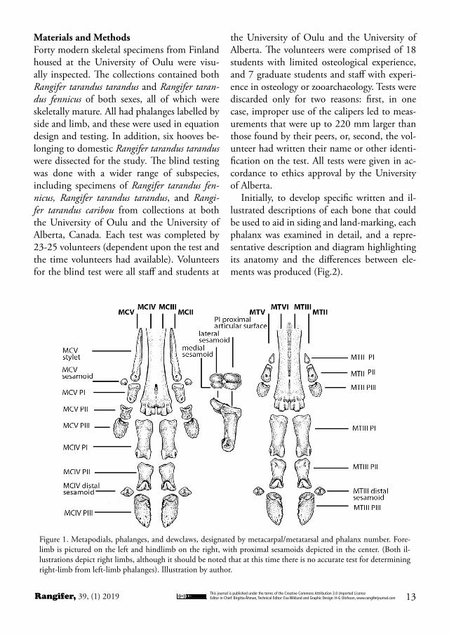
Initially, to develop specific written and il-lustrated descriptions of each bone that could be used to aid in siding and land-marking, each phalanx was examined in detail, and a repre-sentative description and diagram highlighting its anatomy and the differences between ele-ments was produced (Fig.2).
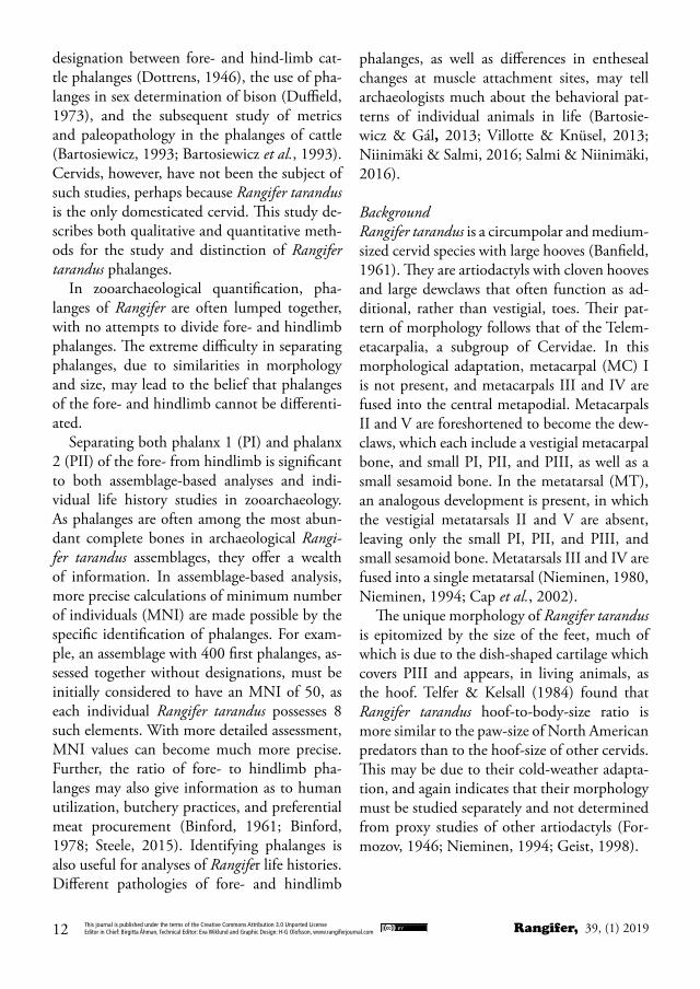
Figure 1. Metapodials, phalanges, and dewclaws, designated by metacarpal/metatarsal and phalanx number. Fore-limb is pictured on the left and hindlimb on the right, with proximal sesamoids depicted in the center. (Both il-lustrations depict right limbs, although it should be noted that at this time there is no accurate test for determining right-limb from left-limb phalanges). Illustration by author.
Rangifer, 39, (1) 2019This journal is published under the terms of the Creative Commons Attribution 3.0 Unported LicenseEditor in Chief: Birgitta Åhman, Technical Editor: Eva Wiklund and Graphic Design: H-G Olofsson, www.rangiferjournal.com14
Secondly, equations were devised systemati-cally distinguish PI and PII from those of the hindlimb. The primary measurements were adapted from those described for the measure-ment of long bones by von den Driesch (1976) but were supplemented with other measure-ments to capture more variation of shape ob-served. Equations were derived and tested based on each phalanx’s most distinct, defini-tive, and consistent morphological features. Six measurements were taken on each PI, and twelve on each PII, based on the most distinc-tive features of the bone. More measurements were taken on PII because of their extreme dif-ficulty to separate visually. An additional goal was to ensure that the resulting equations were simple, straightforward, and require no mensu-ration that could not be expediently achieved with calipers and a calculator. To this end, no more than four measurements were eventually selected for each equation. The overall objective was simplicity and utility in an archaeological context. While both osteometric and morpho-logical techniques are presented in this study, it is hoped that these techniques may be used in conjunction, as visual assessments by morphol-ogy are intrinsically subjective, while osteomet-ric techniques are more reliable.
Measurements were collected in a spread-sheet, and trial and error equations, developed with consideration to shape dynamics, were used to find the greatest degree of separation in results. Initial results were also analyzed for dif-ferences between the sexes, however, all differ-ences were found to be in size, not in shape. The size difference also included significant overlap, so was deemed unreliable for sexing without ad-ditional context.
General anatomy of the phalanges
Figure 2. PI, PII, and PIII depicted from multiple an-gles, in reverse anatomical position. As each hoof con-tains two digits, and as the differentiation between the analogous digit of the opposite hoof cannot yet be quantified, each hoof will be presented as the en-tire subject of study, rather than the entire body of the animal. For this reason, it is important to clarify directional terminology. Medial and lateral sides are designated as medial and lateral to the center of the hoof, not to the animal’s body. Thus, the medial side of a phalanx would be the side that faces the center of the hoof, towards the other digit of the same hoof. (Both illustrations depict right limb bones, although it should be noted that at this time there is no ac-curate test for determining right-limb from left-limb phalanges). Illustration by author.

This journal is published under the terms of the Creative Commons Attribution 3.0 Unported LicenseEditor in Chief: Birgitta Åhman, Technical Editor: Eva Wiklund and Graphic Design: H-G Olofsson, www.rangiferjournal.com 15Rangifer, 39, (1) 2019
the phalanx, facing the centerline of the hoof. Additionally, on the proximal articular surface, the medial articular facet is broader and deeper than the lateral articular facet, which often ap-pears as a slightly raised platform.
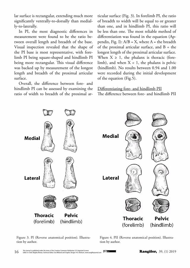
Differentiating medial from lateral sides of PIIOn the distal articular surface of PII are two circular, concave areas just proximal to the dis-tal articular surface on the sides of the bone. The more distinct, concave area marks the me-dial side of the phalanx. The lateral side will of-ten be quite smooth, with minor or indistinct concavity (Fig. 4). Additionally, on the ventral aspect of the proximal articular surface are two protuberances divided by the central ridge bi-secting the articular surface. The side with the longer dorsal to ventral length is the lateral side. This projection will also be generally more ro-bust and protuberant than the medial side.
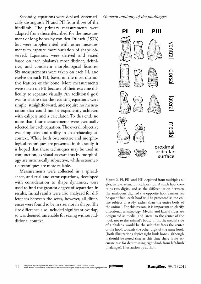
Differentiating fore- and hindlimb PIIn the same individual, forelimb PI may be distinguished as consistently shorter in length with a more robust base than hindlimb PI, which have a noticeably longer diaphysis (Fig. 3). While the distal articular condyles are quite analogous between the fore- and hindlimb, the proximal articular surfaces at the base of the phalanges are a key distinguishing feature. When viewed from the proximal aspect, di-rectly at the articular surface with the metapo-dial, the base of forelimb PI is roughly square or circular, with generally equal length and breadth. By contrast, the medial articular facet of hindlimb PI includes a styloid-like protuber-ance, which extends along the length of the me-dial articular surface significantly farther than the lateral surface. While the medial articular facet of forelimb PI is often slightly longer than the lateral facet, this difference is not so differ-ent to obscure the squared or circular shape of the proximal articular surface of the forelimb phalanx. In the hindlimb PI, the entire articu-
Phalangeal anatomy may be divided into four sections (Fig. 1): Phalanges I and II, PIII (or the terminal phalanx), sesamoids, and bones of the dewclaws.
Phalanges I and IIWhile very different in detail, PI and PII follow a general morphological form. These phalanges consist of a distally-oriented head, diaphyseal body, and a concave, proximal articular base.
While it may seem obvious to more experi-enced zooarchaeologists, it is important to dif-ferentiate PI from PII, as this may not be clear to novices (Fig.2). PII is a much shorter, small-er bone than PI, and can be identified by the heart shaped profile of its head when observed from the distal aspect. While the shape of the distal articular surface on PI resembles a spool or a bow with two rounded articular condyles separated by a central groove, the heart-shaped profile of PII is formed by two condyles, again separated by a central groove, which meet at a rounded point on the dorsal side of the pha-lanx. On the proximal articular surface, PI has a generally rectangular surface, with a central sulcus running dorsally to ventrally, while PII’s proximal articular surface is again an inverted heart-shape, with a central ridge running from a small flat surface (often with vascular forami-na) at the ventral aspect of the articular surface; this runs through the length of the articular surface before curling upwards to a pointed protuberance on the dorsal side of the phalanx. This surface articulates with the spool-shaped distal articular surface of PI.
Differentiating medial from lateral sides of PIOn the distal articular surface of PI are two ar-ticular condyles (Fig. 3). One condyle is higher and has a steeper angle than the other. This condyle also typically has much more develop-ment on the tendon attachment site just proxi-mal to the articular condyle on the side of the phalanx. This condyle marks the medial side of
Rangifer, 39, (1) 2019This journal is published under the terms of the Creative Commons Attribution 3.0 Unported LicenseEditor in Chief: Birgitta Åhman, Technical Editor: Eva Wiklund and Graphic Design: H-G Olofsson, www.rangiferjournal.com16
lar surface is rectangular, extending much more significantly ventrally-to-dorsally than medial-ly-to-laterally.
In PI, the most diagnostic differences in measurement were found to be the ratio be-tween overall length and breadth of the base. Visual inspection revealed that the shape of the PI base is most representative, with fore-limb PI being square-shaped and hindlimb PI being more rectangular. This visual difference was backed up by measurement of the longest length and breadth of the proximal articular surface.
Overall, the difference between fore- and hindlimb PI can be assessed by examining the ratio of width to breadth of the proximal ar-
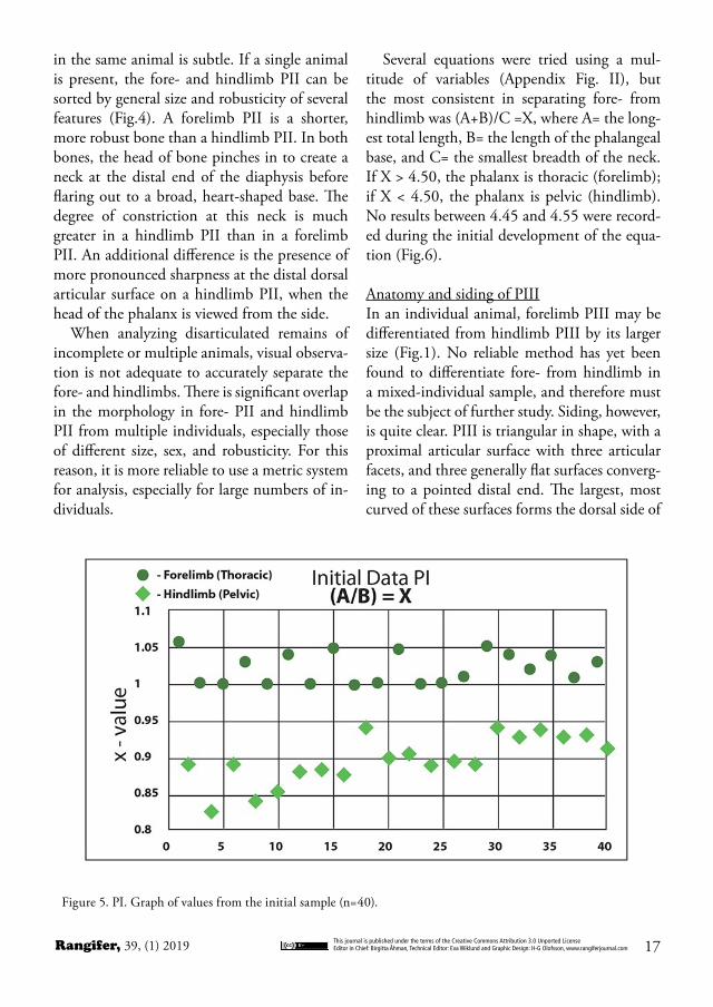
ticular surface (Fig. 3). In forelimb PI, the ratio of breadth to width will be equal to or greater than one, and in hindlimb PI, this ratio will be less than one. The most reliable method of differentiation was found in the equation (Ap-pendix, Fig. I): A/B = X, where A = the breadth of the proximal articular surface, and B = the longest length of the proximal articular surface. When X ≥ 1, the phalanx is thoracic (fore-limb), and when X < 1, the phalanx is pelvic (hindlimb). No results between 0.94 and 1.00 were recorded during the initial development of the equation (Fig.5).
Differentiating fore- and hindlimb PIIThe difference between fore- and hindlimb PII
Figure 3. PI (Reverse anatomical position). Illustra-tion by author.
Figure 4. PII (Reverse anatomical position). Illustra-tion by author.
This journal is published under the terms of the Creative Commons Attribution 3.0 Unported LicenseEditor in Chief: Birgitta Åhman, Technical Editor: Eva Wiklund and Graphic Design: H-G Olofsson, www.rangiferjournal.com 17Rangifer, 39, (1) 2019
in the same animal is subtle. If a single animal is present, the fore- and hindlimb PII can be sorted by general size and robusticity of several features (Fig.4). A forelimb PII is a shorter, more robust bone than a hindlimb PII. In both bones, the head of bone pinches in to create a neck at the distal end of the diaphysis before flaring out to a broad, heart-shaped base. The degree of constriction at this neck is much greater in a hindlimb PII than in a forelimb PII. An additional difference is the presence of more pronounced sharpness at the distal dorsal articular surface on a hindlimb PII, when the head of the phalanx is viewed from the side.
When analyzing disarticulated remains of incomplete or multiple animals, visual observa-tion is not adequate to accurately separate the fore- and hindlimbs. There is significant overlap in the morphology in fore- PII and hindlimb PII from multiple individuals, especially those of different size, sex, and robusticity. For this reason, it is more reliable to use a metric system for analysis, especially for large numbers of in-dividuals.
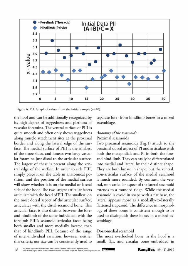
Several equations were tried using a mul-titude of variables (Appendix Fig. II), but the most consistent in separating fore- from hindlimb was (A+B)/C =X, where A= the long-est total length, B= the length of the phalangeal base, and C= the smallest breadth of the neck. If X > 4.50, the phalanx is thoracic (forelimb); if X < 4.50, the phalanx is pelvic (hindlimb). No results between 4.45 and 4.55 were record-ed during the initial development of the equa-tion (Fig.6).
Anatomy and siding of PIIIIn an individual animal, forelimb PIII may be differentiated from hindlimb PIII by its larger size (Fig.1). No reliable method has yet been found to differentiate fore- from hindlimb in a mixed-individual sample, and therefore must be the subject of further study. Siding, however, is quite clear. PIII is triangular in shape, with a proximal articular surface with three articular facets, and three generally flat surfaces converg-ing to a pointed distal end. The largest, most curved of these surfaces forms the dorsal side of
Figure 5. PI. Graph of values from the initial sample (n=40).
Rangifer, 39, (1) 2019This journal is published under the terms of the Creative Commons Attribution 3.0 Unported LicenseEditor in Chief: Birgitta Åhman, Technical Editor: Eva Wiklund and Graphic Design: H-G Olofsson, www.rangiferjournal.com18
the hoof and can be additionally recognized by its high degree of ruggedness and plethora of vascular foramina. The ventral surface of PIII is quite smooth and often only shows ruggedness along muscle attachment sites at the proximal border and along the lateral edge of the sur-face. The medial surface of PIII is the smallest of the three sides, and houses two large vascu-lar foramina just distal to the articular surface. The largest of these is present along the ven-tral edge of the surface. In order to side PIII, simply place it on the table in anatomical po-sition, and the position of the medial surface will show whether it is on the medial or lateral side of the hoof. The two largest articular facets articulate with the head of PII. The smallest, on the most dorsal aspect of the articular surface, articulates with the distal sesamoid bone. This articular facet is also distinct between the fore- and hindlimb of the same individual, with the forelimb PIII’s sesamoid articular facet being both smaller and more medially located than that of hindlimb PIII. Because of the range of inter-individual variation, however, neither this criteria nor size can be consistently used to
separate fore- from hindlimb bones in a mixed assemblage.
Anatomy of the sesamoidsProximal sesamoidsTwo proximal sesamoids (Fig.1) attach to the proximal dorsal aspect of PI and articulate with both the metapodials and PI in both the fore- and hind-limb. They can easily be differentiated into medial and lateral by their distinct shape. They are both lunate in shape, but the ventral, non-articular surface of the medial sesamoid is much more rounded. By contrast, the ven-tral, non-articular aspect of the lateral sesamoid extends to a rounded ridge. While the medial sesamoid is ovoid in shape with a flat base, the lateral appears more as a medially-to-laterally flattened trapezoid. The difference in morphol-ogy of these bones is consistent enough to be used to distinguish these bones in a mixed as-semblage.
Dorsomedial sesamoidThe most overlooked bone in the hoof is a small, flat, and circular bone embedded in
Figure 6. PII. Graph of values from the initial sample (n=40).
This journal is published under the terms of the Creative Commons Attribution 3.0 Unported LicenseEditor in Chief: Birgitta Åhman, Technical Editor: Eva Wiklund and Graphic Design: H-G Olofsson, www.rangiferjournal.com 19Rangifer, 39, (1) 2019
the extensor tendons of the front hooves. The dorsomedial sesamoid is located dorsally at the joint between PI and PII on the forelimbs. At this junction, it acts analogously to a miniature patella, but more study is needed to understand its full function and development.
Distal/navicular sesamoidThe distal sesamoid bone makes up the heel of the hoof. It articulates with PIII on the proxi-mal dorsal aspect and can be identified by its unique shape. This bone is shaped differently in the fore- and hindlimb hooves. The forelimb distal sesamoid is small and has the general shape of an equilateral triangle, with two dorsal articular facets of equal size articulating with PII. A round articulation at the distal end, at the opposite face from the apex of its triangular shape, articulates with PIII.
In the hindlimb, the distal sesamoid bone is larger, with uneven articular facets; the lat-eral facet has a larger surface area and creates the general shape of an obtuse triangle. Like the distal sesamoid of the forelimb, it has three articular facets in the same configuration: two articulating with PII, and one articulating with PIII. Despite these disparities, inter-individual variation makes these differences inappropriate for the determination of fore- from hindlimb phalanges in a multi-individual setting.
DewclawsDewclaws of Rangifer tarandus (Fig. 2) contain their own unique skeletal anatomy, analogous to but distinct from the primary metapodials and digits of the hoof. They do not directly ar-ticulate at any point with the metapodial but are instead held in place by a network of con-nective tissue and ligaments. The forelimb dew-claws contain vestigial MCII and MCV which appear as sharp, linear stylet with a rounded distal articular surface (Barone, 1986). At this point, a rudimentary PI, PII, and PIII all artic-ulate in succession beginning with the MCII/
MCV and MCIIPI/MCVPI. In hindlimb dew-claws, the MCII/MCV stylet component is no longer present, and the complex contains only the phalangeal bones of MCIIPI/MCVPI, MCIIPII/MCVPII, and MCIIPIII/MCVPIII.
Differentiation between primary PIII and dewclaw PIIIThe bones of the dewclaw are unlikely to be mistaken for any other bones of the hoof with one exception: PIII. While size is an important distinguishing factor between the PIII of the dewclaws and the primary PIII bones, it is im-portant to note morphological differences, as the dewclaw PIII bones of a large adult animal may be close in size to the primary PIII bones of a small, young animal. Morphologically, PIII of the dewclaws have rough, vascularized edges around the entire border of the bone apart from the proximal articular surface, and is bifacial, having a front and a back surface running the length of the bone. By contrast, primary PIII bones have a triangular shape and have a rough, serrated edge only on the external margin. The internal border of the dewclaw PIII is smooth, straight, and flat, emerging nearly perpendicu-larly from the dorsal surface. Both PIII bones have large vascular foramina, which occurs on the dorsal surface of the dewclaw PIII and the interior surface of primary PIII bones.
Blind testsStudents all used digital calipers to diminish errors that might be made while reading tradi-tional dial calipers. Each bone was marked with a number or letter on tape, which also covered their collection specimen numbers, as these could have provided bias to the experienced osteologists.
Test A: Qualitative testVolunteers (n=25) were given ten randomly numbered PI and PII phalanges with red and blue dots randomly placed on the sides of each.
Rangifer, 39, (1) 2019This journal is published under the terms of the Creative Commons Attribution 3.0 Unported LicenseEditor in Chief: Birgitta Åhman, Technical Editor: Eva Wiklund and Graphic Design: H-G Olofsson, www.rangiferjournal.com20
They were asked to use the diagrams (Fig. 3 and 4) and the descriptions above to designate them as PI and PII as well as to identify the medial and lateral sides of each bone. The purpose of this test was two-fold: first, to assess the useful-ness of the illustrated guide and descriptions, and second, to allow the novice volunteers to become more comfortable observing the pha-langes.
Test B: Qualitative and quantitative differen-tiation between forelimb (TPI) and hindlimb (PPI) (Appendix; Fig. I)Volunteers (n=23) were given a randomly num-bered sample of ten PI phalanges and assigned (in separate sub-tests) to use illustrations and diagrams (provided in the Appendix) to divide them into fore- and hindlimb bones first, and then to use equations to do the same. This was done to compare the effectiveness of observa-tion versus quantitative analysis.
Test C: Qualitative and quantitative differen-tiation between forelimb (TPII) and hindlimb (PPII) (Appendix; Fig. II)
Volunteers (n=23) were given a randomly num-bered sample of ten PII phalanges and asked (in separate sub-tests) to use illustrations and diagrams (provided in the Appendix) to divide them into fore- and hindlimb bones first, and then to use equations to do the same. This was done to compare the effectiveness of observa-tion versus quantitative analysis.
ResultsProjected test resultsIt was expected that the Test A would produce consistently good results, as the differences be-tween PI and PII, and medial and lateral as-pects were quite distinct once identified. It was projected that Test B, differentiating fore- and hindlimb PI, would result in a high rate of cor-rect assessments as the equation is quite simple and the differences between the elements are often observable to the eye. It was thought that Test C would produce a lower rate of correct assessments, as the differences are very subtle to observe and the equation involves somewhat more complex measurements.
Blind test results
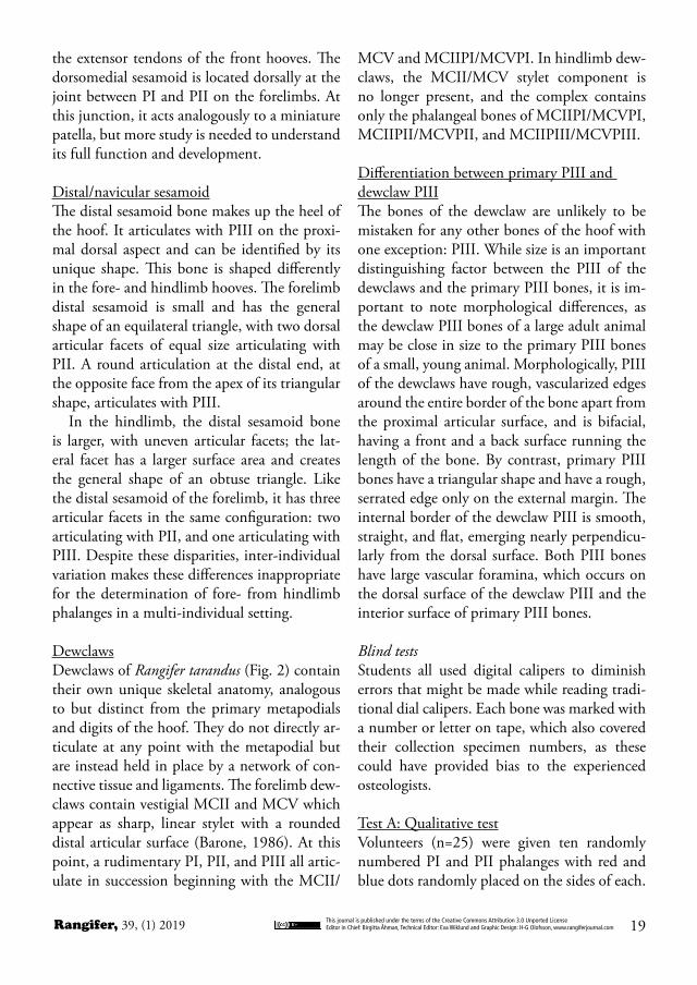
Figure 7. Graph of blind test results. Black bars represent Test A; light green, PI Test B; dark green, PII Test C.
This journal is published under the terms of the Creative Commons Attribution 3.0 Unported LicenseEditor in Chief: Birgitta Åhman, Technical Editor: Eva Wiklund and Graphic Design: H-G Olofsson, www.rangiferjournal.com 21Rangifer, 39, (1) 2019
DiscussionWhile the blind tests did support the higher accuracy of metric determinations versus ob-servation, the projected comparative accuracies of each test were somewhat unexpected. Des-ignation between PI and PII, as well as the de-termination of medial and lateral aspects were very consistent. Any errors may be explained by the inexperience of some of the volunteers. The unexpected results appear in the metric determination between PI and PII. Because of the simplicity and observability of fore- versus hindlimb PI, it was expected that both observa-tion and metric tests of this digits would yield the highest accuracy. The actual results, how-ever, belied this hypothesis (Fig.7). Results of observation were nearly indistinguishable be-tween PI and PII (62% and 63%, respectively),
and the PII metric blind tests yielded higher accuracy than the PI tests, with PII metric tests yielding a mean of 92% (mode= 100%) accuracy, and PI metric tests a mean of 87% (mode= 90%) accuracy. To check observer reli-ability, measurements from the volunteers were assessed by calculating Intraclass Correlation Coefficients. According to generally accepted standards, an Intraclass Correlation Coefficient (ICC) with a value over .750 is considered ex-cellent, while an ICC value of between 0.60 and 0.74 is considered good (Cicchetti, 1993;
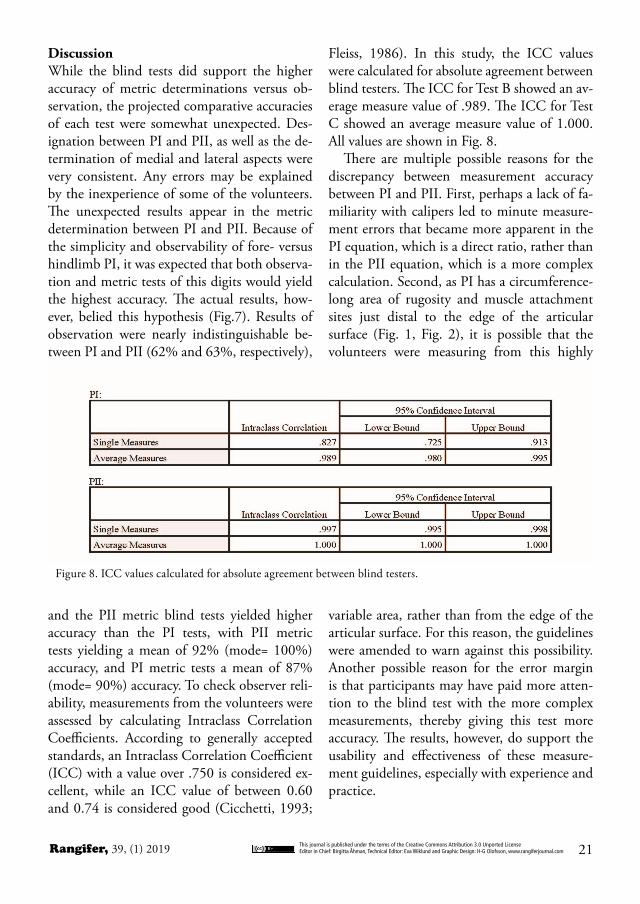
Fleiss, 1986). In this study, the ICC values were calculated for absolute agreement between blind testers. The ICC for Test B showed an av-erage measure value of .989. The ICC for Test C showed an average measure value of 1.000. All values are shown in Fig. 8.
There are multiple possible reasons for the discrepancy between measurement accuracy between PI and PII. First, perhaps a lack of fa-miliarity with calipers led to minute measure-ment errors that became more apparent in the PI equation, which is a direct ratio, rather than in the PII equation, which is a more complex calculation. Second, as PI has a circumference-long area of rugosity and muscle attachment sites just distal to the edge of the articular surface (Fig. 1, Fig. 2), it is possible that the volunteers were measuring from this highly
variable area, rather than from the edge of the articular surface. For this reason, the guidelines were amended to warn against this possibility. Another possible reason for the error margin is that participants may have paid more atten-tion to the blind test with the more complex measurements, thereby giving this test more accuracy. The results, however, do support the usability and effectiveness of these measure-ment guidelines, especially with experience and practice.
Figure 8. ICC values calculated for absolute agreement between blind testers.
Rangifer, 39, (1) 2019This journal is published under the terms of the Creative Commons Attribution 3.0 Unported LicenseEditor in Chief: Birgitta Åhman, Technical Editor: Eva Wiklund and Graphic Design: H-G Olofsson, www.rangiferjournal.com22
ConclusionsWhile many current studies produce detailed results with advanced morphometrics, it was important in the design of this study to utilize simple measurements and to produce equations that could be done in the field or lab with only a set of calipers. The blind tests were done by students and staff who, with few exceptions, had never before studied or done metrics on Rangifer tarandus remains, and many had never practiced metrics analysis of any kind. The level of accuracy during their initial attempts sug-gests that with practice, accuracy would only increase. In the creation of these descriptions, diagrams, and equation-based determina-tions, the focus was on non-destructive usabil-ity, and this was demonstrated to be the case in the blind tests. With these guidelines and tools, more precise determination of fore- and hindlimb phalanges is clearly possible. Tradi-tional zooarchaeology and assemblage-based analysis could utilize this technique for more precise determination of MNI, butchery prac-tices, and preferential transport of meat. In studies of domestication, it has been shown that reindeer involved in different activities show different entheseal changes and patholo-gies; this technique could benefit this study by allowing the differential analysis of the habitual stressors on fore- versus hindlimb (Niinimäki & Salmi, 2016; Salmi & Niinimäki, 2016). Fi-nally, in the emerging and expanding research areas of human-animal relationships, individu-al animal life histories, and animal ontologies, a more distinct understanding of the bones of the hooves may help elucidate topics from habitat, foraging techniques, and individual pathology. This technique has the potential to be an extra tool in the study of the osteology and archaeol-ogy of Rangifer tarandus in both modern and ancient America and Eurasia.
AcknowledgementsI would like to acknowledge the incredible sup-port and guidance of Dr. Robert Losey of the University of Alberta, as well as Dr. Anna-Kaisa Salmi, Dr. Sirpa Niinimäki, and Hanna-Leena Puolakka of the University of Oulu. Invaluable assistance was provided by my research assis-tant, Mitchell Semeniuk. Additional thanks are due to all the wonderful volunteers who partic-ipated in testing the metrics and methodology.
This journal is published under the terms of the Creative Commons Attribution 3.0 Unported LicenseEditor in Chief: Birgitta Åhman, Technical Editor: Eva Wiklund and Graphic Design: H-G Olofsson, www.rangiferjournal.com 23Rangifer, 39, (1) 2019
anatomie, Domestikationsforschung und Ge-schichte der Tiermedizin of the University of Munich (Vol. 1). Cambridge: Peabody Mu-seum Press.
Duffield, L.F. 1973. Aging and sexing the post-cranial skeleton of bison. — Plains An-thropologist 18(60):132-139. https://doi.org/10.1080/2052546.1973.11908656
Fleiss, J.L. 1986. The design and analysis of clinical experiments. John Wiley & Sons, Inc.
Formozov, A. N. 1946. Snow cover as an in-tegral factor of the environment and its im-portance in the ecology of mammals and birds (No. 1). Edmonton, Alberta: Boreal Insti-tute, University of Alberta.
Geist, V. 1998. Deer of the world: their evolu-tion, behavior, and ecology. Mechanicsburg, PA: Stackpole Books.
Nieminen, M. 1980. Evolution and taxonomy of the genus Rangifer in northern Europe. – In: Reimers, E., Gaare, E., and Skjenneberg, S. (Eds.). Proceedings Second International Reindeer/Caribou Symposium. Direktorate for vilt og ferskvannisfisk, Trondheim, Nor-way. pp. 379-391.
Nieminen, M. 1990. Hoof and foot loads for reindeer (Rangifer tarandus). — Rangifer 10(3):249-254. https://doi.org/10.7557/2.10.3.865
Nieminen, M. 1994. Poro: ruumiinrakenne ja elintoiminnat. Helsinki: Riista-ja kala-talouden tutkimuslaitos.
Niinimäki, S. and Salmi, A.K. 2016. En-theseal Changes in Free‐Ranging Versus Zoo Reindeer - Observing Activity Status of Reindeer. — International Journal of Os-teoarchaeology 26(2):314-323. https://doi.org/10.1002/oa.2423
Salmi, A.K. & Niinimäki, S. 2016. Entheseal changes and pathological lesions in draught reindeer skeletons – Four case studies from present-day Siberia. — International Jour-nal of Paleopathology 14:91-99. https://doi.org/10.1016/j.ijpp.2016.05.012
ReferencesBanfield, A. W. F. 1961. A revision of the rein-
deer and caribou genus Rangifer (No. 66). Ottawa: Queen’s Printer.
Barone, R. 1986. Anatomie Comparée des Mammifères Domestiques, tome 1, Ostéologie. Paris: Vigot Freres.
Bartosiewicz, L. 1993. The anatomical posi-tion and metric traits of phalanges in cattle. — Revue de Paléobiologie 12(2):21-43.
Bartosiewicz, L., & Gál, E. 2013. Shuffling Nags, Lame Ducks: The Archaeology of Ani-mal Disease. Oxford: Oxbow Books. https://doi.org/10.2307/j.ctvh1djdq
Bartosiewicz, L., Van Neer, W. and Len-tacker, A. 1993. Metapodial asymmetry in draft cattle. — International Journal of Osteoarchaeology 3(2):69-75. https://doi.org/10.1002/oa.1390030203
Binford, L. R. 1977. For theory building in ar-chaeology: Essays on faunal remains, aquatic resources, spatial analysis, and systemic mod-eling. New York: Academic Press.
Binford, L. R. 1978. Nunamiut: Ethnoarchae-ology. New York: Academic Press.
Binford, L. R. 1981. Bones: ancient men and modern myths. New York: Academic Press.
Cap, H., Aulagnier, S., & Deleporte, P. 2002. The phylogeny and behaviour of Cer-vidae (Ruminantia Pecora). – Ethology, Ecol-ogy, & Evolution 14(3):199-216. https://doi.org/10.1080/08927014.2002.9522740
Cicchetti, D. V. 1994. “Guidelines, criteria, and rules of thumb for evaluating normed and standardized assessment instruments in psychology”. — Psychological Assessment 6 (4):284–290. https://doi.org/10.1037//1040-3590.6.4.284
Dottrens, E. 1946. Étude préliminaire: les phalanges osseuses de Bos taurus domesticus. — Rev. suisse de Zool 53(33):739-774.
von den Driesch, A. 1976. A guide to the meas-urement of animal bones from archaeological sites: as developed by the Institut für Palaeo-
Rangifer, 39, (1) 2019This journal is published under the terms of the Creative Commons Attribution 3.0 Unported LicenseEditor in Chief: Birgitta Åhman, Technical Editor: Eva Wiklund and Graphic Design: H-G Olofsson, www.rangiferjournal.com24
Steele, T. E. 2015. The contributions of animal bones from archaeological sites: The past and future of zooarchaeology. — Journal of Archaeological Science 56:168-176. https://doi.org/10.1016/j.jas.2015.02.036
Telfer, E. S., & Kelsall, J. P. 1984. Adaptation of some large North American mammals for survival in snow. — Ecology 65(6):1828-1834. https://doi.org/10.2307/1937779
Villotte, S., & Knüsel, C. J. 2013. Under-standing entheseal changes: definition and life course changes. — International Journal of Osteoarchaeology 23(2):135-146. https://doi.org/10.1002/oa.2289
Manuscript recieved 16 Jan 2019 revision accepted 5 June 2019 manuscript published 3 July 2019
This journal is published under the terms of the Creative Commons Attribution 3.0 Unported LicenseEditor in Chief: Birgitta Åhman, Technical Editor: Eva Wiklund and Graphic Design: H-G Olofsson, www.rangiferjournal.com 25Rangifer, 39, (1) 2019
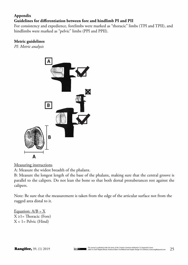
AppendixGuidelines for differentiation between fore and hindlimb PI and PII For consistency and expedience, forelimbs were marked as “thoracic” limbs (TPI and TPII), and hindlimbs were marked as “pelvic” limbs (PPI and PPII).
Metric guidelinesPI: Metric analysis
Measuring instructionsA: Measure the widest breadth of the phalanx.B: Measure the longest length of the base of the phalanx, making sure that the central groove is parallel to the calipers. Do not lean the bone so that both dorsal protuberances rest against the calipers.
Note: Be sure that the measurement is taken from the edge of the articular surface not from the rugged area distal to it.
Equation: A/B = XX ≥1= Thoracic (Fore) X < 1= Pelvic (Hind)
Rangifer, 39, (1) 2019This journal is published under the terms of the Creative Commons Attribution 3.0 Unported LicenseEditor in Chief: Birgitta Åhman, Technical Editor: Eva Wiklund and Graphic Design: H-G Olofsson, www.rangiferjournal.com26
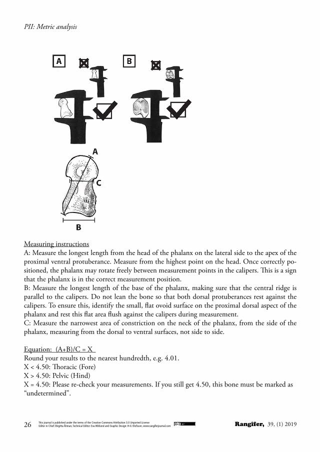
PII: Metric analysis
Measuring instructionsA: Measure the longest length from the head of the phalanx on the lateral side to the apex of the proximal ventral protuberance. Measure from the highest point on the head. Once correctly po-sitioned, the phalanx may rotate freely between measurement points in the calipers. This is a sign that the phalanx is in the correct measurement position.B: Measure the longest length of the base of the phalanx, making sure that the central ridge is parallel to the calipers. Do not lean the bone so that both dorsal protuberances rest against the calipers. To ensure this, identify the small, flat ovoid surface on the proximal dorsal aspect of the phalanx and rest this flat area flush against the calipers during measurement.C: Measure the narrowest area of constriction on the neck of the phalanx, from the side of the phalanx, measuring from the dorsal to ventral surfaces, not side to side.
Equation: (A+B)/C = X Round your results to the nearest hundredth, e.g. 4.01.X < 4.50: Thoracic (Fore) X > 4.50: Pelvic (Hind) X = 4.50: Please re-check your measurements. If you still get 4.50, this bone must be marked as “undetermined”.