Embed Size (px)
Citation preview
Metal Binding Domains 3 and 4 of the Wilson Disease Protein: Solution Structureand Interaction with the Copper(I) Chaperone HAH1†,‡
Lucia Banci,§ Ivano Bertini,*,§ Francesca Cantini,§ Amy C. Rosenzweig,| and Liliya A. Yatsunyk|
Magnetic Resonance Center (CERM), UniVersity of Florence, Via L. Sacconi 6, 50019 Sesto Fiorentino, Italy, Department ofChemistry, UniVersity of Florence, Via della Lastruccia 3, 50019 Sesto Fiorentino, Italy, and Department of Biochemistry,
Molecular Biology, and Cell Biology and Department of Chemistry, Northwestern UniVersity, EVanston, Illinois 60208
ReceiVed March 19, 2008; ReVised Manuscript ReceiVed May 22, 2008
ABSTRACT: The Wilson disease protein or ATP7B is a P1B-type ATPase involved in human copperhomeostasis. The extended N-terminus of ATP7B protrudes into the cytosol and contains six Cu(I) bindingdomains. This report presents the NMR structure of the polypeptide consisting of soluble Cu(I) bindingdomains 3 and 4. The two domains exhibit ferredoxin-like folds, are linked by a flexible loop, and actindependently of one another. Domains 3 and 4 tend to aggregate in a concentration-dependent mannerinvolving nonspecific intermolecular interactions. Both domains can be loaded with Cu(I) when providedas an acetonitrile complex or by the chaperone HAH1. HAH1 forms a 70% complex with domain 4 thatis in fast exchange with the free protein in solution. The ability of HAH1 to form a complex only withsome domains of ATP7B is an interesting property of this class of proteins and may have a signaling rolein the function of the ATPases.
Human ATP7B (WLN)1 is a member of the P1B-typeATPase family that plays a crucial role in copper transportand homeostasis in the body (1). The WLN protein, like theother human copper ATPase, the Menkes protein (MNKhereafter), delivers copper to the secretory pathway of thetrans-Golgi network (TGN) where the metal ion is incorpo-rated into copper-dependent enzymes (2, 3). Both proteinscan also translocate from the Golgi membrane to the plasmamembrane for copper efflux from the cell (4–6). Thepredicted topological organization of WLN and MNKincludes four major regions or domains: the N-terminalcopper binding domain, the transmembrane domain, the ATPbinding domain, and the actuator domain.
The ∼650-amino acid N-terminal domain of WLN(WLN16, hereafter) contains six soluble domains with aconserved metal binding motif GMT/HCxxCxxxIE, eachcapable of binding 1 equiv of Cu(I) with similar affinity (7–12).
The N-termini of WLN homologues from other organismscontain between one and five metal binding domains (13).Although the structure of WLN16 has yet to be determined,the NMR structure of a construct containing domains 5 and6 (WLN56) is available (14). The two domains each have aferredoxin-like fold (�R��R�), are connected by a shortlinker, and are found in a fixed reciprocal orientation.
The six soluble domains of both WLN and MNK receivecopper from the cytoplasmic metallochaperone HAH1 (15).The exact role and interplay of the six soluble domains arestill unclear. In the case of MNK, the six soluble domainsinteract differently with HAH1. The interactions have beeninvestigated by a variety of techniques, ranging from yeasttwo-hybrid assays (16–18) to NMR (19, 20). These dataindicate that the MNK first and fourth domains form a metal-mediated adduct with HAH1 whereas the sixth domain issimultaneously loaded with Cu(I) without formation of theadduct. In the case of WLN, both the second and fourthdomains can form a copper-dependent adduct with HAH1(14, 16, 18, 21). By contrast, WLN56 can receive Cu(I) fromdomain 4, but not from HAH1 (14). In another report, a seriesof six-domain constructs in which five of the six metalbinding CXXC motifs were mutated to SXXS was generated(11). In all constructs, the intact domain was able to receiveCu(I) from HAH1. We report here the structural anddynamical characterization of a construct containing WLNdomains 3 and 4 (WLN34 hereafter) as well as theirinteraction with Cu(I) and Cu(I) HAH1.
MATERIALS AND METHODS
Preparation of Protein Samples. A DNA segment corre-sponding to residues 238-439 of WLN was amplified byPCR, cloned into the pET32Xa/LIC vector, and transformedinto Rosetta(DE3)pLysS cells. The resulting construct is 202
† This work was supported by National Institutes of Health GrantGM58518, Integrated Project SPINE2-COMPLEXES n° 031220,UPMAN n° LSHG-CT-2004-512052, and Ente Cassa di Risparmio diFirenze.
‡ Resonance assignments and the atomic coordinates for the NMRstructures of apo-WLN34 have been deposited in BioMagResBank(entry 11041) and Protein Data Bank, respectively (entry 2ROP).
* To whom correspondence should be addressed: Magnetic Reso-nance Center, University of Florence, Via L. Sacconi, 6 50019 SestoFiorentino, Italy. Fax: +39 055 4574271. Telephone: +39 055 4574272.E-mail: [email protected].
§ University of Florence.| Northwestern University.1 Abbreviations: rmsd, root-mean-square deviation; NOE, nuclear
Overhauser effect; HSQC, heteronuclear single-quantum coherence;WLN, human Wilson disease protein or ATP7B; MNK, human Menkesdisease protein or ATP7A; WLN16, N-terminal metal binding domainof WLN; WLN56, two-domain construct comprising the fifth and sixthmetal binding domains of WLN; WLN34, two-domain constructcomprising the third and fourth metal binding domains of WLN; TGN,trans-Golgi network.
Biochemistry 2008, 47, 7423–7429 7423
10.1021/bi8004736 CCC: $40.75 2008 American Chemical SocietyPublished on Web 06/18/2008
amino acids long and contains domains 3 and 4, theinterdomain linker (31 amino acids), a 17-amino acid portionof the linker connecting domains 2 and 3, a 12-amino acidportion of the linker connecting domains 4 and 5, and notags. For the sake of simplicity, in the following text, residueswill be numbered from 1 to 202, rather than starting from238. Cells were grown at 37 °C in M9 minimal mediumcontaining (NH4)2SO4 as the sole nitrogen source and glucoseas the sole carbon source. The medium was supplementedwith a vitamin mix (Sigma), 100 µg/mL carbenicillin, and40 µg/mL chloramphenicol. The 15N- and 13C-enrichedsamples were produced using >95% enriched nitrogenand carbon sources, as appropriate. When the OD600 reached0.7-0.9, the flasks were cooled to 18 °C, and proteinexpression was induced with 1 mM isopropyl �-D-thioga-lactopyranoside (IPTG). Cells were harvested after anovernight growth by centrifugation for 15 min at 8000g,resuspended in 50 mM HEPES (pH 8.0), 150 mM NaCl,0.1% Triton X-100, and 1 mM phenylmethanesulfonylchloride (PMSF), and stored at -80 °C.
For purification, the cell suspension was thawed and stirredat ambient temperature for 30-60 min with EDTA-freeprotease inhibitor tablets (Roche, one tablet for 40 mL ofcell suspension), 1 mM PMSF, and a pinch of solid DNaseI.The solution was centrifuged at 125000g for 30-60 min,and the supernatant was applied to a 20 mL nickel-loadedChelating Sepharose column (Pharmacia) rinsed with bufferA [50 mM HEPES (pH 7.5), 500 mM NaCl, and 10%glycerol]. WLN34 was eluted with a 4 column volumegradient from 90 to 300 mM imidazole (pH 7.5) with 500mM NaCl and dialyzed twice (with at least one overnightdialysis) at 4 °C versus 2 L of buffer H [50 mM HEPES(pH 7.5) and 200 mM NaCl]. The protein solution was thenconcentrated to 50 mL (∼40 µM), and cleavage of affinitytags was initiated by the addition of 120 units of Factor Xa(Novagen) in buffer H supplemented with 5 mM CaCl2 atroom temperature. The cleavage was stopped after 3 h byaddition of 1 mM PMSF. The protein was loaded onto thenickel column preequilibrated with buffer A and eluted with∼36 mM imidazole. Fractions containing the cleaved proteinwere concentrated in the presence of 1 mM EDTA, 10 mMDTT, and 50 µM bathocuproine disulfonate (BCS), appliedto a Superdex 200 gel filtration column (Pharmacia), andeluted with 20 mM sodium phosphate (pH 6.5) and 150-200mM NaCl. The typical yield was ∼5 and ∼11 mg of pureprotein per liter of Escherichia coli culture for 13C- and 15N-labeled and 15N-labeled samples, respectively. The identityof the protein was confirmed by mass spectrometry, and thepurity was assessed by SDS-PAGE. The Cu(I) HAH1sample was prepared as described previously, always withouta His6 tag (22).
The copper-loaded WLN34 was prepared by adding upto 2 equiv of the Cu(I) acetonitrile complex [Cu(CH3CN)4-PF6)] to apoprotein that had been reduced with 2-3 molarequiv of dithiothreitol (DTT). Cu(CH3CN)4PF6 was addedin a N2 atmosphere chamber and the titration followed via1H-15N HSQC NMR spectra. Binding of Cu(I) was con-firmed by atomic absorption spectroscopy. Titrations of[15N]WLN34 with both unlabeled Cu(I) HAH1 and Cu(I)-[15N]HAH1 with a molar ratio of HAH1 to WLN34 of upto 3:1 were performed similarly. In addition, 15N-labeled
Cu(I) WLN34 was titrated with unlabeled apo-HAH1 at anHAH1:WLN34 molar ratio of up to 2:1.
NMR Experiments and Structure Calculations. The NMRspectra were recorded at 298 K on Avance 500, 700, and900 Bruker spectrometers, all equipped with cryogenicallycooled probes. Resonance assignments of apo-WLN34 andCu(I) WLN34 were performed by conventional multidimen-sional NMR techniques based on triple-resonance experi-ments (Table S1) (23). All residues were assigned, with theexception of Ser17, His30, Lys32, Arg76, and Thr132 forapo-WLN34 and Met29, His30, Lys32-Cys34, and Arg76for Cu(I) WLN34. Resonance assignments are reported asSupporting Information (Tables S2 and S3) and have beendeposited in the BioMagResBank (entry 11041). Distanceconstraints for structure determination of apo-WLN34 wereobtained from 15N-edited and 13C-edited three-dimensionalNOESY-HSQC experiments (Table S1). Structure calcula-tions were performed using CYANA (24). Each of the 20conformers with the lowest residual CYANA target functionvalue was then energy-minimized in explicit solvent withAMBER-8 (25). NOE and torsion angle constraints wereapplied with force constants of 32 kcal mol-1 Å-2 and 32kcal mol-1 rad-2, respectively.
Heteronuclear Relaxation Data. The dynamic propertiesof apo-WLN34 and Cu(I) WLN34 have been directlysampled through 15N relaxation measurements. 15N longitu-dinal and transverse relaxation rates (26) and 15N(1H) NOEs(27) were recorded at 298 K at 500 MHz, using a proteinconcentration of 0.1 mM for both apo-WLN34 and Cu(I)WLN34. Relaxation measurements for apo-WLN34 wereperformed also at a protein concentration of 1 mM. R1 andR2 relaxation rates were obtained by fitting the cross-peakvolumes (I), measured as a function of the relaxation delay,to a single-exponential decay as described in the literature(28). Heteronuclear NOE values were calculated as the ratioof peak volumes in spectra recorded with and withoutsaturation. In all experiments, the water signal was sup-pressed with the “water flipback” scheme (27).
The average backbone 15N longitudinal R1 and transverseR2 relaxation rates and 15N{1H} NOE value at 0.1 mM are1.8 ( 0.1 s-1, 11.7 ( 1.5 s-1, and 0.71 ( 0.04, respectively,for domain 3 and 1.9 ( 0.2 s-1, 10.7 ( 1.8 s-1, and 0.72 (0.04 for domain 4, respectively (Figure 1). For Cu(I)WLN34, the average relaxation rates and 15N{1H} NOEvalue at 0.1 mM are 1.9 ( 0.1 s-1, 12.0 ( 2.0 s-1, and0.73 ( 0.04, respectively, for domain 3 and 2.3 ( 0.2s-1, 8.5 ( 1.4 s-1, and 0.73 ( 0.05 for domain 4,respectively (Figure 1).
RESULTS AND DISCUSSION
Protein Structural and Dynamical Characterization. Aconstruct containing soluble metal binding domains 3 and 4of the human WLN protein (WLN34) has been overex-pressed, and its structural and Cu(I) binding properties aswell as its interaction with HAH1 have been studied byNMR. The NMR spectra of apo-WLN34 and copper-loadedWLN34 are indicative of a folded protein. The two domainsconsist of ∼70 amino acids each and are connected by alinker of 31 residues. The 1H-15N HSQC spectra show well-dispersed amide signals with few peaks clustered in therandom-coil region. Peak assignment indicates that the
7424 Biochemistry, Vol. 47, No. 28, 2008 Banci et al.
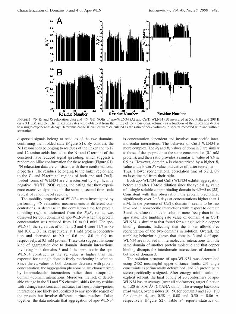
dispersed signals belong to residues of the two domains,confirming their folded state (Figure S1). By contrast, theNH resonances belonging to residues of the linker and to 17and 12 amino acids located at the N- and C-termini of theconstruct have reduced signal spreading, which suggests arandom-coil-like conformation for these regions (Figure S1).15N relaxation data are consistent with these conformationalproperties. The residues belonging to the linker region andto the C- and N-terminal regions of both apo and Cu(I)-loaded forms of WLN34 are characterized by significantlynegative 15N{1H} NOE values, indicating that they experi-ence extensive dynamics on the subnanosecond time scaletypical of random-coil regions.
The mobility properties of WLN34 were investigated byperforming 15N relaxation measurements at different con-centrations. A decrease in the correlation time for proteintumbling (τm), as estimated from the R2/R1 ratios, wasobserved for both domains of apo-WLN34 when the proteinconcentration was reduced from 1.0 to 0.1 mM. For apo-WLN34, the τm values of domains 3 and 4 were 11.7 ( 0.9and 10.6 ( 0.8 ns, respectively, at 1 mM protein concentra-tion and decreased to 9.0 ( 0.6 and 8.0 ( 0.9 ns,respectively, at 0.1 mM protein. These data suggest that somekind of aggregation due to domain-domain interactions,involving both domains 3 and 4, is operative in the apo-WLN34 construct, as the τm value is higher than thatexpected for a single domain freely reorienting in solution.Since the τm values of both domains decrease with proteinconcentration, the aggregation phenomena are characterizedby intermolecular interactions rather than intraproteindomain-domain interactions. Moreover, the lack of detect-able change in the 1H and 15N chemical shifts for any residuewithachangeinconcentrationindicatesthattheseprotein-proteininteractions are likely not localized to any specific region ofthe protein but involve different surface patches. Takentogether, the data indicate that aggregation of apo-WLN34
is concentration-dependent and involves nonspecific inter-molecular interactions. The behavior of Cu(I) WLN34 ismore complex. The R1 and R2 values of domain 3 are similarto those of the apoprotein at the same concentration (0.1 mMprotein), and their ratio provides a similar τm value of 8.9 (0.9 ns. However, domain 4 is characterized by a higher R1
value and a lower R2 value, indicative of faster reorientation.Thus, a lower reorientational correlation time of 6.2 ( 0.9ns is estimated from their ratio.
Both apo-WLN34 and Cu(I) WLN34 exhibit aggregationbefore and after 10-fold dilution since the typical τm valueof a single soluble copper binding domain is 4.5-5 ns (22).Consistent with this observation, the protein precipitatedsignificantly over 2-3 days at concentrations higher than 1mM. In the presence of Cu(I), domain 4 seems to be lessinvolved in nonspecific interactions with respect to domain3 and therefore tumbles in solution more freely than in theapo state. The tumbling rate value of domain 4 in Cu(I)WLN34 is similar to that found for a single soluble copperbinding domain, indicating that the linker allows freereorientation of the two domains in solution. Overall, thetumbling behavior suggests that domains 3 and 4 of apo-WLN34 are involved in intermolecular interactions with thesame domain of another protein molecule and that copperbinding disrupts the interdomain interactions of domain 4but not of domain 3.
The solution structure of apo-WLN34 was determinedusing 2452 meaningful upper distance limits, 231 angleconstraints experimentally determined, and 28 proton pairsstereospecifically assigned. After energy minimization inexplicit solvent, the final bundle of 20 conformers of apo-WLN34 has an average (over all conformers) target functionof 1.80 ( 0.08 Å2 (CYANA units). The average backbonermsd values, over residues 20-90 for domain 3 and 120-190for domain 4, are 0.58 ( 0.08 and 0.50 ( 0.06 Å,respectively (Figure S2). Table S4 reports statistics on
FIGURE 1: 15N R1 and R2 relaxation data and 15N{1H} NOEs of apo-WLN34 (A) and Cu(I) WLN34 (B) measured at 500 MHz and 298 Kon a 0.1 mM sample. The relaxation rates were obtained from the fitting of the cross-peak volumes as a function of the relaxation delaysto a single-exponential decay. Heteronuclear NOE values were calculated as the ratio of peak volumes in spectra recorded with and withoutsaturation.
Characterization of Domains 3 and 4 of Apo-WLN Biochemistry, Vol. 47, No. 28, 2008 7425
restraint violations in the final structure together withstructural quality parameters from PROCHECK-NMR (29)and WHATIF (30). The two domains of apo-WLN34 arewell-folded with the ferredoxin-like fold common to all othercopper ATPase soluble domains characterized up to now(14, 31–34). By contrast, the linker lacks long-range NOEs,consistent with its high flexibility (vide supra) (Figure S3).
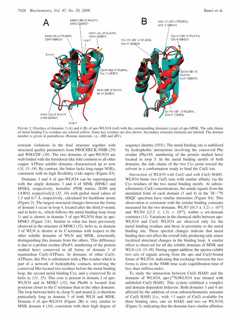
Domains 3 and 4 of apo-WLN34 can be superimposedwith the single domains 3 and 4 of MNK [MNK3 andMNK4, respectively, hereafter (PDB entries 2G90 and1AWO, respectively)] (32, 34) with global rmsd values of1.3 and 0.7 Å, respectively, calculated for backbone atoms(Figure 2). The largest structural changes between the formsof domain 3 occur in loop 4, located after the third �-strand,and in helix R1, which follows the metal binding loop (loop1) and is shorter in domain 3 of apo-WLN34 than in apo-MNK3 (Figure 2A). Similar to what has been previouslyobserved in the structure of MNK3 (32), helix R2 in domain3 of WLN is shorter at its C-terminus with respect to theother soluble domains of WLN and MNK, structurallydistinguishing this domain from the others. This differenceis due to a proline residue (Pro83, numbering of the proteinstudied here) conserved in all forms of domain 3 ofmammalian Cu(I)-ATPases. In domains of other Cu(I)-ATPases, this Pro is substituted with a Phe residue which ispart of a network of hydrophobic contacts involving theconserved Met located two residues before the metal bindingloop, the second metal binding Cys, and a conserved Ile inhelix R1 (31, 35). This core is present in domain 3 of apo-WLN34 and in MNK3 (32), but Phe86 is located fourpositions closer to the C-terminus than in the other domains.The loop between helix R2 (loop 5) and strand �4 is thereforeparticularly long in domain 3 of both WLN and MNK.Domain 4 of apo-WLN34 (Figure 2B) is very similar toMNK domain 4 (34), consistent with their high degree of
sequence identity (55%). The metal binding site is stabilizedby hydrophobic interactions involving the conserved Pheresidue (Phe185, numbering of the protein studied here)located in loop 5. In the metal binding motifs of bothdomains, the side chains of the two Cys point toward thesolvent in a conformation ready to bind the Cu(I) ion.
Interaction of WLN34 with Cu(I) and with Cu(I) HAH1.WLN34 binds two Cu(I) ions with similar affinity via theCys residues of the two metal binding motifs. At substo-ichiometric Cu(I) concentrations, the amide signals from themetalated form of each domain (3 and 4) in the 1H-15NHSQC spectrum have similar intensities (Figure S4). Thisobservation is consistent with the similar binding constantsmeasured for the two domains, WLN3 [(6.3 ( 3.2) × 1010]and WLN4 [(2.5 ( 1.3) × 1010], within a six-domainconstruct (11). Variations in the chemical shifts between apo-WLN34 and Cu(I) WLN34 are observed only for themetal binding residues and those in proximity to the metalbinding site. These spectral changes indicate that metalbinding does not affect the overall fold, producing only minorlocalized structural changes in the binding loop. A similareffect is observed for all the soluble domains of MNK andWLN (14, 33–36). During copper addition, the spectra exhibittwo sets of signals arising from the apo and Cu(I)-boundforms of WLN34, indicating that exchange between the twoforms is slow on the NMR time scale (equilibration time ofless than milliseconds).
To study the interaction between Cu(I) HAH1 and thedomains of WLN34, apo-[15N]WLN34 was titrated withunlabeled Cu(I) HAH1. This system exhibited a complexand domain-dependent behavior. Both domains 3 and 4 areaffected by the addition of even substoichiometric amountsof Cu(I) HAH1 [i.e., with <1 equiv of Cu(I) available forthree binding sites, one on HAH1 and two on WLN34](Figure 3), indicating that the domains have similar affinities
FIGURE 2: Overlays of domains 3 (A) and 4 (B) of apo-WLN34 (red) with the corresponding domains (cyan) of apo-MNK. The side chainsof metal binding Cys residues are colored yellow. Some key residues are also shown. Secondary structure elements are labeled. The domainnumber is given in parentheses (Roman numerals, i.e., dIII and dIV).
7426 Biochemistry, Vol. 47, No. 28, 2008 Banci et al.
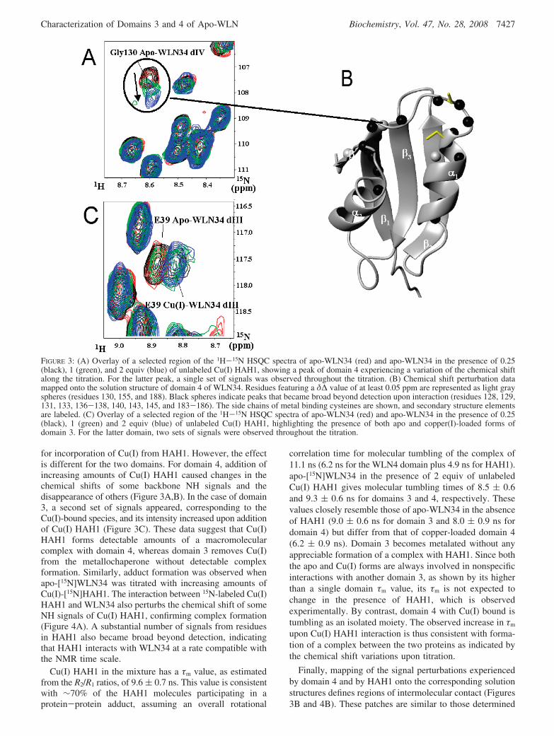
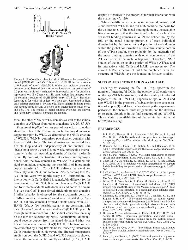
for incorporation of Cu(I) from HAH1. However, the effectis different for the two domains. For domain 4, addition ofincreasing amounts of Cu(I) HAH1 caused changes in thechemical shifts of some backbone NH signals and thedisappearance of others (Figure 3A,B). In the case of domain3, a second set of signals appeared, corresponding to theCu(I)-bound species, and its intensity increased upon additionof Cu(I) HAH1 (Figure 3C). These data suggest that Cu(I)HAH1 forms detectable amounts of a macromolecularcomplex with domain 4, whereas domain 3 removes Cu(I)from the metallochaperone without detectable complexformation. Similarly, adduct formation was observed whenapo-[15N]WLN34 was titrated with increasing amounts ofCu(I)-[15N]HAH1. The interaction between 15N-labeled Cu(I)HAH1 and WLN34 also perturbs the chemical shift of someNH signals of Cu(I) HAH1, confirming complex formation(Figure 4A). A substantial number of signals from residuesin HAH1 also became broad beyond detection, indicatingthat HAH1 interacts with WLN34 at a rate compatible withthe NMR time scale.
Cu(I) HAH1 in the mixture has a τm value, as estimatedfrom the R2/R1 ratios, of 9.6 ( 0.7 ns. This value is consistentwith ∼70% of the HAH1 molecules participating in aprotein-protein adduct, assuming an overall rotational
correlation time for molecular tumbling of the complex of11.1 ns (6.2 ns for the WLN4 domain plus 4.9 ns for HAH1).apo-[15N]WLN34 in the presence of 2 equiv of unlabeledCu(I) HAH1 gives molecular tumbling times of 8.5 ( 0.6and 9.3 ( 0.6 ns for domains 3 and 4, respectively. Thesevalues closely resemble those of apo-WLN34 in the absenceof HAH1 (9.0 ( 0.6 ns for domain 3 and 8.0 ( 0.9 ns fordomain 4) but differ from that of copper-loaded domain 4(6.2 ( 0.9 ns). Domain 3 becomes metalated without anyappreciable formation of a complex with HAH1. Since boththe apo and Cu(I) forms are always involved in nonspecificinteractions with another domain 3, as shown by its higherthan a single domain τm value, its τm is not expected tochange in the presence of HAH1, which is observedexperimentally. By contrast, domain 4 with Cu(I) bound istumbling as an isolated moiety. The observed increase in τm
upon Cu(I) HAH1 interaction is thus consistent with forma-tion of a complex between the two proteins as indicated bythe chemical shift variations upon titration.
Finally, mapping of the signal perturbations experiencedby domain 4 and by HAH1 onto the corresponding solutionstructures defines regions of intermolecular contact (Figures3B and 4B). These patches are similar to those determined
FIGURE 3: (A) Overlay of a selected region of the 1H-15N HSQC spectra of apo-WLN34 (red) and apo-WLN34 in the presence of 0.25(black), 1 (green), and 2 equiv (blue) of unlabeled Cu(I) HAH1, showing a peak of domain 4 experiencing a variation of the chemical shiftalong the titration. For the latter peak, a single set of signals was observed throughout the titration. (B) Chemical shift perturbation datamapped onto the solution structure of domain 4 of WLN34. Residues featuring a δ∆ value of at least 0.05 ppm are represented as light grayspheres (residues 130, 155, and 188). Black spheres indicate peaks that became broad beyond detection upon interaction (residues 128, 129,131, 133, 136-138, 140, 143, 145, and 183-186). The side chains of metal binding cysteines are shown, and secondary structure elementsare labeled. (C) Overlay of a selected region of the 1H-15N HSQC spectra of apo-WLN34 (red) and apo-WLN34 in the presence of 0.25(black), 1 (green) and 2 equiv (blue) of unlabeled Cu(I) HAH1, highlighting the presence of both apo and copper(I)-loaded forms ofdomain 3. For the latter domain, two sets of signals were observed throughout the titration.
Characterization of Domains 3 and 4 of Apo-WLN Biochemistry, Vol. 47, No. 28, 2008 7427
for all the other MNK or WLN domains as well as the solubledomains of ATPases from other organisms (14, 20, 37, 38).
Functional Implications. As part of our efforts to under-stand the roles of the N-terminal metal binding domains incopper transport by WLN, we determined the NMR structureof WLN34. WLN34 comprises two distinct domains withferredoxin-like folds. The two domains are connected by aflexible loop and act independently of one another, like“beads on a string”, even if some weak, nonspecific interac-tions with the corresponding domain of another moleculeoccur. By contrast, electrostatic interactions and hydrogenbonds hold the two domains in WLN56 in a defined andrigid orientation, precluding the possibility of interdomaincopper transfer (14). Cu(I) HAH1 delivers the metal ionefficiently to WLN34, but not to WLN56 according to NMR(14) or the yeast two-hybrid assay (16). Furthermore, theinteraction with Cu(I) HAH1 is remarkably different for thetwo domains of WLN34. It is not clear why Cu(I) HAH1can form stable adducts with domain 4 and not with domain3, given that Cu(I) is transferred efficiently to both domains.Similar behavior is observed for the six-domain constructof MNK, in which domains 3 and 4 were metalated by Cu(I)HAH1, but only domain 4 formed a stable adduct with Cu(I)HAH1 (20). A few possible scenarios are consistent withdata. Domain 3 could be metalated by Cu(I) HAH1, possiblythrough weak interactions. The adduct concentration maybe too low for detection by NMR. Alternatively, domain 3could receive copper from metalated domain 4 [that wasloaded via interaction with Cu(I) HAH1]. The two domainsare connected by a long flexible linker, rendering interdomainCu(I) transfer possible. However, site-directed mutagenesisstudies on both the MNK16 and WLN16 constructs showedthat all the domains can be directly metalated by Cu(I) HAH1
despite differences in the properties for their interaction withthe chaperone (11, 20).
While the differences in behavior between domains 3 and4 and between WLN34 and WLN56 could be the basis forthe distinct roles of the metal binding domains, the availableliterature suggests that the functional roles of each of thesix metal binding domains in WLN are defined not by thefold or the metal binding properties of each individualdomain but by the properties and position of each domainwithin the global conformation of the entire soluble portionof the ATPase and/or, most probably, by the interaction ofthe metal binding domains with other components of theATPase or with the metallochaperone. Therefore, NMRstudies of the entire soluble portion of Wilson ATPase andits interactions with Cu(I) and HAH1 are necessary. Thecurrent NMR structure of WLN34 combined with thestructure of WLN56 lays the foundation for such studies.
SUPPORTING INFORMATION AVAILABLE
Four figures showing the 15N-1H HSQC spectrum, thenumber of meaningful NOEs, the overlay of 20 conformersof the apo-WLN34 structure, and the overlay of selectedregions of the 1H-15N HSQC spectra of apo-WLN34 andapo-WLN34 in the presence of substoichiometric concentra-tion of copper(I) and four tables showing the experimentsperformed, the chemical shift assignments, and the statisticson restraint violations in the final structure of apo-WLN34.This material is available free of charge via the Internet athttp://pubs.acs.org.
REFERENCES
1. Bull, P. C., Thomas, G. R., Rommens, J. M., Forbes, J. R., andCox, D. W. (1993) The Wilson disease gene is a putative coppertransporting P-type ATPase similar to the Menkes gene. Nat. Genet.5, 327–337.
2. Harrison, M. D., Jones, C. E., Solioz, M., and Dameron, C. T.(2000) Intracellular copper routing: The role of copper chaperones.Trends Biochem. Sci. 25, 29–32.
3. Puig, S., and Thiele, D. J. (2002) Molecular mechanisms of copperuptake and distribution. Curr. Opin. Chem. Biol. 6, 171–180.
4. Cater, M. A., La Fontaine, S., Shield, K., Deal, Y., and Mercer,J. F. (2006) ATP7B mediates vesicular sequestration of copper:Insight into biliary copper excretion. Gastroenterology 130, 493–506.
5. La Fontaine, S., and Mercer, J. F. (2007) Trafficking of the copper-ATPases, ATP7A and ATP7B: Role in copper homeostasis. Arch.Biochem. Biophys. 463, 149–167.
6. Petris, M. J., Voskoboinik, I., Cater, M., Smith, K., Kim, B. E.,Llanos, R. M., Strausak, D., Camakaris, J., and Mercer, J. F. (2002)Copper-regulated trafficking of the Menkes disease copper ATPaseis associated with formation of a phosphorylated catalytic inter-mediate. J. Biol. Chem. 277, 46736–46742.
7. Lutsenko, S., Petrukhin, K., Cooper, M. J., Gilliam, C. T., andKaplan, J. H. (1997) N-Terminal domains of human copper-transporting adenosine triphosphatases (the Wilson’s and Menkesdisease proteins) bind copper selectively in vivo and in vitro withstoichiometry of one copper per metal-binding repeat. J. Biol.Chem. 272, 18939–18944.
8. DiDonato, M., Narindrasorasak, S., Forbes, J. R., Cox, D. W., andSarkar, B. (1997) Expression, purification, and metal bindingproperties of the N-terminal domain from the Wilson diseaseputative copper-transporting ATPase (ATP7B). J. Biol. Chem. 272,33279–33282.
9. Bull, P. C., and Cox, D. W. (1994) Wilson disease and Menkesdisease: New handles on heavy-metal transport. Trends Genet. 10,246–252.
10. Banci, L., and Rosato, A. (2003) Structural genomics of proteinsinvolved in copper homeostasis. Acc. Chem. Res. 36, 215–221.
FIGURE 4: (A) Combined chemical shift differences between Cu(I)-bound [15N]HAH1 and Cu(I)-bound [15N]HAH1 in the presenceof 0.5 equiv of apo-[15N]WLN34. White bars indicate peaks thatbecame broad beyond detection upon interaction. A ∆δ value of0.2 ppm was arbitrarily assigned to those peaks only for graphicalrepresentation. (B) Chemical shift perturbation data mapped ontothe solution structure of HAH1 [PDB entry 1TL5 (22)]. Residuesfeaturing a δ∆ value of at least 0.1 ppm are represented as lightgray spheres (residues 8, 59, and 62). Black spheres indicate peaksthat became broad beyond detection upon interaction (residues 9-23and 58). The side chains of metal-binding cysteines are shown,and secondary structure elements are labeled.
7428 Biochemistry, Vol. 47, No. 28, 2008 Banci et al.
11. Yatsunyk, L. A., and Rosenzweig, A. C. (2007) Copper(I) bindingand transfer by the N-terminus of the Wilson disease protein.J. Biol. Chem. 282, 8622–8631.
12. Wernimont, A. K., Yatsunyk, L. A., and Rosenzweig, A. C. (2004)Binding of copper(I) by the Wilson disease protein and its copperchaperone. J. Biol. Chem. 279, 12269–12276.
13. Arnesano, F., Banci, L., Bertini, I., Ciofi-Baffoni, S., Molteni, E.,Huffman, D. L., and O’Halloran, T. V. (2002) Metallochaperonesand metal transporting ATPases: A comparative analysis ofsequences and structures. Genome Res. 12, 255–271.
14. Achila, D., Banci, L., Bertini, I., Bunce, J., Ciofi-Baffoni, S., andHuffman, D. L. (2006) Structure of human Wilson protein domains5 and 6 and their interplay with domain 4 and the copper chaperoneHAH1 in copper uptake. Proc. Natl. Acad. Sci. U.S.A. 103, 5729–5734.
15. Hamza, I., Schafer, M., Klomp, L. W., and Gitlin, J. D. (1999)Interaction of the copper chaperone HAH1 with the Wilson diseaseprotein is essential for copper homeostasis. Proc. Natl. Acad. Sci.U.S.A. 96, 13363–13368.
16. Larin, D., Mekios, C., Das, K., Ross, B., Yang, A. S., and Gilliam,C. T. (1999) Characterization of the interaction between the Wilsonand Menkes disease proteins and the cytoplasmic copper chaperone,HAH1p. J. Biol. Chem. 274, 28497–28504.
17. Strausak, D., Howie, M. K., Firth, S. D., Schlicksupp, A., Pipkorn,R., Multhaup, G., and Mercer, J. F. (2003) Kinetic analysis of theinteraction of the copper chaperone Atox1 with the metal bindingsites of the Menkes protein. J. Biol. Chem. 278, 20821–20827.
18. van Dongen, E. M., Klomp, L. W., and Merkx, M. (2004) Copper-dependent protein-protein interactions studied by yeast two-hybridanalysis. Biochem. Biophys. Res. Commun. 323, 789–795.
19. Banci, L., Bertini, I., Cantini, F., Chasapis, C., Hadjiliadis, N., andRosato, A. (2005) A NMR study of the interaction of a three-domain construct of ATP7A with copper(I) and copper(I)-HAH1:The interplay of domains. J. Biol. Chem. 280, 38259–38263.
20. Banci, L., Bertini, I., Cantini, F., Della Malva, N., Migliardi, M.,and Rosato, A. (2007) The different intermolecular interactions ofthe soluble copper-binding domains of the Menkes protein, ATP7A.J. Biol. Chem. 282, 23140–23146.
21. Walker, J. M., Huster, D., Ralle, M., Morgan, C. T., Blackburn,N. J., and Lutsenko, S. (2004) The N-terminal metal-binding site2 of the Wilson’s disease protein plays a key role in the transferof copper from Atox1. J. Biol. Chem. 279, 15376–15384.
22. Anastassopoulou, J., Banci, L., Bertini, I., Cantini, F., Katsari, E.,and Rosato, A. (2004) Solution structure of the apo- and copper(I)loaded human metallo-chaperone HAH1. Biochemistry 43, 13046–13053.
23. Grzesiek, S., and Bax, A. (1993) Amino acid type determinationin the sequential assignment procedure of uniformly 13C/15N-enriched proteins. J. Biomol. NMR 3, 185–204.
24. Guntert, P. (2004) Automated NMR structure calculation withCYANA. Methods Mol. Biol. 278, 353–378.
25. Case, D. A., Darden, T. A., Cheatham, T. E., Simmerling, C. L.,Wang, J., Duke, R. E., Luo, R., Merz, K. M., Wang, B., Pearlman,D. A., Crowley, M., Brozell, S., Tsui, V., Gohlke, H., Mongan, J.,Hornak, V., Cui, G., Beroza, P., Schafmeister, C. E., Caldwell,
J. W., Ross, W. S., and Kollman, P. A. (2004) AMBER 8,University of California, San Francisco.
26. Farrow, N. A., Muhandiram, R., Singer, A. U., Pascal, S. M., Kay,C. M., Gish, G., Shoelson, S. E., Pawson, T., Forman-Kay, J. D.,and Kay, L. E. (1994) Backbone dynamics of a free andphosphopeptide-complexed Src homology 2 domain studied by 15NNMR relaxation. Biochemistry 33, 5984–6003.
27. Grzesiek, S., and Bax, A. (1993) The importance of not saturatingH2O in protein NMR. Application to sensitivity enhancement andNOE measurements. J. Am. Chem. Soc. 115, 12593–12594.
28. Mandel, M. A., Akke, M., and Palmer, A. G., III (1995) Backbonedynamics of Escherichia coli ribonuclease HI: Correlations withstructure and function in an active enzyme. J. Mol. Biol. 246, 144–163.
29. Laskowski, R. A., MacArthur, M. W., Moss, D. S., and Thornton,J. M. (1993) PROCHECK: A program to check the stereochemicalquality of protein structures. J. Appl. Crystallogr. 26, 283–291.
30. Vriend, G. (1990) WHAT IF: A molecular modeling and drugdesign program. J. Mol. Graphics 8, 52–56.
31. Banci, L., Bertini, I., Cantini, F., Migliardi, M., Rosato, A., andWang, S. (2005) An atomic level investigation of the disease-causing A629P mutant of the Menkes protein ATP7A. J. Mol. Biol.352, 409–417.
32. Banci, L., Bertini, I., Cantini, F., Della Malva, N., Rosato, A.,Herrmann, T., and Wuthrich, K. (2006) Solution structure andintermolecular interactions of the third metal-binding domain ofATP7A, the Menkes disease protein. J. Biol. Chem. 281, 29141–29147.
33. Banci, L., Bertini, I., Chasapis, C., Ciofi-Baffoni, S., Hadjiliadis,N., and Rosato, A. (2005) An NMR study of the interactionbetween the human copper(I) chaperone and the second and fifthmetal-binding domains of the Menkes protein. FEBS J. 272, 865–871.
34. Gitschier, J., Moffat, B., Reilly, D., Wood, W. I., and Fairbrother,W. J. (1998) Solution structure of the fourth metal-binding domainfrom the Menkes copper-transporting ATPase. Nat. Struct. Biol.5, 47–54.
35. Banci, L., Bertini, I., Del Conte, R., D’Onofrio, M., and Rosato,A. (2004) Solution structure and backbone dynamics of the Cu(I)and apo-forms of the second metal-binding domain of the Menkesprotein ATP7A. Biochemistry 43, 3396–3403.
36. DeSilva, T. M., Veglia, G., and Opella, S. J. (2005) Solutionstructures of the reduced and Cu(I) bound forms of the first metalbinding sequence of ATP7A associated with Menkes disease.Proteins 61, 1038–1049.
37. Banci, L., Bertini, I., Cantini, F., Felli, I. C., Gonnelli, L.,Hadjiliadis, N., Pierattelli, R., Rosato, A., and Voulgaris, P. (2006)The Atx1-Ccc2 complex is a metal-mediated protein-proteininteraction. Nat. Chem. Biol. 2, 367–368.
38. Arnesano, F., Banci, L., Bertini, I., Cantini, F., Ciofi-Baffoni, S.,Huffman, D. L., and O’Halloran, T. V. (2001) Characterization ofthe binding interface between the copper chaperone Atx1 and thefirst cytosolic domain of Ccc2 ATPase. J. Biol. Chem. 276,41365–41376.
BI8004736
Characterization of Domains 3 and 4 of Apo-WLN Biochemistry, Vol. 47, No. 28, 2008 7429