Embed Size (px)
Citation preview
Membrane Traffic: ControllingMembrane Fusion by Modifying NSF
Dispatch
Alan Morgan and Robert D. Burgoyne
Recent studies show that NSF, isolated over 15 yearsago as a protein required for membrane fusion invitro, can be reversibly inactivated by both S-nitrosy-lation and tyrosine phosphorylation. Different celltypes use distinct post-translational modifications ofNSF for localized regulation of membrane fusion.
The idea that intracellular membrane fusion events alluse the same basic protein machinery is now gener-ally accepted. The first direct evidence for this camewhen N-ethylmaleimide-sensitive fusion protein (NSF),a soluble protein required for membrane fusion in themammalian Golgi in vitro, was shown to be theortholog of Sec18, a budding yeast protein essentialfor membrane fusion in vivo [1]. Recruitment of NSF tomembranes involves adaptor proteins known assoluble NSF attachment proteins (SNAPs), which bindto membrane-bound SNAP receptors (SNAREs) [2].SNAREs were first isolated from brain membranes bytheir ability to bind recombinant NSF and SNAPs, andidentified as VAMP/synaptobrevin, syntaxin andSNAP-25 [3]. These are all proteolytic substrates ofbotulinum neurotoxins, potent blockers of synapticvesicle fusion, indicating that they are part of auniversal docking/fusion machinery, conserved fromyeast to the mammalian brain [4]. Homology betweenthe synaptic SNAREs and yeast proteins involved invarious membrane trafficking steps, coupled with therecognition that αα-SNAP is the ortholog of yeastSec17, further reinforced this idea [5].
NSF is a homo-hexameric ATPase and eachsubunit has three domains (Figure 1). The amino-ter-minal domain is required for SNAP/SNARE binding;the D1 domain provides ATPase activity; and the D2domain induces oligomerisation [6]. Binding of NSFto SNAPs does not occur in solution; rather, a con-formational change induced by SNARE bindingenables NSF recruitment, whereupon SNAPs stimu-late NSF’s normally low intrinsic ATPase activity [7].The energy liberated is used to disassemble theSNARE complex [3].
Although the precise point at which this ATP-dependent function of NSF acts was initiallycontroversial, it rapidly became clear that NSF doesnot directly drive membrane fusion, but instead actsas a molecular chaperone to disaggregate SNAREcomplexes and thereby maintain a pool of freeSNAREs for subsequent fusion events [8]. NSF can
thus be thought of as a housekeeping protein, the jobof which is continually to recycle dead-end SNAREcomplexes throughout the cell. Two recent studies[9,10], however, show that cellular NSF activity is notuniform and is in fact regulated by post-translationalmodifications to enable both temporal and spatialcontrol over membrane fusion.
The first of these studies [9] focused on theexocytosis of so-called Weibel-Palade bodies fromendothelial cells. Secretion of these organellesmediates vascular thrombosis and inflammation, andthe anti-inflammatory action of nitric oxide (NO) isthought to be due, in part, to inactivation of theexocytosis machinery by NO. Having first establisheda functional requirement for NSF, αα-SNAP and theSNAREs VAMP3, syntaxin 4 and SNAP-23 in Weibel-Palade body exocytosis, Matsushita et al. [9] went onto show that treatment of NSF with NO donorsblocks its ability to disassemble this SNAREcomplex. Furthermore, the inhibition of exocytosis byNO donors in permeabilized endothelial cells wasrescued by introduction of recombinant NSF, but not
Current Biology, Vol. 14, R968–R970, November 23, 2004, ©2004 Elsevier Ltd. All rights reserved. DOI 10.1016/j.cub.2004.10.045
The Physiological Laboratory, School of Biomedical Sciences,University of Liverpool, Crown St., P.O. Box 147, LiverpoolL69 3BX, UK.E-mail: [email protected]; [email protected]
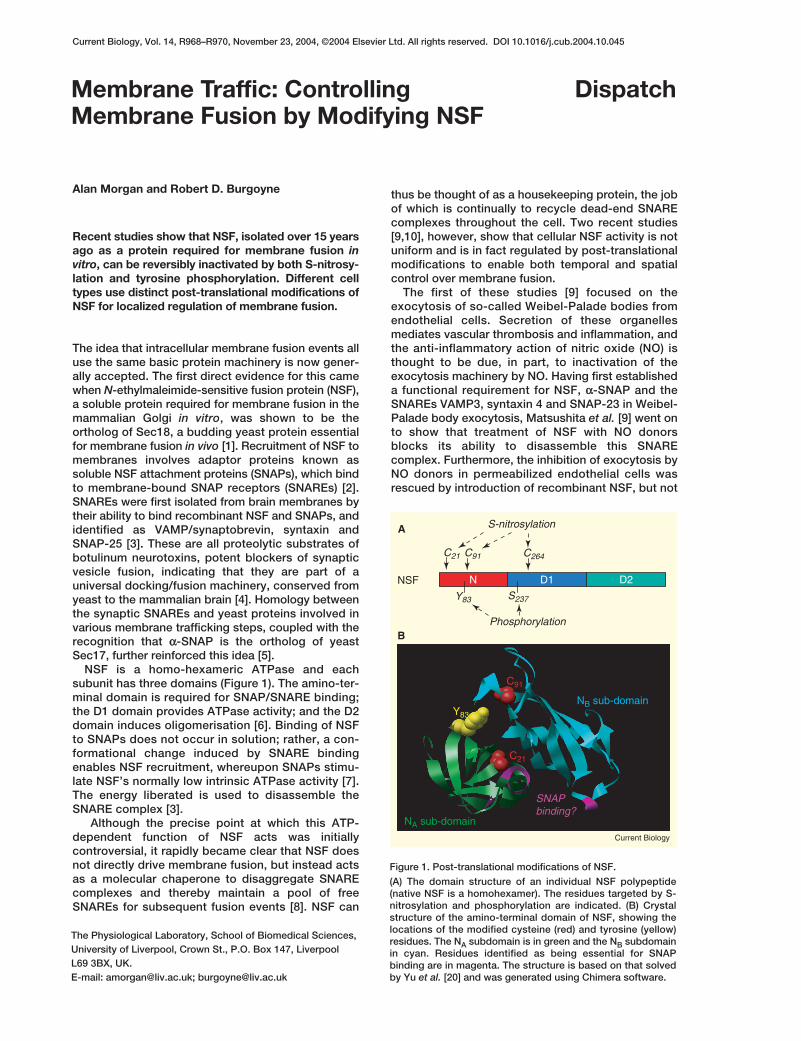
Figure 1. Post-translational modifications of NSF.
(A) The domain structure of an individual NSF polypeptide(native NSF is a homohexamer). The residues targeted by S-nitrosylation and phosphorylation are indicated. (B) Crystalstructure of the amino-terminal domain of NSF, showing thelocations of the modified cysteine (red) and tyrosine (yellow)residues. The NA subdomain is in green and the NB subdomainin cyan. Residues identified as being essential for SNAPbinding are in magenta. The structure is based on that solvedby Yu et al. [20] and was generated using Chimera software.
D1NNSF D2
C21
A
C91 C264
Y83
S-nitrosylation
Phosphorylation
S237
B
NA sub-domain
NB sub-domainY83
C21
C91
SNAPbinding?
Current Biology
NO-treated NSF, demonstrating that NSF is a majorcellular target of NO.
Importantly, S-nitrosylation of endogenous cellularNSF was inhibited by NO synthase antagonists andabsent in cells from endothelial NO synthase knockoutmice, strongly arguing that this post-translationalmodification is physiologically relevant. Mutagenesisof the nine cysteines in NSF implicated amino-domainresidues 21 and 91 and D1-domain residue 264 aslikely sites of S-nitrosylation (Figure 1). Analysis ofthese mutants in assays of SNARE complexdisassembly, however, while consistent with animportant role for Cys91 and Cys264 in this process,failed to identify convincingly the functionally impor-tant residue(s) targeted by NO.
Similarly, the mechanism by which NO preventsNSF-mediated SNARE complex disassembly remainsunclear. NO had no effect on SNAP binding or onNSF’s intrinsic ATPase activity, though the effect ofNO on the stimulation of NSF ATPase activity bySNAP was not examined. This biochemically distinctATPase activation is essential for efficient SNAREdisassembly, and indeed mutants of αα-SNAP [11] andNSF [12] that are defective in this function fail todisassemble SNARE complexes, despite havingnormal levels of intrinsic ATPase activity. The positionof Cys21 and Cys91 in the amino-domain crystalstructure is compatible with a role in the SNAP-induced stimulation of NSF ATPase activity (Figure 1),while Cys264 is located in the heart of the D1 domainWalker A box that is involved in ATP binding. So itmay be that NO blocks ATPase activation by SNAPs[13] (Figure 2), although this speculation remains tobe tested.
The focus for the second study [10] was the regula-tion of secretory vesicle size in cells of haemopoieticlineage. The starting point was the observation thatexpression of a secretory vesicle-localized proteintyrosine phosphatase, PTP-MEG2, induced theformation of enlarged secretory vesicles in RBL cellsand Jurkat T cells as a result of increased homotypicfusion of immature vesicles [14]. A search for tyrosine-phosphorylated substrates of PTP-MEG2 that mightunderlie this effect identified NSF and revealed Tyr83as the likely phosphorylation site [10].
Phosphorylation of NSF in vitro by the tyrosinekinase Fes had two effects: increased intrinsic ATPaseacitivity and decreased affinity for αα-SNAP. ReplacingTyr83 with glutamate had the same effect, strength-ening the in vitro phosphorylation data and also con-firming the phosphomimetic nature of this Y83Emutant. Tyr83 is located at the junction betweenNSF’s two amino-terminal sub-domains, as is theCys91 nitrosylation site (Figure 1), and phosphoryla-tion of this residue would likely disrupt the interfacebetween the sub-domains and hence affect SNAPbinding [10]. Treatment with the PTP inhibitor per-vanadate transiently reverses the increased vesicularsize induced by PTP-MEG2, indicating that tyrosinephosphorylation/dephosphorylation regulates adynamic cycle of homotypic vesicle fusion and fission.The observation that reformation of large vesicles fol-lowing washout of pervanadate was abolished by
NEM provided indirect evidence that tyrosine phos-phorylation of NSF might inhibit homotypic fusion.
The really key observation, however, was thatexpression of the putative dephospho-mimeticmutant Y83F induced the formation of large vesiclesin a strikingly similar manner to PTP-MEG2. Althoughit would have been interesting to push this further bytesting if the Y83F-expressing cells were now resis-tant to pervanadate, these results neverthelessstrongly suggest that homotypic fusion of secretoryvesicles is regulated by phosphorylation of NSF onTyr83. Presumably, inhibition of NSF by tyrosinekinases causes an accumulation of dead-end cis-SNARE complexes and thereby inhibits homotypicfusion, while local dephosphorylation via vesicularPTP-MEG2 antagonises this effect. The same phos-phorylation event appears also to affect heterotypicfusion in exocytosis, as expression of phospho-mimetic NSF-Y83E in Jurkat cells partially inhibitedinterleukin-2 secretion.
Taken together, these two new studies [9,10] firmlyestablish post-translational modification of NSF as animportant mechanism enabling localized inhibition ofmembrane fusion in endothelial and haemopoietic
Current BiologyR969
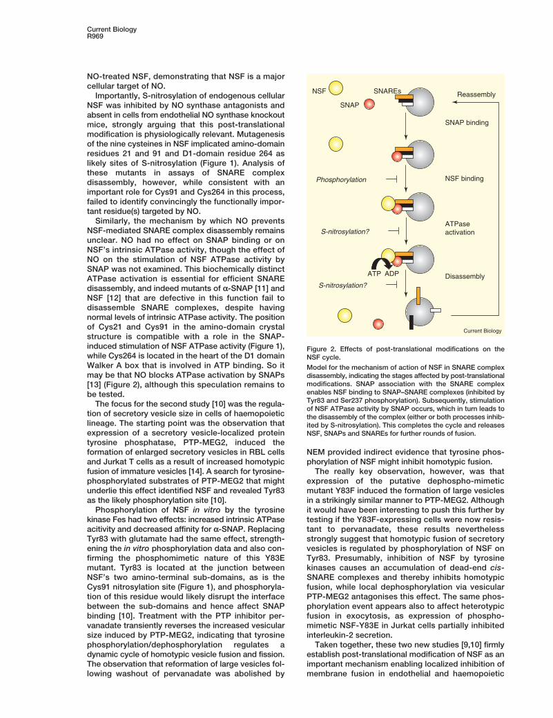
Figure 2. Effects of post-translational modifications on theNSF cycle.
Model for the mechanism of action of NSF in SNARE complexdisassembly, indicating the stages affected by post-translationalmodifications. SNAP association with the SNARE complexenables NSF binding to SNAP–SNARE complexes (inhibited byTyr83 and Ser237 phosphorylation). Subsequently, stimulationof NSF ATPase activity by SNAP occurs, which in turn leads tothe disassembly of the complex (either or both processes inhib-ited by S-nitrosylation). This completes the cycle and releasesNSF, SNAPs and SNAREs for further rounds of fusion.
NSF
SNAP
Phosphorylation
S-nitrosylation?
ATP ADP
SNAP binding
NSF binding
ATPaseactivation
Disassembly
ReassemblySNAREs
S-nitrosylation?
Current Biology
DispatchR970
cells. Interestingly, earlier work demonstrateddepolarisation-induced NSF phosphorylation in brainsynaptosomes [15], suggesting that such regulation ofNSF may also occur in neurons. In this case,phosphorylation of Ser237 by protein kinase Cinhibited NSF binding to the SNAP–SNARE complex[15] — the same biochemical effect as Tyr83 phos-phorylation [10]. Even transient and/or highly localizedinhibition of presynaptic NSF activity might potentiallyhave profound effects on synaptic transmission andplasticity, and intriguingly, both NO donors and Srcfamily tyrosine kinases have been shown to inhibitneurotransmitter release [16,17].
In addition to its presynaptic function in SNAREcomplex disassembly, NSF has an importantpostsynaptic role in regulating the surface expressionof GluR2 glutamate receptors [18], and has also beenshown to interact with various other proteins,including GABARAP, GATE-16, and ββ-arrestin [19].The recent work has shown how the specific localisa-tion of an appropriate enzyme involved in post-trans-lational modification can allow local regulation of aprotein with a wide spectrum of cellular roles. It will beinteresting to see if other membrane traffic steps orthe SNARE-independent functions of NSF are alsolocally regulated by nitrosylation, phosphorylation andindeed other post-translational modifications of NSF.
References1. Wilson, D.W., Wilcox, C.A., Flynn, G.C., Chen, E., Kuang, W.,
Henzel, W.J., Block, M.R., Ullrich, A., and Rothman, J.E. (1989). Afusion protein required for vesicle-mediated transport in both mam-malian cells and yeast. Nature 339, 355–359.
2. Wilson, D.W., Whiteheart, S.W., Wiedmann, M., Brunner, M., andRothman, J.E. (1992). A multisubunit particle implicated in mem-brane fusion. J. Cell Biol. 117, 531–538.
3. Sollner, T., Whiteheart, S.W., Brunner, M., Erdjument-Bromage, H.,Geromanos, S., Tempst, P., and Rothman, J.E. (1993). SNAP recep-tors implicated in vesicle targeting and fusion. Nature 362, 318–324.
4. Ferro-Novick, S., and Jahn, R. (1994). Vesicle fusion from yeast toman. Nature 370, 191–193.
5. Rothman, J.E. (1994). Mechanisms of intracellular protein transport.Nature 372, 55–62.
6. Whiteheart, S.W., Schraw, T., and Matveeva, E.A. (2001). N-ethyl-maleimide sensitive factor (NSF) structure and function. Int. Rev.Cytol. 207, 71–118.
7. Morgan, A., Dimaline, R., and Burgoyne, R.D. (1994). The ATPaseactivity of N-Ethylmaleimide-sensitive fusion protein (NSF) is regu-lated by soluble NSF attachment proteins. J. Biol. Chem. 269,29347–29350.
8. Morgan, A., and Burgoyne, R.D. (1995). Is NSF a fusion protein?Trends Cell Biol. 5, 335–339.
9. Matsushita, K., Morrell, C.N., Cambien, B., Yang, S.X., Yamakuchi,M., Bao, C., Hara, M.R., Quick, R.A., Cao, W., O’Rourke, B., et al.(2003). Nitric oxide regulates exocytosis by S-nitrosylation of N-eth-ylmaleimide-sensitive factor. Cell 115, 139–150.
10. Huynh, H., Bottini, N., Williams, S., Cherepanov, V., Musumeci, L.,Saito, K., Bruckner, S., Vachon, E., Wang, X., Kruger, J., et al. (2004).Control of vesicle fusion by a tyrosine phosphatase. Nat. Cell Biol.6, 831–839.
11. Barnard, R.J.O., Morgan, A., and Burgoyne, R.D. (1997). Stimulationof NSF ATPase activity by a-SNAP is required for SNARE complexdisassembly and exocytosis. J. Cell Biol. 139, 875–883.
12. Horsnell, W.G., Steel, G.J., and Morgan, A. (2002). Analysis of NSFmutants reveals residues involved in SNAP binding and ATPasestimulation. Biochemistry 41, 5230–5235.
13. Sollner, T.H., and Sequeira, S. (2003). S-nitrosylation of NSF con-trols membrane trafficking. Cell 115, 127–129.
14. Wang, X., Huynh, H., Gjorloff-Wingren, A., Monosov, E., Stridsberg,M., Fukuda, M., and Mustelin, T. (2002). Enlargement of secretoryvesicles by protein tyrosine phosphatase PTP-MEG2 in ratbasophilic leukemia mast cells and Jurkat T cells. J. Immunol. 168,4612–4619.
15. Matveeva, E.A., Whiteheart, S.W., Vanaman, T.C., and Slevin, J.T.(2001). Phosphorylation of the N-ethylmaleimide-sensitive factor isassociated with depolarization-dependent neurotransmitter releasefrom synaptosomes. J. Biol. Chem. 276, 12174–12181.
16. Stanton, P.K., Winterer, J., Bailey, C.P., Kyrozis, A., Raginov, I.,Laube, G., Veh, R.W., Nguyen, C.Q., and Muller, W. (2003). Long-term depression of presynaptic release from the readily releasablevesicle pool induced by NMDA receptor-dependent retrogradenitric oxide. J. Neurosci. 23, 5936–5944.
17. Ohnishi, H., Yamamori, S., Ono, K., Aoyagi, K., Kondo, S., and Taka-hashi, M. (2001). A src family tyrosine kinase inhibits neurotrans-mitter release from neuronal cells. Proc. Natl. Acad. Sci. USA 98,10930–10935.
18. Haas, A. (1998). NSF-fusion and beyond. Trends Cell Biol. 8,471–473.
19. Whiteheart, S.W., and Matveeva, E.A. (2004). Multiple binding pro-teins suggest diverse functions for the N-ethylmaleimide sensitivefactor. J. Struct. Biol. 146, 32–43.
20. Yu, R.C., Jahn, R., and Brunger, A.T. (1999). NSF N-terminal domaincrystal structure: models of NSF function. Mol. Cell 4, 97–107.