Embed Size (px)
Citation preview
Melatonin Attenuates Chronic Cough Mediated by Oxidative Stress 1
via Transient Receptor Potential Melastatin-2 in Guinea Pigs 2
Exposed to Particulate Matter 2.5 3
Zhenjun Ji1, Zhen Wang1, Zhe Chen2, Hao Jin1, Chen Chen1, Senlin Chai1, Haining 4
Lv1, Ling Yang1, Yakun Hu1, Rong Dong1*, Kefang Lai2* 5
1Medical School, Southeast University, Nanjing, China 6
2State Key Laboratory of Respiratory Disease, Guangzhou Institute of Respiratory 7
Disease, The First Affiliated Hospital of Guangzhou Medical University, Guangzhou, 8
China 9
Corresponding authors: 10
*Kefang Lai, State Key Laboratory of Respiratory Disease, The First Affiliated 11
Hospital of Guangzhou Medical University, Guangzhou Institute of Respiratory 12
Disease, 151 Yanjiang Rd., Guangzhou, China. Email: [email protected]. 13
*Rong Dong, Medical School of Southeast University, 87 Dingjiaqiao, Nanjing 14
210009. Email: [email protected]. 15
Short Title: Melatonin Attenuates Chronic Cough induced by PM2.5 Exposure 16
Conflict of Interest 17
There is no conflict of interest. 18
Acknowledgements 19
This work was supported by Grants 2016OP015 from Open Project of State Key 20
Laboratory of Respiratory Disease. Authors would like to thank Xinjian Zhu for his 21
assistance of experiment. 22
23
Summary 24
The aim of this study was to investigate the effects of melatonin on oxidative stress, 25
the expression of transient receptor potential melastatin-2 (TRPM2) in guinea pig 26
brains, and the influence of melatonin on oxidative stress in lungs and airway 27
inflammation induced by particulate matter 2.5 (PM2.5). A particle suspension (0.1 28
g/ml) was nasally administered to the guinea pigs to prepare a PM2.5 exposure model. 29
Cough frequency and cough incubation period were determined through RM6240B 30
biological signal collection and disposal system. Oxidative stress markers, including 31
malondialdehyde (MDA), total antioxidant capacity (T-AOC), total superoxide 32
dismutase (T-SOD), and glutathione peroxidase (GSH-Px), in the medulla oblongata 33
were examined through spectrophotometer. Reactive oxygen species (ROS) were 34
detected in the hypoglossal nucleus, cuneate nucleus, Botzinger complex, dorsal vagal 35
complex, and airway through dihydroethidium fluorescence. Hematoxylin-eosin (HE) 36
staining and substance P expression via immunohistochemistry revealed the 37
inflammatory levels in the airway. TRPM2 was observed in the medulla oblongata 38
through immunofluorescence and western blot. The ultrastructure of the blood–brain 39
barrier and neuronal mitochondria was determined by using a transmission electron 40
microscope. Our study suggests that melatonin treatment decreased PM2.5-induced 41
oxidative stress level in the brains and lungs and relieved airway inflammation and 42
chronic cough. TRPM2 might participate in oxidative stress in the cough center by 43
regulating cough. 44
Key words: melatonin, oxidative stress, cough sensitivity 45
46
Introduction 47
Ambient particulate matter 2.5 (PM2.5) is defined as airborne particulate matter with 48
aerodynamic diameters less than 2.5μm (Chen et al. 2016). PM2.5 can transport 49
directly into the alveoli which is mainly derived from oil and coal combustion and 50
vehicle emissions; this pollutant is composed of elemental carbon, organic carbon, 51
aluminum, copper, nickel, sulfates, nitrates, polycyclic aromatic hydrocarbons (PAH) 52
and other compounds (WHO 2013). Air pollutants, including PM2.5, are closely 53
related to respiratory symptoms and diseases, such as chronic cough, chronic 54
obstructive pulmonary diseases (COPD), asthma, and lung cancer (Falcon-Rodriguez 55
et al. 2016; Zhang et al. 2015). Chronic cough is defined as cough lasting more than 56
eight weeks without abnormal pulmonary function, and cough hypersensitivity is a 57
key pathophysiological mechanism of chronic cough (Lai et al. 2013). However, the 58
exact mechanism of cough hypersensitivity is unclear. 59
Cachon et al (Cachon et al. 2014) found that human lung epithelial cells secreted 60
more cytokines including IL-1β、IL-6 and TNF-α after exposed to PM2.5. PM2.5 61
could enter the blood through the alveolus–capillary barrier and penetrate the 62
blood–brain barrier (BBB) or migrate via the olfactory nerve pathway into the brain 63
and cause neuroinflammation, oxidative stress, and neuronal damage (Bos et al. 2014). 64
PM2.5, is also related to central nervous system (CNS) diseases, such as 65
neurodegenerative diseases. Levesque et al.(Levesque et al. 2011) demonstrated that 66
subchronic exposure to diesel engine exhaust causes neuroinflammation and thus 67
increases the expression of α-synuclein, an early marker of neurodegenerative 68
diseases. Calderon-Garciduenas et al. (Calderon-Garciduenas et al. 2015a; 69
Calderon-Garciduenas et al. 2015b) found that neurodegenerative diseases in children 70
are triggered by exposure to particulate matters and ozone, and environmental factors 71
and gene factors promote Alzheimer’s disease. Cough is a special kind of respiratory 72
activity and the medulla oblongata receives sensory signals from the cough receptors 73
in airways via vagus nerves and superior laryngeal nerves as the respiratory center. 74
The respiratory center comprises the Botzinger complex, pons respiratory group, 75
solitary nucleus group, and raphe nucleus group. These neurons in respiratory nuclei 76
which participate in the integration of the afferent cough information are thought as 77
central cough generator, and further regulate the motion of muscles in airways and 78
generate cough (Chung and Pavord 2008). Signals from periphery are also 79
subsequently transmitted into the superior center above the brainstem and it alters 80
emotion and cognition brain regions. Further studies have not yet to clarify whether 81
central nuclei generate pathological changes induced by PM2.5 and affect chronic 82
cough development. 83
84
PM2.5 stimulates the release of endogenous and exogenous free radicals, including 85
reactive oxygen species (ROS) in the CNS; as a result, the cause of 86
oxidant/antioxidant systems imbalance (Fagundes et al. 2015; Liu et al. 2015). Direct 87
PM2.5 exposure likely induces oxidative stress and neurotoxicity in the hippocampus 88
and cerebellum (Fagundes et al. 2015). As a multifunctional non-selective cation 89
channel with N-domain, the transient potential receptor melastatin-2 (TRPM2) 90
channel exhibits a pyrophosphatase activity, which can be activated by ROS and 91
adenosine diphosphatase ribose (ADPR), and functions as a sensor for oxidative stress. 92
TRPM2 is distributed mainly in different parts of the mammalian brain, including the 93
hippocampus, cortex, thalamus, midbrain, and medulla oblongata; this channel is also 94
abundant in neurons and microglia (Naziroglu 2011; Ru and Yao 2014). High TRPM2 95
expression mediates extracellular calcium entry and induces cell death. TRPM2 is 96
also involved in many disorders, such as traumatic brain injury (Yuruker et al. 2015), 97
cerebral ischemia (Akpinar et al. 2016), type II diabetes (Sozbir and Naziroglu 2016), 98
cancers, inflammation, and neurodegenerative diseases (Naziroglu 2011). As such, 99
TRPM2 is a potentially effective target of many diseases. However, studies have not 100
yet to determine whether TRPM2 could mediate the generation of oxidative stress in 101
the respiratory center and airway, and further induce airway inflammation and cough 102
hypersensitivity. 103
Melatonin, secreted by the pineal gland, regulates the sleep, circadian rhythms and act 104
as an effective antioxidant. In various pathological states, melatonin and its 105
metabolites can function as endogenous free radical scavengers and broad-spectrum 106
antioxidants to scavenge ROS. Melatonin also elicits a neuroprotective effect and 107
protects the vascular endothelium by scavenging ROS and inhibiting 108
pro-inflammatory cytokine release and Ca2+ overload (Akpinar et al. 2016; Kahya et 109
al. 2017; Kaisar et al. 2015; Ma et al. 2015). Melatonin was hypothesized to alleviate 110
chronic cough which serve as a potential therapeutic drug for chronic cough. 111
112
This study was aimed to investigate whether melatonin could alleviate chronic cough 113
induced by PM2.5 through relieving oxidative stress and also determine whether 114
TRPM2 participates in oxidative stress in the brain and influences airway oxidative 115
stress and airway inflammation. 116
117
Materials and Methods 118
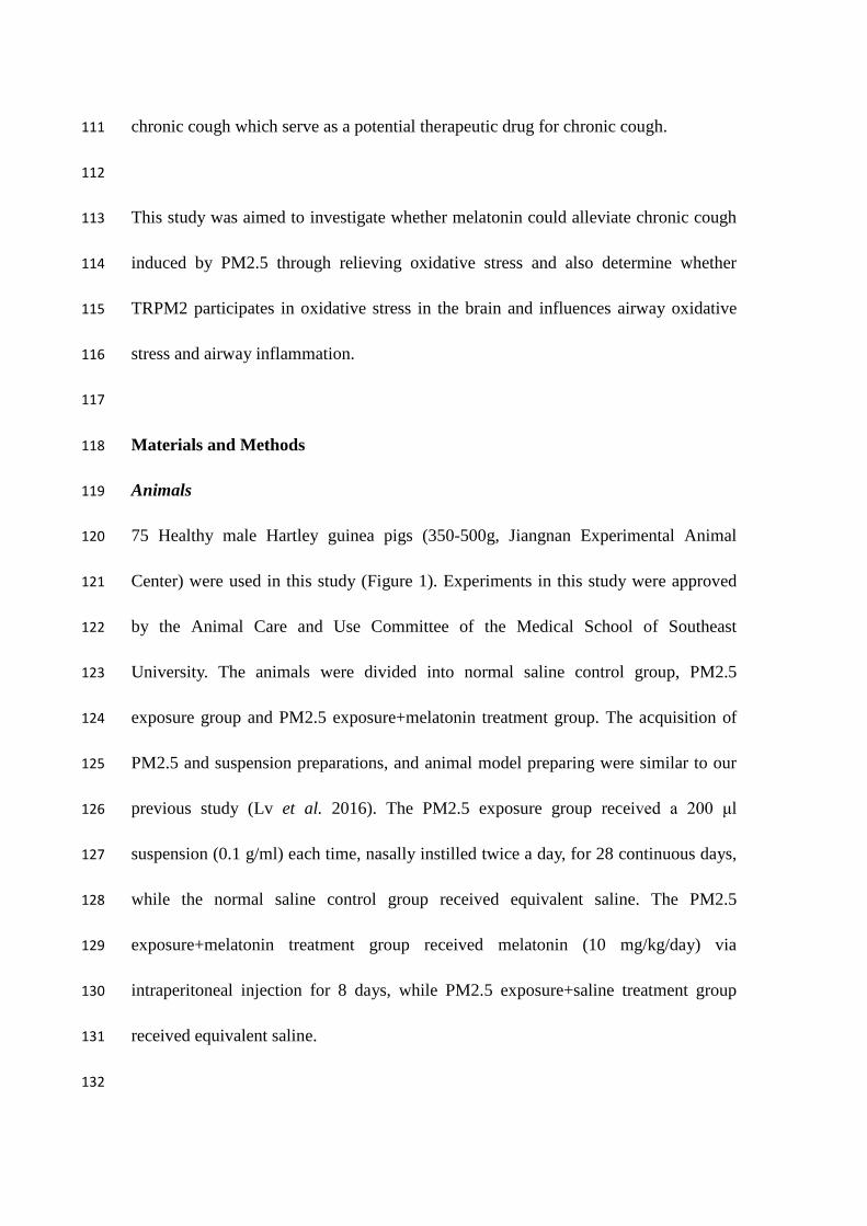
Animals 119
75 Healthy male Hartley guinea pigs (350-500g, Jiangnan Experimental Animal 120
Center) were used in this study (Figure 1). Experiments in this study were approved 121
by the Animal Care and Use Committee of the Medical School of Southeast 122
University. The animals were divided into normal saline control group, PM2.5 123
exposure group and PM2.5 exposure+melatonin treatment group. The acquisition of 124
PM2.5 and suspension preparations, and animal model preparing were similar to our 125
previous study (Lv et al. 2016). The PM2.5 exposure group received a 200 μl 126
suspension (0.1 g/ml) each time, nasally instilled twice a day, for 28 continuous days, 127
while the normal saline control group received equivalent saline. The PM2.5 128
exposure+melatonin treatment group received melatonin (10 mg/kg/day) via 129
intraperitoneal injection for 8 days, while PM2.5 exposure+saline treatment group 130
received equivalent saline. 131
132

Cough sensitivity examination 133
Cough frequency and cough incubation period were measured by RM6240B 134
biological signal collection and disposal system (Chengdu, China). We prepared a 135
0.45 M citric acid solution. Guinea pigs were placed in a cough recorder of sound 136
energy, and the normal waveform was recorded for 10 s after the ultrasonic atomizer 137
was opened and sprayed for 15 s into the recorder. We recorded cough frequency of 138
guinea pigs within 10 min and time from the citric acid spray to the first cough of 139
animals (cough incubation periods) to assess the cough reflex sensitivity in the airway 140
of guinea pigs. 141
142
Tissue preparation 143
For immunofluorescence and dihydroethidium (DHE) fluorescence, guinea pigs were 144
anesthetized by intraperitoneal injection of pentobarbital sodium (40 mg/kg), and then 145
perfused and fixed with phosphate buffer containing 4% paraformaldehyde through 146
the heart after chest opening. Then, the brain and lung tissues were placed in the fixed 147
liquid for 8 h, which were immersed in 30% sucrose solution for using after that. 148
149
HE staining 150
Lung tissues were embedded in paraffin wax after fixation in 4% paraformaldehyde 151
for HE staining, and observed under light microscopy. 152
153
Immunofluorescence 154
The medulla oblongata was cut into continuous coronal slices (30 μm thickness per 155
slice) from 2 mm below to 3 mm above the obex using a microtome (Leica). The 156
slices were placed in sequence into phosphate buffered solution (PBS) containing 157
0.4% Triton X-100, PBS containing 10% goat serum (Abcam), followed by 158
incubation with a rabbit polyclonal anti-TRPM2 antibody (1:200, Abcam) at 4 °C 159
overnight, then rinsed in PBS for 5 min. Subsequently, the sections were incubated 160
with a goat anti-rabbit IgG/AlexaFluor 594 secondary antibody (1:400, Invitrogen) at 161
room temperature in the dark for 2 h, followed by PBS rinsing (5 min, three times), 162
then dyed by DAPI for 1 min and rinsed in PBS for 5 min. Finally, the slices were 163
sealed with glycerol for observation by fluorescence microscope. The slices were 164
observed by an Olympus fluorescence microscope (Olympus LSM-GB200, Japan) 165
and cell number analysis was performed by Image-Pro Plus (Media Cybernetics, 166
USA). Five random slices per animal were chosen for counting at a higher 167
magnification (200×). The average number of five slices represented the value per 168
animal. 169
170
Immunohistochemistry 171
Lung tissue (size of 0.8 cm×0.6 cm×0.2 cm) was cut into continuous coronal slices 172
(45 μm thickness per slice). Briefly, the slices were placed in deionized water 173
containing 3% H2O2, rinsed in PBS (5 min, three times), incubated in rabbit serum 174
(Abcam) at room temperature for 4 h, followed by incubation with goat anti-SP 175
polyclonal antibody (1:200, Santa Cruz) at 4 °C overnight, then rinsed in PBS for 5 176
min. The slices were sequentially incubated with biotinylated rabbit anti-goat IgG 177
(Boster) for 2 h, rinsed in PBS (5 min, three times), incubated with SABC (Boster) for 178
1 h, and followed by PBS rinsing (5 min, three times). The slices were stained with 179
DAB (ZSGB-BIO) and dehydrated using graded ethanol and dimethylbenzene. The 180
slices were sealed with a neutral resin under a microscope. Image-Pro Plus was used 181
to calculate mean optical density (MOD) of SP in the lungs. The five random slices 182
per animal were chosen for calculation and the mean data of five slices represented 183
the value per animal. 184
185
DHE fluorescence 186
Tissues were prepared as discussed above. The slices were sequentially placed into 187
PBS containing 0.4% Triton X-100, followed by incubation with 2 μM DHE working 188
solution at 37 °C for 40 min in the dark, and rinsing with PBS (5 min, three times). 189
Finally, the slices were sealed with glycerol for observation by fluorescence 190
microscope. Cell counting and fluorescence intensity were performed as described 191
above. 192
193
Measurement of peroxide and antioxidant enzymes 194
The guinea pigs were decapitated directly and the medulla oblongata was rapidly 195
stripped. Tissues were mechanically homogenized and mixed with saline using a ratio 196
of weight (g): volume (ml)=1:9 under an ice-water bath. The homogenates were 197
centrifuged at 2500 rpm for 10 min. Furthermore, protein content of supernatant was 198
measured by a 720 spectrophotometer. All procedures referred to the specifications of 199
malondialdehyde (MDA)/total antioxidant capacity (T-AOC)/total superoxide 200
dismutase (T-SOD)/glutathione peroxidase (GSH-Px) kits (Jiancheng, Nanjing, 201
China). 202
203
Western blot 204
The medulla oblongata tissues of guinea pigs were homogenized in a lysis buffer 205
(Beyotime, China) and were then centrifuged at 12,000 rpm for 20 min. Then, the 206
samples were placed in 12% acrylamide denaturing gels (SDS-PAGE) and transferred 207
to nitrocellulose membranes (Sigma) after electrophoresis, followed by incubation for 208
1 h at room temperature with 5% non-fat dry milk in Tris Buffered Saline Tween 209
(TBST). Then, the membranes were sequentially incubated with rabbit anti-TRPM2 210
(1:2000, Abcam, USA) and HRP-linked goat anti-rabbit antibody (1:5000, Invitrogen). 211
Signals were captured by Microchemi chemiluminescent image analysis system 212
(DNR Bio-imaging Systems, Jerusalem, Israel) after handling with the enhanced 213
chemiluminescence method. Blots were quantified by Image-Pro Plus. 214
215
Transmission electron microscopy 216
Guinea pigs were anesthetized with 10% chloral hydrate (3 ml/kg to 4 mL/kg, ip) and 217
perfused with normal saline, followed by phosphate buffer containing 4% 218
paraformaldehyde and 0.5% glutaraldehyde (Sigma). Tissues in dorsal vagal complex 219
(DVC) were diced and fixed in 2.5% chilled glutaraldehyde. A 1 mm3 tissue block 220
was post-stained with uranyl acetate and lead citrate. Tissue sections were cut to 221
50-nm thicknesses and observed under a transmission electron microscope (JEOL, 222
JEM-1010, Japan). 223
224
Statistical analysis 225
All data are analyzed by SPSS 19.0 and presented as the means ± S.E.M. An 226
independent-samples T-test was used to compare between two groups. Values of 227
p<0.05 were considered statistically significant. 228
229
Results 230
Cough sensitivity examination 231
The cough frequency of the saline group, PM2.5 exposure group, and PM2.5 232
exposure+melatonin treatment groups were 8.46±1.02, 25.92±2.74, 18.77±1.77, 233
respectively, while the cough incubation periods were 68.46±8.38, 35.69±6.17, 234
57.77±7.26. Compared with the saline group, the PM2.5 exposure group showed 235
increased cough frequency (p<0.001) and decreased cough incubation period 236
(p<0.01). After melatonin treatment, the PM2.5 exposure+melatonin treatment group 237
showed decreased cough frequency (p<0.05) and increased cough incubation period 238
(p<0.05) (Fig 2). The results indicated that PM2.5 exposure increased cough 239
sensitivity which was prevented by melatonin. 240
241
Airway neurogenic inflammation and oxidative stress 242
HE staining showed mucosa edema of the trachea and inflammatory cells infiltration, 243
such as increased neutrophils, monocytes, lymphocytes and eosinophils, indicating 244
exacerbated airway inflammation, which was relieved after melatonin treatment (Fig. 245
3A). Immunoreactive substances of airway neurogenic inflammatory mediator 246
substance P were dyed brown granules with cytoplasm staining and were mainly 247
distributed around the airway. The MOD of substance P in the saline group, PM2.5 248
exposure group, and PM2.5 exposure+melatonin group were 0.19±0.03, 0.42±0.05, 249
0.21±0.04, respectively. Compared with that in the saline group, the substance P 250
expression increased after PM2.5 exposure (p<0.01), while substance P expression 251
decreased after melatonin treatment (p<0.05) (Fig. 3B 3D). These results 252
demonstrated that PM2.5 induced airway inflammation and melatonin had an 253
antagonistic effect towards inflammation. DHE fluorescence of airway showed that 254
fluorescence intensity of airway in PM2.5 exposure group is higher than that of the 255
control group, which were decreased by melatonin (p<0.05) (Fig. 3C 3E). The MOD 256
of ROS in the saline group, PM2.5 exposure group, and PM2.5 exposure+melatonin 257
group were 0.05±0.00, 0.06±0.02, 0.05±0.02, respectively. These results indicate that 258
PM2.5 exposure elevates the level of oxidative stress in the airway of guinea pigs, 259
which were decreased by melatonin. 260
261
Injury of BBB and neurons 262
The ultrastructure in the DVC observed by transmission electron microscope showed 263
normal microvascular endothelium, astrocytes, and mitochondria structure with 264
double membrane, and parallel arrangement of mitochondrial cristae in control group. 265
The PM2.5 exposure group showed isolated microvascular endothelium, mild edema 266
of astrocytes, and narrow vessels, which indicated injury of BBB. Besides, 267
mitochondria were swollen and vacuolate,and were in disarray, with a decreased 268
number of mitochondria and mitochondrial cristae. Essentially, damage to the BBB 269
and mitochondria was relieved after intervention of melatonin (Fig. 4). 270
271
Oxidative stress in medulla oblongata 272
MDA represented the level of free radicals as lipid peroxides, while levels of 273
T-AOC/T-SOD/GSH-Px represented the ability of scavenging free radical as 274
antioxidant enzymes. Compared with the saline group, the PM2.5 exposure group 275
showed increased levels of MDA representing the level of oxidation and decreased 276
levels of T-AOC/GSH-Px representing antioxidant levels. However, decrease of 277
T-SOD was not obvious significantly (p<0.1). PM2.5 exposure+melatonin group 278
showed obviously decreased level of MDA and increased levels of 279
T-AOC/T-SOD/GSH-Px (Fig 5A). 280
DHE marks the cytoplasm and nuclei of dead neurons producing ROS red (excitation 281
535 nm, emission 610 nm). Compared with the saline group, the PM2.5 exposure 282
group showed an increased production of ROS in the hypoglossal nucleus, cuneate 283
nucleus, Botzinger complex, and DVC (p<0.05). Meanwhile, ROS production in 284
PM2.5 exposure+melatonin group was decreased in relevant nuclei (p<0.05). Our 285
data showed that PM2.5 exposure induced oxidative stress in the medulla oblongata 286
and melatonin treatment decreased the generation of ROS (Fig. 5B 5C). 287
288
TRPM2 expression in medulla oblongata 289
The cytoplasms of immunoreactive neurons of TRPM2 were stained red under a 290
fluorescence microscope. Compared with the saline group, the PM2.5 exposure group 291
showed an increased expression of TRPM2 in the DVC and Botzinger complex 292
(p<0.01). Melatonin treatment decreased the expression of TRPM2 (p<0.05) (Fig. 6A 293
6B). Western blot showed that the protein level of TRPM2 in PM2.5 exposure group 294
was higher than the saline and melatonin groups (p<0.05) (Fig. 6C 6D). 295
296
Discussion 297
In our study, the particles were collected from five urban areas, including commercial 298
areas, business areas, busy traffic intersections, industrial districts, and suburbs (Lv et 299
al. 2016), and were mixed to represent the actual constituents of environmental 300
PM2.5. 301
302
The increased cough sensitivity of guinea pigs after PM2.5 exposure showed that 303
PM2.5 elicited chronic cough. We also found that PM2.5 exposure promoted the 304
infiltration of inflammatory cells, including neutrophils, monocytes, lymphocytes, and 305
eosinophils. Substance P expression was also increased which consequently induced 306
the neurogenic inflammation in the airway; as a result, cause of airway inflammation 307
which was consistent with the inflammatory phenomena of gastroesophageal reflux 308

cough (GERC), cough variant cough (CVA) and chronic cough with definite etiology. 309
Generally, guinea pigs after PM2.5 exposure showed cough hypersensitivity and the 310
same pathological characteristics as chronic cough with definite etiology, which 311
demonstrated that PM2.5 induced chronic cough. Airway neurogenic inflammation 312
was induced by neuropeptides or neurotransmitters released by airway sensory nerve 313
terminal, characterized by increased vascular permeability, plasma extravasation, and 314
tissue edema. Substance P, Calcitonin Gene-Related Peptide (CGRP), and other 315
substances stimulate vasodilation, plasma leakage, and mucus secretion, increase 316
cough sensitivity, and trigger chronic cough. We further demonstrated that PM2.5 317
could increase oxidative stress levels, which could enhance the expression of 318
inflammatory factors, such as interleukin (IL), via the NF-ΚB pathway and 319
accumulate inflammatory cells. Airway inflammation and oxidative stress are two 320
possible mechanisms exacerbating chronic cough, and ROS and inflammation are 321
closely associated. 322
323
Cough is a special respiratory activity and respiratory defensive reflex. The 324
respiratory center is located mostly in the medulla oblongata of brainstem, comprising 325
the dorsal respiratory group with the solitary nucleus, and the ventral respiratory 326
group containing the Botzinger complex. The stimuli of cough receptors including 327
slowly and rapidly adapting stretch receptors (SAR and RAR) and C-fiber in the 328
airway, are transmitted to the solitary nucleus via the glossopharyngeal and vagus 329
nerves to regulate the basic rhythm of inhalation. The DVC is composed of the dorsal 330
nucleus of the vagus nerve (DMV), the nucleus of the solitary tract (NTS), and the 331
area postrema. The DMV, hypoglossal nucleus, and nucleus ambiguus receive afferent 332
fibers from the NTS (Kubin et al. 2006). The neuronal axons of the Botzinger 333
complex projections are extensive in various regions of the medulla oblongata and 334
spinal cord associated with breathing, and these axons inhibit the discharge of 335
inhalation neurons during the expiratory phase to participate in maintaining the 336
expiratory phase. Therefore, the mutual association between the central nervous 337
system and airway has the neuroanatomical foundation. Afferent stimuli in the airway 338
can reach the cough center in the brainstem including the DVC and the ventral 339
Botzinger complex in the medulla oblongata through the sensory afferent nerve. The 340
cough center also regulates respiratory and throat muscles after signals are integrated 341
and further generates cough (Mazzone. and Undem. 2017). 342
343
BBB consists of vascular endothelial cell, tight junction between endothelium, 344
astrocyte, pericyte and basement membrane. In the ultrastructure observed with a 345
transmission electron microscope, isolated microvascular endothelium, mild edema of 346
astrocytes, and narrow vessels were present, and this observation indicated that PM2.5 347
could damage the BBB. Fang et al. (Liu et al. 2015) found that PM2.5 disrupts the 348
tight junction of endothelial cells, increases penetrability, and enhances monocyte 349
migration ability and demonstrated that PM2.5 can reach the CNS through the BBB; 350
the neurotoxicity of PM2.5 is also mediated by glutamate. Swollen and vacuolate 351
mitochondria were also observed in the ultrastructure, and the number of the 352
mitochondria and mitochondrial cristae was decreased. These findings suggested that 353
PM2.5 induced mitochondrial injury, which could trigger imbalance in calcium 354
homeostasis and energy metabolism in cells, and neuronal injury or apoptosis. The 355
leakage of the mitochondrial electron transport chain is also a vital source of 356
intracellular ROS, leading to oxidant/antioxidant imbalance (Redza-Dutordoir and 357
Averill-Bates 2016). We also found that the MDA level was increased, whereas 358
T-SOD, GSH-Px, and T-AOC levels were decreased after PM2.5 exposure. DHE 359
fluorescence demonstrated that ROS production was increased in DVC, Botzinger 360
complex, hypoglossal nucleus, and cuneate nucleus. These observations showed that 361
the medulla oblongata was in a state of high oxidative stress after PM2.5 exposure. It 362
was reported that cerebral ischemia, epilepsy, and trauma result in the generation of 363
oxidative stress (Halliwell 2006), which was related to BBB endothelial damage and 364
was relieved by antioxidants (Kaisar et al. 2015). Oxidative stress is also relevant to 365
neurodegenerative diseases. Children exposed to air pollutants, including PM2.5, in 366
Mexico City Metropolitan Area manifest signs and symptoms of early oxidative stress, 367
inflammation, innate and adaptive immunity-related genes, and BBB disruption, 368
resulting in the early neurodegenerative changes in children (Calderon-Garciduenas et 369
al. 2015b). Hence, during the process of air pollutants inducing chronic cough, the 370
activity of the cough reflex is strengthened, and oxidative stress in the center is 371
enhanced as the generation of airway inflammation. Further, the cough center is 372
remodeled, and chronic cough generated. 373
374
TRPM2 is a cation channel that exhibits oxidative stress sensitivity and mediates 375
oxidative stress-induced cell death via Ca2+ overload (Ru and Yao 2014). Our 376
immunofluorescence results revealed that PM2.5 exposure increased the TRPM2 377
expression in respiratory nuclei, Botzinger complex, and DVC, whereas oxidative 378
stress levels were increased. These findings were also confirmed through western blot. 379
The TRPM2 channel consists of six transmembrane segments with a pore-forming 380
loop between segments 5 and 6. The N-terminal of TRPM2 contains an IQ-like 381
calmodulin-binding motif and the C-terminal comprises a Nudix-like domain, which 382
can be bound by ADPR. Calcium overload after TRPM2 activation in microglia and 383
astrocytes can lead to mitochondrial dysfunction, and calcium overoad is also related 384
to synaptic plasticity change and dementia (Wang et al. 2016a). TRPM2-mediated 385
Ca2+ influx activated by oxidative stress inhibits autophagy (Wang et al. 2016b). 386
387
We utilized antioxidant melatonin to elucidate the role of ROS and TRPM2 in the 388
guinea pig model of PM2.5 exposure. Melatonin treatment decreased the oxidative 389
stress level in the medulla oblongata, the TRPM2 expression in the Botzinger 390
complex and DVC. These findings indicated that ROS might be involved in TRPM2 391
activation. Vehbi Yürüker et al. (Yuruker et al. 2015) also found that melatonin 392
alleviates oxidative stress and apoptosis by inhibiting Ca2+ and TRPM2 channels in 393
the hippocampus of rats with traumatic brain injury; this observation suggests that 394
melatonin exhibits neuroprotective activity. The GSH-Px levels changed most 395
obviously possibly because melatonin maintains the glutathione balance by 396
stimulating the generation of glutathione peroxidase, glutathione reductase, and 397
glucose-6-phosphate dehydrogenase (Reiter et al. 2000). Hence, melatonin could 398
upregulate the levels of T-SOD/GSH-Px/glutathione reductase by eliciting an indirect 399
antioxidant effect and by directly scavenging free radicals and reducing MDA 400
production. Mehmet Cemal et al. (Kahya et al. 2017) demonstrated that melatonin 401
and selenium can prevent the apoptosis of neurons in the hippocampus and dorsal root 402
ganglion of diabetic rats by reducing ROS and calcium influx associated with TRPM2 403
and TRPV1. Melatonin can also function at the genetic level to prevent DNA 404
degradation and activate DNA repair enzyme. Melatonin binding sites are also found 405
in cell nuclei. Melatonin metabolites, namely, 406
N-1-acetyl-N-2-formyl-5-methoxykynuramine and N-1-acetyl-5-methoxykynuramine, 407
induce a similar antioxidant effect to melatonin. Transmission electron microscopy 408
revealed that melatonin relieved the injuries of BBB and mitochondria in the DVC. 409
The melatonin transporter located on the mitochondrial outer membrane and 410
melatonin can be secreted by mitochondria and induce an agglomeration effect. The 411
concentration of melatonin in the mitochondria is higher than that in other organelles. 412
Melatonin can inhibit the mitochondrial permeability transition pore (MPTP) in the 413
mitochondrial membrane and activate uncoupling proteins (UCPs). The inhibition of 414
MPTP can maintain the mitochondrial membrane potential ( △Ψ), which can 415
reduced by UCPs; thus, electron transfer can be accelerated, the efficiency of the 416
mitochondrial electron transport chain can be improved, and electron leakage and 417
ROS production can be reduced. Moreover, melatonin can prevent neuronal apoptosis 418
by decreasing the levels of pro-apoptotic factors and by activating JAK2/STAT3 and 419
BCL2 pathways (Ganie et al. 2016; Tan et al. 2016). The antioxidant edaravone can 420
also alleviate brain injuries of acute CO poisoning rats (Li et al. 2016). In cerebral 421
ischemia rat models, dexmedetomidine generates a neuroprotective effect by reducing 422
oxidative stress and inhibiting Ca2+ entry and apoptosis (Akpinar et al. 2016). Our 423
experiment showed a decrease in cough sensitivity and reduced substance P 424
expression and inflammation and oxidative stress levels in the airway after melatonin 425
treatment. Therefore, melatonin could alleviate chronic cough induced by PM2.5 to 426
some extent. Melatonin also shows an efficient anti-inflammation ability in airway 427
hyper-reactivity (Chen et al. 2011), acute lung injury (Zhang et al. 2016), and 428
neurogenic pulmonary edema (Chen et al. 2015) in rat or mouse models. 429
430
Our study demonstrated airway-generated inflammation, oxidative stress, and cough 431
hypersensitivity in guinea pigs exposed to PM2.5. The cough center also showed high 432
levels of oxidative stress and TRPM2, and injury of BBB and mitochondrial, but these 433
conditions were alleviated by melatonin treatment. Therefore, TRPM2 might be 434
involved in oxidative stress in the brain and regulation of peripheral inflammation by 435
increasing calcium influx and neuronal apoptosis. 436
437
In conclusion, our findings mainly suggested that melatonin treatment could relieve 438
cough by decreasing the expression of TRPM2 in the cough center, alleviating 439
oxidative stress in the brain or airway, and relieving airway inflammation. However, 440
the exact role of TRPM2 in the brain region that regulates airway oxidative stress and 441
inflammation should be further investigated on the basis of antagonist intervention or 442
TRPM2 knockout mice. 443
References 444
AKPINAR H, NAZIROGLU M, OVEY IS, CIG B, AKPINAR O: The neuroprotective action of 445 dexmedetomidine on apoptosis, calcium entry and oxidative stress in cerebral 446 ischemia-induced rats: Contribution of TRPM2 and TRPV1 channels. Sci. Rep. 6: 37196, 447 2016. 448
BOS I, DE BOEVER P, INT PANIS L, MEEUSEN R: Physical activity, air pollution and the brain. 449 Sports Med. 44: 1505-18, 2014. 450
CACHON BF, FIRMIN S, VERDIN A, AYI-FANOU L, BILLET S, CAZIER F, MARTIN PJ, AISSI F, 451 COURCOT D, SANNI A, SHIRALI P: Proinflammatory effects and oxidative stress within 452 human bronchial epithelial cells exposed to atmospheric particulate matter (PM(2.5) and 453 PM(>2.5)) collected from Cotonou, Benin. Environ. Pollut. 185: 340-51, 2014. 454
CALDERON-GARCIDUENAS L, MORA-TISCARENO A, MELO-SANCHEZ G, 455 RODRIGUEZ-DIAZ J, TORRES-JARDON R, STYNER M, MUKHERJEE PS, LIN W, 456 JEWELLS V: A Critical Proton MR Spectroscopy Marker of Alzheimer's Disease Early 457 Neurodegenerative Change: Low Hippocampal NAA/Cr Ratio Impacts APOE varepsilon4 458 Mexico City Children and Their Parents. J. Alzheimers Dis. 48: 1065-75, 2015a. 459
CALDERON-GARCIDUENAS L, VOJDANI A, BLAUROCK-BUSCH E, BUSCH Y, FRIEDLE A, 460 FRANCO-LIRA M, SARATHI-MUKHERJEE P, MARTINEZ-AGUIRRE X, PARK SB, 461 TORRES-JARDON R, D'ANGIULLI A: Air pollution and children: neural and tight junction 462 antibodies and combustion metals, the role of barrier breakdown and brain immunity in 463 neurodegeneration. J. Alzheimers Dis. 43: 1039-58, 2015b. 464
CHEN CF, WANG D, REITER RJ, YAN LE J: Oral melatonin attenuates lung inflammation and 465 airway hyperreactivity induced by inhalation of aerosolized pancreatic fluid in rats. J. Pineal 466 Res. 50: 46-53, 2011. 467
CHEN JY, QIAN C, DUAN HY, CAO SL, YU XB, LI JR, GU C, YAN F, WANG L, CHEN G: 468 Melatonin attenuates neurogenic pulmonary edema via the regulation of inflammation and 469 apoptosis after subarachnoid hemorrhage in rats. J. Pineal Res. 59: 469-477, 2015. 470
CHEN R, HU B, LIU Y, XU J, YANG G, XU D, CHEN C: Beyond PM2.5: The role of ultrafine 471 particles on adverse health effects of air pollution. Biochim. Biophys. Acta. 1860: 2844-55, 472 2016. 473
CHUNG KF, PAVORD ID: Prevalence, pathogenesis, and causes of chronic cough. The Lancet. 371: 474 1364-1374, 2008. 475
FAGUNDES LS, FLECK ADA S, ZANCHI AC, SALDIVA PH, RHODEN CR: Direct contact with 476 particulate matter increases oxidative stress in different brain structures. Inhal. Toxicol. 27: 477 462-7, 2015. 478
FALCON-RODRIGUEZ CI, OSORNIO-VARGAS AR, SADA-OVALLE I, SEGURA-MEDINA P: 479 Aeroparticles, Composition, and Lung Diseases. Front. Immunol. 7: 3, 2016. 480
GANIE SA, DAR TA, BHAT AH, DAR KB, ANEES S, ZARGAR MA, MASOOD A: Melatonin: A 481 Potential Anti-Oxidant Therapeutic Agent for Mitochondrial Dysfunctions and Related 482 Disorders. Rejuvenation Res. 19: 21-40, 2016. 483
HALLIWELL B: Oxidative stress and neurodegeneration: where are we now? J. Neurochem. 97: 484 1634-58, 2006. 485
KAHYA MC, NAZIROGLU M, OVEY IS: Modulation of Diabetes-Induced Oxidative Stress, 486 Apoptosis, and Ca2+ Entry Through TRPM2 and TRPV1 Channels in Dorsal Root Ganglion 487 and Hippocampus of Diabetic Rats by Melatonin and Selenium. Mol. Neurobiol. 54: 488 2345-2360, 2017. 489
KAISAR MA, PRASAD S, CUCULLO L: Protecting the BBB endothelium against cigarette 490 smoke-induced oxidative stress using popular antioxidants: Are they really beneficial? Brain 491 Res. 1627: 90-100, 2015. 492
KUBIN L, ALHEID GF, ZUPERKU EJ, MCCRIMMON DR: Central pathways of pulmonary and 493 lower airway vagal afferents. J Appl Physiol (1985). 101: 618-27, 2006. 494
LAI KF, CHEN RC, ZHONG NS: Air Pollution and Chronic Cough in China Response. Chest. 144: 495 363-364, 2013. 496
LEVESQUE S, SURACE MJ, MCDONALD J, BLOCK ML: Air pollution & the brain: Subchronic 497 diesel exhaust exposure causes neuroinflammation and elevates early markers of 498 neurodegenerative disease. J. Neuroinflammation. 8: 105, 2011. 499
LI Q, BI MJ, BI WK, KANG H, YAN LE J, GUO YL: Edaravone attenuates brain damage in rats after 500 acute CO poisoning through inhibiting apoptosis and oxidative stress. Environ. Toxicol. 31: 501 372-9, 2016. 502
LIU F, HUANG Y, ZHANG F, CHEN Q, WU B, RUI W, ZHENG JC, DING W: Macrophages treated 503 with particulate matter PM2.5 induce selective neurotoxicity through glutaminase-mediated 504 glutamate generation. J. Neurochem. 134: 315-26, 2015. 505
LV H, YUE J, CHEN Z, CHAI S, CAO X, ZHAN J, JI Z, ZHANG H, DONG R, LAI K: Effect of 506 transient receptor potential vanilloid-1 on cough hypersensitivity induced by particulate matter 507 2.5. Life Sci. 151: 157-66, 2016. 508
MA P, LIU X, WU J, YAN B, ZHANG Y, LU Y, WU Y, LIU C, GUO J, NANBERG E, BORNEHAG 509 CG, YANG X: Cognitive deficits and anxiety induced by diisononyl phthalate in mice and the 510 neuroprotective effects of melatonin. Sci. Rep. 5: 14676, 2015. 511
MAZZONE. SB, UNDEM. BJ: Vagal afferent innervation of the airways in health and disease. Physiol. 512 Rev. 96: 975-1024, 2017. 513
NAZIROGLU M: TRPM2 cation channels, oxidative stress and neurological diseases: where are we 514 now? Neurochem. Res. 36: 355-66, 2011. 515
REDZA-DUTORDOIR M, AVERILL-BATES DA: Activation of apoptosis signalling pathways by 516 reactive oxygen species. Biochim. Biophys. Acta. 1863: 2977-2992, 2016. 517
REITER RJ, TAN DX, OSUNA C, GITTO E: Actions of Melatonin in the Reduction of Oxidative 518 Stress. J. Biomed. Sci. 7: 444-458, 2000. 519
RU XC, YAO XQ: TRPM2: a multifunctional ion channel for oxidative stress sensing. Acta 520 Physiologica Sinica. 66: 7-15, 2014. 521
SOZBIR E, NAZIROGLU M: Diabetes enhances oxidative stress-induced TRPM2 channel activity and 522 its control by N-acetylcysteine in rat dorsal root ganglion and brain. Metab. Brain Dis. 31: 523 385-93, 2016. 524
TAN DX, MANCHESTER LC, QIN L, REITER RJ: Melatonin: A Mitochondrial Targeting Molecule 525 Involving Mitochondrial Protection and Dynamics. Int J Mol Sci. 17, 2016. 526
WANG J, JACKSON MF, XIE YF: Glia and TRPM2 Channels in Plasticity of Central Nervous System 527 and Alzheimer's Diseases. Neural Plast. 2016: 1680905, 2016a. 528
WANG Q, GUO WJ, HAO BX, SHI XL, LU YY, WONG CWM, MA VWS, YIP TTC, AU JSK, HAO 529 Q, CHEUNG KH, WU WT, LI GR, YUE JB: Mechanistic study of 530 TRPM2-Ca2+-CAMK2-BECN1 signaling in oxidative stress-induced autophagy inhibition. 531 Autophagy. 12: 1340-1354, 2016b. 532
WHO: Health Effects of Particulate Matter. Policy implications for countries in eastern Europe, 533 Caucasus and central Asia. Copenhagen: WHO Regional Office for Europe. 2013. 534
YURUKER V, NAZIROGLU M, SENOL N: Reduction in traumatic brain injury-induced oxidative 535 stress, apoptosis, and calcium entry in rat hippocampus by melatonin: Possible involvement of 536 TRPM2 channels. Metab. Brain Dis. 30: 223-31, 2015. 537
ZHANG Q, QIU M, LAI K, ZHONG N: Cough and environmental air pollution in China. Pulm. 538 Pharmacol. Ther. 35: 132-6, 2015. 539
ZHANG Y, LI XR, CRAILLER JJ, WANG N, WANG MM, YAO JF, ZHONG R, GAO GF, WARD 540 PA, TAN DX, LI XD: Melatonin alleviates acute lung injury through inhibiting the NLRP3 541 inflammasome. J. Pineal Res. 60: 405-414, 2016. 542
543
Figure legends 544
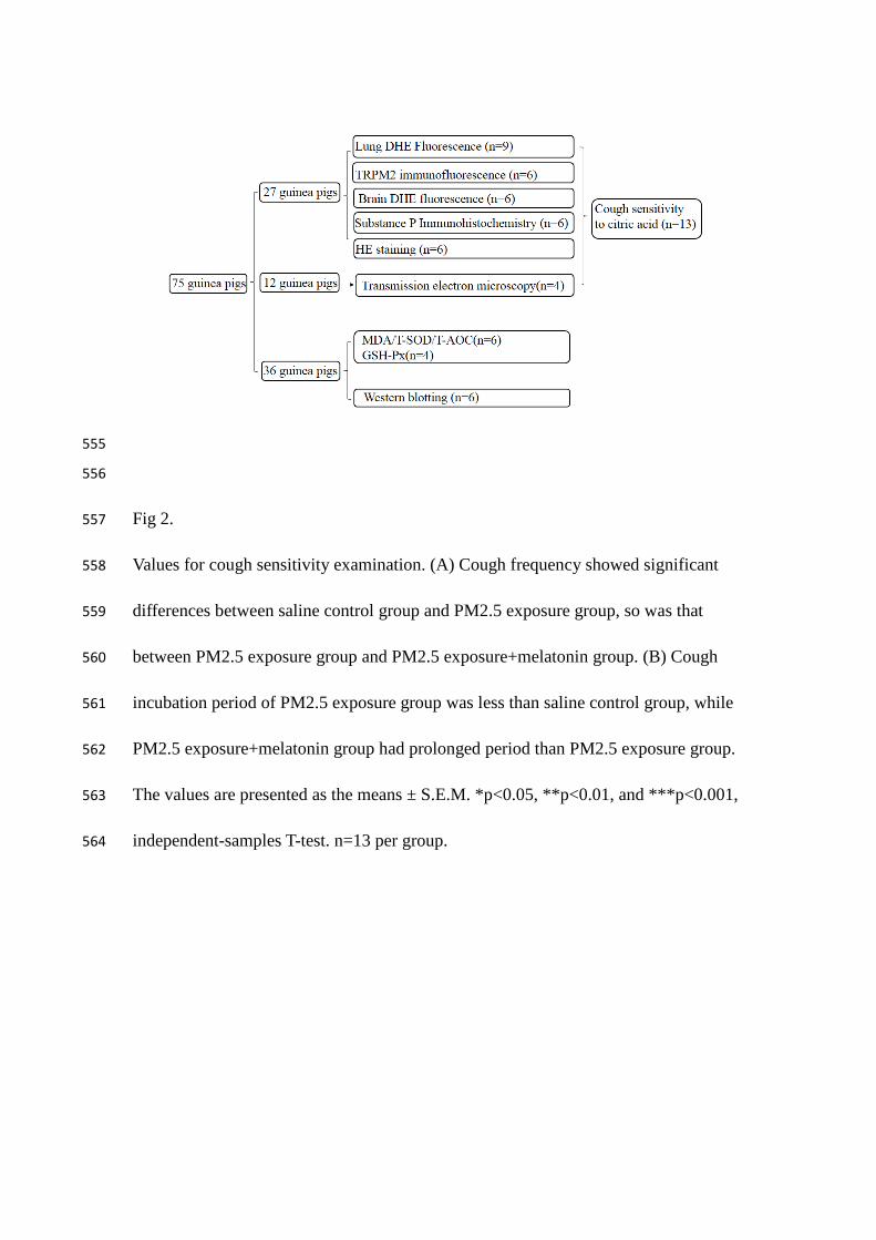
Fig 1. 545
The distribution of guinea pigs in various groups are shown in the figure. There are in 546
total 75 guinea pigs in our study. All guinea pigs were randomly divided into 3 groups 547
including normal saline control group, PM2.5 exposure group and PM2.5 548
exposure+melatonin treatment group. According to different handling methods, 27 549
guinea pigs were used for examination of DHE fluorescence, TRPM2 550
immunofluorescence, SP immunohistochemistry, and HE staining. 36 guinea pigs 551
were used for western blotting and examination of MDA/T-SOD/T-AOC/GSH-Px. 552
Another 12 guinea pigs were used for observation by transmission electron 553
microscopy. 554
555
556
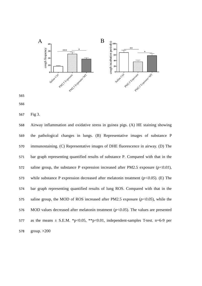
Fig 2. 557
Values for cough sensitivity examination. (A) Cough frequency showed significant 558
differences between saline control group and PM2.5 exposure group, so was that 559
between PM2.5 exposure group and PM2.5 exposure+melatonin group. (B) Cough 560
incubation period of PM2.5 exposure group was less than saline control group, while 561
PM2.5 exposure+melatonin group had prolonged period than PM2.5 exposure group. 562
The values are presented as the means ± S.E.M. *p<0.05, **p<0.01, and ***p<0.001, 563
independent-samples T-test. n=13 per group. 564
565
566
Fig 3. 567
Airway inflammation and oxidative stress in guinea pigs. (A) HE staining showing 568
the pathological changes in lungs. (B) Representative images of substance P 569
immunostaining. (C) Representative images of DHE fluorescence in airway. (D) The 570
bar graph representing quantified results of substance P. Compared with that in the 571
saline group, the substance P expression increased after PM2.5 exposure (p<0.01), 572
while substance P expression decreased after melatonin treatment (p<0.05). (E) The 573
bar graph representing quantified results of lung ROS. Compared with that in the 574
saline group, the MOD of ROS increased after PM2.5 exposure (p<0.05), while the 575
MOD values decreased after melatonin treatment (p<0.05). The values are presented 576
as the means ± S.E.M. *p<0.05, **p<0.01, independent-samples T-test. n=6-9 per 577
group. ×200 578
579
580
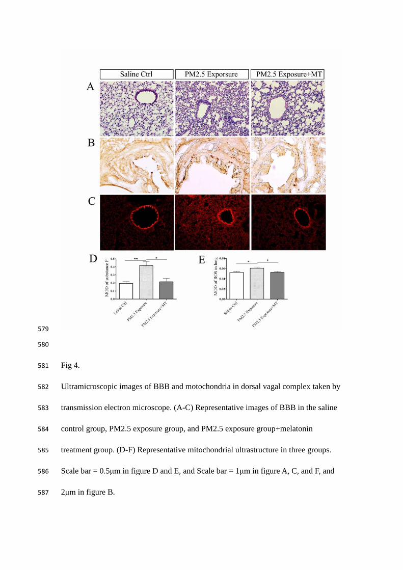
Fig 4. 581
Ultramicroscopic images of BBB and motochondria in dorsal vagal complex taken by 582
transmission electron microscope. (A-C) Representative images of BBB in the saline 583
control group, PM2.5 exposure group, and PM2.5 exposure group+melatonin 584
treatment group. (D-F) Representative mitochondrial ultrastructure in three groups. 585
Scale bar = 0.5μm in figure D and E, and Scale bar = 1μm in figure A, C, and F, and 586
2μm in figure B. 587
588
589
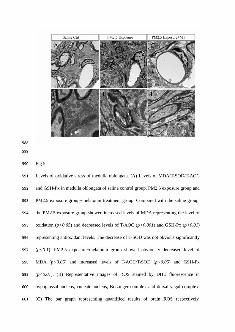
Fig 5. 590
Levels of oxidative stress of medulla oblongata. (A) Levels of MDA/T-SOD/T-AOC 591
and GSH-Px in medulla oblongata of saline control group, PM2.5 exposure group and 592
PM2.5 exposure group+melatonin treatment group. Compared with the saline group, 593
the PM2.5 exposure group showed increased levels of MDA representing the level of 594
oxidation (p<0.05) and decreased levels of T-AOC (p<0.001) and GSH-Px (p<0.01) 595
representing antioxidant levels. The decrease of T-SOD was not obvious significantly 596
(p<0.1). PM2.5 exposure+melatonin group showed obviously decreased level of 597
MDA (p<0.05) and increased levels of T-AOC/T-SOD (p<0.05) and GSH-Px 598
(p<0.01). (B) Representative images of ROS stained by DHE fluorescence in 599
hypoglossal nucleus, cuneate nucleus, Botzinger complex and dorsal vagal complex. 600
(C) The bar graph representing quantified results of brain ROS respectively. 601
Compared with the saline group, the PM2.5 exposure group showed an increased 602
production of ROS in the hypoglossal nucleus (p<0.001), cuneate nucleus (p<0.001), 603
Botzinger complex (p<0.05), and DVC (p<0.01). Meanwhile, ROS production in 604
PM2.5 exposure+melatonin group was decreased in Botzinger complex (p<0.05), 605
cuneate nucleus (p<0.05), hypoglossal nucleus (p<0.01) and DVC (p<0.01). The 606
values are presented as the means ± S.E.M. *p<0.05, **p<0.01, ***p<0.001, and 607
#p>0.05, independent-samples T-test. n=4-6 per group. cc: central canal. ×200 608
609
610
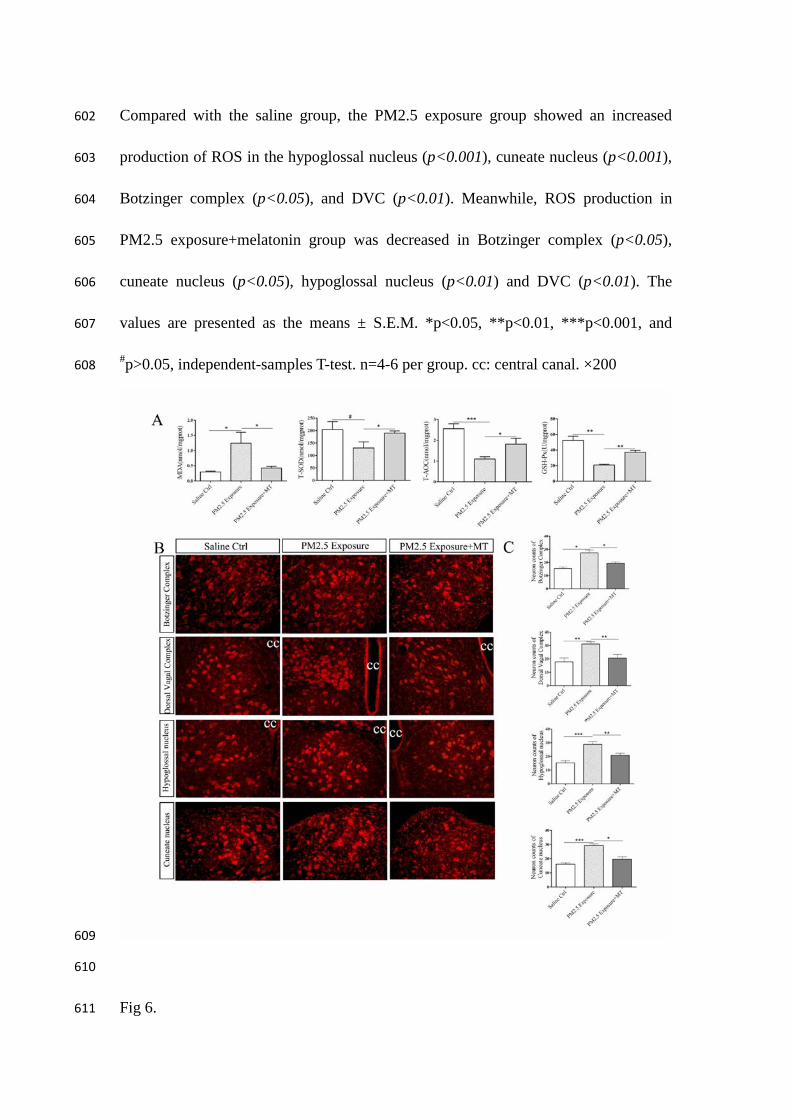
Fig 6. 611
Levels of TRPM2 in medulla oblongata of saline control group, PM2.5 exposure 612
group and PM2.5 exposure group+melatonin treatment group. (A) Representative 613
images of TRPM2 expression (red) in the Botzinger complex and dorsal vagal 614
complex. Nuclei were counterstained with DAPI (blue). (B) The bar graphs 615
representing quantified results of cell counting. Compared with the saline group, the 616
PM2.5 exposure group showed an increased expression of TRPM2 in the DVC and 617
Botzinger complex (p<0.01). Melatonin treatment decreased the expression of 618
TRPM2 (p<0.05) (Fig. 5A 5B). (C) Western blot showing protein level of TRPM2 in 619
medulla oblongata. (D) The bar graphs representing quantified results of TRPM2. The 620
protein level of TRPM2 in PM2.5 exposure group was higher than the saline group 621
(p<0.001) and PM2.5 exposure+melatonin group showed decreased protein level in 622
the DVC (p<0.01) and Botzinger complex (p<0.05). The values are presented as the 623
means ± S.E.M. *p<0.05, **p<0.01, and ***p<0.001, independent-samples T-test. 624
(n=6). cc: central canal. ×200 625
626