Embed Size (px)
Citation preview
729DOS Times - Vol. 11, No. 10 April, 2006
Amblyopia is defined as unilateral or bilateral decreaseof visual acuity caused by vision deprivation or abnormalbinocular interaction for which no organic causes can bedetected by the physical examination of the eye and whichin appropriate cases is reversible by therapeuticmeasures1,2,3.
For detection purposes, on routine examination,amblyopia is defined as a minimum of two Snellen linesdifference in visual acuity between either eyes, butamblyopia is truly a spectrum of visual loss ranging frommissing a few letters on the 20/20 line to hand movement.
Anisometropia is a very common cause of amblyopia.Refractive condition of the two eyes is usually not the same.Anisometropia is the condition in which the refractivestatus of the two eyes shows a considerable difference. Thevision in anisometropia may be binocular, alternating orit may be entirely uniocular. Each 0.25 D difference betweenthe refraction of the two eyes causes 0.5% difference in sizebetween the two retinal images and a difference of 5% isprobably the limit, which can be tolerated. If the defect inone eye is high and more especially if the visual acuity isnot good, it may be excluded altogether from vision at anearly stage in life ,the better eye is alone relied upon and ifit is not so already, the more ametropic eye tends to becomeamblyopic, the image from it being suppressed.
Aims and ObjectivesTo determine the relationship between anisometropia
and depth of amblyopia.
Material and MethodsThe present study was carried out on patients
attending the OPDs and IPD clinics of Gandhi Eye HospitalAligarh, U.P. from June 2002 to July 2003. Both male andfemale patients in the age group of 6-40 years presentingwith anisometropia of 1.0D and more of spherical and or1.0D or more of cylinder were included in the study. Patientswith decreased visual acuity due to any ocular or systemicpathology were excluded from the study and so withhistory of trauma and patients with manifest strabismus.
After obtaining a proper history regarding diminutionof vision, its duration, use of glasses or contact lens, a
A Study of Amblyopia among Anisometropic PatientsRasna Sharma, MS
Dr. Mohan Lal Memorial Gandhi Eye Hospital,Aligarh, UP
complete ocular examination was done. This includedvisual acuity (unaided visual acuity, visual acuity withpinholes using Snellen chart for literates and E-charts forilliterates and children.), torch light examination, covertest, slit lamp examination, retinoscopy under mydriaticfollowed by post mydriatic test (PMT) to get best correctedvisual acuity. The difference in refraction between the twoeyes was documented. Fundus examination (using direct/indirect ophthalmoscope) was done. Ocular movementswere checked in all 9 gazes. Worth 4 dot test andsynoptophore examination was done to check for binocularvision. All types of Anisometropia (sphericalhypermetropic and spherical myopic, mixed, simple,compound and mixed astigmatic) were taken into the study.
To correlate the degree of anisometropia with thedepth of amblyopia, all the cases were divided into 5 groupsin the increasing degree of anisometropia in the followingmanner:-
Group 1 1. Dioptre-2 DioptreGroup 2 2. 25 Dioptre-3 DioptreGroup 3 3. 25 Dioptre-4 DioptreGroup 4 4. 25 Dioptre-5 DioptreGroup 5 5. 25 Dioptre and aboveAmblyopia was defined as a difference of vision of two
Snellen lines or greater with the acuity of the amblyopiceye being less than 6/12. Depth of amblyopia was gradedinto mild moderate and severe depending upon the visualacuity in terms of Snellen lines 6/12-6/18, 6/24-6/36 and 6/60 or less respectively for mild, moderate and severeamblyopia.
ResultsA total of 102 anisometropic patients were included in
the study group, majority of them were males (69.6%), themale to female ratio being 2.28:1 (Table No.1). It wasobserved that out of 102 cases, incidence among 16-20 yearsgroup was higher (25.50%) followed by 17.64% in the agegroup of 11-15 years (Table No. 1).
Most common type of anisometropia reported in thestudy is spherical anisometropia (simple +compound)(42.15%) followed by compound astigmatic type i.e..34.31%. A total of 74 patients, out of 102 patients werefound having Amblyopia (Table No.2).
It has been observed that in smaller degrees ofanisometropia as in group 1 (1-2D) and 2 (2.25-3D), therewas a higher percentage of patients having mild amblyopia
MEDICAL OPHTHALMOLOGY
DOS Times - Vol. 11, No. 10April, 2006 730
(57-58%) while only small percentage (7-9%) had severeAmblyopia (vision 6/60 or less) and about 35-36% hadmoderate depth of amblyopia, but in higher degrees (Group5) of anisometropia, a large percentage (68%) of patientshad severe amblyopia and only 13% had mild Amblyopia(Table Nos. 3 &4)
It was observed clearly as depicted in table no 4 that,as the degree of anisometropia increased from 1 Dioptre to5 Dioptre or above, there was a proportional increase inthe depth of amblyopia.
DiscussionEarly detection of anisometropia is essential to prevent
the development of amblyopia in more ametropic eye.In the present study it was observed that
anisometropia was more prevalent in the age group of 16-20 years (25-50%). It could be explained on the basis of the
fact that patients in this age group are more involved instudies, are more aware of their problems related to visionand eye strain.
Rustein (1999)7 in his study presented a data from apatient group comprising of 32 males and 28 females out of60 anisometropes. The present study showed that in allthe age groups, anisometropia was more amongst themales as compared to their female counterparts. The maleand female ratio was found to be 2.3:1. Higher prevalencein males could be because of an easy and independent accessto hospital and clinics.
Attebo et al. (1998) 1 in his study of 118 personsclassified as having amblyopia, 109 (92%) gave a history ofa diagnosis of “lazy eye” or said that they were aware thatone eye had always been weaker and nine persons gave nosuch history. In the present study, it was observed thatout of 102 patients, 44 (43.13%) noticed decreased vision inthe eye of closing the other eye while 23 (22.54%) gave thehistory of a diagnosis of “lazy eye” or long standing poorvisual acuity in one or both eyes and only 7.84%complained of asthenopic symptoms.
Anisometropia has been historically considered to bea significant amblyopiogenic factor. The term amblyopiadenoted unilateral or bilateral reduction of vision for whichno cause could be detected by physical examination of theeye and which in appropriate cases is correctable bytherapeutic measures. For detection purposes, on routineexamination, amblyopia is defined as a minimum of twoSnellen lines difference in visual acuity. The term
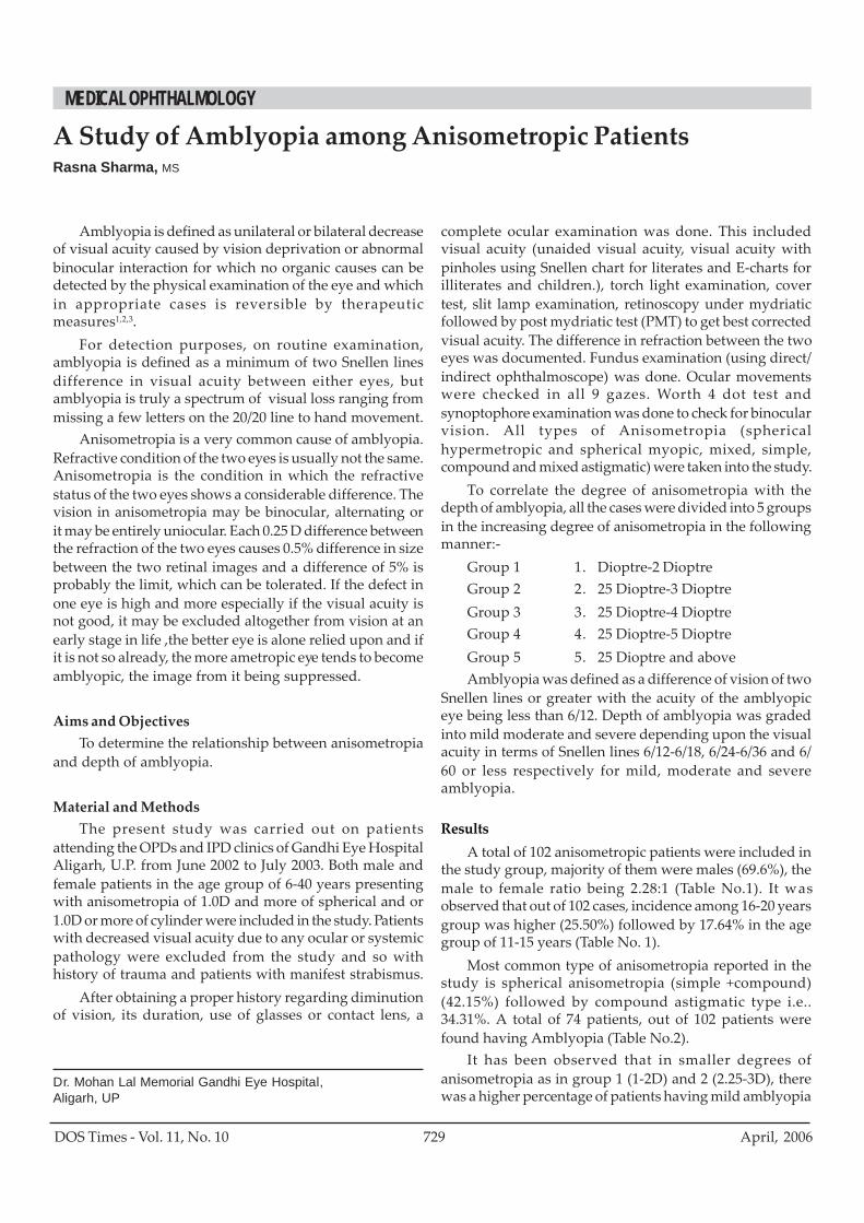
Table 1: Age & Sex wise distribution of Anisometropicpatients (n = 102)
Age Groups Males Females Total Percentage(years)
6 - 10 6 3 9 8.82 %11 – 15 13 5 18 17.64 %16 – 20 19 7 26 25.50 %21 – 25 10 5 15 14.70 %26 – 30 9 6 15 14.70 %31 – 35 7 2 9 8.82 %36 – 40 7 3 10 9.80 %
Total 71 31 102
Percentage 69.60 % 30.40 % 100 % 100 %
Table –3: Amblyopia in various group (Degrees) ofAnisometropia (n= 74)
Age groups Total No. of Total No of Percentage(years) Anisometropic Amblyopic
patients patients
Group I 34 14 41.171 - 2D
Group I 19 12 63.15I2.25 – 3.0D
Group II 15 14 93.33I3.25 – 4.0D
Group IV 12 12 100.004.25 – 5D
Group V 22 22 1005.25 D & above
Total 102 74 72.54
Percentage 100 72.54
Table 2: Distribution of Amblyopia amongAnisometropic Patients (n= 74)
Age groups Total No. of Total No of Percentage(years) Anisometropic Amblyopic
patients patients
6 – 10 9 7 77.7711- 15 18 13 72.2216 – 20 26 18 69.2321 – 25 15 8 53.3326 – 30 15 10 66.6631 – 35 9 6 66.6636 – 40 10 10 100.00
Total 102 74 72.54
731DOS Times - Vol. 11, No. 10 April, 2006
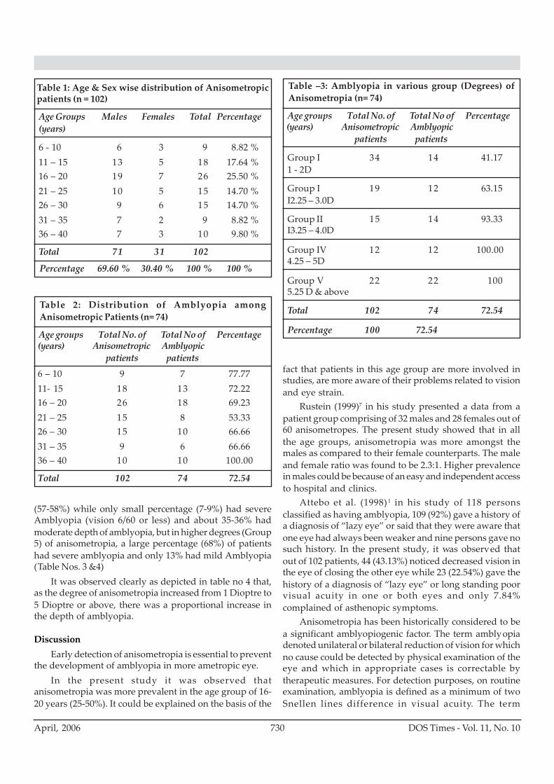
Table–4: Relationship of Degree of Anisometropia with the depth ofAmblyopia (n = 74)
Degree of Amblyopic Depth of AmblyopiaAnisometropia patients —————————————————
Mild Moderate Severe6/12 – 6/18 6/24 – 6/36 6/60 or less
Group I No.% 14 857.14 535.71 17.141 – 2 D
Group II No.% 12 758.33 436.36 19.092.25 – 3.0 D
Group IIID No.% 14 750.00 533.33 213.333.25 – 4.0
Group IVD No.% 12 541.66 433.33 325.004.25 – 5.0
Group V No.% 22 313.63 418.18 1568.185.25 D & above
Total 74 30 22 22
Percentage 100 40.54 29.72 29.72
anisometropic “amblyopia: is widelyaccepted to describe amblyopiapresumed to be caused byanisometropia alone.
Copps2, was the first to attemptto confirm an association betweenanisometropia and amblyopia in theabsence of strabismus which wasaccepted by several other authorslater. Incidence of amblyopia innonstrabismic anisometropia asreported by many workers is variable:60% by Ainsworth (1966), 86% byYuksel et.al.11, 80% by Krazystkona(1967), 53% by Vries (1985)9, whileAttebo et al. (1998)1 found it to be 50%.In the present study, out of 102orthoptropic anisometropes, 74(72.54%) were amblyopic4,5,6,10.
Amblyopia is found to be beingmore common and severe inanisohypermetropia oranisoastigmation. Mitchell and Atteboet al. (1998) also supported the fact ofRustein (1998)8 that amblyopia ismore prevalent in patients with hyperopic than myopicamisometropia. Later Rutstein and Corliss (1999)7 in theirstudy reported that higher degrees of anisometropiagenerally causes deeper amblyopia for hyperopes, but notfor myopes.
Conforming to the findings of various studies donepreviously, the present study also shows amblyopia to bemuch more common (40.53%) in sphericalanisohypermetropes (simple and compound), closelyfollowed by compound hypermetropic anisoastigmatism(37.84%) while very low prevalence (5.40%) was observedin spherical anisomyopes (simple and compound) and inmyopic anisoastigmatics (5.40%).
ConclusionDepth of Amblyopia was determined to vary
proportionally with the degree of Anisometropia.
Bibliography1. Attebo K., Mitchell P, Cumming R. et al. Prevalence and causes of
amblyopia in an adult population. Ophthalmology 1998; 105: 154-
59.2. Copps L.A. Vision in Anisometropia. Am. J. Ophthalmia 1944;
27(6): 641-644.3. Duke –Elder Stewart and Abrams D. Anisometropia. Duke Elder
Stewart. System of Ophthalmology. London. Henry Kimpton,1970; V : 505-11.
4. Goss DA and Tanlamai T. Prevalence of Monocular amblyopiaamong Anisometropes. American J. of Optometry andPhysiological optics 1979; Vol. 56 (II): 704-715.
5. Kutschke PJ, Scott WE and Keech RV. Anisometropic amblyopia.Ophthalmology Feb. 1991; 98(2): 258-63.
6. Mitchell P. Attebo K. et al. Prevalence and causes of Amblyopiain an adult population. Ophthalmology 1998; 105: 154-159.
7. Rutstein R.P. and Corliss D. Relationship between Anisometropia,Amblyopia, and Binocularity. Optometry and vision Science April.1999; 76 (4): 229-233.
8. Rutstein RP and Daum KM. Anomalies of Binocular Vision:Diagnosis and Management. St. Louis: Mosby, 1998: 14.
9. Vries JD. Anisometropia in children : analysis of a hospitalpopulation. British J. Ophthal 1985; 69: 504-507.
10. Weakley D.R. The association between nonstrabismicAnisometropia, Amblyopia and subnormal binocularity.Ophthalmology 2001; 108: 163-171.
11. Yuksel D., Spiritus M, Vamdelannoitte S. and Hoffman D. BullSoc. Belge Ophthalmol 1996; 263: 69-73.
DOS Times - Vol. 11, No. 10April, 2006 732
Eitiopathogenesis, clinical features and investigationswere discussed in the previous issue, now we will discussthe treatment.
Treatment Medical treatment
Laser photocoagulationThe Macular Photocoagulation Study Group showed
that laser photocoagulation was effective in the treatmentof well-defined extrafoveal or juxtafoveal choroidalneovascularization secondary to AMD. In patients withsubfoveal choroidal neovascularization, however, laserphotocoagulation was not beneficial in eyes that had largelesions and moderate-to-good initial visual acuity.Procedure
Angiogram <96 hrs oldLocate centre of FAZ on pretreatment FFAOutline CNVM on FFA using landmarks200 - 500 u spot size confluent burns0.2 -0.5s duration, uniform white burnExtrafoveal lesions treatment to be extended 100ubeyond the margin of lesionJuxtafoveal lesions avoid extension on the foveal sidePost treatment photograph has to be taken to ensurecomplete treatment.
Follow - upPatient after laser treatment needs to be followed upat 2, 4 and 6 weeksRepeat FFA on follow-up visits
Macular Photocoagulation Study (MPS)In Patients with well-defined extrafoveal CNVM after
a follow-up of 5 years, 64% of eyes assigned to no treatmentcompared with 46% of eyes randomized to argon laserexperienced severe visual loss (six or more lines of visualacuity loss using Bailey-Lovie visual acuity charts). Thedifference was statistically significant. Although the riskof severe visual loss was reduced in treated patients, ahigh rate of persistent and recurrent CNVM was observed.The recurrence rate observed in treated eyes at 12, 24, and
Age Related Macular Degeneration - ManagementAjay Kapoor MS, Nishank Mittal MBBS, MS, Ankur Gupta MBBS, DNB
60 months were of 41%, 51%, and 54%, respectively.Patients with well-defined juxtafoveal CNV were
treated with krypton red laser. At 3 years afterrandomization, 49% of laser-treated eyes experiencedsevere visual loss compared with 58% of untreated eyes.
Laser to drusenThere have been attempts for drusen reduction by laser
to decrease the risk of geographic atrophy and CNVM. Nosignificant difference in the development of CNVM wasnoted in the treated and untreated groups. To date,however, prophylactic laser photocoagulation in patientswith high-risk ARMD remains an experimental treatmentand should not be performed outside randomized clinicaltrials.
Feeder-Vessel Laser PhotocoagulationFeeder vessels are defined as vessels that are seen in
the earliest phases of the indocyanine angiogram, andappear to originate from a definite spot in the choroid andbranch into a CNV with distinct blood vessels. Feedervessels are identified in only a small percentage of patientsexamined with subfoveal CNV, so the treatment can beused in only a small number of cases. The first seriespublished on the use of indocyanine green-guided feeder-vessel photocoagulation to treat subfoveal CNV in patientswith ARMD was published by Shiraga and colleagues. Todate, there are not enough data to support the use of feeder-vessel photocoagulation as a routine treatment for patientswith CNV and ARMD.
Photodynamic therapyPhotodynamic therapy (PDT) involves the intravenous
infusion of a drug (photosensitizer) and the application ofa continuous nonthermal laser light directed at the CNVM.The wavelength of the laser light used corresponds to theabsorption peak of the drug, but it is not strong enough toproduce any thermal (photocoagulation) damage.
Mechanism of action: The drug gets concentrated in theimmature endothelium of CNVM, and light-activationinduces a photochemical reaction in the target area thatcauses immunologic and cellular damage, includingendothelial damage of new vessels. Endothelial damageand the resulting platelet adhesion, degranulation, andsubsequent thrombosis and occlusion of the vasculaturemight be the predominant mechanism by which light-activated drugs work. Since the photosensitizer
Venu Eye Institute & Research Centre1/31, Sheikh Sarai Institutional Area,New Delhi-110 017
MEDICAL OPHTHALMOLOGY
733DOS Times - Vol. 11, No. 10 April, 2006
accumulates predominantly in the CNV, a fairly selectivedamage to the CNV is expected.
To date, only PDT with the photosensitizer Verteporfinhas been proven to decrease the risk of visual loss inpatients with neovascular ARMD. Verteporfin (abenzoporphyrin derivative monoacid, BPD-MA; Visudyne,Novartis AG) is a light-activated drug. The application ofphotodynamic therapy withverteporfin involves two main steps:intravenous infusion of the drug andactivation of the drug by light at aspecific wavelength (689 nm) with alow-power, nonthermal laser. Thetherapy includes retreatment as oftenas every 3 months if leakage fromchoroidal neovascularization isdetected on follow-up fluoresceinangiograms.
ProcedureThe intravenous infusion of
verteporfin is given throughout a 10-minute period.
Then, 15 minutes after the start ofthe infusion the laser light is appliedfor 83 seconds.
Guidelines for the treatment ofpatients with ARMD and subfovealCNV with PDT have been recentlypublished. In these guidelines,
treatment with PDT is recommendedfor patients with predominantlyclassic CNV and for those with occultand no classic CNV with recentdisease progression (e.g., presence ofblood associated with the CNV,growth of the CNV, or deterioration ofthe visual acuity within the past 12weeks) and a lesion size of four or fewerdisk areas or a lesion size greater thanfour disk areas associated with lowlevels of vision (i.e., approximately inthe level of 20/50 Snellen vision). Inthese guidelines, it is alsorecommended to treat juxtafoveallesions that are so close to the foveathat conventional laserphotocoagulation almost certainlywould extend under the center of theFAZ, and extrafoveal lesions that arecontiguous to the optic nerve providedthat treatment spots do not overlie theoptic nerve. The recommendations
included a 3-month interval follow-up for at least 2 yearsfrom the time of initial treatment in all patients, except inthose in whom no treatment was recommended for twoconsecutive visits (6-month period). Patients shouldreceive retreatments as often as every 3 months if there isany fluorescein leakage from CNV noted. Although no dataare currently available on the treatment of pregnant or
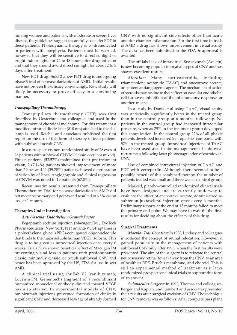
Colour fundus photograph of Classic SubfovealCNVM – Pre TTT
Colour fundus photograph of Regressed CNVM– Post TTT
FFA of the same patient showing leakage ofdye from CNVM before TTT
FFA showing staining of CNVM after TTT
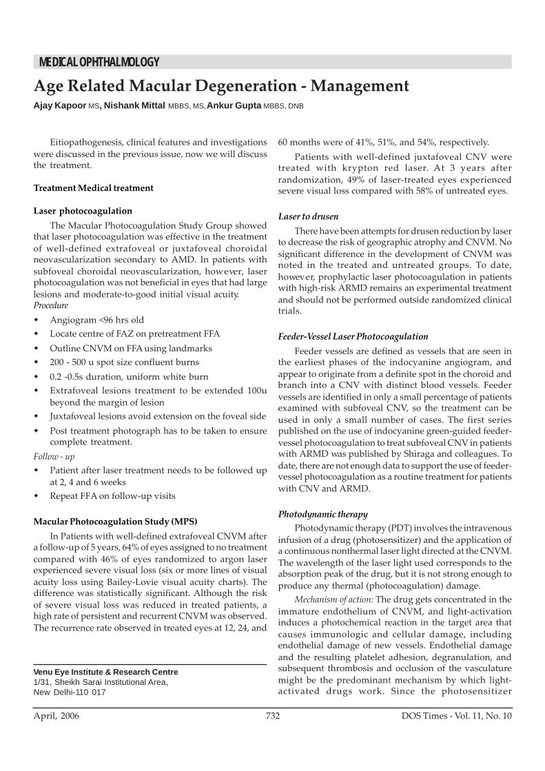
Classic Juxtafoveal CNVM – Pre Laser Regressed CNVM – Post Laser
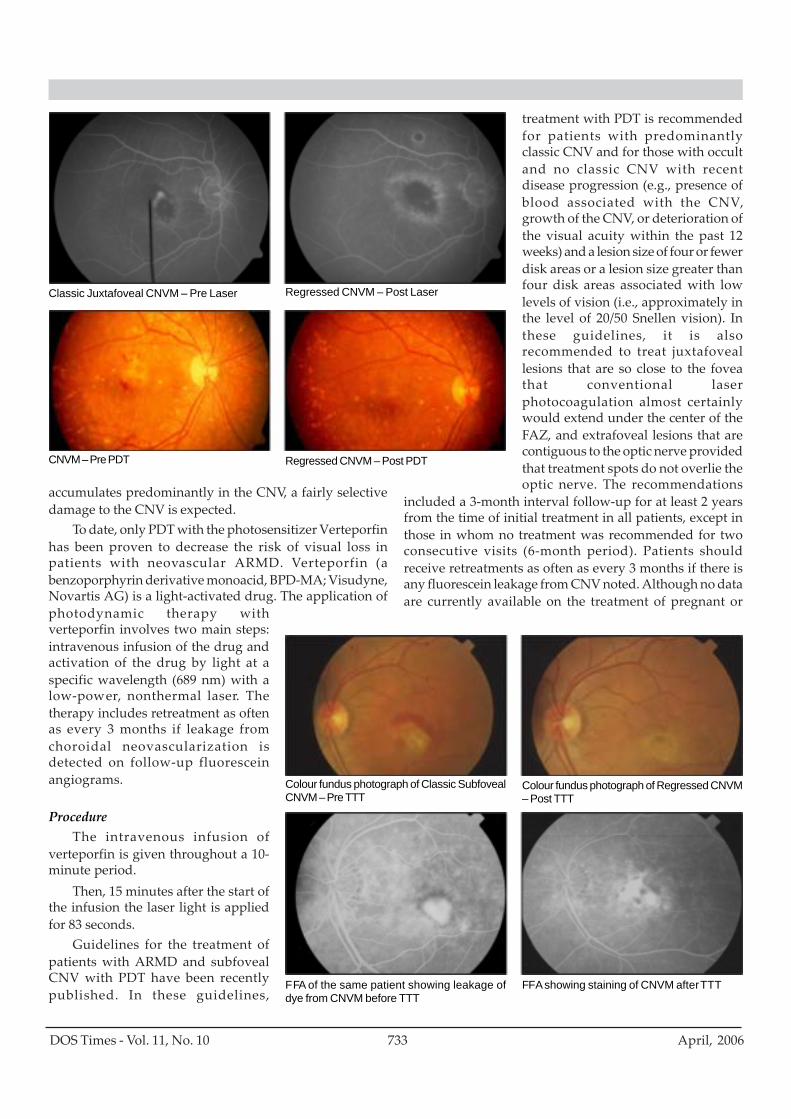
CNVM – Pre PDT Regressed CNVM – Post PDT
DOS Times - Vol. 11, No. 10April, 2006 734
nursing women and patients with moderate or severe liverdisease, the guidelines suggest to carefully consider PDT inthese patients. Photodynamic therapy is contraindicatedin patients with porphyria. Patients must be warned,however, that they will be sensitive to direct sunlight orbright indoor lights for 24 to 48 hours after drug infusionand that they should avoid direct sunlight for about 2 to 5days after treatment.
New PDT drug: SnET2 a new PDT drug is undergoingphase 3 trial of neovascularization of AMD. Initial resultshave not proven the efficacy convincingly. New study willlikely be necessary to prove efficacy in a convincingmanner.
Transpupillary ThermotherapyTranspupillary thermotherapy (TTT) was first
described by Oosterhuis and colleagues and used in themanagement of choroidal melanoma. For this treatment amodified infrared diode laser (810 nm) attached to the slit-lamp is used. Reichel and associates published the firstreport on the use of this form of therapy to treat patientswith subfoveal occult CNV.
In a retrospective, non-randomized study of 28 eyes of28 patients with subfoveal CNVM (classic, occult or mixed).Fifteen patients (53.57%) maintained their pre-treatmentvision, 2 (7.14%) patients showed improvement of morethan 2 lines and 11 (39.28%) patients showed deteriorationof vision by >2 lines. Angiographic and clinical regressionof CNVM was noted in 19 patients (67.8%).
Recent interim results presented from TranspupillaryThermotherapy Trial for neovascularization in AMD didnot meet the primary end points and resulted in a 5% visionloss at 1 month.
Therapies Under InvestigationAnti-Vascular Endothelium Growth FactorPegaptanib sodium injection (MacugenTM , EyeTech
Pharmaceuticals, New York, NY) an anti-VEGF aptamer isa polyethylene glycol (PEG)-conjugated oligonucleotidethat binds to the major soluble human VEGF isoform. Thisdrug is to be given as intravitreal injection once every 6weeks. Trials have shown beneficial effect of MacugenTMpreventing visual loss in patients with predominantlyclassic, minimally classic, or occult subfoveal CNV andhence has been approved by the US, FDA for use in wetAMD.
A clinical trial using rhuFab V2 (ranibizumab,LucentisTM, Genentech) fragment of a recombinanthumanized monoclonal antibody directed toward VEGFhas also started. In experimental models of CNV,ranibizumab injections prevented formation of clinicallysignificant CNV and decreased leakage of already formed
CNV with no significant side effects other than acuteanterior chamber inflammation. For the first time in trialsof AMD a drug has shown improvement in visual acuity.The data has been submitted to the FDA & approval isawaited.
The off-label use of intravitreal Becacizunab (Avastin)is new becoming popular to treat all types of CNV and hasshawn excellent results.
Steroids: Many corticosteroids, includingtriamcinolone acetonide (TAAC) and anecortave acetate,are potent antiangiogenic agents. The mechanism of actionof steroids may be due to their effect on vascular endothelialcell turnover, inhibition of the inflammatory response, oranother means.
In a study by Danis et al using TAAC, visual acuitywas statistically significantly better in the treated groupthan in the control group at 6 months' follow-up. Nopatients in the control group had increased intraocularpressure, whereas 25% in the treatment group developedthis complication. In the control group 22% of all phakicpatients developed increased lens opacities compared with57% in the treated group. Intravitreal injections of TAAChave been used also in the management of subfovealrecurrences following laser photocoagulation of extrafovealCNV.
Use of combined intravitreal injection of TAAC andPDT with verteporfin. Although there seemed to be apossible benefit of this combined therapy, the number ofpatients treated was small and there was no control group.
Masked, placebo-controlled randomized clinical trialshave been designed and are currently underway toevaluate the effect of anecortave acetate, administered assubtenon juxtascleral injection once every 6 months.Preliminary reports at the end of 12 months failed to meetthe primary end point. We may have to wait till the finalresults for deciding about the efficacy of this drug.
Surgical TreatmentsMacular Translocation: In 1983, Lindsey and colleagues
introduced the concept of retinal relocation. However, itgained popularity in the management of patients withsubfoveal CNV only after 1993, when the first results werepresented. The aim of the surgery is to relocate the centralneurosensory retina (fovea) away from the CNV, to an areaof healthier RPE, Bruch's membrane, and choroid. This isstill an experimental method of treatment as it lacksrandomized prospective clinical trials to support this formof treatment.
Submacular Surgery: In 1992, Thomas and colleagues,Berger and Kaplan, and Lambert and associates presentedtheir results after surgical excision of CNV. The techniquefor CNV removal was as follows: After complete pars plana
735DOS Times - Vol. 11, No. 10 April, 2006
vitrectomy CNVM is removed from subretinal space bymaking retinotomy temporal to fovea (usually) andinducing localized retinal detachment. Fluid-air exchangeis performed at the end of surgery and gas tamponade isgiven.
Recently, the first results of the Submacular SurgeryTrial, a randomized clinical trial comparing laserphotocoagulation to surgical removal of subfoveal CNVhave been published. All patients enrolled in this trial hada subfoveal recurrent CNV following prior laserphotocoagulation for extrafoveal or juxtafoveal CNV. Nostatistically significant differences in visual acuity wereobserved between patients randomized to laserphotocoagulation and surgical excision of CNV in this pilottrial. Similarly, health-related quality of life was notstatistically significant different between the two treatedgroups.
A new trial to evaluate the benefit of CNV removal incases of newly developed subfoveal CNV is currentlyunderway (Submacular Surgery Trial, Group N). Patientsare being randomized to either surgical excision of the CNVor observation. In this study, patients with lesions largerthan those eligible for laser photocoagulation followingMPS guidelines or with minimally classic lesions in whichlaser photocoagulation or PDT have not shown anytreatment benefit are eligible for the trial. Patients withpredominantly classic subfoveal lesions are being enrolledalso if after detailed explanation of the benefits of PDT theystill prefer to participate in the trial.
Iris/Retinal Pigment Epithelium TransplantationSeveral reports on RPE transplantation in patients
with neovascular ARMD have been published. Isolated cellsand RPE-cell sheets have been used. Fetal or mature RPEhave been transplanted. Only rarely have good levels ofvision been achieved following RPE transplantation.
Due to possible difficulties in obtaining RPE cells fortransplantation and complications related to thisprocedure, researchers have investigated the possibilityof substituting RPE cells for iris pigment epithelial (IPE)cells. Iris pigment epithelial and RPE cells have a commonembryonic origin, and some of the RPE functions have beendemonstrated in IPE. Few series on IPE transplantationhave been reported in the literature. In these series, visualacuity after transplantation remained low, in the level of20/100.
Prophylactic TreatmentsVitamin and Mineral Supplements
A randomized clinical trial, part of the Age-RelatedEye Disease Study (AREDS), was conducted in order to tryto evaluate the effect of antioxidants and zinc in patientswith ARMD. At 5 years, a statistically significant reduction
in the risk of progression to advanced ARMD and a 15-letter decrease in visual acuity score was found in thosepatients randomized to antioxidants plus zinc in categoriesthree and four. No statistically significant adverse eventswere found with any of the formulations. However, possiblecomplications of the study medications have beenidentified. Those with extensive intermediate size drusen(63 μ -124μ), at least 1 large druse (>125 μ), noncentralgeographic atrophy in 1 or both eyes, or advanced AMD orvision loss due to AMD in 1 eye, and withoutcontraindications such as smoking, should considered forsupplementation of antioxidants plus zinc.
Results from AREDS continue to be gathered andstudied. Moreover, a new AREDS is being proposed. A fewof the findings from the current AREDS include thefollowing
Patients who ate fish more than once per week had a40% reduction in neovascularization compared withpatients who ate fish less than once per month;Zeaxanthin and lutein reduced the risk ofneovascularization in AMD; andPatients taking the zinc regimen appeared to have alower mortality rate than those patients not takingzinc.
Carotenoids: Lutein and Zeaxanthin:Lutein and Zeaxanthin are the main constituents of
the luteal pigment. This yellow pigment, present at themacula, absorbs blue light. Whereas zeaxanthin is the mainpigment present at the fovea, lutein is more abundant inthe rest of the macula. Lutein and zeaxanthin are localizedmainly in Henle's fiber layer. It is possible that lutein andzeaxanthin may protect the retina from the damage causedby blue light exposure and subsequently decrease the riskfor ARMD. In this respect, a case-control study in whichplasma levels of lutein and zeaxanthin in patients withARMD were compared to those in an age-matched controlgroup showed an inverse relationship between plasmalevels of these two carotenoids and the risk for ARMD.However, to date, no well controlled intervention trialswith lutein and zeaxanthin have been performed. Thus, itis not clear to what degree these pigments may decreasethe risk of neovascular complications in ARMD.
ConclusionIncreasing knowledge about the pathogenesis of this
disease has led to new therapeutic strategies. As on todaythe treatment modalities have developed to arrest thedisease process to some extent. Future treatments shouldlikely concentrate in preventing the development of CNVin patients at risk, rather than in treating it onceestablished.
DOS Times - Vol. 11, No. 10April, 2006 736
References1) Argon laser photocoagulation for neovascular maculopathy. Five-
year results from randomized clinical trials. MacularPhotocoagulation Study Group. Arch Ophthalmol 1991;109:1109-14.
2) Krypton laser photocoagulation for neovascular lesions of age-related macular degeneration. Results of a randomized clinicaltrial. Macular Photocoagulation Study Group. Arch Ophthalmol1990;108:816-24.
3) Laser photocoagulation of subfoveal neovascular lesions in age-related macular degeneration. Results of a randomized clinicaltrial. Macular Photocoagulation Study Group. Arch Ophthalmol1991;109:1220-31.
4) Desatnik H, Treister G, Alhalel A, et al: ICGA-guided laserphotocoagulation of feeder vessels of choroidal neovascularmembranes in age-related macular degeneration. Indocyaninegreen angiography. Retina 20: 143-50, 2000
5) Shiraga F, Ojima Y, Matsuo T, et al: Feeder vessel photocoagulationof subfoveal choroidal neovascularization secondary to age-relatedmacular degeneration. Ophthalmology 105: 662-9, 1998
6) Photodynamic therapy of subfoveal choroidal neovascularizationin age-related macular degeneration with verteporfin: one-yearresults of 2 randomized clinical trials-TAP report. Treatment ofage-related macular degeneration with photodynamic therapy (TAP.Arch Ophthalmol 117: 1329-45, 1999
7) Bressler NM: Photodynamic therapy of subfoveal choroidalneovascularization in age-related macular degeneration withverteporfin: two-year results of 2 randomized clinical trials-tapreport 2. Arch Ophthalmol 119: 198-207, 2001
8) Verteporfin therapy of subfoveal choroidal neovascularization inage-related macular degeneration: two-year results of a randomizedclinical trial including lesions with occult with no classic choroidalneovascularization-verteporfin in photodynamic ther. Am JOphthalmol 131: 541-60, 2001
9) Guidelines for using verteporfin (visudyne) in photodynamictherapy to treat choroidal neovascularization due to age-relatedmacular degeneration and other causes. Retina 22: 6-18, 2002
10) Reichel E, Berrocal AM, Ip M, et al: Transpupillary thermotherapyof occult subfoveal choroidal neovascularization in patients withage-related macular degeneration. Ophthalmology 106: 1908-14,1999
11) Agarwal M, Shanmugam MP, Gopal L, Shetty N, Bhende M,Gopal L, Sharma T, Thakur S, Raman R, Nizamuddin SH, MoorthyKR. Transpupillary thermotherapy for choroidal neovascularmembrane in age related macular degeneration. Indian J Ophthalmol.2004 Mar;52(1):45-9.
12) Preclinical and phase 1A clinical evaluation of an anti-VEGFpegylated aptamer (EYE001) for the treatment of exudative age-related macular degeneration. Retina 22: 143-52, 2002
13) Krzystolik MG, Afshari MA, Adamis AP, et al: Prevention ofexperimental choroidal neovascularization with intravitreal anti-vascular endothelial growth factor antibody fragment. ArchOphthalmol 120: 338-46, 2002
14) Danis RP, Ciulla TA, Pratt LM, Anliker W: Intravitreal triamcinoloneacetonide in exudative age-related macular degeneration. Retina20: 244-50, 2000.
15) Spaide RF, Sorenson J, Maranan L: Combined photodynamictherapy with verteporfin and intravitreal triamcinolone acetonidefor choroidal neovascularization. Ophthalmology 110: 1517-25,2003.
16) Lindsey P, Finkelstein D, D'Anna S: Experimental retinal relocation.Invest Ophthalmol Vis Sci 24: 242 (Suppl), 1983
17) Submacular surgery trials randomized pilot trial of laserphotocoagulation versus surgery for recurrent choroidalneovascularization secondary to age-related macular degeneration:I. Ophthalmic outcomes submacular surgery trials pilot study reportnumber. Am J Ophthalmol 130: 387-407, 2000 .
18) Submacular surgery trials randomized pilot trial of laserphotocoagulation versus surgery for recurrent choroidalneovascularization secondary to age-related macular degeneration:II. Quality of life outcomes submacular surgery trials pilot studyreport n. Am J Ophthalmol 130: 408-18, 2000
19) A randomized, placebo-controlled, clinical trial of high-dosesupplementation with vitamins C and E, beta carotene, and zinc forage-related macular degeneration and vision loss: AREDS reportno. 8. Arch Ophthalmol 119: 1417-36, 2001
20) Bone RA, Landrum JT, Friedes LM, et al: Distribution of lutein andzeaxanthin stereoisomers in the human retina. Exp Eye Res 64:211-8, 1997
21) Antioxidant status and neovascular age-related maculardegeneration. Eye Disease Case-Control Study Group. ArchOphthalmol 111: 104-9, 1993
737DOS Times - Vol. 11, No. 10 April, 2006
Meibomian gland dysfunction (MGD) is more commonan entity than we diagnose it. Many a times our patientscomplain of irritation, foreign body sensation and are givennonspecific medications. A careful examination willdiagnose MGD and a simple treatment can have a morecomfortable patient.
Meibomian Gland dysfunction was first noted byCasserius in 1609 and was described by Heinrich Meibomin1666. Posterior blepharitis is associated with variousdisorders of the meibomian glands, which are knowncollectively as meibomian gland dysfunction. There arevarious classifications of the meibomian gland disease, butnone widely adopted.
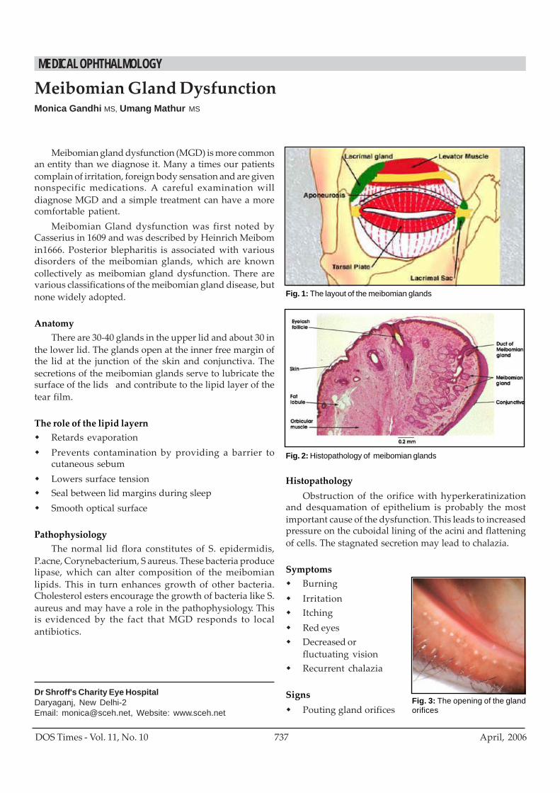
AnatomyThere are 30-40 glands in the upper lid and about 30 in
the lower lid. The glands open at the inner free margin ofthe lid at the junction of the skin and conjunctiva. Thesecretions of the meibomian glands serve to lubricate thesurface of the lids and contribute to the lipid layer of thetear film.
The role of the lipid layernRetards evaporationPrevents contamination by providing a barrier tocutaneous sebumLowers surface tensionSeal between lid margins during sleepSmooth optical surface
PathophysiologyThe normal lid flora constitutes of S. epidermidis,
P.acne, Corynebacterium, S aureus. These bacteria producelipase, which can alter composition of the meibomianlipids. This in turn enhances growth of other bacteria.Cholesterol esters encourage the growth of bacteria like S.aureus and may have a role in the pathophysiology. Thisis evidenced by the fact that MGD responds to localantibiotics.
Meibomian Gland DysfunctionMonica Gandhi MS, Umang Mathur MS
Dr Shroff's Charity Eye HospitalDaryaganj, New Delhi-2Email: [email protected], Website: www.sceh.net
HistopathologyObstruction of the orifice with hyperkeratinization
and desquamation of epithelium is probably the mostimportant cause of the dysfunction. This leads to increasedpressure on the cuboidal lining of the acini and flatteningof cells. The stagnated secretion may lead to chalazia.
SymptomsBurningIrritationItchingRed eyesDecreased orfluctuating visionRecurrent chalazia
SignsPouting gland orifices
Fig. 1: The layout of the meibomian glands
Fig. 2: Histopathology of meibomian glands
Fig. 3: The opening of the glandorifices
MEDICAL OPHTHALMOLOGY
DOS Times - Vol. 11, No. 10April, 2006 738
Change in the numberOrifices displaced posteriorlyFoam in tear meniscusCapping with solidified excretaToothpaste like secretionLid margin roundedThickeningErythemaHyperkeratinizationVascularization-"brush marks"TelangiectasiaNotchingBulbar and tarsal conjunctival injectionPapillary reaction on inferior tarsusCorneal and conjunctival stainingCorneal pannusUlceration
Associated sequelaeContact lens intoleranceGPCChalazia
TreatmentLid hygiene is currently the mainstay of treatment for
meibomian gland dysfunction. This involves warmcompress followed by massage and expression of themeibum. Lid scrubs are effective also. This can be tediousand messy task but if the requirement and logic of thetreatment is explained to the patient they would be morecompliant with the recommended regimen.
There is a need for tear substitutes to stabilize the tearfilm till the normal secretions are regularized.
Other proven therapies for meibomian glanddysfunction include oral Tetracycline, Doxycycline orMinocycline, topical Erythromycin or Bacitracin, andtopical steroids. For refractory cases, topical cyclosporine
can be used. The doses recommendedare as follows:
Tetracycline 250mg QIDDoxycycline 50-100 mg BDMinocyline 50-100 mg BD
Role of TetracyclinesNot as antibiotics
Decrease bacterial lipaseAlter the fatty acid composition of the meibomiangland secretionsImprove their solubilityInhibit collagenaseEffective in protecting the cornea from impendingperforation secondary to inflammatory responses
Adverse effects of TetracyclinesGastrointestinal--diarrhea, pancreatitis andpseudomembranous colitisBenign intracranial hypertensionRenal tubular damageCross the placenta- Permanent discoloration of teeth- Retardation of fetal bone growth
DoxycyclineTwice a dayNot affected by dairy productsPregnant females need to be cautioned
Associated conditionsMeibomian gland dysfunction has been associated
with lacrimal insufficiency, Rosacea and seborrheicdermatitis.
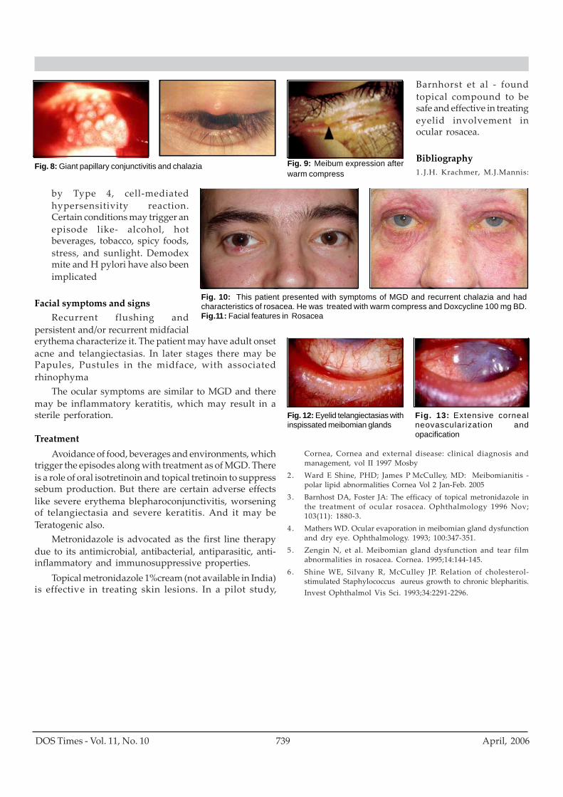
Rosacea is a condition that may not be easily diagnosedin people with dark skin. The patient may present withrecurrent episodes of meibomitis and chalazia. Borrie
found that nearly 100% ofrosacea patients hadposterior blepharitis, andocular disease precededcutaneous manifestationin 20%.
Pathophysiology The etiology of rosacea
is unknown. It is acutaneous vasculardisorder characterized
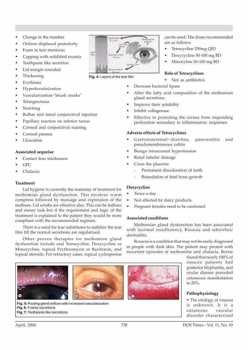
Fig. 4: Layers of the tear film
Fig. 5: Pouting gland orifices with increased vascularizationFig. 6: Foamy secretionsFig. 7: Toothpaste like secretions
5 6 7
739DOS Times - Vol. 11, No. 10 April, 2006
by Type 4, cell-mediatedhypersensitivity reaction.Certain conditions may trigger anepisode like- alcohol, hotbeverages, tobacco, spicy foods,stress, and sunlight. Demodexmite and H pylori have also beenimplicated
Facial symptoms and signsRecurrent flushing and
persistent and/or recurrent midfacialerythema characterize it. The patient may have adult onsetacne and telangiectasias. In later stages there may bePapules, Pustules in the midface, with associatedrhinophyma
The ocular symptoms are similar to MGD and theremay be inflammatory keratitis, which may result in asterile perforation.
TreatmentAvoidance of food, beverages and environments, which
trigger the episodes along with treatment as of MGD. Thereis a role of oral isotretinoin and topical tretinoin to suppresssebum production. But there are certain adverse effectslike severe erythema blepharoconjunctivitis, worseningof telangiectasia and severe keratitis. And it may beTeratogenic also.
Metronidazole is advocated as the first line therapydue to its antimicrobial, antibacterial, antiparasitic, anti-inflammatory and immunosuppressive properties.
Topical metronidazole 1%cream (not available in India)is effective in treating skin lesions. In a pilot study,
Fig. 10: This patient presented with symptoms of MGD and recurrent chalazia and hadcharacteristics of rosacea. He was treated with warm compress and Doxcycline 100 mg BD.Fig.11: Facial features in Rosacea
Fig. 9: Meibum expression afterwarm compress
Fig. 8: Giant papillary conjunctivitis and chalazia
Cornea, Cornea and external disease: clinical diagnosis andmanagement, vol II 1997 Mosby
2 . Ward E Shine, PHD; James P McCulley, MD: Meibomianitis -polar lipid abnormalities Cornea Vol 2 Jan-Feb. 2005
3 . Barnhost DA, Foster JA: The efficacy of topical metronidazole inthe treatment of ocular rosacea. Ophthalmology 1996 Nov;103(11): 1880-3.
4 . Mathers WD. Ocular evaporation in meibomian gland dysfunctionand dry eye. Ophthalmology. 1993; 100:347-351.
5 . Zengin N, et al. Meibomian gland dysfunction and tear filmabnormalities in rosacea. Cornea. 1995;14:144-145.
6 . Shine WE, Silvany R, McCulley JP. Relation of cholesterol-stimulated Staphylococcus aureus growth to chronic blepharitis.Invest Ophthalmol Vis Sci. 1993;34:2291-2296.
Fig. 12: Eyelid telangiectasias withinspissated meibomian glands
Fig. 13: Extensive cornealneovascularization andopacification
Barnhorst et al - foundtopical compound to besafe and effective in treatingeyelid involvement inocular rosacea.
Bibliography1 . J.H. Krachmer, M.J.Mannis:
DOS Times - Vol. 11, No. 10April, 2006 740
Anisocoria means an inequality of the pupils (a – privative;iso – equal; coria – pupil).
Under normal conditions, the pupils remain equal atall times in all levels of light. When pupillary inequality isseen, it usually means that damage has occurred to the irissphincter, the iris dilator or to their innervation.
Pupil size is determined by the local state of the iristissue, the chemical events occurring at the myoneuraljunctions, the tone of the pupillary sphincter(parasympathetic) and the pupillary dilator muscles(sympathetic) in response to nervous stimuli and thegeneral emotional state of the individual.
The retina, optic nerve, chiasm and optic tractsconstitute the afferent pathway of the light reflex.Approximately half of the fibers of the optic nervedecussate in the optic chiasm, and the input to each ofthe parasympathetic nuclei in the brain stem remainsequal. Therefore, relative afferent pupillary defects donot cause anisocoria because any changes in lightinput are distributed equally to both pupils.1
The parasympathetic (pupilloconstrictor) efferentpathway originates in the Edinger-Westphal nucleusin the dorsal mid brain and exits with the motor fibersof the oculomotor nerve to the ciliary ganglion via thecavernous sinus. Postganglionic fibers then travel tothe pupilloconstrictor or sphincter muscle of the iris.Most diseases affecting the efferent pathway areunilateral or asymmetric and cause anisocoria.2
The sympathetic (pupillodilator) pathway leaves thespinal column at the eighth cervical level (C8) throughthe second thoracic level (T2) via the ventral roots tothe paravertebral sympathetic chain. These fibers thenpass through the stellate ganglion near the apex of thelung; then synapse in the superior cervical ganglion.The postganglionic fibers then travel via the carotidartery to innervate the dilator pupillae.3
Approach to the patient of anisocoriaThe first step is to establish which muscle is not
working properly. Anisocoria always increases in the direction ofaction of the paretic iris muscle. If the iris sphincter is paretic,the lighting condition that normally brings that muscle
AnisocoriaJ.L.Goyal MD,DNB, Snigdha Mahajan MBBS
Neuro-Ophthalmic ServicesGuru Nanak Eye CentreNew Delhi 110002
into action (bright light) will accentuate the weakness andincrease the anisocoria. Conversely, if the iris dilator wereparetic, the anisocoria would be expected to increase indarkness, as reflex dilatation is impaired.4
Pupillary inequality that increases in bright lightThere are many causes of anisocoria that increases in
bright light.Iris abnormalities on slit lamp examination: Sphinctertears, traumatic mydriasis, and iris sphincter atrophysuch as by previous herpes zoster uveitis.Medications such as atropineOculomotor nerve palsyPost ganglionic lesion – tonic pupilAngle closure glaucomaIOFB – iron mydriasis.Adrenergic mydriasisSegmental paralysis of the iris sphincter – Adie’s pupil,partial oculomotor nerve palsyFixed pupil after anterior segment surgery
Pupillary inequality that increases in the darkThe two most important causes of anisocoria that
increases in the dark are simple or physiological anisocoriaand Horner’s syndrome.
Local ophthalmologic conditions: Any conditionresulting in an inflammatory response within the anteriorchamber may cause spasm within the sphincter muscle,resulting in anisocoria.15
Some medications for glaucoma cause miosis (eg,pilocarpine).
Important conditions causing anisocoriaPharmacologic mydriasis
An important cause for an isolated dilated pupil ispharmacologic mydriasis. 1% pilocarpine in the affectedwill reverse the mydriasis caused by atropine and can beused as a diagnostic test in suspicious cases.
Sympathomimetics, which are used to facilitatenasotracheal intubation or ophthalmologic examination,also cause mydriasis.
Inadvertent ocular exposure to anticholinergic agentsalso has been reported. Patients using scopolamine patcheshave been noted to have self-limited mydriasis, which hasbeen dubbed “cruise ship anisocoria.” 6,7
MEDICAL OPHTHALMOLOGY
741DOS Times - Vol. 11, No. 10 April, 2006
Oculomotor palsy: The most worrisome cause of anenlarged pupil is oculomotor nerve dysfunction.
Anisocoria in the setting of a head injury with decreasedlevel of consciousness may be caused by uncalherniation due to an expanding supratentorial lesionwhich forces the uncus against the edge of thetentorium, compressing the adjacent mid brain andoculomotor nerve. This results in ipsilateral third-nervepalsy and decreased level of consciousness. In addition,the contralateral cerebral peduncle is compressedagainst the free edge of the tentorium, resulting inipsilateral hemiparesis. Such patients require urgentintervention to lower intracranial pressure and havea poor prognosis without rapid surgical intervention.8
When a parasympathetic pupillary defect coexistswith ptosis and extraocular muscle palsies in a patientwith a normal level of consciousness, the diagnosis isoculomotor palsy with pupillary involvement. In thesesituations, emergent neuroimaging is indicated to ruleout a compressive lesion, such as aneurysm, tumor,etc.9
Oculomotor palsy due to metabolic diseases, such asdiabetes, generally spares the pupil, but can be totalalso.
Acute closed-angle glaucomaIt results in a red painful eye and visual disturbance.
In this condition, the pupil tends to be fixed in mid position,with an impaired light reflex.
Adie tonic pupil 10
Adie tonic pupil predominately occurs in females aged20-50 years. Patients may complain of photophobia andepisodes of blurred near vision or blurred vision whenswitching from near to far viewing; or they simply maycomplain of unequal pupils without any other symptoms.Typically, the involved pupil displays a poor response tolight, with a relatively preserved response to sustainednear fixation but an abnormally slow or tonic contraction.Slit lamp examination often reveals sector palsies of theiris.
The parasympathetic defect in Adie pupil is believedto occur after the fibers leave the ciliary ganglion. As aresult of denervation supersensitivity, the affected eyedisplays an abnormally brisk response to dilute (1/8%)pilocarpine. Normal eyes generally do not respond to sucha dilute solution, and this test has been suggested as a wayof differentiating preganglionic and postganglionicparasympathetic lesions.5
Recent literature reports many patients who havemydriasis due to oculomotor nerve compression and havedisplayed reactivity to dilute pilocarpine, but these
patients should show other signs of CN III dysfunction.The combination of an idiopathic tonic pupil with
decreased deep tendon reflexes and/or orthostatichypotension is termed Holmes-Adie syndrome. Thesymptoms of a tonic pupil tend to be self-limited. Aidepupil is believed to be idiopathic or viral in etiology.
Other causes of a tonic pupil include neurosyphilis,diabetes, herpes zoster, giant cell arteritis, and alcoholism.11
Simple anisocoriaSimple anisocoria may be found in up to 20% of the
general population and may vary from day to day in thesame individual.
In most patients, the degree of anisocoria is less than 1mm, and no ptosis, dilation lag, or vasomotor dysfunctionis present.
In some patients, simple anisocoria may be provokedby oral medications (eg, pseudoephedrine, selectiveserotonin reuptake inhibitors).
Instillation of 4-10% cocaine solution causes dilationof both eyes.
Old photographs, sometimes from drivers’ licenses, canprovide evidence that the anisocoria has been present forsome time.12
Horner syndromeHorner syndrome is the result of disruption of the
sympathetic innervations to the eye at any place along thepathway.
Dilation lag is a classic finding in Horner syndrome.The affected eye typically has a delayed response toreduced illumination. As a result, the anisocoria of Hornersyndrome is greater 5 seconds after entering a darkenvironment than it is after 15-30 seconds.
In patients with an unestablished diagnosis,instillation of 4-10% cocaine solution is indicated. Cocaineinhibits the reuptake of norepinephrine, causing morenorepinephrine to be available at the neuromuscularjunction of the iris dilator muscle. Assess the pupils atbaseline and at 40-60 minutes. In a positive test, thesympathetically impaired pupil fails to dilate, and thedegree of anisocoria increases. If both pupils dilate,physiologic anisocoria is the diagnosis. In some studies,this test has been both sensitive and specific for Hornersyndrome.14
Further delineation of the level of a lesion causingHorner syndrome has proved more problematic. Thisbecomes important, as patients with postganglionic Hornersyndrome tend to have a very good prognosis, whilepreganglionic lesions are often the hallmark of myelopathyor malignancy.13
DOS Times - Vol. 11, No. 10April, 2006 742
The exception is in patients with carotid dissection,which may result in postganglionic Horner syndrome.However, such lesions also are associated with acute neckpain or other neurologic deficits. Therefore, in cases ofpainful Horner syndrome, emergent evaluation of theanterior cerebral circulation is indicated.
In patients with Horner syndrome without pain, achest x-ray to exclude a Pancoast tumor probably isindicated, but further workup may be pursued on anoutpatient basis.
Horner syndrome also can occur in incipienttranstentorial herniation. Such patients typicallyexperience a rapid deterioration in brainstem function andhave a decreased level of consciousness.
Pharmacologic miosisPharmacologic miosis may be the result of a variety of
cholinergic glaucoma medications. A small pupil also maybe the result of chance exposure to cholinergic agents.Suspect pharmacologic miosis in otherwise asymptomaticpatients who wear contacts and have experienced acuteonset of miosis.
Implicated agents include anticholinesterases (eg, fleacollar anisocoria) and inhaled anticholinergics(Ipratropium bromide). In such cases, withdrawal ofexposure to the agent confirms the diagnosis.
Bibliography1 . Thompson H: Afferent pupillary defects: Pupillary findings
associated with defects of the afferent arm of the papillary lightreflex arc. Am J Ophthalmol 62:860, 1966
2 . Warwick R: The ocular parasympathetic nerve supply and itsmesencephalic sources. J Anat 88:71, 1954
3 . Moses R: Adler ’s Physiology of the Eye. St louis, CV MosbyCo, 1970, chap 11, p396
4 . Multack RF, Lannin WC, Olbum JR: Improving diagnostic acumenin pupillary evaluation: a review for the primary care physician. JAm Osteopath Assoc.1989 Jul;89(7):917-24.
5 . Jacobson DM, Olson KA: Influence of pupil size, anisocoria, andambient light on pilocarpine miosis. Implications forsupersensitivity testing. Ophthalmology. 1993 Feb;100(2):275-80.
6 . Bienia RA, Smith M, Pellegrino T: Scopolamine skin-disks andanisocoria. Ann Intern Med. 1983 Oct;99(4):572-3.
7 . Riddick FA Jr, Jordan JD: Cruise ship anisocoria. Ann Intern Med.1992 Jul 1;117(1):95.
8. Andrews BT, Pitts LH: Functional recovery after traumatictranstentorial herniation. Neurosurgery. 1991 Aug;29(2):227-31.
9 . Fujiwara S, Fujii K, Nishio S, Matsushima T, Fukui M: Oculomotornerve palsy in patients with cerebral aneurysms. Neurosurg Rev.1989;12(2):123-32.
10. Lowenfield I, Thompson H: The tonic pupil: A re-evaluation. AmJ Ophthalmol 63:46, 1967
11. Davis R, Daroff R, Hoyt W: Tonic pupil after temporal arteritis.Lancet 1:822, 1968
12. Lam BL, Thompson HS, Corbett JJ: The prevalence of simpleanisocoria. Am J Ophthalmol. 1987 Jul 15;104(1):69-73.
13. Wilhelm H, Ochsner H, Kopycziok E, Trauzettel-Klosinski S,Schiefer U, Zrenner E: Horner’s syndrome: a retrospective analysisof 90 cases and recommendations for clinical handling. Ger JOphthalmol.1992;1(2):96-102.
14. Kardon RH, Denison CE, Brown CK, Thompson HS Criticalevaluation of the cocaine test in the diagnosis of Horner ’s syndrome.Arch Ophthalmol.1990 Mar;108(3):384-7.
15. Pearson RV, Rose GE: Anisocoria in unilateral ophthalmic disease.Br J Ophthalmol. 1991 Jan;75(1):31-3.
743DOS Times - Vol. 11, No. 10 April, 2006
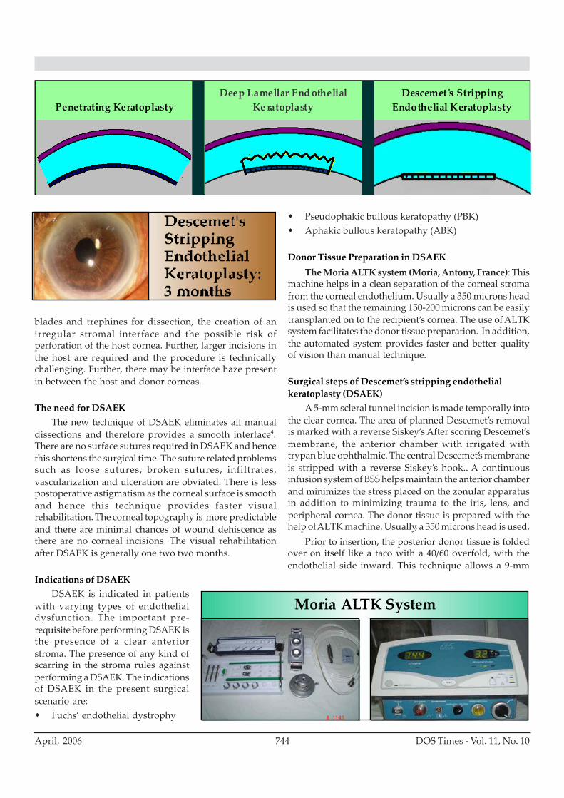
Endothelial dysfunction from disease or trauma is oneof the leading indications for corneal transplantation. Overthe past 100 years, the only solution for endothelialreplacement was through full thickness cornealtransplantation. While penetrating keratoplasty (PKP) hasbeen shown to yield healthy donor tissue with goodendothelial function, this procedure has been plagued bythe inherent problems of unpredictable surfacetopography, retained surface sutures, and poor woundstrength. .
Problems with PKPLong visual recovery time due to the presence ofmultiple suturesPoor quality of vision due to high astigmatism in postoperative period.Risk of wound rupture due to a large wound.PKP can produce anatomically clear corneas, but its
refractive results are abysmal. The sutures remain in placefor a long time and these can cause suture related problemssuch as loose sutures, broken sutures, suture infiltrates,and even graft infection. The patients in developingcountries such as ours come from remote and rural areasand often do not follow up regularly. Poor quality of visionin these eyes has been associated with high astigmatism.Wound dehiscence is another complication, which canoccur in these cases. Patients with bilateral disease oftenwait for a long time for corneal transplantation in theirfellow eye until problems with their first eye resolve.Advantages of Endothelial TransplantationThis technique of endothelial transplantation offers severaladvantages, which include the following:
1. Less postoperative astigmatismThe technique of endothelial transplant is termed as
“sutureless”. At the end of the surgery, only 2-3 suturesare applied on the scleral tunnel. This ensures a quickerpost operative visual recovery as compared to aconventional penetrating keratoplasty.
Descemet’s Stripping Automated Endothelial KeratoplastyVishal Jhanji MD, Namrata Sharma MD, Rasik B Vajpayee MS, FRCS Edin
Dr. R.P.Centre For Ophthalmic Sciences,AIIMS, New Delhi
2. Faster visual recoveryThe presence of a smooth ocular surface in the absence
of corneal sutures ensures a faster visual recovery in thepostoperative period.
3. Stronger wound integrityThere are no large wounds or potential areas of wound
dehiscence after endothelial keratoplasty.
4. Less risk of rejectionTheoretically, there is less risk of allogenic graft
rejection as decreased amount of corneal tissue istransplanted.
Landmarks in Endothelial TransplantationIn 1998, Gerrit Melles1 first described the technique of
Posterior lamellar keratoplasty (PLK) by which the innerlayers of the cornea were replaced using manual dissection.In 2001, Mark A. Terry 2 renamed the technique Deeplamellar endothelial keratoplasty (DLEK). Both optionsrepresent improvements over PKP, but they are tedious,highly surgeon-dependent techniques that requireextensive manual dissection of the donor tissue and hostcornea. Melles developed a technique of stripping theDescemet’s membrane called Descemet’s stripping lamellarendothelial keratoplasty, or DSLEK , which does notrequire manual dissection of a patient’s cornea. The mostrecent version of endothelial keratoplasty is Descemet’sstripping automated endothelial keratoplasty, or DSAEK,which was introduced by Francis Price.
Advantages of DLEKThe technique of DLEK involves manual dissection of a
posterior lamellar disc using a lamellar dissector andcurved corneal scissors (Cindy scissors) or a Terry’strephine. The posterior dissection of the host providesmechanical support to the graft, and therefore there areless chances of postoperative donor lenticule dislocation3
as compared to DSAEK where only the Descemet’s layer ofthe host is removed and no dissection of the posteriorstromal layers is done.
Problems of DLEKHowever, there are problems associated with the DLEK
procedure, which include the need for specially designed
SURGICAL OPHTHALMOLOGY
DOS Times - Vol. 11, No. 10April, 2006 744
blades and trephines for dissection, the creation of anirregular stromal interface and the possible risk ofperforation of the host cornea. Further, larger incisions inthe host are required and the procedure is technicallychallenging. Further, there may be interface haze presentin between the host and donor corneas.
The need for DSAEKThe new technique of DSAEK eliminates all manual
dissections and therefore provides a smooth interface4.There are no surface sutures required in DSAEK and hencethis shortens the surgical time. The suture related problemssuch as loose sutures, broken sutures, infiltrates,vascularization and ulceration are obviated. There is lesspostoperative astigmatism as the corneal surface is smoothand hence this technique provides faster visualrehabilitation. The corneal topography is more predictableand there are minimal chances of wound dehiscence asthere are no corneal incisions. The visual rehabilitationafter DSAEK is generally one two two months.
Indications of DSAEKDSAEK is indicated in patients
with varying types of endothelialdysfunction. The important pre-requisite before performing DSAEK isthe presence of a clear anteriorstroma. The presence of any kind ofscarring in the stroma rules againstperforming a DSAEK. The indicationsof DSAEK in the present surgicalscenario are:
Fuchs’ endothelial dystrophy
Pseudophakic bullous keratopathy (PBK)Aphakic bullous keratopathy (ABK)
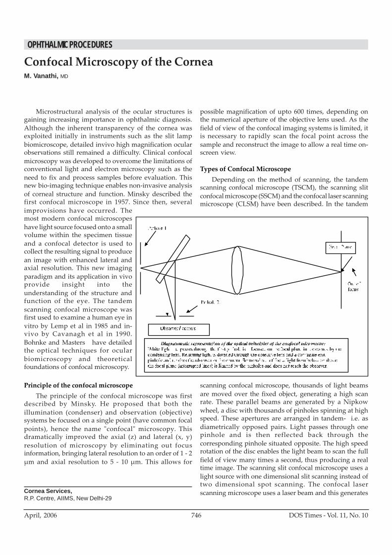
Donor Tissue Preparation in DSAEKThe Moria ALTK system (Moria, Antony, France): This
machine helps in a clean separation of the corneal stromafrom the corneal endothelium. Usually a 350 microns headis used so that the remaining 150-200 microns can be easilytransplanted on to the recipient’s cornea. The use of ALTKsystem facilitates the donor tissue preparation. In addition,the automated system provides faster and better qualityof vision than manual technique.
Surgical steps of Descemet’s stripping endothelialkeratoplasty (DSAEK)
A 5-mm scleral tunnel incision is made temporally intothe clear cornea. The area of planned Descemet’s removalis marked with a reverse Siskey’s After scoring Descemet’smembrane, the anterior chamber with irrigated withtrypan blue ophthalmic. The central Descemet’s membraneis stripped with a reverse Siskey’s hook.. A continuousinfusion system of BSS helps maintain the anterior chamberand minimizes the stress placed on the zonular apparatusin addition to minimizing trauma to the iris, lens, andperipheral cornea. The donor tissue is prepared with thehelp of ALTK machine. Usually, a 350 microns head is used.
Prior to insertion, the posterior donor tissue is foldedover on itself like a taco with a 40/60 overfold, with theendothelial side inward. This technique allows a 9-mm
Penetrating KeratoplastyDeep Lamellar End othelial
Ke ratoplastyDescemet s Stripping
Endothelial Keratoplasty
Moria ALTK System
745DOS Times - Vol. 11, No. 10 April, 2006
donor button to be inserted through a 5-mm scleral tunnelincision. A small amount of viscoelastic is placed on theendothelial side before folding it to help protect it. Thefolded tissue is grasped with a special forceps and thetissue is placed into the eye through the scleral tunnelincision. Air injected into the anterior chamber helpsunfold the donor tissue, with the endothelial sidedownward. The air also presses the donor tissue up againstthe patient’s cornea. The air is allowed to stay for 8 minutes,and then BSS wash is given. The scleral incision is suturedwith the help of 10-0 monofilament sutures.
Postoperative courseThe patient is seen the next morning and the patch is
removed. If the graft is in good position on day one, it willheal in good position. The overlying cornea has a variablerate of clearing, but some patients are able to see as well as20/25 only one week after DSAEK surgery with a clearcentral cornea.
The interface may clinically appear exceptionally clear,but it remains an interface with at least the donor tissuewith a stromal resection. It is this stromal interface of thedonor that likely contributes about one line of visual lossto the macular potential.13-14 Extensive work continues tobe done to improve the interface in DSAEK surgery.Investigators are working in the areas of femtosecond laserpreparation of the donor tissue, but currently, the interfaceafter femtosecond resections in the deep stroma are inferiorto that created by a microkeratome.
The endothelial survival after small incision DSAEKsurgery is quite remarkable. Even with folding the tissueand other donor manipulations the average endothelialcell count after small incision DLEK surgery is comparableto PK surgery.
The postoperative medical therapy after DSAEKsurgery is identical at this time to what is done with PKsurgery patients .Topical prednisolone acetate 1% is usedfour times a day for 3 months, then three times a day until6 months, then twice a day until 9 months, and then oncea day until one year postoperatively. The steroids are thentapered down further until discontinuedentirely. Fluoroquinolone antibiotics are used on a fourtimes a day dosage for the first two weeks after DSAEKsurgery and then slowly tapered over a period of six weeks.
Visual Recovery Time after various techniques ofkeratoplaty4
The visual recovery time is typically four to six weeksafter DSAEK. This is in contrast to the longer recovery timesafter penetrating keratoplasty. The patient is morecomfortable due to the absence of any surface sutures. Theamount and duration of topical antibiotics is less after aDSAEK which further shortens the visual recovery period.The approximate visual recovery time after various
keratoplasty techniques are:PKP : 12 monthsDLEK: 6 monthsDSAEK: 1 month
Our ResultsWe operated on five eyes of five patients using the
technique of DSAEK. All eyes had bullous keratopathy(three Pseudophakic bullous keratopathy, two aphakicbullous keratopathy). The DSAEK was performed usingthe standard surgical technique as described above. Thepatients were followed up regularly for three months. Atthree months follow-up period, 60% had BCVA better than20/40, and 80% had BCVA better than 20/60. The endothelialcell counts were above 2200 cells/ square mm in 60% of theeyes, and above 1800 in 80% of the eyes. There were nocases with graft dislocations.
Challenges of DSAEKPreparation of donor tissue: The use of ALTK machine isanother step towards achieving a smooth interfaceduring lamellar surgeries. Research work is being donein areas of femtosecond laser for preparation of donortissue.Unfolding of donor lenticule inside the recipient eye: Thisis especially important in order to minimize theendothelial cell loss. Various techniques to unfold thetissue have been advocated which include unfoldingwith air or balanced salt solution.Donor dislocation in early postoperative period: A repeatair injection can be tried up to three weeks in cases ofdonor dislocation.
ConclusionDSAEK patients do not require the same degree of
monitoring as standard PK patients and therefore requireless post-op clinic time. With no sutures or corneal incisionsthe wound healing or ulcerations are not an issue.Astigmatism management is also not a problem afterDSAEK surgery. With its superior topography, rapidwound healing and long term safety, the endothelialkeratoplasty of DSAEK is going to be another milestone incorneal transplant surgery.
References1. Melles GR, Eggink FA, Lander F, et al. A surgical technique for posterior lamellar
keratoplasty. Cornea. 1998; 17:618-626.2. Terry MA, Ousley PJ. Deep lamellar endothelial keratoplasty in the first United
States patients: early clinical results. Cornea. 2001; 20:239-243.3. Terry MA, Ousley PJ. Rapid visual rehabilitation after endothelial transplants with
deep lamellar endothelial keratoplasty (DLEK). Cornea. 2004; 23: 143-53. AbdullahA, Mohammad M, Teichmann KD, et al. Presumed Stromal Graft Rejection AfterDeep Anterior Lamellar Keratoplasty. Cornea 2005; 24:241-243.
4. Price FW Jr, Price MO. Descemet’s stripping with endothelial keratoplasty (DSEK)in 50 eyes: a refractive neutral corneal transplant. J Refract Surg. 2005; 21:339-345.
DOS Times - Vol. 11, No. 10April, 2006 746
Microstructural analysis of the ocular structures isgaining increasing importance in ophthalmic diagnosis.Although the inherent transparency of the cornea wasexploited initially in instruments such as the slit lampbiomicroscope, detailed invivo high magnification ocularobservations still remained a difficulty. Clinical confocalmicroscopy was developed to overcome the limitations ofconventional light and electron microscopy such as theneed to fix and process samples before evaluation. Thisnew bio-imaging technique enables non-invasive analysisof corneal structure and function. Minsky described thefirst confocal microscope in 1957. Since then, severalimprovisions have occurred. Themost modern confocal microscopeshave light source focused onto a smallvolume within the specimen tissueand a confocal detector is used tocollect the resulting signal to producean image with enhanced lateral andaxial resolution. This new imagingparadigm and its application in vivoprovide insight into theunderstanding of the structure andfunction of the eye. The tandemscanning confocal microscope wasfirst used to examine a human eye invitro by Lemp et al in 1985 and in-vivo by Cavanagh et al in 1990.Bohnke and Masters have detailedthe optical techniques for ocularbiomicroscopy and theoreticalfoundations of confocal microscopy.
Principle of the confocal microscopeThe principle of the confocal microscope was first
described by Minsky. He proposed that both theillumination (condenser) and observation (objective)systems be focused on a single point (have common focalpoints), hence the name "confocal" microscopy. Thisdramatically improved the axial (z) and lateral (x, y)resolution of microscopy by eliminating out focusinformation, bringing lateral resolution to an order of 1 - 2μm and axial resolution to 5 - 10 μm. This allows for
Confocal Microscopy of the CorneaM. Vanathi, MD
Cornea Services,R.P. Centre, AIIMS, New Delhi-29
possible magnification of upto 600 times, depending onthe numerical aperture of the objective lens used. As thefield of view of the confocal imaging systems is limited, itis necessary to rapidly scan the focal point across thesample and reconstruct the image to allow a real time on-screen view.
Types of Confocal MicroscopeDepending on the method of scanning, the tandem
scanning confocal microscope (TSCM), the scanning slitconfocal microscope (SSCM) and the confocal laser scanningmicroscope (CLSM) have been described. In the tandem
scanning confocal microscope, thousands of light beamsare moved over the fixed object, generating a high scanrate. These parallel beams are generated by a Nipkowwheel, a disc with thousands of pinholes spinning at highspeed. These apertures are arranged in tandem- i.e. asdiametrically opposed pairs. Light passes through onepinhole and is then reflected back through thecorresponding pinhole situated opposite. The high speedrotation of the disc enables the light beam to scan the fullfield of view many times a second, thus producing a realtime image. The scanning slit confocal microscope uses alight source with one dimensional slit scanning instead oftwo dimensional spot scanning. The confocal laserscanning microscope uses a laser beam and this generates
OPHTHALMIC PROCEDURES
747DOS Times - Vol. 11, No. 10 April, 2006
a monochromatic, bright, intense, sharply focused andcoherent light. A novel digital confocal laser scanningmicroscope (CLSM) recently developed, is a combinationof the Heidelberg retina tomography (HRT II) and theRostock cornea module. The LSM has a computer controlledhydraulic linear scanning device and a water contactobjective and a diode laser beam of 670nm wavelength isused as the light source. The Rostock scanning laser confocalmicroscope provides reproducible images of highresolution with uniform illumination and precise depthmeasurements.
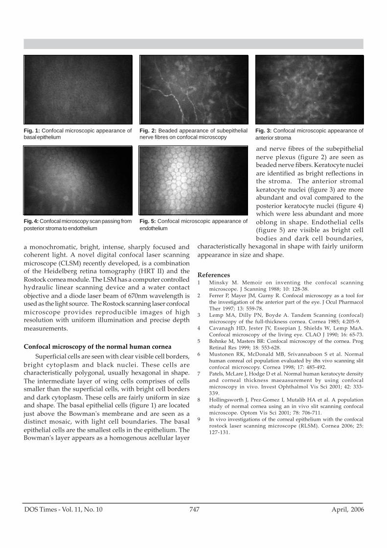
Confocal microscopy of the normal human corneaSuperficial cells are seen with clear visible cell borders,
bright cytoplasm and black nuclei. These cells arecharacteristically polygonal, usually hexagonal in shape.The intermediate layer of wing cells comprises of cellssmaller than the superficial cells, with bright cell bordersand dark cytoplasm. These cells are fairly uniform in sizeand shape. The basal epithelial cells (figure 1) are locatedjust above the Bowman's membrane and are seen as adistinct mosaic, with light cell boundaries. The basalepithelial cells are the smallest cells in the epithelium. TheBowman's layer appears as a homogenous acellular layer
and nerve fibres of the subepithelialnerve plexus (figure 2) are seen asbeaded nerve fibers. Keratocyte nucleiare identified as bright reflections inthe stroma. The anterior stromalkeratocyte nuclei (figure 3) are moreabundant and oval compared to theposterior keratocyte nuclei (figure 4)which were less abundant and moreoblong in shape. Endothelial cells(figure 5) are visible as bright cellbodies and dark cell boundaries,
characteristically hexagonal in shape with fairly uniformappearance in size and shape.
References1 Minsky M. Memoir on inventing the confocal scanning
microscope. J Scanning 1988; 10: 128-38.2 Ferrer P, Mayer JM, Gurny R. Confocal microscopy as a tool for
the investigation of the anterior part of the eye. J Ocul PharmacolTher 1997; 13: 559-78.
3 Lemp MA, Dilly PN, Boyde A. Tandem Scanning (confocal)microscopy of the full-thickness cornea. Cornea 1985; 4:205-9.
4 Cavanagh HD, Jester JV, Essepian J, Shields W, Lemp MaA.Confocal microscopy of the living eye. CLAO J 1990; 16: 65-73.
5 Bohnke M, Masters BR: Confocal microscopy of the cornea. ProgRetinal Res 1999; 18: 553-628.
6 Mustonen RK, McDonald MB, Srivannaboon S et al. Normalhuman conreal cel population evaluated by i8n vivo scanning slitconfocal microscopy. Cornea 1998; 17: 485-492.
7 Patels, McLare J, Hodge D et al. Normal human keratocyte densityand corneal thickness maeaasurement by using confocalmicroscopy in vivo. Invest Ophthalmol Vis Sci 2001; 42: 333-339.
8 Hollingsworth J, Prez-Gomez I, Mutalib HA et al. A populationstudy of normal cornea using an in vivo slit scanning confocalmicroscope. Optom Vis Sci 2001; 78: 706-711.
9 In vivo investigations of the corneal epithelium with the confocalrostock laser scanning microscope (RLSM). Cornea 2006; 25:127-131.
Fig. 1: Confocal microscopic appearance ofbasal epithelium
Fig. 2: Beaded appearance of subepithelialnerve fibres on confocal microscopy
Fig. 3: Confocal microscopic appearance ofanterior stroma
Fig. 4: Confocal microscopy scan passing fromposterior stroma to endothelium
Fig. 5: Confocal microscopic appearance ofendothelium
DOS Times - Vol. 11, No. 10April, 2006 748
Ophthalmology has progressed by leaps and bounds.Refractive surgery has evolved from Radial Keratotomy(RK), to Excimer Laser Vision correction. Refractivesurgeons and researchers have refined earlier techniquesto further improve quantitative and qualitative results.Hence today Excimer laser manufacturers have adaptedWavefront technology to ophthalmology to treat not onlyrefractive errors but also higher order aberrations(Aberropia) captured by Aberrometers which in turnguide the Excimer laser ablations.
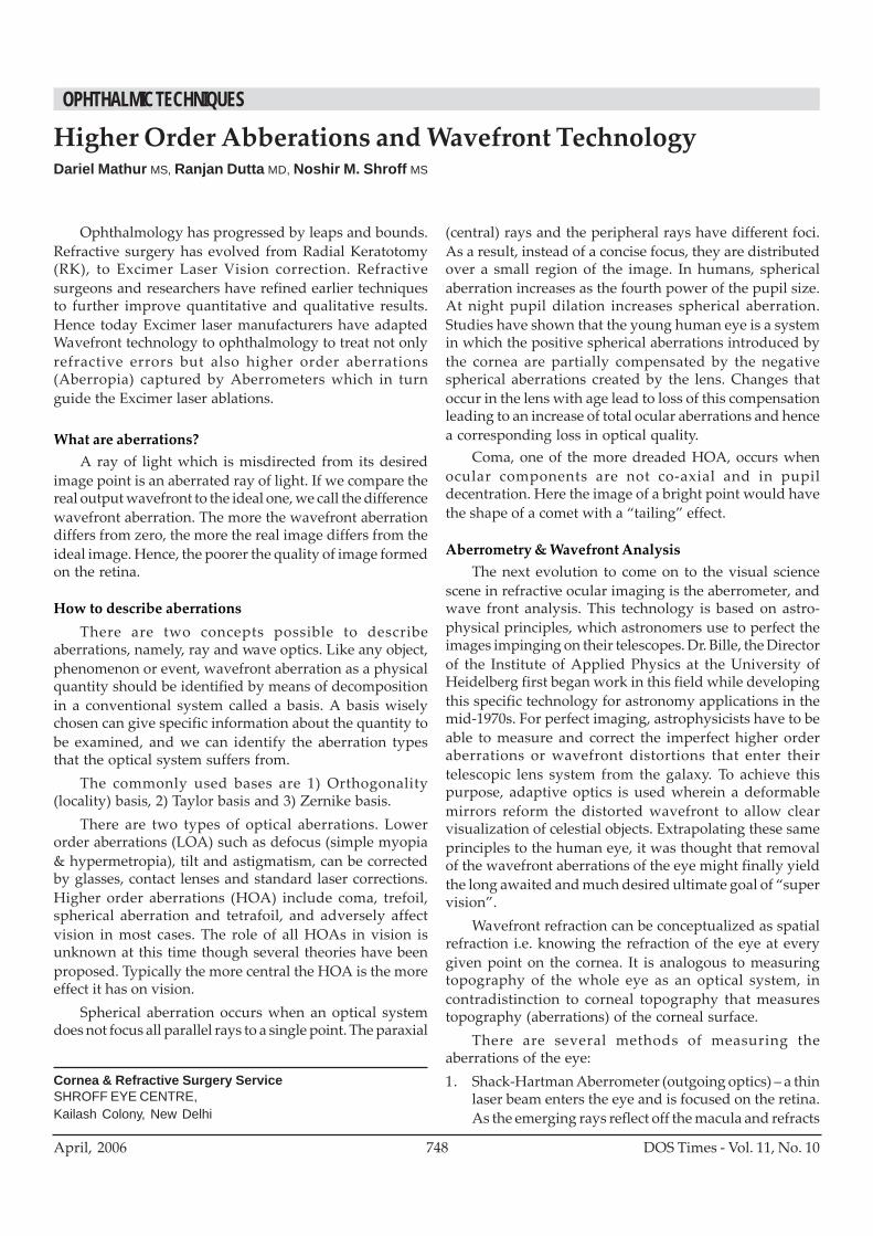
What are aberrations?A ray of light which is misdirected from its desired
image point is an aberrated ray of light. If we compare thereal output wavefront to the ideal one, we call the differencewavefront aberration. The more the wavefront aberrationdiffers from zero, the more the real image differs from theideal image. Hence, the poorer the quality of image formedon the retina.
How to describe aberrationsThere are two concepts possible to describe
aberrations, namely, ray and wave optics. Like any object,phenomenon or event, wavefront aberration as a physicalquantity should be identified by means of decompositionin a conventional system called a basis. A basis wiselychosen can give specific information about the quantity tobe examined, and we can identify the aberration typesthat the optical system suffers from.
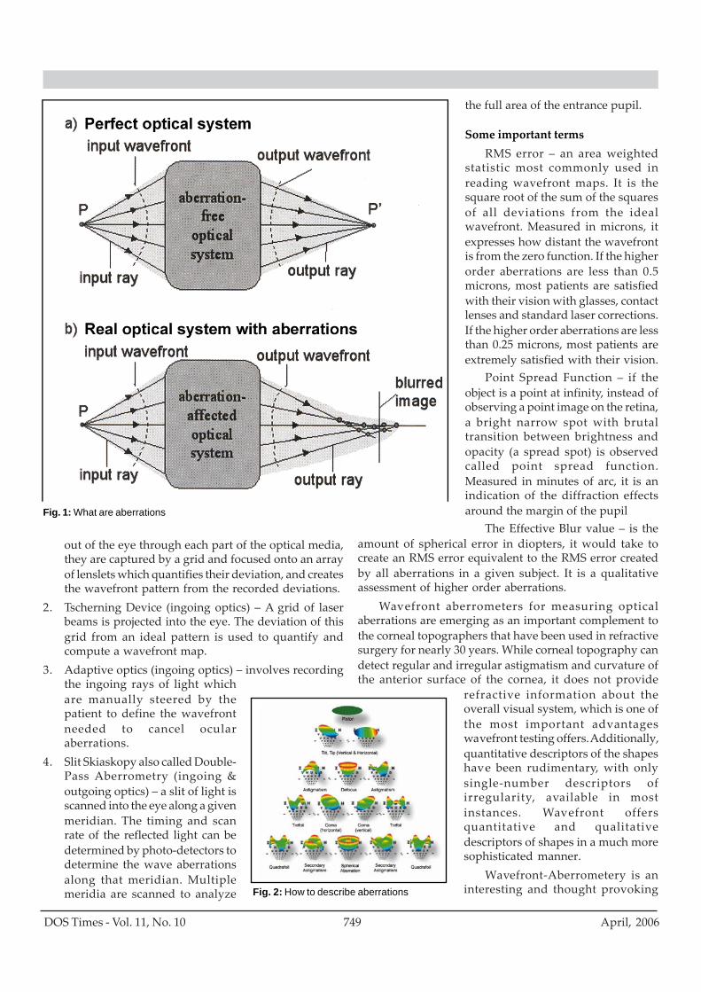
The commonly used bases are 1) Orthogonality(locality) basis, 2) Taylor basis and 3) Zernike basis.
There are two types of optical aberrations. Lowerorder aberrations (LOA) such as defocus (simple myopia& hypermetropia), tilt and astigmatism, can be correctedby glasses, contact lenses and standard laser corrections.Higher order aberrations (HOA) include coma, trefoil,spherical aberration and tetrafoil, and adversely affectvision in most cases. The role of all HOAs in vision isunknown at this time though several theories have beenproposed. Typically the more central the HOA is the moreeffect it has on vision.
Spherical aberration occurs when an optical systemdoes not focus all parallel rays to a single point. The paraxial
Higher Order Abberations and Wavefront TechnologyDariel Mathur MS, Ranjan Dutta MD, Noshir M. Shroff MS
Cornea & Refractive Surgery ServiceSHROFF EYE CENTRE,Kailash Colony, New Delhi
(central) rays and the peripheral rays have different foci.As a result, instead of a concise focus, they are distributedover a small region of the image. In humans, sphericalaberration increases as the fourth power of the pupil size.At night pupil dilation increases spherical aberration.Studies have shown that the young human eye is a systemin which the positive spherical aberrations introduced bythe cornea are partially compensated by the negativespherical aberrations created by the lens. Changes thatoccur in the lens with age lead to loss of this compensationleading to an increase of total ocular aberrations and hencea corresponding loss in optical quality.
Coma, one of the more dreaded HOA, occurs whenocular components are not co-axial and in pupildecentration. Here the image of a bright point would havethe shape of a comet with a “tailing” effect.
Aberrometry & Wavefront AnalysisThe next evolution to come on to the visual science
scene in refractive ocular imaging is the aberrometer, andwave front analysis. This technology is based on astro-physical principles, which astronomers use to perfect theimages impinging on their telescopes. Dr. Bille, the Directorof the Institute of Applied Physics at the University ofHeidelberg first began work in this field while developingthis specific technology for astronomy applications in themid-1970s. For perfect imaging, astrophysicists have to beable to measure and correct the imperfect higher orderaberrations or wavefront distortions that enter theirtelescopic lens system from the galaxy. To achieve thispurpose, adaptive optics is used wherein a deformablemirrors reform the distorted wavefront to allow clearvisualization of celestial objects. Extrapolating these sameprinciples to the human eye, it was thought that removalof the wavefront aberrations of the eye might finally yieldthe long awaited and much desired ultimate goal of “supervision”.
Wavefront refraction can be conceptualized as spatialrefraction i.e. knowing the refraction of the eye at everygiven point on the cornea. It is analogous to measuringtopography of the whole eye as an optical system, incontradistinction to corneal topography that measurestopography (aberrations) of the corneal surface.
There are several methods of measuring theaberrations of the eye:1. Shack-Hartman Aberrometer (outgoing optics) – a thin
laser beam enters the eye and is focused on the retina.As the emerging rays reflect off the macula and refracts
OPHTHALMIC TECHNIQUES
749DOS Times - Vol. 11, No. 10 April, 2006
out of the eye through each part of the optical media,they are captured by a grid and focused onto an arrayof lenslets which quantifies their deviation, and createsthe wavefront pattern from the recorded deviations.
2. Tscherning Device (ingoing optics) – A grid of laserbeams is projected into the eye. The deviation of thisgrid from an ideal pattern is used to quantify andcompute a wavefront map.
3. Adaptive optics (ingoing optics) – involves recordingthe ingoing rays of light whichare manually steered by thepatient to define the wavefrontneeded to cancel ocularaberrations.
4. Slit Skiaskopy also called Double-Pass Aberrometry (ingoing &outgoing optics) – a slit of light isscanned into the eye along a givenmeridian. The timing and scanrate of the reflected light can bedetermined by photo-detectors todetermine the wave aberrationsalong that meridian. Multiplemeridia are scanned to analyze
the full area of the entrance pupil.
Some important termsRMS error – an area weighted
statistic most commonly used inreading wavefront maps. It is thesquare root of the sum of the squaresof all deviations from the idealwavefront. Measured in microns, itexpresses how distant the wavefrontis from the zero function. If the higherorder aberrations are less than 0.5microns, most patients are satisfiedwith their vision with glasses, contactlenses and standard laser corrections.If the higher order aberrations are lessthan 0.25 microns, most patients areextremely satisfied with their vision.
Point Spread Function – if theobject is a point at infinity, instead ofobserving a point image on the retina,a bright narrow spot with brutaltransition between brightness andopacity (a spread spot) is observedcalled point spread function.Measured in minutes of arc, it is anindication of the diffraction effectsaround the margin of the pupil
The Effective Blur value – is theamount of spherical error in diopters, it would take tocreate an RMS error equivalent to the RMS error createdby all aberrations in a given subject. It is a qualitativeassessment of higher order aberrations.
Wavefront aberrometers for measuring opticalaberrations are emerging as an important complement tothe corneal topographers that have been used in refractivesurgery for nearly 30 years. While corneal topography candetect regular and irregular astigmatism and curvature ofthe anterior surface of the cornea, it does not provide
refractive information about theoverall visual system, which is one ofthe most important advantageswavefront testing offers. Additionally,quantitative descriptors of the shapeshave been rudimentary, with onlysingle-number descriptors ofirregularity, available in mostinstances. Wavefront offersquantitative and qualitativedescriptors of shapes in a much moresophisticated manner.
Wavefront-Aberrometery is aninteresting and thought provoking
Fig. 1: What are aberrations
Fig. 2: How to describe aberrations
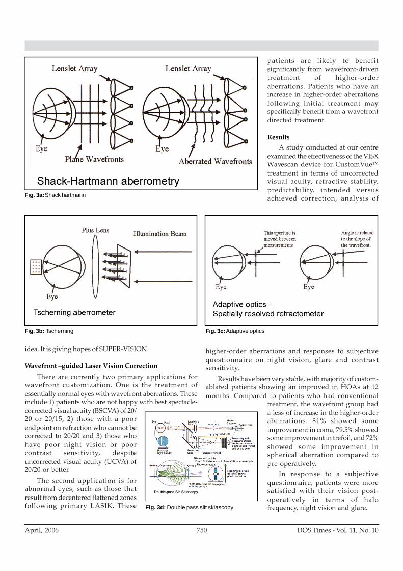
DOS Times - Vol. 11, No. 10April, 2006 750
idea. It is giving hopes of SUPER-VISION.
Wavefront –guided Laser Vision CorrectionThere are currently two primary applications for
wavefront customization. One is the treatment ofessentially normal eyes with wavefront aberrations. Theseinclude 1) patients who are not happy with best spectacle-corrected visual acuity (BSCVA) of 20/20 or 20/15, 2) those with a poorendpoint on refraction who cannot becorrected to 20/20 and 3) those whohave poor night vision or poorcontrast sensitivity, despiteuncorrected visual acuity (UCVA) of20/20 or better.
The second application is forabnormal eyes, such as those thatresult from decentered flattened zonesfollowing primary LASIK. These
patients are likely to benefitsignificantly from wavefront-driventreatment of higher-orderaberrations. Patients who have anincrease in higher-order aberrationsfollowing initial treatment mayspecifically benefit from a wavefrontdirected treatment.
ResultsA study conducted at our centre
examined the effectiveness of the VISXWavescan device for CustomVueTM
treatment in terms of uncorrectedvisual acuity, refractive stability,predictability, intended versusachieved correction, analysis of
higher-order aberrations and responses to subjectivequestionnaire on night vision, glare and contrastsensitivity.
Results have been very stable, with majority of custom-ablated patients showing an improved in HOAs at 12months. Compared to patients who had conventional
treatment, the wavefront group hada less of increase in the higher-orderaberrations. 81% showed someimprovement in coma, 79.5% showedsome improvement in trefoil, and 72%showed some improvement inspherical aberration compared topre-operatively.
In response to a subjectivequestionnaire, patients were moresatisfied with their vision post-operatively in terms of halofrequency, night vision and glare.
Fig. 3a: Shack hartmann
Fig. 3b: Tscherning Fig. 3c: Adaptive optics
Fig. 3d: Double pass slit skiascopy
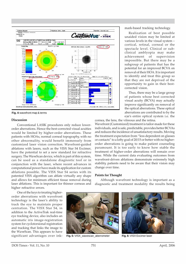
751DOS Times - Vol. 11, No. 10 April, 2006
DiscussionConventional LASIK procedures only reduce lower-
order aberrations. Hence the best-corrected visual acuitieswould be limited by higher-order aberrations. Thesepatients with HOAs, normal corneal topography, with noother abnormality, would benefit immensely fromcustomized laser vision correction. Wavefront-guidedablations with lasers, such as the VISX Star S4 Excimer,have the potential to set a new standard for refractivesurgery. The WaveScan device, which is part of this system,can be used as a standalone diagnostic tool or inconjunction with the laser, where recent advances incomputational power have made its application for customablations possible. The VISX Star S4 series with itspatented VISX algorithm can ablate virtually any shapeand allows for minimum efficient tissue removal duringlaser ablations. This is important for thinner corneas andhigher refractive errors.
One of the keys to treating higher-order aberrations with wavefronttechnology is the laser’s ability totrack the eye to maintain propercentration. The VISX Star S4, inaddition to the ActiveTrak real-timeeye tracking device, also includes anautomatic iris image-registrationsystem for cyclotorsional registrationand tracking that links the image tothe WaveScan. This appears to havesignificant advantages over ink-
mark-based tracking technology.Realization of best possible
unaided vision may be limited atvarious levels in the visual system –cortical, retinal, corneal or thespectacle level. Clinical or sub-clinical amblyopia may makeachievement of supervisionimpossible. But there may be asubgroup of patients that has thepotential for an improved BCVA onremoval of their HOA. It is importantto identify and treat this group sothat they are not deprived of theopportunity to gain in their best-corrected vision.
Thus, there may be a large groupof patients whose best correctedvisual acuity (BCVA) may actuallyimprove significantly on removal ofthe optical aberrations. These opticalaberrations are contributed to by theeye’s entire optical system i.e. the
cornea, the lens, the vitreous and the retina.Wavefront (Customized) treatment is tailor-made for theseindividuals, and is safe, predictable, provides better BCVAsand reduces the incidence of unsatisfactory results. Movingthe treatment expectation from “less dependent on glassesor contacts” to a lofty goal of 20/15 or better with no higher-order aberrations is going to make patient counselingparamount. It is too early to know how stable thetreatment of higher-order aberrations will remain overtime. While the current data evaluating outcomes fromwavefront-driven ablations demonstrate extremely highstability, patients need to be aware that their vision maychange over time.
Points for ThoughtAlthough wavefront technology is important as a
diagnostic and treatment modality the results being
Fig. 6: VISX Excimer laser
Fig. 4: wavefront map & terms
Fig. 5: VISX_wavescan_aberrometer
DOS Times - Vol. 11, No. 10April, 2006 752
achieved are not to the satisfaction level aimed for. Theremay be different reasons for this.1. Let us first look at the precision of our lasers today. Let
us assume that it is plus / minus 0.25D at best (althoughno published study has ever analyzed 0.25D outcomes).With an optical zone of 6 mm it means ±3.5 micronsprecision. To correct higher order aberrations sub-micron precision is required.
2. With wavefront based treatments we are trying tocorrect aberrations of the whole optical system byresculpting the corneal surface i.e. we are introducingnew irregularities on the corneal surface. Theseintroduced corneal irregularities would again needcorrection and so on...
3. We think that we can treat all small cornealirregularities (that compensate for the aberrationsfrom the rest of the system) but we are only assumingthat they would not be evened out by the epithelium,while the practice teaches us the opposite.
4. Concerning LASIK as a modality how can we expectthat a bulky 160 micron flap fit flush with sub-micronprecision sculpting on the stroma. Imagine a 1 cmindentation being covered with a 1.6 meters thick rug!
5. There is also a question of whether we really want tocorrect the intraocular irregularities that areoriginating from a mostly dynamic system (crystallinelens, pupil), with varies greatly with theaccommodative status and the pupil size and age.
6. With wavefront LASIK the microkeratome cut willproduce another aberration – the unreliability of theflap.The achievement of Super Vision is being questioned
and debated. It is necessary to remove all the physiologicalaberrations from the eye? The aberrations have a utility.The theory most accepted about visual perception andparticularly on the attention mechanisms, is the oneproposed by Francis Crick and Christoff Koch. It pointsout that one of the most important recognition factors islocated in the aberrations of the eye, and that it determinesa basic entity called “Multifoco” which is “pre-vision” and“pre-focus”. This is as if retina receives information onwhatever is far or close, all around us all the time &continuously. The eye, by means of a microscanner, beginsto open “Windows” towards the view appearing in frontof it, in an unconscious way, until something in the scenecomes into view reaching a cognitive value enough to callour attention and becoming conscious. The afferent branchof the visual process shoots the efferent arm making theoptic structure to focus rapidly on that part of the scene.This functioning is based on the “multifoco” created bythe eye’s optic aberrations, something that, surely,evolution has taken care of, maintaining and preservingit, and that we actually want to eliminate”.
It is alright to improve the Snellens acuity to Super
Vision level but not at the cost of eliminating the opticelements basic to visual perception and requirements. Thevision is something more than just seeing objects at adistance as if it were a landscape. We might reduce thepresent superior vision to the narrow telescopic so calledSuper Vision!
References1. J Liang, B Grimm, S Goelz, and J Bille, “ Objective measurement of the wave
aberrations of the human eye with the use of a Hartmann-Shack wavefront senson”J Opt Soc Am 11, 1949-1957 (1994)
2. J Liang, D R Williams et al aberrations and retinal image quality of the normal humaneye. J Opt. Soc. Am. A. 1997 Nov; 14(II)2873-83.
3. Oshika T, Klyce SD, Applegate RA et al. Comprison of corneal wavefront aberrationsafter photorefractive keratectony and laser in situkeratomileusis. Am J Ophthalmol1999, 127:1-7
4. MC Campbell, EM Harrison, and P Simonet, “Psychophysical measurement of theblur on the retina due to optical aberrations of the eye,” Vision Res 30, 1587-1602(1990)
5. G Walsh, WN Charman, and HC Howland, “Objective technique for the determinationof monochromatic aberrations of the human eye” J Opt Soc Am. A 1, 987-992 (1984)
6. Oshika T, Klyce SD, Apllygate RA, et al. Changes in corneal wavefront aberrationwith asging, invest Ophthalmol Vis Sci 1999; 40: 1351=1355
7. Guirao A, Gonzalez C, Redondo Metal averge optical performance of the humaneye as a funxtion of age in a normal population. Invest Ophthalmol Vis Sci 1999; 40203-213
8. He JC, Burns SA, Marcos S. Monochromatic aberratuions in the accommodated humaneye. Vis Res 2000;40:41-48
9. B Platt and RV Shack,” Lenticular Hartmann screen,”Opt Sci Center News (Universityof Arizona)5, 15-16 (1971)
10. J Liang, B Grimm, S Goelz, and J Bille, “ Objective measurement of the waveaberrations of the human eye with the use of a Hartmann-Shack wavefront sensor ”J Opt Soc Amll,1949-1957 (1994)
11. J Liang, D R Williams et al Aberrations and retinal image quality of the normalhuman eye. J Opt. Soc. Am. A. 1997 Nov; 14(11):2873-83.
12. Oshika T, Klyce SD, Applegate RA et al. Comparison of corneal wavefront aberrationsafter photorefractive keratectomy and laser in situ keratomileusis. Am J Ophthalmol1999; 127:1-7.
13. Fincham WHA, Freeman MH. Optics. 9th ed. London: Butterworths, 1980. Born M,Wolf E. Principles of Optics. 2nd ed. New York: Macmillan, 1964:203-32.
14. WN Charman, G Walsh. Variations in the local refractive correction of the eye acrossits entrance pupil. Optometry and Vision Science 1989 Jan,;66(1) 34-40
15. WM Rosenblum and JL Christensen, “Objective and subjective spherical aberrationmeasurement of the human eye,” in Progress in Optics, e Wolf, ed. ( North-Holland,Amsterdam,1976),Vo113,pp 69-91
16. MC Campbell, EM Harrison, and P Simonet, “Psychophysical measurement of theblur on the retina due to optical aberrations of the eye,” Vision Res30,15871602(1990)
17. HC Howland and B Howland, “A subjective method for the measurement ofmonochromatic aberrations of the eye, J Opt Soc Am. 67, 1508-1518(1977)
18. G Walsh, WN Charman, and HC Howland, “ Objectiv e technique for thedetermination of monochromatic aberrations of the human eye” J Opt Soc Am. Al,987-992(1984)
19. Ivanoff A. About the spherical aberration of the eye. J Opt Soc Am 1956: 46: 901-320. Jenkins TCA. Aberrations of the eye and their effects on vision. Part 1. Br J Physiol
Opt 1963; 20: 59-9121. Van den Brink G. Measurements of the geometric aberrations of the eye. Vision Res.
1962; 2: 233-4422. Born M, Wolf E. Principles of Optics. 2nd ed. New York: Macmillan, 1964:203-3223. Kaemmerer M, Mrochen M, Mierdel p, et al. Optical aberrations of the human eye.
Nature Medicine (in press)24. Oshika T, Klyce SD, Applegate RA, et al. Changes in corneal wavefront aberration
with aging. Invest Ophthalmol Vis Sci 1999; 40: 1351-135525. Calver RI, Cox MJ, Elliot DB. Effect of aging on the monochromatic aberrations of
the human eye. J Opt Soc Am A 1999; 16: 2069-207826. Guirao A, Gonzalez C, Redondo M et al. Average optical performance of the human
eye as a function of age in a normal population. Invest Ophthalmol Vis Sci 1999; 40203-213
27. Krueger R, Kaemerrer M, Mrochen M et al. Understanding refraction andaccommodation through “ingoing optics” aberrometry: a case report. Ophthalmology(in press)
28. Bogan SJ, Waring GO 3rd, Ibrahim O, et al: Classification of normal corneal topographybased on computer-assisted videokeratography. Arch Ophthalmol. 1990; 1 08(7):945-949.
29. VISX WaveScan Wavefront System Operator’s Manual, VISX, Inc., 2002 Kraff C:Results of six-month wavefront-guided ablation studies for the correction of myopiausing the VISX WaveScan system. J Refract Surg 2003; in press.
30. Liang J, Williams D: Effect of higher order aberrations on image quality in thehuman eye. Vision science and its applications. OSA Technical Digest SeriesWashington DC, Optical Society of America 1995:7073.
31. Shimmick J, et al: Axial and transverse displacement tolerances during excimer lasersurgery for myopia. Ophthalmic Technologies Proc. SPIE. 1991 ;1423: 140-53
753DOS Times - Vol. 11, No. 10 April, 2006
Bioavailability of topical drugsTopically applied drugs have to cross the hydrophilic
mucin layer to penetrate the lipophilic corneal epitheliumand then only can it reach the anterior chamber. All thehydrophilic drugs can cross mucin layer with ease but failto penetrate the lipophilic corneal epithelium. Whenlipophilic drugs come in contact with lipid bilayer ofcorneal epithelium, it gets absorbed easily if it can crossthe mucin layer of the tear film.
Mucin layer is highly hydrophilic which does not allowlipophilic drugs to pass through it. Resultant, less than 5% of the lipophilic drug is available to the corneal epitheliumfor absorption. This can be enhanced by the use ofsurfactants which also act as preservatives in the eye dropsolution. After installation, the drug is present in aqueouslayer of tear film in high concentrations. Benzalkoniumchloride (preservative) breaches the mucin layer by itssurfactant action and opens up the corneal surface for betterabsorption of the lipophilic drug. However, this method isnot safe as BAK is toxic to the corneal epithelium. Resultantfrequent instillation of BAK containing drug solution maylead to breach in corneal epithelial surface. Therefore, abetter drug delivery desire may end up leaving a bad,infection prone epithelial surface.
CyclodextrinsCyclodextrins are a group of structurally related
natural products formed during bacterial digestion ofcellulose. These cyclic oligosaccharides consist of (a-1, 4) -linked a-D-glucopyranose units with a hydrophilic outersurface and inner lipophilic central cavity. The natural a-,b-, and g- cyclodextrins consist of six, seven and eightglucopyranose units respectively. The aqueous solubilityof these natural cyclodextrins is somewhat limited andthus several different water soluble derivatives have beensynthesized. These include hydroxypropyl derivatives ofb- and g- cyclodextrins, randomly methylated andsulfobutylether b-cyclodextrins.
If we take the example of topical steroids, beinglipophilic they are marketed either as hydrophilic prodrugs(usually acetate or phosphate esters) or in suspensionforms. Prodrug fails to achieve high concentration due tolow chemical stability and poor in vivo conversion toparent drug. Suspension has again stability problems andrequires longer contact time to get absorbed on the surface.
Cyclodextrins: A New Approach in Drug DeliveryNeeraj Jain MD, DNB
Goma Bai Nethralaya and Research CentreNeemuch, (458441) Madhya Pradesh,
Use of cyclodextrin molecule overcomes all these problemsby making lipophilic drug soluble in an aqueous vehicle aswell enhancing its absorption at the epithelial surface.Topical Steroid preparations with cyclodextrin moleculehave shown better bioavailability when used.
b- Cyclodextrin and drug molecule do not interactchemically with each other. Either the whole lipophilicmolecule or the lipophilic part of the drug molecule acquiresthe central core of Cyclodextrin molecule and forms a drug-cyclodextrin complex. This complex formation enhancesthe solubility of the lipophilic molecule in aqueous solutionand increases the shelf life of the aqueous eye dropformulation.
Mechanism of actionWhen this complex is instilled in the conjunctival sac,
because of its water solubility it can cross the mucin barriervery easily and comes in contact with the cornealepithelium without harming the mucin layer. TheLipophilic drug molecule has a high affinity for the cornealsurface, so it leaves the Cyclodextrin shell in tear film andgets absorbed in lipid bilayer of corneal epithelium. Thecyclodextrin molecule has very low affinity to bind to thelipid membrane so it remains in the tear film.
Since no covalent bonds are formed or broken duringthis transfer process, the drug-Cyclodextrin complexes arein dynamic equilibrium with free drug and Cyclodextrinmolecules. If high concentration of the drug can bemaintained for a long time in the conjunctival sac, even asmall number of cyclodextrin molecules can deliver a highamount of drug to the epithelial surface. Cyclodextrinmolecule act as true carriers by keeping the lipophilic waterinsoluble drug molecules in solution and delivering themto the membrane surface where they separate from thecyclodextrin cavity into the lipophilic membrane. Theempty central core is filled by another drug molecule or byother suitable lipid molecules present in the tear film.
Studies have shown that up till 5% concentration ofcyclodextrin molecule increases the drug concentration atthe surface. Any further increase in cyclodextrin moleculeconcentration in drug formulation leads to decrease inbioavailability of the drug. But, on the other hand highlevels of cyclodextrin molecules ensure better solubilityand longer shelf life of the preparation. To balance thisequation small amount of water soluble polymer is beingadded. The addition of 0.10% hydroxypropylmethylcellulose not only increases the stability of thepreparation but also holds the drug-cyclodextrin complex
OCULAR PHARMACOLOGY
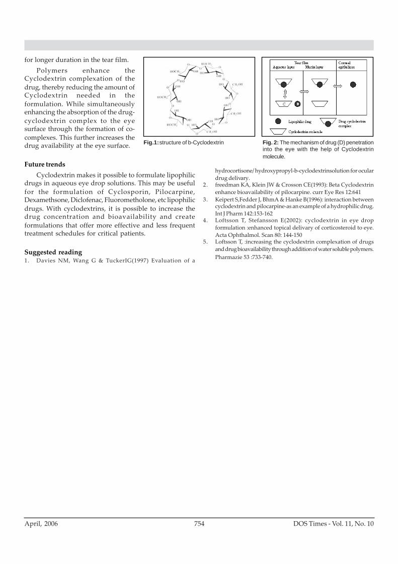
DOS Times - Vol. 11, No. 10April, 2006 754
for longer duration in the tear film.Polymers enhance the
Cyclodextrin complexation of thedrug, thereby reducing the amount ofCyclodextrin needed in theformulation. While simultaneouslyenhancing the absorption of the drug-cyclodextrin complex to the eyesurface through the formation of co-complexes. This further increases thedrug availability at the eye surface.
Future trendsCyclodextrin makes it possible to formulate lipophilic
drugs in aqueous eye drop solutions. This may be usefulfor the formulation of Cyclosporin, Pilocarpine,Dexamethsone, Diclofenac, Fluorometholone, etc lipophilicdrugs. With cyclodextrins, it is possible to increase thedrug concentration and bioavailability and createformulations that offer more effective and less frequenttreatment schedules for critical patients.
Suggested reading1. Davies NM, Wang G & TuckerIG(1997) Evaluation of a
Fig.1: structure of b-Cyclodextrin Fig. 2: The mechanism of drug (D) penetrationinto the eye with the help of Cyclodextrinmolecule.
hydrocortisone/ hydroxypropyl-b-cyclodextrinsolution for oculardrug delivary.
2. freedman KA, Klein JW & Crosson CE(1993): Beta Cyclodextrinenhance bioavailability of pilocarpine. curr Eye Res 12:641
3. Keipert S,Fedder J, BhmA & Hanke B(1996): interaction betweencyclodextrin and pilocarpine-as an example of a hydrophilic drug.Int J Pharm 142:153-162
4. Loftsson T, Stefansson E(2002): cyclodextrin in eye dropformulation :enhanced topical delivary of corticosteroid to eye.Acta Ophthalmol. Scan 80: 144-150
5. Loftsson T, :increasing the cyclodextrin complexation of drugsand drug bioavailability through addition of water soluble polymers.Pharmazie 53 :733-740.
755DOS Times - Vol. 11, No. 10 April, 2006
Over the past decade or so, India has made rapid stridesin prevention and control of blindness activities. With itsnetwork of world-class eye care services and teaching andtraining institutes already serving as global and regionalresource centers, India is poised to make notable progresson all fronts to achieve the goals of VISION 2020. Indianeye care professionals are well known and respected fortheir practical application of advanced techniques.
In spite of these achievements, majority of rural peopledo not have access to comprehensive and quality eye care.In most such areas, the mainstay of eye care service hasbeen through Surgical and screening eye camps. The qualityof services provided through such camps had always beenquestionable but in recent years the initiative taken byNPCB in setting up guidelines for these camps and effortsby Government and NGOs to improve the quality ofcataract surgery in these camps, the scenario is changing.As analysis of the profile of patients awaiting of surgicalcamp services reveals the following:
Quality eye care services are inaccessible,communication and travel is difficult.Cataract occurs relatively early (in the 40’s) in manypatients when patients are very active and are thesole bread earner for the family.They are fairly prone to trauma even in the immediatepostoperative period.The patients find it difficult to come back for sutureremoval and an exposed suture can make the eyeuncomfortable.These patients are less compliant as far as medicationsare concerned.The patients’ demands and expectations haveincreased over the years irrespective of their locationAll these factors need to be addressed while designing
the cataract surgical techniques for these situations. Oneof the critical factors in the success of eye care programmesthat is often overlooked, especially in the third world, isthe quality of the visual results after surgery. Everyonekeeps talking only of numbers.
Very often financial constraints is referred as the reasonfor offering lower quality services to the patient. This isneither a good, nor a valid excuse for poor quality surgeryand inferior results in remote and rural patients.
Feasibility of Phacoemulsification Surgery in CampsSubodh K. Sinha MS, Harsh Goel DO, MS, Abhas Sinha MS, Anil Tara MS
Type of Cataract Surgery Ideal for Indian Patients1. Cataract surgery with secure wound so that patients
can resume their earning activities early.2. Suture less, since many patients will find it difficult to
return for suture removal.3. Surgically Induced astigmatism should be minimal,
as many of these patients will not be using glasses forsome reasons or the other.
4. Quick Healing, since many patients are noncompliantas far as medications are concerned.
5. Surgery should meet the Needs and Expectations ofthe patientPhacoemulsification is the “state of the art” operation
of choice for cataract surgery.The Phacoemulsification technique offers the following
benefits and advantages over planned extra capsularcataract extraction1) It is performed through an incision 3mm or less in
size, which is self-sealing and watertight therebyimproving safety during the procedure.
2) It is significantly less invasive thereby leading to muchless ocular trauma and consequently less postoperativeinflammation which is a significant advantage overother types of cataract surgery in surgical camps asmany of the patients are poorly followed up
3) It results in minimal or no induced astigmatism.4) It provides much more rapid visual and physical
recovery and prompt refractive stability. The visualrecovery is immediate if topical anesthesia is used. Allthese advantages lead to a significant improvement inthe patient’s quality of life.
5) In addition, a smaller incision also may reduce the riskof Endophthalmitis.
Limitations of Phaco Surgery in campsOne of the strong limitations of Phaco has been the
cost of not only the Phaco equipment but also the suppliesrelated to its use.
Initial investment for Phaco machineThere was a time when the equipment or Phaco
machine required a significant investment. At present,most of the companies that manufacture Phaco units arehelping physicians and hospitals to acquire the equipmentand supplies. The equipment is made available at much
COMMUNITY OPHTHALMOLOGY
1/31, Venu Eye Institute,Sheikh Sarai Institutional Area,New Delhi-110 017
DOS Times - Vol. 11, No. 10April, 2006 756
more reasonable prices than their real sales cost, with theunderstanding that the surgeon would utilize a standardminimum of the Phaco supplies of that particularmanufacturer every month.
Running cost for PhacoemulsificationLet us analyze, however, the current situation related
to costs of performing Phacoemulsification, and compareit with the costs of the supplies needed to perform extracapsular extraction. With the latter, there is the cost ofvery fine sutures, which are unnecessary in Phaco; there isthe cost of local anesthesia involved with either a retrobulbar or a Para ocular injection versus Phaco in whichonly topical sometimes with intracameral anesthesia isutilized. The cost of the postoperative injection of steroid-antibiotic in the fornix often administered following extracapsular surgery is also unnecessary with Phaco. The costof even a fairly short stay in the hospital following theextra capsular extraction is higher than in patients withPhacoemulsification.
The significant economical savings to the patient fromlost working hours with ECCE vs. almost immediaterecovery with Phaco and the improved quality of life withPhaco are other major important contributions. All theseare important features to consider when the so-calledexpenses for both operations are taken into account.
The supplies or tubing needed for each patient wasalso a heavy expense when performing several cases. The“tubing” which previously had to be discarded after eachoperation is no longer a problem cost-wise. Now it may beused for as many as 60 cases in the same day. No re-sterilization is needed. The tubing may be used withoutreplacement for a complete day of Phaco surgery.
Limited human resourcesOne of the limitations of large volume Phaco surgery
is unavailability of Ophthalmologists trained in Phacosurgery. This is attributed to the fact that there were onlyfew training centres for Phacoemulsification but withpassing time more and more centers are coming forwardwith Phaco training programmes.
In addition, the manufacturer provides advice andhands-on-training by experts to the surgeon so that he/she will be able to enter into the transition period utilizinghis/ her own personal equipment acquired from thatmanufacturer.
All of this makes the Phaco technique more accessibleto a larger number of surgeons.
Longer learning curve for ophthalmic surgeons andlimited applicability in terms of type of cataract.
These limitations have been partly addressed by betterunderstanding of phacodynamics, availability of betterPhaco machines and better training facilities.
In high volume surgery, the cost of cataract surgeryby Phaco decreases as the numbers of surgeries increase.Over the years, Phaco machines have become cheaper,better, smaller, have multiple capabilities and reusablecomponents, do not need gas to run, are easy to transport,set up and service. The associated procedures(Capsulorrhexis, incision design, lens insertion etc.) havebeen refined and standardized and made easy to learn andcarry out. The consumables such as blades and knives andeven foldable lenses made indigenously are available ataffordable prices. Quite contrary to what people mightsuggest, funding is not the single major barrier to Phacosurgery in camps. Rationalization of expenses, saving onnon-critical components and above all vision and staffmotivation and change in attitude will help a lot in makingPHACOEMULSIFICATON the surgery of choice in campsin majority of cataract patients.