Embed Size (px)
Citation preview

Rev Electron Biomed / Electron J Biomed 2009;1:1
ISSN: 1697-090X
Inicio Home
Comité EditorialEditorial Board
Comité CientíficoScientific Committee
Normas para los autoresInstruction to Authors
Derechos de autor / Copyright
Contacto/Contact:
Rev Electron Biomed / Electron J Biomed 2009;1:1-60
Enero - Abril 2009 / January - April 2009
EDITORIALS /EDITORIALES
3-5.- CONTRIBUTION OF FOOD ANTIOXIDANTS TO HEALTH.
6-9.- CONTRIBUCIÓN A LA SALUD DE LOS ALIMENTOS CON COMPUESTOS ANTIOXIDANTES Pilar Muñiz Rodríguez PhD. Professor of Biochemist and Molecular Biology. Faculty of Sciences. Burgos University. Burgos. España
ORIGINALS / ORIGINALES
10-17.- DEPOSITION STUDIES USING MULTIPURPOSE SOLUTION ON HYDROPHILIC CONTACT LENSESA. Arora, A. Ali, R. K. Khar, M.T. Zzaman. Kiet School of Pharmacy. Ghaziabad-Meerut Road. Ghaziabad. Department of Pharmaceutics. Faculty of Pharmacy. Jamia Hamdard, Hamdard Nagar. New Delhi. India
18-23.- PURIFICATION COLUMN ON SILICA AND CHEMICAL CHARACTERIZATION OF A COUMARIN ISOLATED FROM METHANOL EXCERPT OF THE STEMS OF PLANT Secamone afzelii (ACLEPIEDACEAE) FROM ABIDJAN - IVORY COAST Hervé Zabri, Ahipo Due, Véronique Mambo Laboratoire de Chimie BioOrganique et de Substances Naturelles, Laboratoire de Biochimie et de Nutrition, Laboratoire de Chimie de L'eau. Université d'Abobo-Adjamé. Abidjan. Côte D'Ivoire
24-31.- RELATION BETWEEN GLUCOLIPID PROFILE AND SMALL INTESTINE HISTOLOGICAL PATTERNS IN DIABETIC RATS EXPOSED TO AN INTERMITTENT DIETARY RESTRICTION Noriyuki Hisano Ph.D, Silvana Marisa Montenegro Ph.D, Stella Maris Martinez Ph.D, María Cristina Tarrés Ph.D, Alberto Enrique D'Ottavio Ph.D Medical School, Faculty of Medical Sciences and Scientific Researcher Career. Rosario National University, Rosario. Argentina
CASE REPORTS / CASOS CLÍNICOS
32-35.- OXCARBAZEPINE INDUCED HYPONATREMIA: A POTENTIAL EXPLANATION OF ITS PHYSIOPATHOLOGY.
36-39.- HIPONATREMIA INDUCIDA POR OXCARBAMACEPINA: UNA HIPÓTESIS FISIOPATOLÓGICA Carlos G. Musso, Vilas M, Aparicio C, Bevione P, Reynaldi J, Rojas J, Jauregui R, Algranati L. Nephrology, Neurology Departments, Centro Médico Agustin Rocca. Hospital Italiano de Buenos Aires. Buenos Aires. Argentina
INTERNET REVIEWS / REVISIONES EN INTERNET
40-45.- GANGLIO CENTINELA Y CÁNCER DE MAMA Juan de Dios Díaz-Rosales, MD Servicio de Cirugía. Hospital General de Ciudad Juárez. Ciudad Juárez. México
http://biomed.uninet.edu/2009/n1/index.html

Rev Electron Biomed / Electron J Biomed 2009;1:2
MEDICAL HYPOTHESES / HIPÓTESIS MÉDICAS
46-50.- USO DEL TAMOXIFENO PARA TRATAMIENTO DE LA FIBROSIS SISTÉMICA NEFROGÉNICA
51-55.- USE OF TAMOXIFEN FOR THE TREATMENT OF NEPHROGENIC SYSTEMIC FIBROSIS Enz PA, Musso CG, Fato C, Torre A, Galimberti R, Algranati L. Departments of Dermatology and Nephrology. Hospital Italiano de Buenos Aires - Argentina
LETTERS TO THE EDITOR / CARTAS AL EDITOR
56-58.- MAY A PROBLEM-BASED LEARNING CURRICULUM ENTAIL PROBLEMS? Alberto Enrique D'Ottavio. Facultad de Ciencias Médicas y Consejo de Investigaciones. Universidad Nacional de Rosario. Argentina
59-60.- PROMOCIÓN DE LA DONACIÓN Beatriz Cuevas-Ruiz y María Zamora González-Mariño Servicio de Hematología y Hemoterapia. Complejo Asistencial de Burgos. España
http://biomed.uninet.edu/2009/n1/index.html

Rev Electron Biomed / Electron J Biomed 2009;1:3 Editorial: CONTRIBUTION OF FOOD ANTIOXIDANTS TO HEALTH.
ISSN: 1697-090X
Inicio Home
Indice del volumen Volume index
Comité EditorialEditorial Board
Comité CientíficoScientific Committee
Normas para los autoresInstruction to Authors
Derechos de autor Copyright
Contacto/Contact:
Rev Electron Biomed / Electron J Biomed 2009;1:3-5
Editorial:
CONTRIBUTION OF FOOD ANTIOXIDANTS TO HEALTH.
Pilar Muñiz Rodríguez PhD.
Professor of Biochemist and Molecular Biology Faculty of Sciences. Burgos University. Burgos. España
pmuniz @ ubu.es
Version española
During the last decades, there has been a lot of research work on oxidative stress both due to its involvement in homeostasis of normal cells and its implications in the development of a large number of degenerative diseases such as cardiovascular disease, neurodegenerative disease, chronic inflammation, cancer etc. Although the role of RSON (reactive species of oxygen and nitrogen) in the pathogenesis of different diseases was originally attributed to oxidative damage exerted on the different molecules by altering their function, it is now also known to be involved as an intracellular messenger in gene regulation and in signal transduction pathways. On the other hand, in a very narrow range of concentration The RSONs can lead to the opposite effects such as proliferation or apoptosis.
As a reaction to this increase of free radicals, living organisms have endogenous defense mechanisms consisting of enzymatic antioxidants (superoxide dismutase, catalase, glutathione peroxidase) or non-enzymatic antioxidants (glutathione, thioredoxin, etc). Their function is to eliminate free radicals like superoxide, hydroxyl and peroxides before they react and interact with different biomoléculas (lipids, proteins, DNA), inducing cellular damage.
http://biomed.uninet.edu/2009/n1/editorial-en.html
Rev Electron Biomed / Electron J Biomed 2009;1:4 Editorial: CONTRIBUTION OF FOOD ANTIOXIDANTS TO HEALTH.
As reinforcement for this endogenous antioxidant capacity, there are foods with antioxidant compounds of nutritional interest, with a chemical structure compatible with the in vivo antioxidants /antioxidant properties. Among food compounds with antioxidant properties are polyphenols, vitamins, etc., which can modulate the cellular response to RSON through different mechanisms: stabilizing reactive oxygen species, suppressing their formation by inhibiting enzymes or acting as metal chelators. In this way, these foods can restor the endogenous antioxidant defense or regulate intracellular signals resulting from the cellular antioxidant response.
There are more than 2000 epidemiological studies showing a relationship between the protective effect against various diseases and the consumption of foods with antioxidant capacity. This protective effect has been observed against different diseases (mainly cardiovascular), and is correlated with a high intake of fruits and vegetables. According to WHO reports, if not remedied before, in the year 2020 some diseases (cardiovascular diseases, diabetes, hypertension and some cancers) will be the cause of 73% of deaths and of 60% of the global diseases (WHO, 2001). Therefore, in order to prevent the diseases associated with free radicals, the WHO recommendS an intake of 400 g of fruit and vegetables a day.
Regarding the contribution to health of the antioxidant compounds present in food, others factors should also be considered such as:
1) the presence of other non-antioxidant compounds that contribute indirectly to the reduction of these pathologies (folate, fiber, etc.),
2) the antioxidant capacity of the diet depends on the absorption or the metabolic changes that may alter the antioxidant activity of the original molecule.
3) These compounds can perform their function independently of their ability to act as antioxidant, as they can interact with enzymes, by binding to membrane or nuclear receptors, altering gene expression, etc.
4) The effect of the isolated or pure compound is not the same as when it forms part of the food matrix, which can be synergistic with other components of the food.
REFERENCES
Brownson DM, Azios NG, Fuqua BK, Dharmawardhane TJ. Flavonoid Effects Relevant to Cancer. J Nutr 2002;2132:3482S-3489S.
Cao G, Russell RM, Lischner N, Prior RL. Serum antioxidant capacity is increased by consumption of strawberries, spinach, red wine or
http://biomed.uninet.edu/2009/n1/editorial-en.html

Rev Electron Biomed / Electron J Biomed 2009;1:5 Editorial: CONTRIBUTION OF FOOD ANTIOXIDANTS TO HEALTH.
vitamin C in elderly women. J Nutr 1998;128:2383-90.
Davies KJA. Oxidative stress: The paradox of aerobic life. Biochem Soc Symp. 1995; 61:1-31
Fang YZ, Yang S, PHD W. Free radicals, antioxidants and nutrition. Nutr 2002;18:872-879.
Halliwell B, Gutteridge JM. Free Radicals in Biology and Medicine, second ed. Oxford University Press. New York. 1989.
Heim KE, Taglliaferro AR, Bobilya DJ. Flavonoid antioxidants: Chemistry, metabolism and structure-activity relationships. J Nutr Biochem. 2002;13: 572-584
Jiménez-Escrig A, Santos-Hidalgo AB, Saura-Calixto F. Common sources and estimated intake of plant sterols in the Spanish diet. J Agric Food Chem. 2006;3;54:3462-71
Lindsay DG, Astley SB. European research on the functional effects of dietary antioxidants- EUROFEDA. Mol Aspects Med. 2002;23:1-38.
Serafini M, Villan D, Spera G, Pellegrini N. Redox molecules and cancer prevention: the importance of understanding the role of the antioxidant network. Nutr Cancer 2006;56:232-240.
Udenigwe CC, Ramprasath VR, Aluko RE, Jones PJ. Potential of resveratrol in anticancer and anti-inflammatory therapy. Nutr Rev. 2008;66:445-54
Valko M, Leibfritz D, Moncol J, Cronin MT, Mazur M, Telser J. Free radicals and antioxidants in normal physiological functions and human disease. Int J Biochem Cell Biol. 2007;39:44-84.
Virgili F, Marino M. Regulation of cellular signals from nutritiona molecules : a specific role for phytochemicals beyond antioxidant activity. Free Radical Biology and Medicine. 2008;45:1205-1216.
Yang CS, Landau JM, Huang MT, Newmark HL. Inhibition of carcinogenesis by dietary polyphenolic compounds. Annu Rev Nutr 2001:381-406.
http://biomed.uninet.edu/2009/n1/editorial-en.html

Rev Electron Biomed / Electron J Biomed 2009;1:6 Editorial: ... ALIMENTOS CON COMPUESTOS ANTIOXIDANTES
ISSN: 1697-090X
Inicio Home
Indice del volumen Volume index
Comité EditorialEditorial Board
Comité CientíficoScientific Committee
Normas para los autoresInstruction to Authors
Derechos de autor Copyright
Contacto/Contact:
Rev Electron Biomed / Electron J Biomed 2009;1:6-9
Editorial:
CONTRIBUCIÓN A LA SALUD DE LOS ALIMENTOS CON
COMPUESTOS ANTIOXIDANTES.
Pilar Muñiz Rodríguez PhD.
Profesora Titular del Área de Bioquímica y Biología Molecular de la Facultad de Ciencias
de la Universidad de Burgos. España
pmuniz @ ubu.es
English version
En las últimas decadas, son muchos los trabajos realizado en el campo de las especies reactivas del oxigeno y del nitrógeno (ERON) tanto por su implicación en las homeostasis de las células normales como por su implicación en el desarrollo de un número elevado de patologías degenerativas, como enfermedades cardiovasculares, neurodegenerativas, inflamación crónica, cáncer, etc. El papel de las ERON en la patogénesis de las diferentes enfermedades aunque en un principio fue atribuido al daño oxidativo que podrían ejercer sobre las diferentes biomoléculas alterando su función hoy se sabe que además participan como mensajeros intracelulares participando en la regulación génica así como en vías de transducción de señal. Estos ERON en un rango muy estrecho de concentración pueden generar efectos opuestos como por ejemplo, proliferación o apoptosis.
Frente a este incremento de los radicales libres los organismos vivos disponen de mecanismos de defensa endógena que consta de los antioxidantes endógenos como las enzimas superóxido dismutasa, catalasa, glutation peroxidasa, o los
http://biomed.uninet.edu/2009/n1/editorial-es.html

Rev Electron Biomed / Electron J Biomed 2009;1:7 Editorial: ... ALIMENTOS CON COMPUESTOS ANTIOXIDANTES
antioxidantes tiolicos no enzimáticos glutatión y tiorredoxina. Su función es eliminar los radicales libres como el superóxido, y peróxidos antes de que ellos reaccionen e interaccionen con distintas biomoléculas induciendo el daño celular.
Como refuerzo a esta actividad antioxidante endógena existen varias moléculas, de interés nutricional, con una estructura química compatible con las propiedades antioxidantes in vivo entre ellos se encuentran compuestos como los polifenoles, vitaminas, etc que son capaces de modular la respuesta celular a las ERON a través de diferentes mecanismos, estabilizando las especies oxigénicas reactivas, suprimiendo su formación al inhibir enzimas o actuando como quelantes de metales, regenerando la defensa antioxidante endógena o regulando señales intracelulares resultado en la respuesta antioxidante celular.
Son más de 2000 los estudios epidemiológicos que muestran una relación entre efecto protector frente a diferentes enfermedades y el consumo de alimentos con capacidad antioxidante. Este efecto protector se ha observado contra diferentes enfermedades (principalmente cardiovasculares y cáncer), y está correlacionados con una elevada ingestión de frutas y verduras. Teniendo en cuenta que a Organización Mundial de la Salud (OMS) refleja en sus documentos como, de no actuar adecuadamente, en el año 2.020 las enfermedades no transmisibles (patologías cardiovasculares, diabetes, hipertensión arterial y ciertos tipos de cáncer) serán la causa del 73% de las defunciones y del 60% de la carga mundial de enfermedad (OMS, 2001). De esta forma, la OMS en base a estos estudios recomienda una ingesta de 400 g de frutas y verduras al día, para poder prevenir las patologías asociadas a los radicales libres.
En relación a esta contribución de los compuestos antioxidantes presente sen los alimentos sobre la salud además deben considerarse factores como:
1) la presencia de otros compuestos de naturaleza no antioxidante que contribuyen de forma indirecta a la reducción de estas patologías (folatos, fibra, etc.);
2) la capacidad antioxidante de los alimentos ingeridos depende de la absorción o de las transformaciones metabólicas que pueden modificar la actividad antioxidante original de la molécula.
3) Estos compuestos pueden llevar a cabo su función independiente de su capacidad de actuar como antioxidante al poder interactuar con enzimas, uniéndose a receptores nucleares o de membrana, modificando la expresión de genes, etc.
4) El efecto del compuesto puro o aislado, no es el mismo que cuando forma parte de la matriz alimentaría, donde se pueden establecer efectos sinérgicos con otros componentes del alimento.
http://biomed.uninet.edu/2009/n1/editorial-es.html

Rev Electron Biomed / Electron J Biomed 2009;1:8 Editorial: ... ALIMENTOS CON COMPUESTOS ANTIOXIDANTES
REFERENCIAS
Brownson DM, Azios NG, Fuqua BK, Dharmawardhane TJ. Flavonoid Effects Relevant to Cancer. J Nutr 2002;2132:3482S-3489S.
Cao G, Russell RM, Lischner N, Prior RL. Serum antioxidant capacity is increased by consumption of strawberries, spinach, red wine or vitamin C in elderly women. J Nutr 1998;128:2383-90.
Davies KJA. Oxidative stress: The paradox of aerobic life. Biochem Soc Symp. 1995; 61:1-31
Fang YZ, Yang S, PHD W. Free radicals, antioxidants and nutrition. Nutr 2002;18:872-879.
Halliwell B, Gutteridge JM. Free Radicals in Biology and Medicine, second ed. Oxford University Press. New York. 1989.
Heim KE, Taglliaferro AR, Bobilya DJ. Flavonoid antioxidants: Chemistry, metabolism and structure-activity relationships. J Nutr Biochem. 2002;13: 572-584
Jiménez-Escrig A, Santos-Hidalgo AB, Saura-Calixto F. Common sources and estimated intake of plant sterols in the Spanish diet. J Agric Food Chem. 2006;3;54:3462-71
Lindsay DG, Astley SB. European research on the functional effects of dietary antioxidants- EUROFEDA. Mol Aspects Med. 2002;23:1-38.
Serafini M, Villan D, Spera G, Pellegrini N. Redox molecules and cancer prevention: the importance of understanding the role of the antioxidant network. Nutr Cancer 2006;56:232-240.
Udenigwe CC, Ramprasath VR, Aluko RE, Jones PJ. Potential of resveratrol in anticancer and anti-inflammatory therapy. Nutr Rev. 2008;66:445-54
Valko M, Leibfritz D, Moncol J, Cronin MT, Mazur M, Telser J. Free radicals and antioxidants in normal physiological functions and human disease. Int J Biochem Cell Biol. 2007;39:44-84.
Virgili F, Marino M. Regulation of cellular signals from nutritiona molecules : a specific role for phytochemicals beyond antioxidant activity. Free Radical Biology and Medicine. 2008;45:1205-1216.
http://biomed.uninet.edu/2009/n1/editorial-es.html

Rev Electron Biomed / Electron J Biomed 2009;1:9 Editorial: ... ALIMENTOS CON COMPUESTOS ANTIOXIDANTES
Yang CS, Landau JM, Huang MT, Newmark HL. Inhibition of carcinogenesis by dietary polyphenolic compounds. Annu Rev Nutr 2001:381-406.
http://biomed.uninet.edu/2009/n1/editorial-es.html

Electron J Biomed 2009;1:10. Arora et al... MULTIPURPOSE SOLUTION ON HYDROPHILIC CONTACT LENSES
ISSN: 1697-090X
Inicio Home
Indice del volumen Volume index
Comité Editorial Editorial Board
Comité Científico Scientific Committee
Normas para los autores Instruction to Authors
Derechos de autor Copyright
Contacto/Contact:
DEPOSITION STUDIES USING MULTIPURPOSE SOLUTION ON
HYDROPHILIC CONTACT LENSES
A. Arora1, A. Ali2, R. K. Khar2, M.T. Zzaman1.
1Kiet School of Pharmacy. Ghaziabad-Meerut Road. Ghaziabad 2Department of Pharmaceutics. Faculty of Pharmacy. Jamia Hamdard, Hamdard Nagar.
New Delhi. India
alkamailin @ yahoo.co.in
Rev Electron Biomed / Electron J Biomed 2009;1:10-17
Comment of the reviewer Prof. Pilar Muñiz Rodriguez. PhD. Professor of Biochemistry and Molecular Biology, Faculty of Science. University of Burgos. España
Comment of the reviewer Victoria Valls Bellver PhD. Biochemistry. Department of Pediatry and Ginecology. University of Valencia. España
SUMMARY
In the tears lysozyme and albumin are also present besides other constituents. All these constituents form a biofilm on the hydrophilic contact lenses - minutes after the lens is placed in the eye. These deposits if not removed make the contact lens translucent and impair visual acuity. For the removal of deposit multipurpose solution is used.
In the study, deposits of lysozyme and albumin were made on hydrophilic contact lenses deliberately. These deposits laden contact lenses were then treated with multipurpose solution for 12 hrs. The extent of removal of these deposits by the action of sodium citrate present in multipurpose solution was assessed by measuring albumin and lysozyme quantitatively by using standard analytical procedures.
It was observed that 0.1% of sodium citrate could remove lysozyme and albumin efficiently. Albumin deposited more as compared to lysozyme and non ionic hydrophilic contact lenses are less prone to deposition than ionic. Any further increase in sodium citrate was not desirable.
KEY WORDS: Lysozyme. Albumin. Sodium citrate. Standard tear fluid (STF). Non enzymatic cleaner
RESUMEN
En las lágrimas están presentes lisozima y albúmina, además de otros constituyentes. Todos estos componentes forman una
http://biomed.uninet.edu/2009/n1/arora.html

Electron J Biomed 2009;1:11. Arora et al... MULTIPURPOSE SOLUTION ON HYDROPHILIC CONTACT LENSES
biopelícula en las lentes de contacto hidrofílicas y minutos después de la lente se coloca en el ojo. Estos depósitos, si no se eliminan, hacen el contacto con la lente translúcida y afectan la agudeza visual. Para eliminar los depósitos se utiliza una solución multiuso.
En el estudio, se hicieron deliberadamente depósitos de lisozima y albúmina en lentes de contacto hidrofílicas. Estos depósitos sobre los lentes de contacto fueron tratados con la solución multiuso durante 12 horas. El grado de eliminación de estos depósitos por la acción del citrato de sodio en solución multiuso se evaluó mediante la medición de la albúmina y la lisozima cuantitativamente, mediante procedimientos analíticos.
Se observó que el citrato de sodio al 0,1% podría eliminar la lisozima y albúmina de manera eficiente. La Albúmina se depositó más en comparación con la lisozima y las lentes de contacto hidrofílicas no iónicas son menos proclives al depósito que las iónicas. Cualquier aumento posterior de citrato de sodio es indeseable.
PALABRAS CLAVE: Lisocima. Albúmina. Citrato sódico. Fluído standard de lágrimas (STF). Limpiador no enzimático.
INTRODUCTION
In the eye besides tear, other constituents are also present like proteins, lysozyme, albumin and salts including calcium. All these form a lipoprotein surface film on the hydrophilic contact lenses and other contaminants are adsorbed on this film further. The contaminants may be the environmental pollutants such as nicotine, cosmetic ingredients, finger dirt, chemical vapors, water impurities and preservatives/active ingredient from ophthalmic products1.
Certain other lipid secretions from the eye glands (meibomian glands) can also bind to the lens surface, forming a lipoprotein film that is very difficult to remove. All such deposits if not removed then may cause discomfort and impair visual acuity. Microorganisms may further build up on these deposits and the situation further worsens. To remove such deposits, the lenses are to be treated every day with multipurpose solution (MPS) containing a deproteiniser. The deposits not only cause discomfort but also increase the risk of infection causing giant papillary conjunctivitis (GPC)2.
The enzyme cleaners provide effective cleaning but leave around 25% of the lens surface area still coated. One of the functions of multipurpose solution (MPS) is to remove lens deposits when lenses are soaked in the solution overnight. In this way, it extends the useful life of the lens and keeps the lens free from deposits and thereby provides clear vision, comfort and maintain normal eye health3.
During day time the lenses which are previously rinsed with MPS before being worn and during night, the lenses when not in use are soaked in MPS for 7-8 hours. Lens can be worn continuously for 7-8 hours in a day, after this again rinsed and soaked in MPS for 7-8 hours before being worn again4.
MATERIAL AND METHODS
1. Materials Polyhexanide hydrochloride (PHMB.HCl) was procured from Avecia Biocides, Manchester, U.K. Sodium citrate was obtained from Merck, Mumbai, India and Lysozyme from SRL, Mumbai, India. Albumin was obtained from E Merck, Mumbai, India. FDA group I (Netrafilcon A) hydrophilic contact lenses were used. All other materials were used as received.
2. Preparation of standard tear fluid (STF) containing deposition constituents STF of pH 7.4 was prepared (Table I).
Albumin and lysozyme (Table II) were added into isotonic STF of pH 7.4 by shaking the flask until a clear solution was obtained.
http://biomed.uninet.edu/2009/n1/arora.html

Electron J Biomed 2009;1:12. Arora et al... MULTIPURPOSE SOLUTION ON HYDROPHILIC CONTACT LENSES
The volume was made and pH was adjusted up to 7.4 using pHmeter.
3. Selection of hydrophilic contact lenses and MPS Group I (Netrafilcon A) hydrophilic contact lenses were used for the study. For one MPS, six contact lenses were used. The total contact lenses were 42. Container used: Transparent vial of 10.0 ml capacity were used for the study. MPS tested: Seven selected MPS were subjected to deposition studies i.e. MPS-2, MPS-6, MPS-7, MPS-8, MPS-9, MPS-10 and MPS-11
4. Method
In the deposition studies, two main constituents, which are generally deposited on the surface of the contact lens, are lysozyme and albumin. In the present study the removal of lysozyme and albumin from the deposited hydrophilic contact lenses (CL's) by the action of MPS was studied in order to assess the formulation. Eleven preparations of MPS were prepared and coded (Table III).
All these preparations contain polyhexanide hydrochloride (PMHB.HCl) as the drug and sodium citrate as the deproteiniser and the concentration of the drug varied from 0.0002 to 0.0005% and the concentration of sodium citrate (deproteiniser) varied from 0.05 to 0.30%. In the deposition studies, lysozyme and albumin were added into an isotonic simulated tear fluid (STF) of pH 7.4 in known concentration and the hydrophilic contact lenses were soaked in it for 24 hours at 37°C in order to make coatings of lysozyme and albumin on them i.e. deposits were made on the lenses deliberately and according to the composition as given in tables (Table II and Table III). These lenses were then soaked in MPS for 12 hours and the lysozyme and albumin were estimated in order to assess the deposit removing capacity of MPS 5.
In 42 vials, the artificial tear fluid containing lysozyme and albumin were added (5.0 ml in each vial). In each of 42 vials the hydrophilic contact lens was placed. All the vials were stopper with their respective caps and placed in biological shaker at 37°C. These were shaken for 24 hours. After 24 hours, the lenses were removed with the help of contact lens lifter and placed in separate vial containing 5.0 ml of MPS.
For one MPS, six vials were used and in each vial one lens was placed. These vials were left for 12 hours at room temperature i.e. 25°C. After this these vials were shaken for 5 minutes and lenses were removed. The treated MPS were analyzed for the deposition of lysozyme and albumin, removed from the lenses, by the following methods.
4.1. Estimation of lysozyme
It was determined as per the method of Hu et al7. Different concentrations of lysozyme were prepared in STF of pH 7.4 i.e. 2.0
g/ml to 20.0 g/ml. The absorbance of the solutions were determined at max 280 nm using UV spectrophotometer. From the readings (Table IV), a standard plot was prepared.
http://biomed.uninet.edu/2009/n1/arora.html

Electron J Biomed 2009;1:13. Arora et al... MULTIPURPOSE SOLUTION ON HYDROPHILIC CONTACT LENSES
In deposition studies, the treated MPS preparations were taken and the absorbance were determined at 280 nm. The amount of lysozyme was determined by using the standard plot as per the method. From the readings, a bar chart was plotted for showing the effect of MPS on removal of lysozyme from hydrophilic contact lenses (CL's)6.
4.2. Estimation of albumin
Albumin was determined as per modified Lowry method7. A stock solution of albumin 100 g/ml was prepared in STF of pH 7.4. From this stock solution, an appropriate volume was transferred into a 10.0 ml capacity volumetric flask. To this 1.0 ml of biuret reagent and 1.0 ml of phenol were added. After 5 minutes, volume was adjusted up to 10.0 ml. In this manner, all reaction
mixture were prepared containing different concentrations of albumin i.e. 3.88 g/ml to 42.68 g/ml. Albumin gave intense red color in the presence of biuret agent and phenol. The absorbance of these solutions was measured spectrophotometrically at 700 nm (Table VI).
From the absorbance and concentration value, a standard plot was drawn. In deposition studies, the treated MPS was taken and to this the biuret agent and phenol were added as the method given above. The absorbance was determined and the amount of albumin was calculated using the standard plot.
From the readings a bar chart was plotted, showing the effect of MPS solution on removal of albumin (Table VII).
http://biomed.uninet.edu/2009/n1/arora.html

Electron J Biomed 2009;1:14. Arora et al... MULTIPURPOSE SOLUTION ON HYDROPHILIC CONTACT LENSES
RESULTS:
Seven MPS coded as MPS-2, MPS-6, MPS-7, MPS-8, MPS-9, MPS-10 and MPS-11 were tested for their deposits removal capacity and efficiency upon treatment of the deposits laden hydrophilic contact lenses (Table III).
In the study, the deposits of lysozyme and albumin were made on the contact lenses deliberately by soaking them in STF of pH 7.4 containing the above constituents for 24 hours at 37 ± 0.5°C. Hydrophilic contact lenses of group I (Netrafilcon A) were used. The deposits laden contact lenses were then treated with MPS for 12 hours. Sodium citrate (deproteiniser) present in MPS in concentration of 0.1%, 0.05%, 0.07%, 0.15%, 0.17%, 0.20% and 0.30% in MPS-2, MPS-6, MPS-7, MPS-8, MPS-9, MPS-10 and
MPS-11 respectively removed lysozyme. The average amount of lysozyme in g removed per lens by MPS-2, MPS-6, MPS-7, MPS-8, MPS-9, MPS-10 and MPS-11 were 7.52, 3.63, 5.31, 7.48, 7.87, 8.06 and 8.21 respectively (Table V).
Similarly average amount of albumin removed per lens in g by MPS-2, MPS-6, MPS-7, MPS-8, MPS-9, MPS-10 and MPS-11 were 17.04, 8.27, 10.96, 17.63, 17.54, 17.83 and 17.95 respectively (Table VII).
The removal occurred on the surfaces of non-ionic hydrophilic contact lenses, which are FDA approved, group I and group II types8. The albumin was deposited more than lysozyme and the capability of the MPS with sodium citrate used as deproteiniser was more towards lysozyme.
The lysozyme could be removed easily than albumin by the MPS for hydrophilic contact lenses.
0.1% of sodium citrate in MPS for hydrophilic contact lenses is upto the mark. Sodium citrate 0.1% can be used as deproteiniser for removal of lysozyme and albumin present on the surface of hydrophilic contact lenses.
DISCUSSION:
One reason for removing contact lens deposits is to extend the useful life of the contact lens. The more important reasons for cleaning hydrophilic contact lenses are to maintain clear vision, good comfort, and most importantly normal eye health. Undesirable organic substances within the tear film layer, such as lipids, mucoproteins, albumin, immunoglobulin, glycoproteins, mucin and lysozyme combine with inorganic compounds, bacteria and microorganisms to form a complex biofilm deposit on contact lens surface within minutes of placing the lens on the eye. These deposits continue to build on the contact lens surface with successive wearing period, eventually causing discomfort from mechanical irritation of the ocular tissues, as well as blurred vision as the optical quality of the contact lens surface degrades. This biofilm can also act as an antigenic stimulus causing allergic lid reactions such as giant papillary conjunctivitis (GPC).
GPC causes blurred vision, reduced wearing time, redness, itching, stinging, ocular discomfort and mucous discharge. GPC used to be a frequent occurrence with hydrophilic contact lenses but with the advent of multipurpose solution with non enzymatic cleaners like citrate, tris, hydranateTM the incidence of GPC in hydrophilic contact lens wearer has decreased considerably.
http://biomed.uninet.edu/2009/n1/arora.html

Electron J Biomed 2009;1:15. Arora et al... MULTIPURPOSE SOLUTION ON HYDROPHILIC CONTACT LENSES
Sodium citrate is a non enzymatic deproteiniser used in MPS. It's a trivalent anionic molecule with chelating properties. It is effective in removing protein, lipids and polysaccharide deposits from contact lenses surface and breaks calcium bridges which link protein deposits to each other and to the lens. The cleaning activity and no ocular toxicity promote longer lens life. Citrate is therefore used in MPS for the overnight storage of the lens and for rinsing and soaking of the contact lenses. Citrate is a non enzymatic deproteiniser in the MPS.
Tear proteins such as lysozyme and albumin are large multivalent molecules containing both positive and negative local areas of charge. The positively charged sites on protein molecule can form ionic bonds with the negatively charged surface of the ionic contact lens and thereby binding proteins to the surface. One protein layer on to the other therefore builds up within no time, this ionic building is strong type hence to remove such bindings one has to store the lenses overnight and also to clean, rinse the lens by mechanically rubbing with MPS. This cause the removal of deposit coupled by weekly cleaning using an enzymatic cleaner as well.
In the experiment, the non ionic lenses are used because here the binding is not that strong and non enzymatic cleaner which are less allergy prone like sodium citrate has been used. There is no strong binding hence no need of enzymatic cleaner like papain which induce allergy. Hence sodium citrate could solve the purpose and is therefore used as deproteiniser. Binding occurs but not to that extent as in ionic type and this binding is broken by the compound like citrate8.
In order to ascertain the efficiency of MPS preparation towards the removal of deposits from the contact lenses, a deposition study was performed. In the study, lysozyme and albumin were measured quantitatively i.e. removal of these deposits from the surface of the lens has been studied. Seven MPS preparations i.e. MPS-2, MPS-6, MPS-7, MPS-8, MPS-9, MPS-10 and MPS-11 were tested for their deposits removal efficiency upon treatment of the used lenses by them. These preparations differ in the sodium citrate concentration- the deproteiniser. The concentration of sodium citrate varied from 0.05% to 0.3% in these formulations.
In the study, the deposits of lysozyme and albumin were made on the contact lenses deliberately by soaking them in STF of 7.4 containing these components for 24 hours at 37±0.5°C. Group I (Netrafilcon A) were used for the reasons given above. These lenses are non ionic type. These deposits laden contact lenses were then treated with MPS preparation for 12 hours. During the treatment period, the deposits of lysozyme and albumin were removed by the action of sodium citrate present in the MPS. The extent of removal of these deposits was assessed by measuring albumin and lysozyme quantitatively by using standard analytical procedures discussed in the section. It was observed that the preparation containing less amount of sodium citrate i.e. 0.05% in MPS-6 removed less amount of lysozyme and albumin.
It was further ofserved that(the preparation containing more amount of sodium citrate i.e. MPS-7 and MPS-2 (0.07% of sodium citrate MPS-7 and 0.1% in MPS-2) removeÐ these deposits in increasing orders. However, further increase of sodium citrate i.e. beyond 0.1% in the preparation of MPS-8, MPS-9, MPS-10 and MPS-11 remove the deposits, slightly more as sompared to MPS-12ˆbut not significantly. The result of MPS-2 preparation was comparable with other preparations i.e. MPS-8, MPS-9, MPS-10 and MPS-11. This is due to the fact that entire deposits were removed by MPS-2 preparation containing 0.1% of sodium citrate; therefore further increase in the sodium citrate concentration was not desirable i.e. the concentration of sodium citrate in the preparation MPS-8, MPS-9, MPS-10 and MPS-11 were in excess. Hence MPS-2 is an optimized preparation as far as quantity of sodium citrate is concerned.
It was further observed that deposits due to albumin were more as compared to lysozyme. The solution with 0.1% of sodium citrate could remove lysozyme more efficiently than albumin. Non ionic contact lenses - group I (Netrafilcon A) were used because these lenses actually discourage binding within the polymeric network of hydrophilic contact lenses. The binding is weak enough that non enzymatic cleaner like sodium citrate can break it easily.
From the deposition studies it was concluded that MPS-2, the multipurpose solution preparation, gave better results in the removal of deposits from the surface of the hydrophilic contact lens as compared to the other prepa
REFERENCES
1. Mandell RB. Contact Lens Practice. 3rd Edition, Charles C. Thomas. Springfield. 1981:44-78.
2.- Stewart-Jones JH, Hopkins GA, Phillips AJ. Drugs and solutions in contact lens practice and related microbiology. In: Phillips AJ, Stone J, eds. Contact Lenses. A Textbook for Practitioner and Student. 2nd ed. London; Boston: Butterworths, 1980, 1-59
3.- Vale J and Cox B. Drugs and the Eye. Contact lens solutions. 2nd Ed. Butterworth, London, 1985.
4.- Dada VK. Textbook of Contact Lenses. Ed.4: Jaypee Brothers, 1996.
5.- Hu J, Tartaglia L, Kohler J, Lett J, Shih K. Gentle Touch, a lens material resistant to protein deposition. CLAO J 1985;21:93-95.
6.- Jung J, Rapp J. The efficacy of hydrophilic contact lens cleaning systems in removing protein deposits. CLAO J 1993;19: 47-49.
http://biomed.uninet.edu/2009/n1/arora.html

Electron J Biomed 2009;1:16. Arora et al... MULTIPURPOSE SOLUTION ON HYDROPHILIC CONTACT LENSES
7.- Jones L, Evans K, Sariri R, Franklin V, Tighe B. Lipid and protein deposition of N-vinyl pyrrolidone-containing group II and group IV frequent replacement contact lenses. CLAO J 1997;23: 123-126.
8.- Seal DV, Hay J. Contact lens disinfection and Acanthamoeba: problems and practicalities. Pharm J 1992; 248:717–719.
9.- Bruinsma GM, Rustema-Abbing M, de Vries J, Stegenga B, van der Mei HC, van der Linden ML, Hooymans JM, Busscher HJ. Influence of wear and overwear on surface properties of etafilion. A contact lenses and adhesion of pseudomonas aeruginosa. Invest Ophthalmol Vis Sci. 2002;43:3646-3653.
10.- Jones L, Senchyna M, Glasier MA, Schickler J, Forbes I, Louie D, May C. Lysozyme and lipid deposition on silicone hydrogel contact lenses material. Eye Contact Lens. 2003;29(1 Suppl):S75-9.
11.- Subbaraman LN, Glasier MA, Senchyna M, Jones L. Stabilization of lysozyme mass extracted from lotrafilocone silicone hydrogel contact lenses. Optom Vis Sci 2005;82: 209-214.
12.- Vermeltfoort PB, Rustema-Abbing M, de Vries J, Bruinsma GM, Busscher HJ, van der Linden ML, Hooymans JM, van der Mei HC. Influence of day and night wear on surface properties of silicone hydrogel contact lenses and bacterial adhesion. Cornea. 2006; 25: 516-523.
13.- MA Subbaraman LN, Glasier MA, Senchyna M, Sheardown H, Jones L. Extraction efficiency of an extraction buffer used to quantify lysozyme deposition on conventional and silicone hydogel contact lens materials. Eye Contact Lens. 2007; 33: 169-173.
14.- Glasier MA, Keech A, Sheardown H, Subbaraman LN, Jones L. Conformational and quantitative characterization of lysozyme extracted from galyfilcon and senofilcon silicone hydrogel contact lenses. Curr Eye Res. 2008;33:1–11
15.- Carney FP, Nash WL, Sentell KB. The Adsorption of major tear film deposit in vitro to various silicone hydrogel over time. Invest ophthalmol Vis Sci. 2008;49: 120-124
16.- Santos L, Rodrigues D, Lira M, Real Oliveira ME, Oliveira R, Vilar EY, Azeredo J. Optom Vis Sci. 2008;85:520-525.
ACKNOWLEDGEMENT: The author is thankful to Gaymed Labs Pvt Ltd, Delhi, India for their financial and technical support.
Correspondence: A. Arora. B-264, Derawal Nagar, Opposite Model Town II. Delhi - 11009, India. alkamailin @ yahoo.co.in
Comment of the reviewer Prof. Pilar Muñiz Rodriguez. PhD. Professor of Biochemistry and Molecular Biology, Faculty of Science. University of Burgos. España
In this paper, the authors present the results of a study on the effect of different concentrations of sodium citrate present in a multipurpose solution commonly used for cleaning hydrophilic contact lenses. In the study, the authors observed that a concentration of 0.1% of sodium citrate was optimal in the removal of lisozyme and albumin.
Although the study was in some ways limited, the results obtained are interesting because of their usefulness to both the prolongation of the life of the contact lenses as well as on the eye health.
Comment of the reviewer Victoria Valls Bellver PhD. Biochemistry. Department of Pediatry and Ginecology. University of Valencia. España
The authors describe a method for treating lypophilic contact lenses using the multipurpose solutions (MPS). MPS are the
http://biomed.uninet.edu/2009/n1/arora.html

Electron J Biomed 2009;1:17. Arora et al... MULTIPURPOSE SOLUTION ON HYDROPHILIC CONTACT LENSES
solutions most prescribed with all lenses because of their benefits of convenience, simplicity, and disinfections properties.
It is know that among the organic substances that could deposit on contact lens, are the albumin or lysozyme that combination with bacteria or microorganism to form a biofilm on contact lens. In this work, the authors compare MRP with different concentrations of sodium citrate, showing the best results to the concentration of 0.1% where the release of albumin and lisozyme from the lens was higher.
The applicability of this study could to help prevent complications associated to eye allergies.
Received: June 11, 2008. Received reviewed: December 8, 2008 Published: February 1, 2009.
http://biomed.uninet.edu/2009/n1/arora.html
Electron J Biomed 2009;1:18. Zabri et al. CHARACTERIZAT...OUMARIN ISOLATED FROM ... SECAMONE AFZELII (ACLEPIEDACEAE)
ISSN: 1697-090X
Inicio Home
Indice del volumen Volume index
Comité Editorial Editorial Board
Comité Científico Scientific Committee
Normas para los autores Instruction to Authors
Derechos de autor Copyright
Contacto/Contact:
PURIFICATION COLUMN ON SILICA AND CHEMICAL CHARACTERIZATION OF A
COUMARIN ISOLATED FROM METHANOL EXCERPT OF THE STEMS OF
PLANT SECAMONE AFZELII (ACLEPIEDACEAE) FROM ABIDJAN - IVORY COAST
1Hervé ZABRI, 2Ahipo DUE, 3Véronique MAMBO
1Laboratoire de Chimie BioOrganique et de Substances Naturelles. 2Laboratoire de Biochimie et de Nutrition. 3Laboratoire de Chimie de L'eau.
Université d'Abobo-Adjamé. Abidjan. Côte D'Ivoire
hzabri @ yahoo.fr
Rev Electron Biomed / Electron J Biomed 2009;1:18-23
Comment of the reviewer Prof. Pilar Muñiz Rodriguez. PhD. Professor of Biochemistry and Molecular Biology, Faculty of Science. University of Burgos. España
Comment of the reviewer Pedro del Río Pérez. Community pharmacists. La Quintana de Rueda. León. España.
SUMMARY
The purpose of our study is to isolate the first molecule of Secamone afzelii and caracterize the family molecular of pure product. The research component is distinguished by a TLC, a luminescence to 366 nm with a front, Rf = 0.6. This molecule is not visible to the naked eye, nor to 254 nm, on the TLC.
The column chromatography on silica helped isolate the product search with a yield of purification equal to 18,67 ± 0.72%. The various tests carried out on the phytochemical extracted. The molecule isolated could be considered to coumarin.
KEY WORDS: Secamone afzelii, silica column, purification, coumarin, TLC.
http://biomed.uninet.edu/2009/n1/zabri.html

Electron J Biomed 2009;1:19. Zabri et al. CHARACTERIZAT...OUMARIN ISOLATED FROM ... SECAMONE AFZELII (ACLEPIEDACEAE)
RESUMEN:
La finalidad de nuestro estudio es aislar una molécula de Secamone afzelii y la caracterización de la familia molecular del producto puro. El componente estudiado es identificado por TLC, con luminiscencia a 366nm y con un Rf= 0,6. Esta molécula no es visible a simple vista, ni a 254 nm en el TLC.
La columna de cromatografía de silica permitió aislar el producto, con un rendimiento del 18,67 ± 0.72%. Se realizaron distintas pruebas sobre el extracto fitoquímico La molécula aislada puede ser la cumarina.
PALABRAS CLAVE: Secamone afzelii, columna de sílice, purificación, cumarina,
INTRODUCTION
In the world, 80% of people use medicinal plants for medicine, lack of access to medicines prescribed by modern medicine but also because these plants often have a real impact. Today, traditional knowledge is less and less transmitted and tends to disappear. That is why ethnobotany and Ethnopharmacology working to identify anywhere in the world, plants deemed active and it belongs to modern research to clarify the properties and validate the use1-4.
The search for new molecules should be undertaken within the plant and animal biodiversity using data Ethnopharmacology. This approach allows you to select plants potentially active and increase significantly the number of discovery of new products assets. Interest chemists relate to natural molecules extracted from plants and animals, is increasingly growing. Several authors have studied compounds isolated from plants with multiple interests 5-7. In recent years we are particularly interested in plants recognized by users as having antioxidant properties. Among these plants are Secamone afzelii the family ASCLEPIEDACEAE. The methanol extract was tested and found to have antioxidant properties. The molecules responsible for this important quality would be bioactive flavonoids8-9. So far no author has isolated molecules of this plant.
The originality of this study is to isolate and characterize the chemical family of the first molecule of Secamone afzelii from a methanol extract
MATERIAL AND METHODS
1. Vegetal material:
The vines of Secamone afzelii we studied were harvested in Abidjan in small bush of the University of Abobo-Adjame, which is an extension of the Banco forest. The bodies were washed under running water continuously for fifteen (15) minutes. Then the leaves were separated from the stems. These were dried in an oven at 70 ° C for one week. The body was dry pulverized by a grinder (type RETSCH 811,100) for a powder which were conducted all studies
2. Method:
Extraction by maceration
The extraction method by macerating material in a solvent is to leave a certain amount of plant material in a suitable solvent for a sufficiently long period so that the solvent reaches out molecules based on their polarity. Figure 1 summarizes the extraction method that we used.
http://biomed.uninet.edu/2009/n1/zabri.html

Electron J Biomed 2009;1:20. Zabri et al. CHARACTERIZAT...OUMARIN ISOLATED FROM ... SECAMONE AFZELII (ACLEPIEDACEAE)
Figure 1: summary of the method of extraction maceration rods Seamone afzelii
100 g powder rod is macerated in distilled methanol (500 ml) for a week, then filtered by Büchner. The solution raw methanol S1 is vacuum distilled through an evaporator (Buchi R110 type MKE 6540 / 2) until a solid gross dry R1. The maceration is resumed with M1 to have a solid gross dry R2. Finally mass of R1 and mass R2 gives 9 g.
Purification column of silica.
The thin-layer chromatography (TLC) with developing the mixture chloroform / methanol (9.5 / 0.5) was conducted on the gross extract methanol. This reveals CCM to 366 nm and Rf = 0.6 luminescent a product that is not visible to the naked eye, nor visible to 254 nm. The methanol extract was purified on a column chromatography with silica, in order to isolate the compound luminescent at UV 366 nm. The column is mounted with hexane. The height of silica is 15 cm and the inner diameter of the column is 4 cm.
Phytochemical tests.
Several tests described in the literature6 have been made to characterize the chemical family to which it would be possible to include this molecule.
RESULTS:
1. Purification
Purification column silica is first elected to the well-hexane mixture and then with hexane / chloroform (50 / 50).
The desired product is obtained with pure 100% chloroform. Purification yields are co-signed in Table 1.
http://biomed.uninet.edu/2009/n1/zabri.html

Electron J Biomed 2009;1:21. Zabri et al. CHARACTERIZAT...OUMARIN ISOLATED FROM ... SECAMONE AFZELII (ACLEPIEDACEAE)
Table 1: Values returns to purify the desired product
The yield purification 18.67 ± 0.72% shows that in the methanol extract, this compound appears with a remarkable rate.
2. Characterization
The various tests have been made6, 10 to find the chemical family to which the isolated molecules are summarized in Table 2.
Table 2: Phytohemical Screening realized into pure product
The first test to see whether the isolated molecule is a coumarin was positive. We conducted another experience on the molecule belonging to the family of coumarin.
DISCUSSION
This test is that the cycle of coumarin lactone6, 10. This test was positive. In view of the two experiences, we can say that the isolated molecule may belong to the family of coumarin.
It is worth noting that spectroscopic analysis in 1H and 13C NMR, then SDM, we can confirm that the first approach and infer the structure of this molecule. But already these initial results will help the interpretation of spectra.
In conclusion, a study of thin-layer chromatography of a methanol extract of stem Secamone afzelii, led us to purify the extract methanol. The yield 18.67 ± 0.78% of the product sought to characterize the fact that this compound is revealing that UV visible to 366 nm, was purified.
The phytochemical screening done on the pure compound permits conclude that the isolated molecule is a coumarin.
REFERENCES
1.- Henrotin YE, Labasse AH, Jaspar JM, De Groote DD, Zheng SX, Guillou GB, Reginster JY. Effects of three avocado/soybean unsaponifiable mixtures on metalloproteinases, cytokines and prostaglandin E2 production by human articular chondrocytes. Clin Rheumatol. 1998;17:31-39.
2.- Gurib-Fakhim A. Medicinal plants: traditions of yesterday and drugs of tomorrow.Mol Aspects Med. 2006;27:1-93.
3.- Saucier C, Mirabel M, Daviaud F, Longieras A, Glories Y. Rapid fractionation of grape seed proanthocyanidins. J
http://biomed.uninet.edu/2009/n1/zabri.html

Electron J Biomed 2009;1:22. Zabri et al. CHARACTERIZAT...OUMARIN ISOLATED FROM ... SECAMONE AFZELII (ACLEPIEDACEAE)
Agric Food Chem. 2001 Dec;49(12):5732-5735.
4.- Demierre A, Durand P. La vegetation macrophyte du Léman. Campagne 1997 - 1998, dans Rapports sur les études et recherches entreprises dans le bassin lémanique. Programme quinquennal 1996-2000. Campagne 1998. 129 -217.
5.- Szent György A. Oxydation, Energy transfer, and vitamins. In Nobel Lecture, Physiology or Medicin, Elsevier Publishing Co. 1965, 1922 - 1941.
6.- Dohou N, Yamni S, Tahrourh LM, Hassani I, Badoc A, Gmira N. Screening phytochimique d'une endemique libero-marocaine Thymelaea Lythroides. Bull Soc Pharm Bordeaux. 2003;142:61-78.
7.- Laidman DL, Morton RA. Unsaponifiable constituents of blowfly Larvae (Calliphora erythrocephala). Biochem J. 1962;84:386-389.
8.- Mensah AY, Houghton PJ, Akyirem GNA, Fleischer TC, Mensah MLK, Sarpon K, Adosraku R. Evaluation of Antioxidant and Free Radical Scavenging Properties of Secamone afzelii Rhoem Phytother Res. 2004;18:1031-1032.
9.- Zabri H, Kodjo C, Anoubilé B, Békro M, Békro YA. Screening and determination of flavonoids in Secamone afzelii (Asclepiadaceae) extracts. African J Pure Appl Chem. 2008;2:80-82
10.- Békro AY, Békro JAM, Boua BB, Tra Bi FH, Ehilé EE. Etude ethnobotanique et screening phytochimique de Caesalpina benthamiana (Bail) Herend et Zarucchi (Caesalpiniaceae). Sciences et Nature.2007;4:217-225.
Correspondence: Hervé ZABRI Laboratoire de Chimie BioOrganique et de Substances Naturelles. Université d'Abobo-Adjamé. UFR-SFA. 02 BP 801 Abidjan 02. hzabri @ yahoo.fr
Comment of the reviewer Prof. Pilar Muñiz Rodriguez. PhD. Professor of Biochemistry and Molecular Biology, Faculty of Science. University of Burgos. España
Secamone afzelii is a plant used in traditional medicine against various pains. The antioxidant capacity of methanol extracts obtained from this plant was described previously by Mensah et al (2004) and Houghton et al (2005). In this work, the authors show a simple and rapid method for purify and characterized partially one of the components with antioxidant capacity present in the metanolic extract of S. afzelii. This component was identified as belonging to the family of coumarins. The importance of this study is the potential use of the pure extract by their antioxidant capacity. It is known that compounds of the family of coumarins to act as antioxidants in biological systems. ______________
References: Mensah AY, Houghton PJ, Akyirem GN, Fleischer TC, Mensah ML, Sarpong K, Adosraku R. Evaluation of the antioxidant and free radical scavenging properties of Secamone afzelii Rhoem Phytother Res. 2004;18:1031-1032. Houghton PJ, Hylands PJ, Mensah AY, Hensel A, Deters AM. J (2005) In vitro tests and ethnopharmacological investigations: wound healing as an example. J Ethnopharmacol. 2005;100:100-107.
Comment of the reviewer Pedro del Río Pérez. Community pharmacists. La Quintana de Rueda. León. España.
The coumarins are compounds very abundant in nature. Chemically derived from the cyclization of O-hydroxycinnamic acid and it have different radicals in the 6- and 7-position. The coumarins are synthesized in the roots and it accumulates in young tissues. They are abundant in dicotyledonous (Rutaceae, Umbelliferae, legumes, solanaceae, ...). One of their functions in plants is antigerminative.
From a chemical point of view the coumarins can be simple (umbelliferone, esculetol, fraxetol,...) or compounds (furocoumarins [psolareno, imperatorine, bergapten ...], 7.8-furocoumarins [pimpinellin, angelicin, ...], 3-pyranocoumarins [samidine, visnadine, ...]). Plants with simple coumarins: tonka beans, meliloto, horse-chestnuts. Plants with compound coumarins bergamot, angelica,
http://biomed.uninet.edu/2009/n1/zabri.html

Electron J Biomed 2009;1:23. Zabri et al. CHARACTERIZAT...OUMARIN ISOLATED FROM ... SECAMONE AFZELII (ACLEPIEDACEAE)
Keller ... Its pathway is complex, starting from shikimic acid through of cinnamic acid and resulting in various coumarins.
The coumarins has diverse pharmacological properties (which is not synonymous with therapeutic application): sedatives (tonka bean), antispasmodic (Kella), phlebotonic (Indian horse chestnut).
The furocoumarins are photosensitives (bergamot, angelica, Ammi majus). At present, some plant species are used in therapeutic in form of the dry extract (capsules) or as oral solution.
Received: December 2, 2008. Received reviewed: December 9, 2008 Published: February 1, 2009.
http://biomed.uninet.edu/2009/n1/zabri.html

Electron J Biomed 2009;1:24. Hisano et al. ...SMALL INTESTINE HISTOLOGICAL PATTERNS IN DIABETIC RATS...
ISSN: 1697-090X
Inicio Home
Indice del volumen Volume index
Comité Editorial Editorial Board
Comité Científico Scientific Committee
Normas para los autores Instruction to Authors
Derechos de autor Copyright
Contacto/Contact:
RELATION BETWEEN GLUCOLIPID PROFILE AND SMALL INTESTINE HISTOLOGICAL PATTERNS IN DIABETIC RATS EXPOSED TO AN INTERMITTENT
DIETARY RESTRICTION
Noriyuki Hisano Ph.D1, Silvana Marisa Montenegro Ph.D1,2 Stella Maris Martinez Ph.D1,2, María Cristina Tarrés Ph.D1,2,
Alberto Enrique D'Ottavio Ph.D1,2
1Medical School, Faculty of Medical Sciences and 2Scientific Researcher Career. Rosario National University, Rosario. Argentina
aedottavio @ hotmail.com
Rev Electron Biomed / Electron J Biomed 2009;1:24-31
Comment of the reviewer Noemí Gómez Manero MD. PhD. Servicio de Medicina Interna. Complejo Asistencial de Burgos. España.
Comment of the reviewer Dr. Carlos G. Musso MD. Nephrology Department. Hospital Italiano de Buenos Aires. Buenos Aires. Argentina
SUMMARY
The effects of an intermittent and prolonged dietary restriction on biochemical variables and histological small intestinal patterns in 12-month-old male eSMT rats are examined. These spontaneously diabetic animals were separated in two groups after weaning: 10 rats fed ad libitum with standard rat chow and 10 rats fed a restricted diet by deprivation of the same food for 24 hours every 72. At 12 months of age, animals were weighed and euthanized after tail vein bleeding for plasma analysis (glycemia- both fasting and 120 minutes after an oral glucose challenge-, triglyceridemia and total cholesterolemia). Small intestines were removed, weighed and measured in length.
Intestinal specimens were fixed, embedded in paraffin, semi serially cut at 6 µm and stained with PAS-Hematoxilyn and Hematoxilyn-Eosin. Histometry was performed through a linear devise attached to ocular lens and lectin histochemistry was accomplished employing Canavalis ensiformis, Dolichos biflorus, Arachis hypogea, Ulex europaeus-I, Triticum vulgaris, Ricinus communis and Soy Bean (Glicine Max) Agglutinin. Essentially, eSMT rats, a suitable animal model for studying diabetes and/or its complications, revealed at 12 months of age after undergoing the dietary restriction: 1.- An expected improvement in body weight and determined biochemical variables (fasting and after glucose overload glycemias, triglyceridemia and total cholesterolemia) without reaching euglycemic values. 2.- Changes in most of the analyzed histometric patterns with no relevant reflection on morphometric ones, and 3.- No modifications in lectinhistochemical patterns.
KEY WORDS:Diabetes. Rat. Diet. Digestive tube. Biochemistry. Histology
http://biomed.uninet.edu/2009/n1/hisano.html
Electron J Biomed 2009;1:25. Hisano et al. ...SMALL INTESTINE HISTOLOGICAL PATTERNS IN DIABETIC RATS...
RESUMEN:
Se analizaron los efectos de una restricción dietética intermitente y prolongada sobre variables bioquímicas y patrones histológicos intestinales en ratas macho eSMT. Estos animales, diabéticos espontáneos, fueron separados en dos grupos luego de su destete: 10 fueron alimentados ad libitum con dieta estándar para ratas de laboratorio y 10, sobrellevaron una privación del mismo alimento de 24 horas cada 72. A los 12 meses de edad, los animales fueron pesados y sometidos a eutanasia tras de extraerles sangre de la vena de la cola para análisis plasmáticos (glucemias en ayuno y tras 120 minutos de sobrecarga glúcida, trigliceridemia y colesterolemia total). El intestino delgado fue removido, pesado y medida su longitud.
Los especímenes fueron fijados, incluidos en parafina, cortados a 6 µm de modo semiserial y coloreados con PAS-Hematoxilina y Hematoxilina-Eosina. La histometría fue llevada a cabo mediante un dispositivo lineal ligado al ocular y la lectin histoquímica, empleando Canavalis ensiformis, Dolichos biflorus, Arachis hypogea, Ulex europaeus-I, Triticum vulgaris, Ricinus communis and Soy Bean (Glicine Max) Agglutinin. Esencialmente, las ratas eSMT, un modelo adecuado para el estudio de la diabetes y/o sus complicaciones, revelaron a los 12 meses tras la restricción dietaria seguida: 1.- Una esperada mejoría en el peso corporal, en las glucemias en ayuno y tras sobrecarga glúcida, en la trigliceridemia y en la colesterolemia total aunque sin alcanzar valores euglucémicos. 2.- Cambios en la mayoría de las lectinas analizadas pero sin reflejo sustancial en las variables morfométricas, y 3.- Ausencia de modificaciones en lo que se refiere a la lectinhistoquímica.
PALABRAS CLAVE:Diabetes. Rata. Dieta. Tubo digestivo. Bioquímica. Histología
INTRODUCTION
Diabetes mellitus, long considered a disease of minor significance to world health, is now taking its place as one of the main threats in the 21th.century. The diabetes epidemic, both in developed and developing nations, particularly refers to type 2 diabetes, a metabolic disorder primarily characterized by insulin resistance, relative insulin deficiency and hyperglycemia. Usually associated with overweight and obesity, its prevalence has risen at an alarming pace in the last twenty five years leading to foresee a number of diabetic persons near 366 million for 2030. Consequently, new studies on this topic become strictly necessary1-2.
Distinct animal species have been widely used for investigating the different diabetic types and contributed to the current knowledge on this metabolopathy3-8.
Exceeding induced diabetic animals through diet, alloxan, streptozotocin, surgery and transgenic procedures, those spontaneous or genetically ones (mice and rats) keep being relevant for analyzing the diabetic syndrome6.
Among the spontaneous or genetically animal models for diabetes, the eSMT rat was developed in our laboratory of Biology by crossing eSS rats (a non obese model of type 2 diabetes) with ßrats (a fertile obese model revealing normocholesterolemia, hypertriglyceridemia, and type 2 diabetes). This synthetic line evidences overweight like ß and shows an early beginning of the diabetic syndrome, an enhanced evolution of fasting hyperglycemia and glucose intolerance like eSS9-10.
Continuous or intermittent dietary restriction (DR) diminishes the expression of the diabetic syndrome and has been employed for managing type 2 diabetes and treating obesity11. Likewise, DR also affected the mucosal growth, the morphology and the cell cytokinetics in the small intestine12. Furthermore, DR diminished body and intestinal weights in adult male Lewis rats, put into evidence lower body and liver weights in adult control rats, prevented duodenal hyperplasia and augmentation of ileal villus cell number in 20 months-old -rats, reduced fasting glucose concentrations, triglycerides and cholesterol and increased apoptosis in the small intestine of aging rats13-15.
Although the effects of limiting caloric input depend on the initial age of restriction16-17, chronic caloric restriction augmented longevity, improved insulin sensitivity and lowered lifetime glycemia.
Extending former experiences in eSS and eSMT18-20, the aim of this study was to study the relation between the glucolipid profile and some small intestine histological patterns in male eSMT rats exposed to an intermittent dietary restriction.
MATERIAL AND METHODS
Animals were housed in a room with standard environmental conditions (24ºC, 12 hours light/12 hours dark schedule cycle, air exchange, tap water ad libitum and standard rat chow). 20 eSMT rats were separated in two groups after weaning: 10 rats fed ad libitum (AL) with standard rat chow and 10 rats fed a restricted feeding schedule (R) by deprivation of food for 24 hours every 72.
http://biomed.uninet.edu/2009/n1/hisano.html

Electron J Biomed 2009;1:26. Hisano et al. ...SMALL INTESTINE HISTOLOGICAL PATTERNS IN DIABETIC RATS...
At 11 months of age, animals were placed in metabolism cages. After a 5 - day adaptive period and during 10 days, food intake and body weight were determined for calculating the total food intake, the mean body weight and the relative food intake (mg/100 g body weight).
At 12 months of age, animals were weighed (BW) and euthanized after tail vein bleeding for plasma analysis (glycemia- both fasting - G0 - and 120 min after an oral glucose overload - G120 -, triglyceridemia - TG - and total cholesterolemia - TC -). Abdomen was cut and opened along the midline and the small intestine (from pylorus to the ileocecal junction) was immediately dissected, removed, flushed with PBS (phosphate buffered saline) at 4ºC and subsequently trimmed of fat and mesentery. Weight (SIW) and length (SIL) of the small intestine as well as SIW/BW x100 were then calculated.
Plasma Analysis
Fasting glycemia (18-h fast) and 120 min glycemia after an overload of 10% glucose (200 mg/100 g body weight) via stomach tube were determined by the glucose-oxidase enzymatic method using a commercial kit (Wiener Laboratories, Argentina).
Triglyceridemia and Total Cholesterolemia (both 18-h fast) were registered through an enzymatic-colorimetric method using commercial kits (Wiener Laboratories, Argentina).
Every dosage was carried out at the same hour in the morning for avoiding possible variations due to circadian rhythms.
Histomorphometric Study
Segments of small intestine (25 cm from pylorus) were removed. These portions were cut along the mesentery border, pinned in balsa wood, fixed in Carnoy's fluid, embedded in paraffin, semi serially cut at 6 µm mounted 1 out 40 section and stained with Peryodic Acid Schiff + Hematoxylin (PAS+H). A calibrated eye objective micrometer was employed. Villi heights were measured in those sections showing the entire villus from base to tip and villi width, at the middle of each villus. Total wall thickness, mucosa thickness (distance from villus tip to muscularis mucosae), crypt depth (distance from villous base to muscularis mucosae), goblet cells/villus and enterocytes/villus were complementarily measured. A minimum of ten measurements / rat was achieved.
Lectinhistochemical Analysis
Considering the interactions among lectins, histomorphometry and/or biochemical variables, specimens were successively fixed in Bouin's fluid for 45 min. and 10% formalin in 0.01 M phosphate-buffered saline (PBS-pH 7.2) for 3 hours. Paraffin embedded tissues were cut at 6 µm, stuck on slides with Vectabond, deparaffinised with xylol and hydrated with acetone-alcohol. These slices were incubated in 3% hydrogen peroxide in absolute methanol during 20-30 min for inactivating endogenous peroxidase. Specimens were subsequently rinsed several times in 0.01 M phosphate-buffered saline (PBS-pH 7.2), dried, treated with 0.1% bovine serum albumin in PBS for 15 min and incubated overnight at 4º C with the following lectins: Canavalis ensiformis (Con-A) ( -D-mannose and -D-glucose residues), Dolichos biflorus (DBA) (N-Acetyl Galactosamine -3-N-Acetyl Galactosamine and N-Acetyl Galactosamine -3-Galactose), Arachis hypogea (PNA) (Galactose ß3-N-Acetyl Galactosamine), Ulex europaeus-I (UEA-I) (Fucose 2- Galactose-ß), Triticum vulgaris (WGA) (N-Acetylglucosamine/sialic acid group), Ricinus communis (RCA) (N-Acetylgalactosamine/galactose group), and Soy Bean (Glicine Max) Agglutinin (SBA) ( D-N-Acetyl Galactosamine and ß-3-N-Acetyl Galactosamine). Lectins were employed as horseradish-peroxidase (HRP)-labelled form.
The day after, slices were rinsed with PBS, covered with the Avidin-Biotin Complex (ABC) and incubated in a substrate medium consisting of diaminobenzidine (DAB)-hydrogen-peroxide substrate medium. Reagents and lectins were purchased from Vector Labs. Finally, slices were washed with tap water, counterstained with hematoxylin, dehydrated, mounted and observed in a Zeiss investigation microscope with a color digital video camera (Sony Exwavehad model).
Digital photos in real color were selected and transformed in gray scale images for determining optical densities (OD) through an Image-proPlus Program for Windows 1.1 Version (Media Cibernetics).
For establishing the scale detailed below, structures were measured at 400X (eight counts/structure/animal). The program automatically provided the mean ± standard deviation of lectin fixation for each lectin. Lastly, a mean related with lectin fixation resulting from the total counts/ structures/ rats was determined. Zero was established from a non lectin-reactive zone whilst its maximal value, near to 250, emerged from the higher lectin- reactive zone.
Taking into account the aforesaid means, the lectin OD was graduated in accordance with the following semi-quantitative scale: OD > 200 = negative -0-, OD between 200 and 150 = weakly to moderately positive -1 to 2- and OD <150 = strongly positive -3-.
Statistical Analysis
Data were analyzed applying conventional statistical techniques as the GraphPad Prism Program, version 4.0, April 2003.
http://biomed.uninet.edu/2009/n1/hisano.html
Electron J Biomed 2009;1:27. Hisano et al. ...SMALL INTESTINE HISTOLOGICAL PATTERNS IN DIABETIC RATS...
Bioethical Evaluation
Our School of Medicine Ethical Committee previously examined and approved this project.
RESULTS:
At 11 months of age, mean body weight in R animals was significantly lower than AL ones (R: 318±42 g vs. AL: 427±31 g, p<0.001) exceeding that the total food intake was equivalent during the studied period (R: 225±19 g vs. AL: 230±36 g, p>0.05). In contrast, when the total food intake was compared with body weight (relative food intake), R values were higher (R: 0.70±0.11 vs. AL: 0.53±0.12, p<0.05).
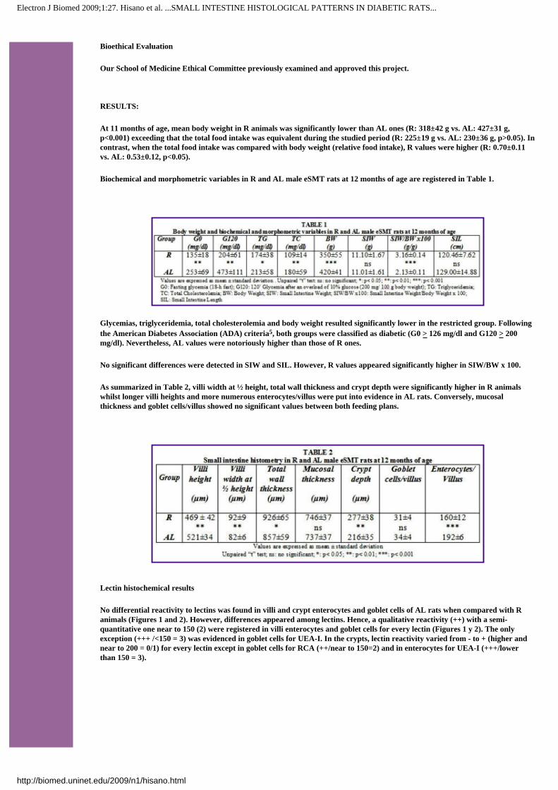
Biochemical and morphometric variables in R and AL male eSMT rats at 12 months of age are registered in Table 1.
Glycemias, triglyceridemia, total cholesterolemia and body weight resulted significantly lower in the restricted group. Following the American Diabetes Association (ADA) criteria5, both groups were classified as diabetic (G0 > 126 mg/dl and G120 > 200 mg/dl). Nevertheless, AL values were notoriously higher than those of R ones.
No significant differences were detected in SIW and SIL. However, R values appeared significantly higher in SIW/BW x 100.
As summarized in Table 2, villi width at ½ height, total wall thickness and crypt depth were significantly higher in R animals whilst longer villi heights and more numerous enterocytes/villus were put into evidence in AL rats. Conversely, mucosal thickness and goblet cells/villus showed no significant values between both feeding plans.
Lectin histochemical results
No differential reactivity to lectins was found in villi and crypt enterocytes and goblet cells of AL rats when compared with R animals (Figures 1 and 2). However, differences appeared among lectins. Hence, a qualitative reactivity (++) with a semi-quantitative one near to 150 (2) were registered in villi enterocytes and goblet cells for every lectin (Figures 1 y 2). The only exception (+++ /<150 = 3) was evidenced in goblet cells for UEA-I. In the crypts, lectin reactivity varied from - to + (higher and near to 200 = 0/1) for every lectin except in goblet cells for RCA (++/near to 150=2) and in enterocytes for UEA-I (+++/lower than 150 = 3).
http://biomed.uninet.edu/2009/n1/hisano.html

Electron J Biomed 2009;1:28. Hisano et al. ...SMALL INTESTINE HISTOLOGICAL PATTERNS IN DIABETIC RATS...
Figure 1: Histochemical reaction to Arachis hypogea (PNA) lectin in the small intestine of 12-month-old male eSMT rats exposed to ad libitum feeding schedule. Enterocytes and goblet cells are qualitatively (++) and semi-quantitatively (Optical Density = 2).
The same occurs with the thin sheath of mucus located on the brush border of the enterocytes. The image is seen at a magnification of 400 X.
Figure 2: Histochemical reaction to Arachis hypogea (PNA) lectin in the small intestine of 12-month-old male eSMT rats exposed to restricted feeding schedule. Enterocytes and the adjacent mucus sheath are qualitatively (++) and semi-quantitatively (Optical
Density = 2). The goblet cells undergo a particular functional stage after secreting the mucus which constitutes the aforesaid sheath [false negativity to PNA lectin in goblet cells with qualitative (++) and semi-quantitative (OD = 2) lectin reaction in mucus
sheath]. The image is seen at a magnification of 400 X.
DISCUSSION
Considering our results, alimentary restriction ameliorated the diabetic syndrome as revealed by the decreased BW, G0, G120, TG and TC, exceeding that G0 and G120 kept showing diabetic values in restricted eSMT rats according to the ADA criteria5. These data appeared in congruence with a better performance of the diabetic genotype in "poor" environments21,22. and agreed with the more benign course of the metabolic syndrome already demonstrated in restricted eSS and OLEFT rats18,23.
On the other hand, the higher relative food intake in R animals pointed out a compensatory consume after the fasting lapse, as opportunely suggested19. This could be particularly involved in the higher relative SIW (joined to a lower BW and a similar SIL-SIW) and the greater total wall thickness registered in R rats. In this regard, Kujalova and Fabry (1960) found that the small intestine became hypertrophic when food was fed intermittently24 whilst Jervis and Levin (1966) reported lower BW and higher SIW and SIL in the small intestine of 1-year-old white rats with severe chronic-alloxan diabetes fed ad libitum25.
R animals also showed shorter villi heights, lesser enterocytes/villus, higher villi width at ½ heights and deeper crypts, In this sense, changes in crypt depth have been associated with alterations in the maturity of cells26.
The presence of distinct quantities of nutrients in the lumen, the endocrine imbalance of chronic diabetes and/or the intestinal hormones (incretins and others)27 could be interacting to produce the aforesaid results.
http://biomed.uninet.edu/2009/n1/hisano.html

Electron J Biomed 2009;1:29. Hisano et al. ...SMALL INTESTINE HISTOLOGICAL PATTERNS IN DIABETIC RATS...
In turn, the histometric differences between eSMT and STZ-induced diabetic rats7,8,26 may be supported in the frequently higher fasting glycemia (400 mg/dl or more) of the last ones, capable of altering the relations between the glucolipid profile and the small intestine histological patterns when long-term effects of type 2 diabetes are taken into account.
Both feeding schedules did not seem to affect lectin binding. Thus, differences detected in villi and crypt enterocytes and goblet cells could be attributed to distinct glucidic constitution of those structures and could be reflecting variations in intestinal function and differentiation28.
Most of these results in eSMT rats appeared coincident with those formerly reported by other workers in non diabetic rats suggesting that, at this age, glucidic residues were similar to those studied in our diabetic line29,30. Conversely, our results were not coincident with obtained in mice where distinct feeding schedules produced different lectin bindings31. In this regard, species-dependent reasons may be put forward.
Whatever the achieved results be, the physiological levels of the epithelial mucins appear necessary for the normal intestinal uptake and the absorption of nutrients.
While the two feeding schedules here employed are in a straight line related with biochemical variables and body weight, its relation with morphometric and histometric findings in the small intestine could be neither confirm nor denied. Exceeding the reasonable interactions among lectins, histomorphometry and/or biochemical variables, other ways of relations remain to be established.
To sum up, eSMT rats, a suitable animal model for studying diabetes and/or its complications, revealed at 12 months of age after undergoing a restricted feeding schedule (R animals):
1. A predictable improvement in body weight and defined biochemical variables related with the diabetic syndrome (G0, G120, TG and TC). Although no category modification could be established according to ADA criteria since GO and G120 did not reach euglycemic values, both showed a eye-catching nearness to the border to impaired fasting glucose (125 mg/dl) and impaired glucose tolerance (199 mg/dl)
2. Changes in certain histometric patterns with the exception of those morphometric related with small intestine length and weight. In this regard, a non-published report32 evidenced that the histometric patterns studied in the small intestine of Wistar rats (a common euglycemic control) did not significantly differ at this age with those of AL- eSMT ones. This could suggest that at this age no adaptive diabetic impacts are still detected in the small intestine of eSMT rats fed ad libitum
3. No modifications in lectinhistochemical patterns between AL and R eSMT rats
REFERENCES
1. Flegal KM. The obesity epidemic in children and adults: current evidence and research issues. Med Sci Sports Exerc. 1999; 31: S509-S514.
2. Bertram CE, Hanson MA. Animal models and programming of the metabolic syndrome. Br Med Bull. 2001; 60:103-21.
3. Buschard K. The Use of Diabetic Animal Models. Meet A Research Group. Diabetes Research Group, Bartholin Instituttets, Copenhagen, Denmark. Scand J Lab Sci. 2001; 28: 58-58.
4. Kaplan JR, Wagner JD. Type 2 Diabetes - An Introduction to the Development and Use of Animal Models. Institute for Laboratory Animal Research (ILAR) Journal. 2006; 47(3):181-185.
5. American Diabetes Association. Diagnosis and Classification of Diabetes Mellitus. Diabetes Care. 2008; 31: S55-S60.
6. Srinivasan K, Ramarao P. Animal models in type 2 diabetes research: An overview. Indian J Med Res. 2007; 125: 451-472.
7. Okamoto J, Kanemoto N, Ohbuchi Y, Okano M, Fukui H et al. Characterization of STZ-induced type 2 diabetes in Zucker fatty rats. Exp. Anim. 2008; 57: 335-345.
8. Tormo MA, Martínez IM, Romero de Tejada A, Gil-Exojo I, Campillo JE. Morphological and enzymatic changes of the small intestine in an n0-STZ diabetes rat model. Exp. Clin. Endocrinol. Diabetes. 2002; 110:119-123.
9. Tarrés MC, Montenegro SM, Martínez SM, Picena JC, Toniolo M et al. The eSMT rat: a murine model of type 2 human diabetes. Animal research and welfare. A partnership proceedings of the international joint meeting XII ICLAS General Assembly & Conference 7th FELASA Symposium. J.A. Tur & JM. Orellana (Managing editors). Laboratory
http://biomed.uninet.edu/2009/n1/hisano.html

Electron J Biomed 2009;1:30. Hisano et al. ...SMALL INTESTINE HISTOLOGICAL PATTERNS IN DIABETIC RATS...
Animals Ltd, pp. 93-94, London, 2000.
10. Picena JC, Montenegro SM, Tarrés MC, Martínez SM. Modificaciones dinámicas en los islotes de Langerhans de dos Líneas de ratas espontáneamente diabéticas. Medicina (Buenos Aires). 2007; 67: 331-340.
11. Dyer RG. Traditional treatment of obesity: does it work? Baillieres Clin Endocrinol Metab. 1994; 8:661-688.
12. Jacobs LR. Effects of dietary fiber on mucosal growth and cell proliferation in the small intestine of the rat: a comparison of oat bran, pectin, and guar with total fiber deprivation. Am J Clin Nutr. 1983; 37:954-960.
13. Duffy PH, Seng JE, Lewis SM, Mayhugh AA, Hattan DG et al. The effects of different levels of dietary restriction on aging and survival in the Sprague-Dawley rat: implications for chronic studies Aging Clin. Exp. Res. 2001; 13:263-272.
14. Heller DT, Holt PR, Richardson A. Food restriction retards age-related histological changes in rat small intestine. Gastroenterology. 1990; 98:387-391.
15. Holt PR, Moss SF, Heydari AR, Richardson A. Diet restriction increases apoptosis in the gut of aging rats. Journals of Gerontology Series A: Biological Sciences and Medical Sciences. 1998; 53:168-172.
16. Cefalu WT, Wang ZQ, Bell-Farrow AD, Terry JG, Sonntag W, Waite M, Parks J: Chronic caloric restriction alters muscle membrane fatty acid content. Experimental Gerontology. 2000; 35:331-341.
17. Lipman RD, Smith DE, Blumberg JB, Bronson RT. Effects of caloric restriction or augmentation in adult rats: longevity and lesion biomarkers of aging. Aging. 1998; 10:463-470.
18. Martínez SM, Tarrés MC, Montenegro SM, Relevant G, D'Ottavio AE et al. Intermittent dietary restriction in eSS diabetic rats. Effects on metabolic control and skin morphology. Acta Diabetológica Latina. 1990; 27:4:329-336.
19. Montenegro SM, Tarrés MC, Picena JC, Martínez SM. Conducta alimentaria y perfil glucémico en dos líneas de ratas con diabetes genética: eSS y eSMT. Biomédica.2005; 25:441-450.
20. Frontini AV, Hisano N, D'Ottavio AE. La densidad óptica aplicada al estudio lectinhistoquímico de radicales glucídicos néfricos en ratas diabéticas tipo 2. Rev Electron Biomed / Electron J Biomed 2008; 1: 36-42.
21. Ohneda M, Inman LR, Unger RH. Caloric restriction in obese pre- diabetic rats prevents beta cell depletion, loss of beta- cell Glut 2 and glucose incompetence. Diabetologia 1995; 38: 173-179.
22. Vuguin P, Ma X, Yang X, Surana M, Liu B, Barzilai N. Food deprivation limits insulin secretory capacity in postpubertal rats. Pediatr Res. 2001; 49: 468-473.
23. Luan Y, Hirashima T, Man ZW, Wang MW, Kawano K et al. Pathogenesis of obesity by food restriction in OLETF rats-increased intestinal monoacylglycerol acyltransferase activities may be a crucial factor. Diabetes Res Clin Pract. 2002; 57:75-82.
24. Lesly JE, Levin RJ. Anatomic adaptation of the alimentary tract of the rat to the hyperphagia of crhronic-alloxan diabetes, Nature. 1966; 5034:391-393.
25. Fabry P, Kujalova V. Enhanced growth of the small intestine in rats as a result of adaptation to intermittent starvation. Acta Anat (Basel). 1960; 43:264-271.
26. Keelan M, Walker K, Thomson ABR. Intestinal brush border membrane marker enzymes, lipid composition and villus morphology: effect of fasting and diabetes mellitus in rats. Comp. Biocem.Physiol. 1985; 82A: 83-89.
27. Simonska-Cichocka E, Gumprecht J, Zelobowska K, Witkowska A, Grzeszczak W. Incretins - new paradigms for the treatment of type 2 diabetes mellitus. Diabetologia Do?wiadczalna i Kliniczna. 2007; 7: 120-126.
28. Gelberg H, Whiteley H, Ballard G, Scott J, Kuhlenschmidt M. Temporal lectin histochemical characterization of porcine intestine Am. J. Vet Res.1992; 53:1873-1880
29. Baintner K, Jakab G, Gyôri Z, Kiss P. Binding of FITC-labelled lectins to the gastrointestinal epithelium of the rat. Pathol Oncol Res. 2000; 6:179-183.
30. Schumacher U, Madry H, Adam E, Peumans WJ, van Damme EJ et al. Analysis of lectin binding sites in the gut of hooded Lister rats with special emphasis on recently detected lectins. Acta Histochem. 1993; 94:163-166.
31. Gupta R, Jaswal VM, Mahmood A. Intestinal epithelial cell surface glycosylation in mice. 1. Effect of low-protein diet. Ann. Nutr. Metabol.1992; 36:71-78
http://biomed.uninet.edu/2009/n1/hisano.html

Electron J Biomed 2009;1:31. Hisano et al. ...SMALL INTESTINE HISTOLOGICAL PATTERNS IN DIABETIC RATS...
32. Liberali A. Histometric patterns in the small intestine of 1 year-old- Wistar rats fed ad libitum. Final Report. Secretariat of Science and Technology. Faculty of Medical Sciences, Rosario National University, 2000.
Correspondence: Alberto Enrique D'Ottavio. Medical School, Faculty of Medical Sciences. Rosario National University. Santa Fe 3100. 2000 Rosario. Argentina aedottavio @ hotmail.com
Acknowledgement: This paper was financially supported by the National University of Rosario (Argentina) and Wiener Lab, Rosario (Argentina).
Comment of the reviewer Noemí Gómez Manero MD. PhD. Servicio de Medicina Interna. Complejo Asistencial de Burgos. España
In the last years, the global incidence of type 2 diabetes mellitus has increased exponentially, becoming a prevalent problem of health.
Classically, type 2 diabetes mellitus has been considered a problem in middle-aged and older people, but lately, many cases have been diagnosed in people younger than 30 and even in children. These data are specially relevant in developed and developing countries. Different factors contribute to this called "type 2 diabetic epidemic", but obesity,overweight, and sedentarism play and undoubtely role in this scene.
In this experimental paper, Hisano et al., show the favourable metabolic effect of intermittent dietary restriction in eSMT rats, diminishing body weight, basal glycemia, glycemia after an overload of glucose, and serum cholesterol and trygliceride levels. These favourable metabolic changes were accompanied with some changes in histologic patterns between the two groups, although no significant differences in lecitinhistochemical patterns were found.
These results will help us to understand the physiopathology of type 2 diabetes mellitus,and focus on the importance of diet in the management of this prevalent disease.
Comment of the reviewer Carlos G. Musso, MD. Nephrology Department. Hospital Italiano de Buenos Aires. Buenos Aires. Argentina
In this paper Hisan et al. show biochemical and histological effects of an intestinal intermittent and prolonged dietary restriction on diabetic rats.
Their findings shed new and valuable information on a topic of great interest is that of food restriction and its favorable consequences.
Received: November 5, 2008. Received reviewed: March 10, 2009 Published: April 4, 2009.
http://biomed.uninet.edu/2009/n1/hisano.html

Electron J Biomed 2009;1:32.- Musso et al. OXCARBAZEPINE INDUCED HYPONATREMIA...
ISSN: 1697-090X
Inicio Home
Indice del volumen Volume index
Comité Editorial Editorial Board
Comité Científico Scientific Committee
Normas para los autores Instruction to Authors
Derechos de autor Copyright
Contacto/Contact:
OXCARBAZEPINE INDUCED HYPONATREMIA: A POTENTIAL EXPLANATION OF ITS
PHYSIOPATHOLOGY.
Carlos G. Musso1-3, Vilas M1-3, Aparicio C1, Bevione P1, Reynaldi J1, Rojas J2, Jauregui R3, Algranati L1.
1Nephrology and 2 Neurology Departments. 3 Centro Médico Agustin Rocca
Hospital Italiano de Buenos Aires. Buenos Aires. Argentina
carlos.musso @ hospitalitaliano.org.ar
Rev Electron Biomed / Electron J Biomed 2009;1:32-35
Version en español
SUMMARY
Carbamazepine and oxcarbazepine are antiepileptic drugs which can induce hyponatremia, being this disorder more frequent with the latter.
In this report we present a clinical case regarding an oxcarbazepine induced hyponatremia, and we hypothesized that oxcarbazepine could induce hyponatremia as a consequence of its influence on distal nephron where it could promote free water retention, urinary sodium loss, or both of them.
Besides, we proposed that a greater tubular sesitivity to oxcarbazepine than to carbamazepine could explain the different hyponatremia inducing capability between these two drugs
KEY WORDS: Oxcarbazepine. Hyponatremia. Physiopathology
RESUMEN
La carbamazepina y la oxcarbazepina son drogas antiepilépticas potencialmente inductoras de hiponatremia, siendo este disturbio más frecuente con ésta última.
En este reporte presentamos un caso de hiponatremia inducida por oxcarbazepina, y proponemos una potencial explicación fisiopatológica para esta entidad, a la cual se la considera consecuencia de un aumento a nivel de los túmulos colectores de la
http://biomed.uninet.edu/2009/n1/musso-en.html

Electron J Biomed 2009;1:33.- Musso et al. OXCARBAZEPINE INDUCED HYPONATREMIA...
reabsorción de agua libre, de la pérdida urinaria de sodio, o de ambos fenómenos combinados.
Además, postulamos que la mayor propensión a la aparición de hiponatremia con el uso de oxcarbazepina respecto del empleo de carbamazepina podría ser consecuencia de una mayor sensibilidad de los túmulos colectores a la primera de las drogas mencionadas.
PALABRAS CLAVE: Oxicarbazepina. Hiponatremia. Fisiopatología
INTRODUCTION
Hyponatremia is one of the most frequent electrolyte disorders associated to medication, being those drugs prescribed for psychological or neurological disorders the most dangerous ones in this sense1.
Carbamazepine and oxcarbazepine are antiepileptic drugs which can induce hyponatremia, an adverse effect usually asymptomatic in the setting of these dugs2,3.
Even though, both drugs are chemically related, since oxcarbazepine is a ketoderivative of carbamazepine, hyponatremia seems to be more associated to oxcarbazepine3-5. There is no explicative hypothesis for such difference in the literature6, then we present a case of oxcarbazepine induced hyponatremia, and potential explanation for its physiopathology in the present clinical report.
CASE REPORT:
Female patient, 71 years old, who had the following antecedents:
❍ Epilepsy under treatment with carbamazepine (600 mg/day) ❍ Hypertension treated with enalapril (10 mg/day) plus atenolol (25 mg/day) ❍ Pulmonary thromboembolism treated with warfarin (10 mg/day)
Since it was difficult to reach an adequate anticoagulation status in this patient, it was interpreted that the inductor enzimatic effect of carbamazepine could explain this refractory to anticoagulation. Then, it was decided to change the antiepileptic drug from carbamazepine (600 mg/day) to oxcarbazepine (600 mg/day) which has a lesser enzimatic induction effect.
Fourty eight hours after this change, the patient developed an asymptomatic hypotonic hyponatremia, with normal extracellular fluid, lowering her serum sodium level from 138 mmol/l progressively to 120 mmol/l.
Since, it was intended to keep the oxacarbazepine treatment, water restriction was effectively implemented (documented by a reduced fractional excretion of urea), but there was no amelioration in her serum sodium level (Table 1).
Then, enalapril, another potetial cause of hyponatremia, was discontinued and hypertension was controlled increasing her previous atenolol dose (25 mg/day to 50 mg/day). However, despite the above described therapeutical changes, her hyponatremia was not solved.
Finally, oxacarbazepine was stopped and carbamazepine was reintroduced as her antiepileptic treatment, while anticoagulation was handled using subcutaneous low weight heparin. Fourty eight hours after these changes serum sodium levels went back to
http://biomed.uninet.edu/2009/n1/musso-en.html

Electron J Biomed 2009;1:34.- Musso et al. OXCARBAZEPINE INDUCED HYPONATREMIA...
normal range.
DISCUSSION:
Although structurally related to carbamazepine, oxcarbazepine has several clinically relevant advantages over the former, including a more favorable pharmacokinetic profile and a better tolerability.
Because the metabolism of oxcarbazepine follows non-oxidative pathways, it has a lower propensity to induce hepatic oxidative enzymes and a reduced potential for drug-drug interactions. Additionally, oxcarbazepine does not undergo autoinduction3,7.
Even though, early reports suggested that carbamazepine could induce excessive release of vasopressin and lead to inappropriate secretion of antidiuretic hormone, more recent studies have not found such hormonal increase8.
It was documented in another study that after a water load, serum sodium and free water clearance were diminished in persons (patients or volunteers) who were on oxcarbazepine. Besides, oxcarbazepine induced hyponatremia was not associated with significant elevation in serum vasopressin levels. These findings indicate that oxcarbazepine-induced hyponatremia is not attributable to the syndrome of inappropiate secretion of antidiuretic hormone7-8.
Moreover, it was also reported that serum natriuretic peptides levels were decreased in these hyponatremic patients7. Actually, in a study performed in patients on oxcarbazepine, serum aldosterone levels were increased in six normonatremic patients while these hormonal levels remained stable in 4 patients who were suffering from hyponatremia, suggesting that an increase in aldosterone levels could involved a compensatory mechanism to prevent hyponatremia in these patients3.
Besides, it is already known from Edelman equation (natremia = free water / sodium + potassium) that hypotonic hyponatremia can be basically a consequence of an increase in free water body content, sodium deplection or a combination of both mechanisms9.
Then, based on the above provided information, it could be hypothesized that the modification of collecting tubules induced by oxcarbazepine could lead them to an increased water reabsorption or/and sodium loss. Besides, a greater induction of these tubular changes in patients on oxcarbazepine respect to the cabamazepine induced ones, could explain the reported higher incidence of hyponatremia in patients on oxcarbazepine.
CONCLUSION:
Oxcarbazepine could induce hyponatremia as a consequence of its influence on distal nephron where it could promote free water retention, urinary sodium loss, or both of them.
A greater tubular sesitivity to oxcarbazepine influence respect to carbamazepine one could explain the different hyponatremia inducing capability between these two drugs.
REFERENCES
1) Cía P, Quero J, Rodrigo MD, Bondía A, Acin P, Aisa O. Hiponatremia sintomática a tratamientos con oxcarbacepina. Revista de la sociedad española del dolor. 2006; 13: 395-398
2.- Pendlebury SC, Moses DK, Eadie MJ. Hyponatraemia during oxcarbazepine therapy. Hum Toxicol. 1989 ; 8: 337- 44
3.- Isojärvi J, Huuskonen U, Pakarinen AJ, Vuolteenaho O, Myllylä V. The regulation of serum sodium after replacing carbamazepine with oxcarbazepine. Epilepsia. 2001; 42: 741-745
4.- Holtmann M, Krause M, Opp J, Tokarzewski M, Korn-Merker E, Boenigk HE. Oxcarbazepine-induced hyponatremia and the regulation of serum sodium after replacing carbamazepine with oxcarbazepine in children. Neuropediatrics. 2002;33:298-300
5.- Dong X, Leppik IE, White J, Rarick J. Hyponatremia from oxcarbazepine and carbamazepine. Neurology.2005; 27;65:1976-1978.
6.- Van Amelsvoort T, Bakshi R, Devaux CB, Schwabe S. Hyponatremia associated with carbamazepine and oxcarbazepine therapy: a review. Epilepsia. 1994;35:181-188
7.- Oxcarbazepine: drug information. Lexi-Comp. 2008
http://biomed.uninet.edu/2009/n1/musso-en.html

Electron J Biomed 2009;1:35.- Musso et al. OXCARBAZEPINE INDUCED HYPONATREMIA...
8.- Sachdeo R, Wasserstein A, Mesenbrink J, D´Souza J. Effects of oxcarbazepine on sodium concentration and water handling. Ann Neurol. 2002; 51: 613-620
9.- Adrogue H, Wesson D. Salt & Water. Houston. Libra & Gemini. 1993
Received: March 9, 2009. Published April 2, 2009.
http://biomed.uninet.edu/2009/n1/musso-en.html

Electron J Biomed 2009;1:36 Musso y col. HIPONATREMIA INDUCIDA POR OXCARBACEPINA...
ISSN: 1697-090X
Inicio Home
Indice del volumen Volume index
Comité Editorial Editorial Board
Comité Científico Scientific Committee
Normas para los autores Instruction to Authors
Derechos de autor Copyright
Contacto/Contact:
HIPONATREMIA INDUCIDA POR OXCARBACEPINA: UNA HIPÓTESIS FISIOPATOLÓGICA
Carlos G. Musso1-3, Vilas M1-3,Aparicio C1, Bevione P1, Reynaldi J1, Rojas J2, Jauregui R3, Algranati L1.
1Servicios de Nefrología2 y Neurología3 Centro Médico Agustin Rocca
Hospital Italiano de Buenos Aires. Buenos Aires. Argentina
carlos.musso @ hospitalitaliano.org.ar
Rev Electron Biomed / Electron J Biomed 2009;1:36-39
English version
RESUMEN
La carbamazepina y la oxcarbazepina son drogas antiepilépticas potencialmente inductoras de hiponatremia, siendo este disturbio más frecuente con ésta última.
En este reporte presentamos un caso de hiponatremia inducida por oxcarbazepina, y proponemos una potencial explicación fisiopatológica para esta entidad, a la cual se la considera consecuencia de un aumento a nivel de los túmulos colectores de la reabsorción de agua libre, de la pérdida urinaria de sodio, o de ambos fenómenos combinados.
Además, postulamos que la mayor propensión a la aparición de hiponatremia con el uso de oxcarbazepina respecto del empleo de carbamazepina podría ser consecuencia de una mayor sensibilidad de los túmulos colectores a la primera de las drogas mencionadas.
PALABRAS CLAVE: Oxicarbazepina. Hiponatremia. Fisiopatología
SUMMARY
Carbamazepine and oxcarbazepine are antiepileptic drugs which can induce hyponatremia, being this disorder more frequent with the latter.
In this report we present a clinical case regarding an oxcarbazepine induced hyponatremia, and we hypothesized that oxcarbazepine could induce hyponatremia as a consequence of its influence on distal nephron where it could promote free water retention, urinary sodium loss, or both of them.
Besides, we proposed that a greater tubular sesitivity to oxcarbazepine than to carbamazepine could explain the different hyponatremia inducing capability between these two drugs
http://biomed.uninet.edu/2009/n1/musso-es.html

Electron J Biomed 2009;1:37 Musso y col. HIPONATREMIA INDUCIDA POR OXCARBACEPINA...
KEY WORDS: Oxcarbazepine. Hyponatremia. Physiopathology
INTRODUCCIÓN
La hiponatremia es uno de los trastornos electrolíticos más frecuentes asociados a medicación, siendo las drogas prescriptas para desórdenes psicológicos y/o neurológicos aquellas de mayor riesgo en este sentido1.
La carbamazepina y la oxcarbazepina son drogas antiepilépticas potencialmente inductoras de hiponatremia, aunque en general de corte asintomático2,3.
Si bien ambos antiepilépticos están químicamente emparentados, la oxcarbazepina es un ceto derivado de la carbamazepina, la incidencia de hiponatremia es mayor con la oxcarbazepina, desconociéndose la razón de esta diferencia3-6. En este reporte presentamos un caso de hiponatremia inducida por oxcabazepina, y postulamos su posible etiopatogenia
CASO CLÍNICO:
Paciente de sexo femenino, de 71 años de edad, portadora de los siguientes antecedentes:
❍ Epilepsia en tratamiento con carbamazepina (600 mg/día) ❍ Hipertensión arterial tratada con enalapril 10 mg/día y atenolol 25 mg/día. ❍ Tromboembolismo pulmonar en tratamiento con warfarina 10 mg/día.
A raíz de no haberse podido alcanzar un estado de anticoagulación, pese a haberse usado dosis adecuadas de warfarina, y que se sospechaba que dicha refractariedad era causada por el efecto inductor enzimático hepático propio de la carbamazepina, se decidió rotar el esquema antiepiléptico de carbamazepina (600 mg/día) a oxcarbazepina (600 mg/día), debido a su menor accionar en este sentido.
Tras 48 horas de realizado el cambio de medicación, la paciente desarrolló una hiponatremia hipo-osmolar, asintomática y con líquido extracelular normal. Su natremia había descendido de un valor inicial de 138 mmol/l a 120 mmol/l.
Dado que se deseaba mantener el tratamiento con oxcarbazepina, se le indicó a la paciente restricción hídrica, sin embargo no hubo mejoría en su natremia, pese a haberse realizado dicha restricción hídrica correctamente (como lo demostró el haber documentado un ascenso de la uremia y descenso de la excreción fraccional de urea (EFU) de la paciente durante la misma (Tabla 1).
Con el objetivo de resolver la hiponatremia de la paciente sin remover la oxcarbazepina, y dado que el enalapril es una droga potencialmente inductora de hiponatremia, se suspendió el enalapril como antihipertensivo, y se lo reemplazó mediante un ascenso de su dosis de atenolol: de 25 mg/día a 50 mg/día.
Sin embargo, a pesar todos de los cambios realizados al esquema terapéutico, su hiponatremia no resolvió, razón por la cual finalmente se decidió suspender la oxcarbazepina, reiniciar la carbamazepina e iniciar heparina de bajo peso molecular subcutánea como anticoagulante. Cuarenta y ocho horas después de estos cambios medicamentosos la paciente retornó a su
http://biomed.uninet.edu/2009/n1/musso-es.html

Electron J Biomed 2009;1:38 Musso y col. HIPONATREMIA INDUCIDA POR OXCARBACEPINA...
natremia basal normal: 138 mmol/l
DISCUSIÓN:
Aunque estructuralmente la oxcarbazepina está relacionada con la carbamazepina, la primera posee ventajas clínicamente relevantes sobre la segunda, incluyendo su mejor perfil farmacocinético y mejor tolerancia.
A raíz de que el metabolismo de la oxcarbazepina sigue vías no oxidativas, tiene una baja propensión a ejercer un efecto inductor sobre las enzimas oxidativas hepáticas y menor número de interacciones farmacológicas. Además, tampoco posee efecto autoinductor3,7.
Aunque reportes tempranos habían sugerido que la carbamazepina podía llegar a inducir liberación de vasopresina y provocar síndrome de secreción inapropiada de hormona antidiurética, otros estudios han desestimado este mecanismo8.
Estudios más recientes han documentado que luego de la sobrecarga de agua, el sodio sérico y el clearence de agua libre disminuyó en personas (sanas o enfermas) medicadas con oxcarbamazepina, sin que hubiese en éstos últimos elevación de la vasopresinemia7,8.
Estos hallazgos indican que la hiponatremia inducida por oxcarbazepina no es atribuible a un síndrome de secreción inadecuada de hormona antidiurética.
También fue documentado que los niveles de péptido natriurético auricular estaban disminuidos en pacientes tratados con oxcarbazepina, y que una respuesta compensadora del sistema renina angiotensina aldosterona podría prevenir la aparición de hiponatremia. En un estudio realizado en pacientes recibiendo este antiepiléptico, los niveles séricos de aldosterona estaban aumentados en 6 pacientes con natremia normal, y permanecieron estables en 4 pacientes en quienes la natremia había caído, sugiriendo que un aumento en los niveles de aldosterona podría involucrar un mecanismo compensatorio para prevenir la hiponatremia en estos pacientes3.
Dado que según la ecuación de Edelman (natremia = agua libre /sodio + potasio) una hiponatremia hipo-osmolar, puede ser consecuencia de un aumento relativo de agua libre, una depleción relativa de sodio, o una combinación de ambas9; en base a la información provista por los estudios antes presentados, podríamos proponer que la etiopatogenia de la hiponatremia secundaria a oxcarbazepina podría deberse a cambios que este fármaco produce a nivel de los túmulos colectores haciéndolos:
❍ más permeables a la recuperación de agua libre (sensibles al accionar de la vasopresina circulante): esto explicaría la instalación de hiponatremia por aumento en la recuperación distal de agua libre en ausencia de vasopresinemia elevada.
❍ más permeables a la pérdida urinaria de sodio: esto explicaría la instalación de hiponatremia con bajos niveles séricos de factores atriales y la activación del sistema renina-angiotensina-aldosterona, así como el hecho de que el uso de diuréticos (aumento de soduria) contribuya a su instalación.
❍ un mecanismo combinado
Que éstos mecanismos se den en forma aislada o combinada, pero de forma más marcada en pacientes bajo tratamiento con oxcarbazepina que bajo carbamazepina, podría explicar la mayor incidencia de hiponatremia en esta primer grupo.
CONCLUSIÓN:
La hiponatremia inducida por oxcarbazepina podría producirse por un aumento de la reabsorción distal de agua libre, de su pérdida urinaria de sodio o una combinación de estos factores.
REFERENCIAS:
1) Cía P, Quero J, Rodrigo MD, Bondía A, Acin P, Aisa O. Hiponatremia sintomática a tratamientos con oxcarbacepina. Revista de la sociedad española del dolor. 2006; 13: 395-398
2.- Pendlebury SC, Moses DK, Eadie MJ. Hyponatraemia during oxcarbazepine therapy. Hum Toxicol. 1989 ; 8: 337- 44
3.- Isojärvi J, Huuskonen U, Pakarinen AJ, Vuolteenaho O, Myllylä V. The regulation of serum sodium after replacing carbamazepine with oxcarbazepine. Epilepsia. 2001; 42: 741-745
4.- Holtmann M, Krause M, Opp J, Tokarzewski M, Korn-Merker E, Boenigk HE. Oxcarbazepine-induced hyponatremia and the regulation of serum sodium after replacing carbamazepine with oxcarbazepine in children. Neuropediatrics. 2002;33:298-300
5.- Dong X, Leppik IE, White J, Rarick J. Hyponatremia from oxcarbazepine and carbamazepine. Neurology.2005; 27;65:1976-1978.
http://biomed.uninet.edu/2009/n1/musso-es.html

Electron J Biomed 2009;1:39 Musso y col. HIPONATREMIA INDUCIDA POR OXCARBACEPINA...
6.- Van Amelsvoort T, Bakshi R, Devaux CB, Schwabe S. Hyponatremia associated with carbamazepine and oxcarbazepine therapy: a review. Epilepsia. 1994;35:181-188
7.- Oxcarbazepine: drug information. Lexi-Comp. 2008
8.- Sachdeo R, Wasserstein A, Mesenbrink J, D´Souza J. Effects of oxcarbazepine on sodium concentration and water handling. Ann Neurol. 2002; 51: 613-620
9.- Adrogue H, Wesson D. Salt & Water. Houston. Libra & Gemini. 1993
Recibido: 9 de marzo de 2009. Publicado 4 de abril de 2009
http://biomed.uninet.edu/2009/n1/musso-es.html
Electron J Biomed 2009;1:40. Díaz-Rosales. GANGLIO CENTINELA Y CÁNCER DE MAMA
ISSN: 1697-090X
Inicio Home
Indice del volumen Volume index
Comité Editorial Editorial Board
Comité Científico Scientific Committee
Normas para los autores Instruction to Authors
Derechos de autor Copyright
Contacto/Contact:
GANGLIO CENTINELA Y CÁNCER DE MAMA
Juan de Dios Díaz-Rosales, MD
Médico Residente de 2º año. Servicio de Cirugía. Hospital General de Ciudad Juárez.
Ciudad Juárez. México
juandedios @ salud.gob.mx
Rev Electron Biomed / Electron J Biomed 2009;1:40-45
Comentario del revisor Dr. Javier Sánchez Manuel, PhD. Servicio de Cirugia General. Complejo Asistencial de Burgos. Burgos, España
Comentario del revisor Dr. Rodrigo Valdés Annunziata. Anatomopatólogo. Prof. Asociado Universidad de Antofagasta. Master en Patología Mamaria Antofagasta. Chile
RESUMEN
La biopsia selectiva del ganglio centinela actualmente es una técnica comprobada que permite evaluar y estadificar a las pacientes con cáncer de mama, así como seleccionar a las candidatas a disección radical axilar. El valor clínico del ganglio centinela reside en su valor predictivo sobre el estado ganglionar axilar, ya que si no contiene células tumorales, tampoco el resto de ganglios axilares presentarán metástasis.
PALABRAS CLAVE: Cáncer de mama, biopsia de ganglio centinela, ganglio centinela
SUMMARY
The sentinel node selective biopsy is currently a proven technique that permits assessment and stating the patients that suffers breast cancer, and allows selecting patients to radical axillary lymphadenectomy. The clinical value of this technique reside in its predictive value about sentinel node status, since if doesn't contain tumor cells; the rest of axillary nodes would be clean.
KEY WORDS: Breast cancer, sentinel node biopsy, sentinel node
INTRODUCCIÓN
La incidencia de cáncer de mama se ha incrementado a nivel mundial, parte de este incremento se debe una detección masiva con el uso rutinario de la mamografía1, detectando casos en estadios tempranos de la enfermedad. El factor pronóstico más
http://biomed.uninet.edu/2009/n1/diaz.html

Electron J Biomed 2009;1:41. Díaz-Rosales. GANGLIO CENTINELA Y CÁNCER DE MAMA
importante en el cáncer de mama temprano continua siendo el estado de los ganglios axilares2, que es una de las mayores determinantes para el tratamiento neoadyuvante. La biopsia selectiva del ganglio centinela (BSGC) actualmente es una técnica comprobada que permite evaluar y estadificar a las pacientes con cáncer de mama, así como seleccionar a las candidatas a disección radical axilar3 (DRA). El valor clínico del ganglio centinela (GC) reside en su valor predictivo sobre el estado ganglionar axilar, ya que si no contiene células tumorales, tampoco el resto de ganglios axilares presentarán metástasis4.
Se define como ganglio centinela al primer ganglio de una cadena linfática que recibe el drenaje desde el tumor primario, y el primero en acoger las células tumorales diseminadas por el sistema linfático4,5. El concepto de ganglio centinela en el cáncer de mama se basa en que las células tumorales se diseminan ordenadamente a través del sistema linfático y de esta forma, la afectación de los ganglios linfáticos se rige por un orden mecánico determinado por el flujo linfático entre el tumor y su primera estación ganglionar4. La BSGC ha emergido como un método mínimamente invasivo, cuyo objetivo es identificar al ganglio que recibe en primer lugar la linfa de la zona afectada, del cual su resultado histopatológico predice el estado de los demás ganglios en la zona linfoportadora6, reemplazando a la DRA, que queda reservada a las pacientes en las que el ganglio centinela es positivo o no se localiza2.
PRINCIPIOS Y LEYES DEL GANGLIO CENTINELA
Siguiendo los principios de la teoría de Halsted, que dictan que el flujo linfático es dinámico, ordenado y previsible5,6 y que el primer relevo ganglionar del sistema linfático sirve de filtro para la diseminación tumoral6, será considerado GC todo aquel ganglio que en vía directa de drenaje linfático del tumor, capte el colorante y/o presente cuentas elevadas de radiactividad en el gammagrama transoperatorio3. Cabe aclarar que el GC no es necesariamente el ganglio más cercano al tumor, ya que ganglios más alejados del tumor pueden recibir la linfa antes que los más cercanos si no existe drenaje directo entre éstos y el tumor3, y que además, puede existir más de un GC.
POBLACIÓN IDEAL DE ESTUDIO, INDICACIONES Y CONTRAINDICACIONES
La población ideal para la realización de BSGC debe cumplir con algunas características, entre las que destacan; mujeres con cáncer de mama invasivo comprobado por biopsia7 y sin clínica de afección de ganglio axilar8,9, en estas existe una eficacia de 97-99% en la predicción del estatus del los ganglios axilares10.
Anteriormente se consideraban como contraindicaciones absolutas para la realización de la BSGC a mujeres con evidencia clínica o histopatológica de afección axilar y alergia a la tinción o al radiocoloide11, y como contraindicaciones relativas a este procedimiento, el hecho que existiera una biopsia previa de la lesión tumoral, antecedentes de cirugía no oncológica en mama y cirugía axilar, enfermedad avanzada, quimioterapia neoadyuvante, pacientes en estadio T3 y T4, lesiones multifocales y
multicéntricas, carcinoma ductal "in situ", edad mayor de 50 años, índice de masa corporal mayor de 30, embarazo11, cáncer de mama inflamatorio.
En la actualidad, la realización de BSGC es posible en la mayoría de los escenarios clínicos de pacientes con cáncer de mama sin afección clínica de ganglios axilares, el tamaño del tumor la localización, la terapia neoadyuvante, incluso se puede realizar en pacientes con sospecha clínica de afección nodular con biopsia guiada por ultrasonido con resultado no diagnóstico10.
LA IDENTIFICACIÓN GANGLIO CENTINELA
De la gran variedad de técnicas para la realización de la BSGC, no existe una técnica única que las agrupe a todas12, tanto en tinción, lugar de inyección, como en otros aspectos. La identificación del GC se puede efectuar con la inyección de una colorante para linfografía (azul de metileno, azul patente13, azul isosulfan, sulfuro coloide, albumina coloide, verde indocianin14), Las desventajas de usar únicamente tinción para la identificación del GC son que el rango de detección del GC depende de las habilidades del cirujano, y que no es posible un mapeo preoperativo del GC14.
La inyección de radioisópos (tecnecio) permite localizar el GC preoperativamente dentro y fuera de la cadena axilar6,14. Las desventajas del uso de radioisotópos es que su uso es regulado, la preparación de los radioisotópo es laboriosa y se requiere de un equipo de detección gamma14. Se usa también la combinación de gammagrafía y biopsia por aspiración con aguja fina dirigida por ultrasonido, y se describe como un procedimiento simple, accesible y mínimamente invasivo, razones por las cuales tiene un uso con potencial dentro de las detecciones preoperatorias de GC14. Según las guías de la Sociedad Americana de Oncología Clínica y las guías de la Sociedad Americana de Cirujanos de Mama, la combinación de tinción y radioisotópos alcanza los más altos rangos de identificación, y los más índices de falsos negativos14, e identifica los sitios de drenaje linfático axilar y extra-axilar6.
La inyección de las sustancias identificadoras puede ser intratumoral, peritumoral, intradérmica o subdérmica suprayacente al tumor, subareolar3 y periareolar14. La inyección subareolar tiene un índice de éxito mayor aunque en teoría no identifica el GC específico de la zona del tumor y tiene la desventaja de no identificar sitios de drenaje extraxilar. Está basada en el concepto de que tanto el parenquima mamario como la piel de la glándula tienen al ectodermo como origen embriológico común, el drenaje linfático mamario parenquimatoso y dérmico confluye en el plexo subdérmico-subareolar de Sappey y drena a un mismo GC en
http://biomed.uninet.edu/2009/n1/diaz.html

Electron J Biomed 2009;1:42. Díaz-Rosales. GANGLIO CENTINELA Y CÁNCER DE MAMA
la axila3, esto ha sido demostrado al encontrarse el GC teñido azul y radiactivo cuando se inyecta el coloide intratumoralmente y el colorante vía subdérmica15.
Los estudios de los microlinfáticos de la piel han mostrado que existe una vasta red linfática que inicia en los prelinfáticos capilares de la capa papilar, que se interconectan con capilares en la dermis y drenan en una extensa red de colectores subdérmicos, el flujo de esta red es alto, se estima en alrededor de 2.8 cm/min, lo que explica en parte porque el GC es más fácilmente localizable cuando la inyección es subareolar que cuando es intratumoral15.
La inyección intratumoral o peritumoral se basa en que se identifican con precisión los sitios de drenaje extraaxilar, sin embargo, tiene la desventaja de no identificar los conductos aferentes hasta en 40% de los casos, el porcentaje de ganglio centinela localizado es menor y disminuye aún más en pacientes con antecedente de biopsia del sitio a inyectar debido a la fibrosis secundaria y a la falta de difusión del colorante3.
En un estudio publicado por Madun y cols. la identificación del ganglio centinela en la gammagrafía fue de un 95% para el grupo con inyecciones simultaneas intradérmica periareolar y peritumoral, obteniendo una identificación del 98% para el drenaje axilar y un 22% para el drenaje mamario interno16. El escaneo nuclear se realiza idealmente en el preoperativo (1 día antes), para identificar el número de ganglios y su posición9. Se inyectan de 50-80 MBq de tecnecio 99m, inyectando volúmenes pequeños de 0.3-0.4 ml9.
Durante el transoperatorio, después de la inducción de la anestesia general, se inyecta azul de metileno o azul patente9. Después de la inyección del colorante se da masaje a la mama durante 10 minutos, lo que facilita la difusión del colorante15. Durante la cirugía, el canal linfático que fue teñido se identifica y nos indica el nódulo centinela9. Si el nódulo centinela no se tiñe, la gammagrafía sola puede indicar el ganglio con radioactividad9. Casi la totalidad de los GC extirpados después de la BSGC se los había visualizado en la imagen previa, lo cual facilitó considerablemente su detección intraoperatoria. Esto nos lleva a pensar que la realización de linfogammagrafía prequirúrgica es imprescindible, como señalan muchos autores y parece indiscutible en la actualidad2. Se puede usar una biopsia múltiple dirigida a 3 ganglios centinela para predecir el estatus del resto de la cadena linfática, aunque sin resultados concluyentes14.
Los ganglios extirpados son estudiados mediante congelamiento y tinción citológica9,14. Es importante el uso de inmunohistoquímica para la mejor identificación de micrometastasis17. El estudio anatomopatológico se puede llevar a cabo mediante la realización de cortes seriados y su tinción con hematoxilina-eosina5 (H/E). Cortes a 3 diferentes niveles con intervalos de 3 micras de espesor6. Adicionalmente se puede utilizar inmunohistoquímica usando anticuerpos monoclonales contra la citoqueratina en el transoperatorio aumentado la detección de micromestastasis14 en lo casos en que la tinción con H/E sea negativa, sobre todo si se plantea una cirugía axilar conservadora17.
USOS, VENTAJAS Y DESVENTAJAS
Un diagnóstico preciso del estatus del GC permite la selección de los pacientes que necesitan una disección axilar linfática durante el intraoperatorio14. Si el GC es positivo, existe seis veces más posibilidades de encontrar metástasis en el vaciamiento axilar17 que si fuera negativo a metástasis.
Entre las ventajas de la BSGC se encuentran: morbilidad quirúrgica prácticamente inexistente7, la incisión axilar es menor, el tiempo operatorio es menor, la exposición a los tejidos es mínima, no se produce linfedema, se puede realizar con anestesia local y se puede plantear en régimen ambulatorio7.
La DRA debe ser realizada cuando el procedimiento de BSGC falle, sea técnicamente insatisfactoria o cuando exista sospecha clínica que existen ganglios no centinela afectados.
METÁSTASIS Y MICROMETÁSTASIS
El valor predictivo de la detección y análisis histopatológico del GC es el de revelar la presencia de metástasis en los ganglios de la axila, sin recurrir a la disección total de la misma5, este no sólo depende de su correcta identificación, de la uni-multifocalidad del tumor primario o de la posibilidad de metástasis "en salto" a otros ganglios posteriores al centinela en el drenaje linfático, sino también, y de manera decisiva, del protocolo utilizado para el estudio intraoperatorio del GC17.
El factor pronóstico más importante en cáncer de mama es el estado histológico ganglionar6 y el factor que más se relaciona con la presencia de metástasis axilares es el tamaño tumoral7; en general, en carcinomas iguales o menores de 5 mm (T1a) la posibilidad de enfermedad metastásica es de aproximadamente 3% al 5%; para tumores T1b varía entre 6% y 17%; para tumores T1c 38%; en tumores T2 entre 23% y 48%, y en tumores T3 del 29% al 64%1.
Además de un tamaño tumoral pequeño, ciertos tipos histológicos favorables (coloide, tubular, papilar, cribiforme y adenoideo quístico), un bajo grado nuclear e histológico y una edad mayor de 65 años parecen disminuir la posibilidad de afectación ganglionar axilar1. Se ha sugerido que la enfermedad metastásica oculta o micrometástasis puede ser un predictor de recurrencia en el 25% de los casos sin metástasis ganglionares que desarrollan recurrencia a lo largo del tiempo, lo cual refuerza
http://biomed.uninet.edu/2009/n1/diaz.html

Electron J Biomed 2009;1:43. Díaz-Rosales. GANGLIO CENTINELA Y CÁNCER DE MAMA
la necesidad de un estudio minucioso y de un diagnóstico preciso17.
Se han encontrado metástasis en ganglios no centinelas en un 10% de los pacientes con metástasis en el GC, y en un 20 a un 35% en pacientes con micrometástasis en el GC. No hay evidencia definitiva de que un GC negativo se correlacione invariablemente con una axila negativa, excepto tal vez en los tumores T1a-b17.
DISCUSIÓN Y CONCLUSIONES
Anterior a la aceptación universal de la BSGC en la practica clínica, la DRA se ofrecía a prácticamente todas las pacientes con cáncer de mama sin distinción, y la remoción de los nódulos linfáticos era aceptada con gras satisfacción por médicos y pacientes.
Con la implementación de la BSGC, el tratamiento del cáncer de mama se modifico fuertemente, reduciendo dramáticamente el número de SRA, y mejorando la calidad de vida de muchas pacientes con cáncer18. Dos factores importantes que afectan el éxito de la BSGC son la experiencia y entrenamiento del cirujano y del patólogo en este tipo de procedimientos. Comparado con la DRA, la BSGC no siempre refleja el verdadero estatus de la axilar13. En series largas, los falsos negativos alcanzan un 11%9.
La incidencia reportada de metástasis axilares en tumores mamarios invasores menores o iguales a 2 cm en su diámetro mayor ha sido reportada de alrededor de un 20%1. La incidencia de metástasis axilares en carcinoma mamario está directamente relacionada al tamaño del tumor mamario.
Continúan vigentes muchos aspectos controvertidos, sobre todo la elección de la vía óptima de inyección, el tipo de radiocoloide, la intervención de cadenas linfáticas extraaxilares, la inclusión de pacientes con cirugía mamaria previa o tras tratamiento neoadyuvante2, etc. Se insiste en la necesidad de validar el método en cada institución en particular para logra una mejor correlación diagnóstica1.
La ventaja fundamental de la BSGC es la de evitar la linfadenectomía axilar en aquellas pacientes donde la biopsia del ganglio indique que no existe afectación tumoral de éste18. La linfadenectomía solo sería necesaria en el caso de afección ganglionar y existen criterios de indicación de tratamiento adyuvante que no dependen del estado axilar7 (factores clínicos dependientes de la paciente o del propio tumor).
Actualmente la BSGC es el método estándar para la estadificación del cáncer de mama operable, y la DRA debe ser limitada a aquellas pacientes con metástasis a los ganglios axilares y debe ser considerada inapropiada e innecesaria cuando los ganglios no estén afectados12. Si el GC es positivo, la axila ya no podrá ser considerada negativa5, aunque no siempre existe metástasis en ganglios no centinela en un paciente con GC positivo.
Debemos siempre considerar que la diseminación hematógena del cáncer de mama puede preceder o coincidir con la diseminación linfática11, aunque esto es en menor proporción, no obstante la BSGC se ha probado como un método seguro para proveer información de los ganglios linfáticos y de ser necesario convertir electivamente o profilácticamente el procedimiento en una DRA11.
REFERENCIAS
1.- Acosta V, Contreras A, Ravelo R, Hurtado O, Marín E, Manzo A, Pérez J, Longobardi I. Biopsia del ganglio centinela en cáncer de mama. Rev Venz Oncol 2002;14:151-158
2.- Blanco I, Moriyón C, Santamarí L, Carrocera A, Díaz D, Lezana M, Álvarez R, Angulo J. Nuestra experiencia en la aplicación clínica de la biopsia del ganglio centinela en cáncer de mama. Cir Esp 2007;82:352-7
3.- Gallegos-Hernández JF. Mapeo linfático y biopsia del ganglio centinela en pacientes con cáncer de mama. Resultados de la primera fase del estudio. Cir Ciruj 2004;72:357-360
4. - Acea-Nebril B, Pais-Silvaa P, Uriarte-Uriartea I, Pradasb C, Guitián-Barreirob D, Candia-Bousoc B, Gómez-Freijoso C. Biopsia del ganglio centinela en enfermas con cáncer de mama. Resultados de la fase de validación y efecto de la técnica de marcaje combinado en la identificación ganglionar y la concordancia histológica. Cir Esp 2002;72:23-27
5.- Barbera L, Illanes L, Terrier F, Dopta G. Ganglio Centinela en cáncer de mama. Abordaje multidisciplinario; análisis crítico de la curva de aprendizaje; resultados obtenidos con un radiofármaco no utilizado anteriormente. Rev Esp Med Nuclear 2001;20:269-275
6.- Gallegos-Hernández JF. Linfadenectomía selectiva del ganglio centinela en pacientes con cáncer de mama. Una alternativa a la disección radical de axila. Act Med Gpo Ang 2003;1:127-131
7.- Román JM, Moreno A, Vidart JA. Fundamentos de la biopsia del ganglio centinela en el cáncer de mama. Piscooncol 2005;2:131-138
http://biomed.uninet.edu/2009/n1/diaz.html

Electron J Biomed 2009;1:44. Díaz-Rosales. GANGLIO CENTINELA Y CÁNCER DE MAMA
8.- Barbera L, Font J, Terrier F. El vaciamiento axilar en el tratamiento del cáncer de mama. Rev Soc Cancerol Pla 1999;10:3-7
9.- Edge J, Nizami A, Whittaker J, Mansel R. Sentinel lymph node biopsy in breast cancer - a modified audit for surgeons in private practice. S Afr Med J 2007;97:451-455
10.- Intraa M, Rotmenszb N, Mattara D, Gentilinia OD, Ventoa A, Veronesia P, Colleonid M, De Ciccoe C, Cassanof E, Luinia A, Veronesia U. Unnecessary axillary node dissections in the sentinel lymph node era. Eur J Cancer 2007;43:2664-2668
11.- Filippakis GM, Zografos G. Contraindications of sentinel lymph node biopsy: Are there any really? W J Surg Oncol 2007, 5:10
12.- Vidal Sicart S. Ganglio centinela en cáncer de mama y tumores ginecológicos. Presente y ¿futuro? Rev Esp Med Nucl 2007;26:135-137
13.- Gómez L, Schwartz R, Iglesis R, Vélez R. Biopsia del ganglio centinela axilar en cáncer de mama. ¿Estamos en condiciones de validar el método? Rev Chil Cir 2003;55:255-258
14.- Motomura K, Egawa C, Komoike Y, Kataoka T, Nagumo S, Koyama H, Inaji H. Sentinel Node Biopsy for Breast Cancer: Technical Aspects and Controversies. Breast Cancer 2007;14:25-30
15.- Gallegos-Hernández JF, Aranda-Soto M, García R, Zarco-Espinosa G, Pichardo P, Melhado-Orellana OA. Linfadenectomía radioguiada del ganglio centinela en cáncer de mama. Cir Ciruj 2002; 70: 339-345
16.- Mudun A, Sanli Y, Ozmen V, Turkmen C, Ozel S, Eroglu A, Igci A, Yavuz E, Tuzlali S, Muslumanoglu M, Cantez S. Comparison of Different Injection Sites of Radionuclide for Sentinel Lymph Node Detection in Breast Cancer: Single Institution Experience. Clin Nucl Med 2008;33:262-267.
17.- Bernet L, Cano R, Bustamante M, Cordero JM. Evaluación intraoperatoria del ganglio centinela en carcinoma infiltrante de mama. Rev Esp Patol 2002;35: 325-330
18.- Ripoll-Orts F, Morera-Ocon F, Olagüe de Ros R, Bernal-Sprekelsen JC. Técnica del ganglio centinela en la cirugía del cáncer de mama en hospital sin servicio de medicina nuclear. Cir Esp 2007;82:312-316
Comentario del revisor Dr. Javier Sánchez Manuel PhD. Servicio de Cirugia General. Complejo Asistencial de Burgos. Burgos, España
El autor realiza con el presente trabajo una revisión global sobre el ganglio centinela y el cáncer de mama.
Es una revisión que incluye todos los aspectos, desde el concepto a su utilidad pronóstica, está bien estructurada, fácil de leer y bastante completa, con lo que puede ser de gran utilidad para aquellos que quieran tener una visión general de lo que significa el ganglio centinela y sus implicaciones.
Comentario del revisor Dr. Rodrigo Valdés Annunziata. Anatomopatólogo. Prof. Asociado Universidad de Antofagasta. Master en Patología Mamaria Antofagasta. Chile.
El tratamiento conservador del cáncer de mama implica obtener el máximo de curabilidad con el mínimo de mutilación. Se acepta que el objetivo quirúrgico es la eliminación total del tumor con márgenes suficientes, conservando una morfología mamaria y simetría lo mejor posible. El vaciamiento axilar, la irradiación y terapias farmacológicas u hormonales completan el tratamiento. El estudio del linfonodo centinela significó una nueva posibilidad en la disminución de la agresión quirúrgica. Esta técnica tiene una curva de aprendizaje que obliga al entrenamiento de los equipos, cirujanos y patólogos.
En muchos centros se ha alcanzado una importante experiencia en la técnica de BSGC, lo que ha llevado a reducir la controversia acerca de su uso. Sin embargo, se mantiene vigente la recomendación de validar el método en cada institución, especialmente al utilizarse distintas técnicas en centros diferentes o incluso entre equipos específicos de cirujanos y patólogos en un mismo centro.
El presente artículo representa una interesante revisión de los aspectos teóricos que fundamentan su uso y de aspectos prácticos de su aplicación. La colaboración entre cirujanos, oncólogos y patólogos, incluyendo el seguimiento de los casos y la discusión de
http://biomed.uninet.edu/2009/n1/diaz.html

Electron J Biomed 2009;1:45. Díaz-Rosales. GANGLIO CENTINELA Y CÁNCER DE MAMA
los resultados, es de primordial importancia para el desarrollo y consolidación las de nuevas técnicas terapeúticas.
Recibido: 9 de marzo de 2009. Publicado 15 de abril de 2009
http://biomed.uninet.edu/2009/n1/diaz.html

Electron J Biomed 2009;1:46. Enz y col. TAMOXIFENO PARA ... LA FIBROSIS SISTÉMICA NEFROGÉNICA
ISSN: 1697-090X
Inicio Home
Indice del volumen Volume index
Comité Editorial Editorial Board
Comité Científico Scientific Committee
Normas para los autores Instruction to Authors
Derechos de autor Copyright
Contacto/Contact:
HIPOTESIS MEDICA
USO DEL TAMOXIFENO PARA TRATAMIENTO DE LA
FIBROSIS SISTÉMICA NEFROGÉNICA
Enz PA1, Musso CG2, Fato C2, Torre A1, Galimberti R1, Algranati L2.
1Servicios de Dermatología y 2Nefología. Hospital Italiano de Buenos Aires.
Buenos Aires. Argentina
carlos.musso @ hospitalitaliano.org.ar
Rev Electron Biomed / Electron J Biomed 2009;1:46-50
Versión en español
Comentario del revisor Ramon Díaz-Alersi MD. Servicio de Medicina Intensiva. Hospital Puerto Real. Cádiz. España
Comentario del revisor Dª Blanca de la Nogal. Servicio de Farmacia. Complejo Asistencial de Burgos. España
RESUMEN
La fibrosis sistémica nefrogénica (FSN) es una entidad infrecuente, que se desarrolla en pacientes insuficientes renales en diálisis, vinculándose su etiopatogenia con diversos factores tales como el uso de gadolinio intravenoso, el antecedente de cirugías vasculares, etc.
El Tamoxifeno, es un modulador selectivo (inhibición competitiva) de los receptores estrogénicos, que posee propiedades antifibróticas las cuales se emplean para el tratamiento de entidades tales como: fibrosis retroperitoneal, esclerosis peritoneal asociada a diálisis peritoneal, tumores desmoides y en algunos casos de esclerodermia.
En el presente articulo presentamos la hipótesis original de que el tamoxifeno, por su propiedades fibrinolíticas, podría ser una terapia no inmunosupresora potencialmente útil para el tratamiento de la fibrosis sistémica nefrogénica, evitándose desde ya su uso en pacientes portadores de contraindicaciones para recibirlo.
PALABRAS CLAVE: Tamoxifeno. fibrosis sistémica nefrogénica
http://biomed.uninet.edu/2009/n1/enz-es.html

Electron J Biomed 2009;1:47. Enz y col. TAMOXIFENO PARA ... LA FIBROSIS SISTÉMICA NEFROGÉNICA
SUMMARY
Nephrogenic systemic fibrosis is (NSF) a rare entity which develops in patients with renal failure undergoing dialysis, and its etiology is related to several factors such as the use of intravenous gadolinium, vascular surgeries, etc.
Tamoxifen, is a selective modulator (competitive inhibition) of estrogen receptors, has antifibrotic properties which are used for treating retroperitoneal fibrosis, peritoneal sclerosis peritoneal associated to peritoneal dialysis, desmoids tumors and in some cases of sclerodermia.
In the present article, it is presented the hypothesis of a potential use of tamoxifen as a non immunosuppressant treatment for NSF in patients who have no contraindication for receiving it.
KEY WORDS: Tamoxifen. Nephrogenic systemic fibrosis.
FIBROSIS SISTÉMICA NEFROGÉNICA
La fibrosis sistémica nefrogénica (FSN) es una entidad infrecuente, que se desarrolla en pacientes insuficientes renales en diálisis, vinculándose su etiopatogenia con diversos factores tales como el uso de gadolinio intravenoso, ciertos medicamentos (eritropoyetina, etc), el antecedente de cirugías vasculares, la presencia de hiperfosfatemia, la enfermedad hepática y los estados de hipercoagulabilidad y proinflamatorios.
Esta patología se caracteriza por la acumulación de tejido conectivo en la piel, por lo que esta se engrosa y clínicamente se caracteriza por progresiva induración de la piel, que se adhiere a los planos profundos, que adquiere primero una consistencia duro-elástica y luego duro-pétrea, y que van ocasionando durante su progresión la contractura en flexión de las articulaciones de los miembros. Generalmente comienza por los miembros inferiores y superiores y progresa en sentido centrípeto, comprometiendo también el tronco. El tejido celular subcutáneo, y en ocasiones hasta el músculo, se ven comprometidos. La FSN se desarrolla en un periodo de días o semanas Y puede tener afectación sistémica de otros órganos, habiéndose estimado que el 5% de los pacientes tienen una evolución rápida, progresiva y fulminante (1-10).
Con respecto al gadolinio, se sospecha que en pacientes con deterioro de la función renal, los quelatos de este elemento químico metálico sufrirían un proceso de transmetilación y este proceso conduciría a un aumento del gadolinio libre en el plasma, y a la precipitación del mismo en la dermis y otros órganos. Se ha formulado la hipótesis de que el depósito de estos compuestos causaría injuria tisular y podría actuar atrayendo fibrocitos circulantes hacia la dermis, provenientes estos de la médula ósea, y que por diversos estímulos pasarían a la sangre, y en los tejidos causarían la fibrosis.
De todas las variantes del gadolinio, la gadodiamida es la que más se ha vinculado con el desarrollo de esta entidad, La gadodiamina tiene una estructura lineal, lo cual facilitaría la liberación del gadolineo de sus sitios de unión. Los pacientes con insuficiencia renal grave (filtrado glomerular < 30 ml/min/1,73 m2) estarían sometidos a un riesgo incrementado de FSN debido a la prolongación del tiempo de eliminación de gadolinio, que se ha estimado de 1,3 horas en voluntarios sanos frente a 34,3 horas en pacientes con insuficiencia renal terminal (11-19)
EL TAMOXIFENO
El tamoxifeno es un modulador selectivo (inhibición competitiva) de los receptores estrogénicos (MSRE o SERMs) que posee efecto estrogénico y antiestrogénico simultáneamente sobre varios tipos de tejidos. Cuando el estrógeno se une a su receptor provoca una serie de modificaciones e interacciones moleculares que culminan con la transcripción de proteínas, entre las cuales se encuentran algunas esenciales para estimular la multiplicación celular, además de que puede inhibir la transcripción de otras que modulan negativamente la progresión del ciclo celular y la división mitótica.
En cuanto al tamoxifeno, cuando éste se une al receptor de estrógeno, el complejo tamoxifeno-receptor se une al ADN, y se desencadena luego un mensaje agonista o antagonista de estrógenos según los elementos promotores presentes según el tipo celular. Así por ejemplo, el tamoxifeno bloquea la actividad del dominio de activación del receptor AF-2, por consiguiente, se manifestará como un antagonista de los estrógenos en todos los ambientes celulares sobre genes que requieran sólo al AF-2; no obstante, en los ambientes donde el AF-1 sea el activador dominante, el tamoxifeno manifestará su habilidad como agonista parcial. Debido a sus características farmacocinéticas, este fármaco puede ser administrado por vía oral, alcanzando concentraciones séricas máximas a las 4-7 horas.
Presenta fuerte unión a la albúmina sérica (>99%), y se metaboliza extensamente en el hígado por el citocromo P450, siendo su metabolito más importante el N-desmetiltamoxifeno, con propiedades terapéuticas similares al tamoxifeno pero del doble de vida media. Sus metabolitos se excretan sobre todo en las heces, y dado que no posee excreción renal, no requiere ajuste de dosis en
http://biomed.uninet.edu/2009/n1/enz-es.html
Electron J Biomed 2009;1:48. Enz y col. TAMOXIFENO PARA ... LA FIBROSIS SISTÉMICA NEFROGÉNICA
nefropatía.
Los principales usos clínicos actuales del tamoxifeno basados en sus propiedades antiestrógenas son: cáncer de mama, melanoma maligno, mastalgias, etc.; mientras que aquellos basados en sus propiedades antifibróticas son: fibrosis retroperitoneal, esclerosis peritoneal asociada a diálisis peritoneal, tumores desmoides y en algunos casos de esclerodermia.
Sus principales efectos adversos reportados en la literatura son:
● depresión de médula ósea ● aparición de trombosis venosas y/o embolias pulmonares ● estimulación de hiperplasia endometrial y/o cáncer de endometrio . La posibilidad de un cáncer de útero es mayor en
mujeres de más de 50 años y con dosis de 20-40 mg/día durante más de 2 años. ● inducción de cataratas o retinopatía ● otros: depresión, cefalea, hipercalcemia, edema, bochornos, irregularidades menstruales, flujo vaginal, constipación,
movilización enzimática hepática e hipetrigliceridemia
Estos efectos adversos antes relatados son tanto el fundamento de las contraindicaciones para su prescripción, así como la guía del tipo de monitoreos que deben realizarse durante su empleo
Esta contraindicado su combinación con warfarina, y su uso en embarazo. Las reacciones de tipo anafiláctico son extremadamente raras. Sus interacciones farmacológicas principales son de tipo inhibitoria de su metabolismo (inhibidores de la proteasa anti-retrovírica, ciclosporina, efavirenz, eritromicina, nevirapina, benzodiazepinas, nifedipina, diltizem), así como con inhibidores de la citocromo P450: ciclofosfamida, isofosfamida, etoposide, paclitaxel y los alcaloides de la vinca.
Los inductores enzimáticos, como la carbamazepina, el fenobarbital, la rifampicina, etc reducen su vida media (20-27).
HIPÓTESIS PROPUESTA:
En el presente articulo presentamos la hipótesis original de que el tamoxifeno, por su propiedades fibrinolíticas, podría ser una terapia no inmunosupresora potencialmente útil para el tratamiento de la fibrosis sistémica nefrogénica, evitándose desde ya su uso en pacientes portadores de contraindicaciones para su uso (estados protrombóticos, etc), así como implementarlo realizando los controles adecuados para su uso: hemograma, lipidograma, evaluación oftalmológica y ginecológica. Por supuesto que esta hipótesis, como cualquier otra, debe ser científicamente corroborada antes de aplicarse a la práctica clínica.
REFERENCIAS
1) Cowper S, Boyer P. Nephrogenic systemic fibrosis: An update. Curr Rheumatol Reports 2006; 8:151-157.
2) Mendoza F, Artlett C, Sandorfi N, Latinis K. Description of twelve cases of Nephrogenic fibrosing dermopathy and review of the literature. Semin Arthritis Rheum 2006; 35: 238-249.
3) Cowper S. Nephrogenic systemic fibrosis: An overview. J Am Coll Radiol 2008; 5: 23-28.
4) Parsons AC. Trasglutaminases: the missing link in nephrogenic systemic fibrosis. Am J Dermophatol 2007; 29:433-436.
5) Brewster UC. Dermatological disease in patients with CKD. Am J kidney Dis 2008: 51; 331-344.
6) Mendoza F, Artlett C, Sandorfi N, Latinis K. Description of twelve cases of Nephrogenic fibrosing dermopathy and review of the literature. Semin Arthritis Rheum 2006; 35: 238-249.
7) Cowper S, Robin H, Steinberg S, Su L, Gupta S, LeBoit P. Scleromixedema-like cutaneous diseases in renal-dialysis patients. Lancet 2000; 356:1000-1001.
8) Cowper S. Nephrogenic systemic fibrosis: An overview. J Am Coll Radiol 2008; 5: 23-28.
9) Ishibe S, Perazella MA, Reilly RF. Nephrogenic fibrosing dermopathy: An Unusual skin condition associated with kidney disease. Seminars in Dialysis 2003; 16:276-280.
10) Maloo M, Abt P, Kashvap R, et al. Nephrogenic systemic fibrosis among liver transplant recipients: a single institution experience and topic update. Am J Transplant 2006; 6: 2212-2217.
11) Lim YL, Lee HY, Low SC, Chan LP. Possible role of gadolinium in nephrogenic systemic fibrosis: a report of two cases and review of the literature. Clin Exp Dermatol 2007; 32: 353-358.
http://biomed.uninet.edu/2009/n1/enz-es.html

Electron J Biomed 2009;1:49. Enz y col. TAMOXIFENO PARA ... LA FIBROSIS SISTÉMICA NEFROGÉNICA
12) Sieber MA, Pietsch H, Walter J, Haider W. A preclinical study to investigate the development of nephrogenic systemic fibrosis: A possible role for gadolinium-based contrast media. Invest Radiol 2008; 43: 65-75.
13) Grobner T. Gadolinium - a specific trigger for nephrogenic fibrosing dermopathy? Nephrol Dial Transplant 2006; 21:1104-1108.
14) High W, Ayers R, Chandler J, Zito G. Gadolinium is detectable within the tissue of patients with nephrogenic systemic fibrosis. J Am Acad Dermatol 2007; 56:21-6.
15) Marckmann P, Rossen K, Heaf JG, Thomsen HS. Case-control study of gadodiamida related nephrogenic systemic fibrosis. Nephrol Dial Transplant 2007; 22:3174-8.
16) Heinrich M, Uder M. Nephrogenic systemic fibrosis after application of gadolinium-based contrast agents. Rofo 2007; 179:613-617.
17) Marckmann P, Skov L, Rossen K, Dupont A. Nephrogenic systemic fibrosis: suspect causative role of gadodiamide used for contrast-enhanced magnetic resonance imaging. J Am Soc Nephrol 2006; 17:2359-2362.
18) Information on Gadolinium-Containing Contrast Agents. FDA. Center for Drug Evaluation and Research-Drug Information (http://www.fda.gov/cder/drug/infopage/gcca/default.htm).
19) Joffe P, Thomsen HS, Meusel M. Pharmacokinetics of gadodiamide injection in patients with severe renal insufficiency and patients undergoing hemodialysis or continuous ambulatory peritoneal dialysis. Acad Radiol 1998;5:491-502.
20) Lacy C, Armstrong L, Goldman, Lance L. Drug Information Handbook. Hudson. Lexi-Comp. 2004
21) Pardo MJ. Tamoxifeno y Cáncer de Mama. Rev Hospital Clínico U de Chile. 2001; 12: 204-207
22) Tsay MJ, O´Malley BW. Molecular mechanisms of action of steroid/thyroid receptor superfamily members. Annual Review of biochemistry; 1994; 63: 451-486.
23) Goodman A y Gilman A. Estrógenos y prostágenos. En: J.G. Hardman, L. E. Limbird, P. B. Molinoff, R. W. Rudalion. (eds) Las bases farmacológicas de la terapéutica. Mc Graw Hill interamericana, México DC, México. 2003: 1511-1513
24) Terán J, Teppa A, Moduladores selectivos de los receptores de estrógeno (SERMs): bioquímica, farmacología, y aplicación clínica en ginecología. Ginecol obstet (On Line), 73, 424-35, 2005. Dirección URL (http://www.medigraphic.com/pdfs/ginobsmex), (Consulta: 29 Oct. 2007).
25) Bergman L, Benraadt J, Leeuwen FE. Tamoxifen, balancing risks and benefits. Neth J Med. 1996; 49, 228-234
26) Fisher, J. Constantino, D. L. Wickerham, C. K. Redmond. Tamoxifen for the prevention of breast cancer. Report of the National Surgical Adjuvant Breast and Bowel Project P-1 Study. J Natl Can Inst. 1998; 81: 1879-1886
27) Kuo DY, Runowicz CD. Gynaecologic effects of tamoxifen. Med Oncol. 1995; 2: 87-94,
Comment of the reviewer Ramón Díaz-Alersi MD. Servicio de Medicina Intensiva. Hospital Puerto Real. Cádiz. España
Aunque la hipótesis es plausible, para el tratamiento de la FSN se estan utilizando ya diversosinmunosupresores y el tamoxifeno, aunque su mecanismo de acción principal sea otro, también tiene efecto inmunosupresor.Serí imposible distinguir, en caso de mostrarse útil, cual sería su efecto real. Además, para preferirlo a los inmunosupresores habría que hacer ensayos clínicos y eso va a ser imposible.
La FSN es una entidad nueva (el primer caso es de1997), solo se han descrito hasta ahora alrededor de 200 casos en todoel mundo y parece que su frecuencia comienza a disminuir debido a lostransplantes renales y el mejor tratamiento de la insuficiencia renalaguda. No está claro que tenga otra causa que el gadolinio, así que con la adecuada prevención puede que en pocos años sea una enfermedad histórica.
http://biomed.uninet.edu/2009/n1/enz-es.html

Electron J Biomed 2009;1:50. Enz y col. TAMOXIFENO PARA ... LA FIBROSIS SISTÉMICA NEFROGÉNICA
Comment of the reviewer Blanca de la Nogal. Servicio de Farmacia. Complejo Asistencial de Burgos. España
El tamoxifeno está autorizado en España para el tratamiento hormonal en pacientes pre y postmenopáusicas diagnosticada de cáncer de mama y receptores hormonales positivos. Es la única indicación aprobada en nuestro pais.
Existen pocos casos publicados en la literatura sobre utilización de tamoxifeno en fibromatosis desmoide, enfermedad de Peyronie, displasia de hueso fibrosa y fibrosis retroperitoneal, con escasa evidencia.
Al tratarse de una hipótesis original, se tiene que considerar como tal, con todas las precauciones y sabiendo que se necesita aplicar el método científico de manera ética para poder contrastar dicha hipótesis.
Recibido: 10 de marzo de 2009 Publicado: 30 de abril de 2009
http://biomed.uninet.edu/2009/n1/enz-es.html

Electron J Biomed 2009;1:51. Enz et al. TAMOXIFEN FOR ... NEPHROGENIC SYSTEMIC FIBROSIS
ISSN: 1697-090X
Inicio Home
Indice del volumen Volume index
Comité Editorial Editorial Board
Comité Científico Scientific Committee
Normas para los autores Instruction to Authors
Derechos de autor Copyright
Contacto/Contact:
MEDICAL HYPOTHESES
USE OF TAMOXIFEN FOR THE TREATMENT OF
NEPHROGENIC SYSTEMIC FIBROSIS
Enz PA1, Musso CG2, Fato C2, Torre A1, Galimberti R1, Algranati L2.
1 Departments of Dermatology and 2 Nephrology. Hospital Italiano de Buenos Aires.
Buenos Aires. Argentina
carlos.musso @ hospitalitaliano.org.ar
Rev Electron Biomed / Electron J Biomed 2009;1:51-55
Versión en español
Comment of the reviewer Ramon Díaz-Alersi MD. Servicio de Medicina Intensiva. Hospital Puerto Real. Cádiz. España
Comment of the reviewer Dª Blanca de la Nogal. Servicio de Farmacia. Complejo Asistencial de Burgos. España
SUMMARY
Nephrogenic systemic fibrosis is (NSF) a rare entity which develops in patients with renal failure undergoing dialysis, and its etiology is related to several factors such as the use of intravenous gadolinium, vascular surgeries, etc.
Tamoxifen, is a selective modulator (competitive inhibition) of estrogen receptors, and it has antifibrotic properties which are used for treating retroperitoneal fibrosis, peritoneal sclerosis associated to peritoneal dialysis, desmoids tumors and in some cases of sclerodermia.
In the present article, it is presented the hypothesis of a potential use of tamoxifen as a non immunosuppressant treatment for NSF in patients who have no contraindication for receiving it.
KEY WORDS: Tamoxifen. Nephrogenic systemic fibrosis.
http://biomed.uninet.edu/2009/n1/enz-en.html

Electron J Biomed 2009;1:52. Enz et al. TAMOXIFEN FOR ... NEPHROGENIC SYSTEMIC FIBROSIS
RESUMEN La fibrosis sistémica nefrogénica (FSN) es una entidad infrecuente, que se desarrolla en pacientes insuficientes renales en diálisis, vinculándose su etiopatogenia con diversos factores tales como el uso de gadolinio intravenoso, el antecedente de cirugías vasculares, etc. El
Tamoxifeno, es un modulador selectivo (inhibición competitiva) de los receptores estrogénicos, que posee propiedades antifibróticas las cuales se emplean para el tratamiento de entidades tales como: fibrosis retroperitoneal, esclerosis peritoneal asociada a diálisis peritoneal, tumores desmoides y en algunos casos de esclerodermia.
En el presente articulo presentamos la hipótesis original de que el tamoxifeno, por su propiedades fibrinolíticas, podría ser una terapia no inmunosupresora potencialmente útil para el tratamiento de la fibrosis sistémica nefrogénica, evitándose desde ya su uso en pacientes portadores de contraindicaciones para su uso.
PALABRAS CLAVE: Tamoxifeno. fibrosis sistémica nefrogénica
NEPHROGENIC SYTEMIC FIBROSIS
Nephrogenic systemic fibrosis is (NSF) a rare entity which develops in patients with renal failure undergoing dialysis, and its etiology is related to several factors such as the use of intravenous gadolinium, certain types of medication (erythropoietin, etc), previous vascular surgeries, hyperphosphatemia, liver disease, hypercoagulability and proinflammatories states.
Clinically, this pathology is characterized by firm plaques in the superior/inferior limbs, which harden and thicken, and could later extend to the trunk, and cause muscle contraction when the joints of such limbs are bended, thus resulting in a progressive reduction of joint mobility. The NSF usually begins in the limbs and then progresses to the trunk, and it can involve the subcutaneous tissue, as well as the muscular one. This disease can developed in a period of days or weeks, and even having a potential systemic compromise, with a fast and severe evolution in 5% of the cases (1-10).
Regarding gadolinium, it is believed that in patients with renal failure, gadolinium chelates would suffer a process of transmetilation, this process would lead to an increase of free gadolinium in the plasma, and to its precipitation in the dermis and other organs. The hypothesis that the deposit of these compounds would cause tissue injury and could attract circulating fibrocytes to the dermis. These fibrocytes could come from the bone marrow. Of all the variants of gadolinium, gadodiamide, has been the one most related to the development of such entity.
This sort of gadolinium has a lineal structure, which would enable the release of gadolinium in its tissue binding sites. Patients suffering from severe renal insufficiency (glomerular filtration rate lower than 30 mi/min/1,73 m2) are in a greater danger of developing FSN due to an increase in gadolinium elimination time: from 1.3 hours in healthy people to 34.3 hours in severe chronic renal failure patients (11-19).
TAMOXIFEN
Tamoxifen is a selective modulator (competitive inhibition) of estrogen receptors (MSRE or SERMs) which has a simultaneous estrogenic and antiestrogenic effect on the various types of tissues. When estrogen binds to its receptor, it causes a series of molecular changes and molecular interaction which finally produce protein transcription, among which we can mention some that are essential to stimulate cellular multiplication, besides, it can inhibit the transcription of other proteins which negatively module the progression of the cellular cycle and mitotic division.
As far as tamoxifen is concerned, when it binds to its estrogen receptor, the complex tamoxifen-receptor binds to DNA, and this causes an agonist or antagonist message of estrogen according to the cellular type. So, for example, tamoxifen blocks the dominium activation activities of the receptor AF-2, thus, it will be an estrogen antagonist in all the cellular environments on genes which only need AF-2; nevertheless, in the environments where AF-1 is the dominant activator, tamoxifen will manifest its ability as a partial agonist. Due to its pharmacocynetic characteristics, this drug can be administered orally, reaching maximum serum concentrations 4-7 hours later.
It shows a strong binding to serum albumina (>99%), and it is extensively metabolized in the liver through the cytochrome P450. Its most important metabolite is N-desmetiltamoxifen, which has similar therapeutic properties to tamoxifene but half of its average life. Its metabolites are excreted mainly through faeces, and since it is not excreted through the kidney, it does not need adjustments of the doses in nephropathy.
The main clinical uses of tamoxifen based on its antiestrogen properties are: breast cancer, malign melanoma, mastalgia, etc.; while those based on antifibrotic properties are: retroperitoneal fibrosis, peritoneal sclerosis associated to peritoneal dialysis, desmoids tumors and in some cases of sclerodermia.
http://biomed.uninet.edu/2009/n1/enz-en.html

Electron J Biomed 2009;1:53. Enz et al. TAMOXIFEN FOR ... NEPHROGENIC SYSTEMIC FIBROSIS
Its principal reported side effects are:
● depression of the bone marrow ● appearance of venous thrombosis and/or pulmonary embolism ● stimulation of the endometrial hyperplasia and/or endometrial cancer. The possibility of having uterine cancer is higher
in women older than 50 years old and with doses of 20-40 mg/day for more than 2 years. ● appearance of cataracts or retinopathies ● other: depression, cephalea, hypercalcemia, oedema, hot flashes, menstrual irregularities, vaginal flux, constipation,
enzymatic mobilization in the liver and hypetriglyceridemia
These side effects are the fundament of contraindication for its use as well as the types or monitoring which should be carried out during its use. It is not advisable to use it in combination with warfarin, or during pregnancy. Anafilactic reactions are extremely rare. Its main pharmacological interactions are of the inhibitor type of its metabolism (inhibitors of the anti-retroviral proteasa, cyclosporin, efavirenz, eritromicin, nevirapin, benzodiazepines, nifedipin, diltizem), as well as inhibitors of cytochrome P450: cyclofosfamide, isofosfamide, etoposide, paclitaxel and the alcaloids of the vinca.
Enzymatic inducers, such as carbamazepine, fenobarbital, rifampicine, etc reduce its average life.
PROPONED HYPOTHESIS:
In this article we present the original hypothesis that tamoxifen, due to its fibrinolitic properties could be a non immunosuppressant therapy, potentially useful for treating nephrogenic systemic fibrosis, but avoiding its use in patients who have contraindications to its use (prothrombotic status, etc), as well as implementing it by doing the corresponding controls: hemogram, lipidogram, oftalmological as well as gynecological evaluation. Of course, this hypothesis, as any one, must be scientifically confirmed before its clinical application.
REFERENCES
1) Cowper S, Boyer P. Nephrogenic systemic fibrosis: An update. Curr Rheumatol Reports 2006; 8:151-157.
2) Mendoza F, Artlett C, Sandorfi N, Latinis K. Description of twelve cases of Nephrogenic fibrosing dermopathy and review of the literature. Semin Arthritis Rheum 2006; 35: 238-249.
3) Cowper S. Nephrogenic systemic fibrosis: An overview. J Am Coll Radiol 2008; 5: 23-28.
4) Parsons AC. Trasglutaminases: the missing link in nephrogenic systemic fibrosis. Am J Dermophatol 2007; 29:433-436.
5) Brewster UC. Dermatological disease in patients with CKD. Am J kidney Dis 2008: 51; 331-344.
6) Mendoza F, Artlett C, Sandorfi N, Latinis K. Description of twelve cases of Nephrogenic fibrosing dermopathy and review of the literature. Semin Arthritis Rheum 2006; 35: 238-249.
7) Cowper S, Robin H, Steinberg S, Su L, Gupta S, LeBoit P. Scleromixedema-like cutaneous diseases in renal-dialysis patients. Lancet 2000; 356:1000-1001.
8) Cowper S. Nephrogenic systemic fibrosis: An overview. J Am Coll Radiol 2008; 5: 23-28.
9) Ishibe S, Perazella MA, Reilly RF. Nephrogenic fibrosing dermopathy: An Unusual skin condition associated with kidney disease. Seminars in Dialysis 2003; 16:276-280.
10) Maloo M, Abt P, Kashvap R, et al. Nephrogenic systemic fibrosis among liver transplant recipients: a single institution experience and topic update. Am J Transplant 2006; 6: 2212-2217.
11) Lim YL, Lee HY, Low SC, Chan LP. Possible role of gadolinium in nephrogenic systemic fibrosis: a report of two cases and review of the literature. Clin Exp Dermatol 2007; 32: 353-358.
12) Sieber MA, Pietsch H, Walter J, Haider W. A preclinical study to investigate the development of nephrogenic systemic fibrosis: A possible role for gadolinium-based contrast media. Invest Radiol 2008; 43: 65-75.
13) Grobner T. Gadolinium - a specific trigger for nephrogenic fibrosing dermopathy? Nephrol Dial Transplant 2006; 21:1104-1108.
14) High W, Ayers R, Chandler J, Zito G. Gadolinium is detectable within the tissue of patients with nephrogenic systemic fibrosis. J Am Acad Dermatol 2007; 56:21-6.
http://biomed.uninet.edu/2009/n1/enz-en.html

Electron J Biomed 2009;1:54. Enz et al. TAMOXIFEN FOR ... NEPHROGENIC SYSTEMIC FIBROSIS
15) Marckmann P, Rossen K, Heaf JG, Thomsen HS. Case-control study of gadodiamida related nephrogenic systemic fibrosis. Nephrol Dial Transplant 2007; 22:3174-8.
16) Heinrich M, Uder M. Nephrogenic systemic fibrosis after application of gadolinium-based contrast agents. Rofo 2007; 179:613-617.
17) Marckmann P, Skov L, Rossen K, Dupont A. Nephrogenic systemic fibrosis: suspect causative role of gadodiamide used for contrast-enhanced magnetic resonance imaging. J Am Soc Nephrol 2006; 17:2359-2362.
18) Information on Gadolinium-Containing Contrast Agents. FDA. Center for Drug Evaluation and Research-Drug Information (http://www.fda.gov/cder/drug/infopage/gcca/default.htm).
19) Joffe P, Thomsen HS, Meusel M. Pharmacokinetics of gadodiamide injection in patients with severe renal insufficiency and patients undergoing hemodialysis or continuous ambulatory peritoneal dialysis. Acad Radiol 1998;5:491-502.
20) Lacy C, Armstrong L, Goldman, Lance L. Drug Information Handbook. Hudson. Lexi-Comp. 2004
21) Pardo MJ. Tamoxifeno y Cáncer de Mama. Rev Hospital Clínico U de Chile. 2001; 12: 204-207
22) Tsay MJ, O´Malley BW. Molecular mechanisms of action of steroid/thyroid receptor superfamily members. Annual Review of biochemistry; 1994; 63: 451-486.
23) Goodman A y Gilman A. Estrógenos y prostágenos. En: J.G. Hardman, L. E. Limbird, P. B. Molinoff, R. W. Rudalion. (eds) Las bases farmacológicas de la terapéutica. Mc Graw Hill interamericana, México DC, México. 2003: 1511-1513
24) Terán J, Teppa A, Moduladores selectivos de los receptores de estrógeno (SERMs): bioquímica, farmacología, y aplicación clínica en ginecología. Ginecol obstet (On Line), 73, 424-35, 2005. Dirección URL (http://www.medigraphic.com/pdfs/ginobsmex), (Consulta: 29 Oct. 2007).
25) Bergman L, Benraadt J, Leeuwen FE. Tamoxifen, balancing risks and benefits. Neth J Med. 1996; 49, 228-234
26) Fisher, J. Constantino, D. L. Wickerham, C. K. Redmond. Tamoxifen for the prevention of breast cancer. Report of the National Surgical Adjuvant Breast and Bowel Project P-1 Study. J Natl Can Inst. 1998; 81: 1879-1886
27) Kuo DY, Runowicz CD. Gynaecologic effects of tamoxifen. Med Oncol. 1995; 2: 87-94,
Comment of the reviewer Ramón Díaz-Alersi MD. Servicio de Medicina Intensiva. Hospital Puerto Real. Cádiz. España
Even though, this hypothesis makes sense, there are already several conventional immunosuppressant drugs in use for treating FSN. Since tamoxifen also has immunosuppressant effect, if this medication would be useful for treating FSN, it would be difficult to identify which of its properties, anti fibrotic or immunosuppressant, would explain its effect. Regarding if tamoxifen could replace conventional immunosuppressant drugs for treating FSN, before that it might perform a clinical trial in this sense, something that seems to be impossible.
FSN is a new entity (first case in 1997), and there are currently just 200 cases described in the world. Its incidence seems to be going down due to kidney transplant practicing and better handling of acute renal failure. Since, there is no clear cause of FSN apart from gadolinium exposition, an adequate medical preventive behaviour could become this entity a historical one in the near future.
Comment of the reviewer Blanca de la Nogal.Servicio de Farmacia. Complejo Asistencial de Burgos. España
Tamoxifen is authorized only for treating pre and post menopausal patients who suffer from breast carcinoma with positive receptors in Spain.
There are few cases in the literature regarding tamoxifen use in desmoid tumors, Peyronie disease, bone fibroses dysplasia, and retroperitoneal fibrosis, with scarce evidence for its use. Since this is just a hypothesis, it must take into account that it has to be
http://biomed.uninet.edu/2009/n1/enz-en.html

Electron J Biomed 2009;1:55. Enz et al. TAMOXIFEN FOR ... NEPHROGENIC SYSTEMIC FIBROSIS
scientifically confirmed before its clinical use.
Received: March 10, 2009 Published: April 30, 2009
http://biomed.uninet.edu/2009/n1/enz-en.html

Electron J Biomed 2009;1:56. Carta. D'Ottavio. MAY A PROBLEM-BASED LEARNING CURRICULUM ENTAIL PROBLEMS?
ISSN: 1697-090X
Inicio Home
Indice del volumen Volume index
Comité Editorial Editorial Board
Comité Científico Scientific Committee
Normas para los autores Instruction to Authors
Derechos de autor Copyright
Contacto/Contact:
Letters to the Editor / Cartas al Editor
MAY A PROBLEM-BASED LEARNING CURRICULUM ENTAIL PROBLEMS?
Alberto Enrique D'Ottavio. Profesor e Investigador. Facultad de Ciencias Médicas y
Consejo de Investigaciones. Universidad Nacional de Rosario. Rosario. Argentina
aedottavio @ hotmail.com
Rev Electron Biomed / Electron J Biomed 2009;1:56-58
Sr. Editor:
Currently, there are in Argentina 26 medical schools, 10 are public and 16 are private1. Public education is free and public universities are autonomous although entirely dependent on the government for income. Students enter to a six-year medical curriculum right after high school (average age = 17 ± 1 years old; mean ± standard deviation) since no intermediate stages (college or similar) exist between secondary education and university. Furthermore, there exists a wide variety of admission policies resulting in a wide range of medical school admission numbers (from about 150 ± 70 to 1.500 ± 500 matriculates/year)2.
When our public medical school at Rosario, Argentina, engaged in implementing a pure problem-based learning (PBL) curriculum, the basic requirements for successful implementation of that format, the difficulties that such curriculum could face, and the need for an unbiased and continuous monitoring and adjustment were made public, proposing simultaneously an alternative hybrid format in view of some arisen problems shortly after its first-year implementation2,3. However, the curriculum planners' decision was to go on with the program assuming that potential impediments for PBL curriculum implementation could be overcome.
After six years of implementation, this letter refers to the fulfillment of the aforesaid basic requirements and to some teachers' working experiences dealing with the first three years of the curriculum design intending to be helpful for curriculum planners, particularly in developing countries, in terms of being aware of potential drawbacks and ways for solving them.
Earlier reports on this subject4-5, university database6and National Commission for the Evaluation and Accreditation of Universities (CONEAU)recommendations7 were considered for comparing the aforesaid basic requirements with our present scenario. In parallel, a personal interview was carried out during 2007 to a representative and reliable group of 20 medical teachers of both sexes (48 ± 12 years old, mean ± standard deviation) selected from a population of approximately 200 teachers.
The obtained results revealed that the referred basic requirements and the improvements recommended during 2005 by CONEAU could not be fulfilled at all by internal and external reasons8-10, despite some efforts made in this regard.
In turn, interviewed teachers identified a set of problems likely to be solved:
● (a) a predominance of triggering enunciates instead of real medical problems, ● (b) a reduced background for understanding physiopathology, pharmacology and its related clinical and therapeutic
contents resulting from a weak morphophysiological core,
http://biomed.uninet.edu/2009/n1/dottavio.html
Electron J Biomed 2009;1:57. Carta. D'Ottavio. MAY A PROBLEM-BASED LEARNING CURRICULUM ENTAIL PROBLEMS?
● (c) poor training for correlating and integrating bio- psycho - social contents, essential for this format, The same occurred in relation with the scientific attitudes and skills despite the inclusion of a 3-month course in scientific research methodology,
● (d) heterogeneous evaluations resulting from different pedagogical, scientific and disciplinary teachers' expertise, ● (e) frequent replacement of self-learning under expert supervision, another key issue, for self-directed learning, ● (f) lecture lacks, ● (g) inadequacy in internet-based searches in many students because of their shortcomings in reading, writing and
managing native and foreign languages, and ● (h) development of cross-disciplinary areas not suitably based on well-defined and balanced disciplines. Attention must
be paid to the agreement existing between our teachers' perceptions and some students' initiatives addressed to solve the aforesaid concerns.
Exceeding the bibliographic pros and cons 11-21 and whatever the underlying reasons for the adopted and sustained decision, an action to be faced is a quick, up-to-date and reality-grounded adjustment of the current curriculum, compatible with the CONEAU recommendations. Summing up, the best of former curricula must be retrieved, and the best of the new trends in medical education must be added, for achieving a reliable curricular hybridization.
To conclude, the lessons to take home were: (a) the greater the curriculum change the better the outcomes to be required, (b) the best ideas and purposes22 and the most promising formats may become problematic when the contextual and operational factors are not fully considered for its implementation, and (c) an advisable flexibility must prevail whatever the curricular design being proposed23.
REFERENCIAS
1. 9th. Argentinean Conference of Medical Education. Rosario (Argentina), 2007
2. Carrera LI, Tellez TE, D'Ottavio AE. Implementing a Problem-Based Learning Curriculum in an Argentinean Medical School: Implications for Developing Countries. Acad. Med. 2003; 78: 798-801
3. Harden RM, Sowden S, Dunn WR. Educational strategies in curriculum development: the SPICES model. Med Educ. 1984;18: 284-297
4. D'Ottavio AE. On being a doctor. Thinking about medical formation and practice. In: Pérez JN, Riestra G (eds.) Rosario, Argentina: Homo Sapiens, 2001
5. D'Ottavio AE. Issues on curricular changes: are they unique to Argentina? Singapore Med J. 2007; 48: 704-704
6. National University of Rosario: Institutional - Statistics (2002-2007) [cited 4 January 2008]Available from http://www.unr.edu.ar/institucional/estadisticas/ estadisticas.htm
7. National Universities Evaluation and Accreditation Commission (CONEAU): Medical Career. Rosario National University. Resolution Nº 954/05 1-39, 2005 [cited 4 January 2008] Available from http://www.coneau.edu.ar/archivos/resoluciones/Res954-05E804629.pdf
8. Haidet P, Stein HF. The Role of the Student-Teacher Relationship in the Formation of Physicians. The Hidden Curriculum as Process. Gen Intern Med. 2006; 21: S16-20
9. Belmartino S: Reorganizing the Health Care System in Argentina. Reshaping health care in Latin America: A Comparative Analysis of Health Care Reform in Argentina, Brazil, and Mexico. In: Fleury S, Belmartino S, Baris E (eds.) The International Development Research Centre, Canada, 2000. [cited 4 January 2008] Available from http://www.idrc.ca/es/ev-9421-201-1-DO_TOPIC.html ISBN 0-88936-923-2
10. Medical College of Santa Fe Province (2nd District): Data actualized at December 31, 2007
11. Dhungel KU, Prajapati R, Pramanik T, Ghosh A, Roychowdhury P. Study habits and attitude of medical students of basic sciences. Nepal Med Coll. J; 2007; 9:129-131
12. Rowan CJ, McCourt C, Bick D, Beake S. Problem based learning in midwifery - the teachers' perspective Nurse Educ. Today; 2007, 27:131-138
13. Beachey WD. A comparison of problem-based learning and traditional curricula in baccalaureate respiratory therapy education. Respiratory Care; 2007, 52: 1497-1506
14. Choon-Huat Koh G, Eng Khoo H, Lian Wong M, Koh D. The effects of problem-based learning during medical school on physician competency: a systematic review. CMAJ 2008; 178: 34-41
http://biomed.uninet.edu/2009/n1/dottavio.html

Electron J Biomed 2009;1:58. Carta. D'Ottavio. MAY A PROBLEM-BASED LEARNING CURRICULUM ENTAIL PROBLEMS?
15. Stockdale A. Medical education must be more patient centred: Good in theory but not in practice. BMJ 2006; 333: 920-d (Letter to the Editor)
16. UNESCO. International Bureau of Education (IBE). 5th Meeting of the Central American Community of Practice in Curriculum Development - Regional Workshop: "Competency-based curriculum design and evaluation". San José (Costa Rica), October 2007
17. Flegel KM, Hébert PC, MacDonald N. Is it time for another medical curriculum revolution? CMAJ 2008; 178: 11-13
18. Zualkernan IA. A framework and a methodology for developing authentic constructivism e-Learning environments. Educational Technology & Society 2006; 9: 198-212
19. Whitcomb ME. Medical Education Reform: Is It Time for a Modern Flexner Report? Academic Medicine 2007; 82:1-2
20. Cruess R, McIlroy JH, Cruess S, Ginsburg S, Steinert Y. The Professionalism Mini-evaluation Exercise: a preliminary investigation. Acad. Med. 2006; 81(10 Suppl.): S74-8
21. Crites GE, Ebert JR, Shuster RJ. Beyond the dual degree: development of a five-year program in leadership for medical undergraduates. Acad. Med. 2008; 83:52-58
22. Maglio F. Ethics and Teaching in Medicine [in Spanish]. Revista del Colegio de Médicos de la Provincia de Santa Fe (2ª Circunscripción) [Santa Fe Medical College (2nd district) Journal] 2007/08; 73: 10-19
23. Drake RL. Anatomy education in a changing medical curriculum. The Anatomical Record 1998; 253: 28-31
Correspondence: Alberto Enrique D'Ottavio Matheu 371 2000 Rosario (Santa Fe) ARGENTINA mail aedottavio @ hotmail.com
Received: September 4, 2008. Received reviewed: January 26, 2009. Published: January 27, 2009
http://biomed.uninet.edu/2009/n1/dottavio.html

Electron J Biomed 2009;1:59. Carta. B Cuevas-Ruiz y M. González-Mariño. PROMOCIÓN DE LA DONACIÓN
ISSN: 1697-090X
Inicio Home
Indice del volumen Volume index
Comité Editorial Editorial Board
Comité Científico Scientific Committee
Normas para los autores Instruction to Authors
Derechos de autor Copyright
Contacto/Contact:
Letters to the Editor / Cartas al Editor
PROMOCIÓN DE LA DONACIÓN
Beatriz Cuevas-Ruiz y María Zamora González-Mariño Servicio de Hematología y Hemoterapia. Complejo Asistencial de Burgos. España
bcuevas @ hgy.es
Rev Electron Biomed / Electron J Biomed 2009;1:59-60
Sr. Editor:
A pesar de los avances médicos, la falta de donaciones de sangre sigue siendo un problema acuciante en todo el mundo. Las iniciativas implementas en las dos décadas pasadas para reducir el riesgo de infecciones transmitidas por transfusión y proteger al donante seleccionando voluntarios seguros, ha supuesto una importante reducción de la población de donantes elegibles.
Es necesario y urgente, por tanto, elaborar una estrategia de incremento de la donación voluntaria de sangre. El objetivo de estas medidas es lograr que un 4% de la población sea donante de sangre activo. Para conseguirlo se deben seguir estos pasos: captar la atención del ciudadano, conseguir su interés, provocar el deseo y desencadenar la acción.
Conseguir promocionar un producto como la donación de sangre es un proceso complejo. Así, es conveniente que el acto de la donación de sangre se asocie y se identifique con la imagen del Banco de Sangre o Centro de Transfusión, dando así una imagen de mayor confianza, de garantía y de seguridad. Esta confianza es un factor importante en la decisión del donante de volver a donar1.
La mayoría de los grandes donantes conciben la donación de sangre como una actividad solidaria más, a la que se orientan por haber tenido acceso a ella en algún momento y considerarla más privada, fácil y rápida de lograr que otras.
Con independencia de que la principal motivación para donar declarada, sea el deseo de ayudar, el factor más determinante en la fidelización ha sido la accesibilidad al lugar de la donación de sangre, debido a la cercanía y frecuencia de las colectas. Así, la facilidad para acceder a la conducta de la donación es el factor más influyente en el desarrollo del compromiso con la donación2. Por ello, las condiciones ofrecidas al público general con la introducción de unidades móviles y un horario más flexible, puede animar a los donantes perdidos y captar nuevos donantes3.
Una cuestión de vital importancia, en relación con la futura estrategia de comunicación, es el grado de información que la población tiene sobre la donación. Las campañas de información deberían enfatizar los aspectos positivos de la donación, neutralizando los aspectos negativos. Y dado que los donantes más fieles son la mejor fuente de donación deben realizarse actuaciones encaminadas a fidelizar a este grupo; así, el lema del Día Mundial del Donante de Sangre en 2008 ha sido "Donemos sangre con regularidad"4.
REFERENCIAS
1.- Ludwig ST, Rodrigues AC. Doação de sangue: uma visão de marketing. Cad Saúde Pública 2005; 21 :932-939
2.- Schreiber GB, Schlumpf KS, Glynn SA, Wright DJ, Tu Y, King MR, Higgins MJ, Kessler D, Gilcher R, Nass CC, Guiltinan AM. Convenience, the bane of our existence, and other barriers to donating. Transfusion, 2006; 46: 545-553
http://biomed.uninet.edu/2009/n1/bcuevas.html

Electron J Biomed 2009;1:60. Carta. B Cuevas-Ruiz y M. González-Mariño. PROMOCIÓN DE LA DONACIÓN
3.- Harrington M, Sweeney MR, Bailie K, Morris K, Kennedy A, Boilson A, O'Riordan J, Staines A. What would encourage blood donation in Ireland? Vox Sang. 2007; 92: 361-367
4.- http://www.who.int/worldblooddonorday/resources/WBDD2008Spanishweb.pdf
Correspondencia: Dra. B. Cuevas-Ruiz Servicio de Hematología y Hemoterapia Hospital General Yagüe - Complejo Asistencial de Burgos. Avda. del Cid 96. 09005, Burgos. España mail bcuevas @ hgy.es
Recibido 12 abril de 2008. Publicado: 30 de abril de 2009
http://biomed.uninet.edu/2009/n1/bcuevas.html