Embed Size (px)
Citation preview

Med TermsCh. 5 - Cardiology
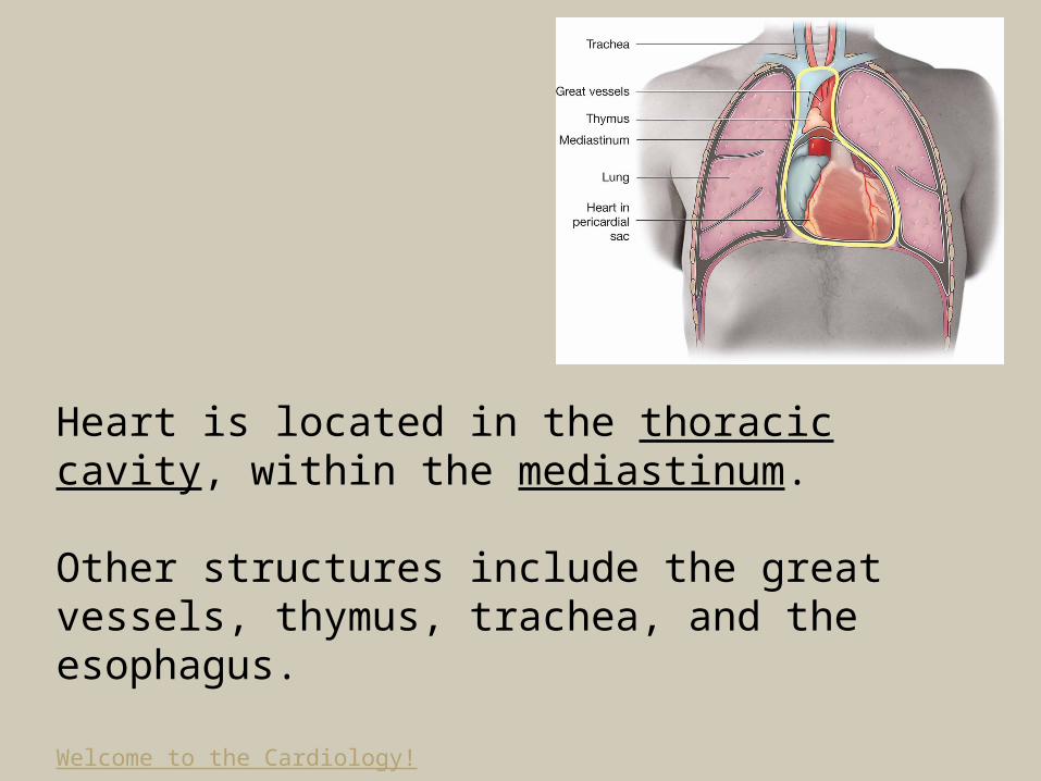
Heart is located in the thoracic cavity, within the mediastinum.
Other structures include the great vessels, thymus, trachea, and the esophagus.
Welcome to the Cardiology!
The Cardiovascular (Circulatory) System
Structures:Heart Blood Vessels (arteries, capillaries, and veins)
Function:-Moves blood throughout the body -Transports oxygen, carbon dioxide, nutrients, and wastes in the blood.

Anatomy of the Cardiovascular System
Heart:-Pumps blood throughout the body-Controlled by electrical signals from the brain
Four chambers:2 atria2 ventricles
SeptumMyocardium (cardiac muscle)Valves

Figure 5-2 Surface of the heart

Figure 5-3 Chambers and valves of the heart

Heart Valves:1. Tricuspid 2. Pulmonary 3. Mitral / Bicuspid4. Aortic
Chordae tendineae

Table 5-1 Layers and Membranes of the Heart

Figure 5-5 Layers and membranes of the heart
2. Blood VesselsFunction: Vascular channels through which blood flows in the body.
Lined with endothelium, a smooth inner layer (intima) that promotes the flow of blood.
Types:Arteries & ArteriolesCapillariesVeins & Venules

Characteristics of Arteries:
- Always carry blood away from the heart to the body.
- They carry bright red blood that has a high level of oxygen. Exception: pulmonary arteries
- Most arteries lie deep beneath the skin.
- All arteries have smooth muscle in their walls.

Figure 5-7 Vasoconstriction and vasodilation

Arteries of the Body:– Coronary artery– Carotid artery– Subclavian artery – Axillary artery (armpit)– Brachial artery (upper arm)– Radial artery (thumb side of the lower arm)– Ulnar artery (little finger side of the lower arm)– Aorta (thoracic and abdominal)– Renal– Iliac– Femoral– Popliteal– Tibial– Peroneal

Figure 5-10 Arteries in the body
Characteristics of Capillaries:
-Smallest blood vessels in the body
-The lumen of a capillary is so small that blood cells must pass through in single file.
Characteristics of Veins:
- They carry blood from the body back to the heart.- They carry dark red-purple blood with a low level of oxygen. Exception: pulmonary veins.- Veins have valves.- Many veins are near the surface of the body;
bluish; bulging lines.

Veins of the body:-Superior vena cava-Inferior vena cava
-Jugular vein-Portal vein-Saphenous and femoral veins

Figure 5-8 Valves in a vein

Figure 5-9 Arteries and veins around the heart

Patterns of Circulation throughout the body:
-Systemic circulation includes the arteries, capillaries, and veins everywhere in the body, except in the lungs.
-Pulmonary circulation includes the arteries, capillaries, and veins going to, within, and coming from the lungs.
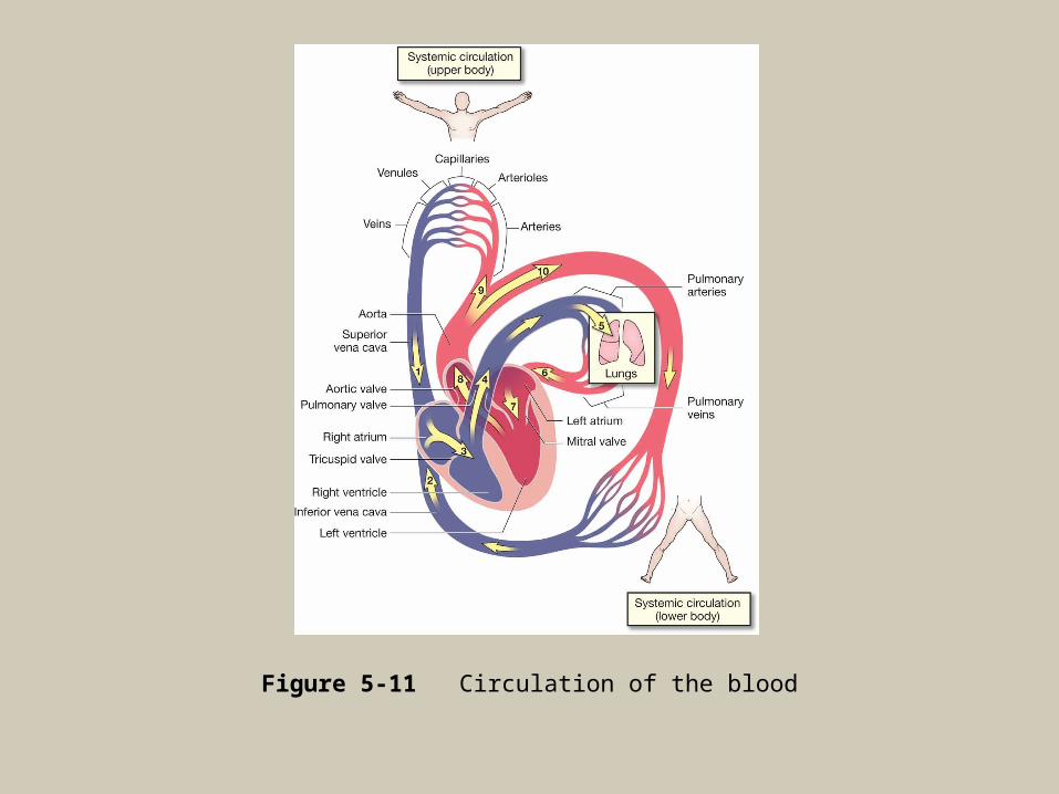
Figure 5-11 Circulation of the blood

Physiology of a Heartbeat
The Heart contracts and relaxes in a regular rhythm coordinated by an electrical conduction system
1. Sinoatrial node (SA node), or pacemaker of the heart, initiates the electrical impulse that begins each heartbeat.2. Atrioventricular node (AV node) receives the impulse to contract from the SA node.-Purkinje fibers, a network of nerves, cause both ventricles to contract simultaneously

Figure 5-12 Conduction system of the heart

Pacemaker• Abnormal
heartbeats can be controlled by an artificial pacemaker that is run on batteries

Heartbeat• Each heartbeat is
called a cardiac cycle: two atria contract then two ventricles contract (systole), and the entire heart relaxes (diastole)
• A normal heart beats 70 times per minute.

Electrocardiogram (ECG or EKG)
• A test that records the electrical activity of the heart.
• The P wave represents contraction (systole) of the atria.
• The QRS wave represents the contraction of the ventricles.
• The T wave represents the relaxation of the heart muscle (diastole).
Question: What are some problems associated with the Sinoatrial Node? How can it be fixed?

Diseases and ConditionsStart of Test 2 Material
Myocardium:-Acute coronary syndrome
-Angina pectoris
-Cardiomegaly

-Cardiomyopathy
-Congestive heart failure (CHF)
-Myocardial infarction (MI)

Heart Valves and Layers of the Heart:-Endocarditis
-Mitral valve prolapse (MVP)
-Murmur
-Pericarditis
-Rheumatic heart disease
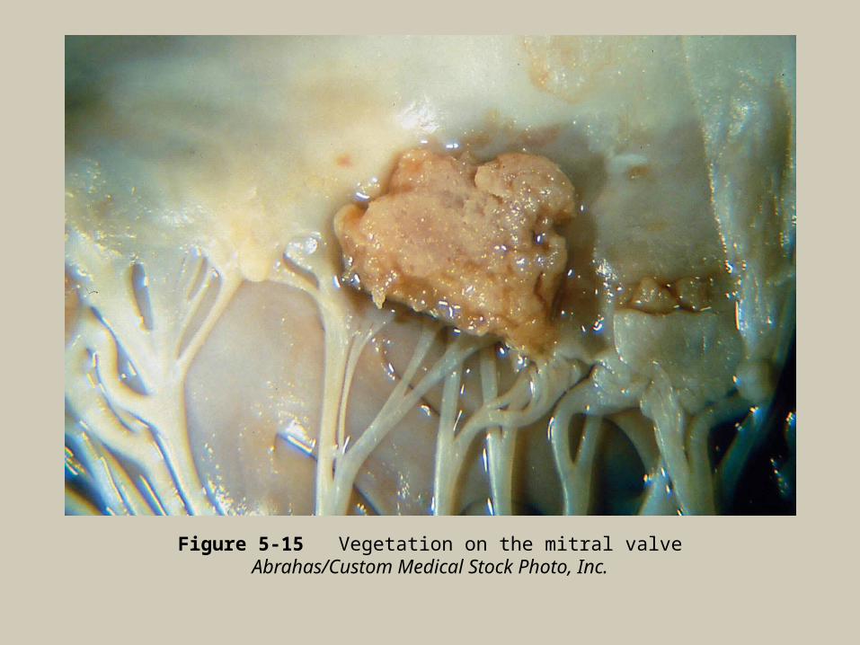
Figure 5-15 Vegetation on the mitral valveAbrahas/Custom Medical Stock Photo, Inc.

Conduction System:-Arrhythmia
-Bradycardia
-Fibrillation
-Flutter
-Heart block
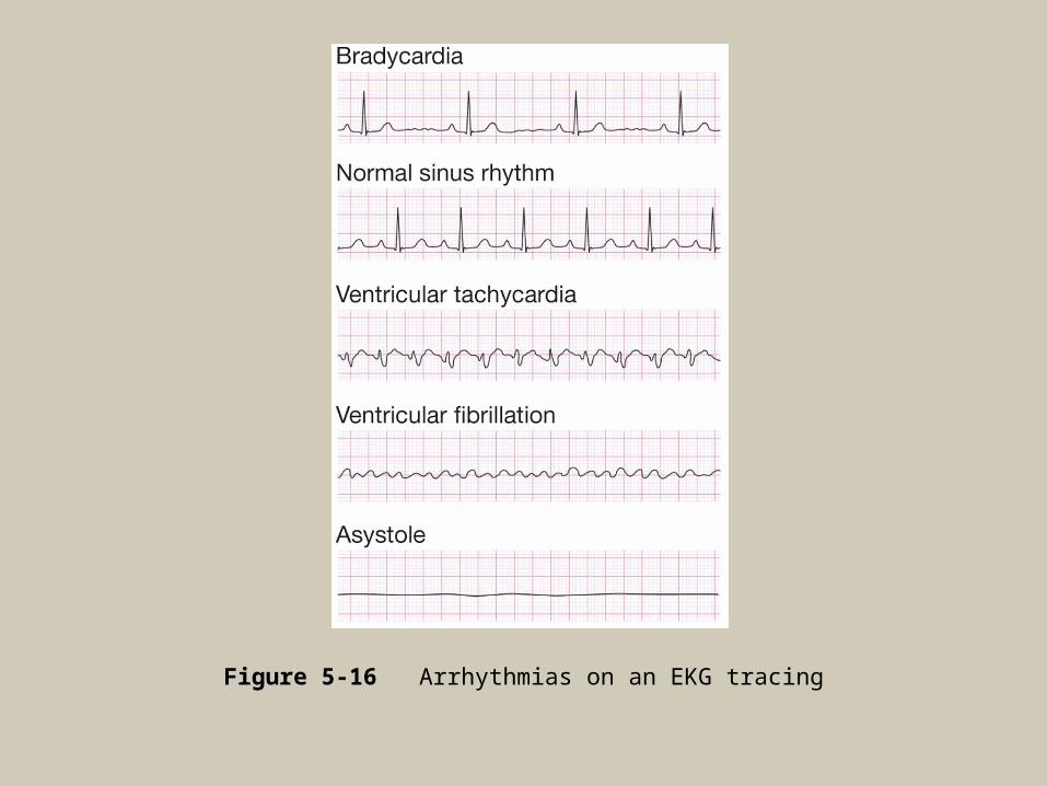
Figure 5-16 Arrhythmias on an EKG tracing

-Premature contraction
-Sick sinus syndrome
-Tachycardia
-Asystole
-Palpitation

Blood Vessels:-Aneurysm
-Arteriosclerosis
-Bruit
-Coronary artery disease (CAD)
-Hyperlipidemia
-Hypertension (HTN)
-Hypotension
-Peripheral artery disease (PAD)

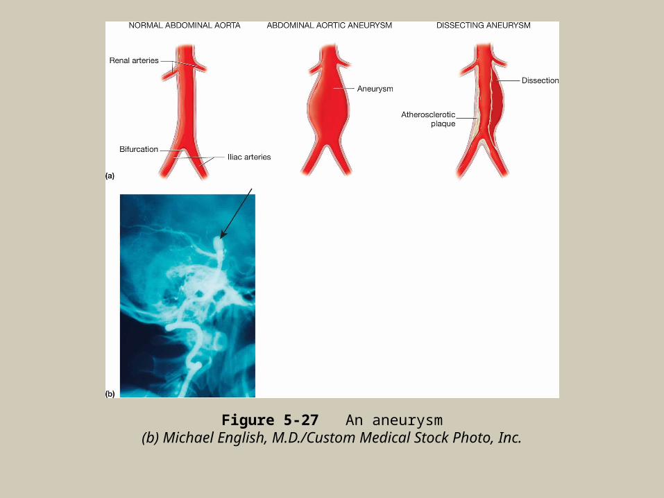
Figure 5-27 An aneurysm(b) Michael English, M.D./Custom Medical Stock Photo, Inc.

Figure 5-18 Mild atheromatous plaqueSIU BioMed/Custom Medical Stock Photo, Inc.

Figure 5-19 Severe atherosclerotic plaque in an arteryC. Abrahams, M.D./Custom Medical Stock Photo, Inc.

-Peripheral vascular disease (PVD)
-Phlebitis
-Raynaud’s disease
-Varicose veins

Figure 5-23 Severe varicose veins in the leg
SPL/Photo Researchers, Inc.

Laboratory and Diagnostic Procedures
Blood Tests:-Cardiac enzymes
-C-reactive protein (CRP)
-Homocysteine
-Lipid profile
-Troponin
Diagnostic Heart Procedures:-Cardiac catheterization
-Cardiac exercise stress test
-Electrocardiography (ECG, EKG)
-Electrophysiologic study (EPS)

Figure 5-22 ElectrocardiographyJupiter Images – PictureArts Corporation/Brand X Pictures-Royalty Free

Figure 5-23 An EKG tracing
-Holter monitor
-Pharmacologic stress test
-Telemetry

Figure 5-21 Treadmill exercise stress testFotopic/Miles Simons/Phototake NYC

Radiology and Nuclear Medicine Procedures:-Angiography
-Echocardiography
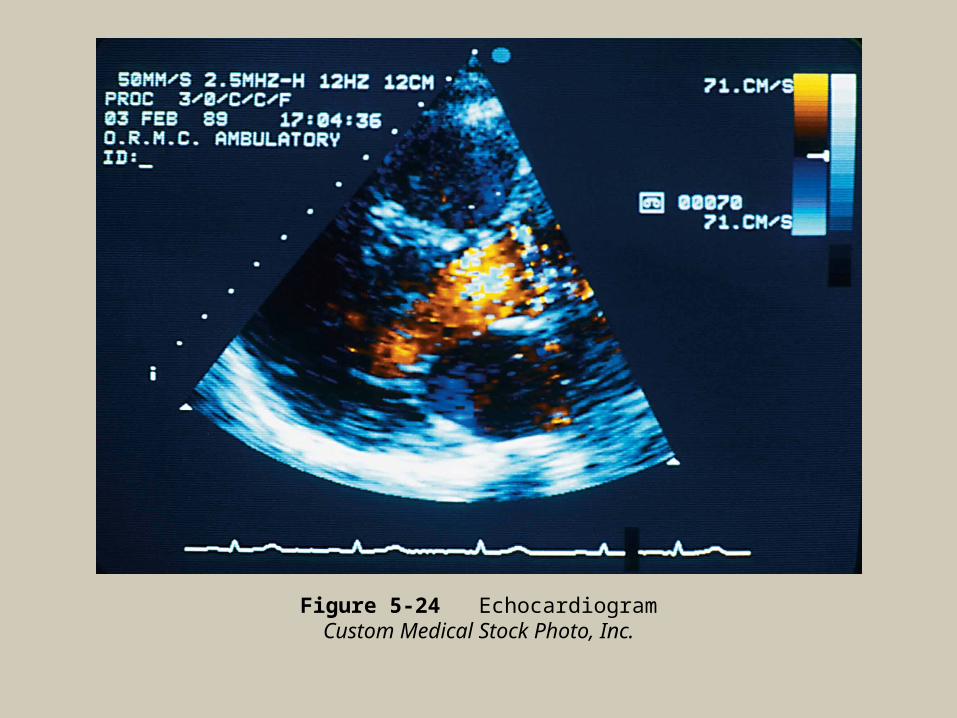
Figure 5-24 EchocardiogramCustom Medical Stock Photo, Inc.

Figure 5-25 Doppler ultrasonographyMatt Meadows/Science Photo Library/Photo Researchers, Inc.
Medical and Surgical Procedures
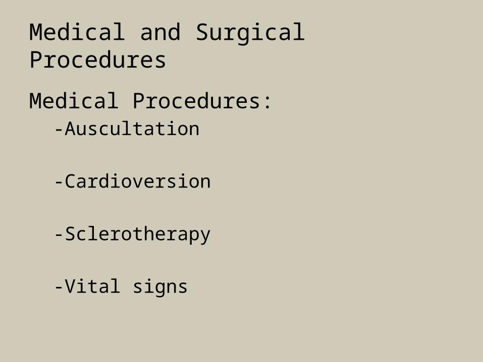
Medical Procedures:-Auscultation
-Cardioversion -Sclerotherapy
-Vital signs
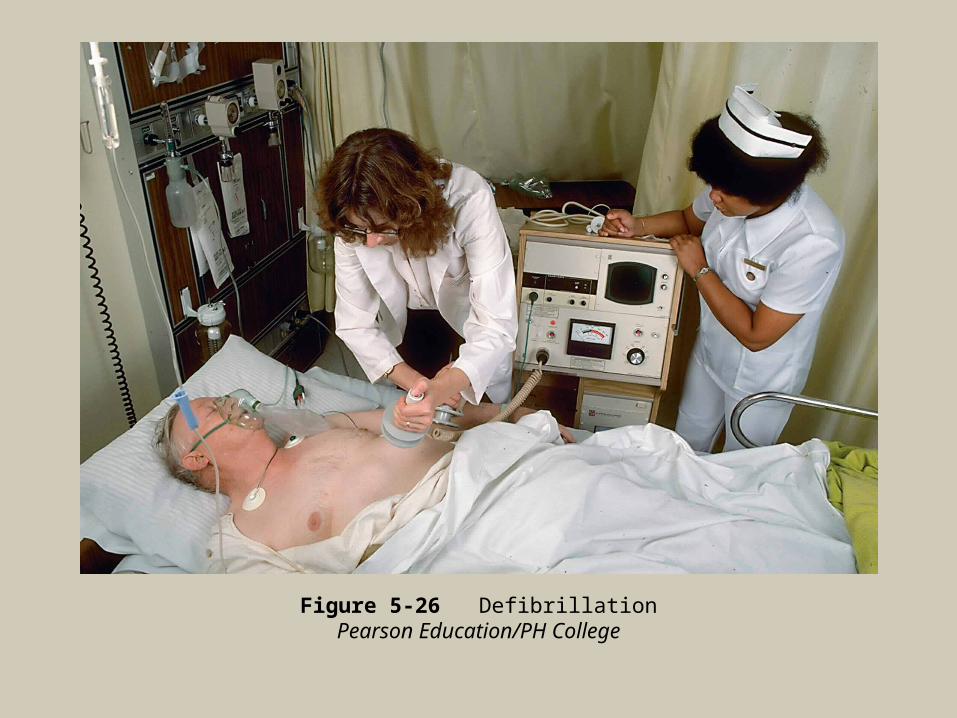
Figure 5-26 DefibrillationPearson Education/PH College

Figure 5-27 Pulse points

Figure 5-28 Carotid pulse
Michal Heron/Pearson Education/PH College

Figure 5-29 Measuring the blood pressure
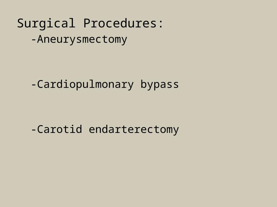
Surgical Procedures:-Aneurysmectomy
-Cardiopulmonary bypass
-Carotid endarterectomy

-Coronary artery bypass graft (CABG)
-Heart transplantation
-Pacemaker insertion

Figure 5-30 Open heart surgery
F. Schussler/PhotoDisc/Getty Images
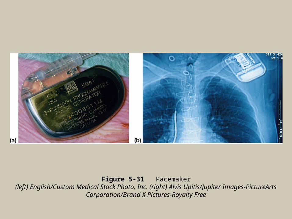
Figure 5-31 Pacemaker(left) English/Custom Medical Stock Photo, Inc. (right) Alvis Upitis/Jupiter Images-PictureArts
Corporation/Brand X Pictures-Royalty Free

-Percutaneous transluminal coronary angioplasty (PTCA)
-Pericardiocentesis
-Valve replacement
-Valvoplasty
-Radiofrequency catheter ablation

Figure 5-32 Balloon angioplasty

Figure 5-33 Stent

Figure 5-34 Valve replacement surgeryCustom Medical Stock Photo, Inc.
![Comparative pathologic analysis of mediastinal B-cell ...s-space.snu.ac.kr/bitstream/10371/164793/1/13000... · [1]. Lymphomas of the mediastinum can originate either from the thymus](https://img.pdfslide.us/doc/110x75/5f0b78017e708231d430ab4d/comparative-pathologic-analysis-of-mediastinal-b-cell-s-spacesnuackrbitstream10371164793113000.jpg)


![superior mediastinum: [Green] Inferior Mediastinum: Below the plane passing from Sternal Angle/Angle Luise Inferior mediastinum has 3 parts: Purple: anterior](https://img.pdfslide.us/doc/110x75/56649c9e5503460f9495e1bf/superior-mediastinum-green-inferior-mediastinum-below-the-plane-passing.jpg)