Embed Size (px)
Citation preview
* Author to whom correspondence should be addressed. E-mail: [email protected]
Mechanistic and kinetic study of singlet O2 oxidation of methionine
by on-line electrospray ionization mass spectrometry
Fangwei Liu, Wenchao Lu, Xunlong Yin, and Jianbo Liu*
Department of Chemistry and Biochemistry
Queens College and the Graduate Center of the City University of New York
Queens, New York 11367
Abstract. We report a reaction apparatus developed to monitor singlet oxygen (1O2) reactions in solution
using on-line ESI mass spectrometry and spectroscopy measurements. 1O2 was generated in the gas phase
by the reaction of H2O2 with Cl2, detected by its emission at 1270 nm, and bubbled into aqueous solution
continuously. 1O2 concentrations in solution were linearly related to the emission intensities of airborne
1O2, and their absolute scales were established based on a calibration using 9,10-anthracene dipropionate
dianion as an 1O2 trapping agent. Products from 1O2 oxidation were monitored by UV-Vis absorption and
positive/negative ESI mass spectra, and product structures were elucidated using collision-induced
dissociation-tandem mass spectrometry. To suppress electrical discharge in negative ESI of aqueous
solution, methanol was added to electrospray via in-spray solution mixing using theta-glass ESI emitters.
Capitalizing on this apparatus, the reaction of 1O2 with methionine was investigated. We have identified
methionine oxidation intermediates and products at different pH, and measured reaction rate constants.
1O2 oxidation of methionine is mediated by persulfoxide in both acidic and basic solutions. Persulfoxide
continues to react with another methionine, yielding methionine sulfoxide as end-product albeit with a
much lower reaction rate in basic solution. Density functional theory was used to explore reaction
potential energy surfaces and establish kinetic models, with solvation effects simulated using the
polarized continuum model. Combined with our previous study of gas-phase methionine ions with 1O2,
evolution of methionine oxidation pathways at different ionization states and in different media is
described.
Key words: On-line ESI mass spectrometry, Reaction kinetics, Methionine, Singlet oxygen, Persulfoxide,
Sulfoxide, Photooxidation, DFT-PCM calculation
2
Introduction
Methionine (Met) is one of the five amino acids (the other four are tryptophan, tyrosine, histidine and
cysteine) that are most susceptible to the attack by reactive oxygen species (ROS) [1-3]. Oxidation of
Met residues in proteins produces sulfoxides (MetO) [4-6]. Under extremely strong oxidative conditions
MetO can be irreversibly oxidized to sulfone (MetOSO) [3, 6]. It is well accepted that hydrophobic
contacts via the Met residues contribute to protein stability. Moreover, Met has the propensity to interact
with aromatic residues, and the resulting Met sulfur-aromatic motif provides additional stabilization over
hydrophobic interactions [7]. Oxidation of Met to MetO decreases hydrophobicity [5] and disrupts both
dispersion and electrostatic interactions present in the sulfur-aromatic motif [7]. Such oxidation-induced
post-translational modifications may cause proteins to change conformations and lose functions [8-10],
and are related to pathophysiological conditions such as cancer, aging, and neurodegenerative diseases [3,
11]. On the other hand, surface-exposed Met residues act as endogenous antioxidants or "molecular
bodyguards" under oxidative stress, protecting other residues important to the functions of proteins [12,
13]. This is due to the fact that cells are capable of reducing MetO back to Met, catalyzed by Met
sulfoxide reductases (Msrs) [14-16]. The reversible Met oxidation is also identified as a trigger for the
functions of two protein kinases CaMKII and transcription factor HypT (HOCl specific) [17, 18].
Consequently, the contents of Met and MetO in cells can be used to indicate the extent of ROS
generation, and the activities of oxidant scavengers and Msrs.
Oxidation kinetics of Met to MetO has been investigated in the presence of various ROSs [3, 19-24],
of which 1O2-mediated kinetics was mostly measured using photooxidation methods [23, 24]. In these
experiments, 1O2 was generated via energy transfer from the triplet excited state of a sensitizer to 3O2
(type II photosensitization) [25]. Photooxidation of Met [24, 26-32] depends on various parameters (e.g.,
pH, O2 concentration, solvent composition, buffer ions, and combination of light and sensitizers).
Determination of 1O2-mediated oxidation kinetics was also challenged by the competition between
radical- and 1O2-mediated reactions, and the interaction between substrates and excited sensitizers.
3
To avoid the interferences in photooxidation experiments, one approach is to use heterogeneous
photosensitization in which sensitizers are isolated from reaction solution and the sensitizer-generated 1O2
is delivered through space [33] or via bubbles [34]. We have adopted another approach based on a
chemical reaction to produce radical-free, "clean" 1O2. Coupled with ion-beam-scattering methods and
electrospray ionization mass spectrometry (ESI MS), we have investigated 1O2-mediated oxidation
dynamics of Met in the gas phase at different ionization and hydration states (including [Met + H]+(H2O)0-2
and [Met - H]-(H2O)0-2) [35, 36]. Study of the 1O2 reactions with dehydrated [Met + H]+/[Met - H]- has
allowed us to identify the intrinsic interactions between Met and 1O2; and that of the 1O2 reactions with
hydrated [Met + H]+(H2O)1-2/[Met - H]-(H2O)1-2 has enabled us to recognize the effects of individual
solvent molecules. On the one hand, gas-phase findings provide fundamentals in understanding the
oxidation mechanism; on the other hand, they suffice as valuable references in examining specific and
continuum solvent effects, and it will be interesting to see how the gas-phase findings can be extrapolated
to solution models. This prompted us to look at 1O2 oxidation of Met in aqueous solution, aimed at
investigating the resemblance and evolution of reaction pathways and products from the gas phase to
aqueous continuum.
Over recent years, applications of ESI MS have spread rapidly in monitoring solution-phase reaction
kinetics and probing the course of chemical transformations [37-39]. The fast growth of such applications
relies on several exclusive advantages of ESI MS: 1) The ESI process is simple, "clean" and "soft" where
no harsh conditions are applied to the sample solution being transferred into mass spectrometer. As a
result, weakly bound species may be detected in the gas phase; 2) On-line ESI MS is a rapid and
sensitive approach that detects multiple products simultaneously at very low concentrations (nanomolar),
and provides chances for capturing short-lived intermediates which might not survive the normal handling
for off-line analysis; 3) Each mass spectrum represents a snapshot of an on-going reaction, and kinetics
can be measured through continuous analyses of the reaction mixture.
However, monitoring 1O2 reaction kinetics in solution puts severe demands on an on-line ESI MS
4
experiment, which warrant careful consideration in the design of the corresponding apparatus and
methodologies. First, the reaction apparatus is continuously pumped in order to replace quenched O2 in
solution with fresh 1O2; consequently, ESI MS has to sample solution from a low-pressure reaction
system. Secondly, we need to record ESI MS of aqueous sample in both positive and negative modes.
Note that water has a high surface tension that requires high ESI spray potentials. The high spray
potentials may lead to the presence of conventional corona discharges at the ESI emitter. For otherwise
equivalent conditions a breakdown due to a corona discharge occurs at significantly lower field strength
in the case of the negative polarity than in the case of the positive one, and the ESI operation becomes
impossible without discharge suppression [40, 41]. However, the method to be used for discharge
suppression should pose inappreciable interference with the aqueous sample. Thirdly, the sample transfer
time must be minimized so that the reaction progress can be tracked promptly. To meet these
specifications, a new on-line ESI MS and spectroscopy monitoring system was developed in the present
work. Capitalizing on this apparatus, we have measured the reaction kinetics between Met and 1O2 in
different pH solutions. The results were interpreted with the aid of electronic structure calculations.
Combined with the recently reported gas-phase dynamics of bare/hydrated Met ions with 1O2 [35, 36], a
sound understanding has been obtained for Met oxidation.
Experimental and computational details
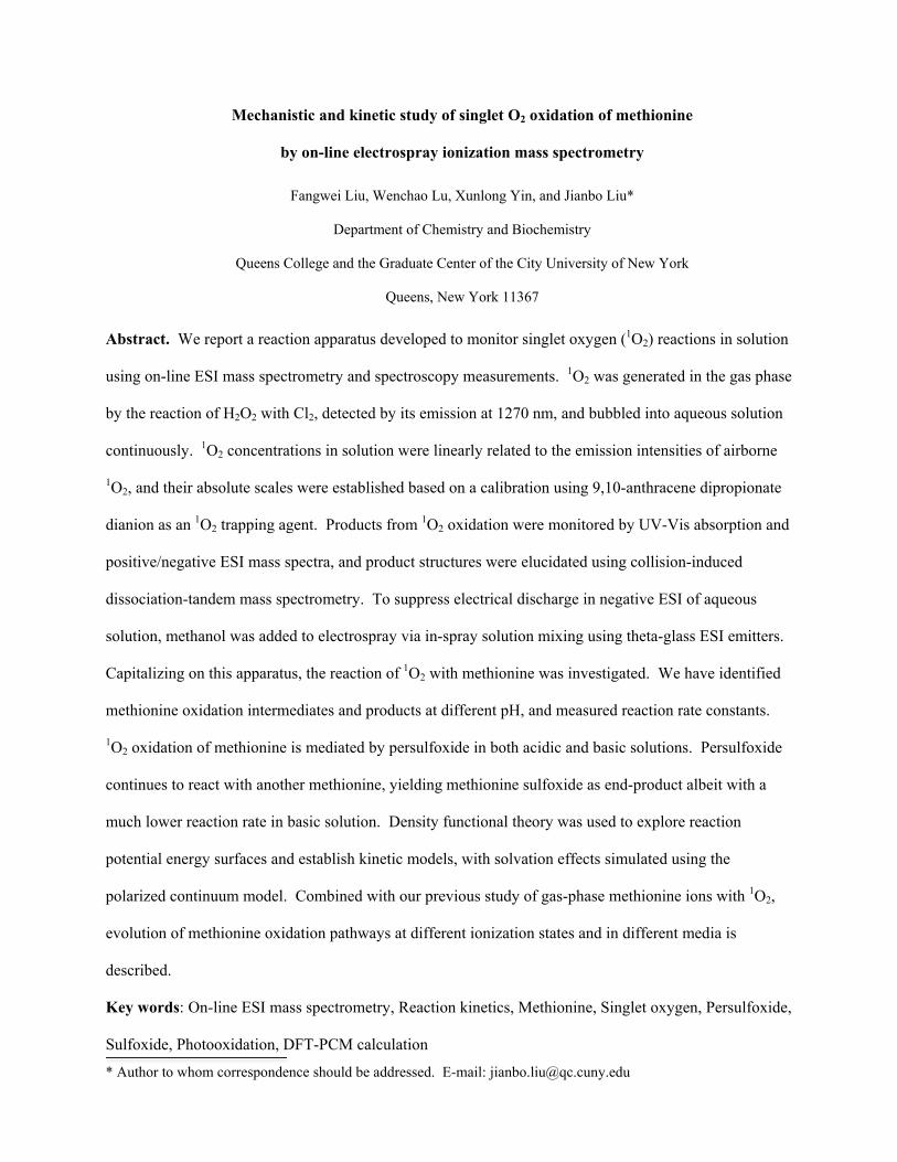
1O2 generation, detection and reaction
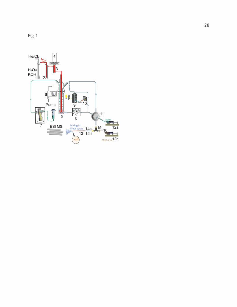
1O2 was generated by the reaction of H2O2 + Cl2 + 2KOH O2 (~ 85% 3g- and ~ 15% a1g) +
2KCl + 2H2O [35, 42, 43]. As shown in Fig. 1, 13 mL of 8 M KOH was added to 20 mL of 35 wt%
aqueous H2O2 in a sparger (1) which was immersed in a chiller held at -19 C. The resulting mixture was
degassed quickly. 2.6 sccm of Cl2 (≥ 99.5%, Sigma-Aldrich) was mixed with 96 sccm of He and bubbled
through the H2O2/KOH slush. All of the Cl2 reacted with H2O2. The gaseous products passed through a
cold trap (2, kept at -70 C) to remove water vapor. Only 1O2, 3O2 and He remained in the downstream
gas. The concentration of 1O2 in the gas was determined by measuring 1O2 emission (a1g 3g-, = 0
5
– 0) [44] at 1270 nm in an optical emission cell (3). Emission from the cell was collected using a plano-
convex lens, passed through an optical chopper (SRS model SR540) and 5-nm bandwidth interference
filter centered at 1270 nm, and focused by another plano-convex lens into a thermoelectrically cooled
InGaAs detector (4, Newport model 71887) coupled with a lock-in amplifier (SRS model SR830). 1O2
(mixed with 3O2 and He) was then bubbled into the aqueous solution in a reaction vessel (5). During the
experiment the entire apparatus was continuously pumped with a mechanical pump, and the pressure of
the apparatus was maintained at 25 (slightly above water vapor pressure at room temperature) through a
pressure relay (6, Cole-Parmer 00244OW). The pumping serves several purposes: reducing the residence
time of 1O2 in the gas phase and therefore minimizing its wall quenching and self-quenching, and
removing quenched O2 and replenishing fresh 1O2 to the reaction solution. Since a significant amount of
water evaporated from 5 at the low operating pressure, extra water was replenished into 5 through an
Ismatec Reglo-CPF rotary piston pump (7) at a precisely controlled flow rate.
To check the reactivity of aqueous substrates towards 3O2/He, control experiments were performed
under the same conditions as those for 1O2, except that Cl2 was replaced by O2 gas at the same flow rate.
On-line sampling for spectroscopy and ESI MS
Our monitoring system includes a UV-Vis spectrometer and an ESI tandem mass spectrometer.
Absorption and mass spectra were recorded over the course of reactions. As illustrated in Fig. 1, the
aqueous reaction solution was circulated by a peristaltic pump (8) through a quartz flow cell (9, Starna
Cells 583.65-Q-10/Z15, 1-cm path length). UV-Vis absorption was monitored using an Ocean Optics
USB4000 diode array spectrometer (10). Spectra were recorded at 30-s intervals using the Ocean Optics
SpectraSuite software.
To the best of our knowledge, most on-line ESI MS approaches sampled reaction solution at a
pressure near atmospheric or higher, where the solution was delivered to ESI by gravity or positive gas
pressure [45-51]. However, our reaction system needs to be maintained at a pressure of 25 as
rationalized above. Therefore, one feature of our sampling system is to transport sample from low-
6
pressure solution to open-air ESI, using a sampling loop (0.035 mL vol.) coupled to a 2-position
switching valve (11). The valve was controlled by a LabVIEW program. For each measurement, the
valve was placed in load position for 2 s to fill the loop with reaction solution through 8, and then
switched to injection position for transferring the solution to electrospray. The time lag between sample
load and MS measurement is determined by the dead volume (~0.1 mL) of the transfer line connecting the
sampling loop to ESI, and the sample transfer rate. Note that a typical flow rate for ESI is 1 µL/min,
which would result in far too long delay for kinetic measurements if this rate is used for sample transfer.
In a previous on-line ESI MS experiment [52], we adopted Cooks and coworkers′ method [51] by adding
a splitter to the sample transfer line, right before ESI. The sample was swept to ESI under N2 gas
pressure. This configuration allowed for a high sample transfer rate (11.4 mL/hr), and in the meantime
delivered only a small fraction of the flow to ESI. We were able to reduce the sample transfer time to less
than 30 s [52]. But this method increased the total sample volume needed (by a factor of ~ 200) for each
measurement. In the present work, we developed another approach to reduce the lag time. The sampling
loop was swept by water plug using a syringe pump (12a) at programmed flow rates. For each sampling,
the flow rate of 12a was first set to 6 mL/hr. During this duration, the sample was loaded, inserted into
the transfer line and transferred to the ESI emitter. After that, the rate of 12a decreased to 0.04 mL/h until
the completion of MS measurement. In the positive ion mode, the ESI emitter was assembled by gluing
the stainless steel hypodermic tubing (35-gauge, 0.005" o.d. 0.002" i.d. 0.5 length, Small Parts Inc.)
to the exit (PEEK tubing, 0.0625" o.d. 0.007" i.d.) of 11, and the total time for sample load and transfer
was minimized to less than 10 s.
An issue for negative ESI of aqueous solution is electrical discharge. Due to the high surface tension
of water, its ESI onset field strengths are 8.9 106 V/m in positive and negative ion modes, respectively
(much higher than those of 5 106 V/m for methanol) [41]. Unfortunately, for the negative ion mode
corona discharge at the stainless steel tip occurred at lower field strength than the ESI onset [41]. As a
result, the stainless steel emitter could not be used in negative ESI. Cassou et al. reported negative nano-
7
ESI of water solution by using borosilicate capillary with an ESI spray potential of -0.7 kV and a 3-mm
distance between the emitter and the counter electrode [53]. We were not able to achieve stable ESI with
the same field strength, presumably due to the higher flow rate we used. Our approach is to utilize a
borosilicate theta-glass capillary (13, Sutter Instrument) as an ESI emitter [54-56], and mix aqueous
sample solution and methanol at the tip of the emitter so as to lower ESI operating potential. Aqueous
solution and methanol were individually delivered by syringe pumps 12a and 12b at a flow rate 0.01 and
0.03 mL/h, respectively, and directed into the two separated channels within the theta capillary through
short PEEK tubing (14a and 14b, 255 m i.d. 510 m o.d.). The theta capillary was laser-pulled to a
tip size of 20 m i.d. 40 m o.d. by a micropipette puller (Sutter Instrument P-2000). A PEEK
microtee (15, Upchurch Scientific, 0.1 µL dead volume) was inserted to the aqueous sample line, and a
platinum wire (16, 0.005" o.d., Alfa Aesar) was inserted into one stem of 15 to supply the electrical
connection (-2.8 kV) for ESI. In this in-spray solution mixing, the encounter of aqueous solution and
methanol took place at the time when solution reaction was terminated by spray. Consequently,
interference with aqueous reactions by methanol was minimized, and diffusion and turbulence were
avoided during fluid mixing.
ESI MS and collision-induced dissociation-tandem MS measurements
A home-built guided-ion-beam tandem mass spectrometer was used for ESI MS measurements. The
operation, calibration and data analysis procedures for the mass spectrometer were described previously
[57]. In brief, the apparatus consists of an ion source, radio frequency (rf) hexapole ion guide, quadruploe
mass filter, rf octopole ion guide surrounded by a scattering cell, second quadrupole mass filter, and a
pulse-counting detector. Both quadrupole mass filters use Extrel 9.5 mm tri-filter rods and were operated
at 2.1 MHz with a detectable mass/charge (m/z) range of 1 - 500.
An ESI emitter was held at 3.7 and -2.8 kV, respectively, for producing positively and negatively
charged species from sample solution. Charged droplets entered the source chamber of the mass
spectrometer through a desolvation capillary (which was heated to 130 C and held at 100 V for positive
8
ions and -120 V for negative ones). The distance between the emitter tip and the entrance of the
desolvation capillary was 1 cm. Liquid droplets underwent desolvation as they passed through the heated
capillary, converted to gas-phase ions in the source chamber. A skimmer with an orifice of 0.99 mm is
located 3 mm from the capillary end, separating the source chamber and the hexapole ion guide. The
skimmer was biased at 20 V for positive ions and -20 V for negative ones. Ions were transported into the
hexapole at a pressure of 26 m, undergoing collisional focusing and cooling to 310 K. In conventional
ESI MS measurements, the first quadrupole mass filter was rendered to an rf-only ion guide, and ions
were mass-analyzed by the second quadrupole.
In order to identify the structures of product ions, collision-induced dissociation (CID) was
performed. The product ions of interest were mass-selected by the first quadrupole, and then injected into
the octopole ion guide which trapped ions in the radial direction. DC bias voltage was applied to the
octopole, allowing control of the kinetic energy (Elab) of ions in the laboratory frame. Elab can be
converted to the collision energy (Ecol) between ions and collision gas in the center-of-mass frame using
Ecol = Elab mneutral/(Mion + mneutral), where mneutral and Mion are the masses of neutral collision gas and ions,
respectively [58]. The octopole runs through the scattering cell filled with Xe gas (99.995%). The
pressure of the cell was controlled using a leak valve and measured by a Baratron capacitance manometer
(MKS model 690 head and 670 signal conditioner). CID was measured at Ecol = 0.5, 1.0 and 1.5 eV with
the cell pressure of 0.3 mTorr. Primary ions and fragment ions were collected by the octopole, and
directed to the second quadrupole for mass analysis.
Electronic structure calculations
To aid in reaction mechanism interpretation, density functional theory (DFT) electronic structure
calculations were performed using Gaussian 09 [59]. All structures were fully optimized at the B3LYP/6-
31+G* level of theory. To take into account the solvation effects for aqueous reactants, transition states
(TSs), intermediates and products, we have employed the polarized continuum model (PCM) [60] which
creates solute cavity via a set of overlapping spheres in the DFT calculations for all species. The DFT-
9
PCM method has been successfully used for probing and reproducing the solution-phase chemistry of 1O2
and peroxides, for example the 1O2 oxidation of 6-thioguanine [61] and the transformation of 8-
oxoguanine [62] in water. Conformation searching was conducted for all reactants, and their most stable
confirmations were used as starting geometries in construction of reaction potential energy surfaces
(PESs). All of the TSs were verified as first-order saddle points, and the vibrational mode with an
imaginary frequency corresponds to the associated reaction pathway. All reported reaction energetics
include zero-point energies (ZPEs) and thermal corrections at 298 K, with ZPEs scaled by a factor of
0.977 [63]. The energy barriers of TSs (with respect to reactants) were refined by single-point
calculations at B3LYP/PCM/aug-cc-pVTZ using the B3LYP/PCM/6-31+G*-optimized reactants and TS
geometries.
Results
1O2 concentrations in solution
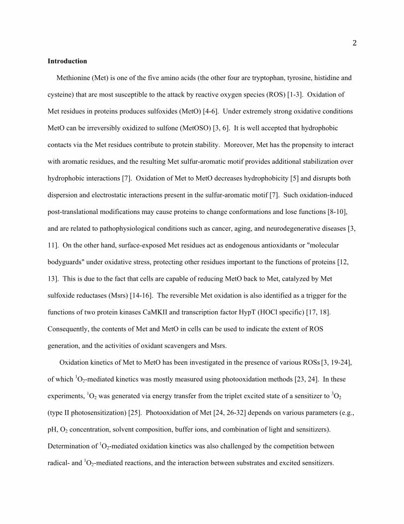
In the experiment chemically generated 1O2 was continuously bubbled into aqueous solution in the
reaction vessel. 1O2 has a longer lifetime in the interior of bubbles (due to reduced encounters with water)
than in bulk solution. After diffusing into the bulk water, 1O2 has a lifetime of ~ 2 s and can travel only
~ 150 nm [64]. Therefore, 1O2 reactions occurred both at the gas/liquid interface of bubbles and in the
bulk solution. Considering the steady concentration of airborne 1O2 (as determined by the 1O2 emission
intensity which varied within 10% over the course of reaction), and the continuous feeding of fresh 1O2
into the reaction solution, a quasi-steady-state [1O2] may be assumed for our reaction system as people
did for bubbled 1O2 in heterogeneous photosensitization [34]. In order to validate this assumption and
determine the average value of [1O2] in solution, two 1O2 chemical trappers, 9,10-anthracene dipropionate
dianion (ADPA, Chemodex) and uric acid (99%, Alfa Aesar), were used as calibration compounds.
ADPA is known to react with 1O2 chemically (i.e. without physical quenching), producing
endoperoxide via [4+2] cycloaddition accompanied by bleaching of the absorption band of ADPA [65].
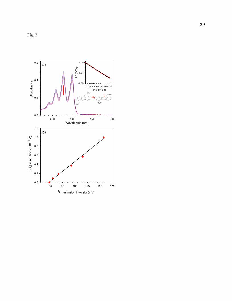
Fig. 2a shows absorption changes of ADPA along the reaction course. The pH of the ADPA solution
10
(0.05 mM) was maintained at 10.0 using borax/NaOH buffer. Shown in the insert of Fig. 2a is the plot of
ln(At/A0) vs. reaction time, where At and A0 are the ADPA peak absorption (at 378 nm) at different
reaction times and time zero, respectively. The observation of a linear relationship between ln(At/A0) and
reaction time indicates that consumption of ADPA obeys first-order rate law. Accordingly, an average
[1O2] in solution can be extracted using the reaction rate kr (8.2 10-7 M-1s-1) for ADPA + 1O2 [65].
During each experiment, emission of airborne 1O2 was continuously monitored. Fig. 2b presents the
correlation between the output of the emission detector and the corresponding ADPA-calibrated [1O2] in
solution. It shows that [1O2] in solution increases linearly with the growth of airborne 1O2 emission. The
calibration curve crosses the x-axis at ~ 50 mV. This indicates a threshold airborne concentration below
which all of the 1O2 quenched in bubbling and diffusion before reaching aqueous substrates. In the
subsequent experiments, the calibration curve of Fig. 2b was used to determine [1O2] in solution.
We also used uric acid [66-70] as a chemical trapper to test our reaction setup, and confirmed that
1O2-induced uric acid oxidation follows first-order kinetics (see supplementary Fig. S1 in the Supporting
Information). This further supports the pseudo-steady-state assumption for [1O2] in our solution reactions.
However, the literature reported kr values for uric acid were based on bleaching of uric acid by
photooxidation and did not represent exclusive chemical reactions with 1O2. Mantana et al. [68] reported
that both 1O2-dependent and -independent mechanisms were involved in photobleaching of uric acid.
Kanofsky [66] and Fischer et al. [67] found that uric acid was able to directly quench sensitizers. Rabello
et al. [70] reported consecutive reaction steps for uric acid oxidation depending on sensitizers. For these
reasons, the reported kr of uric acid was not used for qualitative determination of [1O2] in our experiment.
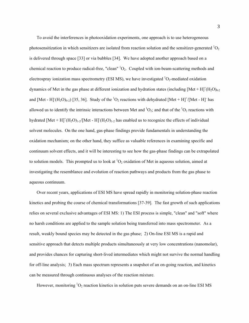
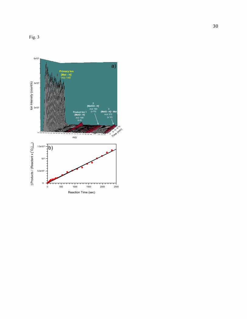
Met oxidation products, mechanism and kinetics in basic solution
We present first the 1O2 oxidation results of Met in basic solution. The reaction solution (pH = 10.4)
was prepared by adding NaOH to 2.5 mM Met aqueous solution. Considering the pK values of Met (pKa
= 2.13 and pKb = 9.28) [71], deprotonated [Met - H]- is the predominant species (i.e. 93% [Met - H]- vs.
7% neutral Met) in this solution. Major product ions were found at m/z 164 and 313, corresponding to
11
deprotonated sulfoxide [MetO - H]- and its Met adduct [MetO - H]--Met, respectively. As shown in Fig.3,
the intensities of both products increase along the reaction time. Note that formation of
dehydromethionine [Met - 3H]- was reported in the photooxidation of Met at pH 6 -10 [24]. But no [Met
- 3H]- was observed in our mass spectra.
It is possible that the product ions of m/z 164 have contribution from doubly charged covalently
bound dimer [Met - H]--OO-[Met - H]-. We have examined the isotope distribution at m/z 165, but did
not observe obvious contribution from 34S-containing [Met - H]--OO-[Met - H]-. For further verification,
we have carried out CID of the ions m/z 164 with Xe. If the ions were from [Met - H]--OO-[Met - H]-,
singly charged [MetOO - H]- would be expected in CID products. However, no fragment ions were
observed in the m/z range higher than 164 at Ecol = 0.5 - 1.5 eV, ruling out this possibility.
CID of the product ions m/z 313 with Xe produced two fragment ions at m/z 164 and 148, which
confirms that this species is an adduct of a neutral Met molecule to [MetO - H] -. Our initial guess for the
structure of [MetO - H]--Met was that the Met is covalently bonded to [MetO - H]- via S(=O)-S
(thiosulfinate) or S-O-S linkage [72]. A grid search was then used to find global minima in their
conformation landscapes. Each of the torsion angles of Met backbones was rotated systematically
through 360 at 30 increments to generate trial staggered confirmations. To our surprise, all of the trial
conformations converged to hydrogen-bonded structures of [MetO - H]-Met. The most stable
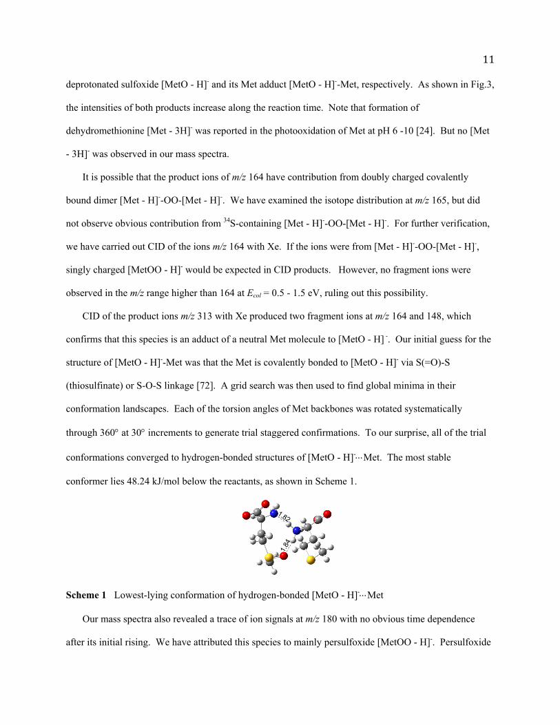
conformer lies 48.24 kJ/mol below the reactants, as shown in Scheme 1.
Scheme 1 Lowest-lying conformation of hydrogen-bonded [MetO - H]-Met
Our mass spectra also revealed a trace of ion signals at m/z 180 with no obvious time dependence
after its initial rising. We have attributed this species to mainly persulfoxide [MetOO - H]-. Persulfoxide
1.82
1.84
12
is a common intermediate in the 1O2 oxidation of dialkyl sulfide R2S. However, persulfoxide readily
reacts with a second R2S to yield sulfoxide [73]. The species observed at m/z 180 could be the remaining
[MetOO - H]- which survived on-line transfer. One complication in the interpretation of m/z 180 is that
the [Met - H]-/CH3OH adduct was expected at the same m/z when methanol was added in negative ESI.
To confirm the presence of [MetOO - H]-, we repeated the experiment using ethanol as a supplementary
solvent for ESI. The peak of m/z 180 was still visible in product mass spectra, verifying formation of
[MetOO - H]-. In Fig. 3a the contribution from [Met - H]-/CH3OH has been subtracted at m/z 180.
Note that an alternative structure for m/z 180 could be sulfone [MetOSO - H]-. We have excluded this
possibility for several reasons. It was reported that formation of sulfone occurs to a much lesser extent in
Met oxidation [3, 6]. Based on DFT calculations, the reaction enthalpy for formation of [MetOSO - H]-
is -367.6 kJ/mol with respect to [Met - H]- + 1O2, which is more exothermic than that of [MetOO - H]-.
[MetOSO - H]-, if it did form in the reaction, would have been observed in product mass spectra with
considerable intensity. However, this is not the case in our measurements. The absence of sulfone could
be due to a tight and high transition state (96.5 kJ/mol above the reactants, calculated at B3LYP/PCM/6-
31+G*) for the interconversion from [MetOO - H]- to [MetOSO - H]-.
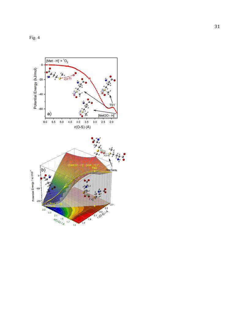
To dissect the oxidation mechanism and elucidate the intermediacy of [MetOO - H]-, we have
mapped out the reaction PESs using relaxed PES scan at B3LYP/6-31+G*. Solvent effects were
simulated using the PCM model. In PES calculations, the following reaction steps were assumed
following Footes sulfide photooxidation mechanism [74], of which the first step corresponds to
formation of a key intermediate persulfoxide, and the persulfoxide can react with another [Met - H]-
leading to two sulfoxides.
[Met - H]- + 1O2 [MetOO - H]- Hrxn = -62.7 kJ/mol (1)
[MetOO - H]- + [Met - H]- 2[MetO - H]- Hrxn = -245.1 kJ/mol (2)
In Fig. 4a the potential energy for the first step is continuously monitored while the O2 moiety is
approaching the Met sulfur atom. The PES scan continuously varied the distance between S and the
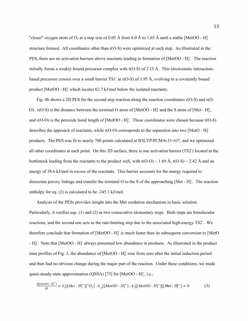
13
closer oxygen atom of O2 at a step size of 0.05 Å from 6.0 Å to 1.65 Å until a stable [MetOO - H]-
structure formed. All coordinates other than r(O-S) were optimized at each step. As illustrated in the
PES, there are no activation barriers above reactants leading to formation of [MetOO - H]-. The reaction
initially forms a weakly bound precursor complex with r(O-S) of 2.15 Å. This electrostatic interaction-
based precursor crosses over a small barrier TS1- at r(O-S) of 1.95 Å, evolving to a covalently bound
product [MetOO - H]- which locates 62.7 kJ/mol below the isolated reactants.
Fig. 4b shows a 2D PES for the second step reaction along the reaction coordinates r(O-S) and r(O-
O). r(O-S) is the distance between the terminal O atom of [MetOO - H]- and the S atom of [Met - H]-,
and r(O-O) is the peroxide bond length of [MetOO - H]-. These coordinates were chosen because r(O-S)
describes the approach of reactants, while r(O-O) corresponds to the separation into two [MetO - H]-
products. The PES was fit to nearly 700 points calculated at B3LYP/PCM/6-31+G*, and we optimized
all other coordinates at each point. On this 2D surface, there is one activation barrier (TS2-) located at the
bottleneck leading from the reactants to the product well, with r(O-O) ~ 1.69 Å, r(O-S) ~ 2.42 Å and an
energy of 38.6 kJ/mol in excess of the reactants. This barrier accounts for the energy required to
dissociate peroxy linkage and transfer the terminal O to the S of the approaching [Met - H]-. The reaction
enthalpy for eq. (2) is calculated to be -245.1 kJ/mol.
Analysis of the PESs provides insight into the Met oxidation mechanism in basic solution.
Particularly, it verifies eqs. (1) and (2) as two consecutive elementary steps. Both steps are bimolecular
reactions, and the second one acts as the rate-limiting step due to the associated high-energy TS2-. We
therefore conclude that formation of [MetOO - H]- is much faster than its subsequent conversion to [MetO
- H]-. Note that [MetOO - H]- always presented low abundance in products. As illustrated in the product
time profiles of Fig. 3, the abundance of [MetOO - H]- rose from zero after the initial induction period
and then had no obvious change during the major part of the reaction. Under these conditions, we made
quasi-steady-state approximation (QSSA) [75] for [MetOO - H]-, i.e.,
d[ MetOO ‐ H] ]
dk1
- [ Met ‐ H] ][1O2] - k‐1-
[ MetOO ‐ H] ] - k2- [MetOO ‐ H] ] Met‐H 0 (3)
14
where the rate constants 1- and
‐1- are for the forward and backward reactions of eq. (1), and 2
- for eq.
(2). The reverse direction of eq. (1) represents a fraction of 1O2 physical quenching that takes place via
decomposition of [MetOO - H]-, partially accounting for the reaction inefficiency [76]. [Met - H]- does
not have an appropriate low-lying excited state to deactivate 1O2 by electronic energy transfer, and a
charge-transfer quenching mechanism is unlikely due to its large reaction endothermicity [36]. On the
other hand, movement along the PES for [Met - H]- + 1O2 [MetOO - H]- may bring the persulfoxide
into a region near the rapidly rising triplet state PES where intersystem crossing could occur [76]. A
similar physical quenching mechanism was observed in the gas-phase collisions of 1O2 with hydrated and
dehydrated [Met - H]- [36].
Considering eq. (2) is rate-limiting, the steady state concentration of [MetOO - H]- implies that a
large fraction of [MetOO - H]- must decay back to reactants rather than converted to [MetO - H]- (i.e.
k‐1-
[[MetOO - H] ]≫ k2- [[MetOO - H] ] Met - H ). We can therefore eliminate
k2- [[MetOO - H] ] Met - H in eq. (3). This leads to a pre-equilibrium for eq. (1) as expressed by eq. (4),
where K- is the equilibrium constant.
MetOO - H] ] 1-[[Met - H] 1O2
-1
--[[Met - H] 1O2 (4)
The rate law for formation of [MetO - H]- can now be given as
1
2
d [MetO - H]
d 2- [ MetOO - H] ][ Met - H] ] -
2- Met - H
2 1O2 - Met - H2 1O2 (5)
where ‐ is the effective rate constant (M-2s-1). Among the three products ([MetO - H]-, [MetOO - H]- and
[MetO - H]-Met), [MetOO - H]- is minimal (< 0.1% over a period of 40 minutes) and can be safely
ignored in the total product concentration. [MetO - H]-Met originates from the secondary reaction of
[MetO - H]-, and was lumped together with [MetO - H]- in calculating the total [MetO - H]- concentration.
As a result, eq. (5) can be approximated as
MetO - H] 2 - Met - H] 0 MetO - H 2 1O2 (6)
15
and its integrated form as
[Met - H] ]0 MetO - H
[Met - H] ]02 - 1O2 dt (7)
or [MetO - H]
[Met - H] 1O2 ave2 - [Met - H]
0t (8)
At the reaction time t, [MetO - H]
[Met - H] can be measured using the relative abundances of reactant and product
intensities in the corresponding mass spectrum. [1O2]ave was averaged over the integration period from 0
to t. As shown in Fig. 3b, the plot of [MetO - H]
Met - H] 12 ave
vs. t fits in to a linear relationship, supporting the
proposed mechanistic scheme. The measured value of - is 1.0 109 M-2s-1.
In the above two-step scheme, formation of [MetO - H]- can also be viewed as a trimolecular reaction
that avoids nearly simultaneous three-body collisions [77]. Assuming - follows the Arrhenius equation
with the activation energy ‐ equal to TS2- (38.6 kJ/mol), the overall activation energy ‐ for this
trimolecular reaction is given by eq. (9) [77]. Since the Hrxn1 for the first step is -62.7 kJ/mol and the
activation energy for the second step is not too high, ‐ for this trimolecular reaction becomes negative:
Ea- =RT2 dlnK-
dT+RT2 dlnk2
-
dT=∆Hrxn1+E2
- =-24.12 kJ/mol (9)
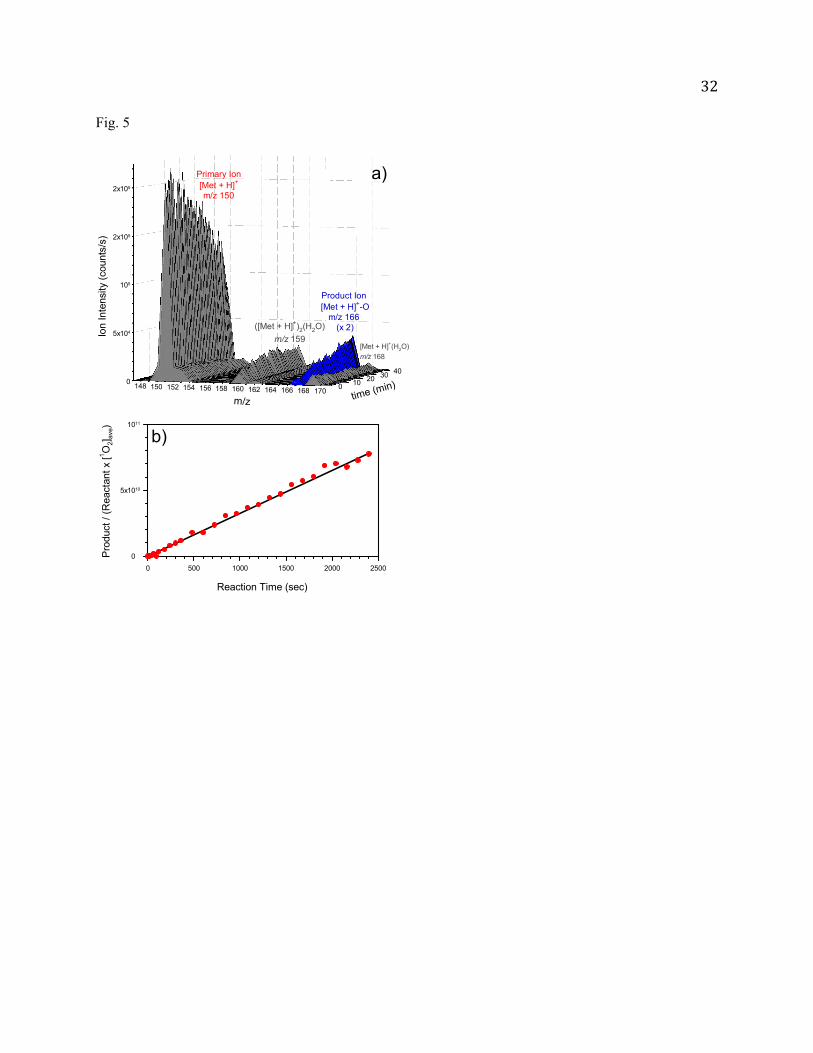
Met oxidation products, mechanism and kinetics in acidic solution
The oxidation product of Met in pH 3.2 solution (prepared by adding equimolar HCl to 0.5 mM Met
aqueous solution) was detected at m/z 166 only, and its intensity increased with the reaction time as
shown in Fig. 5a. This product can be attributed to protonated [MetO + H]+ or [Met + H]+-OO-[Met +
H]+. We have performed CID of m/z 166 with Xe, and expected that [Met + H]+-OO-[Met + H]+ would
lose a proton in CID. However, no [Met + H]+-OO-Met was observed in CID, indicating that no [Met +
H]+-OO-[Met + H]+ was formed in the reaction.
Note that due to the low carboxyl pKa of Met, the percentile populations of neutral and protonated
Met in pH 3.2 solution are 92% and 8%, respectively. As a result, the kinetics at pH 3.2 represents the
16
oxidation chemistry of neutral Met (in a zwitterionic structure), albeit that the reactant and products were
protonated in positive ESI spray. Therefore, the following kinetics discussion focuses on neutral Met.
Similar as its deprotonated counterpart, we may propose two steps for oxidation of neutral Met. Their
reaction enthalpies were calculated at the B3LYP/PCM/6-31+G* level of theory:
Met + 1O2 MetOO Hrxn = -83.0 kJ/mol (10)
MetOO + Met 2MetO Hrxn = -244.1 kJ/mol (11)
Concerning the reaction enthalpies for formation of persulfoxide and sulfoxide, the neutral system has
only moderate changes compared to [Met - H]-. More pronounced effects are observed in their PESs and
associated activation barriers. Unlike the reaction of [Met - H]- + 1O2 (see Fig. 4a), the PES scan for Met
+ 1O2 (see Fig. 6a) results in a covalently bound persulfoxide MetOO barrierlessly, without formation of a
weakly bound precursor. MetOO has the O2 moiety sandwiched between the ammonium group and the
sulfur atom. MetOO has a zwitterionic character [76], so the proton of the ammonium group is shared
between the N and the terminal O, rendering MetOO an S-hydroperoxide compound.
When another Met approaches MetOO, the latter transfers an O atom to Met. As the O-O bond
ruptures and the new O-S bond forms, two molecules of MetO are produced. The PES for this process is
scanned in Fig. 6b along the distance between the terminal O of MetOO and the S of Met from 5.0 Å to
1.5 Å at a decrement of 0.05 Å each step. A transition state TS2 is located at r(O-S) of 2.37 Å and r(O-O)
of 1.69 Å. At TS2, the O-O bond rupture is accompanied by the proton shuttling back to the amino
group. TS2 lies 25.1 kJ/mol above the reactants (For comparison, TS2 is 13.5 kJ/mol lower than the
corresponding TS2- for [MetOO - H]- + [Met - H]-). The overall activation energy Ea for 2Met + 1O2
2MetO is -57.9 kJ/mol according to eq. (9).
Based on the similarities between the PESs for 1O2 with deprotonated and neutral Met, we conclude
that both systems follow similar kinetics, and have applied the pre-equilibrium assumption to eq. (10) and
QSSA to MetOO. As a result, the rate law for formation of MetO can be written as
17
1
2
d MetO
d 2 MetOO Met 1
-12 Met 2[1O2 2 Met 2 1O2 Met 2 1O2 (12)
and its integrated form
MetO
Met 1O2 ave2 Met 0t (13)
where k1 and k-1 are the rate constants for the forward and backward reactions of eq. (10), k2 is the rate
constant for eq. (11), K is the equilibrium constant for eq. (10), k is the effective rate constant for
formation of MetO from 2Met + 1O2, and [Met]0 is the initial Met concentration. Note that, to facilitate
comparisons, all the corresponding rate constants, equilibrium constants and barriers for different
ionization states have been assigned identical names, but with +/- superscripts to indicate ionization
states. Because of the lower TS2 barrier and consequently the larger k2 (compared to TS2- and - ,
respectively), MetOO could react more promptly with another Met to yield MetO, and itself was therefore
missing in product mass spectra. A plot of Met
Met 12 ave
vs. reaction time shown in Fig. 5b is linear, from
which the value of k is calculated to be 3.7 1010 M-2s-1. Remarkably, the k for neural Met is a factor of
37 higher than the - for deprotonated Met.
MetH+ presents similar oxidation behaviors as neutral Met
To further explore the effects of ionization on the Met reaction with 1O2, we have constructed the
PESs for protonated [Met + H]+ + 1O2 in solution, as illustrated in Fig. 6c and d. The oxidation of MetH+
follows the same reaction mechanism and shows nearly identical PES profiles and energetics as those for
neutral Met, i.e., formation of S-hydroperoxide from encounter of [Met + H]+ and 1O2, followed by
transfer of an O atom from [MetOO + H]+ to another [Met + H]+ via an activation barrier TS2+ located at
27.0 kJ/mol above reactants.
[Met + H]+ + 1O2 [MetOO + H]+ Hrxn = -96.5 kJ/mol (14)
[MetOO + H]+ + [Met + H]+ 2[MetO + H]+ Hrxn = -235.4 kJ/mo (15)
Discussion
18
Comparison with photooxidation kinetics
Matheson et al. [22] reported the reactive rate constant kr (1.6 × 10⁷ M-1s-1 ) and the total quenching
rate constant kt (1.7 × 10⁷ M-1s-1) of 1O2 with Met, where 1O2 was generated by direct excitation of 3O2 in
D2O (pD = 8.4). kt was evaluated by competitive inhibition of the chemical reaction of bilirubin with 1O2,
and kr was measured from loss of Met using an amino acid analyzer. Most of the other kinetic
measurements for Met with 1O2 are based on dye-sensitized photooxidations. Kraljic et al. [78] reported a
kr of 8.6 × 10⁶ M-1s-1 in neutral water, according to O2 consumption in steady state photosensitization.
Lindig et al. [79] examined kt in neat D2O using laser flash photolysis. Quenching of 1O2 lay in the
competition for 1O2 between ADPA and Met, and the kt for Met was measured to be 1.5 × 10⁷ M-1s-1. In
the experiment of Miskoski and García [23], 1O2 was generated by rose bengel and tryptophan was used
as a sacrificial substrate. The solution was photolyzed, and tryptophan consumption was monitored by its
fluorescence in the absence and the presence of Met. Through a Stern-Volmer analysis of dynamic
quenching of fluorescence, a kt of 2.1 × 10⁻⁷ M-1s-1 was obtained for Met at pH 7. kr was measured based
on O2 consumption in a solution containing sensitizer and Met, and its value equals kt. Based on a similar
fluorescence quenching approach with diphenylfuran as a fluorescence probe, Sysak et al. reported a kr of
1.15 x 10⁷ M-1s-1 in water (pH = 7) [24].
In above works, no kinetic model was given and all of the kr values were reported as a second-order
rate constant in the unit of M-1s-1. If we assumed that formation of MetOO is the rate-limiting step for
Met oxidation, the rate law for formation of MetO would follow second-order kinetics as eq. (16). Least-
square fitting of our kinetics data to eq. (16) gave a k1 value of 1.6 ×10⁷ M-1s-1, similar as those reported
from photooxidations. But our data were poorly fitted to eq. (16), suggesting eq. (16) does not describe
kinetics correctly.
Met 0
Met 0 MetO2 1
1O2 ave (16)
The pH dependence of Met photooxidation has also been subjected to scrutiny. Earlier work on
methylene blue-sensitized photooxidation of Met by Weil [80] and Spikes et al. [27] showed a remarkable
19
increase of O2 consumption at a high pH range starting from 8. Sysak et al. [24] reported comprehensive
pH dependence of Met photooxidation. According to them, the reaction is initialized by formation of a
persulfoxide intermediate, followed by secondary reactions that are both pH- and buffer-dependent. At
high pH (above 9) and catalyzed by buffer ions, OH- may attack the S of persulfoxide, and the reaction
gives one molecule of sulfoxide and one molecule of H2O2. At intermediate pH of 6 - 10, when Met
carries a free amino group, the dominant pathway leads to dehydromethionine and H2O2 via internal
displacement, where the dehydromethionine was assigned as a five-membered heterocyclic N-S
compound [24]. Dehydromethionine may slowly hydrolyze to Met sulfoxide. At pH below 6, the
persulfoxide oxidizes a second Met by the sulfide trapping mechanism, resulting in a stoichiometry of
2Met + 1O2 2MetO. Competition among the secondary reactions accounts for the variation in O2
uptake at different pH, i.e., the Met to 1O2 ratio is 0.85:1 above pH 8 and 2:1 below pH 5 [24, 65].
It is worth mentioning that formation of dehydromethionine in photooxidations was usually
determined based on -NH2 loss (using fluorogenic detection), but the loss of primary -NH2 reactivity
strongly depended on dyes [81] and buffer ions [24]. For example, little change was observed in the -NH2
reactivity when eosin rather than methylene blue was used as a sensitizer [81]. It was suspected that the
excited dye abstracted H atom from Met to form dehydromethionine [24, 80]. This hypothesis was
corroborated by the fact that H2O2 was formed in methylene blue radical-mediated photooxidation of Met
[80]. This implies that formation of dehydromethionine and H2O2 might not be completely 1O2-specific
[24], and the observed photooxidation pH dependence may reflect the oxidation-reduction potential
between Met and excited dyes[80]. The uncertainty in photooxidation kinetics also remains as to whether
the O2 uptake is dedicated to 1O2-induced oxidation or partially due to formation and reactions of O2-
with substrates. Such uncertainty was eliminated in our measurements, by bubbling clean 1O2 to the
Met solution, and using a straightforward MS technique to identify products.
Evolution of Met oxidation from the gas phase to solution
Preceding the present solution work, we reported the reactions of Met ions with 1O2 in the gas phase.
20
We first investigated the oxidation of protonated and deprotonated Met in the absence of water [35, 36].
The reaction systems were then augmented by adding explicit number of water ligands [35, 36], which
accounts for microsolvation. The parallel gas- and solution-phase study helps us evaluate evolution of
Met oxidation dynamics and kinetics from the gas phase, through microsolvation, to the aqueous solution.
The gas-phase oxidation processes follow an addition/elimination mechanism. The first step is
addition of the O2 moiety to the Met sulfur, yielding persulfoxide. The reaction outcome is determined by
whether the persulfoxide intermediate has accessible product channels or not. In the case of dehydrated
[Met + H]+ + 1O2, the persulfoxide intermediate evolves to an S-hydroperoxide
HN2CH(CO2H)CH2CH2S(OOH+)CH3. The latter eliminates H2O2, yielding a dehydro compound of
MetH+. The reaction is extremely efficient at low Ecol, approaching the collision limit. In the reaction of
dehydrated [Met - H]-, the persulfoxide intermediate interconverts to S-hydroperoxides
NH2CH(CO2-)CH2CH2S(OOH)=CH2 and NH2CH(CO2
-)CH2CH=S(OOH)CH3. Contrary to the
protonated case, H2O2 elimination of deprotonated S-hydroperoxides encounters insurmountable barriers.
Therefore, none of deprotonated S-hydroperoxides could convert to stable end-products but decayed back
to reactants, making [Met - H]- non-reactive towards 1O2.
The fates of S-hydroperoxide intermediates are changed once [Met + H]+ and [Met - H]- become
hydrated. The dissociation of water ligand(s) provides an energy disposal path to release the heat gained
from peroxide formation. It enables the capture of the "relaxed" S-hydroperoxides as stable end-products,
i.e., HN2CH(CO2H)CH2CH2S(OOH+)CH3 for oxidation of [Met + H]+(H2O)1,2, and
NH2CH(CO2-)CH2CH2S(OOH)=CH2 and NH2CH(CO2
-)CH2CH=S(OOH)CH3 for [Met - H]-(H2O)1,2.
And for the reactions of [Met + H]+(H2O)1,2, H2O2 elimination becomes less dominant.
Interestingly, reaction efficiencies of [Met + H]+(H2O) and [Met + H]+(H2O)2 are 15 and 25 times
higher than those of [Met - H]-(H2O) and [Met - H]-(H2O)2, respectively. The effect of ionization state on
gas-phase reaction efficiencies can be traced back to the formation efficiencies of protonated and
deprotonated S-hydroperoxides, whose structures are completely different. Formation of protonated S-
21
hydroperoxide HN2CH(CO2H)CH2CH2S(OOH+)CH3 takes places via intramolecular proton transfer from
the [Met + H]+ ammonium group to -SOO, and there is no activation barrier. On the other hand,
deprotonated S-hydroperoxide NH2CH(CO2-)CH2CH2S(OOH)=CH2 or
NH2CH(CO2-)CH2CH=S(OOH)CH3 is formed by taking an H from the terminal -SCH3 or from the -CH2;
neither of these two routes is facile.
Similar to gas-phase reactions, the first step of Met oxidation in solution is formation of persulfoxide.
However, only in acidic solution the initially formed persulfoxide evolves to S-hydroperoxide; in basic
solution, the structure of initial persulfoxide remains stable presumably due to bulk solvent stabilization.
In aqueous solution, persulfoxide or S-hydroperoxide could be thermalized through solute-solvent
interactions. However, a downstream route opens for persulfoxide/S-hydroperoxide through the
participation of other Met molecules, leading to sulfoxide.
Different than the gas-phase reactions where the reaction efficiencies mostly depend on the formation
efficiencies of S-hydroperoxides, the reaction efficiencies in solution are also related to the second step
which leads to formation of sulfoxides by O-transfer between initially formed persulfoxide and second
Met. Based on DFT-PCM calculations, the barriers for this reaction are 27.0, 25.1 and 38.6 kJ/mol for
protonated, neutral and deprotonated systems, respectively.
Conclusions
A new on-line reaction setup was developed to couple solution-phase reactions of electronically
excited 1O2 with an ESI mass spectrometer and an absorption spectrometer. This apparatus enables us to
measure 1O2 oxidation kinetics in aqueous solution. To determine [1O2] in solution on an absolute scale, a
method was established to correlate the [1O2] in aqueous solution with airborne 1O2 emission intensity.
This on-line monitoring system was utilized to study the reaction kinetics of Met with 1O2 at different pH.
In both acidic and basic solutions, sulfoxide [MetO - H]- and [MetO + H]+ were confirmed to be major
products. We have captured the persulfoxide intermediates in basic solution. The reaction mechanism
and pH dependence were elucidated with the assistance from DFT calculations, using the polarized
22
continuum model to simulate bulk solvation effects. It was found that the reactions in acidic and basic
media follow the same mechanism composed of two elementary steps, formation of persulfoxide for
deprotonated Met or S-hydroperoxide for neutral/ protonated Met, followed by transfer of an O atom from
persulfoxide or S-hydroperoxide to a second Met, producing two sulfoxides. The second step is rate-
limiting, and formation of sulfoxide follows third-order rate law with respect to [Met]2[1O2]. A
remarkable result concerns the pH dependence of Met oxidation. The effective rate constants for
formation of Met sulfoxide are 3.7 1010 M-2s-1 at pH 3.2, decreasing to 1.0 109 M-2s-1 at pH 10.4.
The significantly lower rate constant in basic solution is due to the high activation barrier leading to
formation of sulfoxide from deprotonated persulfoxide, and the latter overwhelmingly decayed back to
reactants accompanied by physical quenching of 1O2.
Combined with our findings in the gas-phase 1O2 oxidation of hydrated/dehydrated Met ions, a
panorama can be created for evolution of the Met oxidation mechanism from the gas phase, through
microsolvation, to aqueous solution. Reactions in different media and at different ionization states are all
mediated by persulfoxides, which carry high internal energy and thus are very reactive. In the gas phase,
the fates of persulfoxides are determined by whether they have exit product channels or not. In the
reaction of protonated Met, transfer of two H from [Met + H]+ to O2 provides an energetically favored
dissociation path. To the contrary, H2O2 elimination is inhibited in deprotonated persulfoxide, and the
latter could only decay back to reactants. The fates of persulfoxide intermediates are altered upon
microsolvation in the gas phase. Dominant hydration effect is the suppression of persulfoxide
decomposition. This can be attributed to energy dissipation from excited persulfoxide intermediates via
water dissociation, yielding persulfoxides themselves as stable products. Finally, in solution-phase
reactions, persulfoxide intermediates, once formed, continue to react with another molecule of Met,
leading to formation of sulfoxides as final products.
23
Acknowledgements
This work was supported by the National Science Foundation CAREER Award (Grant No. CHE-
0954507), Queens College Research Enhancement Funds, and PSC-CUNY Research Awards.
Electronic supplementary material: UV-Vis absorption spectra showing 1O2-induced oxidation of
uric acid.
References
1. Davies, M.J.: Singlet Oxygen-Mediated Damage to Proteins and Its Consequences. Biochem. Biophys. Res. Commun. 305, 761-770 (2003)
2. Davies, M.J.: Reactive Species Formed on Proteins Exposed to Singlet Oxygen. Photochem. Photobiol. Sci. 3, 17-25 (2004)
3. Davies, M.J.: The Oxidative Environment and Protein Damage. Biochim. Biophys. Acta. 1703, 93-109 (2005)
4. Lavine, T.F.: Formation, Resolution, and Optical Properties of the Diastereoisomeric Sulfoxides Derived from L-Methionine. J. Biol. Chem. 169, 477-491 (1947)
5. Vogt, W.: Oxidation of Methionyl Residues in Proteins: Tools, Targets, and Reversal. Free Radicals Biol. Med. 18, 93-105 (1995)
6. Nielsen, H.K., Löliger, J., Hurrell, R.F.: Reactions of Proteins with Oxidizing Lipids. 1. Analytical Measurements of Lipid Oxidation and of Amino Acid Losses in a Whey Protein-Methyl Linolenate Model System. British J. Nutrition. 53, 61-73 (1985)
7. Valley, C.C., Cembran, A., Perlmutter, J.D., Lewis, A.K., Labello, N.P., Gao, J., Sachs, J.N.: The Methionine-Aromatic Motif Plays a Unique Role in Stabilizing Protein Structure. J. Biol. Chem. 287, 34979-34991 (2012)
8. Brot, N., Weissbach, L., Werth, J., Weissbach, H.: Enzymic Reduction of Protein-Bound Methionine Sulfoxide. Proc. Natl. Acad. Sci. U.S.A. 78, 2155-2158 (1981)
9. Caldwell, P., Luk, D.C., Weissbach, H., Brot, N.: Oxidation of the Methionine Residues of Escherichia Coli Ribosomal Protein L12 Decreases the Protein's Biological Activity. Proc. Natl. Acad. Sci. U.S.A. 75, 5349-5352 (1978)
10. Sacksteder, C.A., Whittier, J.E., Xiong, Y., Li, J., Galeva, N.A., Jacoby, M.E., Purvine, S.O., Williams, T.D., Rechsteiner, M.C., Bigelow, D.J., Squier, T.C.: Tertiary Structural Rearrangements Upon Oxidation of Methionine145 in Calmodulin Promotes Targeted Proteasomal Degradation. Biophys. J. 91, 1480-1493 (2006)
11. Chao, C.-C., Ma, Y.-S., Stadtman, E.R.: Modification of Protein Surface Hydrophobicity and Methionine Oxidation by Oxidative Systems. Proc. Natl. Acad. Sci. U.S.A. 94, 2969-2974 (1997)
12. Garner, B., Waldeck, A.R., Witting, P.K., Rye, K.-A., Stocker, R.: Oxidation of High Density Lipoproteins II. Evidence for Direct Reduction of Lipid Hydroperoxides by Methionine Residues of Apolipoproteins AI and AII. J. Biol. Chem. 273, 6088-6095 (1998)
13. Levine, R.L., Mosoni, L., Berlett, B.S., Stadtman, E.R.: Methionine Residues as Endogenous Antioxidants in Proteins. Proc. Natl. Acad. Sci. U.S.A. 93, 15036-15040 (1996)
14. Glaser, C., Schoeneich, C.: Special Issue: Methionine Oxidation and Methionine Sulfoxide Reductases. [In: Biochim. Biophys. Acta; 2005, 1703(2)]. Elsevier B.V., Amsterdam, Netherlands (2005)
15. Brot, N., Weissbach, H.: Biochemistry and Physiological Role of Methionine Sulfoxide Residues in Proteins. Arch. Biochem. Biophys. 223, 271-281 (1983)
16. Grimaud, R., Ezraty, B., Mitchell, J.K., Lafitte, D., Briand, C., Derrick, P.J., Barras, F.: Repair of Oxidized Proteins: Identification of a New Methionine Sulfoxide Reductase. J. Biol. Chem. 276, 48915-48920 (2001)
17. Drazic, A., Miura, H., Peschek, J., Le, Y., Bach, N.C., Kriehuber, T., Winter, J.: Methionine Oxidation Activates a Transcription Factor in Response to Oxidative Stress. Proc. Natl. Acad. Sci. U.S.A. 110, 9493-9498 (2013)
24
18. Erickson, J.R., Joiner, M.-l.A., Guan, X., Kutschke, W., Yang, J., Oddis, C.V., Bartlett, R.K., Lowe, J.S., O'Donnell, S.E., Aykin-Burns, N., Zimmerman, M.C., Zimmerman, K., Ham, A.-J.L., Weiss, R.M., Spitz, D.R., Shea, M.A., Colbran, R.J., Mohler, P.J., Anderson, M.E.: A Dynamic Pathway for Calcium-Independent Activation of CaMKII by Methionine Oxidation. Cell. 133, 462-474 (2008)
19. Pattison, D.I., Davies, M.J.: Absolute Rate Constants for the Reaction of Hypochlorous Acid with Protein Side Chains and Peptide Bonds. Chem. Res. Toxicol. 14, 1453-1464 (2001)
20. Peskin, A.V., Winterbourn, C.C.: Kinetics of the Reactions of Hypochlorous Acid and Amino Acid Chloramines with Thiols, Methionine, and Ascorbate. Free Radical Biol. Med. 30, 572-579 (2001)
21. Buxton, G.V., Greenstock, C.L., Helman, W.P., Ross, A.B.: Critical Review of Rate Constants for Reactions of Hydrated Electrons, Hydrogen Atoms and Hydroxyl Radicals (·OH/·O-) in Aqueous Solution. J. Phys. Chem. Ref. Data. 17, 513-886 (1988)
22. Matheson, I.B.C., He, J.: Chemical Reaction Rates of Amino Acids with Singlet Oxygen. Photochem. Photobiol. 29, 879-881 (1979)
23. Miskoski, S., Garcia, N.A.: Influence of the Peptide Bond on the Singlet Molecular Oxygen-Mediated (O2[
1]) Photooxidation of Histidine and Methionine Dipeptides. A Kinetic Study. Photochem. Photobiol. 57, 447-452 (1993)
24. Sysak, P.K., Foote, C.S., Ching, T.-Y.: Chemistry of Singlet Oxygen. XXV. Photooxygenation of Methionine. Photochem. Photobiol. 26, 19-27 (1977)
25. Schweitzer, C., Schmidt, R.: Physical Mechanisms of Generation and Deactivation of Singlet Oxygen. Chem. Rev. 103, 1685-1757 (2003)
26. Weil, L., Gordon, W.G., Buchert, A.R.: Photooxidation of Amino Acids in the Presence of Methylene Blue. Arch. Biochem. . 33, 90-109 (1951)
27. Spikes, J.D., MacKnight, M.L.: Dye-Sensitized Photooxidation of Proteins Ann. N. Y. Acad. Sci. 171, 149-162 (1970)
28. Cohen, S.G., Ojanpera, S.: Photooxidation of Methionine and Related Compounds. J. Am. Chem. Soc. 97, 5633-5634 (1975)
29. Rougee, M., Bensasson, R.V., Land, E.J., Pariente, R.: Deactivation of Singlet Molecular Oxygen by Thiols and Related Compounds, Possible Protectors against Skin Photosensitivity. Photochem. Photobiol. 47, 485-489 (1988)
30. Bertolotti, S.G., Garcia, N.A., Arguello, G.A.: Effects of the Peptide Bond on the Singlet-Molecular-Oxygen-Mediated Sensitized Photo-Oxidation of Tyrosine and Tryptophan Dipeptides. A Kinetic Study. J. Photochem. Photobiol. B. 10, 57-70 (1991)
31. Stadtman, E.R., Berlett, B.S.: Free-Radical-Mediated Modification of Proteins. In: Wallace K.B. (ed.) Free Radical Toxicology, p 71-87. Taylor & Francis, Washington, DC (1997)
32. Frimer, A.A.: Singlet O2, Vol III, Reaction Modes and Products, Part 2. CRC Press, Boca Raton, FL (1985) 33. Choudhury, R., Greer, A.: Synergism between Airborne Singlet Oxygen and a Trisubstituted Olefin
Sulfonate for the Inactivation of Bacteria. Langmuir. 30, 3599-3605 (2014) 34. Bartusik, D., Aebisher, D., Ghafari, B., Lyons, A.M., Greer, A.: Generating Singlet Oxygen Bubbles: A
New Mechanism for Gas-Liquid Oxidations in Water. Langmuir. 28, 3053-3060 (2012) 35. Fang, Y., Liu, F., Bennett, A., Ara, S., Liu, J.: Experimental and Trajectory Study on Reaction of
Protonated Methionine with Electronically Excited Singlet Molecular Oxygen (a1g): Reaction Dynamics and Collision Energy Effects. J. Phys. Chem. B. 115, 2671-2682 (2011)
36. Liu, F., Liu, J.: Oxidation Dynamics of Methionine with Singlet Oxygen: Effects of Methionine Ionization and Microsolvation. J. Phys. Chem. B. 119, 8001-8012 (2015)
37. Bonchio, M., Licini, G., Modena, G., Bortolini, O., Moro, S., Nugent, W.A.: Enantioselective Ti(IV) Sulfoxidation Catalysts Bearing C3-Symmetric Trialkanolamine Ligands: Solution Speciation by 1H NMR and ESI-MS Analysis. J. Am. Chem. Soc. 121, 6258-6268 (1999)
38. Kang, Y.-B., Gade, L.H.: The Nature of the Catalytically Active Species in Olefin Dioxygenation with Phi(Oac)2: Metal or Proton? J. Am. Chem. Soc. 133, 3658-3667 (2011)
39. Santos, L.S., Editor: Reactive Intermediates: MS Investigations in Solution. Wiley-VCH Verlag GmbH & Co. KGaA (2010)
40. Yamashita, M., Fenn, J.B.: Negative Ion Production with the Electrospray Ion Source. J. Phys. Chem. 88, 4671-4675 (1984)
25
41. Wampler, F.M., Blades, A.T., Kebarle, P.: Negative Ion Electrospray Mass Spectrometry of Nucleotides: Ionization from Water Solution with SF6 Discharge Suppression. J. Am. Soc. Mass Spectrom. 4, 289-295 (1993)
42. Midey, A., Dotan, I., Viggiano, A.A.: Temperature Dependences for the Reactions of O- and O2- with
O2(a1g) from 200 to 700 K. J. Phys. Chem. A. 112, 3040-3045 (2008)
43. Liu, F., Fang, Y., Chen, Y., Liu, J.: Dissociative Excitation Energy Transfer in the Reactions of Protonated Cysteine and Tryptophan with Electronically Excited Singlet Molecular Oxygen (a1g). J. Phys. Chem. B. 115, 9898-9909 (2011)
44. Lafferty, W.J., Solodov, A.M., Lugez, C.L., Fraser, G.T.: Rotational Line Strengths and Self-Pressure-Broadening Coefficients for the 1.27 µm, a1g-X
3g-, V=0-0 Band of O2. Appl. Opt. 37, 2264-2270 (1998)
45. Lee, E.D., Muck, W., Henion, J.D., Covey, T.R.: Real-Time Reaction Monitoring by Continuous-Introduction Ion-Spray Tandem Mass Spectrometry. J. Am. Chem. Soc. 111, 4600-4604 (1989)
46. Fürmeier, S., Metzger, J.O.: Detection of Transient Radical Cations in Electron Transfer-Initiated Diels-Alder Reactions by Electrospray Ionization Mass Spectrometry. J. Am. Chem. Soc. 126, 14485-14492 (2004)
47. Fabris, D.: Mass Spectrometric Approaches for the Investigation of Dynamic Processes in Condensed Phase. Mass Spectrom. Rev. 24, 30-54 (2005)
48. Amarante, G.W., Benassi, M., Milagre, H.M.S., Braga, A.A.C., Maseras, F., Eberlin, M.N., Coelho, F.: Brønsted Acid Catalyzed Morita–Baylis–Hillman Reaction: A New Mechanistic View for Thioureas Revealed by ESI-MS(/MS) Monitoring and DFT Calculations. Chem. Eur. J. 15, 12460-12469 (2009)
49. Huvaere, K., Sinnaeve, B., Bocxlaer, J.V., Skibsted, L.H.: Flavonoid Deactivation of Excited State Flavins: Reaction Monitoring by Mass Spectrometry. J. Agric. Food Chem. 60, 9261-9272 (2012)
50. Yunker, L.P.E., Stoddard, R.L., McIndoe, J.S.: Practical Approaches to the ESI-MS Analysis of Catalytic Reactions. J. Mass Spectrom. 49, 1-8 (2014)
51. Yan, X., Sokol, E., Li, X., Li, G., Xu, S., Cooks, R.G.: On-Line Reaction Monitoring and Mechanistic Studies by Mass Spectrometry: Negishi Cross-Coupling, Hydrogenolysis, and Reductive Amination. Angew. Chem.Int. Ed. 53, 5931-5935 (2014)
52. Liu, F., Lu, W., Fang, Y., Liu, J.: Evolution of Oxidation Dynamics of Histidine: Non-Reactivity in the Gas Phase, Peroxides in Hydrated Clusters and pH Dependecne in Solution. Phem. Chem. Chem. Phys. 16, 22179-22191 (2014)
53. Cassou, C.A., Sterling, H.J., Susa, A.C., Williams, E.R.: Electrothermal Supercharging in Mass Spectrometry and Tandem Mass Spectrometry of Native Proteins. Anal. Chem. 85, 138-146 (2013)
54. Mark, L.P., Gill, M.C., Mahut, M., Derrick, P.J.: Dual Nano-Electrospray for Probing Solution Interactions and Fast Reactions of Complex Biomolecules. Eur. J. Mass Spectrom. 18, 439-466 (2012)
55. Fisher, C.M., Kharlamova, A., McLuckey, S.A.: Affecting Protein Charge State Distributions in Nano-Electrospray Ionization Via in-Spray Solution Mixing Using Theta Capillaries. Anal. Chem. 86, 4581-4588 (2014)
56. Mortensen, D.N., Williams, E.R.: Theta-Glass Capillaries in Electrospray Ionization: Rapid Mixing and Short Droplet Lifetimes. Anal. Chem. 86, 9315-9321 (2014)
57. Fang, Y., Liu, J.: Reaction of Protonated Tyrosine with Electronically Excited Singlet Molecular Oxygen (a1g): An Experimental and Trajectory Study. J. Phys. Chem. A. 113, 11250-11261 (2009)
58. Armentrout, P.B.: Fundamental of Ion-Molecule Chemistry. J. Anal. At. Spectrom. 19, 571-580 (2004) 59. Frisch, M.J., Trucks, G.W., Schlegel, H.B., Scuseria, G.E., Robb, M.A., Cheeseman, J.R., Scalmani, G.,
Barone, V., Mennucci, B., Petersson, G.A., Nakatsuji, H., Caricato, M., Li, X., Hratchian, H.P., Izmaylov, A.F., Bloino, J., Zheng, G., Sonnenberg, J.L., Hada, M., Ehara, M., Toyota, K., Fukuda, R., Hasegawa, J., Ishida, M., Nakajima, T., Honda, Y., Kitao, O., Nakai, H., Vreven, T., J. A. Montgomery, J., Peralta, J.E., Ogliaro, F., Bearpark, M., Heyd, J.J., Brothers, E., Kudin, K.N., Staroverov, V.N., Keith, T., Kobayashi, R., Normand, J., Raghavachari, K., Rendell, A., Burant, J.C., Iyengar, S.S., Tomasi, J., Cossi, M., Rega, N., Millam, J.M., Klene, M., Knox, J.E., Cross, J.B., Bakken, V., Adamo, C., Jaramillo, J., Gomperts, R., Stratmann, R.E., Yazyev, O., Austin, A.J., Cammi, R., Pomelli, C., Ochterski, J.W., Martin, R.L., Morokuma, K., Zakrzewski, V.G., Voth, G.A., Salvador, P., Dannenberg, J.J., Dapprich, S., Daniels, A.D., Farkas, O., Foresman, J.B., Ortiz, J.V., Cioslowski, J., Fox, D.J.: Gaussian 09, Revision D.01, Gaussian, Inc: Wallingford, CT, (2013).
26
60. Tomasi, J., Mennucci, B., Cammi, R.: Quantum Mechanical Continuum Solvation Models. Chem. Rev. 105, 2999-3093 (2005)
61. Zou, X., Zhao, H., Yu, Y., Su, H.: Formation of Guanine-6-Sulfonate from 6-Thioguanine and Singlet Oxygen: A Combined Theoretical and Experimental Study. J. Am. Chem. Soc. 135, 4509-4515 (2013)
62. Munk, B.H., Burrows, C.J., Schlegel, H.B.: An Exploration of Mechanisms for the Transformation of 8-Oxoguanine to Guanidinohydantoin and Spiroiminodihydantoin by Density Functional Theory. J. Am. Chem. Soc. 130, 5245-5256 (2008)
63. Alecu, I.M., Zheng, J., Zhao, Y., Truhlar, D.G.: Computational Thermochemistry: Scale Factor Databases and Scale Factors for Vibrational Requencies Obtained from Electronic Model Chemistries. J. Chem. Theory Comput. 6, 2872-2887 (2010)
64. Skovsen, E., Snyder, J.W., Lambert, J.D.C., Ogilby, P.R.: Lifetime and Diffusion of Singlet Oxygen in a Cell. J. Phys. Chem. B. 109, 8570-8573 (2005)
65. Lindig, B.A., Rodgers, M.A.J., Schaap, A.P.: Determination of the Lifetime of Singlet Oxygen in D2O Using 9,10-Anthracenedipropionic Acid, a Water-Soluble Probe. J. Am. Chem. Soc. 102, 5590-5593 (1980)
66. Kanofsky, J.R.: Quenching of Singlet Oxygen by Human Plasma. Photochem. Photobiol. 51, 299-303 (1990)
67. Fischer, F., Graschew, G., Sinn, H.J., Maier-Borstl, W., Lorenzl, W.J., Schlag, P.M.: A Chemical Dosimeter for the Determination of the Photodynamic Activity of Photosensitizers. Clin. Chim. Acta. 274, 89-104 (1998)
68. Montaña, M.P., Massad, W.A., Amat-Guerri, F., García, N.A.: Scavenging of Riboflavin-Photogenerated Oxidative Species by Uric Acid, Xanthine or Hypoxanthine: A Kinetic Study. J. Photochem. Photobiol. A. 193, 103-109 (2008)
69. Krasnovsky, A.A., Kozlov, A.S., Roumbal, Y.V.: Photochemical Investigation of the IR Absorption Bands of Molecular Oxygen in Organic and Aqueous Environment. Photochem. Photobiol. Sci. 11, 988-997 (2012)
70. Rabello, B.R., Gerola, A.P., Pellosi, D.S., Tessaro, A.L., Aparício, J.L., Caetano, W., Hioka, N.: Singlet Oxygen Dosimetry Using Uric Acid as a Chemical Probe: Systematic Evaluation. J. Photochem. Photobiol. A. 238, 53-62 (2012)
71. Voet, D., Voet, J.G., Pratt, C.W.: Fundamentals Biochemistry (Upgrade Edition). John Wiley & Sons, Inc., Hoboken, NJ (2002)
72. Block, E., O'Connor, J.: Chemistry of Alkyl Thiosulfinate Esters. VI. Preparation and Spectral Studies. J. Am. Chem. Soc. 96, 3921-3929 (1974)
73. Foote, C.S., Peters, J.W.: Chemistry of Singlet Oxygen. XIV. Reactive Intermediate in Sulfide Photooxidation. J. Amer. Chem. Soc. 93, 3795-3796 (1971)
74. Liang, J.J., Gu, C.L., Kacher, M.L., Foote, C.S.: Chemistry of Singlet Oxygen. 45. Mechanism of the Photooxidation of Sulfides. J. Am. Chem. Soc. 105, 4717-4721 (1983)
75. Steinfeld, J.I., Francisco, J.S., Hase, W.L. (2nd ed.): Chemcial Kinetcis and Dynamics. Prentice Hall, Upper Saddle River, NJ (1999)
76. Clennan, E.L.: Persulfoxide: Key Intermediate in Reactions of Singlet Oxygen with Sulfides. Acc. Chem. Res. 34, 875-884 (2001)
77. Silbey, R.J., Alberty, R.A., Bawendi, M.G. (4th ed.): Physical Chemistry. Wiley, (2005) 78. Kraljic, I., Sharpatyi, V.A.: Determination of Singlet Oxygen Rate Constants in Aqueous Solutions
Photochem. & Photobiol. 28, 583-586 (1978) 79. Lindig, B.A., Rodgers, M.A.J.: Rate Parameters for the Quenching of Singlet Oxygen by Water-Soluble
and Lipid-Soluble Substrates in Aqueous and Micellar Systems. Photochem. Photobiol. 33, 627-634 (1981) 80. Weil, L.: On the Mechanism of the Photo-Oxidation of Amino Acids Sensitized by Methylene Blue. Arch.
Biochem. Biophys. 110, 57-68 (1965) 81. Straight, R., Spikes, J.D.: Sensitized Photooxidation of Amino Acids: Effects on the Reactivity of Their
Primary Amine Groups with Fluorescamine and O-Phthalaldehyde. Photochem. Photobiol. 27, 565-569 (1978)
27
Figure captions
Fig. 1 Schematic depiction of on-line coupling of 1O2 generation, detection and reaction to spectroscopy and
ESI MS. The configuration of ESI is shown for negative ion mode. For positive ESI, aqueous
solution from sampling loop is directly transferred to an ESI needle through PEEK tubing. 1) Sparger;
2) cold trap; 3) optical emission cell; 4) InGaAs detector; 5) reaction vessel; 6) pressure relay; 7)
piston pump; 8) peristaltic pump; 9) quartz flow cell; 10) diode array spectrometer; 11) 2-position
switching valve; 12a/b) syringe pump; 13) borosilicate theta-glass capillary; 14a/b) PEEK tubing; 15)
PEEK microtee; and 16) platinum wire.
Fig. 2 (a) UV-Vis absorption spectra of ADPA over the course of the reaction with 1O2, recorded at pH 10.
The insert shows the plot of ln(At/A0) vs. reaction time, where At and A0 are the absorbance at 378 nm
at different times and time zero, respectively; and (b) The linear relationship between the emission
intensity of airborne 1O2 and the [1O2] in solution determined using ADPA trapping.
Fig. 3 (a) Product mass spectra for Met oxidation in pH 10.4 solution at different reaction times; and (b) Plot
of [MetO - H]
Met-H 1O2 ave vs. reaction time t.
Fig. 4 (a) PES for the reaction of [Met - H]- + 1O2 [MetOO - H]-, plotted against the distance between the S
atom of [Met - H]- and the closer O atom of O2; and (b) 2D PES for the reaction of [MetOO - H]- +
[Met - H]- 2[MetO - H]-, plotted against the O-O bond length and the distance between the terminal
O of [MetOO - H]- and the S of [Met - H]-. Calculations were carried out at B3LYP/PCM/6-31+G*.
Fig. 5 (a) Product mass spectra for Met oxidation in pH 3.2 solution at different reaction times; and (b) Plot
of MetO
Met] 1O2 ave vs. reaction time t.
Fig. 6 (a) PES for the reaction of Met + 1O2 MetOO, plotted against the distance between the Met S atom
and the closer O atom of O2; (b) PES for the reaction of MetOO + Met 2MetO, plotted against the
distance between the terminal O of MetOO and the S of Met; (c) and (d) are similar to (a) and (b),
except that the Met, persulfoxide and sulfoxide are protonated in (c) and (d). Calculations were
carried out at B3LYP/PCM/6-31+G*.
28
Fig. 1
29
Fig. 2
0 20 40 60 80 100 120
-0.08
-0.04
0.00
350 400 450 5000.0
0.2
0.4
0.6
Wavelength (nm)
Abs
orb
ance
Ln (
At/A
0)
Time (x 10 s)CO2
-
-O2C
OO
-O2C
CO2-
1O2
1O2 emission intensity (mV)
50 75 100 125 150 175
[1O
2]
in s
olut
ion
(x 1
0-12 M
)
0.0
0.2
0.4
0.6
0.8
1.0
1.2
a)
b)
30
Fig. 3
2x10⁵
4x10⁵
6x10⁵
144 146 148 150 152 154 156 158 160 162 164 166 168 170 172 174 176 178 180 182 184 186 188 310 312 314 010
2030
40
Ion
Inte
nsity
(co
unt/s
)
m/z Time (m
in)
m/z 164(x 4)
m/z 180(x 10)
m/z 313Product Ion 1
[MetO - H]-
2[MetOO - H]-
3[MetO - H]- Met
m/z 148
Primary Ion[Met - H]-
(x 10)
...
Reaction Time (sec)
0 500 1000 1500 2000 2500
Pro
duct
s / (
Rea
ctan
t x
[1O
2]av
e)
0
5.0x10⁹
1010
1.5x10¹⁰
a)
b)
31
Fig. 4
32
Fig. 5
0
5x10⁴
105
2x10⁵
2x10⁵
148 150 152 154 156 158 160 162 164 166 168 1700 10 20 30 40
m/z time (min)
Product Ion [Met + H]+-O
m/z 166 (x 2)
Primary Ion[Met + H]+
m/z 150
a)
[Met + H]+(H2O)
m/z 168
Reaction Time (sec)
0 500 1000 1500 2000 2500
Pro
duct
/ (R
eact
ant
x [1 O
2] a
ve)
0
5x10¹⁰
1011
b)
([Met + H]+)2(H2O)
m/z 159
Ion
Inte
nsity
(co
unts
/s)

33
Fig. 6
r(O-S) (Å)
2.02.53.03.54.04.55.05.56.0
Po
ten
tial E
nerg
y (k
J/m
ol)
-80
-60
-40
-20
0
r(O-S) (Å)
1.52.02.53.03.54.04.55.0
Po
ten
tial E
ne
rgy
(kJ/
mo
l)
-250
-200
-150
-100
-50
0
50
O
MetOO + Met
MetOO
Met + 1O2
SO
S O
O
2.37
1.69
2 x MetO
r(O-S) S
SO
a)
b)
TS2+
([MetOO + H]+ [Met + H]+)*
r(O-S) (Å) 2.02.53.03.54.04.55.0
-100
-80
-60
-40
-20
0
r(O-S) (Å)
1.52.02.53.03.5
-250
-200
-150
-100
-50
0
50
OS
O
OS
2 x [MetO + H]+
[MetOO + H]+ + [Met + H]+
convert to S-hydroperoxide
[Met + H]+ + 1O2
[MetOO + H]+
r(O-S)S
O
2.38
1.68
OS
...
Sr(O-S)
O
c)
d)
S O
TS2[MetOO Met]*...
S
O
r(O-S)


![Alteration of fatty acid oxidation by increased CPT1A on ......hydroxyl radicals, and singlet oxygen, as byproducts of the normal cellular metabolism [10]. Excess of ROS can cause](https://img.pdfslide.us/doc/110x75/60ff0b2695d780127d56e636/alteration-of-fatty-acid-oxidation-by-increased-cpt1a-on-hydroxyl-radicals.jpg)