Embed Size (px)
Citation preview
P1: APR/ary P2: ARS/dat QC: ARS/APM T1: ARS
August 29, 1998 9:42 Annual Reviews AR066-03
Annu. Rev. Cell Dev. Biol. 1998. 14:59–88Copyright c© 1998 by Annual Reviews. All rights reserved
MECHANISMS OF WNTSIGNALING IN DEVELOPMENT
Andreas WodarzInstitut fur Genetik, Universit¨at Dusseldorf, Universit¨atsstrasse 1, 40225 D¨usseldorf,Germany; e-mail: [email protected]
Roel NusseHoward Hughes Medical Institute and Department of Developmental Biology,Stanford University, Stanford, CA 94305-5428; e-mail: [email protected]
KEY WORDS: Wnt, wingless, frizzled, catenin, signal transduction
ABSTRACT
Wnt genes encode a large family of secreted, cysteine-rich proteins that playkey roles as intercellular signaling molecules in development. Genetic studies inDrosophilaandCaenorhabditis elegans, ectopic gene expression inXenopus, andgene knockouts in the mouse have demonstrated the involvement of Wnts in pro-cesses as diverse as segmentation, CNS patterning, and control of asymmetric celldivisions. The transduction of Wnt signals between cells proceeds in a complexseries of events including post-translational modification and secretion of Wnts,binding to transmembrane receptors, activation of cytoplasmic effectors, and,finally, transcriptional regulation of target genes. Over the past two years ourunderstanding of Wnt signaling has been substantially improved by the identifi-cation of Frizzled proteins as cell surface receptors for Wnts and by the findingthatβ-catenin, a component downstream of the receptor, can translocate to thenucleus and function as a transcriptional activator. Here we review recent datathat have started to unravel the mechanisms of Wnt signaling.
CONTENTS
INTRODUCTION . . . . . . . . . . . . . . . . . . . . . . . . . . . . . . . . . . . . . . . . . . . . . . . . . . . . . . . . . . . 60
GENETIC ANALYSIS OF WINGLESS/WNT SIGNALING INDROSOPHILAAND CAENORHABDITIS ELEGANS. . . . . . . . . . . . . . . . . . . . . . . . . . . . . . . 61
Drosophila . . . . . . . . . . . . . . . . . . . . . . . . . . . . . . . . . . . . . . . . . . . . . . . . . . . . . . . . . . . . . . 61C. elegans. . . . . . . . . . . . . . . . . . . . . . . . . . . . . . . . . . . . . . . . . . . . . . . . . . . . . . . . . . . . . . . 61
591081-0706/98/1115-0059$08.00
P1: APR/ary P2: ARS/dat QC: ARS/APM T1: ARS
August 29, 1998 9:42 Annual Reviews AR066-03
60 WODARZ & NUSSE
ASSAYS FOR WNT SIGNALING . . . . . . . . . . . . . . . . . . . . . . . . . . . . . . . . . . . . . . . . . . . . . . 63Axis Duplication in Xenopus. . . . . . . . . . . . . . . . . . . . . . . . . . . . . . . . . . . . . . . . . . . . . . . . 63Tissue Culture Assays for Wingless/Wnt Signaling. . . . . . . . . . . . . . . . . . . . . . . . . . . . . . . 64
WNT SIGNALING UPSTREAM OF THE RECEPTOR. . . . . . . . . . . . . . . . . . . . . . . . . . . . . 65A Role for Porcupine in Post-Translational Modification and Secretion of Wnts. . . . . . . . 65Proteoglycans Facilitate Wnt Signaling. . . . . . . . . . . . . . . . . . . . . . . . . . . . . . . . . . . . . . . 66
INTERACTIONS BETWEEN WNTS AND PROTEINS OF THE FRIZZLED FAMILY. . . 67Frizzleds Are Receptors for Wnts. . . . . . . . . . . . . . . . . . . . . . . . . . . . . . . . . . . . . . . . . . . . 67Genetic Analysis of Frizzled-Wnt Interactions. . . . . . . . . . . . . . . . . . . . . . . . . . . . . . . . . . 68Structure of Frizzled Proteins. . . . . . . . . . . . . . . . . . . . . . . . . . . . . . . . . . . . . . . . . . . . . . . 71Frizzled/Wnt Interactions: How Many Pathways Are There?. . . . . . . . . . . . . . . . . . . . . . . 74FRPs Are Structurally Related to the CRD of Frizzleds and Act as Secreted
Antagonists of Wnts. . . . . . . . . . . . . . . . . . . . . . . . . . . . . . . . . . . . . . . . . . . 75Are Frizzleds the Only Receptors for Wnts?. . . . . . . . . . . . . . . . . . . . . . . . . . . . . . . . . . . . 76
WNT SIGNALING DOWNSTREAM OF THE RECEPTOR. . . . . . . . . . . . . . . . . . . . . . . . . 76Dishevelled. . . . . . . . . . . . . . . . . . . . . . . . . . . . . . . . . . . . . . . . . . . . . . . . . . . . . . . . . . . . . . 76Zeste-White 3/Glycogen Synthase Kinase 3. . . . . . . . . . . . . . . . . . . . . . . . . . . . . . . . . . . . 78Axin . . . . . . . . . . . . . . . . . . . . . . . . . . . . . . . . . . . . . . . . . . . . . . . . . . . . . . . . . . . . . . . . . . . 78A Role for Zw3/GSK3 and APC in Regulation of Arm/β-Catenin Turnover. . . . . . . . . . . 79Wnt Signaling Regulates Transcription of Target Genes Through a Complex of
Arm/β-Catenin with HMG-Box Proteins. . . . . . . . . . . . . . . . . . . . . . . . . . . 80TARGET GENES OF WNT SIGNALING. . . . . . . . . . . . . . . . . . . . . . . . . . . . . . . . . . . . . . . . 82
Do Wnts Act as Morphogens?. . . . . . . . . . . . . . . . . . . . . . . . . . . . . . . . . . . . . . . . . . . . . . . 82PERSPECTIVE. . . . . . . . . . . . . . . . . . . . . . . . . . . . . . . . . . . . . . . . . . . . . . . . . . . . . . . . . . . . . 83
INTRODUCTION
Together with other families of secreted factors such as FGF, TGF-beta, andHedgehog proteins, Wnt proteins are implicated in a wide variety of biologicalprocesses. The first Wnt gene, mouse Wnt-1, was discovered in 1982 as a proto-oncogene activated by integration of mouse mammary tumor virus in mammarytumors (Nusse & Varmus 1982). Consequently, the potential involvement ofWnt genes in cancer was the main area of research in the 1980s (reviewed byNusse & Varmus 1992). With the molecular identification of theDrosophilasegment polarity genewingless(wg) as the orthologue of Wnt-1 (Cabrera et al1987, Rijsewijk et al 1987) and the phenotypic analysis of Wnt-1 mutations inthe mouse (McMahon & Bradley 1990, Thomas & Capecchi 1990), it becameclear that Wnt genes are important regulators of many developmental decisions(reviewed in Nusse & Varmus 1992, Parr & McMahon 1994, Cadigan & Nusse1997) (Table 1). At this moment, close to 100 Wnt genes have been isolatedfrom species ranging from human to the nematodeCaenorhabditis elegans(a comprehensive and regularly updated list of Wnt genes can be viewed onthe Wnt homepage at http://www.stanford.edu/∼rnusse/wntwindow.html). Allthese genes encode proteins with a signal sequence and a nearly invariant patternof 23 cysteines. Presumably, all Wnt proteins are secreted from cells and actthrough cell surface receptors either on the producing or on adjacent cells todetermine cell fate or other differentiation parameters.
P1: APR/ary P2: ARS/dat QC: ARS/APM T1: ARS
August 29, 1998 9:42 Annual Reviews AR066-03
WNT SIGNALING 61
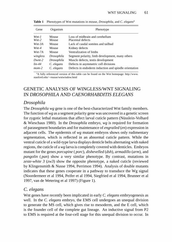
Table 1 Phenotypes of Wnt mutations in mouse,Drosophila, andC. elegansa
Gene Organism Phenotype
Wnt-1 Mouse Loss of midbrain and cerebellumWnt-2 Mouse Placental defectsWnt-3A Mouse Lack of caudal somites and tailbudWnt-4 Mouse Kidney defectsWnt-7A Mouse Ventralization of limbswingless Drosophila Segment polarity, limb development, many othersDwnt-2 Drosophila Muscle defects, testis developmentlin-44 C. elegans Defects in asymmetric cell divisionsmom-2 C. elegans Defects in endoderm induction and spindle orientation
aA fully referenced version of this table can be found on the Wnt homepage: http://www.stanford.edu/∼rnusse/wntwindow.html
GENETIC ANALYSIS OF WINGLESS/WNT SIGNALINGIN DROSOPHILAAND CAENORHABDITIS ELEGANS
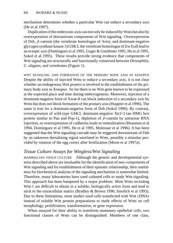
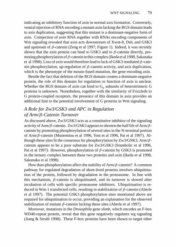
DrosophilaTheDrosophila wggene is one of the best-characterized Wnt family members.The function ofwgas a segment polarity gene was uncovered in a genetic screenfor zygotic lethal mutations that affect larval cuticle pattern (N¨usslein-Volhard& Wieschaus 1980). In theDrosophilaembryo,wg is required for formationof parasegment boundaries and for maintenance ofengrailed(en) expression inadjacent cells. The epidermis ofwg mutant embryos shows only rudimentarysegmentation, which is reflected in an abnormal cuticle pattern. While theventral cuticle of a wild-type larva displays denticle belts alternating with nakedregions, the cuticle of awglarva is completely covered with denticles. Embryosmutant for the genesporcupine( porc), dishevelled(dsh), armadillo (arm), andpangolin ( pan) show a very similar phenotype. By contrast, mutations inzeste-white 3(zw3) show the opposite phenotype, a naked cuticle (reviewedby Klingensmith & Nusse 1994, Perrimon 1994). Analysis of double mutantsindicates that these genes cooperate in a pathway to transduce the Wg signal(Noordermeer et al 1994, Peifer et al 1994, Siegfried et al 1994, Brunner et al1997, van de Wetering et al 1997) (Figure 1).
C. elegansWnt genes have recently been implicated in earlyC. elegansembryogenesis aswell. In theC. elegansembryo, the EMS cell undergoes an unequal divisionto generate the MS cell, which gives rise to mesoderm, and the E cell, whichis the founder cell of the complete gut lineage. An inductive signal from P2to EMS is required at the four-cell stage for this unequal division to occur. In

P1: APR/ary P2: ARS/dat QC: ARS/APM T1: ARS
August 29, 1998 9:42 Annual Reviews AR066-03
62 WODARZ & NUSSE
Figure 1 Wnt signaling pathways are conserved betweenDrosophila, C. elegans, and vertebrates.In Drosophila, anterior epidermal cells (A) express Wg and signal to adjacent posterior cells(P). This signal is required for establishment of parasegment boundaries and for maintenance ofengrailedexpression in posterior cells. InC. elegansa very similar pathway is used to induce theasymmetric division of the EMS cell. Note that only the first three components of this pathway,mom-1, mom-2, andmom-5are also involved in orientation of mitotic spindles during development.Formation of the primary body axis inXenopusdepends on a Wnt pathway comprising GSK3,β-catenin, and XTcf-3. Although overexpression of various Wnts and of Dsh leads to duplicationof the body axis, they do not appear to be required for formation of the primary axis. Instead, theWnt pathway may be triggered at the level of GSK3 by a hypothetical dorsalizing signal. Whetheraxin, APC, and itsC. eleganscounterpartapr-1are integral components of a common Wnt signalingpathway remains to be shown.
the absence of signal, EMS divides symmetrically and gives rise to two MS-like daughters that form mesoderm but no gut. In a screen for mutants thatinterrupt signaling from P2 to EMS, five genes calledmom1-5were identified.Molecular analysis of three revealed that they encode proteins similar toporc(mom-1), Wnt (mom-2), andfrizzled( fz), a Wnt receptor (mom-5; Rocheleauet al 1997, Thorpe et al 1997). Mutation ofpop-1, an HMG box protein similarto pan, has the opposite effect ofmommutations: Both EMS daughters adoptthe E fate and produce exclusively gut (Lin et al 1995). These results indicatethat a Wnt signaling cascade mediates induction of EMS by P2 (reviewed inHan 1997; see Figure 1).
P1: APR/ary P2: ARS/dat QC: ARS/APM T1: ARS
August 29, 1998 9:42 Annual Reviews AR066-03
WNT SIGNALING 63
ASSAYS FOR WNT SIGNALING
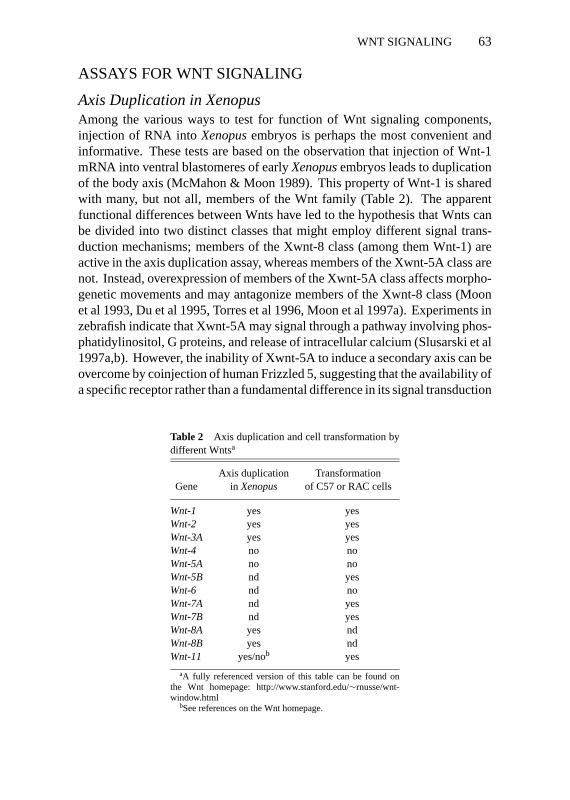
Axis Duplication in XenopusAmong the various ways to test for function of Wnt signaling components,injection of RNA intoXenopusembryos is perhaps the most convenient andinformative. These tests are based on the observation that injection of Wnt-1mRNA into ventral blastomeres of earlyXenopusembryos leads to duplicationof the body axis (McMahon & Moon 1989). This property of Wnt-1 is sharedwith many, but not all, members of the Wnt family (Table 2). The apparentfunctional differences between Wnts have led to the hypothesis that Wnts canbe divided into two distinct classes that might employ different signal trans-duction mechanisms; members of the Xwnt-8 class (among them Wnt-1) areactive in the axis duplication assay, whereas members of the Xwnt-5A class arenot. Instead, overexpression of members of the Xwnt-5A class affects morpho-genetic movements and may antagonize members of the Xwnt-8 class (Moonet al 1993, Du et al 1995, Torres et al 1996, Moon et al 1997a). Experiments inzebrafish indicate that Xwnt-5A may signal through a pathway involving phos-phatidylinositol, G proteins, and release of intracellular calcium (Slusarski et al1997a,b). However, the inability of Xwnt-5A to induce a secondary axis can beovercome by coinjection of human Frizzled 5, suggesting that the availability ofa specific receptor rather than a fundamental difference in its signal transduction
Table 2 Axis duplication and cell transformation bydifferent Wntsa
Axis duplication TransformationGene inXenopus of C57 or RAC cells
Wnt-1 yes yesWnt-2 yes yesWnt-3A yes yesWnt-4 no noWnt-5A no noWnt-5B nd yesWnt-6 nd noWnt-7A nd yesWnt-7B nd yesWnt-8A yes ndWnt-8B yes ndWnt-11 yes/nob yes
aA fully referenced version of this table can be found onthe Wnt homepage: http://www.stanford.edu/∼rnusse/wnt-window.html
bSee references on the Wnt homepage.
P1: APR/ary P2: ARS/dat QC: ARS/APM T1: ARS
August 29, 1998 9:42 Annual Reviews AR066-03
64 WODARZ & NUSSE
mechanism determines whether a particular Wnt can induce a secondary axis(He et al 1997).
Duplication of the embryonic axis can not only be induced by Wnts but also byoverexpression of downstream components of Wnt signaling. Overexpressionof Dsh,β-catenin (the vertebrate homologue of Arm), and dominant-negativeglycogen synthase kinase 3 (GSK3, the vertebrate homologue of Zw3) all lead toan ectopic axis (Dominguez et al 1995, Guger & Gumbiner 1995, He et al 1995,Sokol et al 1995). These results provide strong evidence that components ofWnt signaling are structurally and functionally conserved betweenDrosophila,C. elegans, and vertebrates (Figure 1).
WNT SIGNALING AND FORMATION OF THE PRIMARY BODY AXIS IN XENOPUS
Despite the ability of injected Wnts to induce a secondary axis, it is not clearwhether an endogenous Wnt protein is involved in the establishment of the pri-mary body axis inXenopus. So far there is no Wnt gene known to be expressedat the expected place and time during embryogenesis. Moreover, injection of adominant-negative form of Xwnt-8 can block induction of a secondary axis byWnts but does not block formation of the primary axis (Hoppler et al 1996). Thesame is true for a dominant-negative form of Dsh (Sokol 1996). By contrast,overexpression of wild-type GSK3, dominant-negative Xtcf-3 (an HMG boxprotein similar to Pan and Pop-1), depletion ofβ-catenin by antisense RNAinjection, or overexpression of cadherins leads to ventralization (Heasman et al1994, Dominguez et al 1995, He et al 1995, Molenaar et al 1996). It has beensuggested that the Wnt signaling cascade may be triggered downstream of Dshby an unknown dorsalizing signal unrelated to Wnts, possibly a stimulus pro-vided by rotation of the egg cortex after fertilization (Moon et al 1997a).
Tissue Culture Assays for Wingless/Wnt SignalingMAMMALIAN TISSUE CULTURE Although the genetic and developmental sys-tems described above are invaluable for the identification of new components ofWnt signaling and for establishment of their epistatic relationship, their useful-ness for biochemical analysis of the signaling mechanism is somewhat limited.Therefore, many laboratories have used cultured cells to study Wnt signaling.This approach has been hampered by a major problem: Most Wnts includingWnt-1 are difficult to obtain in a soluble, biologically active form and tend tostick to the extracellular matrix (Bradley & Brown 1990, Smolich et al 1993).Due to these limitations, most studies used cells transfected with Wnt cDNAsinstead of soluble Wnt protein preparations to study effects of Wnts on cellmorphology, proliferation, transformation, or gene expression.
When assayed for their ability to transform mammary epithelial cells, twofunctional classes of Wnts can be distinguished: Members of one class,
P1: APR/ary P2: ARS/dat QC: ARS/APM T1: ARS
August 29, 1998 9:42 Annual Reviews AR066-03
WNT SIGNALING 65
including Wnt-1, Wnt-3A, and Wnt-7A, readily transform cells at high fre-quency, whereas members of the other class, Wnt-4, Wnt-5A, and Wnt-6, donot (Wong et al 1994) (Table 2). Interestingly, the two functional classes definedby this assay are similar to the functional classes defined by the axis duplicationassay inXenopus(Du et al 1995) (Table 2).
DROSOPHILATISSUE CULTURE In contrast to mammalian Wnts, Wg producedin transfectedDrosophilaSchneider S2 cells is secreted into the medium andis biologically active when added to Wg-responsive cells (van Leeuwen et al1994). The assay used for Wg activity is derived from the observation that intheDrosophilaembryo, Arm protein accumulates in the cytoplasm of cells thatare exposed to Wg (Riggleman et al 1990, Peifer et al 1994). ADrosophilaimaginal disc cell line, cl-8, shows the same response upon incubation withWg-conditioned medium (van Leeuwen et al 1994). Overexpression of Dsh ordominant-negative Zw3 also leads to Arm accumulation (Yanagawa et al 1995,1997). By contrast, S2 cells do not show increased Arm levels after exposure toWg although they respond to overexpression of Dsh, which led to the suggestionthat S2 cells lack a functional Wg receptor (Yanagawa et al 1995).
WNT SIGNALING UPSTREAM OF THE RECEPTOR
A Role for Porcupine in Post-Translational Modificationand Secretion of WntsDrosophilaembryos mutant for the geneporcshow a phenotype similar to thatof wg mutants (van den Heuvel et al 1993, Kadowaki et al 1996). Clones ofporc mutant cells display non-cell-autonomous effects, similar towg clones,indicating that Porc is required in the cell that produces the signal rather thanin the receiving cell (Kadowaki et al 1996). Inporc embryos, Wg protein isconfined to the narrow stripe of cells where thewggene is transcribed, insteadof spreading to adjacent cells as in wild-type embryos (van den Heuvel et al1989, 1993). Several mutations inwg itself also cause retention of mutant Wgprotein, indicating that changes of the protein structure can lead to misfoldingand impair secretion (van den Heuvel et al 1993, Bejsovec & Wieschaus 1995,Hays et al 1997).
Molecular cloning ofporc revealed that it encodes a multi-transmembraneprotein predominantly found in the endoplasmic reticulum, consistent with arole in processing of Wg (Kadowaki et al 1996). Biochemical studies in cul-tured cells revealed increased N-linked glycosylation of Wg after coexpressionwith Porc (Kadowaki et al 1996). Glycosylation appears to be a common mod-ification of Wnts and may be important for folding, secretion, and biologicalactivity (Smolich et al 1993).
P1: APR/ary P2: ARS/dat QC: ARS/APM T1: ARS
August 29, 1998 9:42 Annual Reviews AR066-03
66 WODARZ & NUSSE
In C. elegans, the porc homologuemom-1is required in the P2 cell forsignaling to the EMS cell bymom-2(a Wnt) (Rocheleau et al 1997, Thorpeet al 1997). Apart from this example, it is not known whetherporc is essentialfor every Wnt protein to become secreted, mainly because there are few reagentsto study the distribution of Wnt proteins in vivo.
Another ER-resident protein, the molecular chaperone BiP, associates withWnt-1 (Kitajewski et al 1992). Although the functional significance of thisinteraction has not been tested, it is likely that BiP assists in proper folding ofWnts.
Proteoglycans Facilitate Wnt SignalingTheDrosophila sugarless(sgl) gene encodes UDP-glucose dehydrogenase, akey enzyme required for synthesis of proteoglycans. Mutants ofsglshow a lar-val phenotype similar towg (Binari et al 1997, H¨acker et al 1997, Haerry et al1997). Phenocopies ofsgl can be generated by injection of heparinase, whichselectively degrades heparin-like glycosaminoglycans, but not by chondroiti-nase, which degrades chondroitin sulfate, hyaluronic acid, and dermatan sulfate.Conversely, injection of heparan sulfate is sufficient to rescue thesgl mutantphenotype (Binari et al 1997). Mutations insulfateless(sfl ), a gene encodingheparan sulfateN-deacetylase/N-sulfotransferase, another enzyme required forheparan sulfate biosynthesis, also cause awg-like phenotype, underpinning theimportance of proteoglycans for Wg signaling (X Lin & N Perrimon, personalcommunication).
Sulfated glycosaminoglycans also affect Wg signaling in tissue culture.Treatment of cl-8 cells with heparinase, heparitinase, chondroitin ABC lyase,or perchlorate leads to reduced Wg-induced accumulation of Arm. This effectcan be reversed by addition of chondroitin sulfate or heparin. Heparin bindsto Wnt-1 (Bradley & Brown 1990) and Wg, and addition of heparin to Wg-conditioned medium leads to increased Wg activity (Reichsman et al 1996).However, heparin has also been reported to inhibit the activity of Wnt-1 in acell transformation assay (Jue et al 1992). InXenopusanimal caps, removalof heparan sulfate proteoglycans blocks mesoderm induction by Xwnt-8, againsuggesting that proteoglycans are important for the function of Wnts (Itoh &Sokol 1994).
Little is known about the role of proteoglycans in Wnt signaling. In analogyto FGF signaling, proteoglycans may be low-affinity coreceptors for Wnts,which would serve to increase the local concentration of ligand available forbinding to high-affinity receptors. Alternatively, proteoglycans in the ECM maycrosslink Wnts to induce clustering of Wnt receptors in the plasma membrane.In the absence of soluble, sufficiently pure, biologically active preparations ofWnts, this issue will be difficult to address.
P1: APR/ary P2: ARS/dat QC: ARS/APM T1: ARS
August 29, 1998 9:42 Annual Reviews AR066-03
WNT SIGNALING 67
INTERACTIONS BETWEEN WNTS AND PROTEINSOF THE FRIZZLED FAMILY
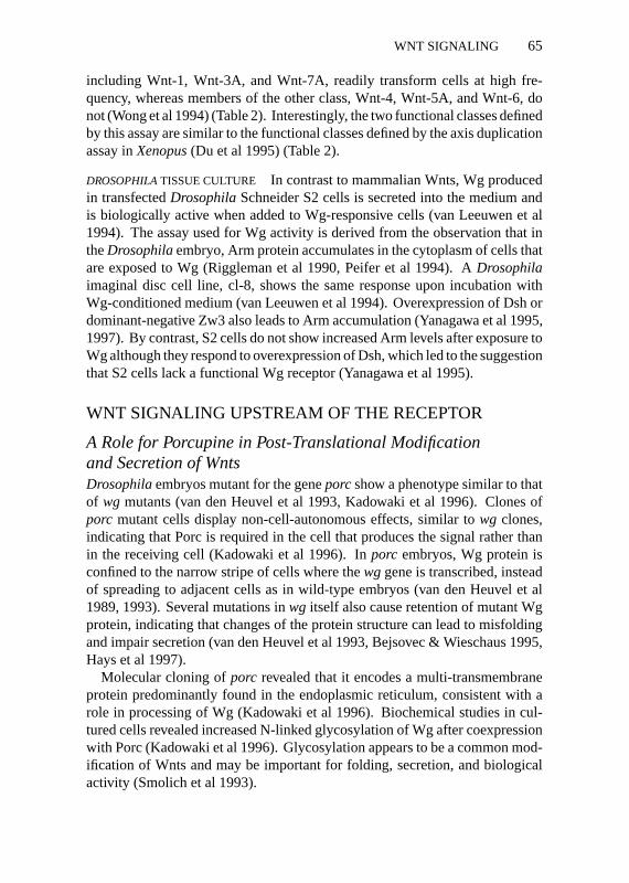
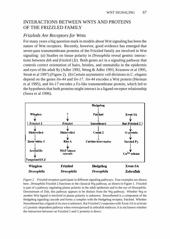
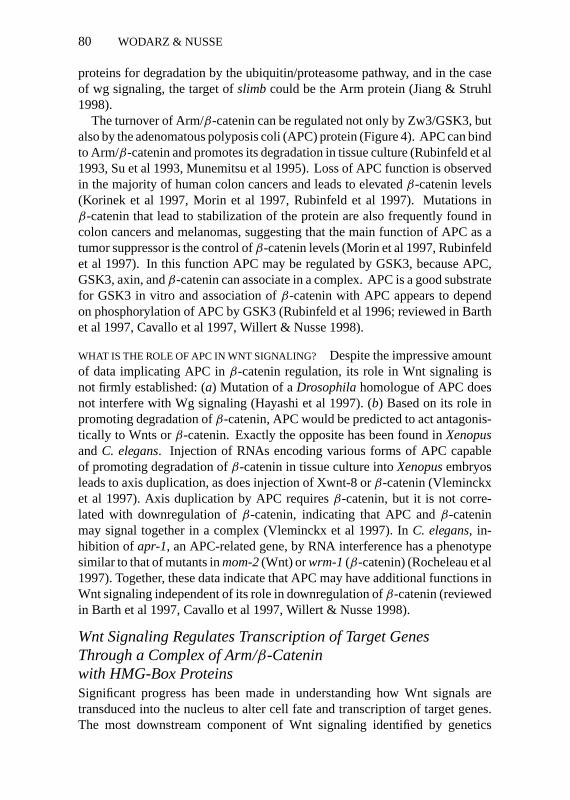
Frizzleds Are Receptors for WntsFor many years a big question mark in models about Wnt signaling has been thenature of Wnt receptors. Recently, however, good evidence has emerged thatseven-pass transmembrane proteins of the Frizzled family are involved in Wntsignaling: (a) Studies on tissue polarity inDrosophilareveal genetic interac-tions betweendshandfrizzled( fz). Both genes act in a signaling pathway thatcontrols correct orientation of hairs, bristles, and ommatidia in the epidermisand eyes of the adult fly (Adler 1992, Wong & Adler 1993, Krasnow et al 1995,Strutt et al 1997) (Figure 2). (b) Certain asymmetric cell divisions inC. elegansdepend on the geneslin-44 andlin-17. lin-44 encodes a Wnt protein (Hermanet al 1995), andlin-17 encodes a Fz-like transmembrane protein, which led tothe hypothesis that both proteins might interact in a ligand-receptor relationship(Sawa et al 1996).
Figure 2 Frizzled receptors participate in different signaling pathways. Four examples are shownhere.DrosophilaFrizzled 2 functions in the classical Wg pathway, as shown in Figure 1. Frizzledis part of a pathway regulating planar polarity in the adult epidermis and in the eye ofDrosophila.Downstream of Dsh, this pathway appears to be distinct from the Wg pathway. Whether Wg oranother Wnt ligand is involved in planar polarity is unknown. Smoothened is a component of theHedgehog signaling cascade and forms a complex with the Hedgehog receptor, Patched. WhetherSmoothened has a ligand of its own is unknown. Rat Frizzled 2 cooperates with Xwnt-5A to activatea G protein–dependent pathway when overexpressed in zebrafish embryos. It is not known whetherthe interaction between rat Frizzled 2 and G proteins is direct.
P1: APR/ary P2: ARS/dat QC: ARS/APM T1: ARS
August 29, 1998 9:42 Annual Reviews AR066-03
68 WODARZ & NUSSE
This issue has been directly addressed in tissue culture. Transfection of Dfz2,a member of the Fz family, fromDrosophila into S2 cells (S2Dfz2) confersresponsiveness to Wg to these cells. Like cl-8 cells, S2Dfz2 cells accumulateArm when incubated with Wg-conditioned medium. Moreover, S2Dfz2 cellsand human 293 cells transfected with Dfz2 bind Wg on their cell surfaces,whereas untransfected cells of both cell lines do not (Bhanot et al 1996).
Cadigan et al (1998) also showed that a dominant-negative form of theDfz2gene blocks signaling bywg in theDrosophilaimaginal disc and that overex-pression of this receptor causes phenotypes similar to those brought about byectopicwg, in awg-dependent way.
Additional evidence for Frizzleds as Wnt receptors comes from studies inXenopus. Coinjection of rat Frizzled 1 and Xwnt-8 into frog embryos results inrecruitment of Xwnt-8 to the plasma membrane and in increased expression ofXwnt-8 target genes, compared with injection of Xwnt-8 alone (Yang-Snyderet al 1996). Also, as mentioned above, coinjection of rat Frizzled 5 and Xwnt-5Aleads to axis duplication, whereas injection of either rat Frizzled 5 or Xwnt-5Adoes not (He et al 1997).
Genetic Analysis of Frizzled-Wnt InteractionsDROSOPHILA Genetic data on the requirement for Frizzleds in Wnt signalingare limited inDrosophila. Loss-of-function mutants of the originalfzare viableand show misorientation of hairs and bristles in the adult epidermis (Vinson &Adler 1987). Rotation of ommatidia in the eye imaginal disc is also defective infzmutants, resulting in a rough eye phenotype (Zheng et al 1995). Clones offzmutant cells induced in wing imaginal discs show an interesting feature termeddirectional nonautonomy, which describes the fact that cells within the clone, aswell as wild-type cells located outside the clone, have misoriented wing hairs.Those “shadows” are found in a distal direction (Vinson & Adler 1987), whichled to the suggestion that Fz is required for not only reception of a polaritysignal but also for generation or propagation of a signal. This signal, whosenature remains to be determined, would spread in a proximal-distal directionacross the wing imaginal disc (Adler 1992).
Although a physiological ligand for Fz has not been identified, it is anticipatedthat this ligand may be a Wnt. It has been suggested that there is a gradientof Fz activity across the wing that is caused by a graded distribution of theligand and that hair polarity follows the slope of that gradient. Support for thisnotion comes from the observation that creation of an artificial gradient of Fzexpression with the high point at the distal tip of the wing leads to polarityreversal (Adler et al 1997).
Interestingly, some alleles offz do not show directional nonautonomy inclones and instead behave strictly cell autonomously. Molecular analysis of four
P1: APR/ary P2: ARS/dat QC: ARS/APM T1: ARS
August 29, 1998 9:42 Annual Reviews AR066-03
WNT SIGNALING 69
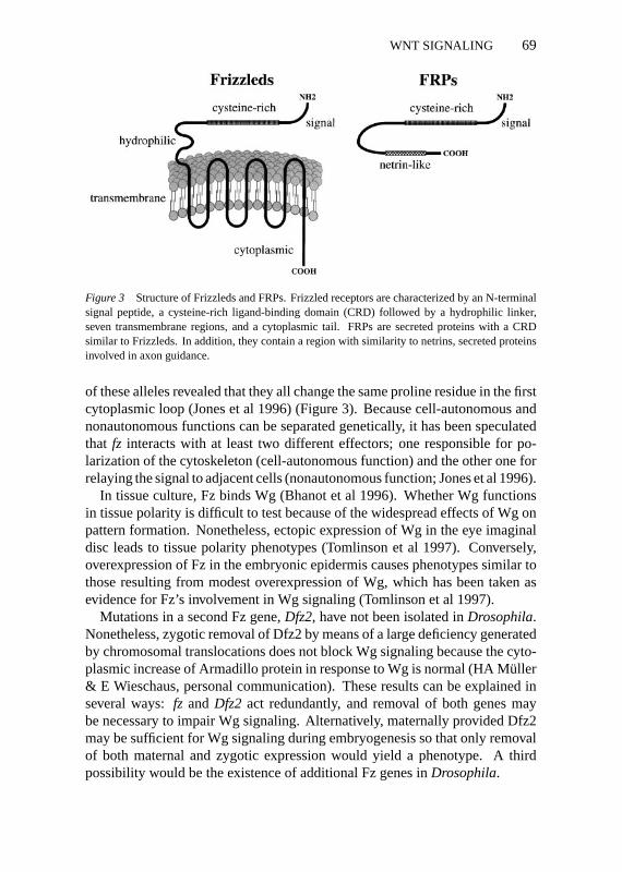
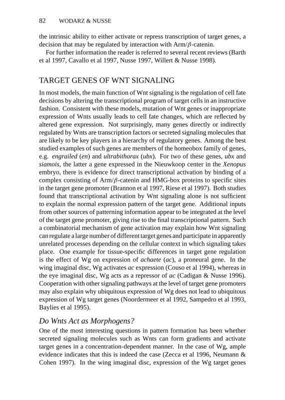
Figure 3 Structure of Frizzleds and FRPs. Frizzled receptors are characterized by an N-terminalsignal peptide, a cysteine-rich ligand-binding domain (CRD) followed by a hydrophilic linker,seven transmembrane regions, and a cytoplasmic tail. FRPs are secreted proteins with a CRDsimilar to Frizzleds. In addition, they contain a region with similarity to netrins, secreted proteinsinvolved in axon guidance.
of these alleles revealed that they all change the same proline residue in the firstcytoplasmic loop (Jones et al 1996) (Figure 3). Because cell-autonomous andnonautonomous functions can be separated genetically, it has been speculatedthat fz interacts with at least two different effectors; one responsible for po-larization of the cytoskeleton (cell-autonomous function) and the other one forrelaying the signal to adjacent cells (nonautonomous function; Jones et al 1996).
In tissue culture, Fz binds Wg (Bhanot et al 1996). Whether Wg functionsin tissue polarity is difficult to test because of the widespread effects of Wg onpattern formation. Nonetheless, ectopic expression of Wg in the eye imaginaldisc leads to tissue polarity phenotypes (Tomlinson et al 1997). Conversely,overexpression of Fz in the embryonic epidermis causes phenotypes similar tothose resulting from modest overexpression of Wg, which has been taken asevidence for Fz’s involvement in Wg signaling (Tomlinson et al 1997).
Mutations in a second Fz gene,Dfz2, have not been isolated inDrosophila.Nonetheless, zygotic removal of Dfz2 by means of a large deficiency generatedby chromosomal translocations does not block Wg signaling because the cyto-plasmic increase of Armadillo protein in response to Wg is normal (HA M¨uller& E Wieschaus, personal communication). These results can be explained inseveral ways:fz and Dfz2 act redundantly, and removal of both genes maybe necessary to impair Wg signaling. Alternatively, maternally provided Dfz2may be sufficient for Wg signaling during embryogenesis so that only removalof both maternal and zygotic expression would yield a phenotype. A thirdpossibility would be the existence of additional Fz genes inDrosophila.
P1: APR/ary P2: ARS/dat QC: ARS/APM T1: ARS
August 29, 1998 9:42 Annual Reviews AR066-03
70 WODARZ & NUSSE
Besidesfz andDfz2, a third family member,smoothened(smo), has beenidentified inDrosophila(Alcedo et al 1996, van den Heuvel & Ingham 1996).Genetic analysis indicates thatsmois required for Hedgehog (Hh) signalingrather than for Wg signaling (Figure 2). These findings, together with thepredicted protein structure of Smo, led to the suggestion that Smo is an Hhreceptor, implicating direct protein-protein interaction between Smo and Hh(Alcedo et al 1996, van den Heuvel & Ingham 1996). This hypothesis has beenquestioned by the finding that Hh and a vertebrate homologue, Sonic Hedgehog,can bind to Patched (Ptc), a multi-transmembrane protein, whereas no directbinding to Smo was detected (Chen & Struhl 1996, Marigo et al 1996, Stone et al1996). However, Ptc can form a complex with Smo, suggesting that Hh activatesSmo indirectly via Ptc (Chen & Struhl 1996, Stone et al 1996) (Figure 2).
C. ELEGANS In the worm, more genetic data are available for fz/Wnt interac-tions. The geneslin-44 (a Wnt) andlin-17 (an Fz) interact genetically in thecontrol of certain asymmetric cell divisions. Inlin-44 mutants, the polarity oftwo asymmetric daughter cells is reversed, whereas inlin-17 mutants, polarityis lost, resulting in two symmetric daughter cells (Herman et al 1995, Sawa et al1996). lin-44/lin-17 double mutants also produce symmetric daughter cells,indicating thatlin-17 acts downstream oflin-44. The fact that the phenotypesof lin-44 andlin-17 are not identical has led to the hypothesis that Lin-17 actsas a receptor for two signals coming from different directions—one Lin-44 andthe other an unknown ligand X, possibly another Wnt (Sawa et al 1996).
Mutants inmom-5, a second Fz-like gene fromC. elegans, also show someunexpected features.mom-5interacts withmom-2(a Wnt) to specify the gut pre-cursor cell E, which is generated in the asymmetric division of EMS (Rocheleauet al 1997, Thorpe et al 1997; see above). The penetrance of the gutless phe-notype ofmom-5is low,≈5%. Mutations inmom-2have a much higher pen-etrance,≈40%. Surprisingly, the double mutantmom-2/mom-5shows a pene-trance of only 8%, so the low penetrance of themom-5phenotype is epistaticover the high penetrancemom-2phenotype, which is not compatible with themodel that the receptor Mom-5 is simply activated by its ligand Mom-2 tospecify the E fate. One explanation for these data would be that Mom-5 con-stitutively represses specification of E fate in the absence of the ligand Mom-2and that this repression can be overcome by binding of Mom-2 (Rocheleau et al1997).
In contrast to the low penetrance of endoderm defects inmom-5andmom-2/mom-5double mutant embryos,mom-1(Porc-like),mom-2, andmom-5mu-tants show a fully penetrant defect in orientation of the mitotic spindle in theABar cell. This kind of defect is not observed in mutants forapr-1 (APC),wrm-1 (β-catenin), andpop-1(HMG box protein), indicating a branching of
P1: APR/ary P2: ARS/dat QC: ARS/APM T1: ARS
August 29, 1998 9:42 Annual Reviews AR066-03
WNT SIGNALING 71
the Wnt pathway downstream ofmom-5(Rocheleau et al 1997, Thorpe et al1997) (Figure 1). Interestingly, the function ofmom-1, mom-2, andmom-5in spindle orientation may not require transcriptional regulation of Wnt targetgenes, since inhibition of the large subunit of RNA polymerase orα-amanitintreatment of embryos does not affect spindle orientation until the 26-cell stage(Rocheleau et al 1997 and references therein).
Together, these data indicate that Frizzled receptors can participate in severaldistinct signal transduction pathways (Figure 2). Moreover, there is evidencethat an individual Frizzled can have more than one physiological ligand and,vice versa, that a Wnt may bind to more than one Frizzled. Regulation of Friz-zleds may occur within the plane of the plasma membrane by association withother transmembrane proteins such as Ptc. In addition, Frizzleds may possessconstitutive activity, allowing both activation and repression of downstreamsignaling components, depending on availability of ligand(s).
Structure of Frizzled ProteinsAll members of the Fz family are characterized by the following features (begin-ning at the N terminus): a putative signal sequence, followed by a sequence of120 amino acids (aa) containing 10 highly conserved cysteine residues (CRD),a highly divergent region of 40–100 aa predicted to form a flexible linker,seven transmembrane segments separated by short extracellular and cytoplas-mic loops, and a cytoplasmic tail (Vinson et al 1989, Wang et al 1996) (Figure 3).The CRD appears to be the ligand-binding site of Frizzleds. Expression of theCRD of Dfz2 anchored in the membrane by a glycosyl-phosphatidylinositol(GPI) anchor is sufficient to provide binding sites for Wg on the surface of cells(Cadigan et al 1998). By contrast, expression of a mouse Fz4 construct lack-ing the CRD, but containing the signal sequence and all seven transmembranedomains, does not confer binding of Wg (Bhanot et al 1996).
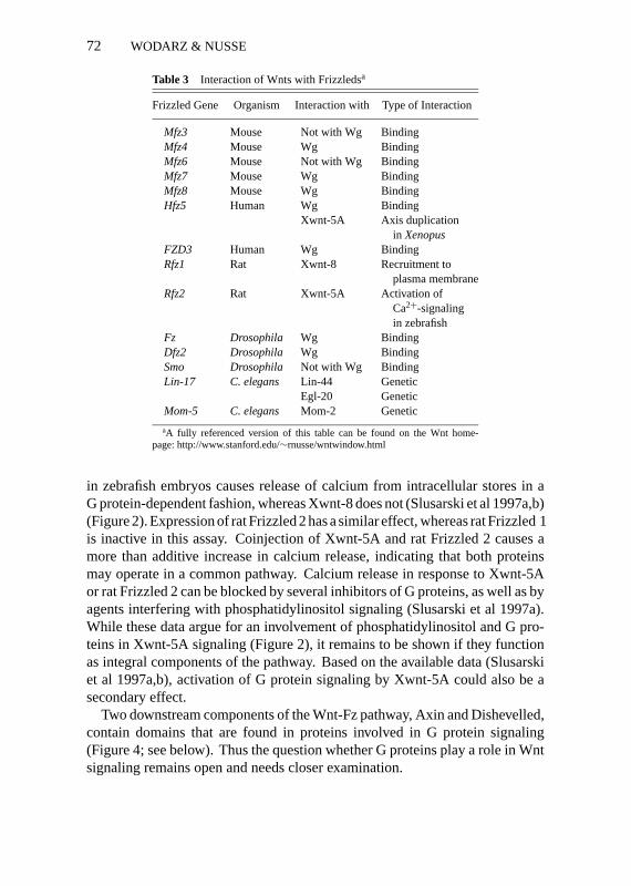
The Wg-binding assay provides a means to test the specificity of ligand-receptor interactions. Several Fz proteins from human, mouse, andDrosophilahave been tested for their ability to allow cell surface binding of Wg. Manyof the Frizzleds tested, including the original Fz fromDrosophila, do conferWg binding, whereas others, e.g. mouse Fz3 and Fz6 and Smoothened, do not(Bhanot et al 1996, Nusse et al 1997, YK Wang et al 1997; Table 3). Althoughthis assay does not provide measurement of binding affinities between Wg anddifferent Frizzleds, it indicates that there is considerable promiscuity in theinteraction of Wnts with their receptors.
The overall structure of Frizzleds resembles that of G protein-coupled recep-tors, which also have seven transmembrane regions. However, this similarity ismostly restricted to the membrane topology of both protein families, and thereis little sequence identity between them. Nonetheless, expression of Xwnt-5A
P1: APR/ary P2: ARS/dat QC: ARS/APM T1: ARS
August 29, 1998 9:42 Annual Reviews AR066-03
72 WODARZ & NUSSE
Table 3 Interaction of Wnts with Frizzledsa
Frizzled Gene Organism Interaction with Type of Interaction
Mfz3 Mouse Not with Wg BindingMfz4 Mouse Wg BindingMfz6 Mouse Not with Wg BindingMfz7 Mouse Wg BindingMfz8 Mouse Wg BindingHfz5 Human Wg Binding
Xwnt-5A Axis duplicationin Xenopus
FZD3 Human Wg BindingRfz1 Rat Xwnt-8 Recruitment to
plasma membraneRfz2 Rat Xwnt-5A Activation of
Ca2+-signalingin zebrafish
Fz Drosophila Wg BindingDfz2 Drosophila Wg BindingSmo Drosophila Not with Wg BindingLin-17 C. elegans Lin-44 Genetic
Egl-20 GeneticMom-5 C. elegans Mom-2 Genetic
aA fully referenced version of this table can be found on the Wnt home-page: http://www.stanford.edu/∼rnusse/wntwindow.html
in zebrafish embryos causes release of calcium from intracellular stores in aG protein-dependent fashion, whereas Xwnt-8 does not (Slusarski et al 1997a,b)(Figure 2). Expression of rat Frizzled 2 has a similar effect, whereas rat Frizzled 1is inactive in this assay. Coinjection of Xwnt-5A and rat Frizzled 2 causes amore than additive increase in calcium release, indicating that both proteinsmay operate in a common pathway. Calcium release in response to Xwnt-5Aor rat Frizzled 2 can be blocked by several inhibitors of G proteins, as well as byagents interfering with phosphatidylinositol signaling (Slusarski et al 1997a).While these data argue for an involvement of phosphatidylinositol and G pro-teins in Xwnt-5A signaling (Figure 2), it remains to be shown if they functionas integral components of the pathway. Based on the available data (Slusarskiet al 1997a,b), activation of G protein signaling by Xwnt-5A could also be asecondary effect.
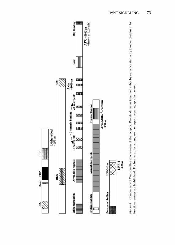
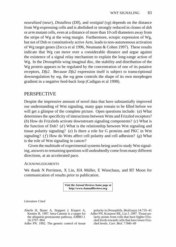
Two downstream components of the Wnt-Fz pathway, Axin and Dishevelled,contain domains that are found in proteins involved in G protein signaling(Figure 4; see below). Thus the question whether G proteins play a role in Wntsignaling remains open and needs closer examination.
P1: APR/ary P2: ARS/dat QC: ARS/APM T1: ARS
August 29, 1998 9:42 Annual Reviews AR066-03
WNT SIGNALING 73
Fig
ure
4C
ompo
nent
sof
Wnt
sign
alin
gdo
wns
trea
mof
the
rece
ptor
.Pr
otei
ndo
mai
nsid
entifi
edei
ther
byse
quen
cesi
mila
rity
toot
her
prot
eins
orby
func
tiona
lass
ays
are
high
light
ed.
For
furt
her
expl
anat
ions
,see
the
resp
ectiv
epa
ragr
aphs
inth
ete
xt.
P1: APR/ary P2: ARS/dat QC: ARS/APM T1: ARS
August 29, 1998 9:42 Annual Reviews AR066-03
74 WODARZ & NUSSE
The C terminus of many, but not all, Frizzleds has the sequence S/TXV, whichhas been described as a potential binding site for PDZ domains (Songyanget al 1997). Dsh is an obvious candidate for binding to the C terminus ofFrizzleds because genetically it is most proximal to the receptor and containsa PDZ domain. However, attempts to show such a direct interaction have beenunsuccesful (Nusse et al 1997). Moreover, the PDZ domain of Dsh lacks crucialamino acid residues predicted to interact with the S/TXV motif and thereforeprobably binds to a different sequence motif (Doyle et al 1996). The functionalimportance of the C terminus of Frizzleds is also questioned by an in vivoexperiment inC. elegans: A Lin-17-GFP fusion protein lacking the 8 mostC-terminal residues of Lin-17 rescues thelin-17 mutant phenotype (Sawa et al1996).
Frizzled/Wnt Interactions: How Many Pathways Are There?One of the most important questions arising from the data summarized aboveis the following: How can individual Wnts elicit specific biological responsesthat are distinct from the effects of other members of the Wnt family? Oneway to achieve specificity is provided by restricting the expression pattern ofdifferent Wnts to discrete, nonoverlapping regions of the organism. This isoften the case, but there are many examples for overlapping expression patternsof Wnts. The next level to discern between Wnts may be the specificity ofligand-receptor interactions. While the experiments described above revealthat not every Wnt can bind to every Frizzled, they also show that there isa high degree of promiscuity in Wnt/Frizzled interactions. However, manyFrizzleds show highly specific expression patterns themselves (Bhanot et al1996, Wang et al 1996), which further restricts the number of potential ligand-receptor interactions in any given tissue.
Once a Wnt has bound to a suitable Frizzled receptor, what happens next?Several scenarios are possible:
1 Every Wnt/Frizzled interaction leads to activation of the same downstreamsignaling cassette, comprising Dsh, GSK3,β-catenin, and HMG boxproteins.
2 There are different signaling pathways downstream of Frizzleds, but eachmember of the Frizzled family can couple to only one of these pathways.If an individual Frizzled can bind different Wnts, binding of any one willactivate the same pathway.
3 There are different signaling pathways downstream of Frizzleds, and a givenFrizzled can couple to several of these pathways. Which pathway is activateddepends on the Wnt ligand bound to the receptor.
P1: APR/ary P2: ARS/dat QC: ARS/APM T1: ARS
August 29, 1998 9:42 Annual Reviews AR066-03
WNT SIGNALING 75
4 There are different signaling pathways downstream of Frizzleds, but onlysome can be triggered by Wnts, while others are ligand independent or aretriggered by ligands unrelated to Wnts.
This list can of course be extended, and the true situation may not be reflectedcorrectly by any of these statements. Fortunately, we now have an increasingnumber of experimental tools to study these questions in various systems.
FRPs Are Structurally Related to the CRD of Frizzledsand Act as Secreted Antagonists of WntsRecently, a family of secreted proteins containing a CRD similar to that ofFrizzleds has been identified in vertebrates (Hoang et al 1996, Finch et al 1997,Leyns et al 1997, Mayr et al 1997, Rattner et al 1997, S Wang et al 1997). Inaddition to the CRD, these frizzled-related proteins (FRPs) contain a C-terminalregion with similarity to netrins, secreted proteins involved in axon guidance(Leyns et al 1997, S Wang et al 1997) (Figure 3). Some evidence has beenobtained that FRPs can bind Wnts directly and that the CRD is necessary andsufficient for this interaction (Leyns et al 1997, Lin et al 1997, Rattner et al1997, S Wang et al 1997) (Table 4). Coinjection of different FRP mRNAstogether with Xwnt-8 mRNA into ventral blastomeres inXenopusblocks axisduplication by Xwnt-8 (Finch et al 1997, Leyns et al 1997, Mayr et al 1997,S Wang et al 1997). In the case of Frzb-1 (also called FRP-3 or Fritz), thisinhibition is also observed when Frzb-1 and Xwnt-8 are injected into differentblastomeres, consistent with Frzb-1’s being a secreted, diffusible antagonist of
Table 4 Interaction of Wnts with FRPsa
FRP Gene Organism Interaction with Type of Interaction
FRP-1 Human Wg, Wnt-1, Xwnt-8 Inhibition of axis duplication in frogs
FRP-2(SDF-5) Mouse Wg Binding
FRP-3(Frzb-1) Mouse Wg, Xwnt-8 Binding, inhibition of axisduplication in frogs
Xenopus Xwnt-8, Wnt-1 Binding, coimmuno-precipitation,inhibition of axis duplication in frogs
Bovine Wnt-1, Wnt-5A Coimmuno-precipitation, inhibition ofWnt-1-induced accumulationof β-catenin in cells, no effecton Wnt-5A in frog assay
Sizzled Xenopus Xwnt-8 Antagonism to Xwnt-8 in severalfrog assays
aA fully referenced version of this table can be found on the Wnt homepage: http://www.stanford.edu/∼rnusse/wntwindow.html
P1: APR/ary P2: ARS/dat QC: ARS/APM T1: ARS
August 29, 1998 9:42 Annual Reviews AR066-03
76 WODARZ & NUSSE
Xwnt-8 (Leyns et al 1997, Mayr et al 1997, S Wang et al 1997). A similarconclusion was drawn from tissue culture experiments with mammalian cells,where Frzb-1 inhibits Wnt-1-mediated accumulation ofβ-catenin (Lin et al1997). Interestingly, although Frzb-1 can also bind to Wnt-5A, as shown bycoimmunoprecipitation, it does not block the activity of Wnt-5A in aXenopusassay (Lin et al 1997) (Table 4).
Although a function for FRPs during normal development has not been rig-orously shown owing to the absence of mutants, Frzb-1 shows an intriguingexpression pattern duringXenopusembryogenesis. Expression is restricted tothe region of the Spemann organizer and thus is complementary to the patternof Xwnt-8, which is expressed in the ventral marginal zone (Leyns et al 1997,S Wang et al 1997). Because Xwnt-8 appears to specify ventrolateral mesoderm,it is tempting to speculate that Frzb-1 expression in the Spemann organizer spec-ifies dorsal cell fates by antagonizing Xwnt-8 (Christian & Moon 1993, Hoppleret al 1996, Leyns et al 1997, Moon et al 1997b, S Wang et al 1997).
Are Frizzleds the Only Receptors for Wnts?Based on genetic interaction studies inDrosophila, it has been speculated thatNotch (N), a transmembrane protein involved in many developmental processes,might be a receptor for Wg (Couso & Martinez-Arias 1994, Hing et al 1994).However, in the complete absence of N, Wg signaling appears unaffected inthe embryo (Cadigan & Nusse 1996). In the wing imaginal disc, the effect ofN on Wg signaling may be indirect. Removal of N by means of a temperature-sensitive mutation or in clones leads to loss of Wg expression, providing anexplanation for the genetic interaction between these two genes. Nonetheless,clones of N mutant cells show a normal response to Wg produced by wild-typecells adjacent to the clone (Rulifson & Blair 1995). Transfection of S2 cellswith N does not lead to significant cell surface binding of Wg and does notconfer responsiveness to Wg (Bhanot et al 1996). Taken together, these datado not support a direct role for N in reception of the Wg signal.
WNT SIGNALING DOWNSTREAMOF THE RECEPTOR
DishevelledGenetic analysis inDrosophilareveals an absolute requirement fordsh in re-ception of the Wg signal (Klingensmith et al 1994, Theisen et al 1994).dshacts cell autonomously and has been placed genetically betweenwgandzw3inthe Wg signaling cascade (Klingensmith et al 1994, Noordermeer et al 1994,Siegfried et al 1994, Theisen et al 1994) (Figure 1).dshis also part of a tissue
P1: APR/ary P2: ARS/dat QC: ARS/APM T1: ARS
August 29, 1998 9:42 Annual Reviews AR066-03
WNT SIGNALING 77
polarity signaling pathway, where it is required cell autonomously downstreamof fzand upstream ofrhoA (Theisen et al 1994, Krasnow et al 1995, Strutt et al1997) (Figure 2). Furthermore, Dsh has been reported to suppress N signalingwhen overexpressed and to directly bind to the cytoplasmic domain of N in ayeast two-hybrid assay (Axelrod et al 1996).
Knockout mice lackingdvl-1, a mouse homologue ofdsh, do not have anyobvious anatomical abnormalities and are viable and fertile. However, oncloser examination, these animals show abnormal social behavior and severalneurological defects (Lijam et al 1997). Similar defects are commonly seenin human patients suffering from certain neurological disorders. The some-what surprising lack of gross morphological defects in thedvl-1knockout micemay be explained by a redundant function provided by at least two otherdshhomologues in the mouse.
dshencodes a ubiquitously expressed cytoplasmic protein containing fourdomains that are highly conserved among all known Dsh homologues, rangingfrom C. elegansto human (Figure 4). At the N terminus, Dsh contains a stretchof 50 aa similar to a region in axin, a protein implicated in Wnt signaling invertebrates (Zeng et al 1997) (Figure 4). The three other conserved regions area short basic domain, a centrally located PDZ domain, and a more C-terminalDEP-domain, which is also found in several proteins interacting with proteinkinase C (PKC) (Klingensmith et al 1994, Theisen et al 1994, Ponting & Bork1996). The function of Dsh is unknown, but the presence of two domains im-plicated in protein-protein interactions suggests that Dsh may be an adaptorprotein required for assembly of a signaling complex, analogous to Grb-2 inthe Ras pathway.
Dsh is a phosphoprotein that becomes more highly phosphorylated on ser-ine and threonine residues when Wg signaling is activated. Moreover, themost highly phosphorylated form of Dsh is enriched in the membrane frac-tion, suggesting that Wg signaling leads to recruitment of Dsh to a membranecompartment. Overexpression of Dsh in the absence of Wg also leads to hyper-phosphorylation and to accumulation of Arm and thus mimics activation of Wgsignaling (Yanagawa et al 1995). The latter conclusion has also been confirmedin vivo (Axelrod et al 1996, Cadigan & Nusse 1996).
Affinity purification of a complex containing Dsh and several associatedproteins revealed the association of Dsh with casein kinase 2 (CK2), a serine-threonine-specific protein kinase. CK2 efficiently phosphorylates Dsh in vitroand in vivo, but the functional significance of this phosphorylation is not clear(Willert et al 1997).
Frizzled expression can affect the phosphorylation status of Dsh (Willertet al 1997) and alter its subcellular localization. InXenopus, overexpressed
P1: APR/ary P2: ARS/dat QC: ARS/APM T1: ARS
August 29, 1998 9:42 Annual Reviews AR066-03
78 WODARZ & NUSSE
Xdsh-GFP is localized in the cytoplasm in a punctate pattern. By contrast,overexpression of Xdsh-GFP together with Rat Frizzled 1 results in relocaliza-tion of Xdsh-GFP to the plasma membrane (Yang-Snyder et al 1996).
Zeste-White 3/Glycogen Synthase Kinase 3Mutations inzeste-white 3(zw3), a gene encoding theDrosophilahomologueof vertebrate glycogen synthase kinase 3 (GSK3), a serine-threonine proteinkinase, have the opposite phenotypes of the mutations inwg, dsh, andarm(Siegfried et al 1992). By genetic epistasis analysis,zw3has been placed be-tweendshandarmin the Wg signaling pathway (Peifer et al 1994, Siegfried et al1994) (Figure 1). The consensus model is thatzw3acts as a constitutive repres-sor of the signaling activity of Arm, further implying that Wg signaling leads torepression of this constitutive activity of Zw3, thus allowing activation of Arm.
Results consistent with this model were obtained after injection of RNAsencoding wild-type and dominant-negative forms of GSK3 intoXenopusem-bryos, supporting the view that GSK3 is an integral component of Wnt signalingin vertebrates (Dominguez et al 1995, He et al 1995) (Figure 1).
These findings raise two major questions: (a) What is the mechanism forsuppression of Zw3/GSK3 activity by Wg/Wnt, and (b) what are the targetsfor Zw3/GSK3 activity responsible for suppressing the signaling activity ofArm/β-catenin?
In cultured mouse 10T1/2 fibroblast cells, the enzymatic activity of GSK3 isinhibited by incubation of cells with Wg-conditioned medium (Cook et al 1996).Several inhibitors of established signal transduction pathways were tested fortheir ability to block Wg-induced inhibition of GSK3 activity. Treatment ofcells with wortmannin, an inhibitor of insulin-mediated repression of GSK3activity, had no effect on inhibition by Wg. By contrast, treatment of cells withdifferent inhibitors of PKC blocked the effect of Wg on GSK3, indicating aninvolvement of PKC signaling in Wg-mediated inhibition of GSK3 (Cook et al1996). Because Dsh is required for transduction of the Wg signal and containsa potential PKC-binding site in its DEP domain, there is increasing evidencefor PKC playing a role in Wg signaling. Interestingly, several PKC isoformscan inhibit GSK3-β by direct phosphorylation in vitro (Goode et al 1992).
AxinA novel and unexpected player in Wnt signaling has emerged from studyinga classical mouse mutation,fused(now calledaxin). Mutations in this gene,which encodes a protein with sequence similarity to the conserved N-terminalregion of Dsh and to regulators of G protein signaling (RGS) proteins result inaxial duplications in mouse embryos (Zeng et al 1997). Ectopic expression ofwild-type axin in dorsal blastomeres ofXenopusembryos causes ventralization,
P1: APR/ary P2: ARS/dat QC: ARS/APM T1: ARS
August 29, 1998 9:42 Annual Reviews AR066-03
WNT SIGNALING 79
indicating an inhibitory function of axin in normal axis formation. Conversely,ventral injection of RNA encoding a mutant axin lacking the RGS domain leadsto axis duplication, suggesting that this mutant is a dominant-negative form ofaxin. Coinjection ofaxin RNA together with RNAs encoding components ofWnt signaling revealed that axin acts downstream of Xwnt-8, Dsh, and GSK3and upstream ofβ-catenin (Zeng et al 1997; Figure 1). Indeed, it was recentlyshown that the axin protein can bind to GSK3 and toβ-catenin directly, pro-moting phosphorylation ofβ-catenin in this complex (Ikeda et al 1998, Sakanakaet al 1998). Loss of axin would therefore lead to lack of GSK3-mediatedβ-cate-nin phosphorylation, up-regulation ofβ-catenin activity, and axis duplication,which is the phenotype of the mouse-fused mutation, the gene encoding axin.
Beside the fact that deletion of the RGS domain creates a dominant-negativeprotein, the role of this domain for regulation or function of axin is unclear.Whether the RGS domain of axin can bind to Gα subunits of heterotrimeric Gproteins is unknown. Nonetheless, together with the similarity of Frizzleds toG protein-coupled receptors, the presence of this domain in axin provides anadditional hint to the potential involvement of G proteins in Wnt signaling.
A Role for Zw3/GSK3 and APC in Regulationof Arm/β-Catenin TurnoverAs discussed above, Zw3/GSK3 acts as a constitutive inhibitor of the signalingactivity of Arm/β-catenin. Zw3/GSK3 appears to shorten the half life of Arm/β-catenin by promoting phosphorylation of several sites in the N-terminal portionof Arm/β-catenin (Munemitsu et al 1996, Yost et al 1996, Pai et al 1997). Al-though these sites fit the consensus for phosphorylation by Zw3/GSK3, Arm/β-catenin appears to be a poor substrate for Zw3/GSK3 (Stambolic et al 1996,Pai et al 1997). However, phosphorylation ofβ-catenin by GSK3 is promotedin the ternary complex between these two proteins and axin (Ikeda et al 1998,Sakanaka et al 1998).
How does phosphorylation affect the stability of Arm/β-catenin? A commonpathway for regulated degradation of short-lived proteins involves ubiquitina-tion of the protein, followed by degradation in the proteasome. In line withthis mechanism,β-catenin is ubiquitinated, and its turnover is slowed afterincubation of cells with specific proteasome inhibitors. Ubiquitination is re-duced in Wnt-1 transfected cells, resulting in stabilization ofβ-catenin (Aberleet al 1997). The potential GSK3 phosphorylation sites mentioned above arerequired for ubiquitination to occur, providing an explanation for the observedstabilization of mutantβ-catenin lacking these sites (Aberle et al 1997).
Moreover, mutations in theDrosophilageneslimb, which encodes an F-boxWD40-repeat protein, reveal that this gene negatively regulates wg signaling(Jiang & Struhl 1998). These F-box proteins have been shown to target other
P1: APR/ary P2: ARS/dat QC: ARS/APM T1: ARS
August 29, 1998 9:42 Annual Reviews AR066-03
80 WODARZ & NUSSE
proteins for degradation by the ubiquitin/proteasome pathway, and in the caseof wg signaling, the target ofslimbcould be the Arm protein (Jiang & Struhl1998).
The turnover of Arm/β-catenin can be regulated not only by Zw3/GSK3, butalso by the adenomatous polyposis coli (APC) protein (Figure 4). APC can bindto Arm/β-catenin and promotes its degradation in tissue culture (Rubinfeld et al1993, Su et al 1993, Munemitsu et al 1995). Loss of APC function is observedin the majority of human colon cancers and leads to elevatedβ-catenin levels(Korinek et al 1997, Morin et al 1997, Rubinfeld et al 1997). Mutations inβ-catenin that lead to stabilization of the protein are also frequently found incolon cancers and melanomas, suggesting that the main function of APC as atumor suppressor is the control ofβ-catenin levels (Morin et al 1997, Rubinfeldet al 1997). In this function APC may be regulated by GSK3, because APC,GSK3, axin, andβ-catenin can associate in a complex. APC is a good substratefor GSK3 in vitro and association ofβ-catenin with APC appears to dependon phosphorylation of APC by GSK3 (Rubinfeld et al 1996; reviewed in Barthet al 1997, Cavallo et al 1997, Willert & Nusse 1998).
WHAT IS THE ROLE OF APC IN WNT SIGNALING? Despite the impressive amountof data implicating APC inβ-catenin regulation, its role in Wnt signaling isnot firmly established: (a) Mutation of aDrosophilahomologue of APC doesnot interfere with Wg signaling (Hayashi et al 1997). (b) Based on its role inpromoting degradation ofβ-catenin, APC would be predicted to act antagonis-tically to Wnts orβ-catenin. Exactly the opposite has been found inXenopusandC. elegans. Injection of RNAs encoding various forms of APC capableof promoting degradation ofβ-catenin in tissue culture intoXenopusembryosleads to axis duplication, as does injection of Xwnt-8 orβ-catenin (Vleminckxet al 1997). Axis duplication by APC requiresβ-catenin, but it is not corre-lated with downregulation ofβ-catenin, indicating that APC andβ-cateninmay signal together in a complex (Vleminckx et al 1997). InC. elegans, in-hibition of apr-1, an APC-related gene, by RNA interference has a phenotypesimilar to that of mutants inmom-2(Wnt) orwrm-1(β-catenin) (Rocheleau et al1997). Together, these data indicate that APC may have additional functions inWnt signaling independent of its role in downregulation ofβ-catenin (reviewedin Barth et al 1997, Cavallo et al 1997, Willert & Nusse 1998).
Wnt Signaling Regulates Transcription of Target GenesThrough a Complex of Arm/β-Cateninwith HMG-Box ProteinsSignificant progress has been made in understanding how Wnt signals aretransduced into the nucleus to alter cell fate and transcription of target genes.The most downstream component of Wnt signaling identified by genetics

P1: APR/ary P2: ARS/dat QC: ARS/APM T1: ARS
August 29, 1998 9:42 Annual Reviews AR066-03
WNT SIGNALING 81
was Arm/β-catenin, a cadherin-associated protein required for assembly ofadherens junctions (Noordermeer et al 1994, Peifer et al 1994, Siegfried et al1994) (Figure 4). Studies inDrosophilareveal that Wg signaling leads to post-transcriptional stabilization of Arm in the cytoplasm of embryonic and culturedcells (Riggleman et al 1990, Peifer et al 1994, van Leeuwen et al 1994). Similarobservations were made forβ-catenin in Wnt-1-transfected mammalian cells(Hinck et al 1994). Upon closer examination it became clear that Wg/Wnt sig-naling predominantly stabilizes a soluble, cytoplasmic form of Arm/β-cateninthat is not associated with cadherins (Peifer et al 1994, Papkoff et al 1996; AWodarz, DB Stewart, WJ Nelson & R Nusse, unpublished data). Genetically,the functions of Arm in Wg signaling and in cadherin-mediated cell adhesioncan be separated, arguing that they are, to some extent, independent of eachother (Orsulic & Peifer 1996, Sanson et al 1996). These findings led to theconclusion that the non-cadherin bound form of Arm/β-catenin, stabilized byWg/Wnt signaling, is crucial for transduction of the signal to the nucleus.
According to recent data, the stabilized form ofβ-catenin forms a complexwith the HMG-box transcription factor LEF-1 and acts as a transcriptionalactivator in the nucleus (Behrens et al 1996, Huber et al 1996). This ratherunexpected finding has been corroborated by the identification of the LEF-1homologuepan/dTCFas a segment polarity gene inDrosophila, which actsdownstream ofarm in Wg signaling (Brunner et al 1997, van de Weteringet al 1997). InXenopus, injection of RNA encoding a dominant-negative formof the LEF-1 homologXTcf-3blocks axis duplication byβ-catenin and leadsto ventralization, indicating thatXTcf-3acts downstream ofβ-catenin and isessential for formation of the endogenous axis (Molenaar et al 1996).
All these findings imply a function of Arm/β-catenin in the nucleus, whichis supported by in situ analysis of Arm/β-catenin protein localization inDrosophilaandXenopusembryos and in cultured cells (Funayama et al 1995,Behrens et al 1996, Huber et al 1996, Molenaar et al 1996, Orsulic & Peifer1996, Schneider et al 1996, Larabell et al 1997, Miller & Moon 1997).
In conclusion, Wg/Wnt signaling stabilizes uncomplexed Arm/β-catenin inthe cytoplasm, which can then translocate to the nucleus to associate withtranscription factors of the HMG-box protein family to directly regulate tran-scription of Wg/Wnt target genes.
However, HMG-box proteins do not always act synergistically with Wnts:The pop-1gene encodes a HMG-box protein participating in a Wnt cascadethat regulates the asymmetric division of the EMS cell inC. elegans. Mutationof this gene leads to the opposite phenotype of those phenotypes produced bymutations inmom-2(Wnt) or wrm-1 (β-catenin) (Lin et al 1995, Rocheleauet al 1997, Thorpe et al 1997) (Figure 1). Constitutive repression of a targetgene promoter in the absence of nuclearβ-catenin has also been demonstratedfor XTcf-3 (Brannon et al 1997). Thus, HMG-box proteins appear to have
P1: APR/ary P2: ARS/dat QC: ARS/APM T1: ARS
August 29, 1998 9:42 Annual Reviews AR066-03
82 WODARZ & NUSSE
the intrinsic ability to either activate or repress transcription of target genes, adecision that may be regulated by interaction with Arm/β-catenin.
For further information the reader is referred to several recent reviews (Barthet al 1997, Cavallo et al 1997, Nusse 1997, Willert & Nusse 1998).
TARGET GENES OF WNT SIGNALING
In most models, the main function of Wnt signaling is the regulation of cell fatedecisions by altering the transcriptional program of target cells in an instructivefashion. Consistent with these models, mutation of Wnt genes or inappropriateexpression of Wnts usually leads to cell fate changes, which are reflected byaltered gene expression. Not surprisingly, many genes directly or indirectlyregulated by Wnts are transcription factors or secreted signaling molecules thatare likely to be key players in a hierarchy of regulatory genes. Among the beststudied examples of such genes are members of the homeobox family of genes,e.g. engrailed(en) andultrabithorax (ubx). For two of these genes,ubxandsiamois, the latter a gene expressed in the Nieuwkoop center in theXenopusembryo, there is evidence for direct transcriptional activation by binding of acomplex consisting of Arm/β-catenin and HMG-box proteins to specific sitesin the target gene promoter (Brannon et al 1997, Riese et al 1997). Both studiesfound that transcriptional activation by Wnt signaling alone is not sufficientto explain the normal expression pattern of the target gene. Additional inputsfrom other sources of patterning information appear to be integrated at the levelof the target gene promoter, giving rise to the final transcriptional pattern. Sucha combinatorial mechanism of gene activation may explain how Wnt signalingcan regulate a large number of different target genes and participate in apparentlyunrelated processes depending on the cellular context in which signaling takesplace. One example for tissue-specific differences in target gene regulationis the effect of Wg on expression ofachaete(ac), a proneural gene. In thewing imaginal disc, Wg activatesacexpression (Couso et al 1994), whereas inthe eye imaginal disc, Wg acts as a repressor ofac (Cadigan & Nusse 1996).Cooperation with other signaling pathways at the level of target gene promotersmay also explain why ubiquitous expression of Wg does not lead to ubiquitousexpression of Wg target genes (Noordermeer et al 1992, Sampedro et al 1993,Baylies et al 1995).
Do Wnts Act as Morphogens?One of the most interesting questions in pattern formation has been whethersecreted signaling molecules such as Wnts can form gradients and activatetarget genes in a concentration-dependent manner. In the case of Wg, ampleevidence indicates that this is indeed the case (Zecca et al 1996, Neumann &Cohen 1997). In the wing imaginal disc, expression of the Wg target genes
P1: APR/ary P2: ARS/dat QC: ARS/APM T1: ARS
August 29, 1998 9:42 Annual Reviews AR066-03
WNT SIGNALING 83
neuralized(neur), Distalless(Dll ), andvestigial(vg) depends on the distancefrom Wg-expressing cells and is abolished or strongly reduced in clones ofdshorarmmutant cells, even at a distance of more than 10 cell diameters away fromthe stripe of Wg at the wing margin. Furthermore, ectopic expression of Wg,but not of Dsh or constitutively active Arm, leads to non-autonomous activationof Wg target genes (Zecca et al 1996, Neumann & Cohen 1997). These resultsindicate that Wg can move over a considerable distance and argue againstthe existence of a signal relay mechanism to explain the long-range action ofWg. In theDrosophilawing imaginal disc, the stability and distribution of theWg protein appears to be regulated by the concentration of one of its putativereceptors,Dfz2. BecauseDfz2 expression itself is subject to transcriptionaldownregulation bywg, thewg gene controls the shape of its own morphogengradient in a negative feed-back loop (Cadigan et al 1998).
PERSPECTIVE
Despite the impressive amount of novel data that have substantially improvedour understanding of Wnt signaling, many gaps remain to be filled before wewill get a glimpse of the complete picture. Open questions include: (a) Whatdetermines the specificity of interactions between Wnts and Frizzled receptors?(b) How do Frizzleds activate downstream signaling components? (c) What isthe function of Dsh? (d ) What is the relationship between Wnt signaling andtissue polarity signaling? (e) Is there a role for G proteins and PKC in Wntsignaling? (f ) How do Wnts affect cell polarity and cell adhesion? (g) Whatis the role of Wnt signaling in cancer?
Given the multitude of experimental systems being used to study Wnt signal-ing, answers to remaining questions will undoubtedly come from many differentdirections, at an accelerated pace.
ACKNOWLEDGMENTS
We thank N Perrimon, X Lin, HA M¨uller, E Wieschaus, and RT Moon forcommunication of results prior to publication.
Visit the Annual Reviews home pageathttp://www.AnnualReviews.org
Literature Cited
Aberle H, Bauer A, Stappert J, Kispert A,Kemler R. 1997. beta-Catenin is a target forthe ubiquitin-proteasome pathway.EMBO J.16:3797–804
Adler PN. 1992. The genetic control of tissue
polarity inDrosophila. BioEssays14:735–41Adler PN, Krasnow RE, Liu J. 1997. Tissue po-
larity points from cells that have higher Friz-zled levels towards cells that have lower Friz-zled levels.Curr. Biol. 7:940–49
P1: APR/ary P2: ARS/dat QC: ARS/APM T1: ARS
August 29, 1998 9:42 Annual Reviews AR066-03
84 WODARZ & NUSSE
Alcedo J, Ayzenzon M, Von Ohlen T, Noll M,Hooper JE. 1996. The Drosophila smooth-ened gene encodes a seven-pass membraneprotein, a putative receptor for the hedgehogsignal.Cell 86:221–32
Axelrod JD, Matsuno K, Artavanis-TsakonasS, Perrimon N. 1996. Interaction betweenWingless and Notch signaling pathways me-diated by dishevelled.Science271:1826–32
Barth AI, Nathke IS, Nelson WJ. 1997. Cad-herins, catenins and APC protein: interplaybetween cytoskeletal complexes and signal-ing pathways.Curr. Opin. Cell Biol.9:683–90
Baylies MK, Martinez-Arias A, Bate M. 1995.winglessis required for the formation of asubset of muscle founder cells duringDro-sophila embryogenesis.Development121:3829–37
Behrens J, von Kries J, K¨uhl M, Bruhn L,Wedlich D, et al. 1996. Functional interactionof beta-catenin with the transcription factorLEF-1.Nature382:638–42
Bejsovec A, Wieschaus E. 1995. Signaling ac-tivities of theDrosophila winglessgene areseparately mutable and appear to be trans-duced at the cell surface.Genetics139:309–20
Bhanot P, Brink M, Harryman Samos C, HsiehJC, Wang Y, et al. 1996. A new member of thefrizzled family fromDrosophilafunctions asa Wingless receptor.Nature382:225–30
Binari RC, Staveley BE, Johnson WA, Go-davarti R, Sasisekharan R, Manoukian AS.1997. Genetic evidence that heparin-like gly-cosaminoglycans are involved in winglesssignaling.Development124:2623–32
Bradley RS, Brown AM. 1990. The proto-oncogene int-1 encodes a secreted protein as-sociated with the extracellular matrix.EMBOJ. 9:1569–75
Brannon M, Gomperts M, Sumoy L, Moon RT,Kimelman D. 1997. A beta-catenin/XTcf-3complex binds to the siamois promoter toregulate dorsal axis specification inXenopus.Genes Dev.11:2359–70
Brunner E, Peter O, Schweizer L, Basler K.1997.pangolinencodes a Lef-1 homologuethat acts downstream of Armadillo to trans-duce the Wingless signal inDrosophila. Na-ture385:829–33
Cabrera CV, Alonso MC, Johnston P, PhillipsRG, Lawrence PA. 1987. Phenocopies in-duced with antisense RNA identify thewing-lessgene.Cell 50:659–63
Cadigan KM, Nusse R. 1996.winglesssignalingin theDrosophilaeye and embryonic epider-mis.Development122:2801–12
Cadigan K, Fish M, Rulifson E, Nusse R. 1998.Wingless repression of Drosophila frizzled2expression shapes thewinglessmorphogen
gradient in the wing.Cell. In pressCadigan KM, Nusse R. 1997. Wnt signaling:
a common theme in animal development.Genes Dev.11:3286–305
Cavallo R, Rubenstein D, Peifer M. 1997. Ar-madillo and dTCF: a marriage made in thenucleus.Curr. Opin. Genet. Dev.7:459–66
Chen Y, Struhl G. 1996. Dual roles for patchedin sequestering and transducing Hedgehog.Cell 87:553–63
Christian JL, Moon RT. 1993. Interactions be-tween Xwnt-8 and Spemann organizer sig-naling pathways generate dorsoventral pat-tern in the embryonic mesoderm ofXenopus.Genes Dev.7:13–28
Cook D, Fry MJ, Hughes K, Sumathipala R,Woodgett JR, Dale TC. 1996. Wingless inac-tivates glycogen synthase kinase-3 via an in-tracellular signalling pathway which involvesa protein kinase C.EMBO J.15:4526–36
Couso JP, Bishop SA, Martinez-Arias A. 1994.Thewinglesssignalling pathway and the pat-terning of the wing margin inDrosophila.De-velopment120:621–36
Couso JP, Martinez-Arias A. 1994. Notch is re-quired for wingless signaling in the epidermisof Drosophila. Cell 79:259–72
Dominguez I, Itoh K, Sokol SY. 1995. Role ofglycogen synthase kinase 3 beta as a nega-tive regulator of dorsoventral axis formationin Xenopusembryos.Proc. Natl. Acad. Sci.USA92:8498–502
Doyle DA, Lee A, Lewis J, Kim E, Sheng M,MacKinnon R. 1996. Crystal structures of acomplexed and peptide-free membrane pro-tein-binding domain: molecular basis of pep-tide recognition by PDZ.Cell 85:1067–76
Du SJ, Purcell SM, Christian JL, McGrewLL, Moon RT. 1995. Identification of dis-tinct classes and functional domains of Wntsthrough expression of wild-type and chimericproteins inXenopusembryos.Mol. Cell. Biol.15:2625–34
Finch PW, He X, Kelley MJ, Uren A, Schaud-ies RP, et al. 1997. Purification and molecularcloning of a secreted, Frizzled-related antag-onist of Wnt action.Proc. Natl. Acad. Sci.USA94:6770–75
Funayama N, Fagotto F, McCrea P, GumbinerBM. 1995. Embryonic axis induction by thearmadillo repeat domain of beta-catenin: ev-idence for intracellular signaling.J. Cell Biol.128:959–68
Goode N, Hughes K, Woodgett JR, ParkerPJ. 1992. Differential regulation of glycogensynthase kinase-3 beta by protein kinase Cisotypes.J. Biol. Chem.267:16878–82
Guger KA, Gumbiner BM. 1995. beta-Cateninhas Wnt-like activity and mimics the Nieuw-koop signaling center inXenopusdorsal-ven-tral patterning.Dev. Biol.172:115–25
P1: APR/ary P2: ARS/dat QC: ARS/APM T1: ARS
August 29, 1998 9:42 Annual Reviews AR066-03
WNT SIGNALING 85
Hacker U, Lin X, Perrimon N. 1997. TheDrosophila sugarlessgene modulates Wing-less signaling and encodes an enzyme in-volved in polysaccharide biosynthesis.De-velopment124:3565–73
Haerry TE, Heslip TR, Marsh JL, O’ConnorMB. 1997. Defects in glucuronate biosynthe-sis disrupt Wingless signaling inDrosophila.Development124:3055–64
Han M. 1997. Gut reaction to Wnt signaling inworms.Cell 90:581–84
Hayashi S, Rubinfeld B, Souza B, Polakis P,Wieschaus E, Levine AJ. 1997. ADrosophilahomolog of the tumor suppressor geneadenomatous polyposis colidown-regulatesbeta-catenin but its zygotic expression is notessential for the regulation of Armadillo.Proc. Natl. Acad. Sci. USA94:242–47
Hays R, Gibori GB, Bejsovec A. 1997. Winglesssignaling generates pattern through two dis-tinct mechanisms.Development124:3727–36
He X, Saint JJ, Wang Y, Nathans J, Dawid I,Varmus H. 1997. A member of the Frizzledprotein family mediating axis induction byWnt-5A. Science275:1652–54
He X, Saint JJ, Woodgett JR, Varmus HE,Dawid IB. 1995. Glycogen synthase kinase-3and dorsoventral patterning inXenopusem-bryos.Nature374:617–22
Heasman J, Crawford A, Goldstone K, Gar-ner HP, Gumbiner BM, et al. 1994. Overex-pression of cadherins and underexpression ofbeta-catenin inhibit dorsal mesoderm induc-tion in early Xenopus embryos.Cell 79:791–803
Herman MA, Vassilieva LL, Horvitz HR, ShawJE, Herman RK. 1995. The C. elegans genelin-44, which controls the polarity of cer-tain asymmetric cell divisions, encodes a Wntprotein and acts cell nonautonomously.Cell83:101–10
Hinck L, Nelson WJ, Papkoff J. 1994. Wnt-1modulates cell-cell adhesion in mammaliancells by stabilizing beta-catenin binding tothe cell adhesion protein cadherin.J. CellBiol. 124:729–41
Hing HK, Sun X, Artavanis-Tsakonas S. 1994.Modulation of wingless signaling by Notchin Drosophila. Mech. Dev.47:261–68
Hoang B, Moos MJ, Vukicevic S, Luyten FP.1996. Primary structure and tissue distribu-tion of FRZB, a novel protein related toDro-sophila frizzled, suggest a role in skeletalmorphogenesis.J. Biol. Chem.271:26131–37
Hoppler S, Brown JD, Moon RT. 1996. Expres-sion of a dominant-negative Wnt blocks in-duction of MyoD inXenopusembryos.GenesDev.10:2805–17
Huber O, Korn R, McLaughlin J, Ohsugi M,
Herrmann BG, Kemler R. 1996. Nuclear lo-calization of beta-catenin by interaction withtranscription factor LEF-1.Mech. Dev.59:3–10
Ikeda S, Kishida S, Yamamoto H, Murai H,Koyama S, Kikuchi A. 1998. Axin, a neg-ative regulator of the Wnt signaling pathway,forms a complex with GSK-3beta and beta-catenin and promotes GSK-3beta-dependentphosphorylation of beta-catenin.EMBO J.17:1371–84
Itoh K, Sokol SY. 1994. Heparan sulfate pro-teoglycans are required for mesoderm for-mation in Xenopusembryos.Development120:2703–11
Jiang J, Struhl G. 1998. Regulation of theHedgehog and Wingless signalling pathwaysby the F-box/WD40-repeat protein Slimb.Nature391:493–96
Jones KH, Liu J, Adler PN. 1996. Molecu-lar analysis of EMS-induced frizzled muta-tions in Drosophila melanogaster. Genetics142:205–15
Jue SF, Bradley RS, Rudnicki JA, Varmus HE,Brown AM. 1992. The mouseWnt-1gene canact via a paracrine mechanism in transforma-tion of mammary epithelial cells.Mol. Cell.Biol. 12:321–8
Kadowaki T, Wilder E, Klingensmith J, ZacharyK, Perrimon N. 1996. The segment polar-ity geneporcupineencodes a putative multi-transmembrane protein involved in Winglessprocessing.Genes Dev.10:3116–28
Kitajewski J, Mason JO, Varmus HE. 1992. In-teraction of Wnt-1 proteins with the bindingprotein BiP.Mol. Cell. Biol.12:784–90
Klingensmith J, Nusse R. 1994. Signaling bywinglessin Drosophila. Dev. Biol.166:396–414
Klingensmith J, Nusse R, Perrimon N. 1994.The Drosophila segment polarity genedi-shevelledencodes a novel protein required forresponse to the wingless signal.Genes Dev.8:118–30
Korinek V, Barker N, Morin PJ, van Wichen D,de Weger R, et al. 1997. Constitutive trans-criptional activation by a beta-catenin-Tcfcomplex in APC−/− colon carcinoma.Sci-ence275:1784–87
Krasnow RE, Wong LL, Adler PN. 1995. Di-shevelled is a component of the frizzled sig-naling pathway inDrosophila. Development121:4095–102
Larabell CA, Torres M, Rowning BA, Yost C,Miller JR, et al. 1997. Establishment of thedorso-ventral axis inXenopusembryos is pre-saged by early asymmetries in beta-cateninthat are modulated by the Wnt signaling path-way.J. Cell Biol.136:1123–36
Leyns L, Bouwmeester T, Kim SH, Piccolo S,De Robertis E. 1997. Frzb-1 is a secreted
P1: APR/ary P2: ARS/dat QC: ARS/APM T1: ARS
August 29, 1998 9:42 Annual Reviews AR066-03
86 WODARZ & NUSSE
antagonist of Wnt signaling expressed in theSpemann organizer.Cell 88:747–56
Lijam N, Paylor R, McDonald MP, CrawleyJN, Deng CX, et al. 1997. Social interac-tion and sensorimotor gating abnormalitiesin mice lacking Dvl1.Cell 90:895–905
Lin K, Wang S, Julius MA, Kitajewski J, MoosMJ, Luyten FP. 1997. The cysteine-rich friz-zled domain of Frzb-1 is required and suffi-cient for modulation of Wnt signaling.Proc.Natl. Acad. Sci. USA94:11196–200
Lin R, Thompson S, Priess JR. 1995. pop-1encodes an HMG box protein required forthe specification of a mesoderm precursor inearly C. elegans embryos.Cell 83:599–609
Marigo V, Davey RA, Zuo Y, Cunningham JM,Tabin CJ. 1996. Biochemical evidence thatpatched is the Hedgehog receptor.Nature384:176–79
Mayr T, Deutsch U, Kuhl M, Drexler HC,Lottspeich F, et al. 1997. Fritz: a secretedfrizzled-related protein that inhibits Wnt ac-tivity. Mech. Dev.63:109–25
McMahon AP, Bradley A. 1990. The Wnt-1(int-1) proto-oncogene is required for devel-opment of a large region of the mouse brain.Cell 62:1073–85
McMahon AP, Moon RT. 1989. Ectopic expres-sion of the proto-oncogene int-1 in Xenopusembryos leads to duplication of the embry-onic axis.Cell 58:1075–84
Miller JR, Moon RT. 1997. Analysis of thesignaling activities of localization mutantsof beta-catenin during axis specification inXenopus. J. Cell Biol.139:229–43
Molenaar M, van de Wetering M, OosterwegelM, Petersen-Maduro J, Godsave S, et al.1996. XTcf-3 transcription factor mediatesbeta-catenin-induced axis formation in Xeno-pus embryos.Cell 86:391–99
Moon RT, Brown JD, Torres M. 1997a. WNTsmodulate cell fate and behavior during verte-brate development.Trends Genet.13:157–62
Moon RT, Brown JD, Yang-Snyder J, Miller JR.1997b. Structurally related receptors and an-tagonists compete for secreted Wnt ligands.Cell 88:725–28
Moon RT, Campbell RM, Christian JL, Mc-Grew LL, Shih J, Fraser S. 1993. Xwnt-5A:a maternal Wnt that affects morphogeneticmovements after overexpression in embryosof Xenopus laevis. Development119:97–111
Morin PJ, Sparks AB, Korinek V, Barker N,Clevers H, et al. 1997. Activation of beta-catenin-Tcf signaling in colon cancer bymutations in beta-catenin or APC.Science275:1787–90
Munemitsu S, Albert I, Rubinfeld B, PolakisP. 1996. Deletion of an amino-terminal se-quence stabilizes beta-catenin in vivo andpromotes hyperphosphorylation of the ade-
nomatous polyposis coli tumor suppressorprotein.Mol. Cell. Biol.16:4088–94
Munemitsu S, Albert I, Souza B, Rubinfeld B,Polakis P. 1995. Regulation of intracellularbeta-catenin levels by the adenomatous poly-posis coli (APC) tumor-suppressor protein.Proc. Natl. Acad. Sci. USA92:3046–50
Neumann CJ, Cohen SM. 1997. Long-range ac-tion of Wingless organizes the dorsal-ventralaxis of theDrosophila wing. Development124:871–80
Noordermeer J, Johnston P, Rijsewijk F, NusseR, Lawrence PA. 1992. The consequencesof ubiquitous expression of the winglessgene in theDrosophilaembryo.Development116:711–19
Noordermeer J, Klingensmith J, Perrimon N,Nusse R. 1994. Dishevelled and armadilloact in the wingless signalling pathway inDrosophila. Nature367:80–83
Nusse R. 1997. A versatile transcriptional ef-fector of Wingless signaling.Cell 89:321–23
Nusse R, Harryman Samos C, Brink M, WillertK, Cadigan KM, et al. 1997. Cell cultureand whole animal approaches to understand-ing signaling by Wnt proteins inDrosophila.Cold Spring Harbor Symp. Quant. Biol.LXII:185–90
Nusse R, Varmus HE. 1982. Many tumors in-duced by the mouse mammary tumor viruscontain a provirus integrated in the same re-gion of the host genome.Cell 31:99–109
Nusse R, Varmus HE. 1992. Wnt genes.Cell69:1073–87
Nusslein-Volhard C, Wieschaus E. 1980. Muta-tions affecting segment number and polarityin Drosophila. Nature287:795–801
Orsulic S, Peifer M. 1996. An in vivo structure-function study of Armadillo, the beta-cateninhomologue, reveals both separate and over-lapping regions of the protein required for celladhesion and for Wingless signaling.J. CellBiol. 134:1283–300
Pai LM, Orsulic S, Bejsovec A, Peifer M.1997. Negative regulation of Armadillo, aWingless effector inDrosophila. Develop-ment124:2255–66
Papkoff J, Rubinfeld B, Schryver B, Polakis P.1996. Wnt-1 regulates free pools of cateninsand stabilizes APC-catenin complexes.Mol.Cell. Biol.16:2128–34
Parr BA, McMahon AP. 1994. Wnt genes andvertebrate development.Curr. Opin. Genet.Dev.4:523–28
Peifer M, Sweeton D, Casey M, Wieschaus E.1994. Wingless signal and Zeste-white 3 ki-nase trigger opposing changes in the intracel-lular distribution of Armadillo.Development120:369–80
Perrimon N. 1994. The genetic basis of
P1: APR/ary P2: ARS/dat QC: ARS/APM T1: ARS
August 29, 1998 9:42 Annual Reviews AR066-03
WNT SIGNALING 87
patterned baldness inDrosophila. Cell 76:781–84
Ponting CP, Bork P. 1996. Pleckstrin’s repeatperformance: a novel domain in G-proteinsignaling?Trends Biochem. Sci.21:245–46
Rattner A, Hsieh JC, Smallwood PM, GilbertDJ, Copeland NG, et al. 1997. A family ofsecreted proteins contains homology to thecysteine-rich ligand-binding domain of friz-zled receptors.Proc. Natl. Acad. Sci. USA94:2859–63
Reichsman F, Smith L, Cumberledge S. 1996.Glycosaminoglycans can modulate extracel-lular localization of the wingless protein andpromote signal transduction.J. Cell Biol.135:819–27
Riese J, Yu X, Munnerlyn A, Eresh S, Hsu SC,et al. 1997. LEF-1, a nuclear factor coordi-nating signaling inputs from wingless and de-capentaplegic.Cell 88:777–87
Riggleman B, Schedl P, Wieschaus E. 1990.Spatial expression of the Drosophila segmentpolarity gene armadillo is posttranscription-ally regulated by wingless.Cell 63:549–60
Rijsewijk F, Schuermann M, Wagenaar E,Parren P, Weigel D, Nusse R. 1987. TheDrosophila homolog of the mouse mammaryoncogene int-1 is identical to the segment po-larity gene wingless.Cell 50:649–57
Rocheleau CE, Downs WD, Lin R, WittmannC, Bei Y, et al. 1997. Wnt signaling and anAPC-related gene specify endoderm in earlyC. elegans embryos.Cell 90:707–16
Rubinfeld B, Albert I, Porfiri E, Fiol C,Munemitsu S, Polakis P. 1996. Binding ofGSK3beta to the APC-beta-catenin complexand regulation of complex assembly.Science272:1023–26
Rubinfeld B, Robbins P, El GM, Albert I, Por-firi E, Polakis P. 1997. Stabilization of beta-catenin by genetic defects in melanoma celllines.Science275:1790–92
Rubinfeld B, Souza B, Albert I, M¨uller O,Chamberlain SH, et al. 1993. Association ofthe APC gene product with beta-catenin.Sci-ence262:1731–34
Rulifson EJ, Blair SS. 1995. Notch regulateswingless expression and is not required for re-ception of the paracrine wingless signal dur-ing wing margin neurogenesis inDrosophila.Development121:2813–24
Sakanaka C, Weiss JB, Williams LT. 1998.Bridging of beta-catenin and glycogen syn-thase kinase-3beta by axin and inhibitionof beta-catenin-mediated transcription.Proc.Natl. Acad. Sci. USA95:3020–23
Sampedro J, Johnston P, Lawrence PA. 1993. Arole for wingless in the segmental gradient ofDrosophila. Development117:677–87
Sanson B, White P, Vincent JP. 1996. Uncou-pling cadherin-based adhesion from wingless
signalling inDrosophila. Nature383:627–30Sawa H, Lobel L, Horvitz HR. 1996. TheCae-
norhabditis elegansgene lin-17, which isrequired for certain asymmetric cell divi-sions, encodes a putative seven-transmem-brane protein similar to theDrosophilafriz-zled protein.Genes Dev.10:2189–97
Schneider S, Steinbeisser H, Warga RM,Hausen P. 1996. Beta-catenin translocationinto nuclei demarcates the dorsalizing centersin frog and fish embryos.Mech. Dev.57:191–98
Siegfried E, Chou TB, Perrimon N. 1992. wing-less signaling acts through zeste-white 3, theDrosophila homolog of glycogen synthasekinase-3, to regulate engrailed and establishcell fate.Cell 71:1167–79
Siegfried E, Wilder EL, Perrimon N. 1994.Components of wingless signalling inDro-sophila. Nature367:76–80
Slusarski DC, Corces VG, Moon RT. 1997a. In-teraction of Wnt and a Frizzled homologuetriggers G-protein-linked phosphatidylinosi-tol signalling.Nature390:410–13
Slusarski DC, Yang-Snyder J, Busa WB, MoonRT. 1997b. Modulation of embryonic intra-cellular Ca2+ signaling by Wnt-5A.Dev.Biol. 182:114–20
Smolich BD, McMahon JA, McMahon AP, Pap-koff J. 1993. Wnt family proteins are secretedand associated with the cell surface.Mol.Biol. Cell 4:1267–75
Sokol SY. 1996. Analysis of Dishevelled sig-nalling pathways duringXenopusdevelop-ment.Curr. Biol. 6:1456–67
Sokol SY, Klingensmith J, Perrimon N, ItohK. 1995. Dorsalizing and neuralizing proper-ties of Xdsh, a maternally expressed Xeno-pus homolog of dishevelled.Development121:1637–47
Songyang Z, Fanning AS, Fu C, Xu J, Mar-fatia SM, et al. 1997. Recognition of uniquecarboxyl-terminal motifs by distinct PDZ do-mains.Science275:73–77
Stambolic V, Ruel L, Woodgett JR. 1996. Lithi-um inhibits glycogen synthase kinase-3 acti-vity and mimics wingless signalling in intactcells.Curr. Biol. 6:1664–68
Stone DM, Hynes M, Armanini M, SwansonTA, Gu Q, et al. 1996. The tumour-suppressorgene patched encodes a candidate receptor forSonic hedgehog.Nature384:129–34
Strutt DI, Weber U, Mlodzik M. 1997. The roleof RhoA in tissue polarity and Frizzled sig-nalling.Nature387:292–95
Su LK, Vogelstein B, Kinzler KW. 1993. Asso-ciation of the APC tumor suppressor proteinwith catenins.Science262:1734–77
Theisen H, Purcell J, Bennett M, Kansagara D,Syed A, Marsh JL. 1994.dishevelledis re-quired duringwinglesssignaling to establish
P1: APR/ary P2: ARS/dat QC: ARS/APM T1: ARS
August 29, 1998 9:42 Annual Reviews AR066-03
88 WODARZ & NUSSE
both cell polarity and cell identity.Develop-ment120:347–60
Thomas KR, Capecchi MR. 1990. Targeted dis-ruption of the murine int-1 proto-oncogeneresulting in severe abnormalities in mid-brain and cerebellar development.Nature346:847–50
Thorpe CJ, Schlesinger A, Carter JC, Bower-man B. 1997. Wnt signaling polarizes anearly C. elegans blastomere to distinguish en-doderm from mesoderm.Cell 90:695–705
Tomlinson A, Strapps WR, Heemskerk J.1997. Linking Frizzled and Wnt signalingin Drosophila development.Development124:4515–21
Torres MA, Yang-Snyder J, Purcell SM, De-Marais AA, McGrew LL, Moon RT. 1996.Activities of the Wnt-1 class of secreted sig-naling factors are antagonized by the Wnt-5Aclass and by a dominant negative cadherinin early Xenopusdevelopment.J. Cell Biol.133:1123–37
van den Heuvel M, Harryman Samos C, Klin-gensmith J, Perrimon N, Nusse R. 1993. Mu-tations in the segment polarity genes winglessand porcupine impair secretion of the wing-less protein.EMBO J.12:5293–302
van den Heuvel M, Ingham PW. 1996.smooth-enedencodes a receptor-like serpentine pro-tein required forhedgehogsignalling.Nature382:547–51
van den Heuvel M, Nusse R, Johnston P,Lawrence PA. 1989. Distribution of the wing-less gene product in Drosophila embryos: aprotein involved in cell-cell communication.Cell 59:739–49
van de Wetering M, Cavallo R, Dooijes D, vanBeest M, van Es J, et al. 1997. Armadillocoactivates transcription driven by the prod-uct of the Drosophila segment polarity genedTCF.Cell 88:789–99
van Leeuwen F, Harryman Samos C, Nusse R.1994. Biological activity of soluble winglessprotein in culturedDrosophilaimaginal disccells.Nature368:342–44
Vinson CR, Adler PN. 1987. Directional non-cell autonomy and the transmission of po-larity information by the frizzled gene ofDrosophila. Nature329:549–51
Vinson CR, Conover S, Adler PN. 1989. ADrosophila tissue polarity locus encodes aprotein containing seven potential transmem-brane domains.Nature338:263–64
Vleminckx K, Wong E, Guger K, Rubinfeld B,Polakis P, Gumbiner BM. 1997. Adenoma-tous polyposis coli tumor suppressor proteinhas signaling activity inXenopus laevisem-bryos resulting in the induction of an ectopicdorsoanterior axis.J. Cell Biol.136:411–20
Wang S, Krinks M, Lin K, Luyten FP, MoosMJ. 1997. Frzb, a secreted protein expressed
in the Spemann organizer, binds and inhibitsWnt-8.Cell 88:757–66
Wang Y, Macke JP, Abella BS, Andreasson K,Worley P, et al. 1996. A large family of puta-tive transmembrane receptors homologous tothe product of theDrosophilatissue polaritygenefrizzled. J. Biol. Chem.271:4468–76
Wang YK, Harryman Samos C, Peoples R,Perez JL, Nusse R, Francke U. 1997. A novelhuman homologue of theDrosophila friz-zledwnt receptor gene binds wingless proteinand is in the Williams syndrome deletion at7q11.23.Hum. Mol. Genet.6:465–72
Willert K, Brink M, Wodarz A, Varmus H,Nusse R. 1997. Casein kinase 2 associateswith and phosphorylates dishevelled.EMBOJ. 16:3089–96
Willert K, Nusse R. 1998.β-catenin: a key me-diator of Wnt signaling.Curr. Opin. Genet.Dev.8:95–102
Wong GT, Gavin BJ, McMahon AP. 1994. Dif-ferential transformation of mammary epithe-lial cells by Wnt genes.Mol. Cell. Biol. 14:6278–86
Wong LL, Adler PN. 1993. Tissue polaritygenes ofDrosophilaregulate the subcellularlocation for prehair initiation in pupal wingcells.J. Cell Biol.123:209–21
Yanagawa S, van Leeuwen F, Wodarz A, Klin-gensmith J, Nusse R. 1995. The dishevelledprotein is modified by wingless signaling inDrosophila. Genes Dev.9:1087–97
Yanagawa S, Lee J-S, Haruna T, Oda H, Ue-mura T, et al. 1997. Accumulation of Ar-madillo induced by Wingless, Dishevelled,and dominant-negative Zeste-white 3 leadsto elevated DE-cadherin inDrosophilaclone8 wing disc cells.J. Biol. Chem.272:25243–51
Yang-Snyder J, Miller JR, Brown JD, Lai CJ,Moon RT. 1996. A frizzled homolog func-tions in a vertebrate Wnt signaling pathway.Curr. Biol. 6:1302–6
Yost C, Torres M, Miller JR, Huang E, Kimel-man D, Moon RT. 1996. The axis-inducingactivity, stability, and subcellular distributionof beta-catenin is regulated inXenopusem-bryos by glycogen synthase kinase 3.GenesDev.10:1443–54
Zecca M, Basler K, Struhl G. 1996. Direct andlong-range action of a wingless morphogengradient.Cell 87:833–44
Zeng L, Fagotto F, Zhang T, Hsu W, Vasicek TJ,et al. 1997. The mouse Fused locus encodesAxin, an inhibitor of the Wnt signaling path-way that regulates embryonic axis formation.Cell 90:181–92
Zheng L, Zhang J, Carthew RW. 1995. friz-zled regulates mirror-symmetric pattern for-mation in theDrosophilaeye.Development121:3045–55