Embed Size (px)
Citation preview
Cite as: M. Eubelen et al., Science 10.1126/science.aat1178 (2018).
RESEARCH ARTICLES
First release: 19 July 2018 www.sciencemag.org (Page numbers not final at time of first release) 1
Wnts constitute a large family of highly conserved and se-creted proteins that mediate intercellular communication during animal development and in adult tissue homeostasis (1, 2). The ten members of the Frizzled (Fz) family are seven-pass transmembrane proteins that serve as receptors for Wnts (3–5). Greatly contributing to the complexity of Wnt sig-naling, the Wnt/Fz binding relationships are promiscuous, with multiple Wnts competing for binding to individual Fzs and vice versa (3–12). The Wnt/Fz contacts are mediated by conserved residues or common chemical modifications (13). These observations raise the question of how cells can re-spond to specific Wnt ligands when exposed to the overlap-ping expression patterns of multiple Wnt ligands that sometimes have opposing biological functions.
A pertinent example is the exclusive control of mamma-lian forebrain and ventral spinal cord angiogenesis by Wnt7a and Wnt7b (14–16). Specifically, in order to respond to neural progenitor-derived Wnt7 by activating Wnt/β-catenin signal-ing, endothelial cells must express Gpr124, an orphan mem-ber of the adhesion class of G protein-coupled receptors (17–22) as well as the GPI-anchored glycoprotein Reck (22, 23). Gpr124 and Reck physically interact to synergistically stimu-late Wnt7-specific responses (22, 24) but it is unknown how Wnt7 signals are specifically recognized and transduced.
Reck is a Frizzled-independent Wnt7-specific recep-
tor We first sought to determine the Wnt7 recognition mech-
anism, a question inherently complicated by the ubiquitous expression of Fz receptors and their Lrp5/6 co-receptors in vertebrate cells. We therefore generated a set of mutant HEK293 cell lines by targeting (i) all ten FZ genes (FZ1-10
−/−), (ii) LRP5 and/or LRP6 or (iii) GPR124 and RECK, through multiplexed CRISPR/Cas9 mutagenesis (Fig. 1A, and figs. S1 to S3, and supplementary materials, materials and methods). Ectopically expressed V5-tagged Wnt7a (Wnt7a-V5) could be immunodetected at the plasma membrane of WT, FZ1-10
−/− and LRP5−/−;LRP6−/− cells, but not GPR124−/−;RECK−/− cells (Fig. 1B). Ectopic restoration of Reck, alone or in combination with Gpr124, was sufficient to restore Wnt7a-V5 membrane label-ing of GPR124−/−;RECK−/− cells (Fig. 1C). Fz5 also bound Wnt7a-V5, reflecting the competence of this receptor to me-diate baseline Wnt7 signaling (11, 24). Control Wnt3a-V5 did not label WT or mutant cells (Fig. 1B and fig. S4).
Proximity ligation assays (PLAs) allow localized detection of protein interactions at the single-molecule level through DNA rolling circle signal amplification. PLAs in GPR124−/−;RECK−/− cells confirmed the interaction between Wnt7a-V5 and HA-Reck or HA-Fz5 at the plasma membrane. The fraction of PLA-positive cells was identical (28.8 ± 7.5% and 28.3 ± 7.0%, respectively) but the intensity of the PLA
A molecular mechanism for Wnt ligand-specific signaling Marie Eubelen1*, Naguissa Bostaille1*, Pauline Cabochette1, Anne Gauquier1, Patricia Tebabi1, Andra C. Dumitru2, Melanie Koehler2, Philipp Gut1†, David Alsteens2, Didier Y. R. Stainier3, Abel Garcia-Pino4,5, Benoit Vanhollebeke1,5,6‡ 1Laboratory of Neurovascular Signaling, Department of Molecular Biology, ULB Neuroscience Institute, Université libre de Bruxelles (ULB), Gosselies B-6041, Belgium. 2NanoBiophysics lab, Louvain Institute of Biomolecular Science and Technology, Université catholique de Louvain, 1348 Louvain-la-Neuve, Belgium. 3Department of
Developmental Genetics, Max Planck Institute for Heart and Lung Research, 61231 Bad Nauheim, Germany. 4Laboratory of Cellular and Molecular Microbiology, Department
of Molecular Biology, Université libre de Bruxelles (ULB), Gosselies B-6041, Belgium. 5Walloon Excellence in Life Sciences and Biotechnology (WELBIO), Belgium. 6Center for
Microscopy and Molecular Imaging (CMMI), Université libre de Bruxelles (ULB), Gosselies B-6041, Belgium.
*These authors contributed equally to this work.
†Present address: Nestlé Institute of Health Sciences, EPFL Innovation Park, Lausanne, Switzerland
‡Corresponding author. Email: [email protected]
Wnt signaling is key to many developmental, physiological and disease processes, in which cells seem able to discriminate between multiple Wnt ligands. This selective Wnt recognition or “decoding” capacity has remained enigmatic as Wnt/Frizzled interactions are largely incompatible with mono-specific recognition. Gpr124 and Reck enable brain endothelial cells to selectively respond to Wnt7. We show that Reck binds with low micromolar affinity to the intrinsically disordered linker region of Wnt7. Availability of Reck-bound Wnt7 for Frizzled signaling relies on the interaction between Gpr124 and Dishevelled. By polymerization, Dishevelled recruits Gpr124 and the associated Reck-bound Wnt7 into dynamic Wnt/Frizzled/Lrp5/6 signalosomes, resulting in increased local concentrations of Wnt7 available for Frizzled signaling. This work provides mechanistic insights into the Wnt decoding capacities of vertebrate cells and unravels structural determinants of the functional diversification of Wnt family members.
on July 23, 2018
http://science.sciencemag.org/
Dow
nloaded from

First release: 19 July 2018 www.sciencemag.org (Page numbers not final at time of first release) 2
signals generated with HA-Reck was 2-fold higher than with HA-Fz5 (Fig. 1D) (see Methods for quantification protocol). By contrast, HA-Gpr124 did not generate PLA signals. We tested all the Wnt family members and found that PLA inter-action signals with HA-Reck were restricted to Wnt7a-V5 and Wnt7b-V5 (Fig. 1E), reflecting the specificity of the Gpr124/Reck complex for Wnt7 signaling (20–22, 24).
We next determined whether Reck recruits Wnt7a in the absence of Fz. We first performed PLAs between V5-tagged Wnt7a or Wnt3a secreted from WT cells and HA-Reck ex-posed at the surface of neighboring GFP-labeled FZ1-10
−/− cells. PLA signals were detected at the plasma membrane of 57.7 ± 17.4% of the GFP+ cells in Wnt7a+ co-cultures but were unde-tectable in Wnt3a+ co-cultures (Fig. 1F). Accordingly, the co-culture of FZ1-10
−/− cells expressing Reck but not Gpr124ΔICD could drastically reduce Wnt7a-Fz5 signaling in neighboring Super Top Flash (STF) reporter cells (Fig. 1G). In contrast, none of the other tested Wnts, including Wnt3a (Fig. 1G), could be trapped by Reck-expressing cells (fig. S5). Of note, in this ligand capture assay, Gpr124 was lacking most of its C-terminal ICD domain (residues 81 to 337 of the ICD, see Fig. 5E) to restrict the analysis to the extracellular parts of the Gpr124/Reck complex.
Defining the Reck-Wnt7 mono-specific recognition
mechanism Next, we mapped the domains of Reck required for Wnt7a
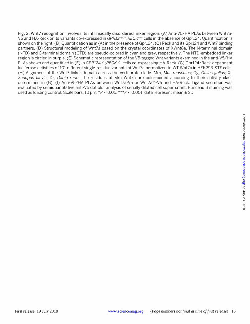
binding. We generated a collection of HA-tagged single-do-main deletion variants of Reck and, after determining that all variants reached the plasma membrane (fig. S6), quantified Wnt7 PLA interaction signals. This analysis revealed that the N-terminal cysteine-knot domain (CK), and more particularly CK4 and CK5, were required for binding (Fig. 2A). Consistent with these results, ReckΔCK4 was also inactive in competition assays (Fig. 1G).
Reck function in Wnt signaling is known to rely on its ca-pacity to form a complex with Gpr124 through its CK domain (20, 22), which we show to be required for Wnt7a binding. Although Gpr124 alone did not bind Wnt7 (Fig. 1, B, D, and G), we detected a 4-fold increase in Wnt7a-V5/HA-Reck PLA signals upon co-expression of untagged Gpr124 (Fig. 2B). In sum, these results suggest that Reck is a Fz-independent Wnt receptor, whose specific and exclusive binding to Wnt7 is re-inforced by the interaction of Gpr124 with its CK domain (Fig. 2C) (24).
In order to better characterize the Reck/Wnt7 interaction, we modeled the 3D structure of Wnt7a based on Xenopus Wnt8a crystallographic analysis (Fig. 2D) (13). Wnt ligands adopt a two-domain structure reminiscent of a human hand pinching the globular Fz cysteine-rich domain (CRD, orange) via the palmitoylated ‘thumb’ of their N-terminal domain
(NTD, cyan) and hydrophobic residues of their ‘index’ C-ter-minal domain (CTD, grey). The structures involved in Fz binding are pseudo-colored in orange in Fig. 2D. The two do-mains are connected through a flexible linker region of the NTD (purple).
We next generated a collection of Wnt7 variants deleted for specific domains or residues, or that carried domains from other Wnt ligands (Fig. 2E). Of note, all analyzed Wnt7 variants could be detected in the cell supernatant (fig. S7). These ligands were each applied to PLAs, and the resulting data showed that Reck binding occurs through the Wnt7a NTD. Wnt7aNTD indeed bound Reck, whereas Wnt7aCTD did not (Fig. 2F). Chimeric ligands made of Wnt7aNTD fused to the CTDs of Xenopus Wnt8a (XWnt8a), murine Wnt4 or Wnt16 were also positive. The palmitoleic acid required for Fz bind-ing was dispensable for the interaction with Reck as revealed by testing a Wnt7aS206A palmitoylation mutant. Altogether, these binding assays reveal that Reck discriminates between Wnt ligands by recognizing a motif embedded in the Wnt7a NTD, at sites distinct from those engaged by Fz.
Smaller NTD variants lacking the linker region, Wnt7a1-212 and Wnt71-237, did not bind Reck. This mutational analysis and the spatial segregation of the Wnt7a linker region from the Fz binding sites, suggest that Reck decodes Wnt7a, at least in part, through this linker. The linker, which is highly diver-gent amongst the different Wnt ligands, exhibits strong evo-lutionary conservation amongst the vertebrate Wnt7 orthologs (Fig. 2H and fig. S8). This region is predicted to be intrinsically disordered (fig. S9), a feature often found in mo-lecular recognition elements providing the necessary struc-tural plasticity to accommodate multiple partners, post-translational modifications, or moonlighting functions. In many cases, intrinsically disordered regions also provide in-teractions with high specificity and moderate-to-high affinity (25).
Although in situ substitution of the XWnt8a linker by the Wnt7 linker (XW8apWnt7a) did not yield detectable PLA signals, presenting the Wnt7 linker at the free N terminus of XWnt8a (pWnt7a-XW8a) was sufficient to confer Reck binding activ-ity to XWnt8a (Fig. 2, E and F). We hypothesize that confor-mational alteration accounts for the lack of binding of XW8apWnt7a. In support of this hypothesis, reciprocal exchange of Wnt linkers between various Wnts abrogates their activity (fig. S10).
To precisely map the Reck interaction site, we analyzed Gpr124/Reck-dependent STF signaling of 101 single residue variants of Wnt7a (Fig. 2G). The mutated residues correspond to surface-exposed NTD residues conserved between Wnt7a and Wnt7b, but not found in XWnt8a or other Wnt ligands. Residues were mutated to alanines, except for endogenous alanine residues which were changed to arginine. Although
on July 23, 2018
http://science.sciencemag.org/
Dow
nloaded from

First release: 19 July 2018 www.sciencemag.org (Page numbers not final at time of first release) 3
~80% of the variants were as active as WT Wnt7a (>70% rel-ative activity, green), eight Wnt7a variants (red) reduced Gpr124/Reck-dependent signaling to less than 10% (Fig. 2G). All but one (I37) critical residues clustered on the “top” or “back” of the predicted Wnt7a structure, with six mapping to the linker domain. All essential residues are strictly con-served amongst Wnt7 orthologs from fish to mammals and absent in other Wnts, including Wnt3a (Fig. 2H and fig. S8).
The linker domain of Wnt3a has been shown to be essen-tial for Wnt3a activity through Lrp6 binding (26). By analogy, the inactivity of the Wnt7 linker variants might therefore re-sult from defective binding to Lrp5/6, Reck or both. In line with a function in Reck binding, Wnt7a4A, a four-residue var-iant of Wnt7a (V241A/F251A/L252A/K262A) within the linker region (Fig. 4H), showed reduced Reck PLA signals as com-pared to WT Wnt7a. Of note, this lower activity occurred de-spite slightly improved secretion rates (Fig. 2I).
Biophysical characterization of the interaction be-
tween the Wnt7 linker domain and Reck To investigate Reck-Wnt7 binding in a cell-free system,
the CK domain of Reck (and variants thereof) were fused to the Fc domain of human IgG1. Fusion proteins were purified from HEK293T cells supernatants (Fig. 3A) and then titrated with synthetic pWnt7a and pWnt7b linker peptides by iso-thermal titration calorimetry (ITC). pWnt7a and pWnt7b bound Reck with affinity values of 7 μM and 1.2 μM, respec-tively (Fig. 3B and fig. S11). As controls, the synthetic peptides corresponding to Wnt7a4A (pWnt7a4A) as well as equivalent linker peptides of Wnt3a (pWnt3a) showed no binding to Reck-CK-Fc. pWnt7b binding required Reck CK4 and CK5, but not CK1, CK2 or CK3, mirroring the PLA results in cul-tured cells (Fig. 2A). To corroborate the results provided by the ITC analysis, we used single molecule force spectroscopy (SMFS) to measure binding affinities at the single-molecule level (Fig. 3C). Binding of pWnt7b to Reck-CK-Fc was detect-able with a measured Kd of 5 μM (Fig. 3, D to F). Despite the fundamental differences between the two techniques, ITC and SMFS thus provided a close match between measured binding affinity values.
Altogether, while not excluding an additional role of the Wnt7 linker in Lrp5/6 binding (26), these data demonstrate that Wnt7 is recognized by Reck at least in part through its ‘signature’ linker motif. The moderate micromolar affinity values measured further suggest that after recognition of the linker, Reck establishes more extensive contacts with Wnt7, in a process that can be potentiated by Gpr124 (Fig. 2B).
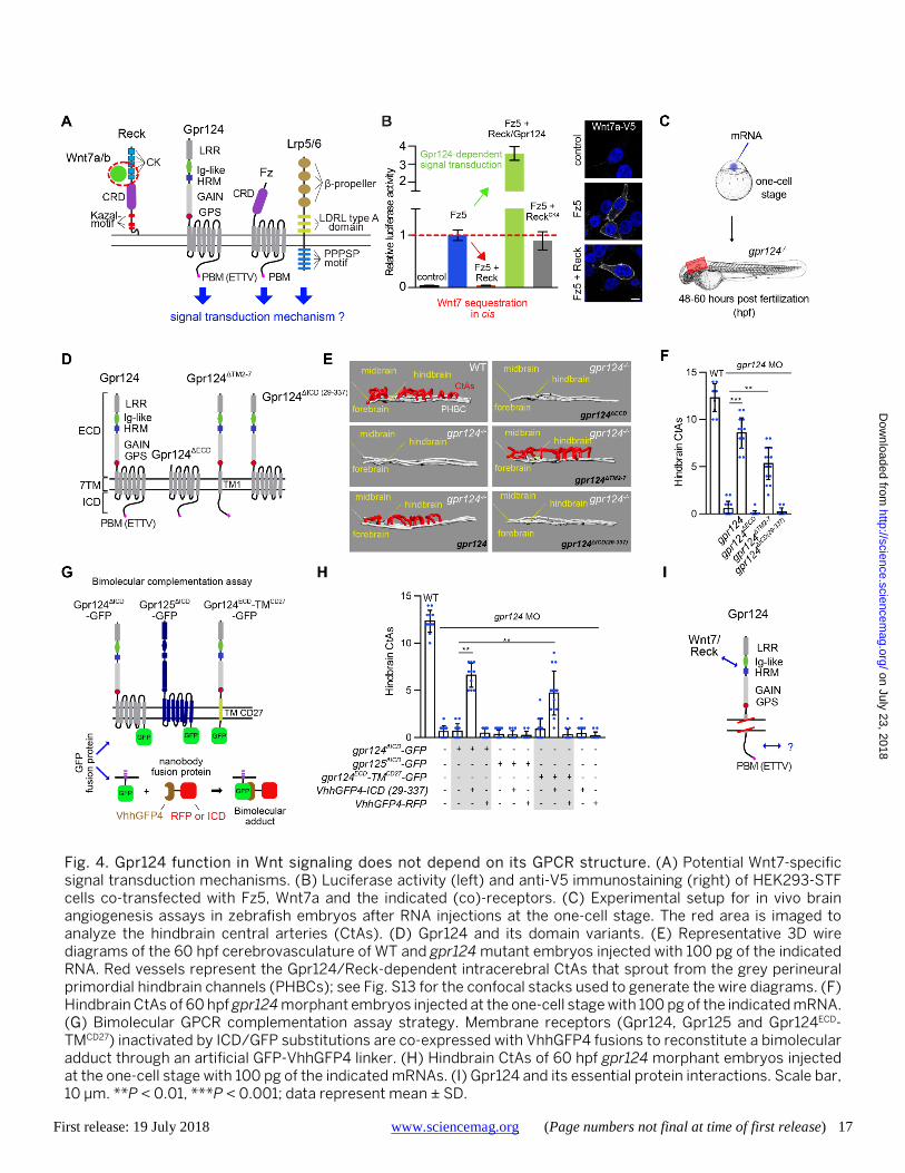
Gpr124 function in Wnt7 signaling does not depend
on its GPCR structure Reck, by virtue of its GPI-anchoring mode, has limited po-
tential to relay Wnt7 signals within the cell. Signal transduc-tion therefore likely relies on other components of the recep-tor complex, i.e. Gpr124 and/or Fz/Lrp5/6 (Fig. 4A).
To uncover the signal transduction mechanism, we first evaluated the functional relationship between Gpr124 and Fz/Lrp5/6 complexes in cultured cells. Using the “Fz-free” and “Lrp5/6-free” cells (Fig. 1A), we found that the function of Gpr124/Reck strictly relies on Fz and Lrp5/6 (fig. S12). We further established that their respective CRD and Dkk-1-sen-sitive Wnt ligand binding domains are essential, implying that Wnt7 binds and activates Fz/Lrp5/6 in a classical man-ner. If Reck mediates Wnt7 binding and Fz/Lrp5/6 trigger sig-naling, what underlies the essential function of Gpr124?
In the absence of Gpr124, Reck acts cell-autonomously as a potent inhibitor of Wnt7/Fz5 signaling, in a CK4-dependent manner (Fig. 4B). The inhibitory function of stand-alone Reck is particularly remarkable in light of the pool of membrane-associated Wnt7 in this Frizzled-positive setting (Fig. 4B). This observation implies that in the absence of Gpr124, Reck scavenges Wnt7 away from Fz5/Lrp5/6 complexes. Strikingly, the presence of Gpr124 switches Wnt7 signaling output from near complete inhibition to potent activation.
To establish how Gpr124 mediates this “on-off” Wnt7 sig-naling switch, we turned to the zebrafish model. In many Wnt-controlled processes, including the Wnt7/Gpr124/Reck-mediated cerebrovascular functions, Wnt input levels are only marginally above the minimal threshold values required for signaling (20). It is therefore important to investigate the signal transduction pathway in vivo, in response to physio-logical Wnt7 inputs. The development of the zebrafish brain vasculature requires Reck/Gpr124 signaling, in a process of angiogenic sprouting that can readily be quantified. It there-fore constitutes an ideal setting to perform structure-function analysis in vivo (22, 27). Using mRNA injections into one-cell stage gpr124−/− embryos (Fig. 4C), we evaluated the activity of three Gpr124 variants lacking the N-terminal extracellular part (Gpr124ΔECD), the seven-span moiety (Gpr124ΔTM2-7) or the C-terminal cytoplasmic extension [Gpr124ΔICD, residues 29-337 (Fig. 5E)] (Fig. 4D). Although ectopic expression of Gpr124ΔECD or Gpr124ΔICD did not restore brain angiogenesis in gpr124 mutants or morphants, Gpr124ΔTM2-7 was sufficient to trigger brain angiogenesis in vivo (Fig. 4, E and F, and fig. S13) and Wnt/β-catenin activity in vitro (fig. S14).
This retained competence of Gpr124ΔTM2-7 was unexpected: Gpr124 is a GPCR, a receptor super-family classically relaying extracellular stimuli by ligand-induced conformational re-modeling of their seven transmembrane spans, which are ab-sent in the engineered Gpr124ΔTM2-7. These data raise the possibility that Gpr124 does not act as a “classical” GPCR when promoting Wnt7 signaling.
To test this hypothesis, we developed a bimolecular com-plementation assay in which the GPR124 ECD and ICD are
on July 23, 2018
http://science.sciencemag.org/
Dow
nloaded from
First release: 19 July 2018 www.sciencemag.org (Page numbers not final at time of first release) 4
linked by a surrogate anti-GFP VhhGFP4 nanobody-GFP con-nector (Fig. 4G). Nanobodies are single-domain antibody fragments that have been used to (mis)-rout intracellular pro-teins (28). We re-purposed them here as conditional tethers for signal transduction analysis. The highly-flexible GFP-VhhGFP4 connector acts a buffering module ensuring that conformational information cannot be exchanged between tethered partners. Based on this idea, we designed Gpr124 ΔICD-GFP and Gpr125 ΔICD-GFP fusions to which VhhGFP4-fu-sions will be recruited (Fig. 4G and fig. S15). Notably, the gpr124 vascular phenotypes were partially suppressed by co-injecting mRNAs encoding Gpr124ΔICD-GFP and VhhGFP4-ICD (residues 29-337) (Fig. 4H). Moreover, a chimeric Gpr124 ECD linked to cytoplasmic GFP via the transmembrane span of the unrelated CD27 receptor was similarly active with VhhGFP4-ICD (Fig. 4, G and H). We used Gpr125, a closely related aGPCR devoid of angiogenic activity, as well as VhhGFP4-RFP as negative controls (20–22).
These results confirm that Gpr124 function in Wnt7 sig-naling does not require signal transduction across the mem-brane through conformational remodeling. Instead, Gpr124 seemingly acts in this module as a signaling-deficient trans-membrane protein whose activity relies on its Reck-binding ECD (22, 24) and its conformationally uncoupled ICD. The function of this latter domain remains to be defined (Fig. 4I).
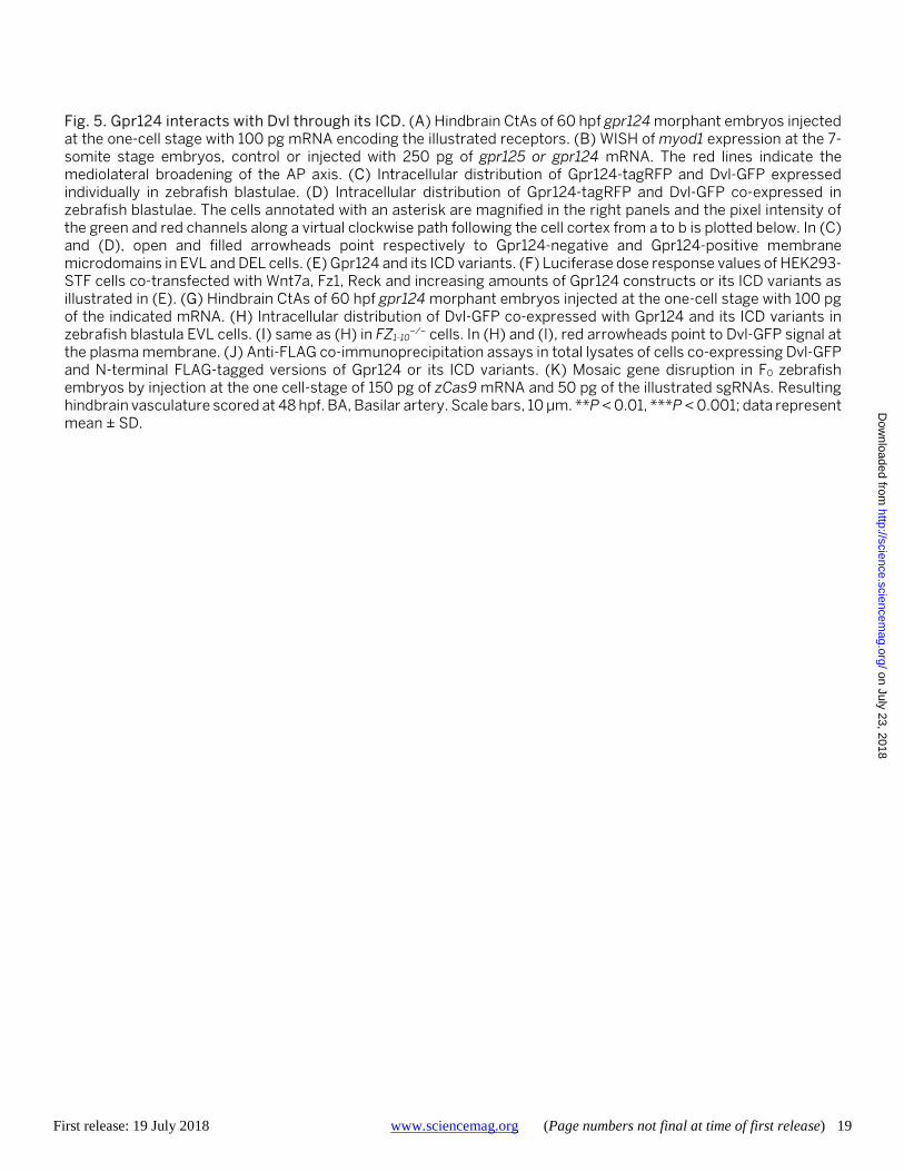
Gpr124 activity requires Dishevelled binding We hypothesized that the Gpr124 ICD might operate
through Dishevelled (Dvl), the necessary effector of Wnt sig-naling which interacts with Fz. This ‘Dvl hypothesis’ is rooted in the findings that Gpr125 physically interacts with Dvl via its C-terminal ICD domain (29) and that Gpr124/125 hybrids in which the ICD of Gpr124 is replaced with the ICD of Gpr125 are able to promote brain angiogenesis (Fig. 5A) (22). Gpr124/Fz2 hybrids (Gpr124ICDFz2) harboring the Fz2 ICD, which is known to bind Dvl, were similarly active. In contrast, full-length Fz2 was not. Notably, the activity of Gpr124ICDFz2 was dependent on its KTxxxW and ETTV Dvl binding motifs (Fig. 5A).
Moreover, in a non-canonical Wnt signaling context, over-expression of Gpr125 impairs convergence and extension movements during zebrafish gastrulation, resulting in a me-diolateral broadening of the AP embryonic axis as well as syn-ophthalmia or cyclopia (29). These phenotypes were linked to the capacity of Gpr125 to modulate Dvl distribution through its ICD. Gpr124 strictly mimicked Gpr125 in these settings (Fig. 5B and fig. S16).
We therefore tested whether Gpr124 affects the intracel-lular distribution of Dvl. We used two distinct cell popula-tions of the zebrafish blastula to address this question: the superficial enveloping cell layer (EVL), where cells maintain continuous intercellular contacts, and the deep layer cells
(DEL), which establish more discrete intercellular junctions (Fig. 5C). Gpr124-tagRFP distribution reflects this differing junctional organization, with near-uniform and discontinu-ous membrane signals in EVL and discontinuous signals in DEL cells (Fig. 5C). When expressed individually, Xenopus Dvl-GFP mainly formed cytoplasmic punctae in both cell pop-ulations, as reported previously (29). Co-expressing Gpr124-tagRFP largely redistributed Dvl-GFP to the Gpr124-positive membrane subdomains in both EVL and DEL cells, where the proteins co-localized (Fig. 5D).
We generated a collection of Gpr124 truncation variants with a range of deletions within the 337 amino acid-long ICD (Fig. 5E), and evaluated their Wnt7/β-catenin functions in vitro (Fig. 5F) and in vivo (Fig. 5G). The Gpr124 and Gpr125 ICDs contain no obvious motifs except for the last four ETTV amino acids that constitute a canonical PDZ binding motif (PBM), also found in a subset of Fz receptors. The ETTV tetrapeptide contributed, but was not essential, for Gpr124 activity. It was similarly dispensable for high-dose Gpr124 or Gpr125-induced planar cell polarity phenotypes (fig. S16) (29). Analysis of increasingly larger C-terminal deletion vari-ants mapped the essential region of the Gpr124 ICD to the interval spanning residues 81 to 213. Notably, the activity of the different ICD variants exactly matched their capacity to recruit Dvl-GFP in EVL cells (Fig. 5H) and FZ1-10
−/− cells (Fig. 5I). This interaction between Gpr124 and Dvl could also be detected by co-immunoprecipitation. In these assays, only Gpr124 variants harboring the 81-213 region interacted with exogenous Dvl-GFP (Fig. 5J) or endogenous DVL2 (fig. S17) in FZ1-10
−/− cells. This interaction is likely direct as purified re-combinant GST-Gpr124-ICD or GST-Gpr124-ICDΔETTV fusion proteins were able to pull-down in vitro translated Dvl-GFP (fig. S18), as previously reported for Gpr125 (29).
To test for the endogenous requirement for Gpr124 ICD interaction with Dvl, we performed gene disruption experi-ments in somatic zebrafish embryos by co-injecting zCas9 mRNA and sgRNAs targeting the Gpr124 coding sequence im-mediately upstream, within or downstream of the Dvl bind-ing region (ICD residues 81-213). Although the injection of four out of five sgRNAs predicted to disrupt Dvl recruitment to Gpr124 generated embryos lacking hindbrain CtAs with a penetrance ranging from 13.6 to 70.8%, none of the seven sgRNAs targeting Gpr124 downstream of the Dvl binding re-gion generated significant brain vascular defects (Fig. 5K).
Taken together, these experiments identify Dvl as a Gpr124 binding partner that could mediate its Wnt7 signaling activities at the plasma membrane. Unlike Fz, Gpr124-medi-ated recruitment of Dvl at the plasma membrane did not yield a detectable increase in phosphorylated Dvl levels, an early indicator of Wnt signaling activation upstream of β-catenin stabilization (fig. S19) (30, 31). This absence of
on July 23, 2018
http://science.sciencemag.org/
Dow
nloaded from
First release: 19 July 2018 www.sciencemag.org (Page numbers not final at time of first release) 5
Gpr124-induced Dvl activation is consistent with the experi-ments shown in Fig. 4, D to I, that showed Gpr124 to be a conformationaly inert Wnt7 signaling mediator.
Dvl polymers assemble ligand-specific Wnt signal-
osomes by linking Gpr124 and Fz Knockdown of DVL2 by siRNA impairs Gpr124/Reck-me-
diated signaling (fig. S20). As Dvl is an essential adaptor of Fz, and Gpr124/Reck signaling relies on Fz (fig. S12), such cell-wide loss-of-function approaches are however of limited value to probe Dvl function specifically as a Gpr124 (and not Fz) effector. We therefore used the nanobody strategy de-scribed in Fig. 4G to selectively modulate Dvl binding to Gpr124. Strikingly, VhhGFP4-mediated recruitment of Dvl to Gpr124ΔICD-GFP, but not to Gpr125ΔICD-GFP, was sufficient to partially reverse the gpr124 mutant vascular phenotype in vivo (Fig. 6A). As additional control, injecting either compo-nent alone or substituting VhhGFP4-Dvl with VhhGFP4-RFP did not rescue brain angiogenesis. These experiments reveal that Dvl is sufficient to mediate Gpr124 intracellular func-tions in Wnt7 signaling.
Gpr124, Reck, and Fz/Lrp5/6 have been reported to form higher-order receptor complexes (24). We reasoned that the Gpr124 ICD might assemble this complex via Dvl. Dvl mole-cules indeed assemble signalosomes through dynamic polymerization (32–34). As Dvl physically interacts with both Gpr124 and Fz, Gpr124 and the associated Reck-bound Wnt7 might thus become trapped in dynamic Wnt signalosomes, thereby increasing the local concentration of Wnt7 ligands available for Fz signaling.
Wnt signalosomes are readily detected by light micros-copy as large, punctate structures enriched in Dvl that form at or below the plasma membrane (33–35). To determine whether Fz and Gpr124 co-distribute in Wnt signalosomes in a Dvl-dependent manner, the localization of individually ex-pressed Fz-GFP and Gpr124-tagRFP was first examined in DEL cells. Fz4 decorated the entire plasma membrane pe-riphery while, in contrast, Gpr124-tagRFP accumulated at cel-lular contacts (Figs. 5, C and D, and 6B). This differential membrane localization was retained upon Dvl expression (Fig. 6B). Consistent with their Dvl binding capacity, both re-ceptors recruited Dvl-GFP from the cytoplasm (Fig. 5C) to their respective membrane compartments (Fig. 6C). How-ever, when Gpr124-tagRFP and Fz4-GFP were co-expressed, Fz4-GFP quantitatively relocalized, in a Dvl-dependent man-ner, to the Gpr124-positive intercellular junctions (Fig. 6D). Gpr124-tagRFP and Fz4-GFP colocalized in Wnt signalosome-reminiscent punctate structures that were particularly evi-dent at EVL cell membranes (Fig. 6E and fig. S21).
We used bimolecular fluorescence complementation as an additional assay to test for Dvl-dependent Fz/Gpr124 interac-tion in DEL cells. Co-injection of Gpr124-VN155 (I152L) and
Fz1-VC155 indeed generated bright junctional signals in a Dvl-dependent manner (Fig. 6F), demonstrating that Fz and Gpr124 indirectly interact via the Dvl scaffold protein. Alto-gether, these data provide a molecular mechanism for spatial enrichment of Wnt7 within Fz/Lrp5/6 signalosomes, permit-ting potentiated and ligand-selective cellular responses (Fig. 6G).
Discussion In summary, this work provides mechanistic insights onto
the Wnt decoding capacities of vertebrate cells. It also demonstrates that the evolutionarily constrained Wnt struc-ture retained enough diversity to allow ligand-specific cellu-lar responses, a property so far thought to require structurally unrelated Frizzled ligands like Norrin (36).
These structural insights into Wnt evolution and function suggest that additional Wnt decoding modules exist, enabling fine-tuning of cellular behaviors in response to other Wnt or Fz family members. The discrete interaction mode of Fz and Wnt leaves large surfaces made of evolutionary conserved residues available to accommodate additional co-receptors. We therefore propose that Wnt decoding modules might have contributed to shaping the evolution of the Wnt ligand fam-ily.
The benefit to promiscuous Wnt/Fz interactions with specificity conferred by accessory proteins rather than mono-specific Wnt/Fz interactions might lie in the increased mod-ularity offered by the binary system. A single-component sys-tem would be limited to an on or off signaling output. The two-component system described here can, context-de-pendently, achieve cell-autonomous Wnt signaling inhibition or act as a tunable rheostat amplifying the signaling output of specific Wnt ligands.
A salient molecular property of the Wnt7 module is the use of Dvl as a common Gpr124 and Fz adaptor. It is tempting to propose that taking advantage of Fz-associated scaffold proteins like Dvl could constitute a generic mechanism for Wnt/Fz modifiers. Accordingly, cells recurrently tailor their responses to Wnt by reshaping the molecular composition of the Dvl-associated proteins, including regulatory kinases, E3 ubiquitin ligases and components of the endocytic machinery (32–34).
Our findings have clinical implications. The pleiotropic functions of Wnt signaling in health and disease make this pathway a conspicuous yet intrinsically challenging thera-peutic target. Manipulating the Wnt/β-catenin pathway at the level of its cytosolic or nuclear components harbors high potential for systemic effects with undesirable outcomes across a range of tissues (37). Interventions focused on spe-cific Wnts, Fz receptors or other signaling components at the cell membrane might, in principle, be more selective and hence better positioned to lead to clinically viable strategies
on July 23, 2018
http://science.sciencemag.org/
Dow
nloaded from
First release: 19 July 2018 www.sciencemag.org (Page numbers not final at time of first release) 6
(4, 38–40). The mechanism uncovered here expands the pro-spects for specific Wnt-targeted interventions. In particular, by giving molecular insights at the heart of Wnt7-specific sig-naling, it provides an opportunity for the targeted treatment of human brain disorders with neurovascular involvement, including stroke and brain cancer (41).
Materials and Methods Zebrafish lines Zebrafish (Danio rerio) were maintained at 28°C on a 14
hours light/10 hours dark cycle. Embryos were obtained and raised under standard conditions in accordance with Euro-pean and national ethical and animal welfare guidelines (pro-tocol approval number: CEBEA-IBMM-2017-22:65). Staging was performed according to Kimmel et al. (42). The trans-genic and mutant lines used in this study are Tg(kdrl:EGFP)s843 (43), Tg(kdrl:HRAS-mCherry)s896 (44) and gpr124s984 (22).
Morpholinos, RNA constructs and microinjection Splice-blocking morpholinos targeting gpr124
(ACTGATATTGATTTAACTCACCACA) (22) were purchased from Gene Tools and injected at the one-cell stage at 2 ng. Synthetic mRNAs were transcribed from NotI linearized pCS2 using the mMessage mMachine SP6 Kit (Thermo Fisher Scientific) and injected into one-cell stage zebrafish embryos.
Somatic gene disruption in zebrafish by
CRISPR/Cas9 The gpr124ICD-targeting sgRNAs constructs (sgRNA 1-12)
were obtained by in vitro annealing of the following primers (1-Fo: TAGGTGGCATGCTACAAAAAGC and 1-Rv: AAACGCTTTTTGTAGCATGCCA; 2-Fo: TAGGTTTGGGTCCGTAGGTGGA and 2-Rv: AAACTCCACCTACGGACCCAAA; 3-Fo: TAGGGTAATGGTTTGGGTCCGT and 3-Rv: AAACACGGACCCAAACCATTAC; 4-Fo: TAGGAGAAGAAGAGTAATGGTT and 4-Rv: AAACAACCATTACTCTTCTTCT; 5-Fo: TAGGAAGCTTGTAGTAGAACCA and 5-Rv: AAACTGGTTCTACTACAAGCTT; 6-Fo: TAGGCCATTCTGGCCCTCTGAA and 6-Rv: AAACTTCAGAGGGCCAGAATGG; 7-Fo: TAGGCCAACGGGCGGCACAGAG and 7-Rv: AAACCTCTGTGCCGCCCGTTGG; 8-Fo: TAGGGGCACAGAGGGGAAGGAC and 8-Rv: AAACGTCCTTCCCCTCTGTGCC; 9-Fo: TAGGTAGTGATGGAGGTAGCAG and 9-Rv: AAACCTGCTACCTCCATCACTA; 10-Fo: TAGGTGCCTTCGGTAGCACGTA and 10-Rv: AAACTACGTGCTACCGAAGGCA; 11-Fo: TAGGGGTATGAAGAGCAGGGTA and 11-Rv:
AAACTACCCTGCTCTTCATACC; 12-Fo: TAGGTCGGTGGGTATGAAGAGC and 12-Rv: AAACGCTCTTCATACCCACCGA) and cloning into pT7-gRNA (Addgene #46759), as described in (45). sgRNA were tran-scribed from the BamHI linearized vector pT7-gRNA using the MEGAshortscript T7 kit (Thermo Fisher Scientific). The synthetic Cas9 mRNA was transcribed from the XbaI linear-ized vector pT3TS-nls-zCas9-nls (Addgene #46757) using the mMessage mMachine T3 Kit (Ambion). sgRNAs 1-12 (50 pg) and nls-zCas9-nls mRNA (150 pg) were injected into one-cell stage zebrafish embryos. The brain vasculature was analyzed and imaged at 48 hpf.
Whole-mount in situ hybridization (WISH) Digoxigenin-labeled antisense riboprobes were synthe-
sized with the digoxigenin (DIG) RNA labeling kit (Roche Di-agnostics GmbH). Embryos were fixed in 4% paraformaldehyde overnight at 4°C and whole-mount in situ hybridization was performed using digoxigenin-labeled myod1 riboprobes as previously described (46, 47).
Expression plasmid constructs Wnt, Frizzled, Reck and Gpr124 and their variants used in
STF and PLA assays were expressed from the CMV promoter of the pCS2 plasmid except for the following plasmids: pRK5-Lrp5-rhotag, Fz4-GFP (Addgene #42197), Fz1 (Addgene #42253) and Fz5 (Addgene #42267), all kindly provided by J. Nathans. Single-point mutation variants, deletions and chi-meras were generated using In-Fusion cloning (ST0345, Takara) and tandem overlapping PCR products. All con-structs were confirmed by Sanger sequencing. Throughout this study, we used Xenopus Dvl-GFP (Addgene #16788, from the R. Moon laboratory), zebrafish Gpr124 and Gpr125 as well as mouse Reck and untagged Wnt ligands (22). The collection of active human Wnt-V5 ligands was kindly provided by the Xi He laboratory via Addgene (#43807 to #43825) (48). The activity of the V5-tagged ligands was verified by STF assays (fig. S22). Reck deletion variants correspond to the following amino acids of mouse Reck (NP_057887.2): ReckΔCK1: 46-93; ReckΔCK2: 113-150; ReckΔCK3: 160-206; ReckΔCK4: 225-272; ReckΔCK5: 301-347; ReckΔCK1-5: 46-347; ReckΔCRD: 352-484 and ReckΔKAZAL: 636-798. Gpr124 deletion variants correspond to the following amino acids of zebrafish Gpr124 (NP_001305000.1): Gpr124ΔECD: 68-725 and Gpr124ΔTM2-7: 764-1030. The Gpr124 ICD used in deletion, swapping or fusion constructs corresponds to residues 1058-1367 of the full length protein (28-337 of the ICD), unless otherwise indi-cated. The ICD of Gpr124 is defined as the segment that fol-lows the last transmembrane segment of the seven pass transmembrane protein as non-ambiguously defined by the InterPro server of EMBL-EBI (based on the Phobius trans-membrane topology and signal peptide predictor from the
on July 23, 2018
http://science.sciencemag.org/
Dow
nloaded from

First release: 19 July 2018 www.sciencemag.org (Page numbers not final at time of first release) 7
Stockholm Bioinformatics Centre). The ICD of zebrafish Gpr125 (NP_001289153) and of zebrafish Fz2 (NP_571215.1) were similarly defined and correspond to the residues 1054-1346 and 526-550, respectively. Gpr124ECD-TMCD27-GFP was generated by replacing the 7-TM span of Gpr124 (residues 743-1030) by the TM of mouse CD27 (NP_001028298.1) (IFVTFSSMFLIFVLGAIL). The VhhGFP4-ICD construct was generated by fusing the Gpr124 ICD to the C terminus of VhhGFP4 [kindly provided by Markus Affolter (28)]. Gpr124ICDFz2ΔKTxxxW/ΔETTV was generated by deleting the KTxxxW (KTLHSW) and ETTV motifs within the ICD of zebrafish Fz2. The BiFC constructs were subcloned from pBiFC-VC155 (Addgene #22011) and pBiFC-VN155 (I152L) (Addgene #27097). The HA tag was inserted after residue 22 in murine Reck, residue 47 in zebrafish Gpr124 and residue 22 in murine Fz5. The FLAG tag was inserted after residue 47 in zebrafish Gpr124. We thank the numerous colleagues that facilitated this work by depositing their plasmids at Addgene.
Cell culture and HEK293(T) mutant cell lines HEK293T cells were obtained from ATCC (CRL-3216) and
the HEK293 STF cell line was kindly provided by J. Nathans. WT and mutant cells were cultured in DMEM/F12 medium (Lonza) supplemented with 10% fetal bovine serum and maintained at 37°C in a humidified incubator equilibrated with 5% CO2. GPR124 and RECK were genetically inactivated using CRISPR/Cas9 approaches in HEK293 STF cells and LRP5, LRP6 and FZ were inactivated in HEK293T cells. The mutant cell lines were obtained by the iterative CRISPR/Cas9-mediated mutagenesis strategy illustrated in Fig. 1A. CRISPR/Cas9 guide sequences were designed using the http://crispr.mit.edu/ website and were cloned into pSpCas9(BB)-2A-GFP (40). The top 1% of GFP+ cells was iso-lated by FACS (AriaIII, BD Biosciences) 48 hours after trans-fection and distributed in 96-well plates for clonal expansion. In order to facilitate the genetic characterization of the mu-tant cell lines, clones generating multiple-peak derivative melt curves in high-resolution melt analyses were first coun-ter-selected. For each gene, two independent PCR products flanking the target site (300-1000 bp PCR products centered on the PAM site) were gel purified and characterized by Sanger sequencing on both strands. The absence of WT al-leles in the uncloned PCR product was verified and the num-ber of different edited alleles (1, 2 or 3 in hypotriploid HEK293 cells) was deduced from the complexity of the chro-matograms (fig. S1). For each gene, the two independent PCR products were then cloned in pCRTM-Blunt II-TOPO® (Thermo Fisher Scientific) and individual colonies were sequenced un-til each edited allele was sequenced at least twice from each individual PCR amplicon. The number of analyzed colonies for the FZ1-10
−/− cell line is provided in table S1. The sequencing chromatograms form the identified individual clones were
then aligned to ensure that the allelic complexity of the un-cloned PCR can exhaustively be accounted for by the identi-fied alleles (fig. S1). Finally, to exclude allelic exclusion by PCR amplification bias, the absence of WT alleles in the FZ1-10
−/− cell line was verified by whole exome sequencing. STF dual luciferase assay Cells were plated into 96-well plates and transfected after
24 hours in triplicate with Lipofectamine 2000 (Thermo Fisher Scientific). The amount of plasmid DNA transfected per well was optimized for each expression vector as follows: Renilla luciferase (0.5 ng), Wnt ligands (20 ng), Fz receptors (5 ng), Lrp5 (2.5 ng), Gpr124 (10 ng), Reck (5 ng) and Dkk-1 (10 ng) unless otherwise indicated. The total amount of DNA was adjusted to 100 ng per well with the empty pCS2 vector. Dual luciferase assays were performed using the STF cell line or by co-transfecting 20 ng of M50 Super 8x TOPFlash plas-mid (Addgene #12456). Cells were harvested in passive lysis buffer (E1980, Promega) and the activities of the Firefly and Renilla luciferases were measured sequentially using the Dual-Luciferase Reporter Assay system (E1980, Promega) 48 hours post transfection. The competition assays in Fig. 1G and fig. S5 were performed by plating cells as a 1:1:1 mixture at 90% confluency in 96-well plates 24 hours after transfec-tion. Luciferase activity was measured 24 hours after co-cul-ture. For siRNA, cells were plated into 96-well plates and transfected after 24 hours in triplicate using Lipofectamine 2000 (Thermo Fisher Scientific) with 1 pmol (per well) of Dvl2 or control siRNAs (SASI_HS01_00104204 and SIC001, Sigma-Aldrich) together with the indicated plasmids.
Immunofluorescence and proximity ligation assay Cells were grown in glass-coated chambers (IBIDI) and
transfected after 24 hours with Lipofectamine 2000 (Thermo Fisher Scientific). Cells were fixed with 4% paraformaldehyde for 10 min at room temperature (RT) 48 hours post transfec-tion. For immunofluorescence staining (IF), cells were blocked in 1% BSA-PBS for 30 min before being exposed to primary antibodies for 1 hour at RT. After three PBS washes, cells were incubated with secondary antibodies for 1 hour at RT. For anti-V5 staining of Wnt ligands, cells were addition-ally washed for 10 min in PBS 0.1% Tween 20 before incuba-tion with the secondary antibody solution. For the proximity ligation assay (PLA) (Sigma-Aldrich), cells were blocked for 30 min at 37°C with the Blocking solution provided by the manufacturer before being incubated with the primary anti-bodies for 1 hour at RT. Cells were washed three times and incubated with the PLA probes anti-rabbit PLUS and anti-mouse MINUS for 1 hour at 37°C. After two PBS washes, cells were incubated with the Duolink Ligation solution for 30 min at 37°C. After two PBS washes, cells were incubated with the
on July 23, 2018
http://science.sciencemag.org/
Dow
nloaded from
First release: 19 July 2018 www.sciencemag.org (Page numbers not final at time of first release) 8
Duolink Amplification solution for 100 min at 37°C. The fol-lowing antibodies were used: monoclonal mouse anti-V5 (R96025, Thermo Fisher Scientific) at 1:500 for IF and PLA, purified polyclonal rabbit anti-HA (H6908, Sigma-Aldrich) at 1:400 for IF and PLA and anti-mouse Alexa488-conjugated secondary antibody (Thermo Fisher Scientific) at 1:5000. Cells were stained for 2 min with Hoechst diluted to 10 μg ml−1 in PBS.
Western blot, Dot blot, GST pull-down and co-im-
munoprecipitation The following antibodies were used with an overnight in-
cubation at 4°C: rabbit anti-DVL2 (1:1000, #3224S, Cell Sig-naling Technology), rabbit anti-DVL2-phospho T224 (1:1000, ab124941, Abcam) and mouse monoclonal anti-β-actin-perox-idase (1:50000, A3854, Sigma-Aldrich), chicken anti-GFP (1:5000, GFP-1010, Aves) and mouse monoclonal anti-FLAG M2 (1:1000, F1804, Sigma-Aldrich).
Dot blot analyses were performed according to manufac-turer’s protocol with a BioDot SF apparatus (Bio-Rad). Serial dilutions of supernatant were spotted onto a nitrocellulose membrane (GE Healthcare). After drying, the membrane was incubated with the antibodies as described above. For the co-immunoprecipitations assays, HEK293T were collected 48 hours after transfection from six-well plates and resuspended after two PBS washes in lysis buffer [150 mM NaCl, 25 mM Tris (pH 7.5) and 1% IGEPAL CA-630 (Sigma-Aldrich)] con-taining EDTA-free protease inhibitor cocktail (Roche) for 30 min at 4°C. After centrifugation at 20000 g for 10 min, the supernatant was incubated with 20 μl of anti-FLAG M2 affin-ity gel (A2220, Sigma-Aldrich) overnight at 4°C. Beads were washed five times with the lysis buffer and boiled in 2x Laemmli Sample buffer. For GST pull-downs, the coding se-quence of Gpr124 ICD and its ∆ETTV variant were fused downstream of GST sequences into pGEX-6P1 (GE Healthcare). The GST-fusion proteins were induced in Esche-richia coli BL21 by exposure to 0.1 mM of IPTG in LB medium at 37°C for 4 hours. Cells were subsequently lysed in a cell disruptor (M110S, Microfluidics) and after centrifugation, the GST-fusion protein from the supernatant were immobilized on Glutathione Sepharose beads (17-0756-01, GE Healthcare). The Dvl-GFP and GFP constructs were transcripted using the mMESSAGE mMACHINE SP6 Transcription Kit (Thermo Fisher Scientific) and in vitro translated using the Rabbit Re-ticulocyte Lysate System provided by Promega (L4960) in the presence of S35-methionine (NEG009T001MC, PerkinElmer). The beads were incubated with the radiolabeled proteins in PBS for 3 hours at 4°C with gentle mixing and then washed five times with cold PBS. The bound complex was resus-pended in 2x Laemmli buffer, separated by SDS-page and an-alyzed by fluorography.
Microscopy and images processing Cells and zebrafish embryos were imaged with a LSM710
confocal microscope and images were processed in ImageJ. Images of eye fusion phenotypes were taken on a Leica M165 FC. Brain vasculature renderings were generated using Imaris software (BitPlane).
The following method was used for the quantification of the PLA signal: The in situ PLA signals are seen as bright flu-orescent dots of characteristic appearance. The signal lined the cell surface, as expected given the membrane localization of the receptors. For quantification, several images were ac-quired on LSM710 confocal microscope with a 20x objective. ImageJ was used to generate binary images by global thresh-olding and to determine the ratio of the total area occupied by the PLA-positive pixels to the DAPI-positive pixels, the lat-ter being indicative of the number of cells in the field of view. The threshold values were manually adjusted to reflect the nuclear localization of DAPI and the focal membrane PLA signals. Identical threshold values were used for all images. On the dot plot, each dot represents the ratio of one image. Each image typically contained 50-100 cells.
Structural modeling The structure of Xenopus Wnt8a (PDB ID: 4F0A) (13) was
used as a starting model for Wnt7a. Missing residues and sub-stitutions were modeled using the program Modeller (49). The modeling strategy included accounting for existing disul-phide bridges as observed in XWnt8a. The initial best models were subjected to a conjugate-gradient energy minimization in vacuum with the Cα restrained and then freed in a second minimization step. These models were then embedded in a water box and electric neutrality was achieved by adding Na+ counter ions at 150 mM. The whole system was again energy minimized in 3000 steps. The molecular dynamics simulation was carried out for 0.5 ns with the program NAMD 2.7 at con-stant temperature (310 K) and constant pressure (1 atm), with periodic boundaries and using CHARMM36 as force field (50). A time step of 2 fs was used to integrate the equations of motion. The short-range interactions were cut at 12 Å and the smooth-particle mesh Ewald method was used to calcu-late electrostatic interactions. Hydrogen atoms were con-strained using the SHAKE algorithm. The resulting model is an average representation of the stable simulation.
Recombinant Reck-Fc fusions and synthetic pep-
tides The HA-tagged CK domain of Reck and its individual CK
motif deletion variants were fused at the N terminus of the Fc region of human IgG1. The fusion vector was kindly pro-vided by J. Nathans (24). Fc fusion proteins were recovered in serum-free FreeStyle 293 Expression Medium (Thermo
on July 23, 2018
http://science.sciencemag.org/
Dow
nloaded from

First release: 19 July 2018 www.sciencemag.org (Page numbers not final at time of first release) 9
Fisher Scientific) from the supernatant of HEK293T cell cul-tures 72 hours post transfection with Lipofectamine 2000 (Thermo Fisher Scientific). After collection, the supernatants were submitted to Protein G affinity purification (Protein G Sepharose 4 Fast Flow, Sigma-Aldrich). After acidic elution, protein purity was assessed by Coomassie Blue staining (Fig. 3A). Synthetic Wnt linker peptides were obtained from Chinapeptide Co., Ltd at purities of over 90%.
Isothermal titration Calorimetry ITC titrations were carried out on an Affinity ITC (TA In-
struments). Prior to the measurement, Reck-CK-Fc and all Reck-CK-Fc fusions were dialyzed to Tris-NaCl buffer (50 mM Tris pH8, 300 mM NaCl). In each case Wnt-derived peptides were prepared with buffer from the last step of protein dial-ysis. The samples were filtered and degassed before being ex-amined in the calorimeter and the titrations were performed at 25°C. All the experiments consisted of injection of constant volumes of 2 μL of titrant into the cell (200 μL) with a stirring rate of 75 rpm. Nominal sample concentrations were between 20 μM and 40 μM in the cell and 400 μM to 1.0 mM in the syringe. Actual sample concentrations were determined after dialysis or buffer exchange by measurement of their absorp-tion at 280 nm or by the BCA method. All data were analyzed using the MicroCal Origin ITC 7.0 and NanoAnalyze software packages.
Atomic force microscopy Glass coverslips coated with a thin gold layer were cleaned
in an ultraviolet radiation and ozone (UV-O) cleaner (Jetlight) for 15 min and subsequently immersed overnight in an ethanol solution containing 1 mM 16-mercaptododecahex-anoic acid and 1-mercapto-1-undecanol at a 1:99 volumetric ratio. Substrates were then rinsed with ethanol, dried with N2 and added to a solution containing equal volumes of 20 mg ml−1 N-hydroxysuccinimide (NHS) and 50 mg ml−1 1-ethyl-3-(3-dimethylaminopropyl)-carbodiimide (EDC) for 30 min. The obtained NHS activated surfaces were rinsed with ul-trapure water and incubated with 100 μl of a 10 μg ml−1 Pro-tein G solution for 1 hour at RT. Samples were then washed with washing buffer (3 × 5 min) and further incubated with 100 μl blocking buffer for 1 hour at RT. Finally, 50 μl of a 0.2 μg ml−1 Reck-CK-Fc solution was added to the substrates for 1 hour, rinsed with washing buffer and subsequently used for AFM experiments. Single-molecule force spectroscopy (SMFS) measurements were performed in PBS buffer at RT using a Nanoscope VIII Multimode AFM (Bruker). Triangular AFM cantilevers (MSCT, Bruker) with silicon nitride tips and a nominal spring constant between 0.01-0.06 N m−1 were used. Cantilevers were calibrated at the end of each experi-ment using the thermal noise method (51). Functionalized
tips were derivatized using a NHS-PEG27-Malemide linker fol-lowing a protocol described elsewhere (52) to covalently at-tach the Pep7b linking the cysteine residue added at the C terminus. After functionalization, the cantilevers were washed with PBS (3 × 5 min) and stored in individual wells of a multiwell dish containing 2 ml of PBS per well at 4°C until used in AFM experiments. Force-distance curves were recorded as 32x32 pixels arrays over 1 × 1 μm2 areas, using an applied force of 250 pN, a contact time of 0.25 s and a con-stant approach and retraction speed of 1 μm s−1. For dynamic force spectroscopy measurements, the retraction speed of the cantilever was varied as follows: 20 nm s−1, 100 nm s−1, 200 nm s−1, 1 μm s−1, 2 μm s−1, 10 μm s−1 and 20 μm s−1. Typically, at least 2000 force-distance curves were performed for each cantilever at a particular retraction speed. The collected data were analyzed using the Nanoscope Analysis software (Bruker). The retraction segment of each curve was analyzed and unbinding events were considered as specific if they oc-curred at a distance between 5-50 nm from the contact point. The minimum adhesion force was further used to calculate binding probabilities and build force distribution histograms. To reconstruct the energy landscape of the measured interac-tions, loading rates were calculated from the force vs time curve, as the slope of the adhesion event before the tip canti-lever jumps off to surface. The dependency of the force with the loading rate was then plotted in dynamic force spectros-copy plots.
Statistical analysis Statistical analysis was performed using GraphPad soft-
ware. Data represent mean ± SD. p-values were calculated by the one-way ANOVA (post hoc Dunnett’s test) and Student’s t test for multiple and single comparisons of normally distrib-uted data (STF and PLA assays) and by the Kruskal–Wallis (post hoc Dunn's test) for multiple comparisons of non-nor-mally distributed data (CtA quantifications); *P < 0.05; **P < 0.01; ***P < 0.001.
REFERENCES AND NOTES 1. C. Y. Logan, R. Nusse, The Wnt signaling pathway in development and disease. Annu.
Rev. Cell Dev. Biol. 20, 781–810 (2004). doi:10.1146/annurev.cellbio.20.010403.113126 Medline
2. R. Nusse, H. E. Varmus, Wnt genes. Cell 69, 1073–1087 (1992). doi:10.1016/0092-8674(92)90630-U Medline
3. P. Bhanot, M. Brink, C. H. Samos, J.-C. Hsieh, Y. Wang, J. P. Macke, D. Andrew, J. Nathans, R. Nusse, A new member of the frizzled family from Drosophila functions as a Wingless receptor. Nature 382, 225–230 (1996). doi:10.1038/382225a0 Medline
4. C. Niehrs, The complex world of WNT receptor signalling. Nat. Rev. Mol. Cell Biol. 13, 767–779 (2012). doi:10.1038/nrm3470 Medline
5. B. T. MacDonald, X. He, Frizzled and LRP5/6 receptors for Wnt/β-catenin signaling. Cold Spring Harb. Perspect. Biol. 4, a007880 (2012). doi:10.1101/cshperspect.a007880 Medline
on July 23, 2018
http://science.sciencemag.org/
Dow
nloaded from
First release: 19 July 2018 www.sciencemag.org (Page numbers not final at time of first release) 10
6. J. C. Hsieh, A. Rattner, P. M. Smallwood, J. Nathans, Biochemical characterization of Wnt-frizzled interactions using a soluble, biologically active vertebrate Wnt protein. Proc. Natl. Acad. Sci. U.S.A. 96, 3546–3551 (1999). doi:10.1073/pnas.96.7.3546 Medline
7. A. Kikuchi, H. Yamamoto, S. Kishida, Multiplicity of the interactions of Wnt proteins and their receptors. Cell. Signal. 19, 659–671 (2007). doi:10.1016/j.cellsig.2006.11.001 Medline
8. C. H. Wu, R. Nusse, Ligand receptor interactions in the Wnt signaling pathway in Drosophila. J. Biol. Chem. 277, 41762–41769 (2002). doi:10.1074/jbc.M207850200 Medline
9. E. J. Rulifson, C. H. Wu, R. Nusse, Pathway specificity by the bifunctional receptor frizzled is determined by affinity for wingless. Mol. Cell 6, 117–126 (2000). doi:10.1016/S1097-2765(05)00018-3 Medline
10. S. L. Holmen, A. Salic, C. R. Zylstra, M. W. Kirschner, B. O. Williams, A novel set of Wnt-Frizzled fusion proteins identifies receptor components that activate β -catenin-dependent signaling. J. Biol. Chem. 277, 34727–34735 (2002). doi:10.1074/jbc.M204989200 Medline
11. H. Yu, X. Ye, N. Guo, J. Nathans, Frizzled 2 and frizzled 7 function redundantly in convergent extension and closure of the ventricular septum and palate: Evidence for a network of interacting genes. Development 139, 4383–4394 (2012). doi:10.1242/dev.083352 Medline
12. Y. K. Wang, C. H. Samos, R. Peoples, L. A. Pérez-Jurado, R. Nusse, U. Francke, A novel human homologue of the Drosophila frizzled wnt receptor gene binds wingless protein and is in the Williams syndrome deletion at 7q11.23. Hum. Mol. Genet. 6, 465–472 (1997). doi:10.1093/hmg/6.3.465 Medline
13. C. Y. Janda, D. Waghray, A. M. Levin, C. Thomas, K. C. Garcia, Structural basis of Wnt recognition by Frizzled. Science 337, 59–64 (2012). doi:10.1126/science.1222879 Medline
14. J. M. Stenman, J. Rajagopal, T. J. Carroll, M. Ishibashi, J. McMahon, A. P. McMahon, Canonical Wnt signaling regulates organ-specific assembly and differentiation of CNS vasculature. Science 322, 1247–1250 (2008). doi:10.1126/science.1164594 Medline
15. R. Daneman, D. Agalliu, L. Zhou, F. Kuhnert, C. J. Kuo, B. A. Barres, Wnt/beta-catenin signaling is required for CNS, but not non-CNS, angiogenesis. Proc. Natl. Acad. Sci. U.S.A. 106, 641–646 (2009). doi:10.1073/pnas.0805165106 Medline
16. S. Liebner, M. Corada, T. Bangsow, J. Babbage, A. Taddei, C. J. Czupalla, M. Reis, A. Felici, H. Wolburg, M. Fruttiger, M. M. Taketo, H. von Melchner, K. H. Plate, H. Gerhardt, E. Dejana, Wnt/β-catenin signaling controls development of the blood-brain barrier. J. Cell Biol. 183, 409–417 (2008). doi:10.1083/jcb.200806024 Medline
17. M. Cullen, M. K. Elzarrad, S. Seaman, E. Zudaire, J. Stevens, M. Y. Yang, X. Li, A. Chaudhary, L. Xu, M. B. Hilton, D. Logsdon, E. Hsiao, E. V. Stein, F. Cuttitta, D. C. Haines, K. Nagashima, L. Tessarollo, B. St Croix, GPR124, an orphan G protein-coupled receptor, is required for CNS-specific vascularization and establishment of the blood-brain barrier. Proc. Natl. Acad. Sci. U.S.A. 108, 5759–5764 (2011). doi:10.1073/pnas.1017192108 Medline
18. F. Kuhnert, M. R. Mancuso, A. Shamloo, H.-T. Wang, V. Choksi, M. Florek, H. Su, M. Fruttiger, W. L. Young, S. C. Heilshorn, C. J. Kuo, Essential regulation of CNS angiogenesis by the orphan G protein-coupled receptor GPR124. Science 330, 985–989 (2010). doi:10.1126/science.1196554 Medline
19. K. D. Anderson, L. Pan, X. M. Yang, V. C. Hughes, J. R. Walls, M. G. Dominguez, M. V. Simmons, P. Burfeind, Y. Xue, Y. Wei, L. E. Macdonald, G. Thurston, C. Daly, H. C. Lin, A. N. Economides, D. M. Valenzuela, A. J. Murphy, G. D. Yancopoulos, N. W. Gale, Angiogenic sprouting into neural tissue requires Gpr124, an orphan G protein-coupled receptor. Proc. Natl. Acad. Sci. U.S.A. 108, 2807–2812 (2011). doi:10.1073/pnas.1019761108 Medline
20. Y. Zhou, J. Nathans, Gpr124 controls CNS angiogenesis and blood-brain barrier integrity by promoting ligand-specific canonical wnt signaling. Dev. Cell 31, 248–256 (2014). doi:10.1016/j.devcel.2014.08.018 Medline
21. E. Posokhova, A. Shukla, S. Seaman, S. Volate, M. B. Hilton, B. Wu, H. Morris, D. A. Swing, M. Zhou, E. Zudaire, J. S. Rubin, B. St Croix, GPR124 functions as a WNT7-specific coactivator of canonical β-catenin signaling. Cell Reports 10, 123–130 (2015). doi:10.1016/j.celrep.2014.12.020 Medline
22. B. Vanhollebeke, O. A. Stone, N. Bostaille, C. Cho, Y. Zhou, E. Maquet, A. Gauquier, P. Cabochette, S. Fukuhara, N. Mochizuki, J. Nathans, D. Y. R. Stainier, Tip cell-specific requirement for an atypical Gpr124- and Reck-dependent Wnt/β-catenin pathway during brain angiogenesis. eLife 4, 1–25 (2015). doi:10.7554/eLife.06489 Medline
23. F. Ulrich, J. Carretero-Ortega, J. Menéndez, C. Narvaez, B. Sun, E. Lancaster, V. Pershad, S. Trzaska, E. Véliz, M. Kamei, A. Prendergast, K. R. Kidd, K. M. Shaw, D. A. Castranova, V. N. Pham, B. D. Lo, B. L. Martin, D. W. Raible, B. M. Weinstein, J. Torres-Vázquez, Reck enables cerebrovascular development by promoting canonical Wnt signaling. Development 143, 147–159 (2016). doi:10.1242/dev.123059 Medline
24. C. Cho, P. M. Smallwood, J. Nathans, Reck and Gpr124 Are Essential Receptor Cofactors for Wnt7a/Wnt7b-Specific Signaling in Mammalian CNS Angiogenesis and Blood-Brain Barrier Regulation. Neuron 95, 1056–1073.e5 (2017). doi:10.1016/j.neuron.2017.07.031 Medline
25. P. E. Wright, H. J. Dyson, Intrinsically disordered proteins in cellular signalling and regulation. Nat. Rev. Mol. Cell Biol. 16, 18–29 (2015). doi:10.1038/nrm3920 Medline
26. M. L. H. Chu, V. E. Ahn, H.-J. Choi, D. L. Daniels, R. Nusse, W. I. Weis, structural Studies of Wnts and identification of an LRP6 binding site. Structure 21, 1235–1242 (2013). doi:10.1016/j.str.2013.05.006 Medline
27. N. Bostaille, A. Gauquier, L. Twyffels, B. Vanhollebeke, Molecular insights into Adgra2/Gpr124 and Reck intracellular trafficking. Biol. Open 5, 1874–1881 (2016). doi:10.1242/bio.021287 Medline
28. S. Harmansa, I. Alborelli, D. Bieli, E. Caussinus, M. Affolter, A nanobody-based toolset to investigate the role of protein localization and dispersal in Drosophila. eLife 6, 1–22 (2017). doi:10.7554/eLife.22549 Medline
29. X. Li, I. Roszko, D. S. Sepich, M. Ni, H. E. Hamm, F. L. Marlow, L. Solnica-Krezel, Gpr125 modulates Dishevelled distribution and planar cell polarity signaling. Development 140, 3028–3039 (2013). doi:10.1242/dev.094839 Medline
30. S. Yanagawa, F. van Leeuwen, A. Wodarz, J. Klingensmith, R. Nusse, The dishevelled protein is modified by wingless signaling in Drosophila. Genes Dev. 9, 1087–1097 (1995). doi:10.1101/gad.9.9.1087 Medline
31. X. Huang, J. C. McGann, B. Y. Liu, R. N. Hannoush, J. R. Lill, V. Pham, K. Newton, M. Kakunda, J. Liu, C. Yu, S. G. Hymowitz, J.-A. Hongo, A. Wynshaw-Boris, P. Polakis, R. M. Harland, V. M. Dixit, Phosphorylation of Dishevelled by protein kinase RIPK4 regulates Wnt signaling. Science 339, 1441–1445 (2013). doi:10.1126/science.1232253 Medline
32. M. Gammons, M. Bienz, Multiprotein complexes governing Wnt signal transduction. Curr. Opin. Cell Biol. 51, 42–49 (2018). doi:10.1016/j.ceb.2017.10.008 Medline
33. M. Bienz, Signalosome assembly by domains undergoing dynamic head-to-tail polymerization. Trends Biochem. Sci. 39, 487–495 (2014). doi:10.1016/j.tibs.2014.08.006 Medline
34. J. Bilic, Y.-L. Huang, G. Davidson, T. Zimmermann, C.-M. Cruciat, M. Bienz, C. Niehrs, Wnt induces LRP6 signalosomes and promotes dishevelled-dependent LRP6 phosphorylation. Science 316, 1619–1622 (2007). doi:10.1126/science.1137065 Medline
35. M. V. Gammons, M. Renko, C. M. Johnson, T. J. Rutherford, M. Bienz, Wnt Signalosome Assembly by DEP Domain Swapping of Dishevelled. Mol. Cell 64, 92–104 (2016). doi:10.1016/j.molcel.2016.08.026 Medline
36. M. B. Lai, C. Zhang, J. Shi, V. Johnson, L. Khandan, J. McVey, M. W. Klymkowsky, Z. Chen, H. J. Junge, TSPAN12 Is a Norrin Co-receptor that Amplifies Frizzled4 Ligand Selectivity and Signaling. Cell Reports 19, 2809–2822 (2017). doi:10.1016/j.celrep.2017.06.004 Medline
37. Z. F. Zimmerman, R. T. Moon, A. J. Chien, Targeting Wnt pathways in disease. Cold Spring Harb. Perspect. Biol. 4, a008086 (2012). doi:10.1101/cshperspect.a008086 Medline
38. A. Koval, V. L. Katanaev, Platforms for high-throughput screening of Wnt/Frizzled antagonists. Drug Discov. Today 17, 1316–1322 (2012). doi:10.1016/j.drudis.2012.07.007 Medline
39. H. Clevers, R. Nusse, Wnt/β-catenin signaling and disease. Cell 149, 1192–1205 (2012). doi:10.1016/j.cell.2012.05.012 Medline
on July 23, 2018
http://science.sciencemag.org/
Dow
nloaded from

First release: 19 July 2018 www.sciencemag.org (Page numbers not final at time of first release) 11
40. E. Driehuis, H. Clevers, WNT signalling events near the cell membrane and their pharmacological targeting for the treatment of cancer. Br. J. Pharmacol. 174, 4547–4563 (2017). doi:10.1111/bph.13758 Medline
41. J. Chang, M. R. Mancuso, C. Maier, X. Liang, K. Yuki, L. Yang, J. W. Kwong, J. Wang, V. Rao, M. Vallon, C. Kosinski, J. J. H. Zhang, A. T. Mah, L. Xu, L. Li, S. Gholamin, T. F. Reyes, R. Li, F. Kuhnert, X. Han, J. Yuan, S.-H. Chiou, A. D. Brettman, L. Daly, D. C. Corney, S. H. Cheshier, L. D. Shortliffe, X. Wu, M. Snyder, P. Chan, R. G. Giffard, H. Y. Chang, K. Andreasson, C. J. Kuo, Gpr124 is essential for blood-brain barrier integrity in central nervous system disease. Nat. Med. 23, 450–460 (2017). doi:10.1038/nm.4309 Medline
42. C. B. Kimmel, W. W. Ballard, S. R. Kimmel, B. Ullmann, T. F. Schilling, Stages of embryonic development of the zebrafish. Dev. Dyn. 203, 253–310 (1995). doi:10.1002/aja.1002030302 Medline
43. S. W. Jin, D. Beis, T. Mitchell, J. N. Chen, D. Y. R. Stainier, Cellular and molecular analyses of vascular tube and lumen formation in zebrafish. Development 132, 5199–5209 (2005). doi:10.1242/dev.02087 Medline
44. N. C. Chi, R. M. Shaw, S. De Val, G. Kang, L. Y. Jan, B. L. Black, D. Y. R. Stainier, Foxn4 directly regulates tbx2b expression and atrioventricular canal formation. Genes Dev. 22, 734–739 (2008). doi:10.1101/gad.1629408 Medline
45. L. E. Jao, S. R. Wente, W. Chen, Efficient multiplex biallelic zebrafish genome editing using a CRISPR nuclease system. Proc. Natl. Acad. Sci. U.S.A. 110, 13904–13909 (2013). doi:10.1073/pnas.1308335110 Medline
46. E. S. Weinberg, M. L. Allende, C. S. Kelly, A. Abdelhamid, T. Murakami, P. Andermann, O. G. Doerre, D. J. Grunwald, B. Riggleman, Developmental regulation of zebrafish MyoD in wild-type, no tail and spadetail embryos. Development 122, 271–280 (1996). Medline
47. C. Thisse, B. Thisse, High-resolution in situ hybridization to whole-mount zebrafish embryos. Nat. Protoc. 3, 59–69 (2008). doi:10.1038/nprot.2007.514 Medline
48. B. T. MacDonald, A. Hien, X. Zhang, O. Iranloye, D. M. Virshup, M. L. Waterman, X. He, Disulfide bond requirements for active Wnt ligands. J. Biol. Chem. 289, 18122–18136 (2014). doi:10.1074/jbc.M114.575027 Medline
49. B. Webb, A. Sali, Current Protocols in Bioinformatics (John Wiley & Sons, Inc., Hoboken, NJ, USA, 2016), vol. 54, p. 5.6.1-5.6.37.
50. J. C. Phillips, R. Braun, W. Wang, J. Gumbart, E. Tajkhorshid, E. Villa, C. Chipot, R. D. Skeel, L. Kalé, K. Schulten, Scalable molecular dynamics with NAMD. J. Comput. Chem. 26, 1781–1802 (2005). doi:10.1002/jcc.20289 Medline
51. H. J. Butt, M. Jaschke, Calculation of thermal noise in atomic force microscopy. Nanotechnology 6, 1–7 (1995). doi:10.1088/0957-4484/6/1/001
52. L. Wildling, C. Rankl, T. Haselgrübler, H. J. Gruber, M. Holy, A. H. Newman, M.-F. Zou, R. Zhu, M. Freissmuth, H. H. Sitte, P. Hinterdorfer, Probing binding pocket of serotonin transporter by single molecular force spectroscopy on living cells. J. Biol. Chem. 287, 105–113 (2012). doi:10.1074/jbc.M111.304873 Medline
53. F. A. Ran, P. D. Hsu, J. Wright, V. Agarwala, D. A. Scott, F. Zhang, Genome engineering using the CRISPR-Cas9 system. Nat. Protoc. 8, 2281–2308 (2013). doi:10.1038/nprot.2013.143 Medline
54. R. W. Friddle, A. Noy, J. J. De Yoreo, Interpreting the widespread nonlinear force spectra of intermolecular bonds. Proc. Natl. Acad. Sci. U.S.A. 109, 13573–13578 (2012). doi:10.1073/pnas.1202946109 Medline
ACKNOWLEDGMENTS
We thank J. Nathans (Johns Hopkins), R. Nusse (Stanford) and V. L. Katanaev (UNIL) for advice and fruitful discussions. We thank E. Dupont, S. Rosar, J. Marchal, V. Micha, S. Denanglaire, J. Declercq, L. Sanderson, M. Boeckstaens, N. De Henau and L. Twyffels (ULB) for their help. Funding: M.E. and N.B. are FRIA fellows from Fonds de la Recherche Scientifique - FNRS. P.C. and D.A. are Postdoctoral Researcher and Research Associate of the FRS.-FNRS, respectively. Work in the B.V. laboratory is supported by the FNRS (MIS F.4543.15), the Concerted Research Action (ARC), the Fondation ULB, the Queen Elisabeth Medical Foundation (Q.E.M.F.), and the FRFS-WELBIO (CR-2017S-05). The CMMI is supported by the European Regional Development Fund and the Walloon Region. Author contributions: All authors performed research and/or analyzed data. All authors discussed results and edited the manuscript. B.V. designed research and wrote the manuscript. Competing interests: The ULB (B.V.) has filed a patent on the neurological applications of the Gpr124/Reck-mediated signaling mechanism. Data and materials availability: All data are available in the main text or the supplementary materials. Correspondence and requests for materials should be addressed to B.V.
SUPPLEMENTARY MATERIALS www.sciencemag.org/cgi/content/full/science.aat1178/DC1 Figs. S1 to S22 Table S1 30 January 2018; accepted 26 June 2018 Published online 19 July 2018 10.1126/science.aat1178
on July 23, 2018
http://science.sciencemag.org/
Dow
nloaded from
First release: 19 July 2018 www.sciencemag.org (Page numbers not final at time of first release) 12
on July 23, 2018
http://science.sciencemag.org/
Dow
nloaded from
First release: 19 July 2018 www.sciencemag.org (Page numbers not final at time of first release) 13
Fig. 1. Reck is a Frizzled-independent Wnt7-specific receptor. (A) Schematics of the Wnt7 receptor complex components and strategy for their genetic inactivation. Cells transfected with a bicistronic construct encoding the sgRNA and SpCas9-2A-GFP were iteratively cloned by FACS (53). (B) Anti-V5 immunostaining of transiently-expressed Wnt7a-V5 or Wnt3a-V5 in cells of the indicated genotype. (C) Same as (B) for cells transiently co-expressing Wnt7a-V5 and the indicated (co)-receptors. (D) Proximity ligation assay (PLA) using anti-V5 and anti-HA antibodies directed against Wnt7a-V5 and the indicated HA-tagged (co)-receptors expressed in GPR124−/−;RECK−/− cells. Signal quantification is on the right (see Methods). (E) Anti-V5/HA PLAs between the indicated V5-tagged Wnt ligand and HA-Reck in GPR124−/−;RECK−/− cells. (F) Anti-V5/HA PLAs in (1:1) co-cultures of Wnt7a-V5 or Wnt3a-V5 secreting WT cells and FZ1-10
−/− HEK293T cells co-expressing HA-Reck and cytosolic GFP as a transfection marker. (G) Ligand capture assay in triple (1:1:1) co-cultures. Luciferase activities of HEK293-STF reporter cells transfected with Fz5 (yellow cell) and stimulated in a paracrine manner by co-cultured Wnt7a or Wnt3a secreting cells in the presence of FZ1-10
−/− HEK293T competing cells transfected with various (co)-receptors as indicated. Scale bars, 10 μm. ***P < 0.001, data represent mean ± SD.
on July 23, 2018
http://science.sciencemag.org/
Dow
nloaded from

First release: 19 July 2018 www.sciencemag.org (Page numbers not final at time of first release) 14
on July 23, 2018
http://science.sciencemag.org/
Dow
nloaded from
First release: 19 July 2018 www.sciencemag.org (Page numbers not final at time of first release) 15
Fig. 2. Wnt7 recognition involves its intrinsically disordered linker region. (A) Anti-V5/HA PLAs between Wnt7a-V5 and HA-Reck or its variants co-expressed in GPR124−/−;RECK−/− cells in the absence of Gpr124. Quantification is shown on the right. (B) Quantification as in (A) in the presence of Gpr124. (C) Reck and its Gpr124 and Wnt7 binding partners. (D) Structural modeling of Wnt7a based on the crystal coordinates of XWnt8a. The N-terminal domain (NTD) and C-terminal domain (CTD) are pseudo-colored in cyan and grey, respectively. The NTD-embedded linker region is circled in purple. (E) Schematic representation of the V5-tagged Wnt variants examined in the anti-V5/HA PLAs shown and quantified in (F) in GPR124−/−;RECK−/− cells co-expressing HA-Reck. (G) Gpr124/Reck-dependent luciferase activities of 101 different single-residue variants of Wnt7a normalized to WT Wnt7a in HEK293-STF cells. (H) Alignment of the Wnt7 linker domain across the vertebrate clade. Mm, Mus musculus; Gg, Gallus gallus; Xl, Xenopus laevis; Dr, Danio rerio. The residues of Mm Wnt7a are color-coded according to their activity class determined in (G). (I) Anti-V5/HA PLAs between Wnt7a-V5 or Wnt7a4A-V5 and HA-Reck. Ligand secretion was evaluated by semiquantitative anti-V5 dot blot analysis of serially diluted cell supernatant. Ponceau S staining was used as loading control. Scale bars, 10 μm. *P < 0.05, ***P < 0.001, data represent mean ± SD.
on July 23, 2018
http://science.sciencemag.org/
Dow
nloaded from

First release: 19 July 2018 www.sciencemag.org (Page numbers not final at time of first release) 16
Fig. 3. The Wnt7 linker peptide binds the Reck CK domain with low micromolar affinity. (A) Coomassie Blue staining of the recombinant Reck-CK-Fc fusion proteins used in biophysical analyses. (B) Interaction between Reck-CK-Fc and synthetic pWnt7 peptides using isothermal titration calorimetry (ITC). ITC of pWnt7a, pWnt7a4A and pWnt3a into Reck-CK-Fc (upper pannels). ITC of Reck-CK-Fc and its CK motif variants to pWnt7b (middle and bottom panels). (C) Probing Reck-CK-Fc/pWnt7b interaction using single molecule force spectroscopy (SMFS). Principle of force-distance curve based atomic force microscopy (FD-based AFM). An AFM tip functionalized with a PEG spacer fused to the pWnt7b peptide is approached and retracted from a surface coated with Reck-CK-Fc. (D) Force-time curve from which the loading rate (LR) can be extracted from the slope of the curve just before bond rupture (LR=ΔF/Δt). (E) Force-distance curves recorded at various retraction speeds (from 20 nm s−1 to 20 μm s−1) and showing no adhesion event (first curve) or specific adhesion events (other curves). (F) Loading rate-dependent interaction forces of single Wnt7 ligand-receptor bonds quantitate the ligand-binding energy landscape of Reck. Fitting the data using the Friddle-Noy-de Yoreo model (thin red line) (54) provides average Feq, xu, ΔGbu, k0
u and KD with errors representing the S.E. (inset). Each circle represents one measurement. Darker shaded areas represent 95% prediction interval.
on July 23, 2018
http://science.sciencemag.org/
Dow
nloaded from
First release: 19 July 2018 www.sciencemag.org (Page numbers not final at time of first release) 17
Fig. 4. Gpr124 function in Wnt signaling does not depend on its GPCR structure. (A) Potential Wnt7-specific signal transduction mechanisms. (B) Luciferase activity (left) and anti-V5 immunostaining (right) of HEK293-STF cells co-transfected with Fz5, Wnt7a and the indicated (co)-receptors. (C) Experimental setup for in vivo brain angiogenesis assays in zebrafish embryos after RNA injections at the one-cell stage. The red area is imaged to analyze the hindbrain central arteries (CtAs). (D) Gpr124 and its domain variants. (E) Representative 3D wire diagrams of the 60 hpf cerebrovasculature of WT and gpr124 mutant embryos injected with 100 pg of the indicated RNA. Red vessels represent the Gpr124/Reck-dependent intracerebral CtAs that sprout from the grey perineural primordial hindbrain channels (PHBCs); see Fig. S13 for the confocal stacks used to generate the wire diagrams. (F) Hindbrain CtAs of 60 hpf gpr124 morphant embryos injected at the one-cell stage with 100 pg of the indicated mRNA. (G) Bimolecular GPCR complementation assay strategy. Membrane receptors (Gpr124, Gpr125 and Gpr124ECD-TMCD27) inactivated by ICD/GFP substitutions are co-expressed with VhhGFP4 fusions to reconstitute a bimolecular adduct through an artificial GFP-VhhGFP4 linker. (H) Hindbrain CtAs of 60 hpf gpr124 morphant embryos injected at the one-cell stage with 100 pg of the indicated mRNAs. (I) Gpr124 and its essential protein interactions. Scale bar, 10 μm. **P < 0.01, ***P < 0.001; data represent mean ± SD.
on July 23, 2018
http://science.sciencemag.org/
Dow
nloaded from

First release: 19 July 2018 www.sciencemag.org (Page numbers not final at time of first release) 18
on July 23, 2018
http://science.sciencemag.org/
Dow
nloaded from
First release: 19 July 2018 www.sciencemag.org (Page numbers not final at time of first release) 19
Fig. 5. Gpr124 interacts with Dvl through its ICD. (A) Hindbrain CtAs of 60 hpf gpr124 morphant embryos injected at the one-cell stage with 100 pg mRNA encoding the illustrated receptors. (B) WISH of myod1 expression at the 7-somite stage embryos, control or injected with 250 pg of gpr125 or gpr124 mRNA. The red lines indicate the mediolateral broadening of the AP axis. (C) Intracellular distribution of Gpr124-tagRFP and Dvl-GFP expressed individually in zebrafish blastulae. (D) Intracellular distribution of Gpr124-tagRFP and Dvl-GFP co-expressed in zebrafish blastulae. The cells annotated with an asterisk are magnified in the right panels and the pixel intensity of the green and red channels along a virtual clockwise path following the cell cortex from a to b is plotted below. In (C) and (D), open and filled arrowheads point respectively to Gpr124-negative and Gpr124-positive membrane microdomains in EVL and DEL cells. (E) Gpr124 and its ICD variants. (F) Luciferase dose response values of HEK293-STF cells co-transfected with Wnt7a, Fz1, Reck and increasing amounts of Gpr124 constructs or its ICD variants as illustrated in (E). (G) Hindbrain CtAs of 60 hpf gpr124 morphant embryos injected at the one-cell stage with 100 pg of the indicated mRNA. (H) Intracellular distribution of Dvl-GFP co-expressed with Gpr124 and its ICD variants in zebrafish blastula EVL cells. (I) same as (H) in FZ1-10
−/− cells. In (H) and (I), red arrowheads point to Dvl-GFP signal at the plasma membrane. (J) Anti-FLAG co-immunoprecipitation assays in total lysates of cells co-expressing Dvl-GFP and N-terminal FLAG-tagged versions of Gpr124 or its ICD variants. (K) Mosaic gene disruption in F0 zebrafish embryos by injection at the one cell-stage of 150 pg of zCas9 mRNA and 50 pg of the illustrated sgRNAs. Resulting hindbrain vasculature scored at 48 hpf. BA, Basilar artery. Scale bars, 10 μm. **P < 0.01, ***P < 0.001; data represent mean ± SD.
on July 23, 2018
http://science.sciencemag.org/
Dow
nloaded from

First release: 19 July 2018 www.sciencemag.org (Page numbers not final at time of first release) 20
on July 23, 2018
http://science.sciencemag.org/
Dow
nloaded from

First release: 19 July 2018 www.sciencemag.org (Page numbers not final at time of first release) 21
Fig. 6. Dvl polymers assemble ligand-specific Wnt signalosomes by linking Fz and Gpr124. (A) Hindbrain CtAs of 60 hpf WT or gpr124 morphant embryos injected at the one-cell stage with 50 pg of the indicated mRNA. Of note, the injected doses of mRNA were reduced compared to Fig. 4H to avoid the developmental toxicity of VhhGFP4-Dvl mRNA injections. Representative hindbrain vasculature shown on the right. (B) Intracellular distribution of Fz4-GFP and Gpr124-tagRFP expressed individually or in the presence of Dvl in zebrafish blastula DEL cells. (C) Intracellular distribution of Dvl-GFP co-expressed with Fz4 or Gpr124 in zebrafish blastula DEL cells. (D) Intracellular distribution of co-expressed Fz4-GFP and Gpr124-tagRFP in the absence or presence of Dvl in zebrafish blastula DEL cells. The cells annotated with an asterisk are magnified in the right panels and the pixel intensity of the green and red channels along a virtual clockwise path following the cell cortex from a to b is plotted below. (E) same as (D) in EVL cells. (F) BiFC signals in zebrafish DEL cells expressing Gpr124-VN155 (I152L) and Fz1-VC155 in the presence or absence of Dvl overexpression. (G) Integrated model for Gpr124/Reck-dependent, Wnt7-specific Fz signaling. Refer to the text for details. Scale bars, 10 μm. ***P < 0.001; data represent mean ± SD.
on July 23, 2018
http://science.sciencemag.org/
Dow
nloaded from
A molecular mechanism for Wnt ligand-specific signaling
Philipp Gut, David Alsteens, Didier Y. R. Stainier, Abel Garcia-Pino and Benoit VanhollebekeMarie Eubelen, Naguissa Bostaille, Pauline Cabochette, Anne Gauquier, Patricia Tebabi, Andra C. Dumitru, Melanie Koehler,
published online July 19, 2018
ARTICLE TOOLS http://science.sciencemag.org/content/early/2018/07/18/science.aat1178
MATERIALSSUPPLEMENTARY http://science.sciencemag.org/content/suppl/2018/07/18/science.aat1178.DC1
REFERENCES
http://science.sciencemag.org/content/early/2018/07/18/science.aat1178#BIBLThis article cites 53 articles, 26 of which you can access for free
PERMISSIONS http://www.sciencemag.org/help/reprints-and-permissions
Terms of ServiceUse of this article is subject to the
registered trademark of AAAS. is aScienceAmerican Association for the Advancement of Science. No claim to original U.S. Government Works. The title
Science, 1200 New York Avenue NW, Washington, DC 20005. 2017 © The Authors, some rights reserved; exclusive licensee (print ISSN 0036-8075; online ISSN 1095-9203) is published by the American Association for the Advancement ofScience
on July 23, 2018
http://science.sciencemag.org/
Dow
nloaded from