Embed Size (px)
Citation preview
1
Mechanics and Structural Stability of the Collagen Triple Helix
Michael W.H. Kirkness,1 Kathrin Lehmann2 and Nancy R. Forde1,2
1Department of Molecular Biology and Biochemistry and 2Department of Physics, Simon Fraser
University. Burnaby, BC, V5A 1S6 Canada
Abstract
The primary building block of the body is collagen, which is found in the extracellular matrix and in many
stress-bearing tissues such as tendon and cartilage. It provides elasticity and support to cells and tissues
while influencing biological pathways including cell signaling, motility and differentiation. Collagen’s
unique triple helical structure is thought to impart mechanical stability. However, detailed experimental
studies on its molecular mechanics have been only recently emerging. Here, we review the treatment of
the triple helix as a homogeneous flexible rod, including bend (standard worm-like chain model), twist,
and stretch deformations, and the assumption of backbone linearity. Additionally, we discuss protein-
specific properties of the triple helix including sequence dependence, and relate single-molecule
mechanics to collagen’s physiological context.
Introduction
Collagen is the most abundant protein in the human body, accounting for more than 30% of the total
protein.1,2 Collagens are defined by their unique right-handed triple helical structure, comprised of three
left-handed polyproline-like helices, each with a (Gly-X-Y) repeating sequence where X and Y are often
proline and hydroxyproline.2 In the body, collagen forms ordered, hierarchical structures such as fibrils
(types I, II and III) and networks (type IV).3 The primary function of collagen is to provide elasticity,
stability and support to tissues,1 properties that contributed to its foundational role in the evolution of
multicellular life.3,4 Through its role in the extracellular matrix, collagen’s mechanics are also involved in
regulating cell signalling, differentiation and migration.1,5–7 At the molecular level, collagen’s flexibility
imposes cellular requirements for trafficking and secretion and may regulate protein binding and
hierarchical assembly.8–13
At the molecular level, collagen is typically viewed as a uniform triple helix, with its properties
dominated by its unique quaternary structure. However, this overlooks the complexity of the native
2
protein: the X and Y amino acid substituents vary throughout the length of each of the three constituent
α-chains; the sequence varies among organisms and between collagen types (of which there are 28 in
humans, localized to different tissues and consisting of homo- and heterotrimers); and there is a variety
of functionally relevant post-translational modifications.1,14–17 Collagen’s size (~300 kDa; ~300 nm in
length) and propensity to undergo self-association lead to challenges working with collagen in vitro, in
situ, in vivo and in silico. For example, no atomistic structures of the full-length protein are available;
instead, its structure has been determined using short polypeptide model systems called collagen
mimetic peptides, or CMPs. The considerable difference in length between these (~10 kDa; ~10 nm) and
the full-length protein is illustrated in Figure 1.18,19 Thus, in spite of the known complexity of collagen as
a protein, a reasonable initial assumption is to treat collagen as a uniform triple helix, modelled
mechanically as a semiflexible rod.
Although collagen’s mechanical properties are of key relevance for its physiological functions, they are
surprisingly not well understood.17 Addressing this point requires baseline information regarding the
structural stability of collagen: How rigid is its signature triple helix structure? Does this rigidity vary in
different chemical environments? Is this structure maintained under force? Recent years have seen the
application of a variety of single-molecule approaches that aim to answer these questions (Figures 2).
This review highlights these studies, most of which treat collagen as a homogeneous, flexible rod-like
structure. However, collagen is not homogeneous throughout its length, and its internal structure likely
influences its response.14,20 Local structural differences shown in Figure 1 such as sequence variations,20
mutations18 and post-translational modifications15 may alter the local flexibility of the collagen molecule.
Thus, we also highlight recent work that begins to investigate the question of its sequence-specific
mechanical stability and function.
Collagen as a Homogeneous, Deformable Rod
When describing the mechanics of the collagen triple helix, a convenient assumption is that it behaves
as a homogeneous, deformable rod, as often assumed for other helical biopolymers such as DNA and
filamentous actin. The mechanics of a homogeneous rod are governed by three deformation modes –
bend, stretch and twist – as illustrated in Figure 2A. The structural response of the rod to an applied
force, or simply to thermal fluctuations, is determined by the elastic energy cost for each of these
deformation modes, as well as potential coupling terms such as twist–stretch or twist–bend coupling.21–
23
To extract these energy scales from experimental data, various models can be applied. The most
widespread model for describing homogeneous semiflexible polymers is the worm-like chain (WLC)
model.24 In its simplest form, the standard WLC assumes an inextensible yet continuously flexible rod
that is inherently linear and can undergo bending deformations: stretch and twist deformations are
considered to cost considerably more energy relative to thermal energy scales and thus are disregarded
when analyzing mechanical response.25 These can be incorporated in extensions of the WLC model,26
and we discuss them following a summary of collagen’s standard WLC response.
Bending Deformations
Collagen’s bending stiffness regulates its compactness in solution (e.g. for secretory transport from the
cell) and ability to conform to packing requirements of different hierarchical structures.11,13 Bending
3
stiffness is characterized, at a given temperature, by the persistence length, the length scale over which
the directionality of collagen’s backbone persists: a long persistence length correlates with a high
bending stiffness (e.g. more rigid-rod-like and straight) while a shorter persistence length indicates a
lower energetic cost to bend (and more flexible, compact structures). Collagen’s persistence length has
been evaluated using various techniques, including atomic force microscopy (AFM) imaging17,27, optical
tweezers28–30, coarse-grained molecular dynamics (MD)31,32, atomistic MD33,34, electron microscopy,35
viscometry36 and dynamic light scattering37 (e.g. Figure 2B). Unlike DNA, where such diverse techniques
converge on a consistent value for persistence length (~50 nm),21 estimates of collagen’s persistence
length vary by over an order of magnitude, from 10-170 nm.17 Considering collagen’s contour length of
300 nm, these estimates provide descriptions ranging from flexible and compact in solution to semi-rigid
and quite extended. Given the physiological implications for this result, ranging from cellular transport
to the material properties of collagen-based structures, it is essential to determine the cause of this
discrepancy and properly describe the mechanics of this protein.
One possible source of variability is the chemical composition of collagen. Collagen can exist as homo-
and hetero-trimers, and, depending on its physiological form and age, can accumulate diverse enzymatic
and nonenzymatic post-translational modifications. These have been suggested to influence collagen
flexibility.16,35,38,39 Building on an early tour-de-force single-molecule study of collagen’s flexibility,35
Rezaei et al. recently used AFM imaging to evaluate the variability of persistence length with collagen
type and source.17 They found collagen’s bending flexibility to vary little among samples, obtaining
persistence lengths of ~90 nm in high-salt conditions for types I, II and III fibrillar collagens,
encompassing homotrimers (II and III) and heterotrimers (I) expressed in yeast and derived from
tissue.17 This finding suggests that chemical composition does not have a strong influence on overall
flexibility of collagen’s triple helix.
Alternatively, chemical environment is emerging as a candidate to explain variation in persistence
lengths.17,27 Using AFM imaging, Lovelady et al. showed that collagen’s structure was much more rigid in
the presence of salt than in water.27 In a more detailed study Rezaei et al. independently varied both pH
and ionic strength.17 The structures of collagen observed were strongly affected by the change in
solution deposition conditions, with collagen adopting more compact configurations when deposited
from solutions of lower ionic strength.17 Persistence lengths obtained from fits to the standard WLC
model appeared to decrease, from ~100 nm to ~40 nm, with decreasing ionic strength, spanning a large
portion of the range of previously reported persistence lengths. However, the apparently flexible
conformations at low ionic strength were not well represented by the standard WLC model, a point to
which we return later in this review.
Extensibility and Twist
Stretch- and twist-induced deformations of collagen have been less thoroughly investigated than
bending. To our knowledge, there have been no direct studies of collagen’s twist elasticity, with
inferences about over- and underwinding of the triple helix being reached only in connection with
stretch-twist coupling.33,40,41 Extensibility of the collagen structure has been evaluated to some degree
using single-molecule techniques such as optical tweezers,42 AFM,43 magnetic tweezers,39–41 centrifuge
force microscopy (CFM)44 and steered MD simulations15,45–47 (Figure 2B).
4
There are two general experimental approaches to establishing the stretch elasticity of collagen. The
first is a more direct approach, which involves determining whether its force-extension curve is well
described by the inextensible WLC model. If this model agrees with the data, and if the parameters of
contour length and persistence length do not vary with the force range used for fitting, then additional
deformations supplied by extensibility and twist are not needed. Most experimental collagen studies
have not evaluated the force-dependence of fitting parameters and provide fits to the standard WLC up
to forces of 10 pN,28–30 the range where DNA is well described by this model.21 In this force range, force-
extension profiles provide a persistence length of 10-20 nm,28–30,42 much shorter than the values
obtained from single-molecule imaging.17,35 Persistence lengths derived from force-extension curves
may be underestimated because of the short contour length of collagen,48 but also may not use an
appropriate model for fitting: deviations from inextensible WLC behavior are suggested from
preliminary force-dependent studies of fitting parameters, which find persistence length to decrease
from 65 to 15 nm as the maximum force used for fitting is increased from 2 to 10 pN.42 If validated, this
finding implies a softening of the triple helix even at these low forces, resulting from additional modes
of deformation such as stretch and/or twist.
The second approach uses the force dependence of enzymatic cleavage to infer changes in collagen’s
structure. Force-dependent enzymatic cleavage assays are based on the underlying assumption that
access to a single α-chain is the rate-limiting step for collagen cleavage. Thus, if the tightness of the
triple helix is destabilized by an applied force, then its cleavage rate should increase, and vice versa.
Force-dependent enzymatic cleavage assays of collagen have been performed with three different
proteases: two collagenases (a matrix-metalloprotease – MMP-1 – and bacterial collagenase) and
trypsin. Using magnetic tweezers to stretch single collagen molecules, Adhikari et al. showed a force-
dependent increase of MMP-1’s cleavage rate on both a CMP40 and full-length collagen type I.39 They
also found bacterial collagenase’s cleavage rate to be force-insensitive,39 in contrast to the result of
Camp et al., who also used magnetic tweezers to stretch collagen, yet found force to reduce its rate of
cleavage by bacterial collagenase.41 To address this controversy Kirkness et al. developed high-
throughput single-molecule cleavage assays using centrifuge force microscopy.44 In contrast to the
studies with collagenases, they studied collagen’s cleavage by trypsin, the established enzymatic probe
for collagen’s triple helical structure.44,49 They found the rate of collagen’s cleavage by trypsin to
increase with force, suggesting that a stretching force destabilizes collagen’s triple helical structure.44 Of
note, all of these enzymatic cleavage assays produce contradictory results below 10 pN, the range in
which collagen has generally been assumed to be inextensible.
Deviations from the standard WLC model suggest further modes of mechanical deformation such as
extensibility, twisting, or other force-induced structural changes of the molecule.26,42,47 Insight into some
of these potential deformations have been provided by steered MD simulations: stretching collagen by
its ends results first in straightening and twisting of the triple helix, followed by helix uncoiling and
breaking of hydrogen bonds (changing from a twisted “rod” to a distinct structural phase), then, as force
is further increased, individual α-chain extension.33,46,50 Hillgärtner et al. compared the applicability of
several WLC models with AFM pulling data of collagen, finding that as collagen is stretched beyond the
entropic regime, twist-stretch coupling is needed to describe the data.26 Collagen’s twist and stretch
elasticity could be obtained experimentally via techniques used to uncover these properties of DNA,
though the shorter contour length of collagen provides technical challenges in this regard.22,51
5
Curved Collagen
The conventional descriptions of collagen as a rod or WLC assume that its backbone is intrinsically
straight. Recent studies call this assumption into question. As mentioned above, AFM imaging studies
observed compact configurations of collagen when the collagen was deposited from solutions of low
ionic strength. While initially these were attributed to a short persistence length / high flexibility,17,27
inspection of the data revealed that the standard WLC model did not agree with the length-dependent
trends in flexibility.17 Instead, a curved WLC model better described the data: conformations observed
from all solution conditions could be explained by a salt- and pH-dependent curvature, with minimal
variation in collagen’s persistence length.17 Because these experiments were performed on mica, they
raise the question of whether collagen possesses intrinsic curvature in low-salt solutions, or whether the
curvature is induced by interactions with the surface.17 Curvature can be induced by interactions of
polar, chiral filaments with a surface;52 here, salt and pH could modulate either the strength of
interactions between collagen side-chains and the mica, or the torsional stiffness of collagen, or both.
Alternatively, at low salt the helical structure of collagen could possess intrinsic curvature, as seen for
other coiled proteins such as tropomyosin.53
There is evidence of inherent curvature in collagen from other work. Collagen’s structure in the native
fibril possesses local curvature.54 Also, while most MD simulations have found or assumed collagen to be
intrinsically straight, some studies have found bent structures as their equilibrated CMP
conformations.45,50 Evidence for nonlinearity of CMPs has also come from a recent small angle x-ray
scattering study.55 All of these studies challenge the assumption that collagen’s triple helix is intrinsically
straight, demonstrating the need for further work on collagen’s intrinsic shape and compactness. These
features have important ramifications for the cellular energy required to package it for secretion and to
bend it into a shape compatible with higher-order extracellular structure formation.
Sequence Dependence
By treating collagen as a homogeneous rod, its protein complexity is ignored. Although homogeneous
WLC analysis of collagen conformations found only minor variations with collagen source and type,17 it is
well known that sequence variability along the length of collagen results in local differences in helix
pitch, dynamics, and thermal stability.14,54,56,57 For example, distinct helical structures allow for
localization of key binding proteins such as integrins, the von Willebrand Factor and matrix-
metalloproteases (MMPs).5,58–60 Additionally, a reduced triple helix stability in the MMP binding region
has been proposed to facilitate collagen cleavage at that location.12,56,57 Thermal stability varies with
local sequence and particularly with imino acid content.14,20,61 This suggests that distinct regions of the
triple helix have different propensities to undergo micro-unfolding or breathing,62,63 structural changes
which may affect local bending stiffness and response to force. Dynamical structure fluctuations likely
play a key role in the physiological function of collagen, particularly because the protein is thermally
unstable at body temperature.64
To date, experimental studies of collagen’s sequence-dependent bending flexibility have been limited to
the pioneering work of Hofmann et al.35 They determined flexibility profiles along various types of
collagen imaged using electron microscopy.35 The strongest variations in flexibility they found were
along the length of type IV collagen.35 Collagen IV is a network-forming collagen that possesses
6
interruptions in the repetitive (Gly-X-Y) sequence; these interruptions of the triple helical structure
provide increased flexibility and potential molecular recognition sites.65
As a predictive tool for understanding sequence-dependent flexibility of collagen, coarse-grained models
show promise. Recent models have adapted techniques developed for studying DNA to parametrize
collagen based on imino vs amino acid content, hydrogen-bonding between strands, and
electrostatics.66,67
The force response of collagen has also been suggested to depend on sequence. The primary evidence
for this comes from the conflicting dependence of cleavage rate on force when using MMP-1 versus
bacterial collagenase, as described above.39–41 Cleavage studies using trypsin, instead, gave results that
agreed with the MMP response.44 This finding may result from the presence of a trypsin site within the
MMP region of type III collagen: MMP and trypsin could be interrogating the same region of the triple
helix.44,68 The different force response seen with bacterial collagenase could be explained by its distinct
cleavage sequence and/or enzymatic mechanism.69 How generic the response of the triple helix is to
force could be investigated, for example, by use of a type III mutant in which trypsin cleavage in the
MMP region is abolished.68
Changes in collagen’s chemical composition also impact its mechanical response. Point mutations within
the triple-helix region can give rise to connective-tissue diseases with mechanical phenotypes such as
Osteogenesis Imperfecta and Ehlers-Danlos syndrome.3,70 These sequence modifications can generate a
local kink (packing defect) in the structure;71 how they affect force response at the molecular level has
thus far been explored only through MD simulations.72,73 Additionally, post-translational modifications
such as age-related glycation end-products (AGEs) may affect the local mechanics of collagen.15
Physiologically Relevant Implications and Future Directions
Collagen’s physiological function in connective tissue and extracellular matrix mechanics occurs in the
context of hierarchical structures. How do its mechanics at the molecular level, as discussed in this
review, impact its physiological function? Collagen mechanics continues to be extensively studied at
higher levels of organization,74 but fewer studies integrate molecular level response.30,75–77 For example,
the molecular response of collagen in strained tendons has been studied with a variety of techniques,
including x-ray scattering, trypsin digestion and imaging with collagen-hybridizing peptides.78–82 These
studies have revealed that overloading the tendon results in molecular-level denaturation of collagen.78–
80 At present, it is not possible to distinguish between denaturation resulting from shear-induced
extraction of an α-chain from a triple helix (perhaps via crosslinks to adjacent strained molecules78) and
irreversible structural deformation of the triple helix (such as a change in registration of its α-chains79).
Connecting single-molecule mechanics with higher-order mechanical response is a key challenge and an
area for growth in collagen research.
At the molecular level, much work remains to be done to understand the mechanics of collagen, and
how they are influenced by the local sequence. Most of this review focused on describing collagen in the
context of the widely-used inextensible WLC model. However, evidence is accumulating that
deformations besides bending, such as twist and stretch, are accessible at low forces and perhaps even
contribute noticeably in unloaded collagen. It is likely that couplings between these three deformation
modes are also of importance. It is of interest to compare collagen and DNA mechanics as much has
been learned over the preceding decades about the energy scales governing DNA’s bend, twist and
7
stretch dynamics. These deformations of DNA enable its compaction into viruses, around nucleosomes,
and manipulation by many regulatory proteins. Similarly, understanding the energy scales required to
bend, stretch and twist collagen will provide desperately needed information about its ability to be
trafficked in constrained geometries and its capacity to regulate its binding and manipulation by
regulatory partners. We hope that this brief review stimulates many more studies of collagen, a protein
with intrinsically mechanical roles that possesses many mysteries yet to be unravelled.
Due to limitations of the article size, we recognize that this article is not completely comprehensive and
apologize for any omissions.
Acknowledgements
The authors acknowledge funding from the Natural Sciences and Engineering Council of Canada (NSERC)
(Discovery Grant to NRF) and from the Deutsche Forschungsgemeinschaft (DFG) (postdoctoral
fellowship to KL). We thank many past and current members and collaborators of the Forde lab for their
critical reading of a draft of this manuscript.
Conflict of Interest
MWHK is employed as a consultant for 3Helix.
8
References
1. Bella, J. Collagen structure: new tricks from a very old dog. Biochem. J. 473, 1001–1025 (2016).
2. Shoulders, M. D. & Raines, R. T. Collagen Structure and Stability. Annu. Rev. Biochem. 78, 929–958 (2009).
3. Fidler, A. L., Boudko, S. P., Rokas, A. & Hudson, B. G. The triple helix of collagens – an ancient protein structure that enabled animal multicellularity and tissue evolution. J. Cell Sci. 131, jcs203950 (2018).
4. Fidler, A. L. et al. Collagen IV and basement membrane at the evolutionary dawn of metazoan tissues. Elife 6, 1–24 (2017).
5. Raynal, N. et al. Cell–collagen interactions: the use of peptide Toolkits to investigate collagen–receptor interactions. Biochem. Soc. Trans. 36, 241–250 (2008).
6. Altman, G. H. et al. Cell differentiation by mechanical stress. FASEB J. 16, 270–272 (2002).
7. Wolf, K. et al. Collagen-based cell migration models in vitro and in vivo. Semin. Cell Dev. Biol. 20, 931–941 (2009).
8. Malhotra, V. & Erlmann, P. The Pathway of Collagen Secretion. Annu. Rev. Cell Dev. Biol. 31, 109–124 (2015).
9. Raote, I. et al. TANGO1 builds a machine for collagen export by recruiting and spatially organizing COPII, tethers and membranes. Elife 7, e32723 (2018).
10. Omari, S. et al. Noncanonical autophagy at ER exit sites regulates procollagen turnover. Proc. Natl. Acad. Sci. 115, E10099–E10108 (2018).
11. McCaughey, J., Stevenson, N. L., Cross, S. & Stephens, D. J. ER-to-Golgi trafficking of procollagen in the absence of large carriers. J. Cell Biol. 218, 929–948 (2018).
12. Fields, G. B. A model for interstitial collagen catabolism by mammalian collagenases. J. Theor. Biol. 153, 585–602 (1991).
13. Orgel, J. P. R. O., Persikov, A. V. & Antipova, O. Variation in the helical structure of native collagen. PLoS One 9, 1–11 (2014).
14. Persikov, A. V., Ramshaw, J. A. M. & Brodsky, B. Collagen model peptides: Sequence dependence of triple-helix stability. Biopolym. - Pept. Sci. Sect. 55, 436–450 (2000).
15. Collier, T. A., Nash, A., Birch, H. L. & de Leeuw, N. H. Effect on the mechanical properties of type I collagen of intra-molecular lysine-arginine derived advanced glycation end-product cross-linking. J. Biomech. 67, 55–61 (2018).
16. Chang, S. W., Shefelbine, S. J. & Buehler, M. J. Structural and mechanical differences between collagen homo-and heterotrimers: Relevance for the molecular origin of brittle bone disease. Biophys. J. 102, 640–648 (2012).
**17. Rezaei, N., Lyons, A. & Forde, N. R. Environmentally Controlled Curvature of Single Collagen Proteins. Biophys. J. 115, 1457–1469 (2018).
9
Possible experimental reasons for the wide variation in reported persistence lengths for collagen were
investigated. Using atomic force microscopy imaging, the authors found a persistence length of ~90 nm,
which did not depend strongly on collagen type (I, II and III) or source (recombinant vs tissue-derived).
Surprisingly, they found that collagen possessed (either intrinsic or surface-induced) curvature at low
salt concentrations.
18. Bella, J., Eaton, M., Brodsky, B. & Berman, H. Crystal and molecular structure of a collagen-like peptide at 1.9 A resolution. Science (80-. ). 266, 75–81 (1994).
19. Fields, G. B. Synthesis and biological applications of collagen-model triple-helical peptides. Org. Biomol. Chem. 8, 1237–1258 (2010).
20. Brodsky, B. B. & Persikov, A. V. Molecular Structure of the Collagen Triple Helix. Adv. Protein Chem. 70, 301–309 (2005).
21. Bustamante, C., Smith, S. B., Liphardt, J. & Smith, D. Single-molecule studies of DNA mechanics. Curr. Opin. Struct. Biol. 10, 279–285 (2000).
22. Gore, J. et al. DNA overwinds when stretched. Nature 442, 836–839 (2006).
23. Marko, J. F. & Siggia, E. D. Bending and twisting elasticity of DNA. Macromolecules 27, 981–988 (1994).
24. Kratky, O. & Porod, G. Röntgenuntersuchung gelöster Fadenmoleküle. Recl. des Trav. Chim. des Pays-Bas 68, 1106–1122 (1949).
25. Marko, J. F. & Siggia, E. D. Stretching DNA. Macromolecules 28, 8759–8770 (1995).
*26. Hillgärtner, M., Linka, K. & Itskov, M. Worm-like chain model extensions for highly stretched tropocollagen molecules. J. Biomech. 80, 129–135 (2018).
The standard WLC model and multiple extensions of it were described and applied to a previously
reported force-extension measurement of collagen. Estimates of persistence lengths and stretch and
twist moduli were provided from testing the applicability of each model, though these are based on very
limited experimental data.
27. Lovelady, H. H., Shashidhara, S. & Matthews, W. G. Solvent specific persistence length of molecular type I collagen. Biopolymers 101, 329–335 (2014).
*28. Sun, Y. L., Luo, Z. P., Fertala, A. & An, K. N. Direct quantification of the flexibility of type I collagen monomer. Biochem. Biophys. Res. Commun. 295, 382–386 (2002).
The first single-molecule stretching experiment of collagen using optical tweezers. The reported persistence length of ~15 nm suggested collagen to be a highly flexible polymer.
29. Sun, Y. L., Luo, Z. P., Fertala, A. & An, K. N. Stretching type II collagen with optical tweezers. J. Biomech. 37, 1665–1669 (2004).
30. Shayegan, M. et al. Probing multiscale mechanics of collagen with optical tweezers. Proc. SPIE 8810, 88101P (2013).
31. Gautieri, A., Russo, A., Vesentini, S., Redaelli, A. & Buehler, M. J. Coarse-Grained Model of Collagen Molecules Using an Extended MARTINI Force Field. J. Chem. Theory Comput. 6, 1210–1218 (2010).
10
32. Vesentini, S., Redaelli, A. & Gautieri, A. Nanomechanics of collagen microfibrils. Muscles. Ligaments Tendons J. 3, 23–34 (2013).
33. Buehler, M. J. & Wong, S. Y. Entropic elasticity controls nanomechanics of single tropocollagen molecules. Biophys. J. 93, 37–43 (2007).
34. Varma, S., Orgel, J. P. R. O. & Schieber, J. D. Nanomechanics of Type I Collagen. Biophys. J. 111, 50–56 (2016).
*35. Hofmann, H., Voss, T., Kühn, K. & Engel, J. Localization of flexible sites in thread-like molecules from electron micrographs. Comparison of interstitial, basement membrane and intima collagens. J. Mol. Biol. 172, 325–343 (1984).
A seminal study that was the first to apply statistical interpretations of chain conformations to analyse
the “sequence-dependent” flexibility of a variety of collagen types imaged using rotary shadowing
electron microscopy (though the sequences were not all known at the time).
36. Utiyama, H., Sakato, K., Ikehara, K., Setsuiye, T. & Kurata, M. Flexibility of tropocollagen from sedimentation and viscosity. Biopolymers 12, 53–64 (1973).
37. Claire, K. & Pecora, R. Translational and Rotational Dynamics of Collagen in Dilute Solution. J. Phys. Chem. B 101, 746–753 (1997).
38. Perret, S. et al. Unhydroxylated Triple Helical Collagen I Produced in Transgenic Plants Provides New Clues on the Role of Hydroxyproline in Collagen Folding and Fibril Formation. J. Biol. Chem. 276, 43693–43698 (2001).
**39. Adhikari, A. S., Glassey, E. & Dunn, A. R. Conformational dynamics accompanying the proteolytic degradation of trimeric collagen I by collagenases. J. Am. Chem. Soc. 134, 13259–13265 (2012).
Magnetic tweezer assays were performed to directly compare collagen’s force-dependent cleavage rate
by MMP-1 and bacterial collagenase, which had provided conflicting results in earlier studies using
different substrates.40,41 Here, collagen’s cleavage by MMP-1 was shown to be accelerated with force
while cleavage by bacterial collagenase was not.
40. Adhikari, A. S., Chai, J. & Dunn, A. R. Mechanical load induces a 100-fold increase in the rate of collagen proteolysis by MMP-1. J. Am. Chem. Soc. 133, 1686–1689 (2011).
41. Camp, R. J. et al. Molecular mechanochemistry: Low force switch slows enzymatic cleavage of human type I collagen monomer. J. Am. Chem. Soc. 133, 4073–4078 (2011).
42. Wieczorek, A. et al. Development and characterization of a eukaryotic expression system for human type II procollagen. BMC Biotechnol. 15, 1–17 (2015).
43. Bozec, L. & Horton, M. Topography and mechanical properties of single molecules of type I collagen using atomic force microscopy. Biophys. J. 88, 4223–4231 (2005).
*44. Kirkness, M. W. H. & Forde, N. R. Single-Molecule Assay for Proteolytic Susceptibility: Force-Induced Collagen Destabilization. Biophys. J. 114, 570–576 (2018).
The first application of centrifuge force microscopy to study enzymatic processes. The authors applied
the widely used trypsin cleavage assay for collagen stability and found that the triple helix was
destabilized when stretched by a force of ~10 pN.
11
45. Pradhan, S. M., Katti, D. R., Asce, M. & Katti, K. S. Steered Molecular Dynamics Study of Mechanical Response of Full Length and Short Collagen Molecules. J. Nanomechanics Micromechanics 1, 104–110 (2011).
46. Tang, M. et al. Steered molecular dynamics characterization of the elastic modulus and deformation mechanisms of single natural tropocollagen molecules. J. Mech. Behav. Biomed. Mater. 86, 359–367 (2018).
47. Chang, S. W. & Buehler, M. J. Molecular biomechanics of collagen molecules. Mater. Today 17, 70–76 (2014).
48. Seol, Y., Li, J., Nelson, P. C., Perkins, T. T. & Betterton, M. D. Elasticity of short DNA molecules: Theory and experiment for contour lengths of 0.6-7 μm. Biophys. J. 93, 4360–4373 (2007).
49. Bruckner, P. & Prockop, D. J. Proteolytic enzymes as probes for the triple-helical conformation of procollagen. Anal. Biochem. 110, 360–368 (1981).
*50. Gautieri, A., Buehler, M. J. & Redaelli, A. Deformation rate controls elasticity and unfolding pathway of single tropocollagen molecules. J. Mech. Behav. Biomed. Mater. 2, 130–137 (2009).
One of many steered molecular dynamics studies of stretching collagen, this work investigates the effect
of loading rate, and relates force-extension curves to changes in molecular structure (unwinding,
straightening and loss of hydrogen bonds, and finally backbone stretching).
51. Gross, P. et al. Quantifying how DNA stretches, melts and changes twist under tension. Nat. Phys. 7, 731–736 (2011).
52. Jordens, S. et al. Adsorption at liquid interfaces induces amyloid fibril bending and ring formation. ACS Nano 8, 11071–11079 (2014).
53. Li, X., Lehman, W. & Fischer, S. The relationship between curvature, flexibility and persistence length in the tropomyosin coiled-coil. J. Struct. Biol. 170, 313–318 (2010).
54. Bella, J. A new method for describing the helical conformation of collagen: Dependence of the triple helical twist on amino acid sequence. J. Struct. Biol. 170, 377–391 (2010).
55. Walker, K. T. et al. Non-linearity of the collagen triple helix in solution and implications for collagen function. Biochem. J. 474, 2203–2217 (2017).
56. Mekkat, A. et al. Effects of flexibility of the α2 chain of type I collagen on collagenase cleavage. J. Struct. Biol. 203, 247–254 (2018).
57. Stultz, C. M. Localized unfolding of collagen explains collagenase cleavage near imino-poor sites. J. Mol. Biol. 319, 997–1003 (2002).
58. Lisman, T. et al. A single high-affinity binding site for von Willebrand factor in collagen III, identified using synthetic triple-helical peptides. Blood 108, 3753–3756 (2006).
59. Howes, J. M. et al. The recognition of collagen and triple-helical toolkit peptides by MMP-13: Sequence specificity for binding and cleavage. J. Biol. Chem. 289, 24091–24101 (2014).
60. Emsley, J. et al. Structural Basis of Collagen Recognition by Integrin Alpha 2 Beta 1. Cell 101, 47–56 (2000).
12
61. Rainey, J. K. & Goh, M. C. An interactive triple-helical collagen builder. Bioinformatics 20, 2458–2459 (2004).
62. Ryhänen, L., Zaragoza, E. J. & Uitto, J. Conformational stability of type I collagen triple helix: Evidence for temporary and local relaxation of the protein conformation using a proteolytic probe. Arch. Biochem. Biophys. 223, 562–571 (1983).
63. Kadler, K. E., Hojima, Y. & Prockop, D. J. Assembly of type I collagen fibrils de novo. Between 37 and 41C the process is limited by micro-unfolding of monomers. J. Biol. Chem. 263, 10517–10523 (1988).
64. Leikina, E., Mertts, M. V., Kuznetsova, N. & Leikin, S. Type I collagen is thermally unstable at body temperature. Proc. Natl. Acad. Sci. 99, 1314–1318 (2002).
65. Parkin, J. D. et al. Mapping structural landmarks, ligand binding sites, and missense mutations to the collagen IV heterotrimers predicts major functional domains, novel interactions, and variation in phenotypes in inherited diseases affecting basement membranes. Hum. Mutat. 32, 127–143 (2011).
*66. Xu, F. et al. Parallels between DNA and collagen - Comparing elastic models of the double and triple helix. Sci. Rep. 7, 12802 (2017).
Developed a coarse-grained model for collagen, adapting what had previously been used successfully for
DNA. By treating steps along the triple helix as triangles, the authors showed how proline content
affects local deformation modes.
67. Condon, J. E. & Jayaraman, A. Development of a Coarse-Grained Model of Collagen-Like Peptide (CLP) for Studies of CLP Triple Helix Melting. J. Phys. Chem. B 122, 1929–1939 (2018).
68. Williams, K. E. & Olsen, D. R. Matrix metalloproteinase-1 cleavage site recognition and binding in full-length human type III collagen. Matrix Biol. 28, 373–379 (2009).
69. Eckhard, U., Schönauer, E., Nüss, D. & Brandstetter, H. Structure of collagenase G reveals a chew-and-digest mechanism of bacterial collagenolysis. Nat. Struct. Mol. Biol. 18, 1109–1114 (2010).
70. Dupuis, L. et al. The Collagenopathies: Review of Clinical Phenotypes and Molecular Correlations. Curr. Rheumatol. Rep. 16, (2013).
71. Bryan, M. A., Cheng, H. & Brodsky, B. Sequence environment of mutation affects stability and folding in collagen model peptides of osteogenesis imperfecta. Biopolymers 96, 4–13 (2011).
72. Gautieri, A., Vesentini, S., Redaelli, A. & Buehler, M. J. Single molecule effects of osteogenesis imperfecta mutations in tropocollagen protein domains. Protein Sci. 18, 161–168 (2009).
73. Gautieri, A., Uzel, S., Vesentini, S., Redaelli, A. & Buehler, M. J. Molecular and mesoscale mechanisms of osteogenesis imperfecta disease in collagen fibrils. Biophys. J. 97, 857–865 (2009).
74. Fratzl, P. Collagen: Structure and Mechanics. (Springer, Boston, MA, 2008).
75. Shayegan, M. & Forde, N. R. Microrheological Characterization of Collagen Systems: From Molecular Solutions to Fibrillar Gels. PLoS One 8, e70590 (2013).
76. Shayegan, M., Altindal, T., Kiefl, E. & Forde, N. R. Intact Telopeptides Enhance Interactions
13
between Collagens. Biophys. J. 111, 2404–2416 (2016).
77. Dittmore, A. et al. Internal strain drives spontaneous periodic buckling in collagen and regulates remodeling. Proc. Natl. Acad. Sci. 113, 8436–8441 (2016).
*78. Zitnay, J. L. et al. Molecular level detection and localization of mechanical damage in collagen enabled by collagen hybridizing peptides. Nat. Commun. 8, 1–12 (2017).
Investigated the structural changes of molecular collagen that occur during subfailure loading of
tendons. By applying collagen-hybridizing peptides to the loaded tendons, the authors found that the
collagen triple helix is unfolded by the applied strain, and used steered molecular dynamics to propose a
shear-induced mechanism for the disruption.
79. Veres, S. P., Harrison, J. M. & Lee, J. M. Mechanically overloading collagen fibrils uncoils collagen molecules, placing them in a stable, denatured state. Matrix Biol. 33, 54–59 (2014).
80. Quigley, A. S. et al. In tendons , differing physiological requirements lead to functionally distinct nanostructures. Sci. Rep. 8, 4409 (2018).
81. Sasaki, N. & Odajima, S. Elongation mechanism of collagen fibrils and force-strain relations of tendon at each level of structural hierarchy. J. Biomech. 29, 1131–1136 (1996).
82. Masic, A. et al. Osmotic pressure induced tensile forces in tendon collagen. Nat. Commun. 6, 5942 (2015).
83. Manka, S. W. et al. Structural insights into triple-helical collagen cleavage by matrix metalloproteinase 1. Proc. Natl. Acad. Sci. 109, 12461–12466 (2012).
14
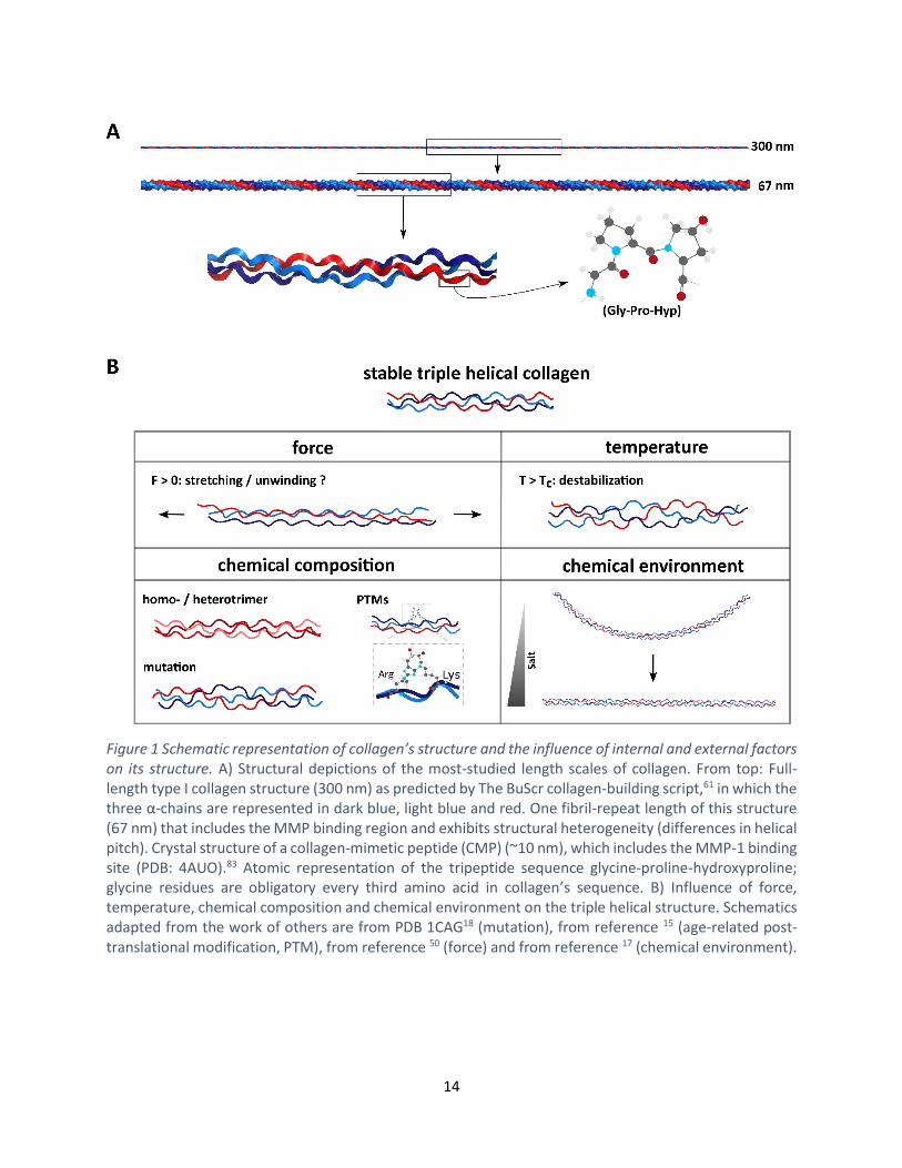
Figure 1 Schematic representation of collagen’s structure and the influence of internal and external factors on its structure. A) Structural depictions of the most-studied length scales of collagen. From top: Full-length type I collagen structure (300 nm) as predicted by The BuScr collagen-building script,61 in which the three α-chains are represented in dark blue, light blue and red. One fibril-repeat length of this structure (67 nm) that includes the MMP binding region and exhibits structural heterogeneity (differences in helical pitch). Crystal structure of a collagen-mimetic peptide (CMP) (~10 nm), which includes the MMP-1 binding site (PDB: 4AUO).83 Atomic representation of the tripeptide sequence glycine-proline-hydroxyproline; glycine residues are obligatory every third amino acid in collagen’s sequence. B) Influence of force, temperature, chemical composition and chemical environment on the triple helical structure. Schematics adapted from the work of others are from PDB 1CAG18 (mutation), from reference 15 (age-related post-translational modification, PTM), from reference 50 (force) and from reference 17 (chemical environment).
15
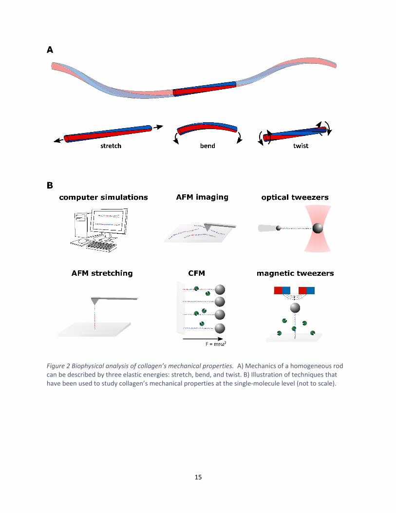
Figure 2 Biophysical analysis of collagen’s mechanical properties. A) Mechanics of a homogeneous rod can be described by three elastic energies: stretch, bend, and twist. B) Illustration of techniques that have been used to study collagen’s mechanical properties at the single-molecule level (not to scale).