Embed Size (px)
Citation preview

MDCT and Pulmonary Embolism
Heber MacMahon
The University of Chicago
Department of Radiology
https://tinyurl.com/hmpe17

Disclosures
• Consultant for Riverain Medical• Minor stockholder in Hologic, Inc.• Consultant for GE Healthcare• Research Support from Philips Healthcare • License and royalty fees from University of Chicago
(UC Tech)
https://tinyurl.com/hmpe17

Pulmonary Embolism
Risk Factors & pre-test probability
CXR findings
CT technique
Dx on contrast and non-contrast CT scans
Acute vs Chronic PE
Non thrombotic PE
https://tinyurl.com/hmpe17

Pulmonary Embolism
500,000 episodes of PE / yr in USA
12 – 64% ICU patients at autopsy
Contributing cause of death in 10-15% ICU pts
Clinical diagnosis difficult
https://tinyurl.com/hmpe17

Risk Factors for PE
Strong Risk Factors
Lower limb Fx, Hip or Knee Replacement
Major Trauma
MI in previous 3 mos.
Spinal Cord Injury
Previous VTE
2014 ESC Guidelines on the diagnosis and management of acute pulmonary
embolism; Task Force for the Diagnosis and Management of Acute Pulmonary
Embolism of the European Society of Cardiology (ESC)
European Heart Journal (2014) 35, 3033–3080
https://tinyurl.com/hmpe17
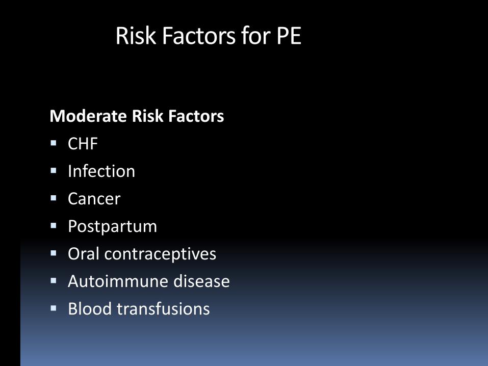
Moderate Risk Factors
CHF
Infection
Cancer
Postpartum
Oral contraceptives
Autoimmune disease
Blood transfusions
Risk Factors for PE

Weak Risk Factors
Bed rest > 3 days
Travel of 4 hr or more in the past month
Pregnancy
Obesity
Advanced age
Risk Factors for PE

Most Common Symptoms of PE
Unexplained: Dyspnea (50%) – Esp. sudden onset
Pleuritic pain (39%)
Substernal (15%)
Hemoptysis (8%)
Syncope/near syncope (6%)More specific

Clinical Signs in PE
Hypoxemia (70%)
Tachypnea/dyspnea (90%)
Tachycardia (40%)
Arrythmia
Wheezing
Hemoptysis
Fever (but < 39.5C)
Signs of DVT

Simplified Wells Criteria (>2: PE likely)
History of DVT or PE — 1 point
Tachycardia (heart rate > 100) — 1 point
Immobilization (≥ 3d)/surgery in previous four weeks — 1 point
Hemoptysis — 1 point
Malignancy (with treatment within 6 months) or palliative — 1 point
Clinically suspected DVT — 1 point
Alternative diagnosis is less likely than PE —3.0 points

D-dimer for PE
Cut-off value: 500 ug/L
Negative predictive value: 98-99%
Positive predictive value: 27-29%
False positives associated with: female sex; increasing age; black (vs. white) race;
cocaine use; immobility; hemoptysis; hemodialysis; active malignancy; rheumatoid arthritis; lupus; sickle cell disease; prior venous thromboembolism (VTE; not under treatment); pregnancy and postpartum state; and abdominal, chest, orthopedic, or other surgery.

Multiple PEs with infarcts

76 y/o with pleuritic chest pain

Multiple Emboli with Small Infarcts

CXR in PE
CXR often normal
Focal subpleural rounded opacity, esp. CP angles
Westermark Sign : Peripheral oligemia, caused by chronic embolism, but often with superimposed new acute episode

Westermark Sign

Large PE causing asymmetric pulmonary edema

CXR in PE
CXR often normal
Focal subpleural rounded opacity, esp. CP angles
Westermark Sign : Peripheral oligemia, caused by chronic embolism, but often with superimposed new acute episode
Fleischner Sign : Enlargement and sharp definition of central PA

Fleischner Sign

CXR in PE
CXR often normal
Focal subpleural rounded opacity, esp. CP angles
Westermark Sign : Peripheral oligemia, caused by chronic embolism, but often with superimposed new acute episode
Fleischner Sign : Enlargement and sharp definition of central PA
Bibasilar atelectasis, subpleural opacity, effusions, suggestive in previously healthy individual

Unexplained Basilar Subsegmental Atelectasis

V/Q Scan accuracy for PE
V/Q Scan Results PE Prevalence
Normal or low probability
+ low clinical suspicion 4%
High probability
+ high clinical suspicion 96%
Still a reasonable test for young women with low PE probability and normal CXR.
or
Patients with contraindication to IV contrast

Pulmonary C.T. Angiography
Rapid contrast infusion (3-6 cc/sec)
Antecubital fossa vein
1 – 1.5 mm collimation
Shallow breath hold
Fixed delay, bolus tracking, timing bolus

Test bolus vs Bolus Tracking
• Test bolus: Visual estimation by tech of optimal contrast timing.
• Makes cursor placement less critical.
• Subjective judgement
• Bolus Tracking: Automatic trigger for scan based on bolus arrival
• Cursor placement critical
• Adds several seconds to scan delay

Test Bolus. Cursor on PA

Normal Circulation Time

Abnormal Test Bolus – Slow Circulation

Main Contrast Bolus – Slow Circulation

Test Bolus using Descending Aorta

Non-Diagnostic CTA Bolus Timing
Motion artifact
Poor contrast enhancement
- Flow obstruction
- Deep inspiration > dilution
- Contrast timing error
Beam hardening and noise –obesity

Dilution by unopacified IVC inflow“Relax, take in a small breath and hold it”

PE CT Results over 12 months at U Chicago

Saddle EmbolusSaddle and Subsegmental Emboli
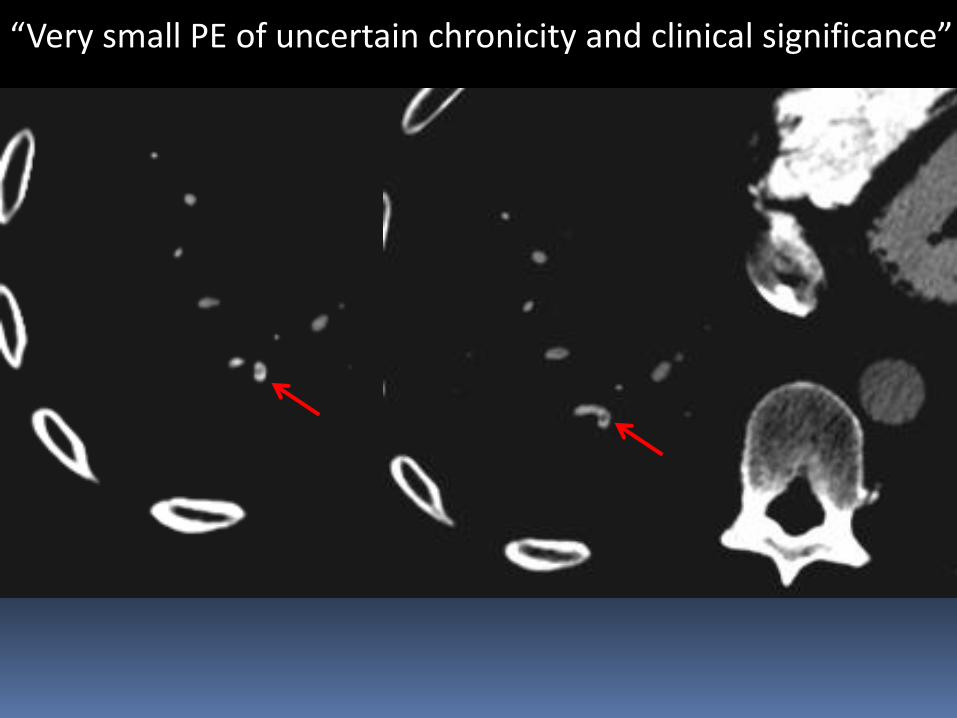
“Very small PE of uncertain chronicity and clinical significance”

PA diameter Septal deviation
Clinical Significance and Prognosis in Acute PE

PE-related mortality predictors
Age above 60 years
RV/LV area >1 (odds ratio 8.6)
RV/LV diameter >1.5 (odds ratio 48.8,P<0.001)
Timing bolus upslope time > 6 seconds (odds ratio 23.3), 50% downslope time >6 seconds (odds ratio 20)
Embolus load score >15 (odds ratio 25)
Li C, Lin CT, Kligerman SJ, Hong SN, White CS. JTI 2014

Pulmonary Infarct - Hampton’s Hump

Only 15% cause true infarction
Most in lower lobes
Usually multiple
Pulmonary Infarct

Pulmonary Infarct
• Sharply defined consolidation• Central lucency• Absence of air bronchogram

Pulmonary Infarcts
Likelihood Ratios: 23.0 for central lucencies
2.9 for vessel sign(enlarged vessel at apex)
0.2 for air bronchograms
Revel et al. Radiology: Volume 244: Number 3 September 2007

PE with HemorrhageR flank pain. Acute PE

Breast cancer patient with lung nodule

Acute PE
Resolving Infarct

Evolving Infarct – Melting Ice cube SignIncidental RUL Nodule

6 months3 weeks
BaselineBaseline
3-4-03 3 months
Evolving Infarct: “Melting Ice Cube Sign”

60 y/o male with hemoptysis

60 y/o male with hemoptysis

60 y/o male with hemoptysis

Non-Contrast Scan
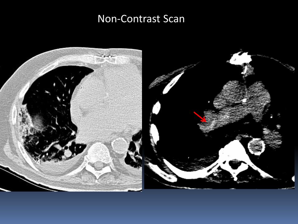
Non-Contrast Scan

Saddle Embolus on Non- Enhanced CT Scan

Chronic PE

Chronic PE

Chronic PE
Recanalized, organized or calcified thrombus

Arterial Web

12-02 1-03Embolus > Web

2 yrs later- arterial web
12-04
Chronic PE
Recanalized, organized or calcified thrombus
Intraluminal webs or bands
Pulmonary hypertension

Chronic PE

Chronic and Acute PE

Chronic PE – Mosaic Perfusion

Chronic PE – Decreased perfusion & bronchiectasis

Enlarged Bronchial Collaterals in Chronic PE

Enlarged Bronchial Collaterals in Chronic PE

Chronic PE
Recanalized, organized, calcified thrombus
Intraluminal webs or bands
Pulmonary hypertension
Mosaic perfusion
Collateral vascularization

CTA for PE: Common Pitfalls
Failure to use proper window & level

Narrow window settingStandard ST Window

CTA for PE: Common Pitfalls
Failure to use correct window &level
Use of lung (sharp) algorithm

Lung Standard
Effect of lung filter

ECG Gated Cardiac Scan

CTA for PE: Common Pitfalls
Failure to optimize window level/width
Use of lung reconstruction filter
Vascular bifurcations (false positives)


5 mm
1mm

CTA for PE: Common Pitfalls
Failure to optimize window level/width
Use of lung reconstruction algorithm
Vascular bifurcations (false positives)
Motion/averaging artifact (false positive or negative)

Motion with Density Averaging

Density Averaging : Crossing Bronchus

Decreased flow with Incomplete Opacification

CTA for PE: Common Pitfalls
Failure to use workstation (window/level)
Use of lung reconstruction algorithm
Vascular bifurcations (false positives)
Motion/averaging artifact (false positive or negative)
Bronchiectasis


Bronchiectasis

Postpneumonectomy. R/O PE

Occurs in up to 12% of cases
Likelihood related to length of arterial stump
May propagate or resolve
Benign course without treatment
Arterial Stump Thrombus

Post-op Lobectomy or Pneumonectomy thrombosis in situ
Non- embolic thrombus &Non-thrombotic emboli

51 y/o woman with severe SOB

Uterine Leiomyosarcoma
Post-op Lobectomy or Pneumonectomy thrombosis in situ
Tumor Embolism
Tumor embolism from remote solid tumors (breast, lung, stomach)
Propagation of tumor via IVC (renal, hepatic, uterine)
Primary arterial sarcomas
Non- embolic thrombus &Non-thrombotic emboli

Asymptomatic Patient

Asymptomatic Patient

Polymethymethacrylate Cement Embolism

Polymethymethacrylate cement embolism• 4-23% of procedures• <1% symptomatic
Polymethymethacrylate Cement Embolism

Post-op Lobectomy or Pneumonectomy thrombosis in situ
Tumor Embolism
Tumor embolism from remote solid tumors (breast, lung, stomach)
Propagation of tumor via IVC (renal, hepatic, uterine)
Primary arterial sarcomas
Foreign material (Surgical cement, filter fragments,
Needle fragments)
Non- embolic thrombus &Non-thrombotic emboli
Take Home Points
CTA is the preferred diagnostic test for PE except in young patients with low clinical probability.
PE can often be recognized on CXRs and non contrast CT scans
Technique is important; use high injection rate with saline chaser, thin sections, and small inspiration.
Use bolus time to aorta to estimate timing
Significance of isolated subsegmental emboli is uncertain
https://tinyurl.com/hmpe17