Embed Size (px)
Citation preview
Res
earc
h R
epo
rt 2
002
MDCBerlin-Buch
Max
Del
brüc
k C
ente
r fo
r M
olec
ular
Med
icin
e
Research Report 2002(covers the period 2000-2001)
Max Delbrück Center forMolecular Medicine (MDC)Berlin-Buch
Robert-Rössle-Str. 10D-13125 Berlin
Tel.: +49-30-9406-0Fax: + 49-30-949-4161e-mail: [email protected]
This Research Report is also available on the World Wide Webhttp://www.mdc-berlin.de
The MDC is a member of the HGF (Helmholtz Association of National Research Centers)
The following Research Reports have been published previously:Research Report 1996 (covers the period 1992-1995)Research Report 1996/97 (covers the period 1996-1997)Research Report 2000 (covers the period 1998-1999)
Editorial board:Walter BirchmeierCarmen BirchmeierThomas BlankensteinUdo HeinemannHelmut KettenmannFriedrich C. LuftPeter M. Schlag
Coordination:Barbara BachtlerHans-Josef LinkensVolker Wunderlich
Book design:Hoch Drei GmbH, 10963 Berlin
Printing:Druckhaus Berlin-Mitte GmbH
Printed in Germany 2002
Legend to Cover Figures:
In situ hybridization with the patched-1 cDNAreveals a block in embryonic hair placodeformation in the absence of �-catenin/wntsignaling. In addition, tissue-specific ablation ofthe �-catenin gene in the skin of mice demon-strates the essential role of �-catenin signalingin the fate of skin stem cells in the adult: in theabsence of �-catenin skin stem cells fail todifferentiate into follicular keratinocytes, takingan epidermal route instead (from Huelsken etal., Cell (2001) 105, 533-545; this report p. 97)

Research Report2002Covers the period 2000/2001
2
Content
Inhalt
IntroductionEinführung .................................................................................................................................................................... 8
OverviewÜberblick .................................................................................................................................................................... 21
Genetics, Bioinformatics and Structural BiologyGenetik, Bioinformatik und Strukturbiologie ........................................................................................................ 40
Molecular Biology and Genetics of Cardiovascular DiseasesMolekularbiologie und Genetik kardiovaskulärer ErkrankungenDetlev Ganten, Norbert Hübner ................................................................................ 43
Molecular Biology of Peptide HormonesMolekularbiologie von Peptid-HormonenMichael Bader .......................................................................................................... 45
Genetics, Etiology, and Pathogenesis of Hypertension, Vascular Injury, and Renal DiseasesGenetik, Ätiologie und Pathogenese von Bluthochdruck, Gefäßschädigung und NierenerkrankungenFriedrich C. Luft ....................................................................................................... 47
Gene Mapping and Identification in Monogenic and Complex DiseasesGenkartierung und Identifizierung bei monogenen und komplexen ErkrankungenPeter Nürnberg ........................................................................................................ 49
Cardiovascular Molecular GeneticsKardiovaskuläre MolekulargenetikLudwig Thierfelder .................................................................................................... 52
Obesity and HypertensionFettleibigkeit und BluthochdruckArya M. Sharma ....................................................................................................... 54
Disorders of the Autonomic Nervous SystemErkrankungen des autonomen NervensystemsJens Jordan (Helmholtz Fellow) ................................................................................ 56
Characterization of newly identified human importin � proteinsCharakterisierung von neuen humanen Importin-alpha ProteinenMatthias Köhler (Helmholtz Fellow) ........................................................................... 57
Tumor Genetics TumorgenetikSiegfried Scherneck ................................................................................................. 58
Mouse GeneticsMausgenetikCarmen Birchmeier .................................................................................................. 61
Developmental GeneticsEntwicklungsgenetikAndreas Schedl ....................................................................................................... 64
Lipids and Experimental Gene TherapyLipide und experimentelle GentherapieThomas Willnow ....................................................................................................... 66
BioinformaticsBioinformatikJens Reich, Peer Bork .............................................................................................. 68
Protein Misfolding: From Basic Biopolymer Physics to Conformational DiseasesProtein-Fehlfaltung: von der Biopolymerphysik zu Konformations-ErkrankungenGregor Damaschun .................................................................................................. 70
Structural Studies of Proteins and Nucleic Acids by X-ray CrystallographyStrukturstudien an Proteinen und Nukleinsäuren mit Röntgenstrahl-KristallographieUdo Heinemann ....................................................................................................... 72
Role of Protein Dynamics in Enzyme FunctionRolle der Proteindynamik bei der EnzymfunktionChristiane Jung ........................................................................................................ 75
Computer Simulation of Nucleic Acid Structure and InteractionsComputersimulation von Nukleinsäure-Strukturen und WechselwirkungenHeinz Sklenar ........................................................................................................... 77
Conformation, Stability and Interaction of Biological MacromoleculesKonformation, Stabilität und Wechselwirkung von biologischen MakromolekülenHeinz Welfle ............................................................................................................. 79
Bioethics and Science Communication Bioethik und WissenschaftskommunikationChristof Tannert ....................................................................................................... 81
Cell Growth and DifferentiationZellwachstum und Differenzierung ................................................................................................................. 84
Growth Control and Gene Regulation in the Hematopoietic SystemWachstumskontrolle und Genregulation im hämatopoetischen SystemAchim Leutz ............................................................................................................. 88
Signal Transduction in Tumor CellsSignaltransduktion in TumorzellenClaus Scheidereit ..................................................................................................... 90
Differentiation and Growth Control in Lymphocyte Development and ImmunopathogenesisDifferenzierung und Wachstumskontrolle bei der Lymphozyten-Entwicklung und ImmunpathogeneseMartin Lipp .............................................................................................................. 92
Initiation of DNA ReplicationInitiierung der DNA-ReplikationManfred Gossen ...................................................................................................... 95
Epithelial Differentiation, Invasion and MetastasisEpitheliale Differenzierung, Invasion und MetastasierungWalter Birchmeier ..................................................................................................... 97
3
4
Surgical OncologyChirurgische OnkologiePeter M. Schlag ..................................................................................................... 100
Molecular Muscle PhysiologyMolekulare MuskelphysiologieIngo L. Morano ...................................................................................................... 103
Cell Biology of Cardiovascular DiseasesZellbiologie kardiovaskulärer ErkrankungenHeinrich Leonhardt, M. Cristina Cardoso ................................................................ 105
Intracellular ProteolysisIntrazelluläre ProteolyseThomas Sommer ................................................................................................... 107
Nucleocytoplasmic Transport in the Yeast Saccharomyces cerevisiaeNukleo-zytoplasmatischer Transport bei der BäckerhefeKatrin Stade (Helmholtz Fellow) .............................................................................. 109
Cytochrome P450 and Endoplasmic ReticulumCytochrom P450 und endoplasmatisches RetikulumWolf-Hagen Schunck ............................................................................................. 110
Cell Polarity and Epithelial Formation in Development and DiseaseZellpolarität und Epithelbildung in Entwicklung und KrankheitSalim Abdelilah-Seyfried ......................................................................................... 112
Electron MicroscopyElektronenmikroskopieGerd Kempermann, Bettina Erdmann ..................................................................... 114
Molecular TherapyMolekulare Therapie ....................................................................................................................................... 118
Myocardial RegenerationRegeneration des MyokardsRainer Dietz ............................................................................................................ 119
Control of Smooth Muscle Cell FunctionKontrolle der Funktion der glatten MuskelzelleMaik Gollasch (Helmholtz Fellow) ............................................................................ 121
Immunology of Cardiovascular DiseasesImmunologie kardiovaskulärer ErkrankungenGerd Wallukat ........................................................................................................ 123
Hematology, Oncology and Tumor ImmunologyHämatologie, Onkologie und TumorimmunologieBernd Dörken ......................................................................................................... 125
Molecular ImmunotherapyMolekulare ImmunotherapieAntonio Pezzutto .................................................................................................... 128
Molecular Immunology and Gene TherapyMolekulare Immunologie und GentherapieThomas Blankenstein ............................................................................................. 130
5
Cellular Immunology of Autoimmune Reactions Zelluläre Immunologie von AutoimmunreaktionenKirsten Falk, Olaf Rötzschke ................................................................................... 132
Molecular and Cell Biology of Hematopoietic CellsMolekular-und Zellbiologie hämatopoietischer ZellenMartin Zenke .......................................................................................................... 134
Cell Cycle Regulation and Gene TherapyZellzyklus-Regulation und GentherapieForschungsgruppe der Humboldt-Universität zu Berlin am MDC ............................. 136
Evolution, Regulation and Genetic Applications of Transposable Elements in VertebratesEvolution, Regulation und genetische Anwendungen von transposablen Elementen bei VertebratenZoltán Ivics ............................................................................................................. 138
Experimental PharmacologyExperimentelle PharmakologieIduna Fichtner ........................................................................................................ 139
Drug Targeting„Drug Targeting“ (Zielgenaue Medikamentenentwicklung)Regina Reszka ....................................................................................................... 141
RNA ChemistryRNA ChemieEckart Matthes ....................................................................................................... 143
Molecular and Developmental NeurosciencesMolekulare Neurowissenschaft und Entwicklungsneurobiologie ............................................................... 146
NeurodegenerationNeurodegenerationChristiane Alexander .............................................................................................. 148
Neuronal Stem CellsNeuronale StammzellenGerd Kempermann ................................................................................................ 150
Cellular NeurosciencesZelluläre NeurowissenschaftenHelmut Kettenmann ............................................................................................... 152
Growth Factor and Regeneration Group Wachstumsfaktoren und RegenerationGary R. Lewin ........................................................................................................ 154
Synapse Formation and Function Bildung und Funktion von SynapsenFrank W. Pfrieger .................................................................................................... 156
Developmental NeurobiologyEntwicklungsneurobiologieFritz G. Rathjen ...................................................................................................... 157
Proteomics and Molecular Mechanisms of Neurodegenerative DisordersProteomik und molekulare Mechanismen neurodegenerativer ErkrankungenErich Wanker .......................................................................................................... 160
6
Structure and OrganizationStruktur und Organisation
Organizational StructureStiftungsorgane ................................................................................................... 164
The Board of Trustees ............................................................................................ 164Kuratorium
Members of the Board of Trustees ............................................................... 164Mitglieder des Kuratoriums
Members of the Scientific Committee .......................................................... 165Mitglieder des Wissenschaftlichen Ausschusses
The Management Board ......................................................................................... 166Stiftungsvorstand
Scientific Council .................................................................................................... 166Wissenschaftlicher Rat
Staff Council .......................................................................................................... 167Personalrat
Supporting Divisions ........................................................................................... 168Stabsstellen
Safety ......................................................................................................... 168Sicherheit
Building Coordination, Engineering and Reconstruction ............................... 168Bauangelegenheiten
Auditing and Legal Affairs ............................................................................ 168Innenrevision und Recht
Patents/Licences ........................................................................................ 169Patente und Lizenzen
Technology Transfer .................................................................................... 169Technologietransfer
Press and Public Relations ................................................................................ 170Presse- und Öffentlichkeitsarbeit
AdministrationVerwaltung ........................................................................................................... 172
Personnel ................................................................................................... 172Personal
Finances .................................................................................................... 172Finanzen
Purchasing and Materials Management ....................................................... 173Einkauf und Materialwirtschaft
7
Central FacilitiesZentrale Dienste .................................................................................................. 174
Library ........................................................................................................ 174Bibliothek
Animal Facilities ........................................................................................... 174Tierhaltung
Campus Net Management ........................................................................... 174Campusnetz-Management
Data and Image Processing ......................................................................... 175Daten- und Bildverarbeitung
Technical Affairs .......................................................................................... 175Technik
Meetings, Workshops and Symposia ................................................................ 176Wissenschaftliche Veranstaltungen
Awards ................................................................................................................. 178Auszeichnungen
Addresses of Scientific Journals at the Berlin-Buch Campus ......................... 179Adressen wissenschaftlicher Zeitschriften auf dem Campus Berlin-Buch
Index .................................................................................................................... 180Index
Organigram .......................................................................................................... 188Organigramm
Campus Map ................................................................................. Inside Back CoverCampusplan ............................................................................. Innenumschlag hinten
How to find your way to the MDC ................................................ Inside Back CoverWie gelangen Sie zum MDC .................................................... Innenumschlag hinten
Introduction
Molecular Medicine – A Concept and its Application atthe MDC in Berlin-Buch
History and Foundation
On January 1, 2002, the Max Delbrück Center for MolecularMedicine (MDC) Berlin-Buch celebrated its 10th birthday. Itwas founded on January 1, 1992, from the Institutes of theEast German Academy of Sciences, the Central Institute forCancer Research, the Central Institute for CardiovascularResearch and the Central Institute for Molecular Biology. Thecenter is named after Max Delbrück, a native of Berlin, andone of the founding fathers of molecular biology who won theNobel Prize in 1969. Delbrück collaborated at the Kaiser Wil-helm Institute for Brain Research in Berlin-Buch with theRussian geneticist, N. W. Timoféev-Ressovsky and the phys-icist, K.G. Zimmer. In 1935, their interdisciplinary researchled to the publication of the epoch-making paper “The Natureof Gene Structure and Gene Mutation”.
Anyone who experienced the reunification of Germanybetween November 9, 1989, and January 1, 1992 – involvingthe break up of the GDR, the unification of the two Germa-nies, with the evaluation of the entire scientific system of theGDR, the many plans for the restructuring of the scientificestablishments and institutional and personal uncertainties ofevery kind – can imagine the problems people experienced,as well as the hope and expectations during this two-year pe-riod. In 1991, in Berlin-Buch, there were over 1,000 staff,physicians, researchers, and technicians working in the insti-tutes of the Academy and the clinics. At its foundation, theMDC had 350 budgeted posts. In such circumstances it wasno simple matter to set up a new organization to match thestandard of the former institutes of the Academy and guaran-tee all qualified staff a future post. The details of this criticaland exciting period of the restructuring of science on theBuch Campus has been comprehensively described else-
Einführung
Molekulare Medizin – Ein Konzept und seine Umsetzungam MDC in Berlin-Buch
Geschichte und Gründung
Am 01. Januar 2002 beging das Max-Delbrück-Centrum fürMolekulare Medizin (MDC) Berlin-Buch sein 10-jährigesBestehen. Es ist am 01. Januar 1992 aus den Instituten derAkademie der Wissenschaften der DDR, dem Zentralinstitutfür Krebsforschung, dem Zentralinstitut für Herz-Kreislauf-forschung und dem Zentralinstitut für Molekularbiologie her-vorgegangen. Die Benennung des Institutes erfolgte nachMax Delbrück, dem aus Berlin stammenden Wegbereiter derMolekularbiologie, der 1969 mit dem Nobelpreis ausgezeich-net worden war. Delbrück hatte am Kaiser-Wilhelm-Institutfür Hirnforschung in Berlin-Buch zusammen mit dem russi-schen Genetiker N. W. Timoféeff-Ressovsky und dem Physi-ker K.G. Zimmer gearbeitet, und bei dieser interdisziplinärenForschung war 1935 die epochemachende Arbeit über die„Natur der Genstruktur und der Genmutation“ entstanden.
Wer die Zeit der deutschen Wiedervereinigung zwischen dem09. November 1989 und dem 1. Januar 1992 persönlich miter-lebt hat – mit dem Zusammenbruch der DDR, mit dem Pro-zess der Vereinigung der beiden deutschen Staaten, mit derEvaluation des gesamten Wissenschaftssystems der DDR, mitden vielen Plänen für die Neustrukturierung der wissenschaft-lichen Einrichtungen und mit den Unsicherheiten in institutio-neller und persönlicher Art –, kann sich vorstellen, welche Be-lastungen, aber auch welche Hoffnungen und Erwartungen indiesen zwei Jahren des Prozesses der deutschen Wiederverei-nigung durchlebt wurden. In Berlin-Buch waren 1991 weitüber 1.000 Mitarbeiter, Ärzte, Wissenschaftler, TechnischeAngestellte in den Akademieinstituten und Kliniken beschäf-tigt. Das MDC hatte bei der Gründung 350 budgetierte Stel-len. Es war in dieser Situation nicht einfach, eine dem Leis-tungsniveau der Akademieinstitute entsprechende neue
8
where. It was only possible because everyone was sympa-thetic to the final aim and worked together to ensure that itwas achieved. The significant and unavoidable problems at personal and in-stitutional levels were made easier by the fact that everyonewas aware of the great tradition of the Buch Institutes whichhad supported outstanding researchers like Oskar and CécileVogt, Walter Friedrich, Karl Lohmann, Arnold Graffi, ErwinNegelein, Albert Wollenberger and Hans Gummel. The realand positive role played by tradition in building new struc-tures at a time of great change can be particularly well ob-served and experienced in Berlin. Attention to tradition wasand still is important as far as setting up the MDC and theBerlin-Buch Campus are concerned.
The clinics of the Academy of Sciences were incorporatedinto the university system – thanks to the energetic efforts ofthe then Senator for Science, Prof. Manfred Erhardt – as theRobert Rössle Cancer Clinic and the Franz Volhard Cardio-vascular Clinic of the Charité, Berlin-Buch Campus. Thanksto particularly well defined cooperation contracts, they werelinked to the MDC in both personnel and institutional terms.BBB Biotechnologie Berlin-Buch Management GmbH set upa biotechnology park. This has allowed the basic researchactivities at the MDC, and clinical research at the Charitéclinics and the Buch Clinic as well as the Technology Park es-tablished by BBB Management GmbH to develop a synergywith respect to all research efforts that has subsequently beenshown to be successful based on any number of criteria. Thepure research carried out on the Berlin-Buch Campus wasboosted in1999 by the arrival of the Forschungsinstitut fürMolekulare Pharmakologie (FMP; Research Institute forMolecular Pharmacology), which is part of the Wilhelm Gott-fried Leibniz Association. The institute collaborates closelywith the MDC on structural research into biologically activemolecules. The privatization of the clinics under the auspicesof Helios Kliniken GmbH in summer 2001 has created evenmore opportunities for cooperation.
Organisationsform zu finden und allen qualifizierten Mitar-beitern die ihnen zukommende Position zuzusichern. ÜberEinzelheiten dieser entscheidenden und aufregenden Monateund Jahre der Neuorientierung der Wissenschaft auf demCampus Buch ist an anderer Stelle ausführlich berichtet undgeschrieben worden. Sie konnten nur bewältigt werden, weiles von allen Seiten viel Verständnis und Unterstützung gab. Die großen und wohl unvermeidbaren Brüche in institutio-neller und personeller Hinsicht wurden unter anderem da-durch erleichtert, dass ganz bewusst die große Tradition derBucher Institute mit so herausragenden Wissenschaftlern wieOskar und Cécile Vogt, Walter Friedrich, Karl Lohmann, Ar-nold Graffi, Erwin Negelein, Albert Wollenberger und HansGummel fortgeführt wurde. Die reale und konkrete positiveBedeutung der Tradition für die Zukunftsgestaltung in einerZeit großer Umbrüche, kann in Berlin besonders gut beob-achtet und erlebt werden. Traditionspflege wurde und wirdbewußt für den Aufbau des MDC und des Campus Berlin-Buch eingesetzt.
Die Kliniken der Akademie der Wissenschaften wurden mittatkräftiger Unterstützung des damaligen Senators für Wis-senschaft, Prof. Manfred Erhardt, in das universitäre Systemeingegliedert als Robert-Rössle-Krebsklinik und Franz-Vol-hard-Herz-Kreislaufklinik der Charité, Campus Berlin-Buch.Durch einen besonders engen Kooperationsvertrag wurdensie mit dem MDC personell und institutionell verbunden. DerAufbau eines Biotechnologieparks wurde durch die BBBBiotechnologie Berlin-Buch Management GmbH begründet.Auf diese Weise wurde mit der Grundlagenforschung amMDC, der klinischen Forschung an den Charité-Kliniken unddem Klinikum Buch sowie mit dem von der BBB Manage-ment GmbH eingerichteten Technologiepark ein Synergienschaffendes komplettes Forschungssystem eingerichtet, dassich in den folgenden Jahren in jeder Hinsicht als erfolgreicherwiesen hat. 1999 wurde die Grundlagenforschung auf demCampus Berlin-Buch durch den Zuzug des Forschungsinsti-tuts für Molekulare Pharmakologie (FMP), das zur Wilhelm-Gottfried-Leibniz-Gemeinschaft gehört, verstärkt. DiesesInstitut arbeitet mit dem MDC insbesondere auf dem Gebietder Strukturforschung für biologisch aktive Moleküle inten-siv zusammen. Die Privatisierung der Kliniken durch dieHelios Kliniken GmbH im Sommer 2001 schafft erweiterteMöglichkeiten der Kooperation.
Aufbruchstimmung in Berlin-Buch
Auf dem Campus Berlin-Buch herrscht Aufbruchstimmung:Das Max Delbrück Communications Center (MDC.C) mitHörsälen, Seminar- und Laborräumen für Kongresse, Fortbil-dung und Kursen ist im Jahre 2001 fertig gestellt und in Betriebgenommen worden. Der erste Bauabschnitt des HelmholtzHauses für Tierlaboratorien und Büroräume wird noch imJahre 2002 bezogen. Ein neues Zentrum für Medizinische Ge-nomforschung des MDC ist im Entstehen. Vier neue Labor-gebäude für Existenzgründer und Biotechnologiefirmen sindfertig gestellt bzw. befinden sich zur Zeit im Bau. Das neue„Helios Klinikum Berlin“ wird auch die universitären Klinikender Charité in einem Neubau für 400 Millionen Mark mit auf-nehmen, so dass eine der modernsten und größten KlinikenBerlins in unmittelbarer Nachbarschaft des Forschungscampus
9
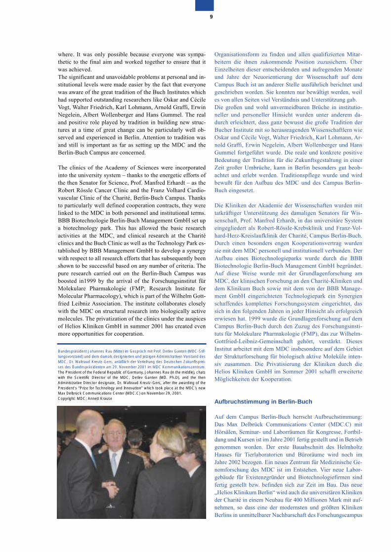
Bundespräsident Johannes Rau (Mitte) im Gespräch mit Prof. Detlev Ganten (MDC-Stif-tungsvorstand) und dem damals designierten und jetzigen Administrativen Vorstand desMDC, Dr. Waltraud Kreutz-Gers, anläßlich der Verleihung des Deutschen Zukunftsprei-ses des Bundespräsidenten am 29. November 2001 im MDC Kommunikationszentrum.The President of the Federal Republic of Germany, Johannes Rau (in the middle), chatswith the Scientific Director of the MDC, Detlev Ganten (MD. Ph.D), and the thenAdministrative Director designate, Dr. Waltraud Kreutz-Gers, after the awarding of thePresident's “Prize for Technology and Innovation” which took place at the MDC`s newMax Delbrück Communications Center (MDC.C) on November 29, 2001. Copyright: MDC; Annett Krause
Developments in Berlin-Buch
Spirits on the Berlin-Buch Campus are high as the site devel-ops: the Max Delbrück Communications Center (MDC.C),with its lecture theaters, seminar rooms and laboratory spacefor congresses, further education and training courses, wasopened in 2001. The first section of the “Helmholtz Haus” foranimal laboratory facilities and offices will be ready in 2002.A new MDC Center for Medical Genome Research is beingcreated. Four new laboratory buildings for start-up compa-nies and biotechnology companies have been completed orare under construction. The new “Helios Klinikum Berlin”will also incorporate the university clinics of the Charité in anew setting costing approximately 200 million Euro so thatone of the most modern and largest clinics in Berlin will belocated right next to the research campus. The space occupiedby the clinics previously represents an area of over 100 haand this will allow the Technology Park to expand. The clinicbuildings designated as being of historic interest will be usedas a “Cité Universitaire”, and will be adopted by the Euro-pean College of Liberal Art (ECLA). A new shopping centerwill be built right next to the S-Bahn station. It is also plannedto build a hotel for visitors and congress delegates as well asrelatives of the patients being treated in the clinics.
The health theme, which has made Berlin-Buch, the “HealthRegion” is becoming more and more important as a part ofthe modern knowledge-based society. In the future, Berlin-Buch will be a center for molecular medicine where the latestadvances in genome research and gene technology will beapplied to the clinical setting and in the field of business,allowing biotech companies and service firms to develop andcreate new jobs.
The Sculpture Park on the Berlin-Buch Campus with works by,among others, Ipousteguy, Szymanski and Kriester, reflects theclose relationship between the Arts and Sciences, which is par-ticularly encouraged in Berlin-Buch. In this way, Buch will beseen as an intellectual focal point where the development ofmolecular medicine and gene technology is being advancedalongside a continuing dialog with people from many differentbackgrounds. In this way Berlin-Buch will become a model ofa future human knowledge-based society. On the MDC Cam-pus over 2000 staff are currently working in the research insti-tutes, clinics and biotech companies, many more than beforereunification in 1989. During the last 10 years many new jobshave been created in this science-based environment.
The rapid connections linking Berlin-Buch to the culturalcenter of central Berlin and the proximity of the Barnim Na-ture Conservation Park have made Berlin-Buch one of themost attractive and dynamic suburbs of the new Berlin.
Many of the staff who work in the research institutes and thebiotech companies live only 15 minutes away in PrenzlauerBerg, one of the favorite parts of Berlin with many theaters,bars and cinemas. Prenzlauer Berg is close to the cultural andspiritual heart of the city with the Humboldt University, theMuseum Island, the main theaters, the Berlin Philharmonicand the Kulturforum.
entstehen wird. Die freiwerdenden Klinikgebäude in einemAreal von über 100 Hektar werden für den expandierendenTechnologiepark zur Verfügung stehen. DenkmalgeschützteKlinikgebäude sollen für eine „Cité Universitaire“, die auchdas European College of Liberal Art (ECLA) aufnehmen wird,genutzt werden. In unmittelbarer Nähe des S-Bahnhofes wirdein neues Einkaufszentrum gebaut. Dazu ist geplant, ein Hotelzu errichten für Besucher und Kongressteilnehmer sowie fürAngehörige von Patienten der Kliniken.
Das Thema Gesundheit, dem sich Berlin-Buch, die „Gesund-heitsregion“, verschrieben hat, wird als Teil der modernenWissensgesellschaft immer weiter an Bedeutung gewinnen.Berlin-Buch soll in Zukunft ein Zentrum für die MolekulareMedizin sein, in dem die Fortschritte der Genomforschungund Gentechnik verantwortungsvoll in Klinik und Wirtschaftangewendet werden und um das herum sich Biotechnologie-firmen und Serviceeinrichtungen entwickeln und neue Ar-beitsplätze entstehen lassen. Der Skulpturenpark auf demCampus Berlin-Buch mit Werken unter anderem von Ipou-steguy, Szymanski und Kriester ist Ausdruck der engen Ver-
10
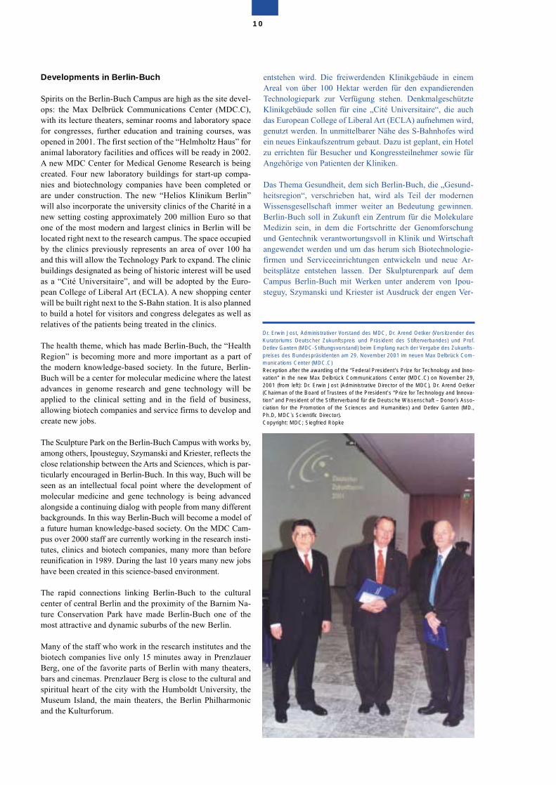
Dr. Erwin Jost, Administrativer Vorstand des MDC, Dr. Arend Oetker (Vorsitzender desKuratoriums Deutscher Zukunftspreis und Präsident des Stifterverbandes) und Prof.Detlev Ganten (MDC-Stiftungsvorstand) beim Empfang nach der Vergabe des Zukunfts-preises des Bundespräsidenten am 29. November 2001 im neuen Max Delbrück Com-munications Center (MDC.C) Reception after the awarding of the “Federal President's Prize for Technology and Inno-vation” in the new Max Delbrück Communications Center (MDC.C) on November 29,2001 (from left): Dr. Erwin Jost (Administrative Director of the MDC), Dr. Arend Oetker(Chairman of the Board of Trustees of the President's “Prize for Technology and Innova-tion“ and President of the Stifterverband für die Deutsche Wissenschaft – Donor´s Asso-ciation for the Promotion of the Sciences and Humanities) and Detlev Ganten (MD.,Ph.D, MDC`s Scientific Director).Copyright: MDC; Siegfried Röpke
Buch is a place of synergies. As a link between town andcountry, between pure research and the clinics as well asscientific applications of this research, living, working andrelaxing in Berlin-Buch are all equally attractive.
The concept of molecular medicine
As far as the success of any scientific institution is concerned,the key factors are the original idea upon which it is based,the efforts of its scientists and other key staff, their commit-ment, the quality of the research activities, projects and itspublication record.
When the MDC was founded, we took advice based on therecommendations of the Scientific Council and the FoundersCommittee regarding ideas about the long-term develop-ments in medicine and the latest results of molecular geneticsand genome research.
There are two reasons for the extraordinary successful andsustained development of the modern life sciences. On theone hand, there were the scientists themselves, who high-lighted the new, often physico-chemical, methods whichcould identify the macromolecules which had key biologicalfunctions in biology and medicine, thereby allowing a betterunderstanding of the interplay between the form and functionof cells. One the other hand, institutions like the RockefellerFoundation play an important role. For example, in 1938, itsupported a research program for the advancement of genet-ics giving it the name “molecular biology”. One aim of thisproject was to raise the level of biosciences at that time andturn it into a quantitative “Science of Man”, which went be-yond simply describing events that happened in Nature. Overthe period 1937–39, the person who gave his name to theMDC, Max Delbrück, held a fellowship from the RockefellerFoundation and their support helped him become one of the
bindung von Kunst und Wissenschaft, die in Berlin-Buch be-sonders gepflegt wird. Buch soll sich auf diese Weise als in-tellektueller Kristallisationspunkt profilieren, in dem die Ent-wicklung der Molekularen Medizin und Gentechnologievorangebracht und durch den Dialog mit breiten Kreisen derGesellschaft kritisch begleitet wird. Auf diese Weise wird Ber-lin-Buch zu einem Modell für die zukünftige humane Wis-sensgesellschaft. Auf dem Campus Berlin-Buch arbeiten zurZeit in den Forschungseinrichtungen, Kliniken und Biotech-nologiefirmen über 2.000 Mitarbeiter, weit mehr als vor derWiedervereinigung 1989. Im Umfeld der Wissenschaft sinddamit in den vergangenen 10 Jahren viele neue anspruchsvolleund zukunftsfähige Arbeitsplätze geschaffen worden.
Die schnelle Anbindung von Berlin-Buch an das kulturelleZentrum in Berlin-Mitte und die direkte Nachbarschaft zumNaturschutzpark Barnim machen Berlin-Buch zu einem derattraktivsten und dynamischsten Vororte des neuen Berlin. Viele Mitarbeiter der Forschungseinrichtungen und Biotechno-logiefirmen wohnen in dem 15 Minuten entfernten PrenzlauerBerg, einem der beliebtesten Stadtteile Berlins mit vielenTheatern, Kneipen und Kinos. Dem Prenzlauer Berg schließtsich direkt das kulturelle und geistige Zentrum der Stadt an mitder Humboldt Universität, der Museumsinsel, den großenTheatern, der Philharmonie und dem Kulturforum.
Buch ist ein Ort der Synergien. Als Nahtstelle zwischen Stadtund Land, zwischen Forschung und klinischer sowie wirt-schaftlicher Anwendung ist das Leben, das Arbeiten und dasErholen in Berlin-Buch gleichsam attraktiv.
Das Konzept der Molekularen Medizin
Entscheidend für den Erfolg einer wissenschaftlichen Institu-tion ist das inhaltliche Konzept, die wissenschaftliche Leis-tung, die tragenden Personen, das Engagement der Wissen-schaftlerinnen, Wissenschaftler und Mitarbeiter, die Qualitätder Forschungstätigkeit, Projekte und Publikationen.
Zur Zeit der Gründung des MDC haben wir uns auf der Basisder Empfehlungen des Wissenschaftsrates und des Gründungs-komitees bei den Konzepten von langfristigen Entwicklun-gen der Medizin und neuesten Ergebnisse der molekularenGenetik und Genomforschung leiten lassen.
Es gibt zwei Quellen für das außerordentlich erfolgreiche undnachhaltige Konzept der modernen Lebenswissenschaft. Zumeinen waren dies die Wissenschaftler selbst, die mit neuen,über die Biologie und Medizin hinausreichenden, häufig phy-sikalisch-chemischen Methoden die Strukturen von Makro-molekülen mit wichtigen biologischen Funktionen erkundenkonnten und sich anschließend daran machten, das Wechsel-spiel von Form und Funktion in der Zelle zu verstehen. Zumzweiten spielten Institutionen wie etwa die Rockefeller Stif-tung eine wichtige Rolle, die im Jahre 1938 einem For-schungsprogramm zur Förderung der Genetik den Namen„Molekularbiologie“ gab. Ein Ziel dieser Initiative lag darin,das Niveau der damaligen Biowissenschaften anzuheben undzu einer quantitativ operierenden „Science of Man“ auszu-weiten, die über die reine Beschreibung der Natur hinaus-ging. Der Namenspatron des MDC, Max Delbrück, gehörte in
11
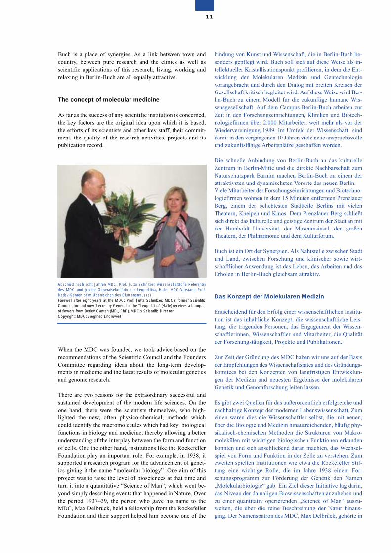
Abschied nach acht Jahren MDC: Prof. Jutta Schnitzer, wissenschaftliche Referentindes MDC und jetzige Generalsekretärin der Leopoldina, Halle. MDC-Vorstand Prof.Detlev Ganten beim Überreichen des Blumenstrausses. Farewell after eight years at the MDC: Prof. Jutta Schnitzer, MDC`s former ScientificCoordinator and now Secretary General of the “Leopoldina” (Halle) receives a bouquetof flowers from Detlev Ganten (MD., PhD), MDC`s Scientific DirectorCopyright: MDC; Siegfried Endruweit
founding fathers of molecular biology; there is much that wecan still learn from him today.
When the MDC was established ten years ago in Berlin-Buchand raised a flag as far as the development of molecular med-icine was concerned, there were only a few institutes aroundthe world engaged in applying the latest results of pure re-search to medical research in a closely-knit institutional set-ting. At that time, no one could have known the kind ofprogress that would take place in genome analysis in the nextten years. The mission of the MDC is to combine modernmedical and clinical research with the methods of molecularbiology and cell biology as well as the expansion in genetechnology, which systematically concentrates on the genomeand the latest results arising from research into it. As far aspure research is concerned, key areas should be those that areof vital importance for the analysis of disease phenomena andwhich lead to new opportunities firmly based on the naturalsciences for diagnosis, treatment and prevention.
den Jahren 1937–39 zu den Stipendiaten der Rockefeller Stif-tung und ist mit ihrer Hilfe zu dem Wegbereiter der Molekul-arbiologie geworden, von dem wir heute noch lernen können.
Als das MDC vor zehn Jahren in Berlin-Buch gegründetwurde und sich die Entwicklung einer Molekularen Medizinauf die Fahnen schrieb, gab es weltweit nur wenige Institute,die so konsequent die Anwendung neuester Methoden in derGrundlagenforschung für medizinische Forschung in engerinstitutioneller Verbindung verfolgten. Niemand konnte da-mals wissen, welche Fortschritte die Genomanalyse in die-sem Zeitraum machen würde. Die Aufgabe des MDC bestanddarin, moderne medizinische und klinische Forschung imVerband mit molekularbiologischen, zellbiologischen sowiezunehmend gentechnischen Methoden zu betreiben, die sichsystematisch auf das Genom konzentrierten und von ihm aus-gingen. Wegweisend für die Grundlagenforschung solltenThemen sein, die von prinzipieller Bedeutung für die Analysevon Krankheitsphänomenen sind und aus denen sich natur-wissenschaftlich begründet neue Möglichkeiten für Diagnos-tik, Therapie und Prävention ableiten lassen.
12
The multitalented molecules of molecular medicine
Touch and Pain: two senses, one ion channel named DRASICOur senses of touch and pain, opposites one might think, have some-thing in common, an ion channel called DRASIC. DRASIC, whichstands for dorsal root ganglion acid sensitive channel, has beenshown to be critical for both these senses. The DRASIC protein formsa channel in the membrane that when opened conducts sodium ionswhich then leads to the electrical excitation of neurons. A first clueabout the possible function of the DRASIC protein came from a ge-netic screen for mutations in the nematode worm C.elegans whichshowed that similar proteins in the worm are necessary for the wormto sense touch. These proteins including DRASIC are all members ofthe Deg/Enac channel superfamily. Thus arose the idea that mechani-cal forces on the membrane could open the channel: this would makeit a mechanical sensor. Thus the same channel obviously helps sen-sory neuron to detect innocuous and painful stimuli in a completely dif-ferent way. It is known that DRASIC can potentially form heteromericion channel complexes with other members of the Deg/Enac family ofproteins. Another aspect of DRASIC channel function is the fact that acidifica-tion ie. protons are a very effective stimulus to open the channel. Acid-ification is a universal consequence of injury or inflammation of periph-eral tissues. Mice lacking DRASIC were much less affected by muscleacidification, in other words the lack of the channel protein had an an-algesic effect. The diversity of function for the DRASIC channel re-ported here for the first time shows directly that the channel does notwork alone but in concert with other as yet unknown proteins. It is pre-cisely this kind of in vivo analysis of protein function that highlights theprinciple that the function of a single protein can vary enormously de-pending on the cellular context. Thus products of single genes are notsimple entities that work alone to accomplish a well-defined functionbut rather execute their function in a multitude of ways depending theother proteins that they can interact with. For further details see GaryLewin´s research group.
Die vielseitigen Moleküle der Molekularen Medizin
Berührung und Schmerz: Zwei Sinne, ein Ionenkanal namensDRASICDer Tastsinn und der Schmerz erscheinen zwar als gegenteilige Sinne,sie haben aber etwas gemeinsam, einen Ionenkanal mit Namen DRA-SIC. DRASIC kürzt den englischen Ausdruck „dorsal root ganglionacid sensitive channel“ ab, womit ein säureempfindlicher Ionenkanalim dorsalen Wurzelganglion gemeint ist. Es konnte gezeigt werden,dass DRASIC für die beiden genannten Sinne wesentlich ist. DasDRASIC Protein formt einen Kanal durch die Membran, durch den imgeöffneten Zustand Natriumionen strömen, die dann zur elektrischenErregung von Neuronen führen. Einen ersten Hinweis auf die möglicheFunktion des DRASIC Proteins ergab eine genetische Reihenuntersu-chung von Mutationen in dem Fadenwurm C. elegans. Sie zeigte,dass ähnliche Proteine nötig sind, damit der Wurm berührungsemp-findlich wird. Diese Proteine – einschließlich DRASIC – sind alle Mit-glieder der Deg/Enac Kanal Großfamilie. Aus dieser Kenntnis leitetsich die Idee ab, dass mechanische Kräfte, die auf die Membran wir-ken, zur Öffnung des Kanals führen, der auf diese Weise ein mechani-scher Sensor würde. Derselbe Kanal hilft offenbar sensorischen Neu-ronen, harmlose und schmerzhafte Reize auf vollständig verschiedeneWeise zu registrieren. Es ist bekannt, dass DRASIC in der Lage ist,heteromere Kanalkomplexe mit anderen Mitgliedern der Deg/EnacFamilie zu bilden.Ein weiterer Aspekt der Funktion des DRASIC Kanals steckt in derTatsache, dass eine Zunahme des Säuregrads – also das Auftretenvon Protonen – als effektiver Reiz zur Kanalöffnung funktioniert. DieZunahme des Säuregrads ist die universelle Konsequenz einer Verlet-zung oder Entzündung des peripheren Gewebes. Mäuse, die nichtüber DRASIC verfügten, waren jedoch viel weniger durch die Muskel-säuerung betroffen. Mit anderen Worten, die Abwesenheit des Kanal-proteins hatte einen analgetischen Effekt. Die Verschiedenheit derFunktionen für den DRASIC Kanal, die hier zum ersten Mal vorgestelltwird, zeigt direkt, dass der Kanal nicht allein, sondern im Verbund mitanderen Proteinen funktioniert, die bislang unbekannt sind. Es ist nungenau diese Art der in vivo Analyse einer Proteinfunktion, die das Prin-zip deutlich macht, demzufolge die Funktion eines einzelnen Proteinsje nach dem zellulären Kontext enorm variieren kann. Produkte einzel-ner Gene sind also nicht bloß einfache Einheiten, die alleine operieren,um wohldefinierte Funktionen zu erfüllen. Sie üben ihre Aufgaben viel-mehr in einer Vielzahl von Wegen aus, die von anderen Proteinen ab-hängen, mit denen sie in Wechselwirkung treten können. WeitereDetails siehe Arbeitsgruppe von Gary Lewin.
Die Interdisziplinarität in der Grundlagenforschung spiegeltesich wider in der Tatsache, dass die Ergebnisse Bedeutunghaben und Anwendung finden in fast allen klassischen klini-schen Fächern. Die gleichen Moleküle der Zelldifferenzie-rung, Wachstumsfaktoren, Ionenkanäle in den Membranensind für Krebs, Herzkreislauf- und Nervenerkrankungen ingleicher Weise wichtig. Die Molekulare Medizin überwindetdamit die traditionellen Grenzen der Disziplinen und schafftdie Voraussetzung für wahrhaft inhaltliche und strukturelleInterdisziplinarität.
Zu Beginn der achtziger Jahre war zum ersten Mal verstandenworden, wie die Werkzeuge der Gentechnik es erlaubten, Gen-karten des Menschen anzufertigen, aber lange Zeit hindurchkonnten viele Wissenschaftler keinen Sinn in dem ungeheurenAufwand erblicken, der mit der Erstellung der menschlichenGensequenz und ihren rund drei Milliarden Bausteinen ver-bunden sein würde. Die Hoffnung auf genetische Analysenvon Krankheiten beruhte mehr auf Optimismus als auf empiri-scher Evidenz. Erst als klinische Beobachtungen in Verbin-dung mit molekularbiologischen Forschungen deutlich vorAugen führten, dass zum Beispiel Krebs eine genetischeKrankheit ist und die Bildung von Tumoren von DNA Varian-ten beeinflusst wird, kam das Projekt in Schwung, in dessenVerlauf das humane Genom und andere Modellgenome se-quenziert wurden. Mit diesen Genomprojekten konnte sich dieIdee der Molekularen Medizin öffentliche Anerkennung ver-schaffen und umfassende Perspektiven für die Zukunft bieten.
In den neunziger Jahren hat dann zunehmend eine neue Formder Zusammenarbeit auf dem Gebiet der Medizin begonnen.Dabei versuchen molekulargenetisch und genomisch orien-tierte Wissenschaftler mit gentechnischen Methoden klinischrelevante Fragestellungen zu beantworten und ihre Lösungenin neue Produkte für den Gesundheitsmarkt umzusetzen.
Es ist heute vielleicht möglich, vom vergangenen Jahrzehntals einem ersten Zeitalter der Biomedizin zu sprechen, in demnicht nur das Spektrum der Gendiagnostik enorm erweitertwurde, sondern in dem auch viele neue Zielstrukturen (Tar-gets) erkannt wurden, die als Ausgangspunkt für die Ent-wicklung künftiger Medikamente dienen. Diese Entwicklungist nicht nur durch ihre ungeheure Dynamik und das konse-quent interdisziplinäre Vorgehen der Wissenschaft charakte-risiert, sondern auch durch die nahtlose Zusammenarbeit vonGrundlagenforschung, Klinik und Industrie, die – so lässtsich vorhersagen – in Zukunft noch intensiviert wird. DerCampus in Berlin-Buch hat von Anfang an diese Entwicklungwahrgenommen, mitgeprägt, zu ihr beigetragen und sein Vor-gehen an diesem wissenschaftlichen, gesellschaftlichen undökonomischen Verbund orientiert.
Das MDC mit seinen mehr als 700 Mitarbeiterinnen und Mit-arbeitern, von denen 300 wissenschaftlich tätig sind, konzen-triert seine Forschungen auf insgesamt sechs Felder: • Herz-Kreislaufforschung, • Krebsforschung, • Neurowissenschaften, • Genetik und Genomforschung, Strukturbiologie, Bioinfor-
matik, • Zellwachstum und Zelldifferenzierung • Molekulare Therapie.
The interdisciplinary nature of pure research is reflected inthe fact that its results are of importance and have applica-tions in almost all classic clinical disciplines. The same mole-cules involved in cell differentiation, growth factors, and ionchannels in membranes also play important roles in cancer,cardiovascular and nervous system diseases. Molecular med-icine overcomes the traditional boundaries of these disci-plines and provides conditions for true content-based andstructural interdisciplinary activities.
At the start of the 1980s, people began to understand for thefirst time how the tools of gene technology could be used tomap the human genes, but for a long time many scientistswere unable to make any sense of the immense efforts asso-ciated with the identification of the human gene sequence andthe three billion or so building stones involved. The hope of agenetic analysis of diseases was based more on optimismthan any empirical evidence. Only when clinical observationsin combination with molecular biology research clearlyshowed that cancer, for example, is a genetic disease and theformation of tumors is affected by DNA variants, did the pro-ject really get underway, leading to the sequencing of the hu-man genome and other model genomes. Due to these genomeprojects the ideas of molecular medicine became accepted bythe general public with their promise of sweeping perspec-tives for the future.
The 1990s saw the start of a new form of collaboration inmedicine whereby scientists involved in molecular geneticsand genome research, using genetic engineering, tried to an-swer clinically relevant questions and develop new productsfor the healthcare market. Today, it is perhaps possible tothink of the past decade as the first period of biomedicine,during which the scope of gene diagnosis was enormouslyextended as well as the identification of many new targets asstarting points for the development of drugs of the future.These developments are not only characterized by immense
13
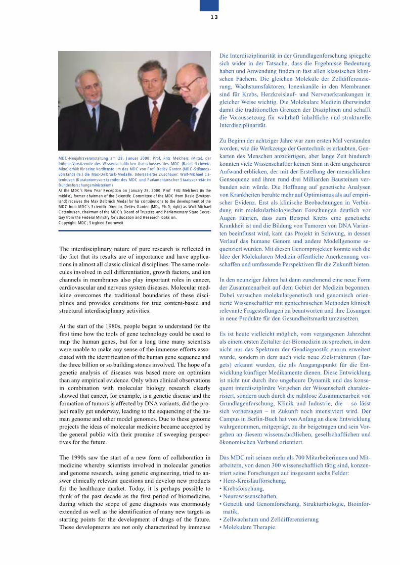
MDC-Neujahrsveranstaltung am 28. Januar 2000: Prof. Fritz Melchers (Mitte), derfrühere Vorsitzende des Wissenschaftlichen Ausschusses des MDC (Basel, Schweiz,Mitte) erhält für seine Verdienste um das MDC von Prof. Detlev Ganten (MDC-Stiftungs-vorstand) (re.) die Max-Delbrück-Medaille. Interessierter Zuschauer: Wolf-Michael Ca-tenhusen (Kuratoriumsvorsitzender des MDC und Parlamentarischer Staatssekretär imBundesforschungsministerium).At the MDC`s New Year Reception on January 28, 2000: Prof Fritz Melchers (in themiddle), former chairman of the Scientific Committee of the MDC from Basle (Switzer-land) receives the Max Delbrück Medal for his contributions to the development of theMDC from MDC`s Scientific Director, Detlev Ganten (MD., Ph.D; right) as Wolf-MichaelCatenhusen, chairman of the MDC`s Board of Trustees and Parliamentary State Secre-tary from the Federal Ministry for Education and Research looks on. Copyright: MDC; Siegfried Endruweit
dynamic and subsequent interdisciplinary scientific activitiesbut also by the ready collaboration between pure research, theclinics and industry, which – as previously said – will be evenmore intense in the future. Right from the start, the Berlin-Buch Campus has recognized the importance of this and beenan enthusiastic participant by adopting a perspective that com-bines scientific, social and economic attitudes.
The MDC, with over 700 staff, 300 of whom are engaged inscientific work, is concentrating its research efforts in sixareas: • Cardiovascular research, • Cancer research, • Neurosciences, • Genetics and Genome Research, Structural-Biology, Bioin-
formatics, • Cell Growth and Cell Differentiation • Molecular Therapy.
These are described in detail in this Research Report. TheForschungsinstitut für Molekulare Pharmakologie with al-most 200 staff is collaborating closely with the MDC in itssearch for targets for new drugs. They are investigating thethree-dimensional structure of biological macromoleculesand using this information to characterize their pharmacolog-
Die Schwerpunkte werden in diesem Forschungsbericht imDetail vorgestellt.
Das Forschungsinstitut für Molekulare Pharmakologie (FMP)mit seinen knapp 200 Mitarbeiterinnen und Mitarbeitern, mitdem das MDC eng zusammenarbeitet, fahndet nach Ziel-punkten für neue Medikamente. Man versucht, biologischeMakromoleküle unter dieser Vorgabe in ihrer Raumstrukturaufzuklären und auf pharmakologische Wirkungen hin zucharakterisieren, ohne dabei die komplexe Biologie einerZelle aus den Augen zu verlieren.
Die Besonderheit des MDC besteht in der Orientierung seinerForschungen an den klinischen Fragestellungen, und die bei-den Kliniken, mit denen die Wissenschaftler kooperieren,sind die Robert-Rössle-Krebs-Klinik und die Franz-Volhard-Herz-Kreislauf-Klinik.
Oben wurde der Hinweis gegeben, dass das Humane Genom-projekt unter anderem deshalb in die Wege geleitet wurde,weil erkannt worden war, dass zahlreiche genetische Kompo-nenten zur Entwicklung von Krebs beitragen können. Inzwi-schen setzt sich die wissenschaftlich begründete Ansichtdurch, dass es für eine Behandlung besser ist, einen Tumor(Krebsgeschwulst) nicht mehr nur durch das Organ zu
14
The multitalented molecules of molecular medicine
C/EBPs flip a switch in chromatin remodelling and gene activa-tionThe human genome contains over 30,000 different genes. Expressionof various combinations of these genes gives rise to specific cell types,such as muscle, skin, blood or nerves, and also determines whethercells remain in a resting state, grow and proliferate or die and self-de-struct. Inappropriate gene regulation causes autoimmune diseases, avariety of other genetic diseases and cancer. Researchers in the MDClaboratory directed by Achim Leutz are interested in how cells makedecisions to differentiate and how these processes are linked to thedevelopment of disease, especially leukemia. By focusing on a familyof transcription factors called C/EBP proteins, which were originallyidentified as regulators of liver-specific genes but which are nowknown to be important regulators for many cell types, the MDC scien-tists have found that the ability of C/EBP proteins to influence variousprograms of differentiation depends on their ability to producechanges in chromatin structure. Chromatin is the higher order struc-ture of chromosomes in a cell that helps keep some genes inaccess-ible and inactive, while others are exposed and activated. The C/EBPproteins work in concert with other transcription factors to change thestructure of chromatin, in essence allowing some genes to switch froman inactive state to an activated one. The MDC research group hasshown that abrogation of the interaction between C/EBP and collabo-rating transcription factors, such as c-Myb, is an important event dur-ing leukemogenesis. Meanwhile, workers in other laboratories haveidentified frequent mutations in C/EBP genes in patients with severalhematological disorders. Researchers at the MDC believe that C/EBPproteins can integrate signals elicited by nutrients or growth factors,translating them into changes in chromatin structure, gene regulationand cell fate. As a key regulator of cellular differentiation, and an im-portant contributor to oncogenesis, C/EBP proteins are ideal candi-dates for the development of novel therapeutic agents.
Die vielseitigen Moleküle der Molekularen Medizin
C/EBP Proteine: Schalter der Chromatinumordnung undGenaktivierung Das menschliche Genom enthält mehr als 30.000 verschiedene Gene.Die Expression von verschiedenen Kombinationen dieser Gene führtnicht nur zu spezifischen Zelltypen wie etwa Muskel-, Haut-, Blut- undNervenzellen, sondern bestimmt auch, ob Zellen in einem Ruhezustandbleiben oder ob sie wachsen, sich teilen, sterben und selbst zerstören.Die ungeeignete Regulation von Genen kann zu Autoimmunkrankhei-ten und einer Reihe von anderen Genkrankheiten bis hin zu Krebsführen. Das von Achim Leutz geleitete MDC-Laboratorium ist an derFrage interessiert, wie Zellen Entscheidungen bei der Differenzierungtreffen und wie diese Vorgänge mit dem Entstehen von Krankheitenverbunden sind, wobei das besondere Augenmerk der Leukämie gilt.Indem sie sich auf eine Familie von Transkriptionsfaktoren mit NamenC/EBP Proteinen konzentriert haben, die ursprünglich als Regulatorenvon leberspezifischen Genen identifiziert worden waren und von denenman inzwischen weiß, dass sie wichtige Regulatoren für viele Zelltypensind, haben die Wissenschaftler am MDC herausfinden können, dassdie Fähigkeit der C/EBP Proteine, verschiedene Differenzierungspro-gramme zu beeinflussen, auf ihrem Vermögen beruht, Änderungen derChromatinstruktur zu bewirken. Chromatin heißt die höhere Ordnungs-struktur der Chromosomen in einer Zelle, mit deren Hilfe einige Geneunzugänglich und inaktiv gehalten werden, während andere exponiertund aktiviert werden. Die C/EBP Proteine arbeiten mit anderen Trans-kriptionsfaktoren zusammen, um die Struktur des Chromatins zuändern, wobei vor allem einige Gene ihren inaktiven Zustand verlassenund aktiv werden. Die MDC Forschungsgruppe hat gezeigt, daß dieAufhebung der Wechselwirkung zwischen den C/EBP Proteinen undanderen Transkriptionsfaktoren wie etwa c-Myb ein wichtiger Schrittwährend der Entstehung der Leukämie ist. In der Zwischenzeit habenandere Laboratorien häufige Mutationen in C/EBP Genen in Patientenmit mehreren hämatologischen Störungen identifiziert. Forscher amMDC glauben, daß C/EBP Proteine Signale integrieren könnten, dievon Nährstoffen oder Wachstumsfaktoren stammen, um sie in Ände-rungen der Chromatinstruktur, der Genregulation und des Schicksalseiner Zelle umzusetzen. Als Schlüsselmoleküle der Zelldifferenzierungund als wichtige Faktoren der Onkogenese sind die C/EBP Proteineideale Kandidaten für die Entwicklung neuartiger therapeutischerSubstanzen.
ical effects, without losing sight of the complex biologicalprocesses that take place within cells. The uniqueness of the MDC lies in the application of its re-search efforts to answer clinical questions, and the two clin-ics with which the researchers collaborate are the RobertRössle Cancer Center and the Franz Volhard Clinic for Cardio-vascular Diseases.
It was mentioned earlier that the Human Genome Project, dueto the way it was conducted, showed that many genetic com-ponents could contribute to the development of cancer. Sincethen, researchers have found that it is better for treatment if atumor is no longer just characterized by the organ in which itoccurs. It is much more desirable to carry out a precise anal-ysis of as many genes as possible in the cancerous trans-formed cells and associated tissues, as is possible today withgene chips. Now scientists talk about the genetic profile of atumor and they are able to use this to develop more specifictreatments which are based on particular situations. The application of molecular medicine has a number of ad-vantages, such as a better choice of available drugs, since agenetic analysis can show which drugs have too narrow aspectrum of activity or highlight cases where undesirable sideeffects are likely to occur. Similar developments are expectedin the treatment of cardiovascular diseases, if researchersmanage to obtain more detailed information on the geneticcauses of cardiomyopathies or learn more about the molecu-lar mechanisms which lead to high blood pressure or heartfailure. These problems are the subject of intensive study at the MDCwhere pure scientists have to rely on simplified systems,models and animal experiments because of the complexity ofthe problems. In our laboratories genes have been identifiedwhich in altered (mutated) form can trigger changes in heartfunction, blood pressure regulation or the nervous systemwhich clinicians see everyday in their patients.
charakterisieren, in dem er entstanden ist. Vielmehr ist es ge-boten, in den krebsartig transformierten Zellen und dem er-krankten Gewebe eine genaue Analyse von möglichst vielenGenen durchzuführen, wie sie heute mit sogenannten Gen-chips gelingt. Man spricht dabei von dem genetischen Profileines Tumors und kann mit seiner Hilfe das Ziel einer Thera-pie in Angriff nehmen, die der individuellen Situation ange-messen und insofern massgeschneidert ist.
Der Ansatz der Molekularen Medizin erlaubt unter anderemeine bessere Auswahl unter den verfügbaren Medikamenten,da eine Genanalyse Hinweise gibt, welche Arzneistoffe übereine zu geringe Wirksamkeit verfügen bzw. in welchen Fällenunzumutbare Nebenwirkungen auftreten können. ÄhnlicheEntwicklungen sind für die Behandlung von Herz-Kreislauf-erkrankungen zu erwarten, wenn es gelingt, die genetischenUrsachen etwa von Kardiomyopathien genauer zu verstehenoder mehr von den molekularen Mechanismen zu erfahren, dieBluthochdruck- oder eine Herzinsuffizienz zur Folge haben.
Am MDC wird intensiv an diesen Fragen gearbeitet, wobeidie Grundlagenforschung wegen der Komplexität der Frage-stellungen auf reduktionistische Systeme, Modelle undTierversuche angewiesen bleibt. Dabei konnten in unserenLaboratorien Gene identifiziert werden, die in veränderter(mutierter) Form Störungen bei der Herzfunktion, Blutdruck-regulation oder im Nervensystem auslösen, wie sie den Klini-kern auch bei erkrankten Menschen bekannt sind.
Wenn die Forscher des MDC in Zusammenarbeit mit den Kli-niken erforschen, wie es möglich wird (und hoffentlich zuverhindern ist), dass aus einer Genvariante eine krankhafteStörung – Krebswachstum oder Herzkreislaufkrankheit –wird, dann legen sie ihren Überlegungen ein Konzept zugrun-de, das sich zum ersten Mal bei Max Delbrück findet. Es gehtum die Idee der Signalumwandlung bzw. der Signalkette und
15
A simple blood test may increase patient safety before genetherapy with adenovirusesAdenoviruses are the most widely used vectors in gene therapy stu-dies and their role is to transport the therapeutic genes to their site ofaction. Viruses are particularly versatile and effective gene transpor-ters. There are great expectations that they will useful in the treatrmentof cancer. However, two years ago, the 18-year-old Jesse Gelsingerdied suddenly in the USA from organ failure after he had received aninjection of a genetically modified adenovirus, used as a gene taxi, di-rectly into his bloodstream. The exact molecular biological cause of hisdeath remains unknown. However, new information about a previouslyunknown immune reaction to adenoviruses, or as “Science” commen-ted on January 25, 2002, “a new piece in the puzzle”, has now comefrom a research team led by Günter Cichon from the Humboldt Uni-versity, Berlin, at the Max Delbrück Center for Molecular Medicine(MDC) and Reinhard Burger from the Robert Koch Institute, Berlin,and published in the journal “Gene Therapy” (Vol. 8 (2001), pp. 1794-1800). Under laboratory conditions, high concentrations of the adeno-viruses used in gene therapy produce an unexpected intense activa-tion of the so-called complement system. The complement systemconsists of a group of proteins which circulate in the blood and act asan initial defense mechanism against an assault by infectious patho-gens. In order to increase patient safety, the authors suggest carryingout a simple blood test to measure the degree of the complementreaction before gene therapy.More detailed information can be obtained in the report of the Hum-boldt University research group at the MDC.
Ein einfacher Bluttest könnte die Sicherheit der Patienten voreiner Gentherapie mit Adenoviren erhöhenAdenoviren sind die am meisten benutzten Vektoren bei Gentherapie-Studien. Ihre Aufgabe ist es, therapeutische Gene an ihren Wirkort zutransportieren. Viren sind besonders vielseitige und wirksame Gen-taxis. Große Hoffnungen, z. B. bei der Behandlung von Krebs, grün-den sich auf deren Einsatz. Vor zwei Jahren starb jedoch plötzlich der18-jährige Jesse Gelsinger in den USA an Organversagen, nachdem ereine Injektion eines genetisch veränderten, als Gentaxi benutztenAdenovirus direkt in den Blutstrom erhalten hatte. Die genaue moleku-larbiologische Ursache seines Todes blieb bisher rätselhaft. Neue Infor-mationen über eine zuvor unbekannte Immunreaktion gegenüberAdenoviren, oder wie die „Science“ am 25. Januar 2002 kommentierte„ein neues Stück des Puzzles“ haben nun ein Team um Günter Cichonvon der Humboldt-Universität zu Berlin am Max-Delbrück-Centrum fürMolekulare Medizin (MDC) und Reinhard Burger vom Robert-Koch-Institut Berlin vorgelegt und in der Zeitschrift „Gene Therapy“ (Band 8(2001), pp. 1794–1800) publiziert. Unter Laborbedingungen könnendie bei der Gentherapie verwendeten hohen Konzentrationen anAdenoviren eine unerwartet heftige Aktivierung des so genanntenKomplementsystems hervorrufen. Dieses System besteht aus einerGruppe von Proteinen, die im Blut zirkulieren und gemeinsam infek-tiöse Pathogene angreifen. Um die Sicherheit von Patienten zu er-höhen, schlagen die Autoren vor, den Grad der Komplementreaktionkünftig vor einer Gentherapie mit einem einfachen Bluttest zu messen.Weitere Einzelheiten im Bericht der Forschungsgruppe der HumboldtUniversität am MDC.
If MDC researchers work in close collaboration with the clin-ics to discover how it is possible (and hopefully how it can beprevented) that a gene variant can cause a serious change –the growth of a tumor or cardiovascular disease – then theirconjectures will form the basis of a concept that was first pro-posed by Max Delbrück. This involves the idea of signaltransformation or the signal chain and signal transduction. Inthis context, Delbrück meant the molecular route that ledfrom an altered gene sequence via the changed gene product(protein), then via another intermediate information carrier tothe final result which manifested itself as a disease. As far asDelbrück was concerned, a mechanism could only be under-stood if the route of signal transfer was completely known.This has often led to a number of major surprises, such as justhow many different effects a signal molecule can exhibit.One example of this is “�-catenin”, which appears to have akey function in the life of cells.
Signaltransduktion. Delbrück meinte damit den molekularenWeg, der von einer veränderten Gensequenz über das dannveränderte Genprodukt (Protein) und weiter über andere Zwi-schenträger der Information bis zum Endergebnis führt, dassich als Krankheit zeigt. Für Delbrück war ein Mechanismuserst dann verstanden, wenn sich der Signalübertragungsweglückenlos nachvollziehen ließ. Dabei treten oft große Überra-schungen zutage, nämlich dann, wenn sich herausstellt, wievielfältig einige Signalmoleküle sein können. Ein Beispielhierfür ist das „Beta-Catenin“, das im Leben von Zellen eineSchlüsselfunktion zu haben scheint.
Unter dem oben genannten Gesichtspunkt der Signalum-wandlung lassen sich auch neurobiologische Arbeiten ausdem MDC anführen, in denen es nicht nur wie früher schonbei den Arbeiten von Timoféeff-Ressovsky um die Neuroge-nese – die Entwicklung des Nervensystems – geht, sondernmit denen die Mechanismen erkundet werden, die dem Ab-
16
The multitalented molecules of molecular medicine
One single signal molecule controls the programs responsiblefor the development of both hair and skin Adult skin stem cells are able to produce two different types of cells:they can develop into skin cells of the outer layer of the skin (epidermalcells) as well as hair follicles with the horn cells of the hair. MDC re-searchers Walter Birchmeier and Jörg Hülsken have now discoveredthe signal that drives the development potential of skin stem cells inthe direction of the skin as well as the signal that drives them in thedirection of hair. The answer is a surprising one in that the choice ofdirection involves a single signal molecule, �-catenin. This key mole-cule is well known to Walter Birchmeier´s group since it plays a centralrole in an important cell communication system, the Wnt/�-cateninsignal pathway. For years the group has been studying this signalpathway which controls the transfer of signals from the cell surface toits nucleus. For some time researchers have believed that �-cateninmust be involved in the formation of skin and hair. �-Catenin plays arole in a type of tissue related to hair, the feathers of birds. In addition,according to Birchmeier, mutations in other components of the Wnt-pathway in the mouse have provided “certain information” about de-fects in teeth, mammary glands and hair. However, absolutely nothingwas known about which cells were involved and how �-cateninworked. It was mice, which had been modified by Jörg Hülsken sothat only the �-catenin gene in their skin had been switched off (so-called conditional knock-out), that provided the explanation: thesemice lost their hair 20 to 30 days after birth. They then remainednaked, because they were unable to produce new hair follicles whichwould have led to the growth of new hair. In the adult, healthy animalthe hair follicles undergo a continuous process of self-renewal, whichis triggered by stem cells producing hair follicles. Only then can thehair continue to grow. In addition, it was found that knock-out micewere not just bald: the skin of these animals contained unusual cystscoated with stem cells. These cysts were produced by a defect instem cell regulation, which resulted in stem cells losing their ability toform hair follicle cells. Therefore, the results obtained by the MDC re-searchers showed two things: 1. Skin stem cells require �-catenin, inorder to produce hair follicles. 2. If there is no �–catenin available, thenthe stem cells cannot produce hair cells, only skin cells. The fact that�-catenin controls the development program for stem cells is a newdiscovery. Investigations by other groups have shown that the Wnt/�-catenin signal pathway also plays an important role in the control ofother stem cells (e.g. the Lieberkühn crypts in the small intestine). For further details see research group of Walter Birchmeier
Die vielseitigen Moleküle der Molekularen Medizin
Ein einziges Signalmolekül steuert die Entwicklungspro-gramme für Haut und Haare Adulte Stammzellen der Haut haben die Fähigkeit, zwei verschiedeneZelltypen auszubilden: Aus ihnen können sowohl Hautzellen der Ober-haut (epidermale Zellen) als auch Haarfollikel mit den Hornzellen derHaare hervorgehen. Welche Signale dieses Entwicklungspotenzialder Hautstammzellen einmal in Richtung Haut und einmal in RichtungHaar regulieren, haben Wissenschaftler des MDC um WalterBirchmeier und Jörg Hülsken herausgefunden. Dabei stellte sichüberraschenderweise heraus, dass für die Wahl der Richtung nur eineinziges Signalmolekül, das �-Catenin, ausschlaggebend ist. DiesesSchlüsselmolekül ist ein alter Bekannter von Walter Birchmeier´sGruppe, da es eine zentrale Rolle in einem wichtigen Kommunika-tionssystem der Zelle, dem Wnt/�-Catenin-Signalweg einnimmt. ZurKenntnis dieses Signalweges, der dafür sorgt, dass Signale von derOberfläche der Zelle in den Zellkern gelangen, hat die Gruppe seitJahren wesentlich beigetragen. Schon seit einiger Zeit hatte man da-her vermutet, dass �-Catenin in die Haut- und Haarbildung eingebun-den sein muss. So spielt �-Catenin bei einem dem Haar verwandtenOrgan, den Federn von Vögeln, eine Rolle. Auch Mutationen in ande-ren Komponenten des Wnt-Weges in der Maus haben, so Birchmeier,„gewisse Hinweise” auf Defekte in Zähnen, den Brustdrüsen undeben in den Haaren ergeben. Völlig unbekannt war jedoch, in welchenZellen und auf welche Weise �-Catenin wirkt. Erst Mäuse, bei denendurch Jörg Hülsken das �-Catenin-Gen nur in der Haut ausgeschaltetwurde (sogenannter konditionaler knock-out), lieferten eine Erklärung:solche Mäuse verloren 20 bis 30 Tage nach der Geburt ihr Haarkleid.Danach blieben sie nackt, weil sich keine neuen Haarfollikel mehr bil-den und damit auch kein Haar mehr nachwachsen konnte. Im er-wachsenen, gesunden Organismus sind die Haarfollikel einem ständi-gen Selbsterneuerungsprozess unterworfen, der durch Stammzellenan den Seiten der Haarfollikel ausgelöst wird. Nur dann kann das Haarständig nachwachsen. Weiterhin wurde festgestellt, dass Knock-out-Mäuse nicht bloß kahl waren: in der Haut der Tiere fanden sich un-gewöhnliche, mit Hautzellen ausgekleidete Zysten. Diese Zystenentstehen aus fehlgeleiteten Stammzellen, die ihre Fähigkeit verlorenhaben, Haarfollikelzellen zu bilden. Mit ihren Forschungen konnten dieMDC-Forscher demnach zweierlei zeigen: 1. Stammzellen der Hautbenötigen �-Catenin, um Haarfollikel zu bilden. 2. Fehlt �-Catenin, sobilden sich aus den Stammzellen keine Haare sondern lediglich Haut-zellen. Die Tatsache, dass �-Catenin die Entwicklungsprogramme vonStammzellen steuert, ist eine neue Erkenntnis. Untersuchungen ande-rer Gruppen haben ergeben, dass der Wnt/�-Catenin-Signalwegauch bei der Kontrolle anderer Stammzellen (z. B. der Lieberkühn-Krypten im Dünndarm) eine wichtige Rolle spielt. Weitere Details siehe Arbeitsgruppe Walter Birchmeier
From the above viewpoint of signal transfer, one can also citeneurobiological studies from the MDC, not only the earlywork involving the research of Timoféev-Ressovsky on neu-rogenesis – the development of the nervous system – but alsoon how those mechanisms are signaled, which are fundamen-tal for the death of brain tissue. For example, at the MDC, re-searchers are investigating the signals that are responsible forthe stability of the connections (synapses) between neuronsor initiate their destruction. Scientists are also carrying outresearch on glial cells and neurons and the sites where thesignals manifest their effects (receptors), as well as the genesthat are involved in the production of the relevant proteins.
The entrepreneurial side
Alongside these research facilities and clinics, over forty bio-tech-oriented companies have set themselves up in Berlin-Buch using the Campus as a biotechnology park and, togetherwith the associated service companies and supply firms, theyemploy over 550 staff. These entrepreneurial activities arecoordinated by “BBB Management GmbH Campus BerlinBuch” which was set up in 1995. Since 1998, it has operatedan Innovation and Start-up Center (German abbrev. IGZ), thesecond stage of which was completed in Spring 2001. Withits help, it should be easier for scientists to transfer the resultsof their basic research into useful applications and assist inthe commercial exploitation of their business ideas. The bio-tech companies, which are located on the Buch Campus offera variety of new techniques, for example, to improve the di-agnosis of myocardial infarction; they are also trying to linkthe vast synthetic ability of micro-organisms with modern
sterben von Hirngewebe zugrunde liegen. Gefragt wird amMDC konkret zum Beispiel nach den Signalen, die für dieStabilität der Verbindungen (Synapsen) zwischen Neuronensorgen oder ihren Abbau in die Wege leiten und wie sichSynapsen bei der Entwicklung des Gehirns entfalten. Er-forscht wird auch die Funktion von Gliazellen und Neuronenund ihren Rezeptoren. Weiter wird erforscht, welche Gene ander Herstellung der entsprechenden Proteine beteiligt sind.
Die unternehmerische Seite
Neben den genannten Forschungseinrichtungen und den Kli-niken haben sich in Berlin-Buch mehr als vierzig biotech-nisch ausgerichtete Firmen eingerichtet, die den Campus alsBiotechnologiepark nutzen und zusammen mit dazugehöri-gen Dienstleistungsunternehmen und Zulieferfirmen mehr als550 Angestellte haben. Koordiniert werden die unternehmeri-schen Aktivitäten durch die BBB Management GmbH, die1995 gegründet worden ist. Sie betreibt seit 1998 ein Innova-tions- und Gründerzentrum (IGZ), dessen zweite Baustufe imFrühjahr 2001 fertig geworden ist. Mit seiner Hilfe soll es fürWissenschaftler leichter werden, Ergebnisse aus der Grundla-genforschung in verwertbare Anwendungen zu überführenund ihrer Geschäftsidee den Weg auf den Markt zu ermögli-chen (Existenzgründungen). Die Biotechfirmen, die auf demCampus in Buch vertreten sind, bieten unter anderem neueVerfahren an, um Herzinfarkte besser diagnostizieren zu kön-nen; sie versuchen, die vielfältigen Syntheseleistungen vonMikroorganismen mit den modernen Methoden der Genom-analyse zu verbinden, um rascher Zugang zu bioaktivenSubstanzen und damit zu neuen Medikamenten zu bekom-
17
The multitalented molecules of molecular medicine
Cholesterol in the brain: Making or breaking synapses“The smooth operation of the nervous system depends on rapid com-munication between nerve cells at meeting areas called synapses. Al-though synapses were first identified 100 years ago, their formation, aprocess called synaptogenesis, has remained something of a mystery.For example, it is still not clear how many synapses a neuron canmake with other nerve cells. Is the number of synapses rigidly prepro-grammed or is it fluid, governed by interactions with neighboring cells?Thanks to a simple model system in which a defined type of neuronfrom the central nervous system is purified and cultured, two intriguingbut unanticipated conclusions about synaptogenesis have beenreached. The first is that neurons by themselves form few synapsesunless they have help from other nerve cells called glial cells, the sec-ond is that the synapse-promoting signal released by glial cells is cho-lesterol.” Cholesterol, hitherto known as an essential neutral lipid con-stituent, makes its “nervous debut”.“These studies suggest that glial cells, long thought to be passive by-standers in the formation and operation of our neural circuitry, activelyparticipate in the making and breaking of synapses.”Excerpted from a comment of B.A. Barres and S.J. Smith (StanfordUniversity) to papers from Pfrieger´s group at the MDC - SCIENCE 294(2001), 1296-97So, cholesterol is not only of interest to cardiovascular researchers be-cause of the way it coats arteries, it is also being studied by neurolo-gists. Further details of this are given in the report of the work of FrankPfrieger and his group.
Die vielseitigen Moleküle der Molekularen Medizin
Das Cholesterol im Gehirn: Aufbau oder Abbau der Nerven-übertragung„Das reibungslose Operieren des Nervensystems hängt von der ra-schen Kommunikation zwischen Nervenzellen ab, die sich an Ortenbegegnen, die Synapsen heißen. Obwohl Synapsen zum ersten Malvor 100 Jahren identifiziert wurden, stellen ihre Formation und der da-zugehörige Prozeß der Synaptogenese nach wie vor ein Geheimnisdar. So ist zum Beispiel nicht klar, wie viele Synapsen ein Neuron mitanderen Nervenzellen ausbilden kann. Ist die Zahl der Synapsen starrprogrammiert, oder ist sie flexibel in Abhängigkeit von der Wechsel-wirkung mit benachbarten Zellen? Dank eines einfachen Modellsys-tems, in dem eine bestimmte Art von Neuron aus dem zentralen Ner-vensystem gereinigt und kultiviert wird, konnten gleich zwei verblüf-fende und unerwartete Schlüsse über die Synaptogenese gezogenwerden. Der erste besteht darin, dass Neuronen selbst nur wenigeneue Synapsen ausbilden, solange sie keine Hilfe von anderen Ner-venzellen namens Gliazellen bekommen, und der zweite bestehtdarin, dass das Signal, das Gliazellen freisetzen, um die Bildung vonSynapsen anzuregen, das Cholesterin ist, das auf diese Weise seinDebüt im Nervensystem macht.„Diese Experimente legen den Gedanken nah, daß die Gliazellen, dieman lange als passive Zuschauer sowohl bei der Bildung von Synap-sen als auch bei der Operation des Nervensystems angesehen hat,sich vielmehr aktiv am Auf- und Abbau der Synapsen beteiligen.“Auszüge aus einem Kommentar von B. A. Barres und S. J. Smith(Stanford Universität) zu Arbeiten der Pfrieger Gruppe vom MDC:SCIENCE 294 (2001), 1296–97 Cholesterin interessiert also nicht nur die Herz-Kreislaufforscher be-züglich der Entstehung von Arterienverkalkung, sondern auch die Hirn-forscher. Näheres im Bericht der Arbeitsgruppe von Frank Pfrieger.
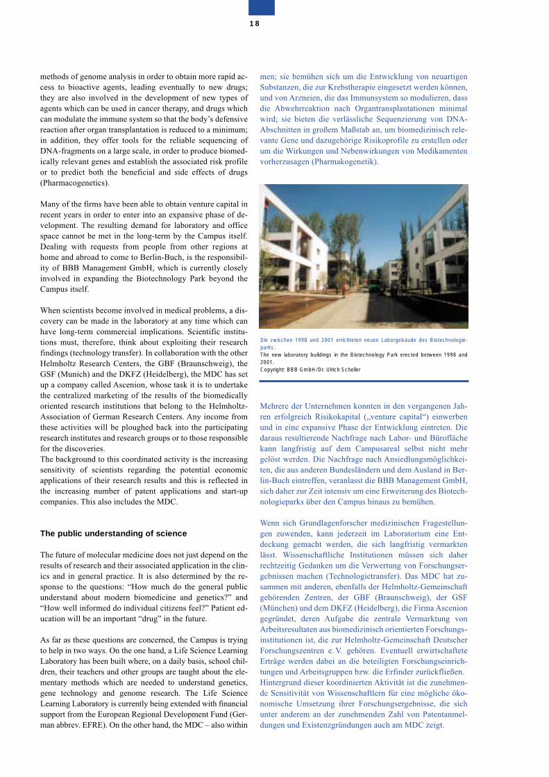
methods of genome analysis in order to obtain more rapid ac-cess to bioactive agents, leading eventually to new drugs;they are also involved in the development of new types ofagents which can be used in cancer therapy, and drugs whichcan modulate the immune system so that the body’s defensivereaction after organ transplantation is reduced to a minimum;in addition, they offer tools for the reliable sequencing ofDNA-fragments on a large scale, in order to produce biomed-ically relevant genes and establish the associated risk profileor to predict both the beneficial and side effects of drugs(Pharmacogenetics).
Many of the firms have been able to obtain venture capital inrecent years in order to enter into an expansive phase of de-velopment. The resulting demand for laboratory and officespace cannot be met in the long-term by the Campus itself.Dealing with requests from people from other regions athome and abroad to come to Berlin-Buch, is the responsibil-ity of BBB Management GmbH, which is currently closelyinvolved in expanding the Biotechnology Park beyond theCampus itself.
When scientists become involved in medical problems, a dis-covery can be made in the laboratory at any time which canhave long-term commercial implications. Scientific institu-tions must, therefore, think about exploiting their researchfindings (technology transfer). In collaboration with the otherHelmholtz Research Centers, the GBF (Braunschweig), theGSF (Munich) and the DKFZ (Heidelberg), the MDC has setup a company called Ascenion, whose task it is to undertakethe centralized marketing of the results of the biomedicallyoriented research institutions that belong to the Helmholtz-Association of German Research Centers. Any income fromthese activities will be ploughed back into the participatingresearch institutes and research groups or to those responsiblefor the discoveries.The background to this coordinated activity is the increasingsensitivity of scientists regarding the potential economicapplications of their research results and this is reflected inthe increasing number of patent applications and start-upcompanies. This also includes the MDC.
The public understanding of science
The future of molecular medicine does not just depend on theresults of research and their associated application in the clin-ics and in general practice. It is also determined by the re-sponse to the questions: “How much do the general publicunderstand about modern biomedicine and genetics?” and“How well informed do individual citizens feel?” Patient ed-ucation will be an important “drug” in the future.
As far as these questions are concerned, the Campus is tryingto help in two ways. On the one hand, a Life Science LearningLaboratory has been built where, on a daily basis, school chil-dren, their teachers and other groups are taught about the ele-mentary methods which are needed to understand genetics,gene technology and genome research. The Life ScienceLearning Laboratory is currently being extended with financialsupport from the European Regional Development Fund (Ger-man abbrev. EFRE). On the other hand, the MDC – also within
men; sie bemühen sich um die Entwicklung von neuartigenSubstanzen, die zur Krebstherapie eingesetzt werden können,und von Arzneien, die das Immunsystem so modulieren, dassdie Abwehrreaktion nach Organtransplantationen minimalwird; sie bieten die verlässliche Sequenzierung von DNA-Abschnitten in großem Maßstab an, um biomedizinisch rele-vante Gene und dazugehörige Risikoprofile zu erstellen oderum die Wirkungen und Nebenwirkungen von Medikamentenvorherzusagen (Pharmakogenetik).
Mehrere der Unternehmen konnten in den vergangenen Jah-ren erfolgreich Risikokapital („venture capital“) einwerbenund in eine expansive Phase der Entwicklung eintreten. Diedaraus resultierende Nachfrage nach Labor- und Büroflächekann langfristig auf dem Campusareal selbst nicht mehrgelöst werden. Die Nachfrage nach Ansiedlungsmöglichkei-ten, die aus anderen Bundesländern und dem Ausland in Ber-lin-Buch eintreffen, veranlasst die BBB Management GmbH,sich daher zur Zeit intensiv um eine Erweiterung des Biotech-nologieparks über den Campus hinaus zu bemühen.
Wenn sich Grundlagenforscher medizinischen Fragestellun-gen zuwenden, kann jederzeit im Laboratorium eine Ent-deckung gemacht werden, die sich langfristig vermarktenlässt. Wissenschaftliche Institutionen müssen sich daherrechtzeitig Gedanken um die Verwertung von Forschungser-gebnissen machen (Technologietransfer). Das MDC hat zu-sammen mit anderen, ebenfalls der Helmholtz-Gemeinschaftgehörenden Zentren, der GBF (Braunschweig), der GSF(München) und dem DKFZ (Heidelberg), die Firma Asceniongegründet, deren Aufgabe die zentrale Vermarktung vonArbeitsresultaten aus biomedizinisch orientierten Forschungs-institutionen ist, die zur Helmholtz-Gemeinschaft DeutscherForschungszentren e. V. gehören. Eventuell erwirtschafteteErträge werden dabei an die beteiligten Forschungseinrich-tungen und Arbeitsgruppen bzw. die Erfinder zurückfließen.Hintergrund dieser koordinierten Aktivität ist die zunehmen-de Sensitivität von Wissenschaftlern für eine mögliche öko-nomische Umsetzung ihrer Forschungsergebnisse, die sichunter anderem an der zunehmenden Zahl von Patentanmel-dungen und Existenzgründungen auch am MDC zeigt.
18
Die zwischen 1998 und 2001 errichteten neuen Laborgebäude des Biotechnologie-parks.The new laboratory buildings in the Biotechnology Park erected between 1998 and2001.Copyright: BBB GmbH/Dr. Ulrich Scheller
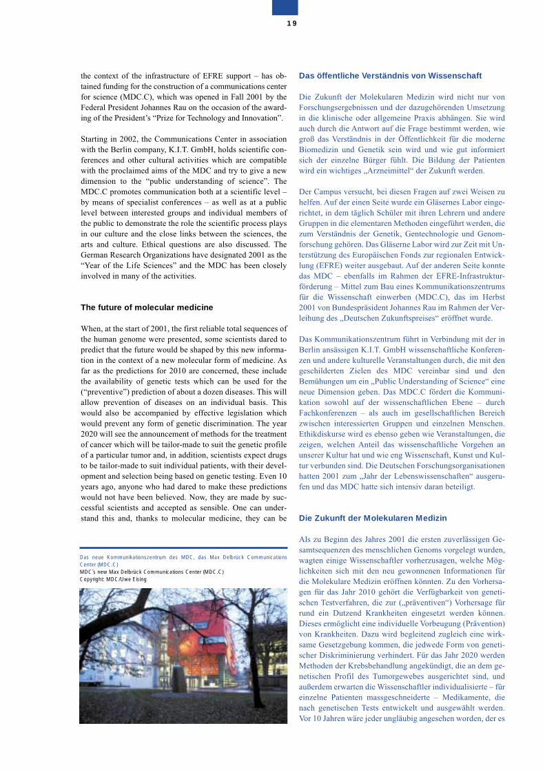
the context of the infrastructure of EFRE support – has ob-tained funding for the construction of a communications centerfor science (MDC.C), which was opened in Fall 2001 by theFederal President Johannes Rau on the occasion of the award-ing of the President’s “Prize for Technology and Innovation”.
Starting in 2002, the Communications Center in associationwith the Berlin company, K.I.T. GmbH, holds scientific con-ferences and other cultural activities which are compatiblewith the proclaimed aims of the MDC and try to give a newdimension to the “public understanding of science”. TheMDC.C promotes communication both at a scientific level –by means of specialist conferences – as well as at a publiclevel between interested groups and individual members ofthe public to demonstrate the role the scientific process playsin our culture and the close links between the sciences, thearts and culture. Ethical questions are also discussed. TheGerman Research Organizations have designated 2001 as the“Year of the Life Sciences” and the MDC has been closelyinvolved in many of the activities.
The future of molecular medicine
When, at the start of 2001, the first reliable total sequences ofthe human genome were presented, some scientists dared topredict that the future would be shaped by this new informa-tion in the context of a new molecular form of medicine. Asfar as the predictions for 2010 are concerned, these includethe availability of genetic tests which can be used for the(“preventive”) prediction of about a dozen diseases. This willallow prevention of diseases on an individual basis. Thiswould also be accompanied by effective legislation whichwould prevent any form of genetic discrimination. The year2020 will see the announcement of methods for the treatmentof cancer which will be tailor-made to suit the genetic profileof a particular tumor and, in addition, scientists expect drugsto be tailor-made to suit individual patients, with their devel-opment and selection being based on genetic testing. Even 10years ago, anyone who had dared to make these predictionswould not have been believed. Now, they are made by suc-cessful scientists and accepted as sensible. One can under-stand this and, thanks to molecular medicine, they can be
Das öffentliche Verständnis von Wissenschaft
Die Zukunft der Molekularen Medizin wird nicht nur vonForschungsergebnissen und der dazugehörenden Umsetzungin die klinische oder allgemeine Praxis abhängen. Sie wirdauch durch die Antwort auf die Frage bestimmt werden, wiegroß das Verständnis in der Öffentlichkeit für die moderneBiomedizin und Genetik sein wird und wie gut informiertsich der einzelne Bürger fühlt. Die Bildung der Patientenwird ein wichtiges „Arzneimittel“ der Zukunft werden.
Der Campus versucht, bei diesen Fragen auf zwei Weisen zuhelfen. Auf der einen Seite wurde ein Gläsernes Labor einge-richtet, in dem täglich Schüler mit ihren Lehrern und andereGruppen in die elementaren Methoden eingeführt werden, diezum Verständnis der Genetik, Gentechnologie und Genom-forschung gehören. Das Gläserne Labor wird zur Zeit mit Un-terstützung des Europäischen Fonds zur regionalen Entwick-lung (EFRE) weiter ausgebaut. Auf der anderen Seite konntedas MDC – ebenfalls im Rahmen der EFRE-Infrastruktur-förderung – Mittel zum Bau eines Kommunikationszentrumsfür die Wissenschaft einwerben (MDC.C), das im Herbst2001 von Bundespräsident Johannes Rau im Rahmen der Ver-leihung des „Deutschen Zukunftspreises“ eröffnet wurde.
Das Kommunikationszentrum führt in Verbindung mit der inBerlin ansässigen K.I.T. GmbH wissenschaftliche Konferen-zen und andere kulturelle Veranstaltungen durch, die mit dengeschilderten Zielen des MDC vereinbar sind und denBemühungen um ein „Public Understanding of Science“ eineneue Dimension geben. Das MDC.C fördert die Kommuni-kation sowohl auf der wissenschaftlichen Ebene – durchFachkonferenzen – als auch im gesellschaftlichen Bereichzwischen interessierten Gruppen und einzelnen Menschen.Ethikdiskurse wird es ebenso geben wie Veranstaltungen, diezeigen, welchen Anteil das wissenschaftliche Vorgehen anunserer Kultur hat und wie eng Wissenschaft, Kunst und Kul-tur verbunden sind. Die Deutschen Forschungsorganisationenhatten 2001 zum „Jahr der Lebenswissenschaften“ ausgeru-fen und das MDC hatte sich intensiv daran beteiligt.
Die Zukunft der Molekularen Medizin
Als zu Beginn des Jahres 2001 die ersten zuverlässigen Ge-samtsequenzen des menschlichen Genoms vorgelegt wurden,wagten einige Wissenschaftler vorherzusagen, welche Mög-lichkeiten sich mit den neu gewonnenen Informationen fürdie Molekulare Medizin eröffnen könnten. Zu den Vorhersa-gen für das Jahr 2010 gehört die Verfügbarkeit von geneti-schen Testverfahren, die zur („präventiven“) Vorhersage fürrund ein Dutzend Krankheiten eingesetzt werden können.Dieses ermöglicht eine individuelle Vorbeugung (Prävention)von Krankheiten. Dazu wird begleitend zugleich eine wirk-same Gesetzgebung kommen, die jedwede Form von geneti-scher Diskriminierung verhindert. Für das Jahr 2020 werdenMethoden der Krebsbehandlung angekündigt, die an dem ge-netischen Profil des Tumorgewebes ausgerichtet sind, undaußerdem erwarten die Wissenschaftler individualisierte – füreinzelne Patienten massgeschneiderte – Medikamente, dienach genetischen Tests entwickelt und ausgewählt werden.Vor 10 Jahren wäre jeder ungläubig angesehen worden, der es
19
Das neue Kommunikationszentrum des MDC, das Max Delbrück CommunicationsCenter (MDC.C)MDC`s new Max Delbrück Communications Center (MDC.C)Copyright: MDC/Uwe Eising
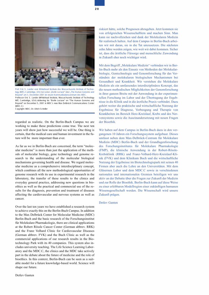
regarded as realistic. On the Berlin-Buch Campus we areworking to make these predictions come true. The next tenyears will show just how successful we will be. One thing iscertain, that the medical care and human investment in the fu-ture will be more important than ever.
As far as we in Berlin-Buch are concerned, the term “molec-ular medicine” is more than just the application of the meth-ods of molecular biology, gene technology and genome re-search to the understanding of the molecular biologicalmechanisms governing health and disease. We regard molec-ular medicine as a comprehensive interdisciplinary concept,which combines all the new methodological opportunities ofgenome research with its use in experimental research in thelaboratory, the transfer of these results to the clinics andeveryday general practice, addressing new questions in bio-ethics as well as the practical and commercial use of the re-sults for the diagnosis, prevention and treatment of diseasesaffecting the cardiovascular and nervous systems as well ascancer.
Over the last ten years we have established a research systemto achieve exactly this on the Berlin-Buch Campus. In additionto the Max Delbrück Center for Molecular Medicine (MDC)Berlin-Buch and the basic research of the Forschungsinstitutfür Molekulare Pharmakologie, there are clinical applicationsat the Robert Rössle Cancer Center (German abbrev. RRK)and the Franz Volhard Clinic for Cardiovascular Diseases(German abbrev. FVK) and the Buch Clinic as well as thecommercial applications of our research results in the Bio-technology Park with its 40 companies. This system also in-cludes university teaching. The Life Science Learning Labor-atory and the MDC.C, the clinics and the MDC take activelypart in the debate about the future of medicine and the role ofbioethics. In this context, Berlin-Buch can be seen as a suit-able model for a future knowledge-based world. Science willshape our future.
Detlev Ganten
riskiert hätte, solche Prognosen abzugeben. Jetzt kommen sievon erfolgreichen Wissenschaftlern und machen Sinn. Mankann sie nachvollziehen und dank der Molekularen Medizinfür realistisch halten. Auf dem Campus in Berlin-Buch arbei-ten wir mit daran, sie in die Tat umzusetzen. Die nächstenzehn Jahre werden zeigen, wie weit wir dabei kommen. Sicherist, dass die ärztliche Fürsorge und menschliche Zuwendungin Zukunft eher noch wichtiger wird.
Mit dem Begriff „Molekulare Medizin“ verbinden wir in Ber-lin-Buch mehr als den Einsatz von Methoden der Molekular-biologie, Gentechnologie und Genomforschung für das Ver-ständnis der molekularen biologischen Mechanismen beiGesundheit und Krankheit. Wir verstehen die MolekulareMedizin als ein umfassendes interdisziplinäres Konzept, dasdie neuen methodischen Möglichkeiten der Genomforschungin ihrer ganzen Breite mit der Anwendung in der experimen-tellen Forschung im Labor und der Übertragung der Ergeb-nisse in die Klinik und in die ärztliche Praxis verbindet. Dazugehört weiter die praktische und wirtschaftliche Nutzung derErgebnisse für Diagnose, Vorbeugung und Therapie vonKrankheiten im Bereich Herz-Kreislauf, Krebs und des Ner-vensystems sowie die Auseinandersetzung mit neuen Fragender Bioethik.
Wir haben auf dem Campus in Berlin-Buch dazu in den ver-gangenen 10 Jahren ein Forschungssystem aufgebaut. Diesesumfasst neben dem Max-Delbrück-Centrum für MolekulareMedizin (MDC) Berlin-Buch und der Grundlagenforschungdes Forschungsinstitutes für Molekulare Pharmakologie(FMP), die klinische Anwendung in der Robert-Rössle-Krebsklinik (RRK) und Franz-Volhard-Herz-Kreislauf-Kli-nik (FVK) und dem Klinikum Buch und die wirtschaftlicheNutzung der Ergebnisse im Biotechnologiepark mit seinen 40Firmen aber auch die Lehre an den Universitäten. Mit demGläsernen Labor und dem MDC.C sowie in verschiedenennationalen und internationalen Gremien beteiligen wir unsaktiv an der Debatte über die Fragen zur Zukunft der Medizinund zur Rolle der Bioethik. Berlin-Buch kann auf diese Weisezu einer erlebbaren Modellregion einer zukünftigen humanenWissensgesellschaft werden. Die Wissenschaft wird unsereZukunft prägen.
Detlev Ganten
20
Prof. Eric S. Lander vom Whitehead Institute des Massachusetts Institute of Techno-logy (MIT), Cambridge, USA bei seiner „Berlin Lecture“ über „The Human Genome andBeyond“ am 5. Dezember 2001 im neuen Kommunikationszentrum des MDC.Professor Eric S. Lander (Whitehead Institute, Massachusetts Institute of TechnologyMIT, Cambridge, USA) delivering his “Berlin Lecture” on “The Human Genome andBeyond” on December 5, 2001 in MDC`s new Max Delbrück Communications Center(MDC.C)Copyright: MDC; Dr. Ulrich Scheller
Overview
Clinical Research
The collaboration between the MDC and the two universityclinics, the Franz Volhard Clinic for Cardiovascular Diseases(FVK) and Robert Rössle Cancer Center (RRK), have devel-oped in an exceedingly satisfactory manner in recent years.Annually, the MDC makes available about 7 million Eurofrom its budget for joint research projects. The following pro-cedures have been set up to allocate MDC research funds toclinics:
The four heads of departments of the FVK and RRK alsohead research groups at the MDC. This means that the clini-cians are part of the MDC research activities and memberswith equal rights on all committees of the MDC, includingthe management working party. This ensures that there is anunhindered institutional link between clinical and basic re-search. The same refers to teaching activities at the univer-sities.
Clinical Research Units (CRUs) form the basis of collabora-tive projects. These CRUs are not involved in routine patientcare but are exclusively intended for diagnostic proceduresand the scientific investigation of patients required as part ofthe collaborative projects. In this respect, they represent anunusual feature of the research structure of a university clinic.Examples of MDC-funded measures include the equipping ofa center for the identification of genetic diseases and for es-tablishing experimental protocols in the field of circulatorydiseases at the FVK as well as setting up a tumor bank at theRRK. This is available to all researchers for their researchprojects and provides a useful service involving future high-throughput procedures based on DNA chip technology tohelp identify altered disease genes.
All the research funding which the MDC provides for Collab-orative Projects (“Twinning Grants”) between clinicians andbasic research groups is monitored by internal and externalexperts.
Überblick
Klinische Forschung
Die Zusammenarbeit zwischen dem MDC und den beidenUniversitätskliniken – der Franz-Volhard-Klinik für Herz-Kreislauferkrankungen (FVK) und der Robert-Rössle-Krebs-klinik (RRK) hat sich in den vergangenen Jahren sehr gut ent-wickelt. Das MDC stellt jährlich etwa sieben Millionen Euroaus seinem Budget für gemeinsame Forschungsvorhaben zurVerfügung. Für die Vergabe von MDC-Forschungsgeldern indie Kliniken wurden folgende Verfahren eingerichtet:
Die vier leitenden Ärzte der FVK und der RRK leiten gleich-zeitig eine eigene Forschungsgruppe am MDC. Als MDC-Forschungsgruppenleiter sind sie zugleich Mitglieder in allenMDC-Gremien einschließlich des Leitungskollegiums. Da-mit wird eine institutionelle Verbindung zwischen Klinik undGrundlagenforschung sichergestellt.
Klinische Forschungsgruppen bilden die Basis der gemeinsa-men Projekte. Sie beteiligen sich nicht an der Routineversor-gung der Patienten, sondern ausschließlich an diagnostischenVerfahren und wissenschaftlichen Untersuchungen von Pati-enten im Rahmen von gemeinsamen Forschungsprojekten.Sie stellen in dieser Hinsicht eine Besonderheit in der For-schungsstruktur einer Universitätsklinik dar.
Zu den Beispielen der vom MDC geförderten Maßnahmengehören die Ausstattung eines Zentrums für die Identifizie-rung genetisch bedingter Krankheiten sowie die Etablierungexperimenteller Protokolle auf dem Gebiet der Herz-Kreis-lauferkrankungen an der FVK oder einer Tumorbank an derRRK. Diese Einrichtungen stehen allen Forschern für ihreArbeiten zur Verfügung. Sie sind ein nützliches Instrumentauch für die künftig verfügbaren Verfahren mit hohen Durch-laufkapazitäten, die auf der Chip-Technologie basieren unddabei helfen, veränderte Gene, die eine Bedeutung für die Ent-stehung von Krankheiten haben könnten, zu identifizieren.
Die gesamte Forschungsförderung, die das MDC für die ge-meinsamen Projekte („Twinning Grants“) von Klinikern und
21
The clinical research supported by MDC funds is based ex-clusively on research projects. These projects obtain financialsupport for research-associated additional expenditure pro-vided that it involves collaborative projects between theMDC and the clinics. These projects are proposed followingclose agreement with the coordination sectors of the MDCand, as far as the topics are concerned, they are classifiedunder the research specialities of molecular therapy, genetics,cell growth and differentiation as well as neurosciences. Ingeneral, such projects run for 2–3 years. The maximum pe-riod of funding is 5 years.
This allows the application of research methods under qual-ity-controlled conditions and the support of collaborationbetween basic research and the clinics. In addition, the twoclinics have the opportunity to gain access to third-partyfunding, independently of the MDC, to help support theirclinical research.
The fundamental idea behind a special clinical training pro-gram for young doctors is to use targeted trainee sponsorshipof doctors who have completed their clinical training to facil-itate the switch to basic research at the MDC and to lay thefoundation for their further scientific development. At present16 young physicians are taking part in this training program.These four MDC-sponsored programs for clinical research
Gruppen der Grundlagenforschung aufbietet, wird von inter-nen und externen Experten überwacht.
Die Finanzierung der klinischen Forschung aus MDC-Mit-teln erfolgt ausschließlich für gemeinsame Forschungspro-jekte mit dem MDC. Diese Projekte werden in enger Über-einstimmung mit dem MDC vorgeschlagen und thematischden Forschungsschwerpunkten des MDC zugeordnet, alsoder Molekularen Therapie, Genetik, Zellwachstum und Zell-differenzierung und den Neurowissenschaften. Im allgemei-nen haben diese Projekte eine Laufzeit von zwei bis drei Jah-ren. Die maximale Förderdauer liegt bei fünf Jahren.
Damit wird die Unterstützung der Zusammenarbeit zwischenGrundlagenforschung und klinischer Forschung sowie derEinsatz von Forschungsmethoden und ihrer Qualitätskon-trolle sichergestellt. Daneben haben die beiden Kliniken dieMöglichkeit, sich Zugang zur Drittmittelförderung – unab-hängig vom MDC – zu verschaffen.
Grundidee eines speziellen Ausbildungsprogramms für jungeÄrzte ist es, Medizinern mit abgeschlossener klinischer Aus-bildung gezielt den Übergang in die Grundlagenforschungam MDC zu erleichtern und die Basis für ihre weitere wis-senschaftliche Entwicklung zu legen. Zur Zeit nehmen 16junge Ärzte an diesem Ausbildungsprogramm teil.
22
The multitalented molecules of molecular medicine
NF-�B: a protein that switches on genesNF-�B is the name for a small group of related proteins that are to befound in all cells in the body. They normally exist in their inactive formin the cytosol but, after the external effect of a variety of signalingagents, like cytokines, growth factors or bacterial pathogens, theNF-�Bs are transported from the cytosol into the cell nucleus wherethey bind to the genomic DNA. This triggers the copying of a group ofgenes which results in a subsequent increase in the proteins coded bythese genes. These proteins, which are under the control of NF-�B, in-clude a number of important switching molecules, some of which con-trol cellular growth properties and programmed cell death, while oth-ers can trigger signal-regulated gene activation in other cells. A num-ber of studies have shown that NF-�B- regulated signal-dependentgene regulation is a pre-requisite for the correct functioning of the cellsof the immune system. No humoral or cellular immune reactions cantake place without NF-�B. Since NF-�B activates the expression ofcertain cytokines, such as tumor necrosis factor and a number ofinterleukins, it also plays a key role in chronic inflammation. NF-�B-as-sociated conditions include a wide spectrum of autoimmune and in-flammatory conditions, such as rheumatoid arthritis, psoriasis andother skin conditions, asthma, Crohn’s disease and atherosclerosis.Due to its anti-apoptotic and cell-division-activating effects, NF-�B isof general importance as a potential therapeutic target as far as the re-sistance of tumors to chemo- or radiotherapy is concerned. Defectivecellular control of NF-�B is considered to be a critical step in the path-ogenesis of certain lymphomas (e.g. Hodgkin’s disease) and leukemia. Biochemical and biological investigations of NF-�B have provided fun-damental information which is making an increasing impact on clinicalresearch. Due to its widespread clinical significance, MDC researchershave been studying NF-�B for a long time at both a basic level and incollaboration with its two clinics.For further details see the report of Claus Scheidereit´s research group
Die vielseitigen Moleküle der Molekularen Medizin
NF-�B: ein Eiweiß, das Gene anschaltetNF-�B bezeichnet eine kleine Gruppe verwandter Proteine, die in allenZelltypen des Körpers vorkommen. Dort befinden sie sich in inaktiverForm im Zytosol. Nach äußerer Einwirkung verschiedener als Signalewirkender Substanzen, wie Zytokine, Wachstumsfaktoren oder bakte-rieller Pathogene, werden die NF-�Bs aus dem Zytosol in den Zellkerntransportiert und binden dort an die genomische DNA. Hierdurch wirddie Abschrift einer Gruppe von Genen aktiviert, was zu einer nachfol-genden Zunahme der von diesen Genen kodierten Proteine führt.Diese durch NF-�B kontrollierten Proteine umfassen eine Anzahl vonwichtigen Schaltermolekülen, von denen einige das zelluläre Wachs-tumsverhalten und den programmierten Zelltod steuern, andere wie-derum eine Signal-vermittelte Genaktivierung in anderen Zellen auslö-sen können. Eine Anzahl von Untersuchungen haben gezeigt, daßNF-�B vermittelte Signal-abhängige Genregulation eine Vorausset-zung für die Funktionsweise von Zellen des Immunsystems ist. Wederhumorale noch zelluläre Immunreaktionen können ohne NF-�B zu-stande kommen. Da NF-�B die Expression von bestimmten Zytoki-nen, wie Tumornekrosisfaktor und eine Reihe von Interleukinen akti-viert, spielt es auch eine Schlüsselrolle bei chronischen Entzündun-gen. NF-�B-assoziierte Erkrankungen schließen ein weites Spektrumvon Autoimmun- und Entzündungserkrankungen ein, wie rheuma-toide Arthritis, Psoriasis und andere Dermatosen, Asthma, MorbusCrohn und Artherosclerose. Wegen seiner anti-apoptotischen sowiezellteilungs-aktivierenden Wirkungen erlangt NF-�B bei der Resistenzvon Tumoren gegen Chemo- oder Radiotherapie als mögliches thera-peutisches Target eine generelle Bedeutung. Eine defekte zelluläreKontrolle von NF-�B wird zudem bei bestimmten Lymphomen (z. B.Morbus Hodgkin) und Leukämien als einer der entscheidendenSchritte der Krankheitsentstehung angenommen. Biochemische und biologische Untersuchungen von NF-�B haben einumfassendes Grundlagenwissen geschaffen, welches in zunehmen-dem Maße auf die klinische Forschung Einfluß nimmt. Wegen der brei-ten klinischen Signifikanz werden am MDC seit längerem NF-�B For-schungsarbeiten nicht nur im Grundlagenlabor, sondern auch in Ko-operation mit den beiden Kliniken durchgeführt.Weitere Einzelheiten im Bericht der Arbeitsgruppe von Claus Scheidereit
form a unit and have been set up by agreed procedures toguarantee that outstanding clinical research projects can besupported for specific periods from MDC resources in theRRK and FVK.
To bring together the two different worlds of basic and clini-cal research was quite unique in Germany in 1992. However,this model of close collaboration between these two worldshas turned out to be very promising.
One example of this is the area of molecular therapy. TheMDC and the clinics have developed various strategies forgene therapeutic approaches. The MDC and the clinics havecome to an agreement with other working groups and institu-tions in conjunction with the Union of Clinical PharmacologyBerlin/Brandenburg and obtained substantial funding fromthe BMBF for a period of 8 years.
Moreover, laboratories meeting the good manufacturingpractice standards (GMP laboratories) have been set up in theMDC in conjunction with one partner of the pharmaceuticalindustry, Schering AG (Berlin). This GMP facility involvesfour laboratories for vector production and two for analysisand cell culture. It is also available for use in collaborativeprojects with other partners e.g. biotechnology companieslocated on the Campus.
Diese vier Programme für die klinische Forschung bildeneine Einheit. Sie sind nach gemeinsam aufgestellten Kriterieneingerichtet und begutachtet worden. Damit wird sicherge-stellt, dass hervorragende klinische Forschungsprojekte inder RRK und FVK zeitlich befristet mit MDC-Mitteln geför-dert werden können.
Es war im Deutschland des Jahres 1992 einzigartig, die bei-den so verschiedenen Welten von Grundlagenforschung undklinischer Forschung zusammenzubringen. Das Modell derengen Zusammenarbeit zwischen diesen beiden Bereichenhat sich jedoch viel versprechend entwickelt.
Ein Beispiel betrifft dabei das Gebiet der Molekularen Thera-pie. Das MDC und die Kliniken haben verschiedene gen-therapeutische Strategien entwickelt. Das MDC und die Kli-niken konnten sich zu diesen Arbeiten mit anderen Gruppenund Institutionen abstimmen und zusammen mit dem Ver-bund für Klinische Pharmakologie Berlin/Brandenburg er-hebliche Fördermittel des Bundes (BMBF) für den Zeitraumvon acht Jahren einwerben.
Darüber hinaus konnte das MDC mit einem Partner aus derpharmazeutischen Industrie – der Schering AG –, GMP-La-boratorien (GMP – Good Manufacturing Practice) einrichten.Diese GMP Einrichtung verfügt über vier Labore zur Vektor-produktion und zwei für Analyse und Zellkultur. Die Labora-torien stehen für Projekte mit anderen Partnern – wie etwaBiotechnologiefirmen auf dem Campus – zur Verfügung.
23
GTB GenTherapeutika Berlin-Buch GmbHGTB GenTherapeutika Berlin-Buch is a company specializing in themanufacture of clinical trial samples for clinical studies in phases I, IIand III. The trial samples are viral and non-viral vectors as well as lipo-somal carrier systems for gene therapy, cellular products and oncolyticviruses. It was founded in August 2000 and is a cooperation betweenthe pharmaceutical company Schering AG Berlin and the MaxDelbrück Center for Molecular Medicine (MDC) in Berlin-Buch. GTB GenTherapeutika is a contract development and manufacturingorganization. Besides manufacturing clinical trial samples, the com-pany also offers manufacture of non-GMP material for pre-clinicalstudies and development of lab scale processes for GMP-compatibleprocesses. As manufacturer of biopharmaceutical products, GTBGenTherapeutika has established a quality management and a qualityassurance system according to German laws and regulations govern-ing the manufacture of drugs. The manufacture facility has been established as a cleanroom facility.The facility consists of several cleanrooms which represent the clean-room classification A, B and D. Cleanroom areas of categories A andB are equipped and devised for aseptic works, the areas of category Dare for analytics (molecular-biological and cell-based analytics) and forproduct storage (cryo cell storage). The entire facility as well as the sin-gle processing steps are being monitored by a complex supervisionand documentation system. The cleanroom facility and the operationalequipment of the clean room areas are qualified, validated where nec-essary and in compliance with the current GMP guidelines of the EUand the FDA (USA). In addition, the facility is suitable for work with ge-netically modified organisms of Risk Group 2 (GenTG).
GTB GenTherapeutika Berlin-Buch GmbHDie GTB GenTherapeutika Berlin-Buch GmbH ist ein Unternehmen,das sich auf die Herstellung von Proben für klinische Versuche spezia-lisiert hat, die für Arzneimittelstudien in den klinischen Phasen I, II undIII eingesetzt werden. Die Versuchsproben enthalten neben viralenund nicht-viralen Vektoren auch liposomale Transportsysteme für dieGentherapie und darüber hinaus zelluläre Produkte und onkolytischeViren. Das Unternehmen ist im August 2000 gegründet worden, undzwar als Kooperation zwischen der Schering AG und dem MDC.Die GTB GenTherapeutika Berlin-Buch GmbH agiert als Organisationim Bereich der Kontraktentwicklung und der Herstellung. Neben derAnfertigung von klinischen Versuchsproben bietet das Unternehmendie Herstellung von Materialien für präklinische Studien an, die nichtden GMP Richtlinien unterliegen, und es entwickelt Abläufe im Labor-maßstab für solche Verfahren, für die der GMP Standard verlangtwird. Als Hersteller von biopharmazeutischen Produkten hat die GTBGenTherapeutika Berlin-Buch GmbH ein Qualitätsmanagement undein Qualitätssicherungssystem entwickelt, das den deutschen Arznei-mittelgesetzen Rechnung trägt.Die Produktionsanlagen sind als „cleanroom facilities“ (Reinräume) an-gelegt. Sie bestehen aus mehreren Reinräumen entsprechend derReinraum-Klassifikation A, B und D. Zonen der Kategorie A und Bsind ausgerüstet für aseptische Arbeiten, während die Kategorie D füranalytische Arbeiten (molekularbiologischer und zellanalytischer Art)und Produktlagerung (eingefrorene Zellen) vorgesehen ist. Sowohl diegesamte Anlage als auch die einzelnen Verarbeitungsschritte unterlie-gen einem komplexen Überwachungs- und Dokumentationssystem.Die Reinraum-Einrichtungen und die operationelle Ausrüstung sindqualifiziert, wenn nötig validiert und in Übereinstimmung mit den der-zeitigen GMP Richtlinien der EU und der amerikanischen FDA. Dane-ben ist die Anlage auch für genetisch modifizierte Organismen derRisikogruppe 2 (GenTG) geeignet.
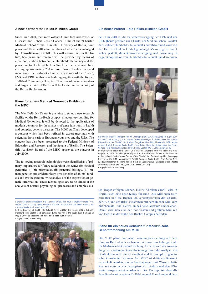
A new partner: the Helios-Kliniken GmbH
Since June 2001, the Franz Volhard Clinic for CardiovascularDiseases and Robert Rössle Cancer Clinic of the “Charité”Medical School of the Humboldt University of Berlin, haveprivatized their health care facilities which are now managedby Helios-Kliniken GmbH. This will ensure that, in the fu-ture, healthcare and research will be provided by means ofclose cooperation between the Humboldt University and theprivate sector. Helios-Kliniken GmbH will erect a new cliniccosting approximately 200 million Euro in Berlin-Buch andincorporate the Berlin-Buch university clinics of the Charité,FVK and RRK, in this new building together with the former1000 bed Community Hospital. Thus, one of the most modernand largest clinics of Berlin will be located in the vicinity ofthe Berlin Buch campus.
Plans for a new Medical Genomics Building at the MDC
The Max Delbrück Center is planning to set up a new researchfacility on the Berlin-Buch campus, a laboratory building forMedical Genomics. It will be devoted to the application ofmodern genomics for the analysis of gene functions in healthand complex genetic diseases. The MDC staff has developeda concept which has been refined in expert meetings withscientists from various European countries and the USA. Theconcept has also been presented to the Federal Ministry ofEducation and Research and the Senate of Berlin. The Scien-tific Advisory Board of the MDC approved the concept inJuly 2000.
The following research technologies were identified as of pri-mary importance for future research in the center for medicalgenomics: (i) bioinformatics, (ii) structural biology, (iii) hu-man genetics and epidemiology, (iv) genetics of animal mod-els and (v) the genome-wide analysis of the expression of ge-netic information. These technologies are to be aimed at theanalysis of normal physiological processes and complex dis-
Ein neuer Partner – die Helios-Kliniken GmbH
Seit Juni 2001 ist die Patientenversorgung der FVK und derRKK (beide gehören zur Charité, der Medizinischen Fakultätder Berliner Humboldt-Universität ) privatisiert und wird vonder Helios-Kliniken GmbH gemanagt. Zukünftig ist damitsicher gestellt, dass Krankenversorgung und Forschung inenger Kooperation von Humboldt-Universität und dem priva-
ten Träger erfolgen können. Helios-Kliniken GmbH wird inBerlin-Buch eine neue Klinik für rund 200 Millionen Euroerrichten und die Bucher Universitätskliniken der Charité,der FVK und des RRK, zusammen mit dem Bucher Klinikummit ehemals 1.000 Betten, in das neue Gebäude einbeziehen.Damit wird sich eine der modernsten und größten Klinikenvon Berlin in der Nähe des Buches Campus befinden.
Pläne für ein neues Gebäude für MedizinischeGenomforschung am MDC
Das MDC plant, eine neue Forschungseinrichtung auf demCampus Berlin-Buch zu bauen, und zwar ein Laborgebäudefür Medizinische Genomforschung. Es wird sich der Anwen-dung der modernen Genomforschung durch die Analyse vonGenfunktionen für die Gesundheit und für komplexe geneti-sche Krankheiten widmen. Am MDC ist dafür ein Konzeptentwickelt worden, das in Fachtagungen mit Wissenschaft-lern aus verschiedenen europäischen Ländern und den USAweiter ausgearbeitet worden ist. Das Konzept ist ebenfallsdem Bundesministerium für Bildung und Forschung und dem
24
Der frühere Wissenschaftssenator Dr. Christoph Stölzl (2. v. l.) besuchte am 3. Juli 2000das MDC. Mit dabei (v.l): Prof. Bernd Dörken (damaliger Ärztlicher Leiter der Robert-Rössle-Klinik der Charité), Dr. Gudrun Erzgräber (Geschäftsführerin der BBB Mana-gement GmbH Campus Berlin-Buch), Prof. Rainer Dietz (Ärztlicher Leiter der Franz-Volhard-Herz-Kreislauf-Klinik) und Prof. Detlev Ganten (MDC-Stiftungsvorstand).Former Berlin Senator for Science, Dr. Christoph Stölzl (2nd from left) visited the MDCon July 3rd, 2000. With him (from left) are: Prof. Bernd Dörken (former Medical Directorof the Robert Rössle Cancer Center of the Charité), Dr. Gudrun Erzgräber (ManagingDirector of the BBB Management GmbH Campus Berlin-Buch), Prof. Rainer Dietz(Medical Director of the Franz Volhard Clinic for Cardiovascular Diseases of the Charité)and Detlev Ganten (MD, Ph.D, MDC`s Scientific Director). Copyright: MDC/Uwe Eising
Bundesgesundheitsministerin Ulla Schmidt (Mitte) mit MDC-Stiftungsvorstand Prof.Detlev Ganten (2.v.re) sowie Klinikern und Wissenschaftlern bei ihrem Besuch desCampus Berlin-Buch am 8. Mai 2001. Federal Secretary of Health, Ulla Schmidt (in the middle), listening to MDC`s ScientificDirector Detlev Ganten (2nd from right) during her visit to the Berlin-Buch Campus onMay 8, 2001, as clinicians and researchers from Buch look on. Copyright: MDC/Uwe Eising
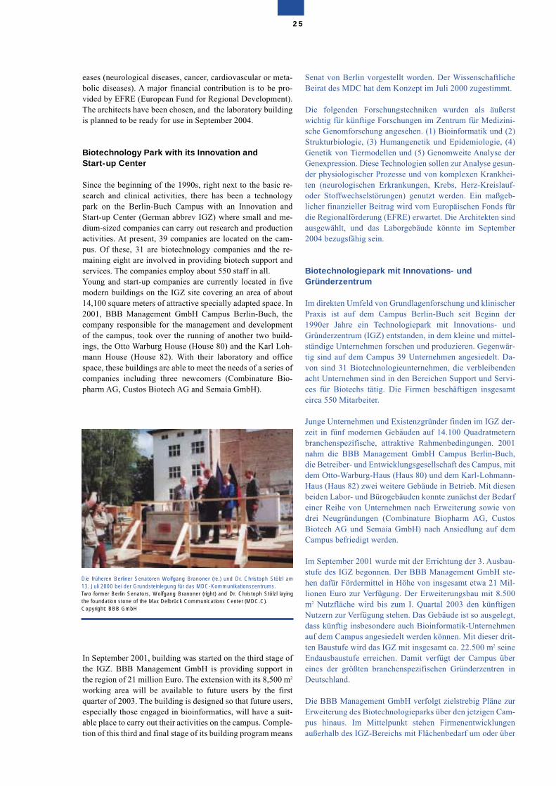
eases (neurological diseases, cancer, cardiovascular or meta-bolic diseases). A major financial contribution is to be pro-vided by EFRE (European Fund for Regional Development).The architects have been chosen, and the laboratory buildingis planned to be ready for use in September 2004.
Biotechnology Park with its Innovation and Start-up Center
Since the beginning of the 1990s, right next to the basic re-search and clinical activities, there has been a technologypark on the Berlin-Buch Campus with an Innovation andStart-up Center (German abbrev IGZ) where small and me-dium-sized companies can carry out research and productionactivities. At present, 39 companies are located on the cam-pus. Of these, 31 are biotechnology companies and the re-maining eight are involved in providing biotech support andservices. The companies employ about 550 staff in all.Young and start-up companies are currently located in fivemodern buildings on the IGZ site covering an area of about14,100 square meters of attractive specially adapted space. In2001, BBB Management GmbH Campus Berlin-Buch, thecompany responsible for the management and developmentof the campus, took over the running of another two build-ings, the Otto Warburg House (House 80) and the Karl Loh-mann House (House 82). With their laboratory and officespace, these buildings are able to meet the needs of a series ofcompanies including three newcomers (Combinature Bio-pharm AG, Custos Biotech AG and Semaia GmbH).
In September 2001, building was started on the third stage ofthe IGZ. BBB Management GmbH is providing support inthe region of 21 million Euro. The extension with its 8,500 m2
working area will be available to future users by the firstquarter of 2003. The building is designed so that future users,especially those engaged in bioinformatics, will have a suit-able place to carry out their activities on the campus. Comple-tion of this third and final stage of its building program means
Senat von Berlin vorgestellt worden. Der WissenschaftlicheBeirat des MDC hat dem Konzept im Juli 2000 zugestimmt.
Die folgenden Forschungstechniken wurden als äußerstwichtig für künftige Forschungen im Zentrum für Medizini-sche Genomforschung angesehen. (1) Bioinformatik und (2)Strukturbiologie, (3) Humangenetik und Epidemiologie, (4)Genetik von Tiermodellen und (5) Genomweite Analyse derGenexpression. Diese Technologien sollen zur Analyse gesun-der physiologischer Prozesse und von komplexen Krankhei-ten (neurologischen Erkrankungen, Krebs, Herz-Kreislauf-oder Stoffwechselstörungen) genutzt werden. Ein maßgeb-licher finanzieller Beitrag wird vom Europäischen Fonds fürdie Regionalförderung (EFRE) erwartet. Die Architekten sindausgewählt, und das Laborgebäude könnte im September2004 bezugsfähig sein.
Biotechnologiepark mit Innovations- undGründerzentrum
Im direkten Umfeld von Grundlagenforschung und klinischerPraxis ist auf dem Campus Berlin-Buch seit Beginn der1990er Jahre ein Technologiepark mit Innovations- undGründerzentrum (IGZ) entstanden, in dem kleine und mittel-ständige Unternehmen forschen und produzieren. Gegenwär-tig sind auf dem Campus 39 Unternehmen angesiedelt. Da-von sind 31 Biotechnologieunternehmen, die verbleibendenacht Unternehmen sind in den Bereichen Support und Servi-ces für Biotechs tätig. Die Firmen beschäftigen insgesamtcirca 550 Mitarbeiter.
Junge Unternehmen und Existenzgründer finden im IGZ der-zeit in fünf modernen Gebäuden auf 14.100 Quadratmeternbranchenspezifische, attraktive Rahmenbedingungen. 2001nahm die BBB Management GmbH Campus Berlin-Buch,die Betreiber- und Entwicklungsgesellschaft des Campus, mitdem Otto-Warburg-Haus (Haus 80) und dem Karl-Lohmann-Haus (Haus 82) zwei weitere Gebäude in Betrieb. Mit diesenbeiden Labor- und Bürogebäuden konnte zunächst der Bedarfeiner Reihe von Unternehmen nach Erweiterung sowie vondrei Neugründungen (Combinature Biopharm AG, CustosBiotech AG und Semaia GmbH) nach Ansiedlung auf demCampus befriedigt werden.
Im September 2001 wurde mit der Errichtung der 3. Ausbau-stufe des IGZ begonnen. Der BBB Management GmbH ste-hen dafür Fördermittel in Höhe von insgesamt etwa 21 Mil-lionen Euro zur Verfügung. Der Erweiterungsbau mit 8.500m2 Nutzfläche wird bis zum I. Quartal 2003 den künftigenNutzern zur Verfügung stehen. Das Gebäude ist so ausgelegt,dass künftig insbesondere auch Bioinformatik-Unternehmenauf dem Campus angesiedelt werden können. Mit dieser drit-ten Baustufe wird das IGZ mit insgesamt ca. 22.500 m2 seineEndausbaustufe erreichen. Damit verfügt der Campus übereines der größten branchenspezifischen Gründerzentren inDeutschland.
Die BBB Management GmbH verfolgt zielstrebig Pläne zurErweiterung des Biotechnologieparks über den jetzigen Cam-pus hinaus. Im Mittelpunkt stehen Firmenentwicklungenaußerhalb des IGZ-Bereichs mit Flächenbedarf um oder über
25
Die früheren Berliner Senatoren Wolfgang Branoner (re.) und Dr. Christoph Stölzl am13. Juli 2000 bei der Grundsteinlegung für das MDC-Kommunikationszentrums. Two former Berlin Senators, Wolfgang Branoner (right) and Dr. Christoph Stölzl layingthe foundation stone of the Max Delbrück Communications Center (MDC.C). Copyright: BBB GmbH
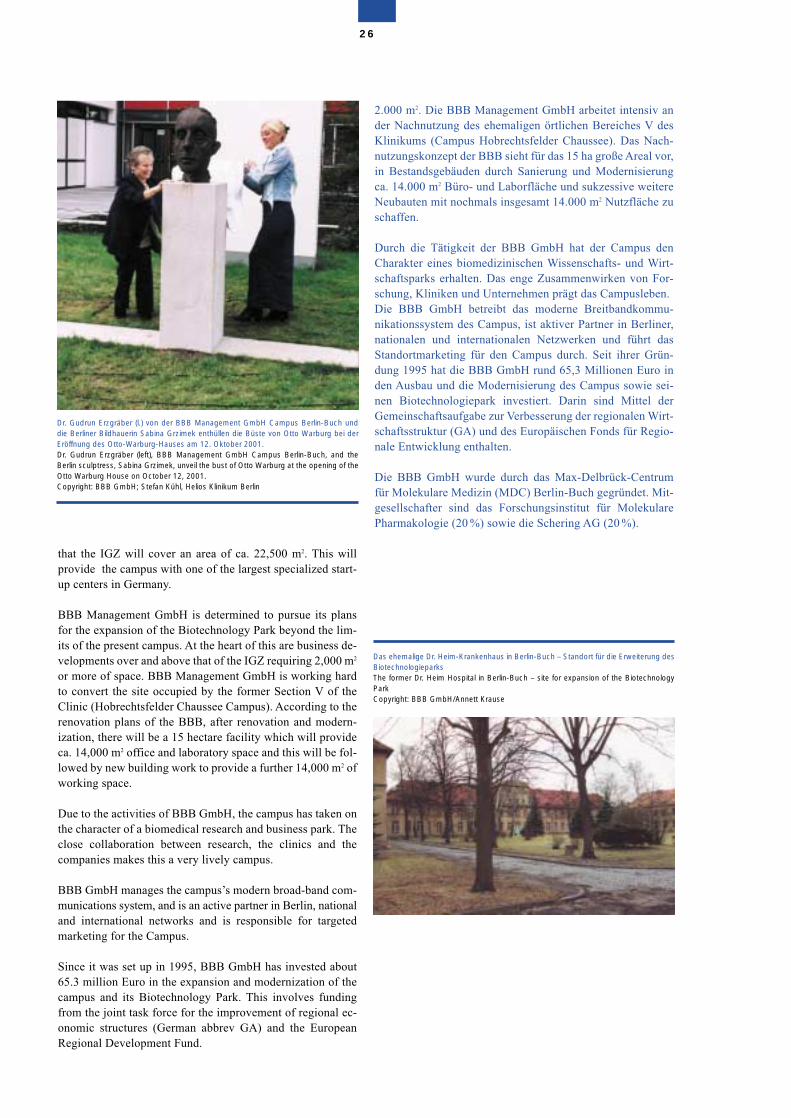
that the IGZ will cover an area of ca. 22,500 m2. This willprovide the campus with one of the largest specialized start-up centers in Germany.
BBB Management GmbH is determined to pursue its plansfor the expansion of the Biotechnology Park beyond the lim-its of the present campus. At the heart of this are business de-velopments over and above that of the IGZ requiring 2,000 m2
or more of space. BBB Management GmbH is working hardto convert the site occupied by the former Section V of theClinic (Hobrechtsfelder Chaussee Campus). According to therenovation plans of the BBB, after renovation and modern-ization, there will be a 15 hectare facility which will provideca. 14,000 m2 office and laboratory space and this will be fol-lowed by new building work to provide a further 14,000 m2 ofworking space.
Due to the activities of BBB GmbH, the campus has taken onthe character of a biomedical research and business park. Theclose collaboration between research, the clinics and thecompanies makes this a very lively campus.
BBB GmbH manages the campus’s modern broad-band com-munications system, and is an active partner in Berlin, nationaland international networks and is responsible for targetedmarketing for the Campus.
Since it was set up in 1995, BBB GmbH has invested about65.3 million Euro in the expansion and modernization of thecampus and its Biotechnology Park. This involves fundingfrom the joint task force for the improvement of regional ec-onomic structures (German abbrev GA) and the EuropeanRegional Development Fund.
2.000 m2. Die BBB Management GmbH arbeitet intensiv ander Nachnutzung des ehemaligen örtlichen Bereiches V desKlinikums (Campus Hobrechtsfelder Chaussee). Das Nach-nutzungskonzept der BBB sieht für das 15 ha große Areal vor,in Bestandsgebäuden durch Sanierung und Modernisierungca. 14.000 m2 Büro- und Laborfläche und sukzessive weitereNeubauten mit nochmals insgesamt 14.000 m2 Nutzfläche zuschaffen.
Durch die Tätigkeit der BBB GmbH hat der Campus denCharakter eines biomedizinischen Wissenschafts- und Wirt-schaftsparks erhalten. Das enge Zusammenwirken von For-schung, Kliniken und Unternehmen prägt das Campusleben.Die BBB GmbH betreibt das moderne Breitbandkommu-nikationssystem des Campus, ist aktiver Partner in Berliner,nationalen und internationalen Netzwerken und führt dasStandortmarketing für den Campus durch. Seit ihrer Grün-dung 1995 hat die BBB GmbH rund 65,3 Millionen Euro inden Ausbau und die Modernisierung des Campus sowie sei-nen Biotechnologiepark investiert. Darin sind Mittel derGemeinschaftsaufgabe zur Verbesserung der regionalen Wirt-schaftsstruktur (GA) und des Europäischen Fonds für Regio-nale Entwicklung enthalten.
Die BBB GmbH wurde durch das Max-Delbrück-Centrumfür Molekulare Medizin (MDC) Berlin-Buch gegründet. Mit-gesellschafter sind das Forschungsinstitut für MolekularePharmakologie (20 %) sowie die Schering AG (20 %).
26
Das ehemalige Dr. Heim-Krankenhaus in Berlin-Buch – Standort für die Erweiterung desBiotechnologieparks The former Dr. Heim Hospital in Berlin-Buch – site for expansion of the BiotechnologyPark Copyright: BBB GmbH/Annett Krause
Dr. Gudrun Erzgräber (l.) von der BBB Management GmbH Campus Berlin-Buch unddie Berliner Bildhauerin Sabina Grzimek enthüllen die Büste von Otto Warburg bei derEröffnung des Otto-Warburg-Hauses am 12. Oktober 2001.Dr. Gudrun Erzgräber (left), BBB Management GmbH Campus Berlin-Buch, and theBerlin sculptress, Sabina Grzimek, unveil the bust of Otto Warburg at the opening of theOtto Warburg House on October 12, 2001.Copyright: BBB GmbH; Stefan Kühl, Helios Klinikum Berlin
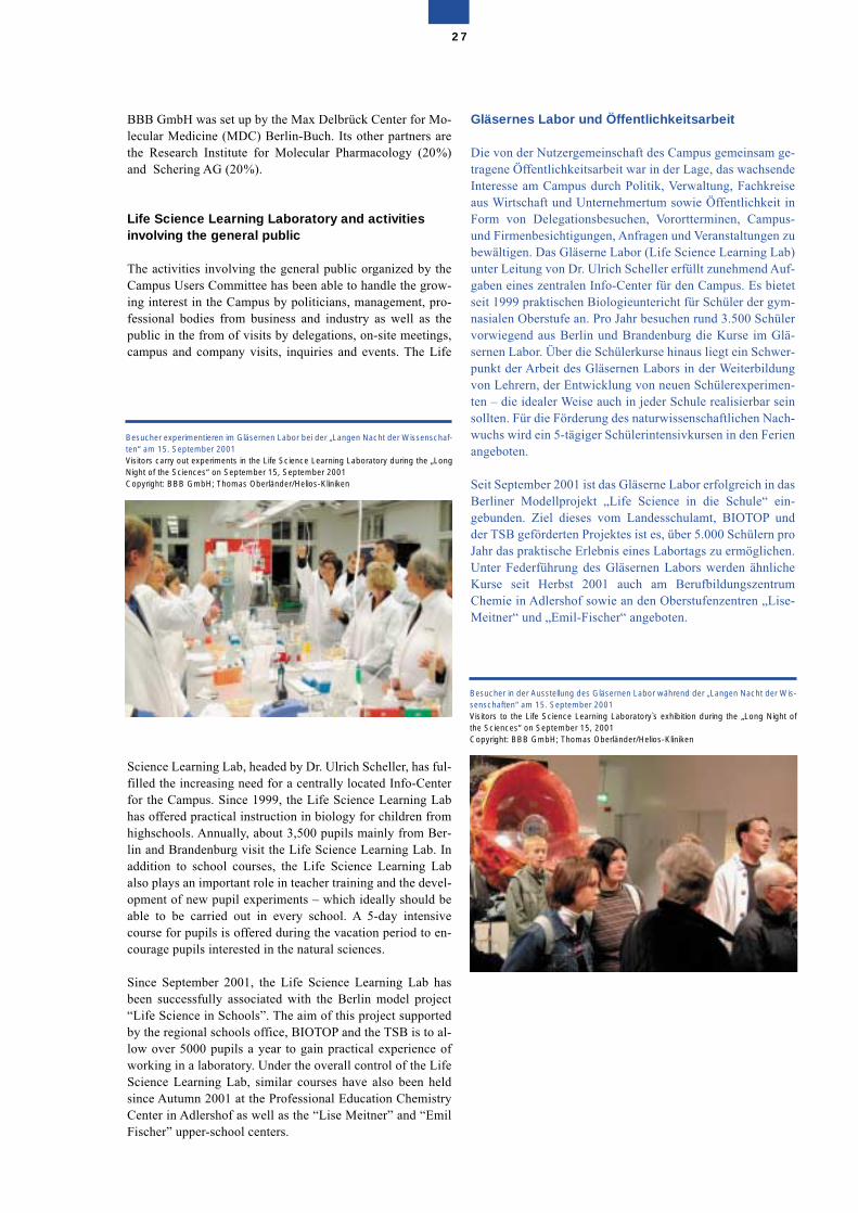
BBB GmbH was set up by the Max Delbrück Center for Mo-lecular Medicine (MDC) Berlin-Buch. Its other partners arethe Research Institute for Molecular Pharmacology (20%)and Schering AG (20%).
Life Science Learning Laboratory and activitiesinvolving the general public
The activities involving the general public organized by theCampus Users Committee has been able to handle the grow-ing interest in the Campus by politicians, management, pro-fessional bodies from business and industry as well as thepublic in the from of visits by delegations, on-site meetings,campus and company visits, inquiries and events. The Life
Science Learning Lab, headed by Dr. Ulrich Scheller, has ful-filled the increasing need for a centrally located Info-Centerfor the Campus. Since 1999, the Life Science Learning Labhas offered practical instruction in biology for children fromhighschools. Annually, about 3,500 pupils mainly from Ber-lin and Brandenburg visit the Life Science Learning Lab. Inaddition to school courses, the Life Science Learning Labalso plays an important role in teacher training and the devel-opment of new pupil experiments – which ideally should beable to be carried out in every school. A 5-day intensivecourse for pupils is offered during the vacation period to en-courage pupils interested in the natural sciences.
Since September 2001, the Life Science Learning Lab hasbeen successfully associated with the Berlin model project“Life Science in Schools”. The aim of this project supportedby the regional schools office, BIOTOP and the TSB is to al-low over 5000 pupils a year to gain practical experience ofworking in a laboratory. Under the overall control of the LifeScience Learning Lab, similar courses have also been heldsince Autumn 2001 at the Professional Education ChemistryCenter in Adlershof as well as the “Lise Meitner” and “EmilFischer” upper-school centers.
Gläsernes Labor und Öffentlichkeitsarbeit
Die von der Nutzergemeinschaft des Campus gemeinsam ge-tragene Öffentlichkeitsarbeit war in der Lage, das wachsendeInteresse am Campus durch Politik, Verwaltung, Fachkreiseaus Wirtschaft und Unternehmertum sowie Öffentlichkeit inForm von Delegationsbesuchen, Vorortterminen, Campus-und Firmenbesichtigungen, Anfragen und Veranstaltungen zubewältigen. Das Gläserne Labor (Life Science Learning Lab)unter Leitung von Dr. Ulrich Scheller erfüllt zunehmend Auf-gaben eines zentralen Info-Center für den Campus. Es bietetseit 1999 praktischen Biologieuntericht für Schüler der gym-nasialen Oberstufe an. Pro Jahr besuchen rund 3.500 Schülervorwiegend aus Berlin und Brandenburg die Kurse im Glä-sernen Labor. Über die Schülerkurse hinaus liegt ein Schwer-punkt der Arbeit des Gläsernen Labors in der Weiterbildungvon Lehrern, der Entwicklung von neuen Schülerexperimen-ten – die idealer Weise auch in jeder Schule realisierbar seinsollten. Für die Förderung des naturwissenschaftlichen Nach-wuchs wird ein 5-tägiger Schülerintensivkursen in den Ferienangeboten.
Seit September 2001 ist das Gläserne Labor erfolgreich in dasBerliner Modellprojekt „Life Science in die Schule“ ein-gebunden. Ziel dieses vom Landesschulamt, BIOTOP undder TSB geförderten Projektes ist es, über 5.000 Schülern proJahr das praktische Erlebnis eines Labortags zu ermöglichen.Unter Federführung des Gläsernen Labors werden ähnlicheKurse seit Herbst 2001 auch am BerufbildungszentrumChemie in Adlershof sowie an den Oberstufenzentren „Lise-Meitner“ und „Emil-Fischer“ angeboten.
27
Besucher experimentieren im Gläsernen Labor bei der „Langen Nacht der Wissenschaf-ten“ am 15. September 2001 Visitors carry out experiments in the Life Science Learning Laboratory during the „LongNight of the Sciences“ on September 15, September 2001 Copyright: BBB GmbH; Thomas Oberländer/Helios-Kliniken
Besucher in der Ausstellung des Gläsernen Labor während der „Langen Nacht der Wis-senschaften“ am 15. September 2001 Visitors to the Life Science Learning Laboratory`s exhibition during the „Long Night ofthe Sciences“ on September 15, 2001 Copyright: BBB GmbH; Thomas Oberländer/Helios-Kliniken
External Evaluation
During the period November 1996 to April 1998, three exter-nal evaluations of its entire research activities were carriedout at the MDC and, in early 2002, there was an evaluation ofthe clinical research programs in which basic MDC research-ers closely collaborated with the clinicians at the RobertRössle and Franz Volhard Clinics. A comprehensive strategicevaluation as part of the HGF Health program took place insummer 2002.
The evaluation were conducted in accordance with the samebasic principle as that employed by special research areas ofthe German Research Society (DFG). The evaluation com-mittee spent two days on-site to allow them the opportunity toget to know the researchers who gave a number of presenta-tions. Following this site visit, the evaluation committee pre-pared a report which included a number of recommendations.Following these recommendations, the MDC has undertakena series of financial and structural measures following de-tailed discussions by the Scientific Management Board. Theresults of these measures are regularly monitored using acheck-list.
Externe Evaluierung
In der Zeit von November 1996 bis April 1998 ist am MDC indrei externen Begutachtungen die gesamte Forschung eva-luiert worden. Im Frühjahr 2002 sind die klinischen For-schungsprogramme begutachtet worden, in denen Grund-lagenwissenschaftler des MDC mit den Klinikern derRobert-Rössle-Klinik und der Franz-Volhard-Klinik eng zu-sammenarbeiten, Eine umfassende strategische Evaluationim Rahmen des Gesundheitsprogramms der HGF erfolgt imSommer 2002.
Die bisherigen Begutachtungen verliefen nach dem gleichenGrundprinzip wie bei den Sonderforschungsbereichen derDeutschen Forschungsgemeinschaft (DFG). Die Gutachter-kommissionen hatten auf der Grundlage ausführlicher Unter-lagen vor Ort zwei Tage lang Gelegenheit, die Wissenschaftle-rinnen und Wissenschaftler bei Vorträgen und Laborbesuchenkennenzulernen. Im Anschluss an die „site visit“ erstellten dieGutachterkommissionen einen Bericht, in dem Empfehlungenabgegeben wurden.
28
The Helmholtz AssociationThe MDC belongs to the Helmholtz Association of German ResearchCentres (Helmholtz-Gemeinschaft Deutscher Forschungszentren,HGF). The Helmholtz Association is Germany´s largest government-funded research organization consisting of 15 national research cen-ters. A total of 10 Helmholtz Centers contribute to research in thehealth field. The major four Helmholtz Centers in health research areGSF-Forschungszentrum für Umwelt und Gesundheit GmbH, DKFZ-Deutsches Krebsforschungszentrum Heidelberg, GBF-Gesellschaftfür Biotechnologische Forschung mbH and the Max Delbrück Centerfor Molecular Medicine (MDC) Berlin-Buch.The members of the Helmholtz Association have a high research pro-file in various fields of medical research, particularly in the areas of ba-sic, clinical and public health research and they have key skills in rese-arch fields with complex, interdisciplinary and long-term scientific pro-grams.Recently, the Helmholtz Association has been reorganized. Under thereforms, most of the Helmholtz Association´s annual budget will beearmarked for six research fields, among them health research. Theseactivities and capacities are structured in programs which are of along-term, but not permanent, nature. In future, the funding of theHelmholtz Centers will be program-oriented. This means that fundingwill take place through the provision of resources for programs aimedat long-term research objectives carried out on the basis of coopera-tion and competition with external evaluation. Program-oriented fun-ding is intended to raise the collaboration between the legally autono-mous Helmholtz Centers to an even higher standard. The MDC takes part in three out of seven programs in the researchfield, Health. These are the programs Cancer Research, Cardiovascu-lar and Metabolic Disease Research and the program Function andDysfunction of the Nervous System. The strategic evaluation of healthresearch of the Helmholtz Centers is scheduled for summer 2002.During the conduct of these programs, the four centres devoting morethan 60 percent of their total budget to the research field, Health(DKFZ, MDC, GBF, GSF), will strive to harmonize their managerialstructures. For example, by setting up a steering committee consistingof the managerial boards of the four centres in order to achieve veryclose coordination. Furthermore, it is being considered to establishone common scientific advisory board for future program evaluations.
Die Helmholtz GemeinschaftDas MDC gehört zur Helmholtz Gemeinschaft Deutscher Forschungs-zentren (HGF). Die HGF ist Deutschlands größte staatlich finanzierteForschungsorganisation, die aus 15 nationalen Zentren besteht.Insgesamt zehn Helmholtz Zentren arbeiten auf den Gebiet derGesundheit. Die vier größten Helmholtz Zentren sind das GSF-For-schungszentrum für Umwelt und Gesundheit GmbH, das DKFZKrebsforschungszentrum Heidelberg, die GBF-Gesellschaft für Bio-technologische Forschung mbH und das MDC. Die Mitglieder derHGF weisen hohe Forschungsprofile in verschiedenen Bereichen dermedizinischen Wissenschaft auf, insbesondere bei der Erforschungder biologischen Grundlagen, der klinischen Anwendung und der all-gemeinen, die Gesundheit fördernden Maßnahmen („public healthresearch“). Sie verfügen über Schlüsselkompetenzen in Forschungs-gebieten mit komplexen, interdisziplinären und langfristigen wissen-schaftlichen Programmen.Vor kurzem ist die HGF restrukturiert worden. Als Konsequenz derReform wird der größte Teil des jährlichen Budgets auf sechs For-schungsfelder verteilt werden, von denen eines die Gesundheit ist.Innerhalb dieser Forschungsfelder werden die Aktivitäten und Kapa-zitäten in Programmen strukturiert, die zwar langfristig, aber nicht aufDauer angelegt sind. In Zukunft wird die Mittelvergabe bei der HGFprogrammorientiert sein. Dies bedeutet, daß die Förderung durch dieVergabe von Mitteln für Programme vor sich geht, die langfristige For-schungsziele haben, die auf der Basis von Kooperation und Kompeti-tion mit externer Evaluierung durchgeführt werden. Das MDC nimmt an drei von sieben Programmen auf dem Gebiet derGesundheit teil. Dies sind die Programme Krebsforschung, Erfor-schung von Herz-Kreislauferkrankungen und Stoffwechselkrankheitensowie das Programm Funktion und Fehlfunktion des Nervensystems.Die strategische Bewertung der Gesundheitsforschung ist für denSommer 2002 vorgesehen. Im Verlauf der Fortschritte in diesen Pro-grammen werden die vier Zentren (DKFZ, MDC, GBF, GSF), die mehrals 60% ihres Gesamtbudgets dem Thema Gesundheit widmen, sichbemühen, ihre Managementstrukturen zu harmonisieren. So lassensich zum Beispiel Lenkungsausschüsse einrichten, zu denen dasManagement der vier Zentren gehört, um eine möglichst enge Koope-ration zu erreichen. Darüber hinaus ist vorgesehen, einen gemein-samen Wissenschaftsausschuss für die kommenden wissenschaft-lichen Evaluierungen einzurichten.
In addition, the MDC has carried out an evaluation of itsmanagement procedures. This involved all aspects of thestructure, organization, quality and financial efficiency of theinfrastructure and management of the MDC. In 1998, 1999and 2000 there were also three rounds of evaluations involv-ing Staff and Social Affairs, Purchasing and Materials as wellas Finance and Accounting Procedures.
The Helmholtz Society centers were also evaluated by theScientific Advisory Board as part of a system evaluation in2000. On March 16 and 17, 2000, the MDC had a visit fromthe Scientific Advisory Board. In January 2001 the results ofthe evaluation by the Scientific Advisory Board were pub-lished. Based on this expert report, a fundamental reorganiza-tion of the entire Helmholtz Association has taken place.
Graduate student education – dean of graduatestudents
The support and structure of graduate student education at theMDC is given extreme importance. We have established agraduate program to accommodate highly qualified candida-tes and prepare them for careers in research science. The pro-gram provides training and research opportunities at the hig-hest level within existing resources of the MDC. Ph.D./M.D.students participate in lectures and seminars held at the MDCand gain broad knowledge in biomedical sciences. A strongstudent-advisor relationship is essential for superior acade-mic performance and is the basis for development of studentsinto independent and creative researchers.
The approximately 120 MDC graduate students elect repre-sentatives who negotiate graduate student affairs with the in-stitute and interact closely with the elected dean of graduatestudents. In addition, the student representatives organizescientific and social meetings for their fellow graduate stu-dents such as the traditional annual Student Symposium.
Helmholtz fellows
Helmholtz Fellowships are intended to allow promisingyoung scientists an early opportunity to carry out their ownindependent research. The Helmholtz Fellowships have beenestablished for young scientists who have already demon-strated that they are capable of carrying out high quality re-search on their own but who do not yet qualify for C3-gradeposts or equivalent positions with their own facilities and per-sonal freedom. The fellowships support the positions for upto a maximum of five years. The fellows are given their ownequipment and the cost of this is borne by the research group.Generally, working space and personal equipment is suppliedby the research group of the institution they join. It is expec-ted that support is provided by the research group to allow theresearch fellow full independence. The aim is to allow theHelmholtz Fellows during the period of their award to setthemselves up as independent researchers and compete on anequal footing. They are also expected to apply for third-partyfunding in their own right. Fellows are chosen following apositive assessment on the basis of suggestions from the re-search groups involved.
Das MDC hat aufgrund der Empfehlungen der Gutachter fi-nanzielle und strukturelle Maßnahmen ergriffen, die im Wis-senschaftlichen Ausschuss des Kuratoriums detailliert disku-tiert worden sind. Die Ergebnisse der Maßnahmen werdenanhand einer Checkliste regelmäßig überprüft.
Darüber hinaus hat das MDC eine Begutachtung der Verwal-tung durchgeführt. Diese bezog sich auf alle Fragen der Glie-derung, Organisation, Qualität und Wirtschaftlichkeit von In-frastruktur und Verwaltung des MDC. Sie fand 1998, 1999und 2000 in drei Runden, jeweils für die Abteilungen Perso-nal- und Sozialwesen, Einkauf und Materialwirtschaft sowieFinanz- und Rechnungswesen statt.
Alle Zentren der Helmholtz Gemeinschaft wurden im Rah-men einer Systemevaluation im Jahre 2000 durch den Wis-senschaftsrat evaluiert. Am 16. und 17. März 2000 wurde dasMDC durch den Wissenschaftsrat besucht. Im Januar 2001wurden die Ergebnisse der Begutachtung durch den Wissen-schaftsrat veröffentlicht. Auf der Basis dieser Begutachtungerfolgte die grundlegende Reorganisation der gesamtenHelmholtz Gemeinschaft.
Ausbildung von Doktoranden – Der Vertrauensdozent
Der Förderung und Struktur der Ausbildung von Doktorandenwird im MDC besondere Aufmerksamkeit geschenkt. Eswurde ein Graduiertenprogramm eingerichtet, um hochquali-fizierte Kandidaten auf ihre Karriere in der Forschung vorzu-bereiten. Das Programm sieht Trainings- und Forschungsmög-lichkeiten auf höchstem Niveau im Rahmen der vorhandenenMDC Ressourcen vor. Doktoranden nehmen an Vorlesungenund Seminaren des MDC teil und erlangen dabei ein breitesWissen auf dem Feld der biomedizinischen Wissenschaften.Eine enge Verbindung zwischen Student und Lehrer ist we-sentlich für eine herausragende akademische Qualifikation,und sie stellt die Grundlage dafür dar, dass aus den Doktoran-den unabhängige und kreative Forscher werden.
Es gibt rund 120 Doktoranden am MDC. Sie wählen ihre Ver-treter, welche die Belange der Doktoranden mit dem Institutverhandeln und die engen Kontakt mit dem gewählten Ver-trauensdozent haben. Darüber hinaus organisieren die Studen-tenvertreter wissenschaftliche und soziale Zusammenkünftefür ihre Kommilitonen wie zum Beispiel das traditionelle jähr-liche Doktoranden-Symposium.
Helmholtz-Stipendiaten
Helmholtz-Stipendien sind vorgesehen, um die frühe Unab-hängigkeit junger, erfolgversprechender Wissenschaftler zuermöglichen. Die Helmholtz-Stipendien werden für jungeWissenschaftler eingerichtet, die bereits nachgewiesen haben,dass sie hervorragende eigenständige wissenschaftliche Arbeitleisten, die aber sich noch nicht für C3-Stellen oder andereäquivalente Positionen mit eigener Ausstattung und persönli-cher Unabhängigkeit qualifizieren. Die Stipendien finanzierendie Stelle des Stipendiaten für einen Zeitraum bis zu maximalfünf Jahren. Sie werden mit eigenen Sachmitteln ausgestattet,
29
Scientific Journals
The MDC is the site of several editorial offices.
Journal of Molecular Medicine (JMM)The Journal of Molecular Medicine (JMM) is publishedmonthly by Springer Verlag Heidelberg since 1995. The fo-cus is in molecular medicine, a field which applies the me-thods and knowledge of molecular biology and gene techno-logy to medical research, therapy, and disease prevention.JMM’s goal is to bring together basic science and clinical me-dicine in the field of molecular and gene technology research,which has become particularly important for the progress ofmedicine in all aspects. The journal’s editor-in-chief is DetlevGanten, Scientific Director of the MDC.
NeuroforumNeuroforum is the Newsletter of the German NeuroscienceSociety (Neurowissenschaftliche Gesellschaft). The journalwas founded in 1994, Neuroforum gives an overview of theactivities in the field of neuroscience research in Germany.The journal publishes review articles covering all aspects ofneuroscience research. Besides that, Neuroforum publishesarticles on key persons in the history of the neurosciences,meeting reports, methodological aspects, book reviews, opi-nions, portraits of industrial research institutions, informationon educational and research programs, and news from theGerman Neuroscience Society. The journal is published bySpektrum Akademischer Verlag, Heidelberg, Germany. Theeditor-in-chief is Helmut Kettenmann.
GliaGlia, founded in 1988, provides a dedicated forum for a broadrange of experimental topics in the field of glial research andis an indispensible medium for scientific exchanges amongresearchers interested in neuroglial research. Original arti-cles, short communications, review articles and Special Issueson the physiology, anatomy, pharmacology, chemistry, andpathology of glia are published. The publisher is WILEY-LISS,New York, USA, editors-in-chief are Bruce Ransom andHelmut Kettenmann.
die sich an den Kosten für die Arbeitsrichtung orientieren. Dieräumliche und personelle Ausstattung soll im Allgemeinendurch die aufnehmende Arbeitsgruppe der Institution gewähr-leistet werden. Es wird erwartet, dass von dieser Arbeits-gruppe Unterstützung geleistet wird und die Unabhängigkeitdes Wissenschaftlers gefördert wird. Ziel ist es, dass dieHelmholtz-Stipendiaten während der Zeit ihres Stipendiumsin unabhängige Positionen hineinwachsen und sich darum be-werben. Für selbst eingeworbene Drittmittel wird eine selbst-ständige Verantwortlichkeit vorgesehen. Das Auswahlverfah-ren erfolgt über geeignete Begutachtung auf Vorschlag deraufnehmenden Arbeitsgruppe.
Wissenschaftliche Journale
Das MDC ist Sitz mehrerer wissenschaftlicher Redaktionen.
Journal of Molecular Medicine (JMM)Das JMM wird seit 1995 monatlich vom Springer Verlag pu-bliziert. Die Zeitschrift konzentriert sich auf das Gebiet dermolekularen Medizin, in dem die Methoden und die Ergeb-nisse der Molekularbiologie und Gentechnologie für die me-dizinische Forschung, Therapie und Prävention genutzt wer-den. Das Ziel des JMM besteht darin, Grundlagenforschungund klinische Medizin zusammenzubringen, weil das Zusam-mengehen von Molekularbiologie und Gentechnologie einebesondere Bedeutung für alle Aspekte des Fortschritt in derMedizin hat. Der verantwortliche Herausgeber der Zeitschriftist Detlev Ganten, der wissenschaftliche Vorstand des MDC.
NeuroforumNeuroforum ist das Mitteilungsblatt der deutschen Neuro-wissenschaftlichen Gesellschaft. Die Zeitschrift ist 1994 ge-gründet worden. Neuroforum bietet einen Überblick über dieAktivitäten auf dem Gebiet der Neurowissenschaften inDeutschland. Die Zeitschrift publiziert Übersichtsartikel zuallen Aspekten der neurowissenschaftlichen Forschung. Darü-ber hinaus publiziert Neuroforum Aufsätze über Schlüsselper-sonen in der Geschichte der Neurobiologie, bringt Berichteüber Konferenzen, stellt methodologische Aspekte vor, bringtBuchrezensionen, Porträts von Industrieforschungsinstitute,informiert über Ausbildungs- und Forschungsprogramme undmeldet die Neuigkeiten der deutschen Neurowissenschaft-lichen Gesellschaft. Die Zeitschrift erscheint im SpektrumAkademischer Verlag, Heidelberg. Der Herausgeber ist HelmutKettenmann.
GliaGlia wurde 1988 gegründet und stellt ein engagiertes Forumfür einen weiten Bereich an experimentellen Themen aus derGliazellenforschung dar. Es ist ein maßgebliches Medium fürden wissenschaftlichen Austausch unter Forschern in diesemFeld. Glia publiziert Originalbeiträge, kurze Mitteilungen,Übersichtsartikel und Sonderhefte über die Physiologie, Ana-tomie, Chemie, Pharmakologie und Pathologie der Gliazellen.Die Zeitschrift erscheint im Verlag Wiley-Liss, New York(USA), die Herausgeber sind Bruce Ransom und HelmutKettenmann.
30
Chinese-German Laboratory for MolecularMedicine in Beijing
The identification of genes which are involved in causing car-diovascular and associated diseases is the aim of the Chinese-German Laboratory for Molecular Medicine which has re-cently been set up in Beijing by the Fu Wai Hospital of theChinese Academy of Medical Sciences (CAMS) and theMDC. This laboratory includes a “Genome Screening Cen-ter” which was set up in 1999 with funds from the GermanFederal Research Ministry (German abbrev. BMBF). There,scientists are trying to identify the genes that are responsiblefor these diseases. This has led to more intensive collabora-tion between researchers at the CAMS and the MDC. FromNovember 2000, the Beijing Laboratory for Molecular Medi-cine (headed by Hui Rutai and Liu Lisheng) has been assistedby the activities of two “Young Investigator PartnershipGroups”. The laboratory in the Fu Wai Hospital is supportedby the CAMS, the Chinese Ministry of Research, the MDCand the Swiss pharmaceutical company, Hoffmann-La Ro-che, in Basle. A number of other groups are also taking part inthis research including the Chinese Human Genome Projectand the Franz Volhard Clinic for Cardiovascular Diseases(Charité University Clinic on the Berlin-Buch Campus). Fel-lowships to support guest scientists in Beijing and Berlin areprovided by the Society of Friends and Sponsors of the MDC.The Fu Wai Hospital, a specialist facility for cardiovasculardiseases on western lines, also acts as a reference center forthese diseases in China. Since 1995, the MDC has provided acenter for gene mapping. This specialist facility is headed byNorbert Hübner and Peter Nürnberg and is funded by theBMBF under its German Human Genome Project.
Congresses
In the years reported, two major conferences took place inBerlin which were organized by scientists from the MDC.
International Conference on “Cell Migration in Developmentand Disease”Cell migration is an important process in development. Fre-quently precursor cells are generated at one site in the embryoand then migrate over long distances to targets where theydifferentiate. Migration processes in the embryo are well con-trolled, and migrating cells are generated during distinct sta-ges and use characteristic and even invariant paths to reachtheir targets. Ordered migration of cells can also be observedin the adult, e.g. in the immune system. Such well controlledprocesses are in contrast to the aberrant migration of cells inpathological processes such as cancer.
Important recent insight into these processes was provided atan international conference on “Cell Migration in Develop-ment and Disease” (November 29 to December 1, 2001),which was held by the MDC in Berlin. The conference wasfunded by the Deutsche Forschungsgemeinschaft (DFG; Ger-man Research Fondation) and the MDC. More than 200scientists from the USA, Australia, Israel, Japan, and many
Chinesisch-deutsches Labor für MolekulareMedizin in Peking
Die Identifizierung von Genen, die an der Entstehung vonHerz-Kreislauferkrankungen und ihren Folgekrankheiten be-teiligt sind, ist das Ziel des chinesisch-deutschen Labors fürMolekulare Medizin, welches das Fu Wai Krankenhaus derChinesischen Akademie der Medizinischen Wissenschaften(CAMS) und das MDC in Peking in den vergangenen Jahrenaufgebaut haben. Zu diesem Pekinger Labor gehört ein 1999mit Mitteln des Bundesforschungsministeriums (BMBF) ein-gerichtetes „Genom-Screening-Zentrum“, in dem die Forschersystematisch nach Genen fahnden, die Krankheiten verursa-chen. Damit konnte die Zusammenarbeit zwischen Wissen-schaftlern der CAMS und dem MDC intensiviert werden. SeitNovember 2000 ist das Pekinger Labor für Molekulare Medi-zin (Leitung Hui Rutai und Liu Lisheng) am Fu Wai Hospitaldurch zwei junge Forschergruppen („Young InvestigatorPartnership Groups“) verstärkt worden. Das Labor im Fu WaiHospital wird gefördert von der CAMS, dem chinesischenForschungsministerium, dem MDC sowie dem SchweizerPharmakonzern Hoffmann-La Roche in Basel. An den For-schungsprojekten beteiligt sind auch das Chinesische Human-genomprojekt und die Franz-Volhard-Klinik für Herz-Kreis-lauferkrankungen (Universitätsklinikum Charité, CampusBerlin-Buch). Mit Stipendien für Forschungsaufenthalte vonGastwissenschaftlern in Peking und Berlin unterstützt derVerein der Freunde und Förderer des MDC diese Initiative.Das Fu Wai Krankenhaus, eine nach westlicher Medizin aus-gerichtete Spezialklinik für Herz-Kreislauf-Erkrankungen, istzugleich ein Referenzzentrum für diese Erkrankungen inChina. Das MDC verfügt seit 1995 über ein Zentrum für Gen-kartierung. Dieses von Norbert Hübner und Peter Nürnberggeleitete Speziallabor wird im Rahmen des DeutschenHumangenomprojekts vom BMBF gefördert.
Kongresse
In den vergangenenen zwei Jahren fanden zwei wichtigeKonferenzen in Berlin statt, die von Wissenschaftlern desMDC organisiert wurden.
Internationaler Kongress zu „Cell Migration in Developmentand Disease“Die Fähigkeit von Zellen, während der Entwicklung einesOrganismus zu wandern, ist ein lebenswichtiger Prozess.Sehr häufig entstehen in einem Embryo so genannte Vorläu-ferzellen, die dann zu weit entfernten Zielorganen wandernmüssen, wo sie differenzieren, das heißt, zu ganz spezifischenKörperzellen werden. Diese Wanderungsprozesse von Zellenim Embryo sind sehr genau gesteuert. Die Fähigkeit von Zel-len, ihren Entstehungsort zu verlassen, wird in bestimmtenEntwicklungsphasen des Organismus benötigt. Sie nutzenganz charakteristische, vorgebene und sich niemals änderndePfade, um ihre Zielpunkte zu erreichen. Eine geordnete Zell-wanderung wird auch im erwachsenen Organismus beobach-tet, zum Beispiel im Abwehrsystem des Körpers, dem Immun-system. Gerät die Zellwanderung aus dem Takt, dann könnenKrankheiten, wie zum Beispiel Krebs, entstehen.
31
European countries attended. The organisers were WalterBirchmeier (MDC), Doris D. Wedlich (Technical Universityof Karlsruhe, Germany), Carmen Birchmeier and MartinLipp (both MDC).
2nd International Symposium on Obesity and HypertensionOverweight and obesity currently affect approximately 100million adults in the countries of the European Union and inmost European countries more than 20 per cent of the popu-lation are obese. In Germany, more than 16 million people areobese while more than 40 million, i.e. half the population, isoverweight. Obesity is now recognized as an important riskfactor for a number of medical problems including cardiovas-cular diseases, such as hypertension, and diabetes. Health-care related costs are around 200 times higher for obese indi-viduals, says Arya Sharma, physician and nephrologist at theFranz Volhard Clinic (FVK) for Cardiovascular Diseases ofthe Charité, Medical School of the Humboldt University ofBerlin. In recent years, there have been remarkable advancesin our understanding of the biology of fat (adipose) tissue. Fatcells (adipocytes) are now seen as an important source of avariety of molecules that can affect blood pressure regulationand kidney function. Scientists and physicians hope that thesefindings will eventually lead to new preventive strategies andtherapies for managing obesity and related cardiovasculardiseases. These diseases were the key topics of the 2nd Inter-national Symposium on Obesity and Hypertension held inBerlin from October 25 to October 27, 2001 at the MDC. 200scientists and physicians from the USA, Canada, Japan, Aus-tralia, Israel and Europe attended this symposium. The organ-isers were Arya Sharma, Friedrich Luft (FVK and MDC) andAchim Leutz (MDC). This symposium was officially endor-sed by the European Society of Hypertension, the EuropeanAssociation for the Study of Obesity and the InternationalObesity Task Force. It was supported by the Deutsche For-schungsgemeinschaft, and by the “High-Level ScientificMeeting Programme” of the European Union.
Academic Appointments
Six new group leaders have been appointed during the lasttwo years (2000 and 2001).
The biologist Christiane Alexander was appointed head ofthe research group “Neurodegeneration” in the Neuroscien-ces section of the MDC for a period of five years in July2001. She received her degree from the University of Düssel-dorf in 1994 and her doctorate four years later. Her postdoc-toral research involved molecular genetics, first one year atTübingen University Clinic, followed by two years at Uni-versity College, London, supported by a DFG-Postdoctoralscholarship.
Gerd Kempermann was appointed head of the “NeuronalStem Cells” research group at the MDC in September 2000.He completed his medical studies in Cologne and Freiburg imBreisgau with a practical year in New York in 1993. In thesame year, he obtained his doctorate in the Department ofNeuropathology, Pathology Institute, University of Freiburg.He spent a period of two years practical training as a physi-cian. He then obtained a DFG scholarship for three years at
Mit diesen Prozessen befasste sich ein Internationaler Kon-gress „Cell Migration in Development and Disease“ (Zell-wanderung in der Entwicklung von Organismen und bei derEntstehung von Krankheiten), den das MDC vom 29. Novem-ber bis 1. Dezember 2001 in Berlin veranstaltet hatte. DerKongress wurde unterstützt von der Deutschen Forschungs-gemeinschaft (DFG) und dem MDC. Über 200 Wissenschaft-ler aus den USA, Australien, Israel, Japan und mehrereneuropäischen Ländern nahmen an dem Kongress teil. Organi-satoren waren Walter Birchmeier (MDC), Doris D. Wedlich(Technische Universität Karlsruhe), Carmen Birchmeier undMartin Lipp (beide MDC).
2. Internationales Symposium zu Fettsucht undBluthochdruckEtwa 100 Millionen Erwachsene in den Ländern der Europäi-schen Union sind übergewichtig oder zu dick. Allein in derBundesrepublik bringen 40 Millionen Menschen, und damitnahezu die Hälfte der Bevölkerung, nach Ansicht von Medi-zinern zuviel Gewicht auf die Waage. 20 Prozent, rund 16Millionen Menschen, gelten als adipös. Menschen mit Über-gewicht und Fettsucht sind häufig auch chronisch krank. Siesind anfällig für Herz-Kreislauf-Erkrankungen, zum BeispielBluthochdruck (Hypertonie), sowie für Diabetes. Sie verursa-chen nach Aussage von Arya Sharma (Franz-Volhard-Klinik(FVK) der Charité, Humboldt-Universität zu Berlin, CampusBerlin-Buch) darüber hinaus 200 mal höhere Gesundheitsko-sten als schlanke Menschen. In den vergangenen Jahren ha-ben Grundlagenforscher sehr viele neue Erkenntnisse überdie Biologie des Fettgewebes gewonnen. So wissen sie in-zwischen, dass eine Vielzahl von Molekülen, die von Fettzel-len produziert werden, direkt das Herz-Kreislaufsystem unddie Nieren schädigen können. Fettsucht und Bluthochdruckstanden im Mittelpunkt des 2. internationalen Symposiumsfür Grundlagenforscher und Kliniker, das Arya Sharma ge-meinsam mit Friedrich Luft (FVK und MDC) und AchimLeutz (MDC) in Berlin-Buch organisiert hatte. An dem Sym-posium vom 25. Oktober bis 27. Oktober 2001 hatten rund200 Wissenschaftler aus den USA, Kanada, Japan, Austra-lien, Israel und Europa teilgenommen. Es wurde unterstütztvon der Europäischen Gesellschaft für Bluthochdruck, derEuropäischen Vereinigung für die Erforschung der Adiposi-tas, der Internationalen Adipositas Task Force sowie derDeutschen Forschungsgemeinschaft (DFG) und dem „High-Level Scientific Conference Programme“ der EuropäischenUnion.
Akademische Berufungen
Sechs neue Gruppenleiter sind in den beiden vergangenenJahren (2000 und 2001) berufen worden :
Die Biologin Christiane Alexander leitet seit Juli 2001 fürfünf Jahre die Forschungsgruppe „Neurodegeneration“ imBereich Neurowissenschaften am MDC. 1994 legte sie an derUniversität Düsseldorf die Diplomprüfung ab und promo-vierte dort vier Jahre später. In ihrer Postdoc-Zeit beschäf-tigte sie sich mit molekularer Genetik, zuerst für ein Jahr amUniversitätsklinikum Tübingen, anschließend zwei Jahre amUniversity-College London/Großbritannien, finanziert miteinem DFG-Postdoc-Stipendium.
32
the Genetics Laboratory of the Salk Institute, La Jolla, Cali-fornia. In October 1998, he was appointed a scientific assist-ant at the Regensburg Neurological University Clinic beforecoming to the MDC.
Peter Nürnberg was appointed head of the research group“Molecular Genetics and Center for Gene Mapping” at theMDC for a period of five years in February 2001. He studiedbiology until 1981 at the Humboldt University of Berlin. Af-ter carrying out research at the Institute of Biochemistry,Charité, and the Central Institute of Molecular Biology inBerlin-Buch, he obtained his doctorate in 1984. He has alsoworked at the Institute of Medical Genetics, Charité, wherehe was appointed head of the Molecular Genetics Departmentin 1989, and the Max Planck Institute of Psychiatry, Martins-ried. In 1997, he qualified as a university lecturer in humangenetics and was appointed senior assistant in September1998.
Erich Wanker was appointed head of the “Proteome Researchand Molecular Mechanisms of Neurodegenerative Diseases”research group following a C4-grade appointment in “Mole-cular Medicine” at the Humboldt University of Berlin and theMDC, in September 2001. He is also responsible for the corefacility “Proteomics” at the MDC. The Austrian-born scien-tist studied biochemistry at the Technical University in Graz,obtaining his doctorate in 1992. After a three-year post-doc-toral fellowship at the University of California in Los Ange-les, he led the “Huntington’s Disease Project” at the MaxPlanck Institute of Molecular Genetics in Berlin-Dahlem. In1999, he was appointed to a C3-grade post there before co-ming to the MDC.
Salim Abdelilah-Seyfried has been head of the research group“Cell Polarity and Epithelium Formation” since September2001. After obtaining his high school leaving certificate inDüsseldorf and studying biochemistry, molecular biology andzoology in Manchester, Tübingen and Harvard MedicalSchool, Boston/USA, the Algerian-born scientist graduatedin biology at the University of Tübingen. He was interested inthe Zebra fish as a model for genetic research and obtainedhis doctorate in 1996 from the Faculty of Biology, Universityof Tübingen. After a short period at the Max Planck Instituteof Biochemistry in Martinsried, he undertook postdoctoral re-search at the University of California in San Francisco, be-fore moving to the MDC for a period of 5 years.
Christof Tannert is principal investigator in a project “Dis-course to the Ethical Questions of Biomedicine”, funded bythe Federal Ministry for Education and Research, and hasheaded a group “Bioethics and Scientific Communication” atthe MDC since January 2002. He studied biology at the Uni-versity of Leipzig. He then carried out research at an Acad-emy Institute in Leipzig, became an assistant at the Charité,Humboldt University, Berlin, studying the destruction of mit-ochondria and lipid peroxidation as well as the biochemistryof erythrocyte aging, and obtained his doctorate in biology in1979. From 1980 to 1984, he studied theology by enrolling ina distance-learning course in the diocese of Saxony and thenworked freelance on questions involving scientific ethics. In1987, he started the Independent Institute for EnvironmentalProblems, Berlin/Halle, which he headed from 1989 to 1993.
Gerd Kempermann ist im September 2000 für fünf Jahre alsLeiter der Forschungsgruppe „Neuronale Stammzellen“ andas MDC berufen worden. Er studierte Medizin in Köln undFreiburg i. Br. und schloß es mit einem praktischen Jahr inNew York 1993 ab. Im gleichen Jahr verteidigte er seine Pro-motion in der Abteilung Neuropathologie des PathologischenInstitutes der Universität Freiburg. Dort arbeitete er auchzwei Jahre als Arzt im Praktikum. Anschließend ging er miteinem Ausbildungsstipendium der DFG für drei Jahre an dasGenetische Labor des Salk Instituts, La Jolla, Kalifornien/USA. Im Oktober 1998 hatte er die Stelle eines wissenschaft-lichen Assistenten an der Neurologischen UniversitätsklinikRegensburg erhalten, bevor er ans MDC kam.
Peter Nürnberg leitet seit Februar 2001 für den Zeitraum vonfünf Jahren die Forschungsgruppe „Molekulare Genetik undZentrum für Genkartierung“. Er studierte bis 1981 an derHumboldt-Universität zu Berlin Biologie. Nach Forschungs-arbeiten am Institut für Biochemie der Charité und am Zen-tralinstitut für Molekularbiologie in Berlin-Buch promovierteer 1984. Stationen seiner weiteren wissenschaftlichen Lauf-bahn waren neben Berlin-Buch, das Institut für MedizinischeGenetik der Charité der Humboldt-Universität zu Berlin, andem er ab 1989 die Abteilung „Molekulargenetik“ leitete, unddas Max-Planck-Institut für Psychiatrie, Martinsried. 1997hatte er sich für das Fach Humangenetik habilitiert.
Erich Wanker hat im September 2001 von der Humboldt-Uni-versität zu Berlin und dem MDC einen Ruf als C4-Professorauf den Lehrstuhl „Molekulare Medizin“ sowie als For-schungsgruppenleiter „Proteomforschung und molekulareMechanismen der neurodegenerativen Erkrankungen“ erhal-ten. Am MDC leitet er zudem die Core-facility „Proteomics“.Der in Österreich geborene Wissenschaftler studierte Bioche-mie an der Technischen Universität in Graz, wo er 1992 auchpromovierte. Nach einer dreijährigen Postdoc-Zeit an derUniversität von Kalifornien in Los Angeles leitete er amMax-Planck-Institut für Molekulare Genetik in Berlin-Dah-lem das „Huntington’s Disease Project“. 1999 wurde er dortin eine C3-Position berufen.
Salim Abdelilah-Seyfried leitet seit September 2001 dieMDC-Forschungsgruppe „Zellpolarität und Epithelbildung“.Nach Abitur in Düsseldorf und Studien der Biochemie, Mole-kularen Biologie und Zoologie in Manchester/Großbritan-nien, Tübingen und an der Harvard Medical School in Boston/USA, legte der in Algerien geborene Wissenschaftler 1993seine Diplomprüfung in Biologie an der Universität Tübingenab. Er beschäftigte sich mit dem Zebrafisch als Modell gene-tischer Untersuchungen und promovierte 1996 an der Fakul-tät für Biologie der Universität Tübingen. Als Postdoc ging ernach kurzem Aufenthalt am Max-Planck-Institut für Bioche-mie in Martinsried, an die Universität von Kalifornien/USAin San Francisco. Von dort wurde er für 5 Jahre an das MDCberufen.
Christof Tannert verantwortet ein vom BMBF gefördertesProjekt „Diskurs zu den ethischen Fragen der Biomedizin“und leitet seit Januar 2002 am MDC die Arbeitsgruppe„Bioethik und Wissenschaftskommunikation“. Er studiertean der Universität Leipzig Biologie und arbeitete danach ineinem Leipziger Akademie-Institut und an der Charité der
33
From 1990 to 1994, he was a member of the Berlin State Par-liament and, from 1994 to 1999, a member of the EuropeanParliament in Strasbourg/France.
Further appointments at the MDCThomas Willnow received a tenured position as researchgroup leader at the MDC in February 2001. A joint C4-gradeappointment with the Free University of Berlin in MolecularCardiovascular Research was made in September of thatsame year. He studied biology in Munich and obtained hisdoctorate in 1991. He then worked at the Southwestern Medi-cal Center in Dallas/USA. He received a Heisenberg-Stipendand has been research group leader at the MDC since 1996.He succeeded Ernst-Georg Krause, who had headed the rese-arch group “Intracellular Signal Transduction in the Cardio-vascular System” until his retirement in December 2000.
Wolfgang Uckert has been offered a joint C3-grade profes-sorship in “Molecular Cell Biology and Gene Therapy” at theMDC and the Humboldt University, Berlin.
Appointments of MDC scientists to other scientificInstitutionsYoung-Ae Lee from the MDC Molecular Genetics and GeneMapping Center has been appointed to a junior professorshipin pediatrics for an initial period of 3 years at the CharitéMedical School of the Humboldt-University of Berlin. Shereceived her doctorate in 1999. Her research focusses on theanalysis of complex genetic diseases.
André Wiesmann Da Silva Reis, research group leader ofMDC’s Molecular Genetics and Gene Mapping Center, wasappointed to the Chair of Human Genetics at the FriedrichAlexander University Erlangen-Nürnberg in June 2000.
Jürgen Behrens from the research group “Epithelial Differen-tiation, Invasion and Metastasis” accepted a C4-grade pro-fessorship in Experimental Medicine (Molecular TumorResearch) in September 2000 at the Friedrich Alexander Uni-versity Erlangen-Nürnberg.
Reinhold Förster from the research group “Molecular TumorGenetics” was appointed to a grade C3 chair in ExperimentalSurgery at the University Erlangen-Nürnberg in January 2001.
Frank W. Pfrieger, research group leader “Formation andFunction of Synapses”, was appointed to a 5-year post tohead a junior group at the Institute of Genetics and Molecularand Cellular Biology in Strasbourg/France. It is jointly fun-ded by the French CNRS and the the Max Planck Societystarting in January 2001.
Andreas Schedl, research group leader “Developmental Ge-netics in the Cardiovascular System”, was appointed to a pro-fessorship in murine genetics at the Institute of Human Gene-tics at the International Centre for Life in Newcastle uponTyne/United Kingdom in March 2001.
Hans-Dieter Royer, research group leader “Cell Cycle Regu-lation”, was appointed group leader and chief physician at theInstitute of Transplantation Diagnosis and Cell Therapeuticsat the University of Düsseldorf in December 2000.
Humboldt-Universität zu Berlin. 1979 promovierte er imFach Biologie. Von 1980 bis 1984 studierte er in einem Fern-kurs der Kirchenprovinz Sachsen Theologie und arbeitete da-nach freiberuflich über Fragen der Wissenschaftsethik. 1987initiierte er das Unabhängige Institut für Umweltfragen Ber-lin/Halle, das er von 1989 bis 1993 leitete. Von 1990 bis 1994war er Mitglied des Berliner Abgeordnetenhauses sowie von1994 bis 1999 Mitglied des Europäischen Parlaments inStraßburg/Frankreich.
Weitere Berufungen im MDCThomas Willnow ist im Februar 2001 als unbefristeter For-schungsgruppenleiter an das MDC berufen worden. Die ge-meinsame Berufung mit der Freien Universität Berlin auf dieC4-Position für das Fachgebiet Molekulare Herz-Kreislauf-Forschung erfolgte im September des gleichen Jahres. Er tratdamit die Nachfolge von Ernst-Georg Krause an. ThomasWillnow studierte Biologie in München. Nach seiner Promo-tion 1991 folgte ein mehrjähriger Forschungsaufenthalt amSouthwestern Medical Center in Dallas/USA. Der Heisen-berg Stipendiat hatte 1996 eine Forschungsgruppe am MDCerhalten.
Wolfgang Uckert von der MDC-Forschungsgruppe „Moleku-lare Immunologie und Gentherapie“ hat im Frühjahr 2002einen Ruf auf eine gemeinsame C 3-Professur „MolekulareZellbiologie und Gentherapie“ am MDC und der Humboldt-Universität zu Berlin erhalten.
Berufungen von MDC-Wissenschaftlern an andere wissen-schaftliche Einrichtungen Young-Ae Lee vom Zentrum für Genkartierung des MDC undder Kinderklinik der Charité der Humboldt-Universität zuBerlin hat den Ruf auf eine vorerst auf 3 Jahre befristeteStelle der Juniorprofessur des Faches Kinderheilkunde an derCharite. Sie hatte 1999 an der Freien Universität (FU) Berlinpromoviert. Ihr Forschungsschwerpunkt ist die genetischeAnalyse multifaktorieller Erkrankungen.
André Wiesmann Da Silva Reis, Forschungsgruppenleiter desZentrum für Genkartierung des MDC hat den Ruf auf denLehrstuhl für Humangenetik der Friedrich-Alexander-Uni-versität Erlangen-Nürnberg im Juni 2000 angenommen.
Jürgen Behrens, Forschungsgruppe „Epitheliale Differenzie-rung, Invasivität und Metastasierung“, hat im September2000 den Ruf an die Friedrich-Alexander-Universität Erlan-gen-Nürnberg als C 4-Professor für Experimentelle Medizin(Molekulare Tumorforschung) angenommen.
Reinhold Förster, Forschungsgruppe „Molekulare Tumorge-netik und Immungenetik“, hat im Januar 2001 den Ruf alsC3-Professor auf den Lehrstuhl für Experimentelle Chirur-gie, an die Universität Erlangen-Nürnberg angenommen .
Frank W. Pfrieger, Forschungsgruppenleiter „Bildung undFunktion von Synapsen“, hat ab Januar 2001 eine auf 5 Jahrebefristete Anstellung bei der Max-Planck-Gesellschaft erhal-ten, um im Rahmen eines Austauschprogramms eine Nach-wuchsgruppe des CNRS am Institut de Genetique et de Bio-logie Moleculaire et Cellulaire in Strasbourg/Frankreich zuleiten.
34
Heinrich Leonhardt research group leader “Cell Biology ofCardiovascular Diseases” was appointed to a grade C3 uni-versity professorship in Molecular Human Genetics at theLudwig Maximilian University, Munich, starting April 1,2002.
RetirementsBrigitte Wittmann-Liebold, research group leader “ProteinChemistry and Proteomic Analysis” since 1992 remainedactive although reaching retirement age until her group wastaken over by Erich Wanker at the end of 2001. Her groupcollaborated closely with and received financial support fromthe Max Planck Society.
Dietrich Arndt, research group leader “Phospholipids”, reti-red from his scientific activities at the MDC on reachingretirement age in December 2001. He had a PhD in chemistryand had worked at the Buch research institutes since 1972.
Uwe Karsten, head of the research group “Tumor-associatedGlycoconjugates” will have complete his scientific work inBerlin-Buch at the end of 2002. He has a PhD in biology andworked in a number of the Academy’s institutes on the cam-pus since 1967 .
Ernst-Georg Krause retired in December 2000 as head of theMDC research group “Intracellular Signal Transduction inthe Cardiovascular System”. His successor is Thomas Will-now. After obtaining his doctorate and qualifying as a univer-sity lecturer, he was appointed a professor in 1979 at theHumboldt University of Berlin. He had worked on the BerlinCampus since 1958 and headed the research group “Intracel-lular Signal Transduction in the Cardiovascular System” un-til his retirement. He is a member of the renowned DeutscheAkademie der Naturforscher Leopoldina in Halle (GermanAcademy of Natural Scientists Leopoldina).
Awards
A number of prestigious prizes have been awarded to scien-tists of the MDC and clinicians of the collaborating universityaffiliated Robert Rössle Cancer Clinic and Franz VolhardClinic for Cardiovascular Diseases in 2000 and 2001.
Leibniz Prize awarded to Carmen Birchmeier Developmental biologist and gene researcher Carmen Birch-meier, research group leader at the MDC, has won the mostvaluable award of the German Research Society (DeutscheForschungsgemeinschaft, DFG), the Gottfried Wilhelm Leib-niz Prize. The prize is awarded for a period of five years. andis worth 1.55 million Euro (3 million Deutsch marks). Car-men Birchmeier’s main research interests center on molecu-lar biology and mammalian embryo and organ development.She is particularly interested in signal transmission which isof critical importance for communication between cells dur-ing development. The ability of cells to maintain a continuousdialogue with each other and coordinate their functions isalso fundamentally important for sustaining all the key func-tions that are essential to human and animal life. A break-down in the communication pathways can lead to severe de-velopmental problems in the embryo and result in serious
Andreas Schedl, Forschungsgruppenleiter „Entwicklungsge-netik des Herz-Kreislauf-Systems“, hat im März 2001 einenRuf auf die Professur für Mausgenetik am Institute of HumanGenetics des International Centre for Life in Newcastle uponTyne/Grossbritannien angenommen.
Hans-Dieter Royer, Forschungsgruppenleiter „Regulationdes Zellzyklus“, hat im Dezember 2000 die Stelle einesArbeitsgruppenleiters und leitenden Oberarztes am Institutfür Transplantationsdiagnostik und Zelltherapeutika der Uni-versität Düsseldorf angetreten.
Heinrich Leonhardt, Forschungsgruppe „Zellbiologie kardio-vaskulärer Erkrankungen“, hat im Juni 2001 einen Ruf alsC3-Universitätsprofessor für Molekulare Humangenetik andie Ludwig-Maximilians-Universität München erhalten undihn zum 1. April 2002 angenommen.
EmeritierungenBrigitte Wittmann-Liebold leitete seit 1992 die Arbeitsgruppe„Proteinchemie und Proteomanalyse“, die in enger Koopera-tion und unter Förderung der Max-Planck-Gesellschaft tätigwurde. Auch nach ihrer Emeritierung war sie am MDC biszur Übernahme der Arbeitsgruppe durch Erich Wanker, Endedes Jahres 2001, wissenschaftlich aktiv.
Dietrich Arndt, Leiter der MDC-Arbeitsgruppe „Phospholi-pide“, hat nach Erreichen des Rentenalters seine wissen-schaftliche Tätigkeit am MDC im Jahre 2002 beendet. Derpromovierte Chemiker hatte seit 1972 in Bucher Forschungs-instituten gearbeitet.
Uwe Karsten hat mit Ablauf des Jahres 2002 seine wissen-schaftliche Tätigkeit in Berlin-Buch beendet. Er leitete dieArbeitsgruppe „Tumorassoziierte Glykokonjugate“. Als pro-movierter Biologe arbeitete er seit 1967 auf dem Wissen-schaftscampus in verschiedenen Akademieinstituten undschließlich am MDC in der Krebsforschung.
Ernst-Georg Krause, Leiter der Forschungsgruppe „Intrazel-luläre Signalumsetzung im Herz-Kreislauf-System“, wurdeam 31. Dezember 2000 emeriert. Er war seit 1958 auf demBucher Campus wissenschaftlich tätig. Nach Promotion undHabilitation an der Humboldt-Universität zu Berlin, wurde er1979 zum Professor berufen. Ernst-Georg Krause ist Mitgliedder Deutschen Akademie der Naturforscher Leopoldina inHalle. Sein Nachfolger ist Thomas Willnow.
Preise
Eine Reihe angesehener Preise wurde in den Jahren 2000 und2001 Wissenschaftlern des MDC und der kooperierendenuniversitären Kliniken zuerkannt.
Leibniz-Preis für Carmen Birchmeier Mit dem höchstdotierten Preis der deutschen Wissenschaft,dem Gottfried Wilhelm Leibniz-Preis der Deutschen For-schungsgemeinschaft (DFG), ist für das Jahr 2002 die Ent-wicklungsbiologin und Genforscherin Carmen Birchmeiervom MDC ausgezeichnet worden. Sie erhielt für die Dauervon fünf Jahren 1,55 Millionen Euro (drei Millionen Mark).
35
conditions in the fully developed animal. Carmen Birchmeieris engaged in the study of the function of a range of growthfactors. The internationally renowned researcher was able toidentify some of these factors as key role players in the devel-opment of the nervous system and heart.
Berlin-Buch and Munich scientists win the Erwin SchrödingerPrize worth 100,000 Deutsch marksMartin Lipp (MDC), Reinhold Förster (formerly MDC, cur-rently professor at the Medical University, Hannover), Eli-sabeth Kremmer (GSF-Research Center for Health and theEnvironment, Neuherberg) and Eckhard Wolf (Gene Center,Ludwig Maximilian University, Munich) have been awardedthe Erwin Schrödinger Prize 2000 for their discovery of thefundamental mechanism that allows immune cells to trackdown and destroy pathogens and which may also play a rolein autoimmune diseases and the rejection of transplanted or-gans. The prize was established by the Donors’ Associationfor German Science (Stifterverband für die Deutsche Wissen-schaft) in 1999 and is awarded by the Helmholtz Associationfor interdisciplinary research.
Friedrich Luft wins the Lingen Prize Friedrich Luft was awarded the Prize from the Helmut andRuth-Lingen Foundation (Cologne), worth 100,000 Deutschmarks on November 30, 2001, for his “outstanding efforts inthe pathophysiology, clinical aspects and treatment of renaland vascular disease”. Luft is an nephrologist at the FranzVolhard Clinic for Cardiovascular Diseases at the Charité,Humboldt University Berlin, and also leads a research groupat the Max-Delbrück Center for Molecular Medicine (MDC)Berlin-Buch. He intends to use his prize money to supportyoung researchers.
Domagk Prize for Jürgen Behrens Jürgen Behrens (formerly of the MDC, currently professor atthe University of Erlangen) has been awarded the GerhardDomagk Prize for his “ground-breaking research” on the eti-ology of cancer. He received the prize worth 20,000 Deutschmarks on June 5, 2000, at the University of Münster. Since1963, the prize has been awarded biannually. Behrens and hisgroup have discovered a number of control molecules which,when they undergo alteration, play an important role in theetiology of intestinal cancer because cellular signal cascadesare not under proper control.
Young-Ae Lee receives the SmithKline Beecham PrizeThe prize for clinical research, worth 25,000 Deutsch marks,donated by the SmithKline Beecham Foundation wasawarded in 2001 to Young-Ae Lee who works at the MDCand the Charité Children’s Clinic of the Humboldt UniversityBerlin. The pediatrician and researcher received the prize onApril 24, 2001, in Wiesbaden for her research into neuroder-mitis, one of the most common chronic diseases of childhood.In a study involving countries across Europe she collaboratedwith researchers from Germany, Italy, Sweden and the Neth-erlands and identified for the first time a region on chromo-some 3 which contains the neurodermitis gene. A significantsystem involved in a predisposition to allergies has beentraced to this same site on chromosome 3.
Carmen Birchmeier befasst sich vor allem mit molekularbio-logischen Fragen der Embryonal- und Organentwicklung derSäuger. Dabei erforscht sie insbesondere die Signalübertra-gung, die für ein geordnetes Zusammenspiel von Zellenwährend dieser Entwicklungsperioden eine wichtige Rollespielen. Die Fähigkeit von Zellen, in ständigem Dialog mit-einander zu stehen und ihre Funktionen aufeinander abzu-stimmen, ist auch für die Aufrechterhaltung der Lebensfunk-tionen von Mensch und Tier von grundlegender Bedeutung.Sind die fein aufeinander abgestimmten Signalwege gestört,kann es im Embryo zu Entwicklungsstörungen und im er-wachsenen Organismus zur Entstehung schwerer Krankhei-ten kommen. Carmen Birchmeier und ihrer Forschungsgruppe ist es gelun-gen, die Rolle einer Reihe von Wachstumsfaktoren bei derEntwicklung des Organismus aufzuklären. Einige von ihnensind entscheidend für die Entstehung von Brustkrebs oder lö-sen Fehlfunktionen des Nervensystems und des Herzens aus.
Berlin-Bucher und Münchner Wissenschaftler erhalten denmit 100.000 Mark dotierten Erwin Schrödinger-Preis Für die Aufklärung eines grundlegenden Mechanismus,durch den Immunzellen ihre Fähigkeit erhalten, Krankheits-erreger aufzuspüren und zu vernichten und der auch bei Auto-immunerkrankungen und der Abstoßung von Organtrans-plantaten eine Rolle spielen könnte, haben Martin Lipp(MDC), Reinhold Förster (früher MDC, jetzt Professor an derMedizinischen Hochschule Hannover), Elisabeth Kremmer(GSF-Forschungszentrum für Umwelt und Gesundheit, Neu-herberg) und Eckhard Wolf (Gen-Zentrum der Ludwig-Maxi-milians-Universität München) den Erwin-Schrödinger-Preis2000 erhalten. Der vom Stifterverband für die Deutsche Wis-senschaft initiierte Preis wird seit 1999 von der Helmholtz-Gemeinschaft für interdisziplinäre Forschung vergeben.
Friedrich Luft erhält Lingen-Preis Für seine „herausragenden Leistungen auf dem Gebiet derPathophysiologie, Klinik und Therapie der Nieren- undGefäßerkrankungen“ hat Friedrich Luft am 30. November2001 den mit 100.000 Mark dotierten Preis der Helmut undRuth-Lingen Stiftung (Köln) erhalten. Luft ist Nephrologe ander Franz-Volhard-Klinik für Herz-Kreislauf-Erkrankungender Charité der Humboldt-Universität zu Berlin und leitetdarüber hinaus eine Forschungsgruppe am Max-Delbrück-Centrum für Molekulare Medizin (MDC) Berlin-Buch. Mitdem Preisgeld will er junge Wissenschaftler fördern.
Domagk-Preis an Jürgen BehrensFür seine „bahnbrechenden Forschungen“ über die Ent-stehung von Krebs ist Jürgen Behrens (damals MDC, jetztProfessor an der Universität Erlangen) mit dem Gerhard-Domagk-Preis ausgezeichnet worden. Er erhielt den mit20.000 Mark dotierten Preis am 5. Juni 2000 in der Univer-sität Münster überreicht. Der Preis wird seit 1963 alle zweiJahre verliehen. Behrens und Mitarbeiter entdeckten ver-schiedene Kontrollmoleküle, deren Veränderung bei der Ent-stehung von Darmkrebs eine wichtige Rolle spielt, weil zellu-läre Signalkaskaden fehlreguliert werden.
36
SmithKline Beecham Prize for Thomas Willnow Thomas Willnow, a research group leader at the MDC, haswon the Basic Medicine Research Prize 2000 awarded by theSmithKline Beecham Foundation for his research into themetabolism of Vitamin D. He has shown how Vitamin D, asteroid hormone which is essential for healthy bone growth,is taken up from the bloodstream and stored in the kidneyswhere it can be activated. The results of his research suggesthow other steroid hormones in the body reach their targetsites, including those that promote the growth of certainbreast and prostate tumors. The prize is worth 25,000Deutsch marks and was presented to him on May 3, 2000, inWiesbaden.
Sofja Kovalevskaja Prize for Michael Gotthardt Physician Michael Gotthardt (32) is one of 29 trainee re-searchers who received the new and valuable Sofja Kovalevs-kaja Prize of the Alexander von Humboldt Foundation, to al-low them to set up their own research groups in Germany.Several years ago he worked at the MDC in the group led byProf. Michael Strauss, before going to the USA. There heworked at the Universities of Dallas (Texas) and Pullman(Washington). With the help of the prize fellowship worth 1.2million Euro, he returned to the MDC in 2002. The programprovides support for a period of three years.
MDC and the Karlsruhe Research Center win the first tech-nology transfer prize of the Federal Research Ministry The 400,000 Deutsch mark “Prize of the Federal Ministry forformation and research for start-up initiatives, awarded by theKarl Heinz Beckurts Foundation” is divided equally betweenthe MDC and the Karlsruhe Research Center. This is the firsttime the prize has been awarded and the Federal ResearchMinistry (Bundesministerium für Bildung und Forschung,BMBF) wishes to support initiatives by extra-university re-search institutions to develop new commercial ideas and en-courage the setting up of technology-based companies. Theprize was awarded to the two members of the HelmholtzAssociation of Helmholtz Association of National ResearchLaboratories (Helmholtz-Gemeinschaft Deutscher Forschungs-zentren, HGF) on December 8, 2000, during a ceremony heldby the Beckurts Foundation in Munich by the ParliamentarySecretary of State to the Federal Research Ministry, Wolf-Michael Catenhusen. The MDC received the prize for “Set-ting up a network of biomedical companies and researchfacilities for venture capital companies leading to a largenumber of companies establishing themselves on the cam-pus”.
Young-Ae Lee erhielt SmithKline Beecham PreisDer mit 25 000 Mark dotierte Preis für klinische Forschungder SmithKline Beecham Stiftung ist im Jahr 2001 an Young-Ae Lee vom MDC und der Charité-Kinderklinik der Hum-boldt-Universität zu Berlin gegangen. Die Kinderärztin undWissenschaftlerin erhielt den Preis am 24. April 2001 inWiesbaden für ihre Forschungen über die Neurodermitis, eineder häufigsten chronischen Krankheiten im Kindesalter. In ei-ner europaweiten Studie hatte sie gemeinsam mit Wissen-schaftlern aus der Bundesrepublik, Italien, Schweden undden Niederlanden erstmals eine Genregion auf Chromosom 3identifiziert, die ein Krankheitsgen der Neurodermitis ent-hält. Eine erbliche Anlage zur Allergieneigung konnte andemselben Genort auf Chromosom 3 nachgewiesen werden.
SmithKline Beecham Preis für Thomas Willnow Für die Erforschung des Vitamin D-Stoffwechsels ist ThomasWillnow, Forschungsgruppenleiter im MDC, mit dem Preisfür medizinische Grundlagenforschung 2000 der SmithKlineBeecham Stiftung ausgezeichnet worden. Er hatte nachge-wiesen, auf welche Weise Vitamin D, ein Steroidhormon, dasfür gesundes Knochenwachstum unerlässlich ist, aus demBlutkreislauf in die Niere geschleust wird, um dort aktiviertzu werden. Die Forschungsergebnisse geben möglicherweiseauch einen Hinweis darauf, wie andere Steroidhormone imKörper an ihren Zielort gelangen, darunter auch solche, diedas Wachstum bestimmter Brust- und Prostatatumoren för-dern. Die mit 25.000 Mark dotierte Auszeichnung wurde ihmam 3. Mai 2000 in Wiesbaden überreicht.
Sofja Kovalevskaja-Preis für Michael Gotthardt Der Mediziner Michael Gotthardt (32) ist einer von 29 jungenForschern, die den neuen Sofja-Kovalevskaja-Preis der Alex-ander von Humboldt-Stiftung erhalten haben, um eine eigeneForschungsgruppe in Deutschland aufzubauen. Einige Jahrezuvor hatte er in der Gruppe von Prof. Michael Strauss amMDC gearbeitet, bevor er in die USA ging. Dort war er anden Universitäten in Dallas (Texas) und Pullman (Washing-ton) tätig. Mit Hilfe des mit 1,2 Millionen Euro dotierten Sti-pendiums kehrte er 2002 für eine Förderperiode von 3 Jahrenan das MDC zurück.
MDC und Forschungszentrum Karlsruhe erhalten erstenTechnologietransferpreis des Bundesforschungsministeriums Der mit insgesamt 400.000 Mark dotierte „Preis des Bundes-ministeriums für Bildung und Forschung für Gründungs-initiativen, vergeben durch die Karl Heinz Beckurts-Stiftung“ist zu gleichen Teilen an das MDC und das Forschungszen-trum Karlsruhe gegangen. Mit dem zum ersten Mal verliehe-nen Preis will das Bundesforschungsministerium (BMBF)Initiativen außeruniversitärer Forschungseinrichtungen beider Entwicklung von Geschäftsideen und bei der Gründungtechnologieorientierter Unternehmen unterstützen. Der Preiswurde den beiden Mitgliedern der Helmholtz-GemeinschaftDeutscher Forschungszentren (HGF) am 8. Dezember 2000im Rahmen einer Festveranstaltung der Beckurts-Stiftung inMünchen vom Parlamentarischen Staatssekretär im Bundes-forschungsministerium, Wolf-Michael Catenhusen, überreicht.Das MDC erhält den Preis für den „Aufbau eines Netzwerkesvon biomedizinischen Firmen und Forschungseinrichtungenbis hin zu Venture-Capital-Gesellschaften, aus dem einegroße Zahl von Firmenansiedlungen resultiert“.
37
InnoRegio project launched in Berlin-Buch
In the InnoRegio competition announced by the Federal Mi-nistry of Education and Research (Bundesministerium fürBildung und Forschung, BMBF) for regional development inthe new German states, Berlin-Buch, represented by the Ge-sundheitsregion (Health Region) Berlin-Buch e.V., was oneof the few winners of the second round. Almost 5 millionEuro was granted by the BMBF to establish a regional net-work of competence in order to encourage the developmentof novel therapeutic approaches, biomedical technologiesand clinical applications that will have a decisive impact onregional development. As a first step in achieving this aim,the network management was set up at the beginning of 2002on the biomedical campus Berlin-Buch. The main task of themanagement team, consisting of a project manager and a pro-ject assistant, is to encourage and coordinate joint projectsbetween science, biotechnology and clinical research. Thekey goal of InnoRegio and its management is to initiate a net-work of experts and technological platforms for the develop-ment and testing of new therapeutic agents, beginning withearly studies of Human Genome Epidemiology, functionalgenomics, screening molecular targets for drugs, drug design,GMP-production of samples for clinical trials and finallytesting new agents in Phase I–III clinical trials. All theseactivities involving local partners from diverse backgroundswill be supported by a company, which will be founded in thenear future to provide site management of the activities of ourpartners in clinically oriented research and development. Pro-jects concerned with education of study personnel and theimprovement of the public understanding of genetics andclinical trials will complement the portfolio of InnoRegio.
The first projects initiated in spring 2002 are focusing on thedevelopment of novel procedures for the screening of mole-cular drug targets and subsequent drug design as well as onthe establishment of a new technological platform for geneassays to allow drug monitoring at a molecular level duringclinical trials.
Ein InnoRegio Projekt für Berlin-Buch
Im InnoRegio Wettbewerb, den das BMBF zur Förderung derregionalen Entwicklung in den neuen Bundesländern ausgeru-fen hatte, gehörte Berlin-Buch zu den wenigen Gewinnern derzweiten Runde. Buch wurde repräsentiert durch die Gesund-heitsregion Berlin-Buch e.V. Das BMBF stellte einen Betragvon nahezu 5 Millionen Euro zur Verfügung, um ein regiona-les Kompetenz-Netzwerk zu etablieren, mit dessen Hilfe dieEntwicklung neuartiger therapeutischer Ansätze, biomedizini-scher Technologien und klinischer Anwendungen erleichtertwerden soll, die einen entscheidenden Einfluss auf die regio-nale Entwicklung ausüben können. Als ersten Schritt zur Rea-lisierung des Programms wurde zu Beginn des Jahres 2002 aufdem biomedizinischen Campus Berlin-Buch das NetzwerkManagement etabliert. Die Hauptaufgabe dieses Manage-ment, zu dem ein Projektmanager und eine Projektassistentingehören, liegt in der Stimulierung und Koordinierung gemein-schaftlicher Projekte zwischen der Wissenschaft, der Biotech-nologie und der klinischen Forschung. Das oberste Ziel derInnoRegio und des dazugehörigen Managements bestehtdarin, ein Netzwerk von Experten und technologischen Platt-formen zu knüpfen, das der Entwicklung und Prüfung vonneuen therapeutischen Agenzien dient, beginnend mit frühenStudien der Humangenom-Epidemiologie, der funktionellenGenomik, der Reihenuntersuchung von molekularen Angriffs-punkten für Medikamente, dem Design von Medikamenten,der Produktion von Proben für klinische Prüfungen nach denGMP-Richtlinien bis hin zu der Prüfung neuer Agenzien inder Klinik von Phase I bis Phase III. Alle diese Aktivitäten vonverschiedenen Partnern mit jeweils verschiedenem Hinter-grund werden von einer Firma unterstützt, die in nächster Zu-kunft gegründet wird, um die verstreuten Aktivitäten der Part-ner in der klinisch orientierten Forschung und Entwicklungvon einer Stelle aus zu managen. Projekte, die der Ausbildungdes Prüfungspersonals und dem verbesserten „public under-standing“ von Genetik und klinischen Prüfungen dienen, wer-den das Portfolio von InnoRegio ergänzen.
Die ersten Projekte, die im Frühjahr 2002 in die Wege gelei-tet wurden, konzentrieren sich sowohl auf die Entwicklungneuartiger Verfahren für die Reihenuntersuchung von mole-kularen Angriffspunkten für Medikamente mit anschließen-dem „drug design“ als auch auf die Errichtung einer neuentechnologischen Basis für Gentests, die auch eine Ermittlungder Medikamente auf der molekularen Ebene während klini-scher Prüfungen erlauben.
38
Genetics, Bioinformatics and Structural Biology
Genetics, Bioinformatics andStructural Biology
This Research Program combines groups that use approachesbased on genetics, functional genomics, bioinformatics andstructural biology to study the genetic and molecular basis ofdisease. Problems addressed in this context range from thegenetic epidemiology of cardiovascular disease and cancer todevelopmental biology, signal transduction and macromolec-ular structure, function, and stability. Animal models are ofparticular importance for elucidating gene function duringembryonic development and in the adult organism. Close col-laboration with the Franz Volhard Clinic promotes researchinto the molecular basis of cardiovascular disease. Otherresearch, carried out within the Genetics, Bioinformatics andStructural Biology Program, is directed at breast cancer. In-depth studies of the function of single genes and gene prod-ucts in cells and tissues are complemented by systematic link-age analyses of genetic markers as carried out in the MDCGene Mapping Center. Along with various functional genom-ics approaches, gene mapping and genetic analyses will beimportant in the planned MDC Center of Medical Genomicsthat is a logical consequence of the work currently carried outin this Research Program. Another research discipline ofgreat importance to the new Center is bioinformatics, whichalready supports and connects the genetic, genomic andstructure-oriented research in the department.
Carmen Birchmeier and colleagues are studying signal trans-duction processes that regulate cell migration and cell lineagedetermination during development. Using recently estab-lished mouse models with ubiquitous or conditional genedefects in the tyrosine kinase receptor ErbB2, its ligand neu-regulin-1, and the transcription factor Sox10, they have iden-tified signaling pathways controlling the migration of neuralcrest cells and their differentiation into the Schwann cell lin-eage. Furthermore, they identified crucial roles for the scatterfactor/hepatocyte growth factor, its receptor c-Met, and thehomeobox gene Lbx1 in the migration of myogenic precur-sors to the limb buds and in skeletal muscle formation.The group of Thomas Willnow also employs mouse modelsand conditional mutagenesis to elucidate the role of novelgene products. The focus of these studies is the functional
Genetik, Bioinformatik undStrukturbiologie
Die in diesem Experimentellen Forschungsprogramm ver-einten Gruppen untersuchen genetische und molekulareGrundlagen von Erkrankungen mit Ansätzen, die auf Genetik,funktioneller Genomforschung, Bioinformatik und Struktur-biologie beruhen. Die bearbeiteten Problemfelder reichen vonder genetischen Epidemiologie kardiovaskulärer Erkrankun-gen und von Krebs zu Aspekten der Entwicklungsbiologie, zel-lulären Signalübertragung und makromolekularen Struktur,Funktion und Stabilität. Von besonderer Bedeutung für dieAufklärung genetischer Faktoren bei Embryonalentwicklungund im adulten Organismus sind Tiermodelle. In enger Koope-ration mit der Franz-Volhard-Klinik der Charité werden mole-kulare Grundlagen kardiovaskulärer Erkrankungen erforscht.Daneben wird im Rahmen des Forschungsprogramms auch dieGenetik von Brustkrebs untersucht. Vertiefte Untersuchungender Funktion einzelner Gene oder Genprodukte in Zellen undGeweben werden im Genkartierungszentrum des MDCs durchsystematische Analyse der Kopplung zwischen genetischenMarkern ergänzt. So wie diverse Ansätze der funktionellen Ge-nomforschung werden auch Genkartierungen und genetischeAnalysen für das geplante Zentrum für Medizinische Genom-forschung des MDCs wichtig sein. Dieses Zentrum darf alslogische Fortentwicklung der jetzt im ForschungsprogrammGenetik, Bioinformatik und Strukturbiologie durchgeführtenArbeiten angesehen werden. Die Forschungsdisziplin Bioin-formatik wird für das neue Zentrum von ähnlich großer Bedeu-tung sein wie für das bestehende Forschungsprogramm, in demsie genetische, genomische und strukturbiologische Forschungschon jetzt unterstützt und verbindet.
Carmen Birchmeier und ihre Kollegen studieren Signalübertra-gungsvorgänge, welche die Zellmigration und Zelltyp-Spezi-fizierung während der Embryonalentwicklung regulieren.Ausgehend von unlängst etablierten Maus-Modellen mit ubi-quitären oder konditionalen Defekten in den Genen des Tyro-sinkinase-Rezeptors ErbB2, seines Liganden Neuregulin-1 unddes Transkriptionsfaktors Sox10 konnten Signalübertragungs-wege aufgezeigt werden, die die Migration von Neuralleisten-zellen und ihre Differenzierung in Schwann-Zellen steuern.Darüber hinaus konnte die entscheidende Bedeutung des Pro-
40
characterization of the LDL receptor gene family, a class ofmultifunctional endocytic receptors. Recent work has uncov-ered an important role of megalin, a member of this genefamily, in the cellular uptake and metabolism of the steroidhormone vitamin D. Megalin defects have been identified asa possible cause of vitamin D deficiency in humans.
Cardiovascular research depends heavily on model systemsto elucidate genetic and non-genetic factors. Mouse, rat, andhuman disease models form the basis of a broad researchspectrum targeted at unravelling critical pathways in cardio-vascular pathology. Key molecules of the renin-angiotensinsystem and its counterplayer, the kallikrein-kinin system,have been modified in transgenic rodent models by MichaelBader’s group to define organ-specific effects. In addition toendocrine systems, unrecognized genetic factors also appearto play a major role in systemic hypertension. Detlev Gantenand his team are addressing the issue of genetic factors bymapping quantitative trait loci in congenic rat strains. Toolssuch as transgenic technologies, linkage maps, genomic li-braries, and expression chips, will provide the necessary re-sources for rat genomics, and not only for hypertension re-search. Genetic mapping information is often limited in thecase of human diseases. However, with the identification oflarge families, twins, and other disease populations, severalgroups (Friedrich Luft, Arya Sharma, Jens Jordan, Maik Gol-lasch, Matthias Köhler, and Ludwig Thierfelder) have suc-cessfully used these resources to map and identify geneticfactors relevant to human hypertension, obesity, metabolicand vascular diseases, as well as heart failure.
Genetic defects that predispose to the onset and progressionof breast cancer in humans are the topics being addressed bySiegfried Scherneck and his coworkers. New studies includethe detailed characterization of germ-line mutations in thebreast cancer susceptibility genes BRCA1 and 2, and the finemapping of novel tumor suppressor genes on chromosomes 6,8 and 17.
The Gene Mapping Center of the MDC (Peter Nürnberg) pro-vides techniques that permit the positional cloning of genesunderlying genetic diseases. This facility provides high-throughput genetic linkage analysis with polymorphic micro-satellite markers to identify the chromosomal location of can-didate genes. Recently, gene mapping has been extendedfrom the localization of genes underlying monogenic traits toidentifying genetic factors involved in complex disease. Thebioinformatics group of Jens Reich and Peer Bork is analyz-ing individual variations in the human genome and their rolein lipid metabolism. By evaluating the importance of singlenucleotide polymorphisms and splicing variants in the humangenome, they are attempting to identify lipid traits as risk fac-tors for arteriosclerosis.
Within the structural biology program of the MDC, three-di-mensional structure analysis of proteins and nucleic acids byX-ray diffraction is a central theme of Udo Heinemann’sresearch. His group has recently addressed problems of struc-ture- and sequence-specific protein-nucleic-acid recognition,electron transfer in cytochrome P450 systems, the conforma-tional stability of the ubiquitous cold shock domain, the func-tion of LG domains in the sex-hormone binding globulin, and
teins Scatter Factor/Hepatozyten-Wachstumsfaktor, seines Re-zeptors c-Met und des Homeobox-Gens Lbx1 für die Migra-tion myogener Vorläufermoleküle zu den Extremitäten-Knos-pen und bei der Skelettmuskelbildung nachgewiesen werden.
Die Gruppe um Thomas Willnow setzt ebenfalls Maus-Mo-delle und konditionelle Mutagenese ein, um die Funktion neuerGenprodukte zu untersuchen. Der Schwerpunkt ihrer Unter-suchungen liegt in der funktionellen Beschreibung der LDL-Rezeptor-Genfamilie, einer Klasse multifunktioneller Trans-portrezeptoren. Unlängst konnte eine wichtige Rolle desMegalins, eines Mitglieds dieser Genfamilie, in der zellulärenAufnahme und dem Metabolismus des Steroidhormons Vita-min D beschrieben werden. Megalindefekte wurden als mögli-che Ursache des Vitamin-D-Mangels beim Menschen entdeckt.
Kardiovaskuläre Forschung bedarf zum Studium genetischerund nicht-genetischer Faktoren geeigneter Modellsysteme.Maus, Ratte und menschliche Erkrankungsmodelle bilden dieGrundlage eines auf die Aufklärung kritischer Mechanismenkardiovaskulärer Pathologie abzielenden Forschungsspek-trums. Schlüsselmoleküle des Renin-Angiotensin-Systems undseines Gegenparts, des Kallikrein-Kinin-Systems, wurden vonMichael Baders Gruppe zur Beschreibung organspezifischerEffekte modifiziert. Zusätzlich zu endokrinen Systemen schei-nen bei der Ausbildung des systemischen Bluthochdrucks nochunbekannte genetische Faktoren eine Rolle zu spielen. DetlevGanten und seine Gruppe untersuchen genetische Faktorenkardiovaskulärer Erkrankungen durch Kartierung quantitativerLoci, die einen Phänotyp in congenen Rattenstämmen bestim-men. Analytische Werkzeuge wie transgene Techniken, geneti-sche Kopplungs-analysen, genomische Bibliotheken und Ex-pressions-Chips versprechen die bei der Genomanalyse derRatte benötigten Ressourcen, nicht nur für die Erforschung desBluthochdrucks, bereit zu stellen. Für Erkrankungen des Men-schen liegen oft nur begrenzte Genkartierungsdaten vor. Mitder Identifizierung großer Familien, von Zwillingen und ande-ren krankheitsrelevanten Populationen haben einige Gruppen(Friedrich Luft, Arya Sharma, Jens Jordan, Maik Gollasch,Matthias Köhler und Ludwig Thierfelder) diese Ressourcenjedoch erfolgreich genutzt, um genetische Faktoren mit Bezugzu Bluthochdruck, Übergewicht, metabolischen und Gefäß-erkrankungen, sowie Herzversagen beim Menschen zu identi-fizieren.
Genetische Anlagen für das Auftreten und Fortschreiten vonBrustkrebs in der Bevölkerung werden von Siegfried Schern-eck und seinen Mitarbeitern untersucht. Neuere Studienbeschäftigten sich mit eingehenden Analysen von Keimbahn-mutationen in den Brustkrebs-Genen BRCA1 und 2, sowie derFeinkartierung neuer Tumorsuppressorgene auf den Chromo-somen 6, 8 und 17.
Das Genkartierungszentrum des MDCs (Peter Nürnberg) stelltTechniken zur Verfügung, die letztendlich die Positions-Klonierung krankheitsauslösender Gene erlaubt. Diese Ein-richtung ist für hohen Durchsatz bei genetischen Kopplungs-analysen mit polymorphen Mikrosatellitenmarkern zurIdentifizierung der chromosomalen Lage eines Kandidaten-gens ausgelegt. Die Anwendbarkeit der Genkartierung wurdein jüngster Zeit von der Genanalyse bei monogenen Defektenauf die Identifizierung genetischer Faktoren, die an komplexen
41
the recognition of cell surface receptors, such as tissue factor,by monoclonal antibodies. The group also plays a leadingrole in the Protein Structure Factory, a Berlin-based structuralgenomics project devoted to establishing a technical basis forhigh-throughput protein structure analysis.
Computer algorithms for the systematic conformational de-scription of nucleic-acid helices and the treatment of electro-statics in simulations of nucleic-acid structure and ligandbinding are being developed in the group led by HeinzSklenar. These tools are being used to predict conformationalproperties of gene regulatory DNA sequences, non-canonicalstructural motifs in RNA, and singlet-oxygen generating dyesbound to DNA.
Protein misfolding and non-native protein conformations re-lated to amyloidoses, diseases characterized by deposits of�-stranded protein aggregates in tissue, are in the focus ofGregor Damaschun’s research. By combining several experi-mental techniques, his group is studying the folding pathwaysand kinetics of a number of polypeptides, including the Alz-heimer A� peptides, to help understand the mechanisms ofpathological protein misfolding.
The group of Christiane Jung is interested in the dynamic be-havior of the thiolate heme proteins, cytochrome P450 andNO synthase, during redox reactions. Using time-resolvedFourier-transform infrared spectroscopy discrete reactionintermediates associated with electron transfer have beenidentified for cytochrome P450, and structural features of theactive site of NO synthase have been characterized. Anti-body-peptide interactions and initiator-tRNA binding by thebacterial initiation factor IF2 have been investigated in thelaboratory of Heinz Welfle. These studies used spectroscopic(circular dichroism, fluorescence, infrared, and Raman), aswell as calorimetric methods.
Carmen Birchmeier-Kohler, Udo Heinemann, Friedrich C.Luft, Jens G. Reich, Ludwig Thierfelder, Thomas Willnow
Erkrankungen beteiligt sind, erweitert. Die Bioinformatik-Gruppe von Jens Reich und Peer Bork beschäftigt sich mit derAnalyse individueller genetischer Variationen beim Menschenund ihrer Rolle im Fettstoffwechsel. Durch Untersuchung derBedeutung von Einzel-Nukleotid-Polymorphismen und Spleiß-varianten im Humangenom wird versucht, Fettstoffwechsel-Anlagen als Risikofaktoren für Arteriosklerose zu identifi-zieren.
Innerhalb der Strukturbiologie am MDC beschäftigt sich UdoHeinemanns Gruppe mit der Analyse dreidimensionalerStrukturen von Proteinen und Nukleinsäuren durch Röntgen-beugungsmethoden. In jüngerer Zeit hat sie Aspekte der struk-tur- und sequenzspezifischen Protein-Nukleinsäure-Erken-nung, des Elektronentransfers im Cytochrom P450-System,der konformationellen Stabilität der ubiquitären Kälteschock-domäne, der Funktion von LG-Domänen, wie sie im Sexual-hormonbindenden Globulin vorkommen, sowie der Erkennungvon Zelloberflächen-Rezeptoren vom Typ des Gewebsfaktorsdurch monoklonale Antikörper untersucht. Darüber hinausspielt die Gruppe eine führende Rolle in der „Proteinstruktur-fabrik“, einem Berliner Gemeinschaftsprojekt der strukturel-len Genomforschung mit dem Ziel der Etablierung einer tech-nischen Infrastruktur für die Proteinstrukturanalyse bei hohemDurchsatz.
Computeralgorithmen für die systematische Konformations-analyse von Nukleinsäurehelices und die Behandlung derElektrostatik in Simulationen der Nukleinsäurestruktur und derLigandenbindung werden in Heinz Sklenars Gruppe ent-wickelt. Diese Werkzeuge dienen der Vorhersage von Konfor-mationseigenschaften genregulatorischer DNA-Sequenzen,nicht-kanonischer Strukturmotive in RNA und der Bindungvon Singulett-Sauerstoff freisetzenden Farbstoffen an DNA.
Die Fehlfaltung von Proteinen und nicht-native Proteinkonfor-mationen mit Bezug zu Amyloiderkrankungen, die durch dieAblagerung �-strukturierter Proteinaggregate in Gewebe cha-rakterisiert sind, stehen im Mittelpunkt der Arbeit bei GregorDamaschun. Durch Kombination verschiedener experimen-teller Techniken untersucht seine Gruppe Faltungswege und-kinetik einer Reihe von Polypeptiden, darunter AlzheimerA�-Peptide, um Mechanismen pathologischer Proteinfehlfal-tungen verstehen zu helfen.
Christiane Jungs Gruppe interessiert sich für das dynamischeVerhalten der Thiolat-Haem-Proteine Cytochrom P450 undNO-Synthase während Redoxreaktionen. Unter Anwendungzeitaufgelöster Fourier-Transform-Infrarotspektroskopie wur-den diskrete Reaktionsintermediate beim Elektronentransferbei Cytochrom P450, sowie strukturelle Eigenschaften des ak-tiven Zentrums der NO-Synthase beschrieben. Wechselwir-kungen zwischen Antikörpern und Peptiden, sowie zwischenInitiator-tRNA und dem bakteriellen Initiationsfaktor IF2 wur-den im Labor von Heinz Welfle untersucht. Dabei kamen spek-troskopische (Circulardichroismus, Fluoreszenz, Infrarot undRaman) und kalorimetrische Methoden zum Einsatz.
Carmen Birchmeier-Kohler, Udo Heinemann, Friedrich C.Luft, Jens G. Reich, Ludwig Thierfelder, Thomas Willnow
42
Molecular Biology and Genetics ofCardiovascular Disease
Detlev GantenNorbert Hübner
Analysis of complex cardiovascular diseases in the rat
The rat is one of the most important model systems for com-plex, polygenic diseases. Since all epidemiologically impor-tant human diseases belong to this category, the potential formajor advances through systematic genetic investigation ofthe rat is substantial.
Over the past years we have demonstrated that multiple chro-mosomal loci in rat models contribute to blood pressure regu-lation and hypertension. Independent of elevated blood pres-sure, additional genetic factors contribute to end-organdamage and stroke in these animals.
Ongoing research in our laboratory is directed towards theidentification of the underlying predisposing genes and thesubsequent identification of their molecular variants whichare causative for different cardiovascular disease phenotypes.
To localize the disease genes within chromosomal regionslinked to quantitative traits (e.g. blood pressure), we are esta-blishing multiple congenic rat strains. These congenic strainsare being developed by introgressing disease alleles encom-passing the quantitative trait locus (QTL) into non-affectedreference strains by successive backcrossing and molecularanalysis. This strategy allows the observation of the effect andthe genetic analysis of a single QTL. We are currently apply-ing this strategy to a number of QTLs for blood pressure re-gulation, stroke, and kidney disease in the stroke-prone spon-taneously hypertensive rat. A similar strategy is currentlybeing adopted in collaboration with our Israeli partners to elu-cidate the genetic basis of salt-sensitive hypertension in theSabra rat model.
The combination of congenic experimentation with the deve-lopment of subcongenic animals, with only a fraction of theinitial congenic segment will allow the successive fine map-ping within a QTL.
Production and high-throughput characterizationof genomic resources for the rat genome
The ultimate identification of disease-relevant genes withinQTLs by positional cloning requires the availability of a va-riety of genomic tools, such as large-insert genomic libraryclones, cDNA libraries and mapping resources. As a partnerof national and international rat genome research efforts ourgroup has produced various tools for the rat genome, amongthem the first rat YAC library, a high-resolution mappingcross, and a hybridization based Interspersed RepetitiveSequence (IRS-)PCR marker system. A set of about 800 IRSmarkers has been assigned to the rat genetic and radiationhybrid (RH) maps. A preliminary physical framework maphas been produced based on hybridization data from this setof markers against high density gridded filters representingabout 90,000 YAC clones (corresponding to 20-fold cover-age) of the rat genome.
The mapping efforts of complex cardiovascular traits by con-genic experimentation and positional cloning will be used inongoing projects jointly with the establishment of gene ex-pression signatures in target organs of congenic animals andtheir parental progenitors. High density arrays of cDNA clo-nes or gene-specific oligonucleotides are used for this ap-proach. A combinatorial approach of positional cloning andexpression profiling will provide a powerful tool to identifypositional candidate genes within chromosomal regions forgenetically determined cardiovascular diseases (see Figure).
Transgenic rat technology
In order to study the functional relevance of genes linked tohypertension and stroke, transgenic rats have been producedwith alterations in the expression of these genes. The powerof this technology has been demonstrated in several transge-nic rat models with modifications in the renin-angiotensinsystem. Rats expressing the mouse renin-2 gene have helpedto reveal the physiological functions of local renin-angioten-sin systems in tissues. Furthermore, transgenic rats carryingthe human renin and angiotensinogen genes are excellent mo-dels to study pregnancy-induced hypertension and hyperten-sion-induced end-organ damage, particularly in the kidney. Inaddition, numerous other transgenic rat models for the studyof cardiovascular physiology have been generated and analy-zed in collaboration with other groups. Furthermore, transge-nic technology in the rat is being developed further by thegeneration of transgenic animals with large genomic con-structs and the establishment of knockout technology for thisspecies. Major efforts are directed towards the establishmentof ES cells in rats.
43
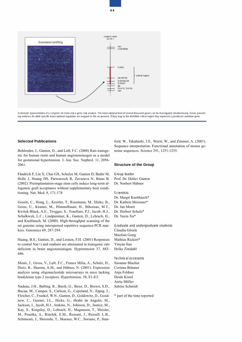
Selected Publications
Bohlender, J., Ganten, D., and Luft, F.C. (2000) Rats transge-nic for human renin and human angiotensinogen as a modelfor gestational hypertension. J. Am. Soc. Nephrol. 11, 2056-2061.
Fändrich F, Lin X, Chai GX, Schulze M, Ganten D, Bader M,Holle J, Huang DS, Parwaresch R, Zavazava N, Binas B.(2002). Preimplantation-stage stem cells induce long-term al-logeneic graft acceptance without supplementary host condi-tioning. Nat. Med. 8, 171-178
Gosele, C., Hong, L., Kreitler, T., Rossmann, M., Hieke, B.,Gross, U., Kramer, M., Himmelbauer, H., Bihoreau, M.T.,Kwitek-Black, A.E., Twigger, S., Tonellato, P.J., Jacob, H.J.,Schalkwyk, L.C., Lindpaintner, K., Ganten, D., Lehrach, H.,and Knoblauch, M. (2000). High-throughput scanning of therat genome using interspersed repetitive sequence-PCR mar-kers. Genomics 69, 287-294.
Huang, B.S., Ganten, D., and Leenen, F.H. (2001) Responsesto central Na(+) and ouabain are attenuated in transgenic ratsdeficient in brain angiotensinogen. Hypertension 37, 683-686.
Monti, J., Gross, V., Luft, F.C., Franca Milia, A., Schulz, H.,Dietz, R., Sharma, A.M., and Hübner, N. (2001). Expressionanalysis using oligonucleotide microarrays in mice lackingbradykinin type 2 receptors. Hypertension. 38, E1-E3.
Nadeau, J.H., Balling, R., Barsh, G., Beier, D., Brown, S.D.,Bucan, M., Camper, S., Carlson, G., Copeland, N., Eppig, J.,Fletcher, C., Frankel, W.N., Ganten, D., Goldowitz, D., Good-now, C., Guenet, J.L., Hicks, G., Hrabé de Angelis, M.,Jackson, I., Jacob, H.J., Jenkins, N., Johnson, D., Justice, M.,Kay, S., Kingsley, D., Lehrach, H., Magnuson, T., Meisler,M., Poustka, A., Rinchik, E.M., Rossant, J., Russell, L.B.,Schimenti, J., Shiroishi, T., Skarnes, W.C., Soriano, P., Stan-
ford, W., Takahashi, J.S., Wurst, W., and Zimmer, A. (2001).Sequence interpretation. Functional annotation of mouse ge-nome sequences. Science 291, 1251-1255.
Structure of the Group
Group leaderProf. Dr. Detlev GantenDr. Norbert Hübner
ScientistsDr. Margit Knoblauch*Dr. Kathrin Meissner*Dr. Jan MontiDr. Herbert Schulz*Dr. Yaxin Xu*
Graduate and undergraduate studentsClaudia GöseleMaolian GongMathias Rickert*Yinyan SunHeike Zimdahl
Technical assistantsSusanne BlachutCorinna BräunerAnja FeldnerHeide KistelAnita MüllerSabine Schmidt
* part of the time reported
44
Schematic representation of a congenic rat strain and a gene chip analysis. The transcriptional level of several thousand genes can be investigated simultaneously. Genes present-ing evidence for allele specific transcriptional regulation are mapped to the rat genome. If they map to the identified critical region they represent a positional candidate gene.
critical region
Molecular Biology of PeptideHormones
Michael Bader
Our group is interested in the molecular biology and functionof hormone systems involved in cardiovascular regulation.Besides the cloning and characterization of genes for theircomponents, the physiological functions of the systems areanalyzed by the production and analysis of transgenic andgene-targeted animal models.
Renin-angiotensin system
The renin-angiotensin system (RAS) is central to blood pres-sure regulation and, therefore, has been studied in detail bytransgenic techniques.
A major focus of our research is the tissue RAS in the brain.Transgenic rats expressing an antisense RNA against angio-tensinogen exclusively in brain astrocytes have been devel-oped and show a decreased local concentration of this proteinand lowered blood pressure and plasma vasopressin levels.These animals are suitable models for the study of the func-tion of local angiotensin production in the brain. Using theserats, we showed that central angiotensin modulates circadianrhythms and the baroreceptor reflex. Furthermore, it is in-volved in the hypertensive and hypertrophic effects of circu-lating angiotensin.
The role of the RAS in hypertension-induced end-organ dam-age is of major clinical importance. In a novel transgenicmouse model, we studied the function of locally produced an-giotensin in the development of cardiac hypertrophy andnephrosclerosis. These mice carried a targeted disruption ofthe angiotensinogen gene which was compensated by a rattransgene exclusively expressed in liver and brain, but not inkidney and heart, where the angiotensinogen gene is expressedin normal mice. Because of elevated plasma angiotensinogenlevels, the animals are hypertensive but they suffer less dam-age to the target organs as a result of the lack of local angio-tensin synthesis.
Kallikrein-kinin system
The kallikrein-kinin system (KKS) is an important hormonesystem for cardiovascular regulation mostly counteracting theeffects of the RAS. As a model to study the functions of theKKS in an intact animal, transgenic rats were developed ex-pressing the human tissue kallikrein gene under the control ofthe heavy-metal responsive metallothionein promoter. Theanimals express the transgene in all organs tested and excretehuman tissue kallikrein in the urine. In these rats, blood pres-sure and its diurnal rhythm, as measured by telemetry, is sig-nificantly reduced compared with control rats. The hearts ofthe animals are protected against ischemic and hypertrophicinjury.
The functions of the kinin B1 receptor are an enigma. To in-vestigate them, we developed mice lacking this subtype. Theresulting animals exhibited analgesia and altered inflamma-tory reactions demonstrating the important role of the B1 re-ceptor in pain transmission and inflammation.
Embryonic stem cell technology
Using embryonic stem cell technology, the gene for thesmooth muscle myosin heavy chain has been deleted by ho-mologous recombination. Lack of this gene still allowedsmooth muscle contraction indicating redundancy of the con-tractile proteins. Furthermore, mice lacking tryptophan hy-droxylase were developed and the genotype of these seroto-nin-deficient animals is under investigation.
In order to also allow gene-targeting experiments in the rat,which is more suitable for research on cardiovascular dis-eases than the mouse, we are establishing embryonic stemcells and cloning technology for this species.
Selected Publications
Baltatu, O., Janssen, B.J., Bricca, G., Plehm, R., Monti, J.,Ganten, D., and Bader, M. (2001). Alterations in blood pres-sure and heart rate variability in transgenic rats with low brainangiotensinogen. Hypertension 37, 408-413.
Fändrich F, Lin X, Chai GX, Schulze M, Ganten D, Bader M,Holle J, Huang DS, Parwaresch R, Zavazava N, Binas B.(2002). Preimplantation-stage stem cells induce long-termallogeneic graft acceptance without supplementary host con-ditioning. Nat. Med. 8, 171-178
Krivokharchenko, A., Galat, V., Ganten, D., and Bader, M.(2001). In vitro formation of tetraploid rat blastocysts afterfusion of two-cell embryos. Mol. Reprod. Dev., 61, 460-465.
Morano, I.L., Chai, G.X., Baltas, L.G., Lamounier-Zepter, V.,Lutsch, G., Kott, M., Haase, H., and Bader, M. (2000).Smooth muscle contraction without smooth muscle myosin.Nature Cell Biol. 2, 371-375.
45
Pesquero, J.B., Araujo, R.C., Heppenstall, P.A., Stucky, C.L.,Silva, J.-A.Jr., Walther, T., Oliveira S.M., Pesquero, J.L.,Paiva, A.C., Calixto, J.B., Lewin, G.R., and Bader, M. (2000).Hypoalgesia and altered inflammatory responses in micelacking kinin B1 receptors. Proc. Natl. Acad. Sci. USA 97,8140-8145.
Silva, J.-A.Jr., Araujo, R.C., Baltatu, O., Oliveira, S.M.,Tschöpe, C., Fink, E., Hoffmann, S., Plehm, R., Chai, K.X.,Chao, L., Chao, J., Ganten, D., Pesquero, J.B., and Bader, M.(2000). Reduced cardiac hypertrophy and altered blood pres-sure control in transgenic rats with the human tissue kallik-rein gene. FASEB J. 14, 1858-1860.
Structure of the Group
Group leaderDr. Michael Bader
ScientistsDr. Ovidiu BaltatuDr. Cécile CaylaDr. Alexander KrivokharchenkoDr. Thomas LangenickelDr. Elena PopowaDr. Mihail TodirasDr. Diego Walther
Graduate and undergraduate studentsNatalia AleninaHeloisa Baptista Allegro*Saleh BashamekhLuciana Aparecida Campos*Cibele Campos CardosoRadu IliescuJens-Uwe PeterClaudia WilhelmIoulia Zaitseva
Technical assistantsAdelheid BöttgerMonika NitzLiselotte Winkler
SecretariatDana Lafuente
* part of the period reported
46
Genetics, Etiology, and Pathogenesis of Hypertension, Vascular Injury, and Renal Diseases
Friedrich C. Luft
Summary
This group is interested in the genetics and pathogenesis ofhypertension, vascular, and renal diseases. Sylvia Bähringleads a team concentrating on the molecular genetics of bloodpressure regulation and lipid metabolism. Here, the aim is tofind important regulatory genes by means of linkage, associa-tion, and positional cloning. Volkmar Gross focuses on so-phisticated cardiovascular physiology in gene-targeted mice.He combines this technology with gene expression arrays.Dominik N. Müller concentrates his attention on the vascularwall. His team uses unique, transgenic rodent models to elu-cidate mechanisms responsible for hypertension-induced end-organ damage. Ralf Dechend is pursuing preeclampsia re-search in patients and in animal models. Ralf Kettritz isstudying human vasculitis while Marek Drab has succeededin disrupting the gene for caveolin-1.
Molecular genetics
Sylvia Bähring has been pursuing the gene responsible for au-tosomal-hypertension and brachydactyly. She, Hakan Toka,and Friedrich C. Luft have studied families from Turkey,France, Canada, and the United States with this problem. Af-fected persons have severe hypertension and die of stroke be-fore the age of 50. The gene is mapped on chromosome 12pand attempts are being made to find the gene by positionalcloning. Hussam Al-Kateb is working on autosomal-recessivehypercholesterolemia. His family has a mutation in the genecoding for an LDL receptor adapter protein on chromosome 1.Interestingly, the family also shows linkage to a site on chro-mosome 13q that the team has previously identified as the siteof a putative “lipid-lowering” gene. Andreas Busjahn has fo-cused on monozygotic and dizygotic twins. He and his asso-ciates recently identified the PPAR� gene locus as the site ofa gene involved in the process of dizygotic twinning.
Vascular research
Volkmar Gross has succeeded in establishing blood pressuretelemetry, cardiac catheterization, and sophisticated methodsof renal function monitoring in gene disrupted mice. He hascharacterized mice lacking the angiotensin AT2 receptor andother mice lacking the bradykinin BK2 receptor. Other novelmodels are currently being studied. Coupling detailed phys-iology with microarray analyses promises to identify novelgenes involved in cardiovascular disease. Dominik N. Müllerhas focused on angiotensin II-induced vascular damage. Re-active oxygen species generation, and activation of associatedtranscription factors, such as NF-�B and AP-1, are pivotal tothis process. In collaboration with Claus Scheidereit, Müllerhas shown that aspirin treatment and other novel anti-inflam-matory approaches can reduce vascular damage. A uniquerole for angiotensin II-induced adaptive immunity has beenrecently identified by the group.
Ralf Dechend, a young cardiologist from the department ofRainer Dietz, has become interested in preeclampsia. Theteam had previously shown that women with preeclampsiadevelop autoantibodies capable of stimulating the angiotensinII AT1 receptor. Dechend has pursued this issue further andhas shown that NADPH oxidase is activated by these antibod-ies in human placental tissue. Ralph Kettritz is an authority onvasculitis, particularly on Wegener’s granulomatosis and herecently observed that anticytoplasmic antibodies signal neu-trophils via the MAP kinase pathway. The process has an im-portant bearing on neutrophil apoptosis.
Finally, Marek Drab spent four years in the laboratory of Tey-muras Kurzchalia at the MDC where they succeeded in dis-rupting the caveolin-1 gene. The absence of caveolae in themice impaired nitric oxide and calcium signaling in the cardi-ovascular system causing aberrations in endothelium-depen-dent relaxation, contractility, and maintenance of myogenictone. In addition, the lungs of knockout animals displayedthickening of the alvaeolar septa caused by uncontrolled en-dothelial cell proliferation and fibrosis, resulting in severephysical limitations in caveolin-1-disrupted mice. The miceshould prove invaluable for clarifying the importance of ca-veolae.
Milestones
Hermann Haller left the Nephrology Hypertension Depart-ment at the Franz Volhard Clinic to assume chairmanship ofthe Nephrology Department at the University of HannoverMedical School in Hannover, Germany. His position has beenfilled by Arya M. Sharma, whose research report is in thisvolume. Herbert Schuster founded the biotech company, IN-FOGEN, which specializes in genetic evaluations and risk as-sessment. He has been joined by Andreas Busjahn and HansKnoblauch and close collaborations with the MDC have beenmaintained by these investigators. Ralf Dechend was awardedthe Jan Brod prize (2000) for his preeclampsia work and Frie-drich C. Luft was awarded the Arthur Corcoran Award of theAmerican Heart Association (2000) and the Lingen Prize(2001) of the Lingen Foundation.
47
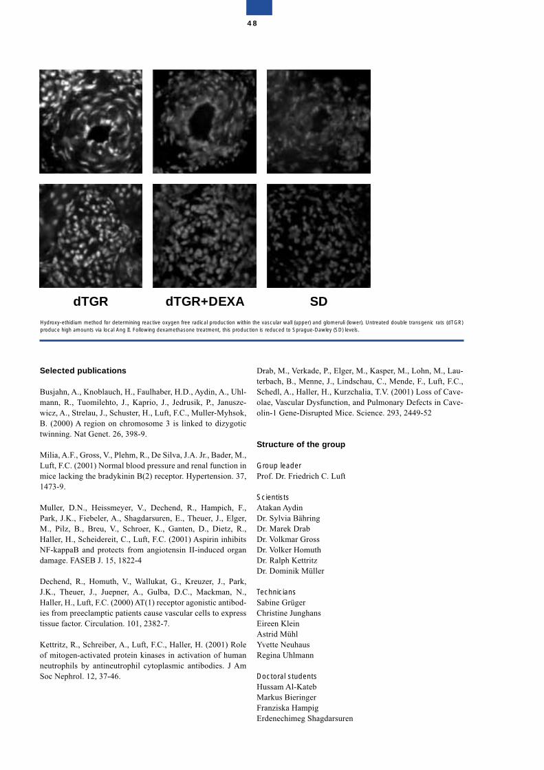
Selected publications
Busjahn, A., Knoblauch, H., Faulhaber, H.D., Aydin, A., Uhl-mann, R., Tuomilehto, J., Kaprio, J., Jedrusik, P., Janusze-wicz, A., Strelau, J., Schuster, H., Luft, F.C., Muller-Myhsok,B. (2000) A region on chromosome 3 is linked to dizygotictwinning. Nat Genet. 26, 398-9.
Milia, A.F., Gross, V., Plehm, R., De Silva, J.A. Jr., Bader, M.,Luft, F.C. (2001) Normal blood pressure and renal function inmice lacking the bradykinin B(2) receptor. Hypertension. 37,1473-9.
Muller, D.N., Heissmeyer, V., Dechend, R., Hampich, F.,Park, J.K., Fiebeler, A., Shagdarsuren, E., Theuer, J., Elger,M., Pilz, B., Breu, V., Schroer, K., Ganten, D., Dietz, R.,Haller, H., Scheidereit, C., Luft, F.C. (2001) Aspirin inhibitsNF-kappaB and protects from angiotensin II-induced organdamage. FASEB J. 15, 1822-4
Dechend, R., Homuth, V., Wallukat, G., Kreuzer, J., Park,J.K., Theuer, J., Juepner, A., Gulba, D.C., Mackman, N.,Haller, H., Luft, F.C. (2000) AT(1) receptor agonistic antibod-ies from preeclamptic patients cause vascular cells to expresstissue factor. Circulation. 101, 2382-7.
Kettritz, R., Schreiber, A., Luft, F.C., Haller, H. (2001) Roleof mitogen-activated protein kinases in activation of humanneutrophils by antineutrophil cytoplasmic antibodies. J AmSoc Nephrol. 12, 37-46.
Drab, M., Verkade, P., Elger, M., Kasper, M., Lohn, M., Lau-terbach, B., Menne, J., Lindschau, C., Mende, F., Luft, F.C.,Schedl, A., Haller, H., Kurzchalia, T.V. (2001) Loss of Cave-olae, Vascular Dysfunction, and Pulmonary Defects in Cave-olin-1 Gene-Disrupted Mice. Science. 293, 2449-52
Structure of the group
Group leaderProf. Dr. Friedrich C. Luft
ScientistsAtakan Aydin Dr. Sylvia BähringDr. Marek DrabDr. Volkmar GrossDr. Volker HomuthDr. Ralph KettritzDr. Dominik Müller
TechniciansSabine GrügerChristine JunghansEireen KleinAstrid MühlYvette NeuhausRegina Uhlmann
Doctoral studentsHussam Al-KatebMarkus BieringerFranziska HampigErdenechimeg Shagdarsuren
48
Hydroxy-ethidium method for determining reactive oxygen free radical production within the vascular wall (upper) and glomeruli (lower). Untreated double transgenic rats (dTGR)produce high amounts via local Ang II. Following dexamethasone treatment, this production is reduced to Sprague-Dawley (SD) levels.
dTGR dTGR+DEXA SD

Gene Mapping and Identification inMonogenic and Complex Diseases
Peter Nürnberg
Positional cloning is now widely used for the identification ofgene defects that are the basis of inherited diseases. A neces-sary first step for positional cloning is the mapping of thegene locus that co-segregates within families with a particulardisease or trait, which allow allocation of a specific chromo-somal position to the responsible gene. Although mappingwas initially developed for monogenic traits, it has now be-come the most widely used strategy to locate genetic factorsinvolved in the etiology of complex diseases. The most pow-erful technique currently available is linkage analysis withhighly polymorphic microsatellite markers, which involvesan examination of the entire genome with a set of evenlyspaced markers. This type of study is usually referred to as awhole genome scan.The Gene Mapping Center is a specialized laboratory for suchhigh-throughput genotyping for gene mapping in monogenicas well as complex diseases. We have developed various setsof well established markers from the Généthon and the CHLCgenetic maps with different marker densities to accommodatethe requirements of special study designs. The laboratory ismainly funded by grants from the German Federal Ministry ofEducation and Research (BMBF). Since January 1997, wehave participated in the German Human Genome Project and,since 2001, in the National Genome Research Network as oneof the major core facilities. The laboratory also undertakesmapping projects for researchers from Germany and abroad.
Mapping of complex diseases
The main focus of the Gene Mapping Center is mapping ofgenetic factors in multifactorial diseases. This type of study in-volves the analysis of large numbers of phenotypically wellcharacterized families. Hundreds of markers are used for gen-otyping and sophisticated biostatistical analyses are subse-quently required to identify genetic loci that contribute to acomplex disease. For this purpose we have established thenecessary techniques and, particularly, automation of the ex-perimental procedures. Our annual capacity is about 2,000,000high-quality genotypes and we are working towards doubling
this capacity by the end of the year. Currently, mapping isbased upon highly informative microsatellite markers but, inthe future, analysis will shift more towards single-nucleotidepolymorphisms (SNPs). Five scientists are involved in projectmanagement, genotyping and technology development whiletwo scientists concentrate on laboratory information manage-ment (LIM) which involves the integration of genotype andphenotype data and the processing of these data for biostatisti-cal analyses. This is done in close collaboration with the bio-informatics group (Dr. K. Rohde) and the University of Bonn(Prof. T. Wienker) who are also involved in data analysis.
A total of seven genome scans for complex diseases havebeen completed and two further studies are underway. In aEuropean collaborative study of the genetics of atopic derma-titis, 199 families with two or more affected siblings each,totalling 839 probands, have been investigated and a majorsusceptibility locus identified on chromosome 3q21. In a sec-ond European collaborative study, 130 families comprising617 probands have been investigated to identify genetic fac-tors involved in susceptibility to common idiopathic general-ized epilepsies (IGE). The linkage results provided significantevidence for a novel IGE susceptibility locus on chromosome3q26 and suggestive evidence for two IGE loci on chromo-somes 14q23 and 2q36.1. The susceptibility loci of both dis-eases are currently being pursued further with refined map-ping and testing of positional candidate genes. We expect togain important insights into the etiology of both diseasegroups.
Ongoing studies include an affected sib-pair study of type 2diabetes in about 400 affected sib pairs from Saxony (PI Dr.Tom Lindner, University Hospital Würzburg) and genotypingof a study of the genetic factors in hypertension in collabora-tion with the Franz-Volhard Clinic on the MDC campus (Prof.Friedrich Luft). The design of the latter study is based on iso-lated populations and takes advantage of the restricted geneticheterogeneity in these populations.
Mapping of monogenic diseases
In contrast to complex diseases, mapping of monogenic traitsrequires less genotyping. Usually, it is sufficient to analyze 30probands or less. The statistical evaluation is different andoften requires skilled interpretation, e.g. haplotyping. In thesix years the laboratory has been in existence, about 50 mono-genic traits have been mapped in humans. The underlyinggene defect has been identified for several of these, complet-ing the process of positional cloning. For instance, an auto-somal dominant bone dysplasia, craniometaphyseal dysplasia(CMD), was found to be caused by heterozygous mutations inANKH, the human ortholog of the mouse progressive ankylo-sis gene. The gene product is a multipass transmembrane pro-tein and seems to form a channel for inorganic pyrophosphate(PPi). These data are important since they link the PPi path-way with bone formation and remodelling. Further analysis ofthe molecular pathology of CMD and related conditions is inprogress. Understanding this group of monogenic diseaseswill provide us with novel insights into more general pro-cesses controlling bone density and may offer new approach-es to the therapy of osteoporosis.
49
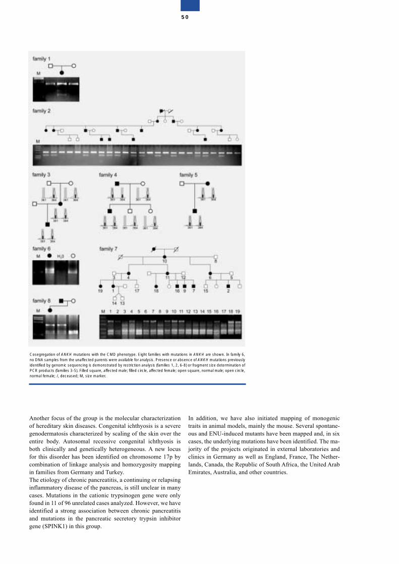
Another focus of the group is the molecular characterizationof hereditary skin diseases. Congenital ichthyosis is a severegenodermatosis characterized by scaling of the skin over theentire body. Autosomal recessive congenital ichthyosis isboth clinically and genetically heterogeneous. A new locusfor this disorder has been identified on chromosome 17p bycombination of linkage analysis and homozygosity mappingin families from Germany and Turkey. The etiology of chronic pancreatitis, a continuing or relapsinginflammatory disease of the pancreas, is still unclear in manycases. Mutations in the cationic trypsinogen gene were onlyfound in 11 of 96 unrelated cases analyzed. However, we haveidentified a strong association between chronic pancreatitisand mutations in the pancreatic secretory trypsin inhibitorgene (SPINK1) in this group.
In addition, we have also initiated mapping of monogenictraits in animal models, mainly the mouse. Several spontane-ous and ENU-induced mutants have been mapped and, in sixcases, the underlying mutations have been identified. The ma-jority of the projects originated in external laboratories andclinics in Germany as well as England, France, The Nether-lands, Canada, the Republic of South Africa, the United ArabEmirates, Australia, and other countries.
50
Cosegregation of ANKH mutations with the CMD phenotype. Eight families with mutations in ANKH are shown. In family 6,no DNA samples from the unaffected parents were available for analysis. Presence or absence of ANKH mutations previouslyidentified by genomic sequencing is demonstrated by restriction analysis (families 1, 2, 6-8) or fragment size determination ofPCR products (families 3-5). Filled square, affected male; filled circle, affected female; open square, normal male; open circle,normal female; /, deceased; M, size marker.
Selected Publications
Krebsová, A., Küster, W., Lestringant, G.G., Schulze, B.,Hinz, B., Frossard, P.M., Reis, A., and Hennies, H.C. (2001).Identification, by homozygosity mapping, of a novel locus forautosomal recessive congenital ichthyosis on chromosome17p, and evidence for further genetic heterogeneity. Am. J.Hum. Genet. 69, 216-222.
Lee, Y.A., Wahn, U., Kehrt, R., Tarani, L., Businco, L.,Gustafsson, D., Andersson, F., Oranje, A.P., Wolkertstorfer,A., v. Berg, A., Hoffmann, U., Küster, W., Wienker, T.,Rüschendorf, F., and Reis, A. (2000). A major susceptibilitylocus for atopic dermatitis maps to chromosome 3q21. NatureGenet. 26, 470-473.
Nürnberg, P., Thiele, H., Chandler, D., Hohne, W., Cunning-ham, M.L., Ritter, H., Leschik, G., Uhlmann, K., Mischung,C., Harrop, K., Goldblatt, J., Borochowitz, Z.U., Kotzot, D.,Westermann, F., Mundlos, S., Braun, H.S., Laing, N., andTinschert, S. (2001). Heterozygous mutations in ANKH, thehuman ortholog of the mouse progressive ankylosis gene, re-sult in craniometaphyseal dysplasia. Nature Genet. 28, 37-41.
Sander, T., Schulz, H., Saar, K., Gennaro, E., Riggio, M.C.,Bianchi, A., Zara, F., Luna, D., Bulteau, C., Kaminska, A.,Ville, D., Cieuta, C., Picard, F., Prud’homme, J.F., Bate, L.,Sundquist, A., Gardiner, R.M., Janssen, G.A.M.A.J., de Haan,G.J., Kasteleijn-Nolst-Trenité, D.G.A., Bader, A., Lindhout,D., Riess, O., Wienker, T.F., Janz, D., and Reis, A. (2000). Ge-nome search for susceptibility loci of common idiopathic gen-eralised epilepsies. Hum. Mol. Genet. 9, 1465-1472.
Witt, H., Luck, W., Hennies, H.C., Classen, M., Kage, A., Lass,U., Landt, O. and Becker, M. (2000). Mutations in the gene en-coding the serine protease inhibitor, Kazal type 1 are associatedwith chronic pancreatitis. Nature Genet. 25, 213-216.
Structure of the Group
Group leaderDr. Peter Nürnberg
ScientistsArif Bülent Ekici*Dr. Hans Christian HenniesDr. Birgit Meyer*Dr. Holger Thiele*Mohammad Reza Toliat*Dr. Kathrin Saar*
Guest ScientistsDr. Katrin HoffmannDr. Young-Ae LeeDr. Franz Rüschendorf
Graduate and undergraduate studentsSilke Appel*Anja Brinckmann*Katja-Martina Eckl*Stefan Ey*Lisa Haucke*Julia KrauseInés Mácha*Dietlind PachaleKaren Uhlmann*Andreas Wollstein*
Technical assistantsFrancoise AndréChristian Becker*Ingelore Bäßmann*Elisabeth Kirst*Barbara Lucke*Susanne Lützkendorf*Gudrun NürnbergRegina Pospiech*Madeleine SkornaInka Szangolies*
Guest assistantsHeike Fischer*Jenny Pech*Monika Schwarz
SecretariatKornelia Dokup
* part of the period reported
51
Cardiovascular Molecular Genetics
Ludwig Thierfelder
Animal models for familial hypertrophic cardiomyopathy disease genes
All disease genes for autosomal dominant familial hyper-trophic cardiomyopathy (FHC) code for proteins of the con-tractile machinery of striated muscle. This led to the defini-tion that FHC is a disease of the sarcomere. In order to betterunderstand functional and molecular disease pathways inFHC caused by alpha-tropomyosin mutations, we overex-press two alpha-tropomyosin mutations (Asp175Asn;Glu180Gly) in a transgenic rat model. The phenotype differsfor each mutation: mutation Glu180Gly is functionally silent,whereas mutation Asp175Asn shows decreased Ca++-sensi-tivity of cardiac myofilaments. Although no gross cardiachypertrophy is observed, animals carrying either mutationdevelop myocyte disarray and reexpress fetal cardiac genes asearly molecular markers of cardiac hypertrophy. This modelis suited to the investigation of early functional and molecularevents in the pathogenesis of alpha-tropomyosin mutations.Because the physiology of cardiac myosin binding protein C(MybP-C; another FHC disease gene), especially its N-ter-minus, is not well known, we have developed a knock-inmouse model to study functional aspects of mutated cardiacMybP-C.
Molecular genetics of familial dilated cardiomyopathy
Dilated cardiomyopathy (DCM) is a genetic disease causedby autosomal dominant mutations in 20-30% of cases. Only afew DCM disease genes are known and these belong to differ-ent classes of molecules (dystrophin; lamin A/C; beta myosinheavy chain, actin; desmin). In two large DCM families, posi-tional cloning efforts in our laboratory have very recently ledto the identification of mutations in titin (TTN), causing oneform of non-syndromic DCM. Titin is the largest knownmolecule in mammals (3-3.7MDa) and encoded by up to114kb cDNA. A truncation mutation of A-band titin and amissense mutation in I-band titin cause a similar phenotype in
two unrelated families. Interestingly, although both mutationsare expressed in cardiac and skeletal muscle, only the heart isclinically affected.
Molecular genetics of arrhythmogenic right ventricular cardiomyopathy
Arrhythmogenic right ventricular cardiomyopathy (ARVC) iscardiac condition and is associated with sudden death andheart failure. We have identified an autosomal dominantfounder mutation on chromosome 3p25 in a large Canadiancardiomyopathy population of ~400 individuals at 50% riskof inheriting the condition. ARVC in these individuals is asso-ciated with a distinct electrocardiographic pattern and a lifeexpectancy of <40y in males. Mutational analyses of approx-imately 15 genes located in a 2Mbp region on chromosome3p25 should identify the responsible mutation.
Genetic studies of isolated non-compaction of the left ventricle in the adult
Isolated non-compaction of the left ventricle (INVC) is a raredisorder characterized by wide intertrabecular spaces due toan arrest of endomyocardial morphogenesis. It is not knownwhether INVC can be caused by germline mutations. Becausea mutation of the G4.5 gene (Barth disease gene) has beenfound in a patient with infantile INVC, the G4.5 gene in over15 adult INVC patients has been screened but no mutation hasbeen found. We have further evaluated the pedigrees of twoINVC index patients and identified several members also af-fected by INVC. This shows that INVC in the adult can betransmitted genetically. Additional candidate genes of the en-domyocardial morphogenesis pathways are currently beingscreened for INVC-causing mutations.
Molecular genetics of pseudoxanthoma elasticum(PXE)
Pseudoxanthoma elasticum is an inherited systemic disorderof the elastic tissue characterized by degenerative calcifica-tion with subsequent disintegration and destruction of theelastic tissue of several organs. Cardiovascular disease en-compasses a wide clinical spectrum from mental fatigue syn-drome to early cardiovascular death due to myocardial infarc-tion or, very rarely, gastrointestinal hemorrhage. We havemapped the PXE locus to a 500 kb interval on chromosome16p13.1. and shown that mutations in a transmembrane trans-porter protein, ABC-C6 (also known as MRP-6), cause PXE.
Selected Publications
Eisenberg, I., Thiel, C., Levi, T., Tiram, E., Argov, Z., Sadeh,M., Jackson, C.L., Thierfelder, L., and Mitrani-Rosenbaum,S. (1999). Fine structure mapping of the hereditary inclusionbody myopathy locus. Genomics 55, 43-48.
52
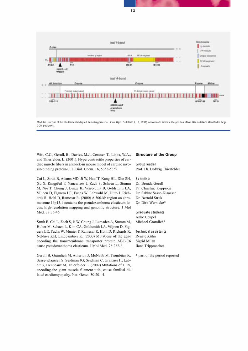
Witt, C.C., Gerull, B., Davies, M.J., Centner, T., Linke, W.A.,and Thierfelder, L. (2001). Hypercontractile properties of car-diac muscle fibers in a knock-in mouse model of cardiac myo-sin-binding protein-C. J. Biol. Chem. 16, 5353-5359.
Cai L, Struk B, Adams MD, Ji W, Haaf T, Kang HL, Dho SH,Xu X, Ringpfeil F, Nancarrow J, Zach S, Schaen L, StummM, Niu T, Chung J, Lunze K, Verrecchia B, Goldsmith LA,Viljoen D, Figuera LE, Fuchs W, Lebwohl M, Uitto J, Rich-ards R, Hohl D, Ramesar R. (2000) A 500-kb region on chro-mosome 16p13.1 contains the pseudoxanthoma elasticum lo-cus: high-resolution mapping and genomic structure. J MolMed. 78:36-46.
Struk B, Cai L, Zach S, Ji W, Chung J, Lumsden A, Stumm M,Huber M, Schaen L, Kim CA, Goldsmith LA, Viljoen D, Fig-uera LE, Fuchs W, Munier F, Ramesar R, Hohl D, Richards R,Neldner KH, Lindpaintner K. (2000) Mutations of the geneencoding the transmembrane transporter protein ABC-C6cause pseudoxanthoma elasticum. J Mol Med. 78:282-6.
Gerull B, Gramlich M, Atherton J, McNabb M, Trombitas K,Sasse-Klaassen S, Seidman JG, Seidman C, Granzier H, Lab-eit S, Frenneaux M, Thierfelder L. (2002) Mutations of TTN,encoding the giant muscle filament titin, cause familial di-lated cardiomyopathy. Nat. Genet. 30:201-4.
Structure of the Group
Group leaderProf. Dr. Ludwig Thierfelder
ScientistsDr. Brenda GerullDr. Christine KupprionDr. Sabine Sasse-KlaassenDr. Bertold StrukDr. Dirk Wernicke*
Graduate studentsAnke GeupelMichael Gramlich*
Technical assistantsRenate KühnSigrid MilanIlona Trippmacher
* part of the period reported
53
Modular structure of the titin filament (adapted from Gregorio et al., Curr. Opin. Cell Biol.11, 18, 1999). Arrowheads indicate the position of two titin mutations identified in largeDCM pedigrees.
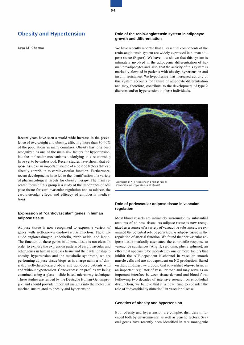
Obesity and Hypertension
Arya M. Sharma
Recent years have seen a world-wide increase in the preva-lence of overweight and obesity, affecting more than 30-40%of the populations in many countries. Obesity has long beenrecognized as one of the main risk factors for hypertension,but the molecular mechanisms underlying this relationshiphave yet to be understood. Recent studies have shown that ad-ipose tissue is an important source of a host of factors that candirectly contribute to cardiovascular function. Furthermore,recent developments have led to the identification of a varietyof pharmacological targets for obesity therapy. The main re-search focus of this group is a study of the importance of adi-pose tissue for cardiovascular regulation and to address thecardiovascular effects and efficacy of antiobesity medica-tions.
Expression of “cardiovascular” genes in humanadipose tissue
Adipose tissue is now recognized to express a variety ofgenes with well-known cardiovascular function. These in-clude angiotensinogen, endothelin, nitric oxide, and leptin.The function of these genes in adipose tissue is not clear. Inorder to explore the expression pattern of cardiovascular andother genes in human adiposes tissue and their relationship toobesity, hypertension and the metabolic syndrome, we areperforming adipose-tissue biopsies in a large number of clin-ically well-characterized obese and non-obese patients withand without hypertension. Gene-expression profiles are beingexamined using a glass – slide-based microarray technique.These studies are funded by the Deutsche Human-Genompro-jekt and should provide important insights into the molecularmechanisms related to obesity and hypertension.
Role of the renin-angiotensin system in adipocytegrowth and differentiation
We have recently reported that all essential components of therenin-angiotensin system are widely expressed in human adi-pose tissue (Figure). We have now shown that this system isintimately involved in the adipogenic differentiation of hu-man preadipocytes and also that the activity of this system ismarkedly elevated in patients with obesity, hypertension andinsulin resistance. We hypothesize that increased activity ofthis system accounts for failure of adipocyte differentiationand may, therefore, contribute to the development of type 2diabetes and/or hypertension in obese individuals.
Role of perivascular adipose tissue in vascularregulation
Most blood vessels are intimately surrounded by substantialamounts of adipose tissue. As adipose tissue is now recog-nized as a source of a variety of vasoactive substances, we ex-amined the potential role of perivascular adipose tissue in theregulation of arterial function. We found that perivascular ad-ipose tissue markedly attenuated the contractile response tovasoactive substances (Ang II, serotonin, phenylephrine), aneffect that appears to be mediated by one or more factors thatinhibit the ATP-dependent K-channel in vascular smoothmuscle cells and are not dependent on NO production. Basedon these findings, we propose that adventitial adipose tissue isan important regulator of vascular tone and may serve as animportant interface between tissue demand and blood flow.Following two decades of intensive research on endothelialdysfunction, we believe that it is now time to consider therole of “adventitial dysfunction” in vascular disease.
Genetics of obesity and hypertension
Both obesity and hypertension are complex disorders influ-enced both by environmental as well as genetic factors. Sev-eral genes have recently been identified in rare monogenic
54
Expression of AT1 receptors on a human fat cell(Confocal microscopy: Gorzelniak/Quass)
forms of obesity and hypertension, but the importance ofthese genes for the development of non monogenic forms ofhypertension remains to be explored. We are currently con-ducting several studies using both a population-based cross-sectional as well as a family-based approach in an attempt toidentify genetic variants that can account for the developmentof these disorders.
Pharmacological management of obesity hypertension
Although obesity accounts for a considerable proportion ofcardiovascular morbidity and mortality, the efficacy and out-come of cardiovascular therapies have not been extensivelystudied in obese patients. Thus, whereas for non-obese pa-tients the utility and efficacy of pharmacological interventionhas been well established, these principles need to be reas-sessed regarding their utility and efficacy in obese patients.This is important, since obesity can affect both the pharma-cokinetics and pharmacodynamics of various cardiovasculardrug treatments. Furthermore, recent antiobesity drugs havebeen found to have a significant impact on cardiovascularregulation, but their role in the management of obesity hyper-tension remains to be defined. We are currently conductingseveral studies addressing these issues
Selected Publications
Boschmann, M., Ringel, J., Klaus, S., and Sharma, A.M.(2001). Metabolic and hemodynamic response of adipose tis-sue to angiotensin II. Obes. Res. 9, 486-491.
Sharma, A.M., Pischon, T., Engeli, S., and Scholze, J. (2001).Choice of drug treatment for obesity-related hypertension:where is the evidence? J. Hypertens. 19, 123-134.
Sharma, A.M., Pischon, T., Hardt, S., Kunz, I., and Luft, F.C.(2001). Beta-adrenergic receptor blockers and weight gain: Asystematic analysis. Hypertension. 37, 250-254.
Kunz, I., Klaus, S., Kallies, B., Schorr, U., and Sharma, A.M.(2000). Kinetic analysis of the thermic effect of food and itsrelationship to body composition in humans. Metabolism 49,1340-1345.
Engeli, S., and Sharma, A.M. (2000). Role of adipose tissuefor cardiovascular-renal regulation in health and disease.Horm. Metab. Res. 32, 485-499.
Brand, E., Schorr, U., Kunz, I., Kertmen, E., Ringel, J., Dis-tler, A., and Sharma, A.M. (2001). Tumor necrosis factor-alpha-308 G/A polymorphism in obese Caucasians. Int. J. Obes. Relat. Metab. Disord. 25, 581-585.
Structure of the Group
Group leaderProf. Dr. Arya M. Sharma
ScientistsStefan EngeliDr. Jürgen JankeDr. Margaret WellnerKerstin GorzelniakDr. Heidrun MehlingDr. Michael StoeterGalina DubrovskaDr. Carmen Kresse
Graduate and undergraduate studentsMareike FeldpauschClaudia GösselFrauke Hartwig
DieticianJana Böhnke
Study NurseIris GottschalkGrit StoffelsAnke Strauß
TechniciansBärbel GirreschHenning DammBettina JunkerUte GerhardtMathilde Schmidt
SecretariatSigrid Rehm
55
Disorders of the AutonomicNervous System
Jens Jordan (Helmholtz fellow)
The main interest of the group is basic (mechanism-oriented)patient-oriented research in the field of clinical autonomicdisorders, arterial hypertension, obesity, and genetics. One in-tention of our group is to combine patient-oriented researchwith basic science and genetics in the field of cardiovasculardiseases. The purpose of our research is to develop new treat-ment strategies for patients with orthostatic hypotension, ortho-static intolerance, multiple system atrophy, pure autonomicfailure, neurally-mediated syncope, and neurogenic hyperten-sion based on a better understanding of the pathophysiologyof these clinical syndromes. To elucidate the potential influ-ence of candidate genes we participate in twin studies.
Genetic influences on cardiovascular regulation inhealth and disease
In a patient with orthostatic intolerance and her identical twin,we conducted extensive tests of autonomic nervous systemregulation. We found evidence for impaired norepinephrineuptake mechanisms. Genetic testing revealed a functional mu-tation of the norepinephrine transporter gene causing the syn-drome. We investigated blood pressure regulation in patientswith monogenic hypertension, brachydactyly, and neurovas-cular brainstem contact. We found no difference in sympa-thetic nerve traffic or activation. However, blood pressureincreases to phenylephrine were markedly augmented, com-pared with controls. Complete ganglionic blockade largely at-tenuated this difference, suggesting marked impairment ofbaroreflex buffering in these patients. In healthy dizygotic andmonozygotic twins, we tested the hypothesis that baroreflexfunction, which has a central role in blood pressure regulation,is influenced by genetic factors. A large part of the variabilityin baroreflex function could be explained by genetic influ-ences. In a subsequent association study, we found that part ofthe genetic influence on baroreflex function may be explainedby a gene encoding a calcium-activated potassium channel.
Selected Publications
Shannon, J. R., Flattern, N., Jordan, J., Jacob, G., Black, B.K., Biaggioni, I., Blakely, R. D., and Robertson, D. (2000).Orthostatic Intolerance intolerance and tachycardia asso-ciated with norepinephrine transporter deficiency. N. Engl. J.Med. 342, 541-548.
Jordan, J., Shannon, J. R., Black, B. K., Ali, Y., Farley, M.,Costa, F., Diedrich, A., Robertson R. M., Biaggioni, I., andRobertson, D. (2000). The pressor response to water drinkingin humans: a sympathetic reflex? Circulation. 101, 504-509.
Jordan, J., Toka, H., Heusser, K., Toka, O., Shannon, J. R.,Tank, J., Diedrich, A., Stabroth, C., Stoffels, M., Oelkers, W.,Schuster, H., Schobel, H. P., Haller, H., and Luft, F. C. (2000).Severely impaired baroreflex-buffering in patients withmonogenic hypertension and neurovascular contact. Circula-tion. 102, 2611-2618.
Jordan, J., Tank, J., Stoffels, M., Franke, G., Luft, F. C., andBoschmann, M. (2001). Interaction of beta-adrenoreceptorstimulation and nitric oxide release on tissue perfusion andmetabolism. J. Clin. Endocrinol. Metab. 86, 2803-2810.
Tank, J., Jordan, J., Diedrich, A., Stoffels, M., Franke, G.,Faulhaber, H. D., Luft, F. C., and Busjahn, A. (2001). Geneticinfluences on baroreflex function in normal twins. Hyperten-sion. 37, 907-910.
Structure of the Group
Group leaderDr. Jens Jordan
ScientistsFrauke AdamsMarina BrinsukDr. Christoph SchröderDr. Jens Tank
Graduate and undergraduate studentsGötz KruppAndreas Birkenfeld
Study NursesMandy StoffelsNadine Krüger
TechniciansGabriele Franke
56
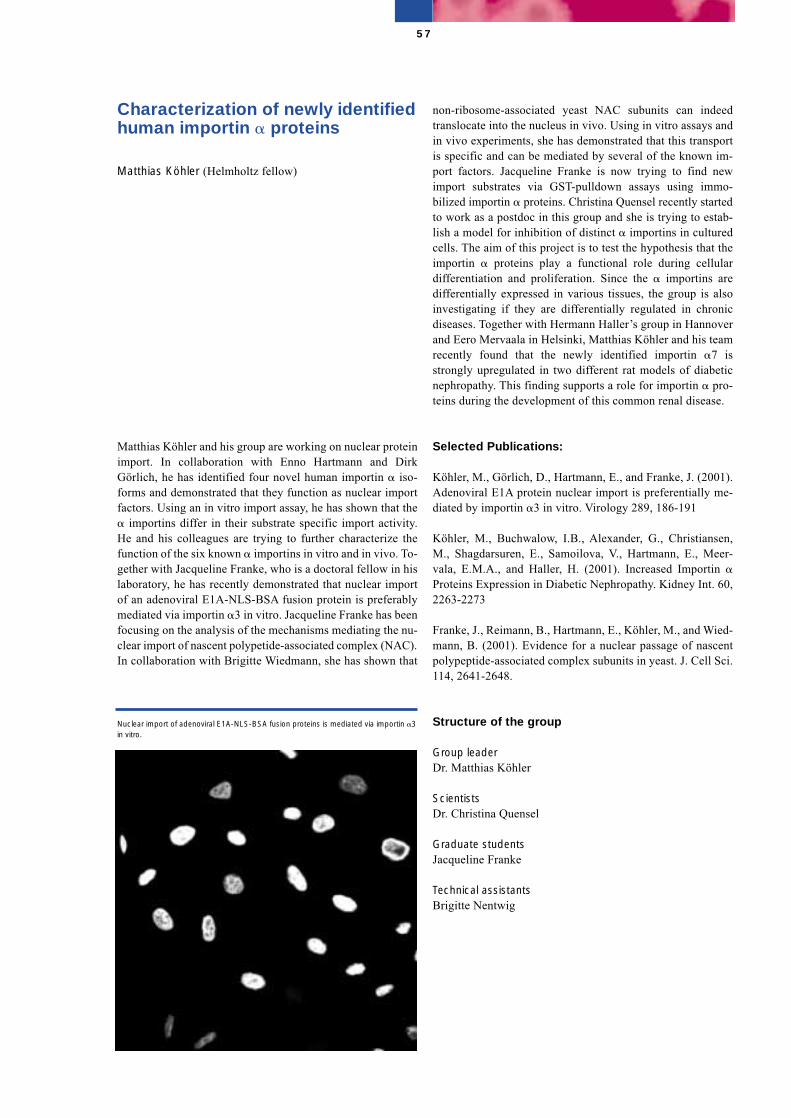
Characterization of newly identifiedhuman importin � proteins
Matthias Köhler (Helmholtz fellow)
Matthias Köhler and his group are working on nuclear proteinimport. In collaboration with Enno Hartmann and DirkGörlich, he has identified four novel human importin � iso-forms and demonstrated that they function as nuclear importfactors. Using an in vitro import assay, he has shown that the� importins differ in their substrate specific import activity.He and his colleagues are trying to further characterize thefunction of the six known � importins in vitro and in vivo. To-gether with Jacqueline Franke, who is a doctoral fellow in hislaboratory, he has recently demonstrated that nuclear importof an adenoviral E1A-NLS-BSA fusion protein is preferablymediated via importin �3 in vitro. Jacqueline Franke has beenfocusing on the analysis of the mechanisms mediating the nu-clear import of nascent polypetide-associated complex (NAC).In collaboration with Brigitte Wiedmann, she has shown that
non-ribosome-associated yeast NAC subunits can indeedtranslocate into the nucleus in vivo. Using in vitro assays andin vivo experiments, she has demonstrated that this transportis specific and can be mediated by several of the known im-port factors. Jacqueline Franke is now trying to find newimport substrates via GST-pulldown assays using immo-bilized importin � proteins. Christina Quensel recently startedto work as a postdoc in this group and she is trying to estab-lish a model for inhibition of distinct � importins in culturedcells. The aim of this project is to test the hypothesis that theimportin � proteins play a functional role during cellulardifferentiation and proliferation. Since the � importins aredifferentially expressed in various tissues, the group is alsoinvestigating if they are differentially regulated in chronicdiseases. Together with Hermann Haller’s group in Hannoverand Eero Mervaala in Helsinki, Matthias Köhler and his teamrecently found that the newly identified importin �7 isstrongly upregulated in two different rat models of diabeticnephropathy. This finding supports a role for importin � pro-teins during the development of this common renal disease.
Selected Publications:
Köhler, M., Görlich, D., Hartmann, E., and Franke, J. (2001).Adenoviral E1A protein nuclear import is preferentially me-diated by importin �3 in vitro. Virology 289, 186-191
Köhler, M., Buchwalow, I.B., Alexander, G., Christiansen,M., Shagdarsuren, E., Samoilova, V., Hartmann, E., Meer-vala, E.M.A., and Haller, H. (2001). Increased Importin �Proteins Expression in Diabetic Nephropathy. Kidney Int. 60,2263-2273
Franke, J., Reimann, B., Hartmann, E., Köhler, M., and Wied-mann, B. (2001). Evidence for a nuclear passage of nascentpolypeptide-associated complex subunits in yeast. J. Cell Sci.114, 2641-2648.
Structure of the group
Group leaderDr. Matthias Köhler
ScientistsDr. Christina Quensel
Graduate studentsJacqueline Franke
Technical assistantsBrigitte Nentwig
57
Nuclear import of adenoviral E1A-NLS-BSA fusion proteins is mediated via importin �3in vitro.
Tumor Genetics
Siegfried Scherneck
The research program of this group is aimed at obtaining abetter understanding of the genetic basis of cancers, particu-larly human breast cancers (BC). BC, one of the most com-mon cancers affecting women, has been demonstrated to arisethrough a multi-step process in which a number of oncogenesand tumor suppressor genes (TSG) contribute to the cancerwhen their function is altered. Although about 90% of BC aresporadic, the remaining cases are heritable and caused by mu-tations of at least two TSG, BRCA1 and BRCA2. It is reason-ably likely that at least one more highly penetrant susceptibil-ity gene will emerge. However, attention is now shiftingtoward more common low-penetrance mutations and theirpossible contribution to BC. Therefore, multiple approaches,such as linkage in high-risk families and association studiesin large BC case-control studies, are being used to identify ad-ditional high- and low penetrance genes.
Furthermore, identification and characterization of a numberof genes that are somatically altered in sporadic BC as well assubsequent analysis of respective alterations in the germlineof individuals of BC families (predisposing genes) will leadto a better understanding of the development and progressionof BC. Advances in these areas may provide informationabout the clinical uses of these genes for predisposition test-ing, early detection, prognostication, and therapy selection.
Comprehensive analysis of German breast/ovariancancer families for BRCA1/BRCA2 germline mutations: mutation profiles and frequencies inthe German populationW. Hofmann, E. Claßen, D. Horn, B. Jandrig, S. Seitz, A.Nothnagel, I. Sümnich, H. Zeidler, K. Krause, the “Berlin Cen-ter for Hereditary Breast and Ovarian Cancer” in cooperationwith the “German Consortium for Hereditary Breast andOvarian Cancer (GCHBOC)” and M. Schwab (DKFZ, Heidel-berg)
Germline mutations in the BC susceptibility genes, BRCA1/BRCA2, jointly explain the most significant part of the famil-ial breast/ovarian cancer syndrome. Within the GCHBOC,which was initiated and supported by the “Deutsche Krebs-hilfe”, we have participated in a comprehensive study to ana-lyze the entire coding sequence of the BRCA1 and BRCA2genes in about 1000 patients from German breast/ovariancancer families. A total of about 80 BRCA1 and about 70BRCA2 distinct deleterious mutants in about 300 patientshave been identified by the GCHBOC. More than one third ofthese mutations are novel and might be specific for the Ger-man population. The mutation study has defined groups ofhigh-risk families. Mutation rates of only 50% and lower inthe higher-risk groups provide evidence for further predispos-ing genes. At present, identified mutations and unclassifiedvariants in the BRCA genes, as well as specific rearrange-ments in mutation carriers have been characterized for clarifi-cation of genotype-phenotype correlations by extended con-trol studies and investigation of the corresponding tumors.The identification of BRCA1 and BRCA2 genes has led tochanges in the management and treatment of high-riskwomen.
Systematic search for genes contributing to thegenesis and progression of sporadic and hereditarybreast cancerB. Jandrig, S. Seitz, I. Lapidous, M. Plaumann, A. Schwartz,K. Wenzel, C. Zeller, R. Frege, K. Poppe, S. Werner, in coop-eration with A. Meindl (Munich), N. Arnold (Kiel), D. Nieder-acher (Düsseldorf), K. Schmutzler (Bonn), B.M. Jockusch(Braunschweig), I. Petersen (Berlin), B. Hinzmann, A. Rosen-thal (Berlin)
Over the last few years, the increasing resolution of geneticand physical maps has allowed the identification of recurrentgenetic alterations in BC development. We have focused ourresearch on genes whose function is impaired or lost duringBC development, with particular emphasis on chromosomeregions 6q23-q25, 8p12-p21 and 17p13.3. Chromosometransfer studies using cell lines as target cells derived frombreast carcinoma suggest the existence of TSG within theseregions. Deletion fine mapping in combination with sequenc-ing, sequence database searches and microarray technologyhave uncovered sequences of several candidate genes on 6q(i.e. MDC 11 and 12 genes), 8p (i.e. TRAIL receptor genes,EXTL3, LOXL2, BZF) and 17p13 (i.e. profilin 1) which arecurrently being tested. To validate breast cancer associatedgenes, several positional and functional approaches are beingused in combination: identification of differentially expressedESTs by electronic- and real Northern blotting and RT-PCR;
58
fine mapping of LOH hotspots; microarray-technology; mu-tation analysis. In addition, transfection assays and functionalcomplementation tests have been applied.
Somatic genetic alterations in breast cancer:correlation of genetic data with clinicopathologicalparametersS. Seitz, A. Nothnagel, K. Poppe, S. Werner in cooperationwith P.M. Schlag (RRK, Berlin), J. Fischer (RRK, Berlin)
Specific genetic alterations could become new diagnosticmarkers for BC prognosis, providing defined individualizedinformation about clinical outcome and response to therapy.In particular, LOH has been shown to occur commonlyamong various alleles at specific loci. A significant correla-tion was found between loss of 17p13 and parameters asso-ciated with more aggressive tumor behaviour, including largetumor size, grade, proliferative activity and estrogen receptorstatus. At present we are examining the contribution andprognostic relevance of genetic alterations on chromosomeregions 8p, 8q, 6q and 17p at the genomic and transcriptionallevel.
Molecular pathology of solid tumorsK. Kölble, B. Barthel, H. Fischer, M. Simon
We have investigated the patterns of chromosomal and micro-satellite instability in human tumors of the breast, gastrointes-tinal and urogenital tracts and have correlated these with theprevalence of germline and somatic mutations in a range ofgenes implicated in tumorigenesis (APC, CTNNB1, AXIN2).The development of a novel instrument and advanced metho-dologies for laser-assisted microdissection of human tissuesand their integration with microscale real-time PCR and im-munohistological expression analyses allows to detect quanti-tative expression patterns in morphologically selected smallnumbers of normal and neoplastic cells. Such multi-dimen-sional assays reflecting the various physiological and patho-logical differentiation states of tissues may help to refine theconcepts and practice of molecular diagnostics.
Antibody engineering B. Micheel, J. Schenk, G. Scharte
Antibody technology has been used to produce and modifyantibodies against different antigens, including tumor-asso-ciated gene products. Several monoclonal antibodies havebeen produced against epitopes of the BRCA1 gene product.The antibodies have been tested by immunohistochemicaltechniques for their value as diagnostic reagents. Metaboliclabelling experiments have been performed to screen forthose antibodies reacting with the intracellular BRCA1 geneproduct that is biologically active. Binding partners of theBRCA1 gene product will be identified with the help of theseantibodies. Monoclonal antibodies have also been producedagainst the products of newly identified tumor associatedgenes and these will be used to study the biological functionof the gene products.
Selected Publications
Janke, J., Schlüter, K., Jandrig, B., Theile, M., Kölble, K., Ar-nold, W., Grinstein, E., Schwartz, A., Estevéz-Schwarz, L.,Schlag, P.M., Jockusch, B., and Scherneck, S. (2000). Sup-pression of tumorigenicity in breast cancer cells by microfila-ment protein profilin 1. J. Exp. Med. 191, 1675-1685.
Seitz, S., Werner, S., Fischer, J., Nothnagel, A., Schlag, P.M.,and Scherneck, S. (2000). Refined deletion mapping in spo-radic breast cancer at chromosomal region 8p12-p21 and as-sociation with clinicopathological parameters. Eur. J. Cancer36, 1507-1513.
Zhang, Y., Siebert, R., Matthiesen P., Harder, S., Theile, M.,Scherneck, S., and Schlegelberger, B. (2000). Feasibility ofsimultaneous fluorescence immunophenotyping and fluores-cence in situ hybridization study for the detection of estrogenreceptor expression and deletions of the estrogen receptorgene in breast carcinoma cell lines. Virchows Arch 436, 271-275.
Seitz, S., Poppe, K., Fischer, J., Nothnagel, A., Estevéz-Schwarz, L., Haensch, W., Schlag, P.M., and Scherneck, S.(2001). Detailed deletion mapping in sporadic breast cancerat chromosomal region 17p13 distal to the TP53 gene: associ-ation with clinicopathological parameters. J. Pathol. 194,318-326.
Salvelyeva, L., Claas, A., Matzner, I., Schlag, P.M., Hofmann,W., Scherneck, S., Weber, B., and Schwab, M. (2001). Consti-tutional genomic instability with inversions, duplications, andamplifications in 9p23-24 in BRCA2 mutation carriers. Can-cer Res. 61, 5179-5185.
Structure of the Group
Group leader Prof. Dr. Siegfried Scherneck
Scientists Dr. Burkhard JandrigDr. Helmuth Prokoph*Dr. Arnfried Schwartz Dr. Susanne Seitz Dr. Elke Burghardt-Göttges*Dr. Denise Horn Dr. Irina Lapidous Dr. Marlies Plaumann* Dr. Katrin Wenzel*Elvira Claßen
Graduate and undergraduate students Unn-Hilde Grasmo-Wendler*Wera Hofmann Constanze Zeller*Peter Waßmuth*Friedrich Wiedemann*
59

Technical assistants Renate FregeKonstanze Poppe Karin Rücker Ingeborg Sümnich Sabine Werner Helga Zeidler Kerstin Krause*
Guest scientists Dr. Dr. Konrad Kölble Dr. Beatrix Barthel*Heike Fischer Dr. Lope Estevez-Schwarz*
Secretariat Renate Galle
Associated Research Group Group leader Prof. Dr. Burkhard Micheel
Graduate student Jörg Schenk
Technical assistantGudrun Scharte
* part of the time reported
60
Mouse Genetics
Carmen Birchmeier
We are using mice as a model organism for the functionalanalysis of genes important for development and disease.Tools for the molecular genetics are well established in mice,and homologous recombination and embryonal stem celltechnology make it possible to introduce targeted deletions orinsertions into the mouse genome. A further development ofthe technique, the Cre-LoxP technology, now allows us to in-troduce subtle alterations like point mutations, or conditionalmutations that are restricted to a particular cell lineage.
The role of Sox10 and the Neuregulin signalingsystem in development of neural crest cells and theperipheral nervous system S. Britsch, M. Woldeyesus, A. Garratt, L. Li, M. Sieber
Neuregulin-1 is an EGF-like growth and differentiation factorthat signals via tyrosine kinase receptors of the ErbB family.Two receptors, ErbB3 and ErbB4, bind Neuregulin-1 withhigh affinity, and are expressed in distinct patterns during de-velopment: ErbB3 is expressed in neural crest and glial cells,whereas ErbB4 is expressed in the heart. The third receptor,ErbB2, is present ubiquitously and acts as an essential co-re-ceptor. The functional receptor complexes in vivo are thus ei-ther ErbB2/3 or ErbB2/4 heteromers, depending on the organor cell type. We have introduced null-mutations into mouseNeuregulin-1, ErbB2 and ErbB3 genes. All these mutationscause embryonic or postnatal lethality. We, therefore, also in-troduced more subtle mutations, for instance point mutationsthat eliminate either the alpha or beta isoform of Neuregulin,or conditional mutations in the ErbB2 receptor. Together, theanalysis of these mutants revealed the important role of thissignaling system in the development of neural crest cells andof the peripheral glial lineage.
Animals with mutations in the Neuregulin/ErbB signalingsystem lack Schwann cells in the late developmental stages.The first changes observed in the development of the periph-eral glial lineage appear very early, i.e. before differentiationof neural crest cells into early glial precursors. Since the sym-
pathogenic neural crest sub-populations require Neuregulin/ErbB2 for migration, we have concluded that a deficit in mi-gration of neural crest cells along the developing axons is thecause of this phenotype. Interestingly, work on the condi-tional mutants demonstrated that Neuregulin/ErbB2 has addi-tional roles during subsequent stages of development of pe-ripheral glial cells and that a single signaling system takesover multiple roles during the development of the lineage.Firstly, the early function in migration of neural crest cells,the precursors of the glial lineage; secondly, an essential rolefor growth and survival of early glial precursors, that allowsan adjustment of cell numbers in the precursor pool. Upon ter-minal differentiation of the lineage, the glia becomes inde-pendent of Neuregulin/ErbB2 signals in terms of growth andsurvival. However, this is when the third role in myelinationof Schwann cells becomes apparent.
Sox10 and ErbB3 have very similar expression patterns inneural crest cells and their derivatives. This prompted us toinvestigate a genetic interaction between the two genes. Fol-lowing the analysis of Sox10 mutant mice, we found that ap-propriate ErbB3 expression in neural crest cells requiresSox10. Accordingly, Sox10 and ErbB3 mutant mice sharephenotypes, for instance reduced numbers of neural crestcells that migrate along the outgrowing peripheral axons.However, analysis of the Sox10 mutants demonstrated thatSox10 has additional roles in development of the glial line-age, that are not mediated by ErbB3. Neuronal cells form indorsal root ganglia in mice that carry a spontaneous or a tar-geted mutation of Sox10 but early Schwann cells or satellitecells are not generated. Sox10 is thus a key regulator of thedifferentiation of peripheral glial cells.
The role of the Neuregulin signaling system in heartdevelopment and functionC. Özcelik and A. Garratt
Mice with null-mutations in Neuregulin/ErbB2 display defi-cits in heart development, that cause embryonic lethalityat mid-gestation. This genetic analysis revealed for the firsttime a role of Neuregulin/ErbB2 in cardiomyocytes, whichwas also of interest for subsequent observations of side-ef-fects of ErbB2 antibody therapy used now in patients for tu-mor treatment: a proportion of such patients develop cardiom-yopathies. To analyze whether ErbB2 has an essential role inadult heart function, we used the Cre-loxP technology to mu-tate ErbB2 specifically in cardiomyocytes. Such conditionalmutant mice develop a severe dilated cardiomyopathy, withsigns of cardiac dysfunction appearing by the second postna-tal month. We infer that signaling from the ErbB2 receptor,which is enriched in T-tubules in cardiomyocytes, is crucialnot only during heart development, but also for the correctfunctioning of the adult heart. As in the development of theperipheral glial lineage, the signaling system is thus essentialat distinct stages in the life of a cardiomyocyte. ConditionalErbB2 mutants provide a novel animal model of dilated cardi-omyopathy, and will allow a rigorous assessment of the ad-verse effects of anti-ErbB2 antibodies on cardiac function.
61
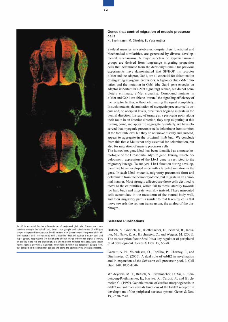
Genes that control migration of muscle precursorcellsH. Brohmann, M. Strehle, E. Vassioutina
Skeletal muscles in vertebrates, despite their functional andbiochemical similarities, are generated by diverse develop-mental mechanisms. A major subclass of hypaxial musclegroups are derived from long-range migrating progenitorcells that delaminate from the dermomyotome. Our previousexperiments have demonstrated that SF/HGF, its receptorc-Met and the adaptor, Gab1, are all essential for delaminationof migrating myogenic precursors. A hypomorphic c-Met mu-tation and the mutation in Gab1 (the Gab1 gene encodes anadaptor important in c-Met signaling) reduce, but do not com-pletely eliminate, c-Met signaling. Compound mutants inc-Met and Gab1 are able to “titrate” the signaling efficiency ofthe receptor further, without eliminating the signal completely.In such mutants, delamination of myogenic precursor cells oc-curs and, on occipital levels, precursors begin to migrate in theventral direction. Instead of turning at a particular point alongtheir route in an anterior direction, they stop migrating at thisturning point, and appear to aggregate. Similarly, we have ob-served that myogenic precursor cells delaminate from somitesat the forelimb level but they do not move distally and, instead,appear to aggregate in the proximal limb bud. We concludefrom this that c-Met is not only essential for delamination, butalso for migration of muscle precursor cells.The homeobox gene Lbx1 has been identified as a mouse ho-mologue of the Drosophila ladybird gene. During muscle de-velopment, expression of the Lbx1 gene is restricted to themigratory lineage. To analyze Lbx1 function during develop-ment, we have developed mice with a targeted mutation in thegene. In such Lbx1 mutants, migratory precursors form anddelaminate from the dermomyotome, but migrate in an abnor-mal manner. Most strongly affected are those cells destined tomove to the extremities, which fail to move laterally towardsthe limb buds and migrate ventrally instead. These misroutedcells accumulate in the mesoderm of the ventral body wall,and their migratory path is similar to that taken by cells thatmove towards the septum transversum, the analog of the dia-phragm.
Selected Publications
Britsch, S., Goerich, D., Riethmacher, D., Peirano, R., Ross-ner, M., Nave, K. A., Birchmeier, C., and Wegner, M. (2001).The transcription factor Sox10 is a key regulator of peripheralglial development. Genes & Dev. 15, 66-78.
Garratt, A. N., Voiculescu, O., Topilko, P., Charnay, P., andBirchmeier, C. (2000). A dual role of erbB2 in myelinationand in expansion of the Schwann cell precursor pool. J. CellBiol. 148, 1035-1046.
Woldeyesus, M. T., Britsch, S., Riethmacher, D. Xu, L., Son-nenberg-Riethmacher, E., Harvey, R., Caroni, P., and Birch-meier, C. (1999). Genetic rescue of cardiac morphogenesis inerbB2 mutant mice reveals functions of the ErbB2 receptor indevelopment of the peripheral nervous system. Genes & Dev.19, 2538-2548.
62
Sox10 is essential for the differentiation of peripheral glial cells. Shown are crosssections through the spinal cord, dorsal root ganglia and spinal nerves of wild-type(upper image) and homozygous Sox10 mutant mice (lower image). Peripheral glial cellsand neuronal cells are visualized with antibodies directed against B-FABP (red) andTuJ-1 (green), respectively. On the left side of each image only the red signal is shown;an overlay of the red and green signals is shown on the mirrored right side. Note that inhomozygous Sox10 mutant animals, neuronal cells within the dorsal root ganglia form,but glial cells in the dorsal root ganglia and along the spinal nerves are not generated.
Birchmeier, C., and Brohmann, H. (2000). Genes that controldevelopment of migrating myogenic precursors in the em-bryo. Curr. Opin. Cell Biol. 12, 725-731.
Brohmann, H., Jagla, K., and Birchmeier, C. (2000). TheLbx1 gene is essential for migration of muscle precursor cellsinto the limb buds. Development 127, 437-445.
Structure of the Group
Group leaderDr. Carmen Birchmeier
ScientistsDr. Stefan BritschDr. Ursula Gaio*Dr. Alistair GarrattDr. Thomas Müller Dr. Cemil Özcelik* Dr. Masresha Woldeyesus*
Graduate and undergraduate studentsMalgorzata Borowiak*Henning BrohmannLi LiMartin SieberMichael StrehleElena Vassioutina*Hendrik Wildner *
Technical AssistantsSven BuchertKarin GottschlingAndrea LeschkeCathrin RudolphVerena SasseTanja Schalow*
SecretariatBrigitta Wedekind
* part of the period reported
63
Developmental Genetics
Andreas Schedl
Studying the molecular mechanisms underlying organ devel-opment is one of the key approaches to understanding thepathways leading to disease. We are particularly interested intwo genes, the Wilms’ Tumor suppressor WT1 and the HMGbox-containing gene SOX9, both of which are involved in anumber of genetic disorders in humans. Using transgenic andknock-out strategies in mice, we are analysing the molecularfunction of these transcription factors, developing modelsystems for human diseases and trying to understand the cel-lular and pathophysiological processes leading to the diseasephenotype in mouse and man.
WT1 function in development and disease
WT1 is required for development of the adrenal gland andnephrogenesis.Wilms’ tumour is a paediatric kidney tumour occurring in 1 inevery 10000 children. A proportion of patients show muta-tions in the Wilms’ tumour suppressor WT1, which encodes azinc finger protein with the ability to bind to both DNA andRNA. WT1 has also been implicated in embryonic develop-ment. Mice carrying homozygous mutations for WT1 fail todevelop kidneys and gonads and die at day 13.5 in utero. Weperformed complementation experiments involving themouse mutation by introducing human WT1 YAC constructsof 280kb or 470kb into the mouse genome. Both constructsrescued the knock-out mutation to different extents, which al-lowed us to look at WT1 function during later stages of devel-opment. Mice complemented with a 280kb construct surviveduntil birth, but died within 48 hours due to defects in kidneydevelopment. Initial steps of nephrogenesis were completed,but mature epithelium never formed indicating a second rolefor WT1 during later stages of kidney development. Surpris-ingly, adrenal glands were also affected demonstrating the re-quirement of WT1 for this organ.
A crucial role for WT1 in the kidney podocyte: reduced ex-pression levels cause glomerular diseaseAs described above, complementation experiments with thehuman 280kb construct were only partially successful, pos-sibly because of the lack of a regulatory region on the con-struct used for our rescue experiments. Indeed, when usingthe 470kb human transgene, all developmental defects werecomplemented. Interestingly, rescued mice developed pro-gressive glomerulosclerosis and died of renal failure a fewmonths after birth. Using quantitative analysis we demon-strated that the observed phenotype is caused by too low lev-els of WT1 expression within the podocytes of the kidney.Moreover, we showed that nephrin and podocalyxin, two ma-jor components of the slit diaphragm of the podocyte, are dra-matically downregulated in rescued animals, offering an at-tractive hypothesis for the molecular mechanism leading torenal failure. Our data have demonstrated for the first time anessential role for WT1 in the maintenance of podocyte func-tion in adult life (Guo et al., in press).
Two splice variants of WT1 gene have distinct functions dur-ing sex determination and nephron formationAlternative splicing of WT1 results in the production of vari-ous isoforms. Of particular interest is an alternative splice do-nor site at the end of exon 9, which introduces the three ami-noacids KTS between zinc fingers 3 and 4. In vitro analysesindicated that + and -KTS isoforms fulfil distinct functions.Whereas –KTS isoforms are likely to act as transcriptionalregulators, proteins harbouring the +KTS sequence have beenproposed to act at the posttranscriptional level. To investigatethese functions in vivo we have introduced subtle mutationsinto the endogenous Wt1 locus using ES cell technology. Thepoint mutations were designed to interfere with splice donorsite selection and, hence, result in the production of only the+KTS or –KTS allele from the targeted allele. Heterozygousmice carrying +KTS ablated ES-cells developed kidney ab-normalities reminiscent of the Frasier/Denys-Drash syndrome(progressive mesangial sclerosis). When crossed to homozy-gosity +KTS mice survived embryonic development, but diedperinatally possibly due to kidney failure. Podocytes com-pletely failed to develop footprocesses demonstrating the im-portance of +KTS variants for the terminal differentiation ofthis cell type. Excitingly, these mice show male-to- femalesex reversal and completely lack Sox9 expression in the de-veloping gonad. In contrast, mice lacking –KTS isoforms(production of +KTS only) show streak gonads with a limitedamount of Sox9. These data demonstrate a different role forthe + and –KTS isoforms in vivo and place +KTS forms be-fore SRY in the sex determination cascade.
SOX9 - a sex-determining geneMutations in SOX9 have been found in patients sufferingfrom the semidominant campomelic dysplasia syndromewhich is characterised by bowing of the long bones, XY sexreversal and perinatal death. The observed sex reversal sug-gests that SOX9 is required for male sex determination and,indeed, it is the first gene with a male-specific expression pat-tern after the expression of the sex-determining gene Sry. Toaddress whether Sox9 is not only required, but also sufficient,to induce the sex determination pathway, we performed ec-topic expression studies in which the mouse Sox9 locus wasbrought under control of Wt1 regulatory regions. Transgenic
64
mice generated with this construct expressed Sox9 in bothmale and female gonads. Interestingly, XX transgenic micedeveloped as phenotypical males and expressed male-specificmarkers such as MIS. Our results demonstrate that Sox9 issufficient to induce the male sex determination pathway andsuggests that Sry acts only as a molecular switch (Vidal et al.,2001).
Selected Publications
Hammes, A., Guo, J. K., Lutsch, G., Leheste, J. R., Landrock,D., Ziegler, U., Gubler, M. C., and Schedl, A. (2001). Twosplice variants of the Wilms' tumor 1 gene have distinct func-tions during sex determination and nephron formation. Cell106, 319-329.
Vidal, V. P., Chaboissier, M. C., de Rooij, D. G., and Schedl,A. (2001). Sox9 induces testis development in XX transgenicmice. Nat. Genet. 28, 216-217.
Drab, M., Verkade, P., Elger, M., Kasper, M., Lohn, M., Lau-terbach, B., Menne, J., Lindschau, C., Mende, F., Luft, F. C.,Schedl, A., Haller, H., and Kurzchalia, T. V. (2001). Loss ofcaveolae, vascular dysfunction, and pulmonary defects in ca-veolin-1 gene-disrupted mice. Science 293, 2449-2452.
Kleinjan, D. A., Seawright, A., Schedl, A., Quinlan, R. A.,Danes, S., and van, H., V (2001). Aniridia-associated translo-cations, DNase hypersensitivity, sequence comparison andtransgenic analysis redefine the functional domain of PAX6.Hum. Mol. Genet. 10, 2049-2059.
Schedl, A. and Hastie, N.D. (2000). Cross-talk in kidney de-velopment. Curr. Opin. Genet. Dev. 10, 543-549.
Moore, A.W., McInnes, L., Kreidberg, J., Hastie, N.D., andSchedl, A. (1999). YAC complementation shows a require-ment for Wt1 in the development of epicardium, adrenalgland and throughout nephrogenesis. Development 126,1845-1857.
Structure of the Group
Group leaderDr. Andreas Schedl
ScientistsDr. Marie-Christine ChaboissierDr. Annette HammesDr. Christian MrowkaDr. Valerie Vidal
Graduate and undergraduate studentsJiankan GuoYiming XingFrances Wong
Technical assistantsDanilo LandrockSabine SchmidtUlrike Ziegler
Present addressProf. Dr. Andreas Schedl, Institute of Human Genetics, Inter-national Centre for Life Sciences, Newcastle upon Tyne, NE13BZ, Central Parkway, e-mail [email protected]
65
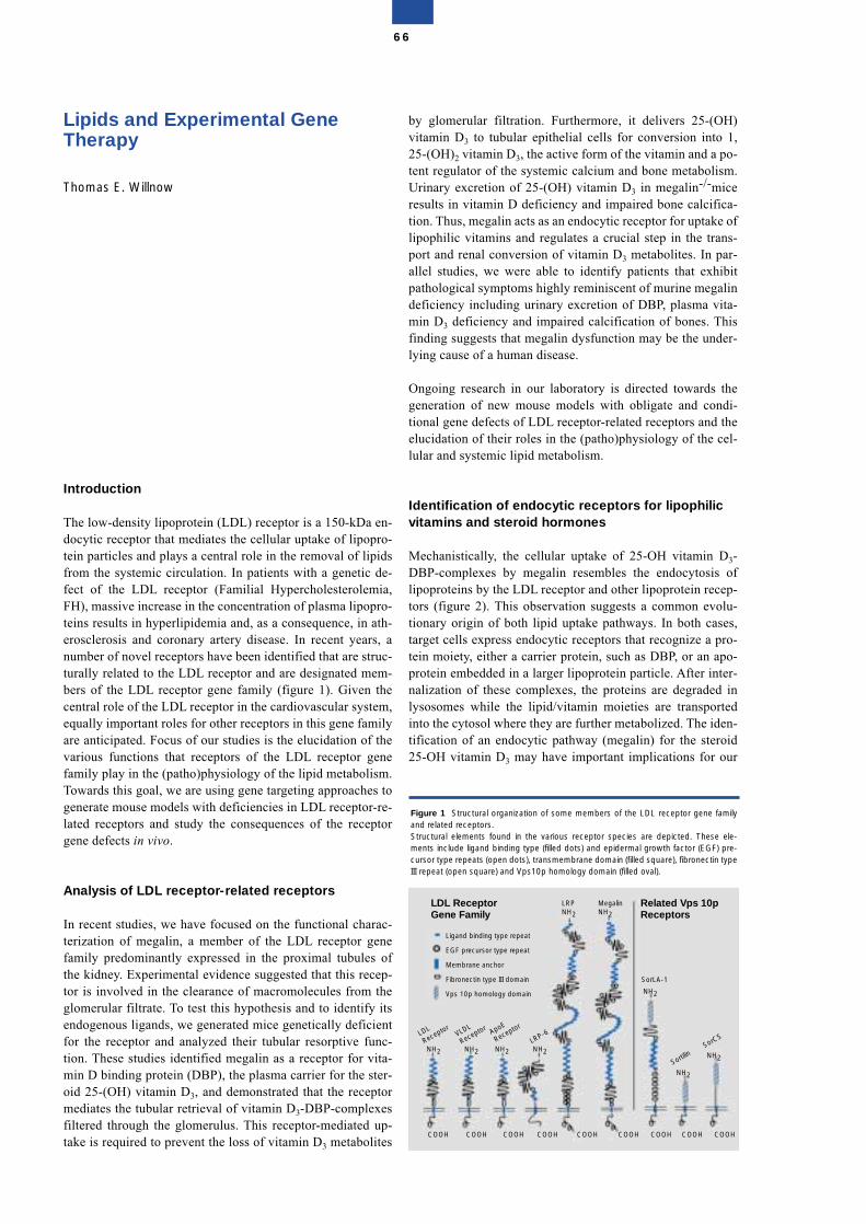
Lipids and Experimental GeneTherapy
Thomas E. Willnow
Introduction
The low-density lipoprotein (LDL) receptor is a 150-kDa en-docytic receptor that mediates the cellular uptake of lipopro-tein particles and plays a central role in the removal of lipidsfrom the systemic circulation. In patients with a genetic de-fect of the LDL receptor (Familial Hypercholesterolemia,FH), massive increase in the concentration of plasma lipopro-teins results in hyperlipidemia and, as a consequence, in ath-erosclerosis and coronary artery disease. In recent years, anumber of novel receptors have been identified that are struc-turally related to the LDL receptor and are designated mem-bers of the LDL receptor gene family (figure 1). Given thecentral role of the LDL receptor in the cardiovascular system,equally important roles for other receptors in this gene familyare anticipated. Focus of our studies is the elucidation of thevarious functions that receptors of the LDL receptor genefamily play in the (patho)physiology of the lipid metabolism.Towards this goal, we are using gene targeting approaches togenerate mouse models with deficiencies in LDL receptor-re-lated receptors and study the consequences of the receptorgene defects in vivo.
Analysis of LDL receptor-related receptors
In recent studies, we have focused on the functional charac-terization of megalin, a member of the LDL receptor genefamily predominantly expressed in the proximal tubules ofthe kidney. Experimental evidence suggested that this recep-tor is involved in the clearance of macromolecules from theglomerular filtrate. To test this hypothesis and to identify itsendogenous ligands, we generated mice genetically deficientfor the receptor and analyzed their tubular resorptive func-tion. These studies identified megalin as a receptor for vita-min D binding protein (DBP), the plasma carrier for the ster-oid 25-(OH) vitamin D3, and demonstrated that the receptormediates the tubular retrieval of vitamin D3-DBP-complexesfiltered through the glomerulus. This receptor-mediated up-take is required to prevent the loss of vitamin D3 metabolites
by glomerular filtration. Furthermore, it delivers 25-(OH)vitamin D3 to tubular epithelial cells for conversion into 1,25-(OH)2 vitamin D3, the active form of the vitamin and a po-tent regulator of the systemic calcium and bone metabolism.Urinary excretion of 25-(OH) vitamin D3 in megalin-/-miceresults in vitamin D deficiency and impaired bone calcifica-tion. Thus, megalin acts as an endocytic receptor for uptake oflipophilic vitamins and regulates a crucial step in the trans-port and renal conversion of vitamin D3 metabolites. In par-allel studies, we were able to identify patients that exhibitpathological symptoms highly reminiscent of murine megalindeficiency including urinary excretion of DBP, plasma vita-min D3 deficiency and impaired calcification of bones. Thisfinding suggests that megalin dysfunction may be the under-lying cause of a human disease.
Ongoing research in our laboratory is directed towards thegeneration of new mouse models with obligate and condi-tional gene defects of LDL receptor-related receptors and theelucidation of their roles in the (patho)physiology of the cel-lular and systemic lipid metabolism.
Identification of endocytic receptors for lipophilicvitamins and steroid hormones
Mechanistically, the cellular uptake of 25-OH vitamin D3-DBP-complexes by megalin resembles the endocytosis oflipoproteins by the LDL receptor and other lipoprotein recep-tors (figure 2). This observation suggests a common evolu-tionary origin of both lipid uptake pathways. In both cases,target cells express endocytic receptors that recognize a pro-tein moiety, either a carrier protein, such as DBP, or an apo-protein embedded in a larger lipoprotein particle. After inter-nalization of these complexes, the proteins are degraded inlysosomes while the lipid/vitamin moieties are transportedinto the cytosol where they are further metabolized. The iden-tification of an endocytic pathway (megalin) for the steroid25-OH vitamin D3 may have important implications for our
66
Figure 1 Structural organization of some members of the LDL receptor gene familyand related receptors. Structural elements found in the various receptor species are depicted. These ele-ments include ligand binding type (filled dots) and epidermal growth factor (EGF) pre-cursor type repeats (open dots), transmembrane domain (filled square), fibronectin typeIII repeat (open square) and Vps10p homology domain (filled oval).
LDL
ReceptorVLDL
Receptor ApoE
Receptor
LRP NH2
Megalin NH2
NH2
COOH COOH COOH COOH COOH COOH COOH COOH COOH
NH2 NH2LRP-6
NH2
NH2
NH2
NH2Sortili
n
SorLA-1
SorCS
Ligand binding type repeat
EGF precursor type repeat
Membrane anchor
Fibronectin type III domain
Vps 10p homology domain
LDL Receptor Gene Family
Related Vps 10p Receptors
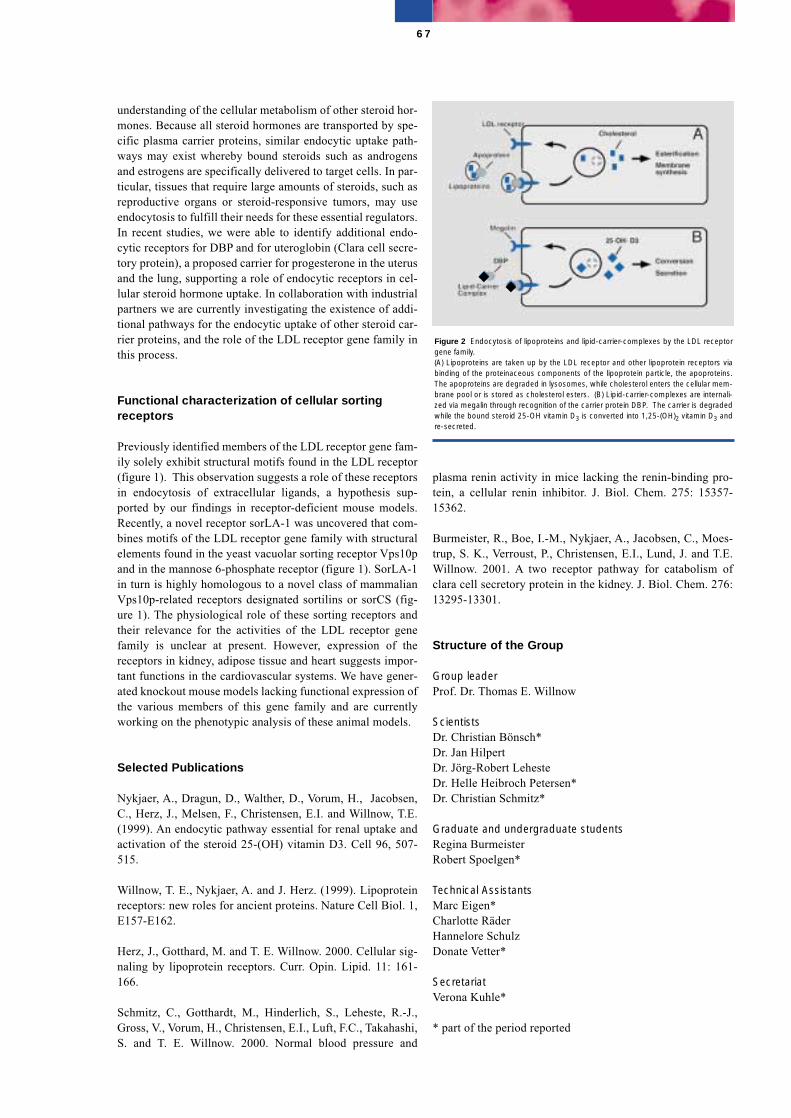
understanding of the cellular metabolism of other steroid hor-mones. Because all steroid hormones are transported by spe-cific plasma carrier proteins, similar endocytic uptake path-ways may exist whereby bound steroids such as androgensand estrogens are specifically delivered to target cells. In par-ticular, tissues that require large amounts of steroids, such asreproductive organs or steroid-responsive tumors, may useendocytosis to fulfill their needs for these essential regulators.In recent studies, we were able to identify additional endo-cytic receptors for DBP and for uteroglobin (Clara cell secre-tory protein), a proposed carrier for progesterone in the uterusand the lung, supporting a role of endocytic receptors in cel-lular steroid hormone uptake. In collaboration with industrialpartners we are currently investigating the existence of addi-tional pathways for the endocytic uptake of other steroid car-rier proteins, and the role of the LDL receptor gene family inthis process.
Functional characterization of cellular sortingreceptors
Previously identified members of the LDL receptor gene fam-ily solely exhibit structural motifs found in the LDL receptor(figure 1). This observation suggests a role of these receptorsin endocytosis of extracellular ligands, a hypothesis sup-ported by our findings in receptor-deficient mouse models.Recently, a novel receptor sorLA-1 was uncovered that com-bines motifs of the LDL receptor gene family with structuralelements found in the yeast vacuolar sorting receptor Vps10pand in the mannose 6-phosphate receptor (figure 1). SorLA-1in turn is highly homologous to a novel class of mammalianVps10p-related receptors designated sortilins or sorCS (fig-ure 1). The physiological role of these sorting receptors andtheir relevance for the activities of the LDL receptor genefamily is unclear at present. However, expression of thereceptors in kidney, adipose tissue and heart suggests impor-tant functions in the cardiovascular systems. We have gener-ated knockout mouse models lacking functional expression ofthe various members of this gene family and are currentlyworking on the phenotypic analysis of these animal models.
Selected Publications
Nykjaer, A., Dragun, D., Walther, D., Vorum, H., Jacobsen,C., Herz, J., Melsen, F., Christensen, E.I. and Willnow, T.E.(1999). An endocytic pathway essential for renal uptake andactivation of the steroid 25-(OH) vitamin D3. Cell 96, 507-515.
Willnow, T. E., Nykjaer, A. and J. Herz. (1999). Lipoproteinreceptors: new roles for ancient proteins. Nature Cell Biol. 1,E157-E162.
Herz, J., Gotthard, M. and T. E. Willnow. 2000. Cellular sig-naling by lipoprotein receptors. Curr. Opin. Lipid. 11: 161-166.
Schmitz, C., Gotthardt, M., Hinderlich, S., Leheste, R.-J.,Gross, V., Vorum, H., Christensen, E.I., Luft, F.C., Takahashi,S. and T. E. Willnow. 2000. Normal blood pressure and
plasma renin activity in mice lacking the renin-binding pro-tein, a cellular renin inhibitor. J. Biol. Chem. 275: 15357-15362.
Burmeister, R., Boe, I.-M., Nykjaer, A., Jacobsen, C., Moes-trup, S. K., Verroust, P., Christensen, E.I., Lund, J. and T.E.Willnow. 2001. A two receptor pathway for catabolism ofclara cell secretory protein in the kidney. J. Biol. Chem. 276:13295-13301.
Structure of the Group
Group leaderProf. Dr. Thomas E. Willnow
ScientistsDr. Christian Bönsch*Dr. Jan HilpertDr. Jörg-Robert LehesteDr. Helle Heibroch Petersen*Dr. Christian Schmitz*
Graduate and undergraduate studentsRegina BurmeisterRobert Spoelgen*
Technical AssistantsMarc Eigen*Charlotte Räder Hannelore Schulz Donate Vetter*
SecretariatVerona Kuhle*
* part of the period reported
67
Figure 2 Endocytosis of lipoproteins and lipid-carrier-complexes by the LDL receptorgene family.(A) Lipoproteins are taken up by the LDL receptor and other lipoprotein receptors viabinding of the proteinaceous components of the lipoprotein particle, the apoproteins.The apoproteins are degraded in lysosomes, while cholesterol enters the cellular mem-brane pool or is stored as cholesterol esters. (B) Lipid-carrier-complexes are internali-zed via megalin through recognition of the carrier protein DBP. The carrier is degradedwhile the bound steroid 25-OH vitamin D3 is converted into 1,25-(OH)2 vitamin D3 andre-secreted.
Bioinformatics
Jens ReichPeer Bork (P. B. is jointly appointed as guest researcher at EMBL)
Individual variation of the human genome and itsrole in lipid metabolism: genetic-epidemiologicalstudy of risk factors for arteriosclerosis
Two subgroups of our team (genome analysis, P.B., metabolicpathway analysis, S. Schuster, and lipid network, J.R.) contin-ued in a joint endeavour to evaluate the importance of SNPsand splicing variants in the human genome. This was com-bined with an application study on 1250 normal probands (re-cruited by INFOGEN-VALIGEN Medical Genetics GmbH [H. Schuster], and Franz-Volhard-Clinic, FVK [F. Luft]), inwhich 30 SNP variants at 6 relevant gene loci (for names andabbreviations see figure legend) were measured together with5 clinically important indicators of human lipoprotein metab-olism. This is the first time that the individual genotype ofcommon SNPs (i.e. > 3% population frequency in a Germanpopulation sample) relevant for a metabolic pathway werecorrelated with the physiological level of the resultant pheno-type. Lipid traits are the major risk factor for arteriosclerosisand its severe complications (myocardial infarction, stroke,a.o.). Physiologically valid phenotypic values (plasma levelsof total cholesterol, TC; triglyceride, TG; low density lipopro-tein, LDL, high density lipoprotein, HDL; and the importantclinical risk factor LDL/HDL) were measured at FVK understandardized conditions and SNP genotyping was done byVALIGEN´s genomic center in Paris. Earlier comparativestudies on homozygotic and dizygotic twins (Busjahn & Luft)had established that a global heritability component of bet-ween 30 and 40% contributes to the individual lipid level inhumans. As SNPs are the most common genetic variants wewanted to learn which of them change the phenotype levels toa statistically significant extent. This genotype-phenotypecorrelation was to be integrated into a pathway model that wehave developed on the basis of theoretical studies of meta-bolic models (S. Schuster et al. 2000, H. Knoblauch et al.,2000). The data obtained were subjected to an exhaustivemathematical-statistical analysis using advanced computertechniques.
The main result is that 25 of the studied polymorphismsshowed common variation in the sample. The allelic associa-tion of these SNPs (expressed as score of linkage disequilib-rium, LD) varied with their distance on the genome. The fullgenomic structure of all loci could be established for all geneloci by reference to the NCBI data base of the human genome(incidentally: Peer Bork and two further members of ourgroup are the only German theorists co-authoring the mile-stone NATURE paper of Lander et al. that published the fullreference sequence of the human genome on February 15th ,see publication list). About half of the SNPs were in the cod-ing region (non-synonymous and synonymous) the others inpromoter and intron segments.
We found that within 1 kbp distance (= 1000 nucleotide let-ters) all SNP pairs showed complete LD, since only 2 or 3 ofthe 4 possible 2-SNP haplotypes were found, pointing to theabsence of recombination effects in the population at suchsmall inter-SNP distance. At larger distances (our gene locispan between 30 and 140 kbp on the genome) the LD levelsoff and reaches full equilibrium due to recombination at about50 kbp.
These data are in agreement with population-genetic modelsfor a fully outbred panmictic population in Germany, and alsowith published data on other genome sections. Such a situa-tion allows the mapping of functional gene alleles only atsmall distances from the marker SNP. However, we could es-tablish the SNP-haplotype structure of all loci by way of anal-ysis of the family pedigrees of the 1250 subjects, since theywere recruited as nuclear families in a systematic field work-ing project of INFOGEN. Haplotyping was achieved with anewly developed computer program (Rohde & Fürst, 2001)that permits the establishment, with high confidence, of thechromosomal phase of SNP positions when diploid genotypedata from nuclear families are available, as was our case. Itwas found that in 4 of the studied gene loci only a few (4-5)haplotypes accounted for the genotype of about 80-90% ofthe population sample (see fig.). This means that chromoso-mal haplotypes of common SNPs are very “old”genomicstructures (i.e. many tens of thousands generations old, notdissolved in the whole population by extensive recombina-tion) and may therefore be reliable markers of functional al-leles that explain the variation and risk status of the pheno-type. This warrants a genotype-phenotype association study ifcontrolled for stratification into subpopulations.
The association was evaluated in combination with linkagetests according to modern models of biometric genetics, whichpartition the total variance into additive genetic, polygenicand environmental components. Comparison of genotype-phenotype correlation between families and within familiespermit to control for stratification (sample inhomogeneity) ef-fects.
We found that at 5 of the loci there are chromosomal haplo-types in the population which change the mean lipid levels oftheir carriers in a significant way. Most impressive is the vari-ation effect of apolipoprotein E (ApoE), which confirms re-sults published for Sweden, America and other countries.ApoE, whose variants play a major role as risk or protectionfactors in lipid disorders as well as in the causation of Alzhei-
68
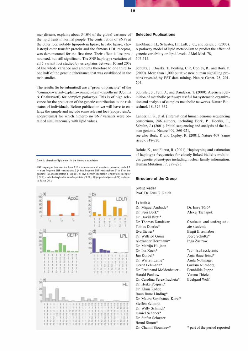
mer disease, explains about 5-10% of the global variance ofthe lipid traits in normal people. The contribution of SNPs atthe other loci, notably lipoprotein lipase, hepatic lipase, cho-lesteryl ester transfer protein and the famous LDL receptor,was demonstrated for the first time. Their effect is less pro-nounced, but still significant. The SNP haplotype variation ofall 5 variant loci studied by us explains between 10 and 20%of the whole variance and amounts therefore to one third toone half of the genetic inheritance that was established in thetwin studies.
The results (to be submitted) are a “proof of principle” of the“common-variant-explains-common-trait”-hypothesis (Collins& Chakravarti) for complex pathways. This is of high rele-vance for the prediction of the genetic contribution to the riskstatus of individuals. Before publication we will have to en-large the sample and include some relevant loci (apoproteinA,apoproteinB) for which hitherto no SNP variants were ob-tained simultaneously with lipid values.
Selected Publications
Knoblauch, H., Schuster, H., Luft, J. C., and Reich, J. (2000).A pathway model of lipid metabolism to predict the effect ofgenetic variability on lipid levels. J.Mol.Med. 78,507-515.
Schultz, J., Doerks, T., Ponting, C.P., Copley, R., and Bork, P.(2000). More than 1,000 putative new human signalling pro-teins revealed by EST data mining. Nature Genet. 25, 201-204.
Schuster, S., Fell, D., and Dandekar, T. (2000). A general def-inition of metabolic pathways useful for systematic organiza-tion and analysis of complex metabolic networks. Nature Bio-technol. 18, 326-332.
Lander, E. S., et al. (International human genome sequencingconsortium, 246 authors, including Bork, P., Doerks, T.,Schultz, J.) (2001). Initial sequencing and analysis of the hu-man genome. Nature 409, 860-921,see also Bork, P. and Copley, R. (2001). Nature 409 (sameissue), 818-820.
Rohde, K., and Fuerst, R. (2001). Haplotyping and estimationof haplotype frequencies for closely linked biallelic multilo-cus genetic phenotypes including nuclear family information.Human Mutation 17, 289-295.
Structure of the Group
Group leaderProf. Dr. Jens G. Reich
ScientistsDr. Miguel Andrade*Dr. Peer Bork*Dr. David Brett*Dr. Thomas DandekarTobias Doerks*Eva Eicher*Dr. Wilfried Gunia Alexander Herrmann*Dr. Martijn HuijnenDr. Ina Koch*Jan Korbel*Dr. Warren Lathe*Gerrit Lehmann*Dr. Ferdinand MoldenhauerHarald PankowDr. Carolina Perez-Iracheta*Dr. Heike Pospisil*Dr. Klaus RohdeRaun Rune Linding*Dr. Mauro Santibanez-Koref*Steffen SchmidtDr. Willy Schmidt*Daniel Schober*Dr. Stefan SchusterBernd Simon*Dr. Chamil Siouniaev*
69
Genetic diversity of lipid genes in the German population
SNP-haplotype frequencies from 616 chromosomes of unrelated persons, coded 1(= more frequent SNP-variant) and 2 (= less frequent SNP-variant) from 5’ to 3’ on thegenome: a) apolipoprotein E (ApoE), b) low density lipoprotein cholesterol receptor(LDLR), c) cholesteryl ester transfer protein (CETP), d) lipoprotein lipase (LPL), e) hepa-tic lipase (HL).
Dr. Imre Törö*Alexej Tschapek
Graduate and undergradu-ate studentsBirgit EisenhaberJoerg Schultz*Inga Zastrow
Technical assistantsAnja Bauerfeind*Anita NothnagelGudrun NürnbergBrunhilde PoppeVerena ThieleEdelgard Wolf
* part of the period reported
Protein Misfolding: From Basic Biopolymer Physics to Conformational Diseases
Gregor Damaschun
Amyloid fibers are the characteristic common to a number ofso-called protein conformational disorders. Systemic or organ-specific amyloidoses, including Alzheimer’s disease, and thetransmissible spongiform encephalopathies belong to thisclass of disorders. More than twenty of these human and ani-mal diseases are known where a specific protein is misfoldedto form amyloid fibers in each case. In spite of the very differ-ent proteins involved, the amyloid fibers have very similarconformations. Slow kinetics of progression is typical of allamyloidoses and the process of fiber formation is only poorlyunderstood up to now.
Protein conformational disorders can be promoted by pointmutations but appear also spontaneously or are caused by in-fectious protein conformations (prions).
In recent years, it has been observed that about thirty proteinsunrelated to so far known diseases can form amyloid fibers.
This observation has led to the suggestion that the ability toform amyloid fibers is a common phenomenon and a genericproperty of polypeptide chains. The ability to form amyloidfibrils from a wide range of proteins allows a large number ofmodel systems to be used to study the process of fibril forma-tion in more detail.
We have recognized that the adage “one sequence, one con-formation” is not strictly true. Depending on solvent condi-tions at folding, probably any protein chain can adopt a va-riety of conformations in which there is a degree of periodicorder, i.e., extensive-regions of secondary structure. A recentsystematic study of the conformations adopted by the glyco-lytic enzyme, phosphoglycerate kinase, shows that in differ-ent media the chain can adopt five distinct states in vitro.However, such alternative states do not have the precisely andtightly packed side-chains, which are the hallmark of the na-tive state of orthodox globular proteins. One of these alternatives is the amyloid state, the result ofprotein conformational disorders in vivo.
A�-Amyloids (in cooperation with E. Krause, FMP)
A critical event in Alzheimer’s disease is the transition of A�peptides from their soluble forms into disease-associated�-sheet-rich conformers. Structural analysis of a completeD-amino acid replacement set of A�(1-42) has enabled us tolocalize in the full-length 42-mer peptide the region respon-sible for the conformational switch into a �-sheet structure.Although NMR spectroscopy of trifluoroethanol-stabilizedmonomeric A�(1-42) revealed two separated helical domains,only the destabilization of helix I, comprising residues 11-24,caused a transition to a �-sheet structure. This conformational�-to-� switch was directly accompanied by an aggregationprocess leading to the formation of amyloid fibrils.
Kinetics of amyloid formation (in cooperation with H. Fabian, MDC)
We have studied the transformation of different model pro-teins into the amyloid conformation by time-resolved simul-taneous measurements by means of dynamic and static lightscattering, X-ray scattering, circular dichroism and Fourier-transform infrared spectroscopy. This combination of meth-ods allows us to determine simultaneously changes in particledimensions and mass as well as changes in the secondarystructure.
The main results are the following. Monomers having the typ-ical amyloid-like cross-� structure do not exist as was postu-lated by Prusiner for the prion. The conformational changefrom �-helices into �-sheets is triggered by the onset ofmonomer aggregation. The amount of �-structure grows untilthe formation of an ensemble of critical oligomers is finished.These critical oligomers assemble into wormlike amyloid fi-bers in a second step. The fractal dimensions of the twogrowth mechanisms are different.
Protein misfolding and structure transformation
Protein molecules adopt their native conformation in solutionand within the cell only under specific environmental condi-tions. Alcohols are suitable for modulating the interactionsbetween the polypeptide chain and the solvent as well as theinteraction between different segments of the polypeptidechain. Recent investigations have dealt with the influence ofcosolvents on the folding kinetics of proteins, the tuning ofsolvent conditions for studies of the amyloid formation ofpeptides and proteins, the dissection and reassembly of amy-loid fibrils, and the �-�-transition of proteins, which plays anessential role in various conformational diseases.
The conformational transitions of bovine �-lactoglobulin Aand phosphoglycerate kinase from yeast induced by hexaflu-oroisopropanol (HFIP) and trifluoroethanol (TFE) have beenstudied by dynamic light scattering and circular dichroismspectroscopy in order to elucidate the potential of fluoroalco-hols to bring about structural changes in proteins. Moreover,pure fluoroalcohol-water mixed solvents were investigated toprove the relationship between hydrophobic cluster formation
70
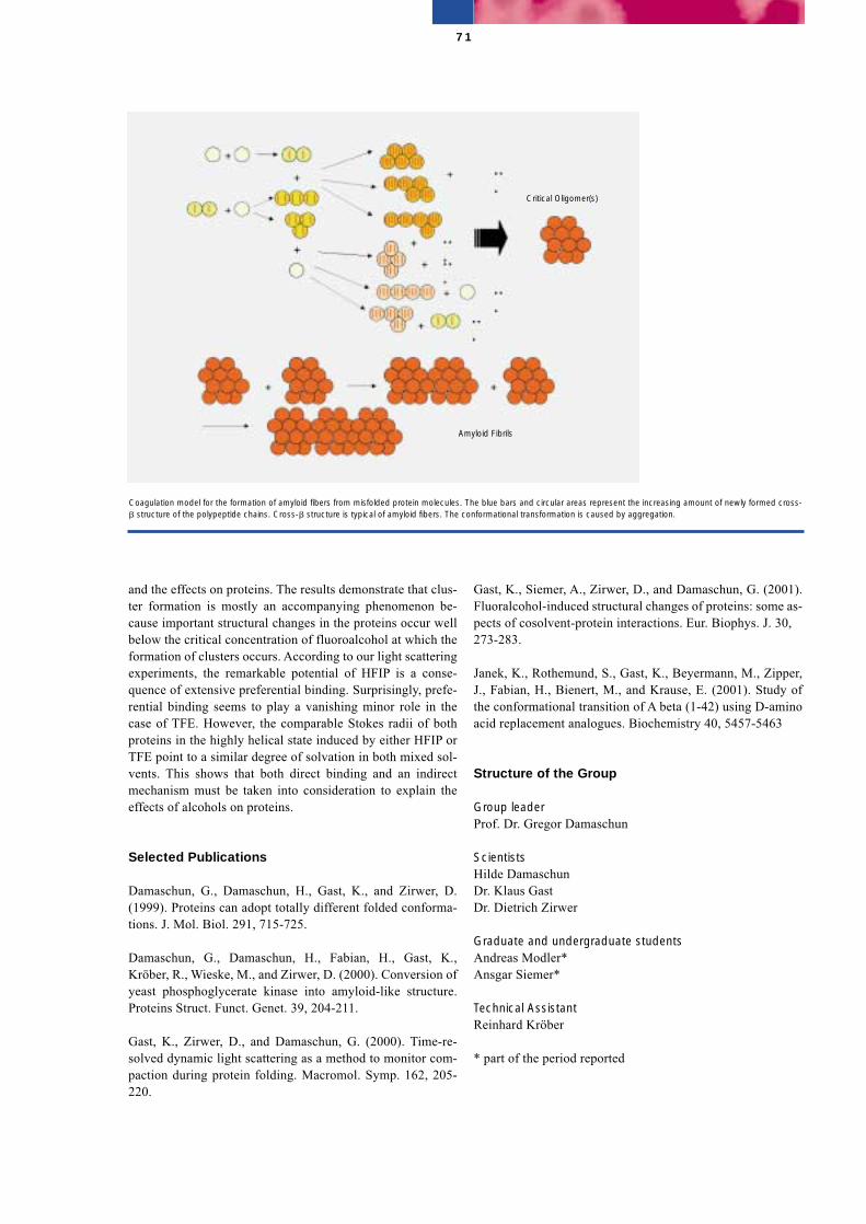
and the effects on proteins. The results demonstrate that clus-ter formation is mostly an accompanying phenomenon be-cause important structural changes in the proteins occur wellbelow the critical concentration of fluoroalcohol at which theformation of clusters occurs. According to our light scatteringexperiments, the remarkable potential of HFIP is a conse-quence of extensive preferential binding. Surprisingly, prefe-rential binding seems to play a vanishing minor role in thecase of TFE. However, the comparable Stokes radii of bothproteins in the highly helical state induced by either HFIP orTFE point to a similar degree of solvation in both mixed sol-vents. This shows that both direct binding and an indirectmechanism must be taken into consideration to explain theeffects of alcohols on proteins.
Selected Publications
Damaschun, G., Damaschun, H., Gast, K., and Zirwer, D.(1999). Proteins can adopt totally different folded conforma-tions. J. Mol. Biol. 291, 715-725.
Damaschun, G., Damaschun, H., Fabian, H., Gast, K.,Kröber, R., Wieske, M., and Zirwer, D. (2000). Conversion ofyeast phosphoglycerate kinase into amyloid-like structure.Proteins Struct. Funct. Genet. 39, 204-211.
Gast, K., Zirwer, D., and Damaschun, G. (2000). Time-re-solved dynamic light scattering as a method to monitor com-paction during protein folding. Macromol. Symp. 162, 205-220.
Gast, K., Siemer, A., Zirwer, D., and Damaschun, G. (2001).Fluoralcohol-induced structural changes of proteins: some as-pects of cosolvent-protein interactions. Eur. Biophys. J. 30,273-283.
Janek, K., Rothemund, S., Gast, K., Beyermann, M., Zipper,J., Fabian, H., Bienert, M., and Krause, E. (2001). Study ofthe conformational transition of A beta (1-42) using D-aminoacid replacement analogues. Biochemistry 40, 5457-5463
Structure of the Group
Group leaderProf. Dr. Gregor Damaschun
ScientistsHilde DamaschunDr. Klaus GastDr. Dietrich Zirwer
Graduate and undergraduate studentsAndreas Modler*Ansgar Siemer*
Technical AssistantReinhard Kröber
* part of the period reported
71
Coagulation model for the formation of amyloid fibers from misfolded protein molecules. The blue bars and circular areas represent the increasing amount of newly formed cross-� structure of the polypeptide chains. Cross-� structure is typical of amyloid fibers. The conformational transformation is caused by aggregation.
Critical Oligomer(s)
Amyloid Fibrils
Structural Studies of Proteinsand Nucleic Acids by X-rayCrystallography
Udo Heinemann
Macromolecular crystallography is a uniquely powerful toolto study the three-dimensional structures of proteins, nucleicacids and their complexes. It permits to determine the precisearrangement of all atoms and the shape and property of allsurfaces in small or medium-sized molecules as well as hugemolecular complexes. This knowledge can be used to explainbiochemical observations, to predict biological functions andto design ligands specific to a given protein molecule. Wecombine X-ray diffraction studies with biochemical and bio-physical experiments of proteins involved in a variety of bio-logical processes. Crucial to this work is the ability to preparecrystallizable samples of nucleic acids, proteins and proteindomains by chemical, biochemical and gene-technologicalmeans. The scientific infra-structure at our disposal is cur-rently being extended with the completion of a state-of-the-art protein crystallography station at the electron storage ringBESSY II in Berlin-Adlershof.
Below, selected examples from the different areas of our re-search are presented. They include work by Yves Muller, whohas recently accepted an offer to join the faculty of the Uni-versity of Sussex, UK, and his colleagues.
Nucleic acid-interacting proteinsR. Bienert, H. Delbrück, D. Khare, Y. Roske, T. Schwartz,E. Werner
Recent work in the group of A. Rich (MIT, Cambridge, USA)had shown that the unusual left-handed Z-form of DNA canbe induced and stabilized by binding of the Z� domain of theRNA-editing enzyme ADAR1. To examine whether ADAR1is part of a larger family of Z-DNA-binding proteins, theweakly sequence-related N-terminal domain of the tumor-as-sociated protein DLM-1 from mouse was bound to a self-complementary six-base-pair oligonucleotide and crystal-lized. The crystal structure of this Z�DLM-DNA complexclearly showed the DNA to adopt the Z-conformation. Z�DLM,as well as Z�ADAR bind the DNA conformation- rather thansequence-specifically by contacting mainly the uniquely
shaped sugar-phosphate backbone. The single base contactmade is with the C8 atom of a guanine which is exposed forintermolecular contact only in the Z-form. The crystal struc-ture of the Z�DLM-Z-DNA complex thus proves the existenceof a family of Z-DNA-binding proteins which includes fur-ther molecules such as the vaccinia virus protein E3L. Inwhich way these proteins use the B↔Z transition of DNA tomodulate biological processes remains to be established.
Electron transport in cytochrome P450 systemsJ. J. Müller
To continue our studies of electron transport to cytochromesP450, carried out in collaboration with K. Ruckpaul (MDC)and R. Bernhardt (Universität des Saarlandes), a complex ofthe [2Fe-2S] ferredoxin adrenodoxin cross-linked to its cog-nate adrenodoxin reductase was crystallized. The structureanalysis of this complex was a challenge, since the crystalsdisplayed an unusually long primitive axis of >600 Å. Thestructure (see figure) confirmed biochemical data regardingresidues of adrenodoxin involved in reductase binding andidentified two additional interaction regions of the ferredoxin.A probable route for the electron transfer from the FAD ofadrenodoxin reductase to the iron cluster of adrenodoxin couldbe delineated based on the crystal structure.
Structural basis of protein stabilityH. Delbrück, U. Mueller
The small, bacterial cold shock proteins (Csp's) are extremelywell suited for studies of the structural basis of protein stabil-ity, since they fold in a reversible two-state process and theiranalysis is not complicated by disulfide bonds, bound cofac-tors or multimerization. In collaboration with F.X. Schmid(Universität Bayreuth) we have studied two Csp's from differ-ent Bacillus strains that differ in only a small number ofamino-acid side chains but show a large difference in thermalstability. We could show that the backbone structure of theseproteins is virtually unchanged and the stability difference ismainly caused by electro-static interactions between sidechains. Studying a set of Csp mutants it could be shown by bi-ophysical experiments that only two surface residues are re-sponsible for the entire stability difference and by crystalstructure analysis that these side chains do not engage in de-fined pairwise salt bridges but rather help establish the globalsurace charge balance of the proteins. This work permits theimportant conclusion that proteins may be easier stabilized bythe removal of electrostatic repulsion between side chainsthan the engineering of salt bridges.
72
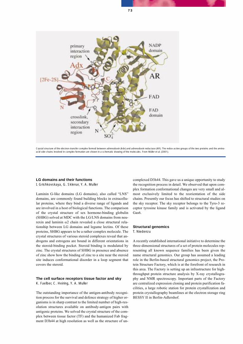
LG domains and their functionsI. Grishkovskaya, G. Sklenar, Y. A. Muller
Laminin G-like domains (LG domains), also called “LNS”domains, are commonly found building blocks in extracellu-lar proteins, where they bind a diverse range of ligands andare involved in a host of biological functions. The comparisonof the crystal structure of sex hormone-binding globulin(SHBG) solved at MDC with the LG/LNS domains from neu-rexin and laminin �2 chain revealed a close structural rela-tionship between LG domains and legume lectins. Of theseproteins, SHBG appears to be a rather complex molecule. Thecrystal structures of various steroid complexes reveal that an-drogens and estrogens are bound in different orientations inthe steroid-binding pocket. Steroid binding is modulated byzinc. The crystal structures of SHBG in presence and absenceof zinc show how the binding of zinc to a site near the steroidsite induces conformational disorder in a loop segment thatcovers the steroid.
The cell surface receptors tissue factor and skyK. Faelber, C. Heiring, Y. A. Muller
The outstanding importance of the antigen-antibody recogni-tion process for the survival and defence strategy of higher or-ganisms is in sharp contrast to the limited number of high-res-olution structures available on antibody-antigen pairs withantigenic proteins. We solved the crystal structure of the com-plex between tissue factor (TF) and the humanized Fab frag-ment D3h44 at high resolution as well as the structure of un-
complexed D3h44. This gave us a unique opportunity to studythe recognition process in detail. We observed that upon com-plex formation conformational changes are very small and al-most exclusively limited to the reorientation of the sidechains. Presently our focus has shifted to structural studies onthe sky receptor. The sky receptor belongs to the Tyro-3 re-ceptor tyrosine kinase family and is activated by the ligandGas6.
Structural genomicsT. Niedenzu
A recently established international initiative to determine thethree-dimensional structures of a set of protein molecules rep-resenting all known sequence families has been given thename structural genomics. Our group has assumed a leadingrole in the Berlin-based structural genomics project, the Pro-tein Structure Factory, which is at the forefront of research inthis area. The Factory is setting up an infrastructure for high-throughput protein structure analysis by X-ray crystallogra-phy and NMR spectroscopy. Important parts of the Factoryare centralized expression cloning and protein purification fa-cilities, a large robotic station for protein crystallization andprotein crystallography beamlines at the electron storage ringBESSY II in Berlin-Adlershof.
73
Crystal structure of the electron-transfer complex formed between adrenodoxin (Adx) and adrenodoxin reductase (AR). The redox-active groups of the two proteins and the amino-acid side chains involved in complex formation are shown in a schematic drawing of the molecules. From Müller et al. (2001).
Selected Publications
Avvakumov, G.V., Grishkovskaya, I., Muller, Y.A., and Ham-mond, G.L. (2001). Resolution of the human sex hormone-binding globulin dimer interface and evidence for two ster-oid-binding sites per homodimer J. Biol. Chem. 276,34453-34457.
Faelber, K., Kirchhofer, D., Presta, L., Kelley, R., and Muller,Y.A. (2001). The 1.85 Å resolution crystal structures of tissuefactor in complex with humanized Fab D3h44 and of free hu-manized Fab D3h44; revisiting the solvation of antigen com-bining sites. J. Mol. Biol. 313, 83-97.
Heinemann, U., Frevert, J., Hofmann, K.-P., Illing, G.,Maurer, C., Oschkinat, H., and Saenger, W. (2001). An inte-grated approach to structural genomics. Prog. Biophys. Mol.Biol. 73, 347-362.
Müller, J.J., Lapko, A., Bourenkov, G., Ruckpaul, K., andHeinemann, U. (2001). Adrenodoxin reductase-adrenodoxincomplex structure suggests electron transfer path in steroidbiosynthesis. J. Biol. Chem. 276, 2786-2789.
Perl, D., Mueller, U., Heinemann, U., and Schmid, F.X.(2001). Two exposed amino acid residues confer thermo-stability on a cold shock protein. Nature Struct. Biol. 7, 380-383.
Schwartz, T., Behlke, J., Lowenhaupt, K., Heinemann, U.,and Rich, A. (2001). Structure of the DLM-1–Z-DNA com-plex reveals a conserved family of Z-DNA-binding proteins.Nature Struct. Biol. 8, 761-765.
Structure of the Group
Group leaderProf. Dr. Udo Heinemann
ScientistsDr. Michael Kolbe*Dr. Jürgen J. MüllerDr. Yves A. Muller*Timo Niedenzu*Dr. Thomas Schwartz*
Graduate and undergraduate studentsRalf BienertHeinrich DelbrückKatja FaelberOlaf GaiserIrina GrishkovskayaDheeraj KhareUwe Mueller*Yvette RoskeGisela TünnemannErik Werner
Technical AssistantsAnette FeskeAndreas KnespelGisela Sklenar
SecretariatBirgit Cloos
74
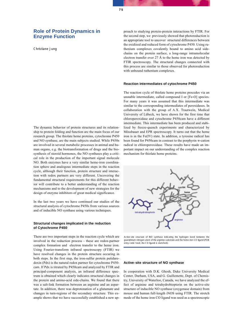
Role of Protein Dynamics inEnzyme Function
Christiane Jung
The dynamic behavior of protein structures and its relation-ship to protein folding and function are the main focus of ourresearch group. The thiolate heme proteins, cytochrome P450and NO synthase, are the main subjects studied. While P450sare involved in several metabolic processes in animal and hu-man organs, e.g. the biotransformation of drugs and the bio-synthesis of steroid hormones, the NO synthases play a criti-cal role in the production of the important signal moleculeNO. Both enzymes have a very similar heme-iron coordina-tion sphere and analogous intermediate steps in the reactioncycle, although their function, protein structure and interac-tion with redox partners are very different. Uncovering thefundamental structural requirements for this different behav-ior will contribute to a better understanding of the reactionmechanisms and to the development of new strategies for thedesign of enzyme inhibitors of great medical significance.
In the last two years we have continued our studies of thestructural analysis of cytochrome P450s from various sourcesand of inducible NO synthase using various techniques.
Structural changes implicated in the reductionof Cytochrome P450
There are two important steps in the reaction cycle which areinvolved in the reduction process - these are redox-partnercomplex formation and electron transfer to the heme iron.Using Fourier-transform infrared spectroscopy (FTIR) wehave resolved changes in the protein structure occuring inboth steps. In the first step, the iron-sulfur protein putidare-doxin (Pdx) is the natural redox partner for cytochrome P450-cam. If Pdx is titrated by P450cam and analyzed by FTIR andprincipal-component analysis, an infrared difference spec-trum is obtained which clearly indicates structural changes inthe protein and amino-acid side-chains. We found that therewas a salt-link formation between an arginine and an aspar-tate. In addition, there was deprotonation of a glutamate andchanges in turn-regions of the secondary structure. This ex-ample shows that we have successfully established a new ap-
proach to studying protein-protein interactions by FTIR. Forthe second step, we previously showed that photoreduction isan appropriate tool to uncover structural differences betweenthe oxidized and reduced form of cytochrome P450. Using ru-thenium complexes covalently bound to amino acid side-chains on the protein surface, a long-range intramolecularelectron transfer over 27 Å to the heme iron was detected byFTIR spectroscopy. The structural changes connected withthis process are similar to those observed for photoreductionwith unbound ruthenium complexes.
Reaction intermediates of cytochrome P450
The reaction cycle of thiolate heme proteins procedes via anunstable intermediate, called compound I or [Fe-O] species.For many years it was assumed that this intermediate wassimilar to the corresponding intermediates of peroxidases. Incollaboration with the group of A.X. Trautwein, MedicalUniversity of Lübeck, we have shown for the first time thatchloroperoxidase and cytochrome P450cam have a differentintermediate. This intermediate has been produced and stabi-lized by freeze-quench experiments and characterized byMössbauer and EPR spectroscopy. It turns out that the hemeiron is in the Fe(IV) state. In addition, a tyrosine radical hasbeen found for P450cam in contrast to the porphyrin �-cationradical in chloroperoxidase. These results have made an im-portant impact on our understanding of the complex reactionmechanism for thiolate heme proteins.
Active-site structure of NO synthase
In cooperation with D.K. Ghosh, Duke University MedicalCenter, Durham, USA, and G. Guillemette, Dept. of Chemis-try, University of Waterloo, Canada, we have analyzed the ef-fect of arginine and tetrahydrobiopterin on the active-sitestructure of inducible NO synthase (oxygenase domain) frommouse and human full-length iNOS using FTIR. The stretchmode of the heme iron CO ligand was used as a spectroscopic
75
Active-site structure of iNO synthase indicating the hydrogen bond between theguanidinium nitrogen atom of the arginine substrate and the heme iron CO ligand (PDBentry code 1nod, the CO ligand is sketched).
probe. We found that arginine forms a hydrogen bond with theCO ligand (Figure). High-pressure FTIR using a membrane-driven sapphire-anvil cell allowed us to study the pressure-in-duced shift of the CO stretch mode and the compressibility ofthe active site was determined. It turns out that the active-sitestructure in arginine-bound iNOSox is less compressible thanin camphor-bound P450cam.
Selected Publications
Jung, C. (2000). Insight into protein structure and protein-ligand recognition by Fourier transform infrared spectroscopy.J. Mol. Recogn. 13, 325-351.
Schünemann, V., Jung, C., Trautwein, A.X., Mandon, D., andWeiss, R. (2000). Intermediates in the reaction of substrate-free cytochrome P450cam with peroxyacetic acid. FEBS Let-ters 479, 149-154.
Jung, C., Stuehr, D., and Ghosh, D.K. (2000). FT-infraredspectroscopic studies of the iron ligand CO stretch mode ofiNOS oxygenase domain: Effect of arginine and tetrahydrobi-opterin. Biochemistry 39, 10163-10171.
Lei, C., Wollenberger, U., Jung, C., and Scheller, F.W. (2000).Clay-bridged electron transfer between cytochrome P450camand electrode. Biochem. Biophys. Res. Commun. 268, 740-744.
Simgen, B., Contzen, J., Schwarzer, R., Bernhardt, R., andJung, C. (2000). Substrate binding to 15�-hydroxylase(CYP106A2) probed by FT infrared spectroscopic studies ofthe iron ligand CO stretch vibration. Biochem. Biophys. Res.Commun. 269, 737-742.
Structure of the Group
Group leader Dr. Christiane Jung
Graduate and undergraduate studentsJörg ContzenAndrei KariakineRolf Schwarzer*
Technical AssistantUrsula Klamm
* part of the time reported
76
Computer Simulation of NucleicAcid Structure and Interactions
Heinz Sklenar
Understanding nucleic acid structures is important due to thevariety of biological functions fulfilled by DNA and differentclasses of RNA molecules. To contribute to the functional an-notation of regulatory regions in genomic sequences we havefocussed on the development of both structural analysis toolsand new simulation techniques. Computer simulations arebased on physical models that describe the driving forces forthe formation of molecular structures. The results lead to abetter understanding of biomolecular structures in terms oftheir physical properties, help to predict what structures areformed and how these structures interact in living systems.The computational approach complements high-resolutionstructure determination using X-ray crystallography andNMR spectroscopy, with the advantage that the full sequencespace can be readily explored.
Structural analysis tools
The accumulation of structural data on nucleic acid fragmentsand nucleic acid complexes with proteins and other moleculeshas emphasized the need for clear and standardized analysistools. In the case of helical structures, helical parametersmake important geometrical features easily readable, pro-vided they faithfully quantify sequence-dependent helicaldeformations which are generally present. The CURVES ap-proach satisfies this goal by providing a full set of helical pa-rameters and by defining an unique, generally curvilinear,helical axis.
The revised version (CURVES 2000) takes into account thedecisions from the 1999 Tsukuba meeting on DNA structuralanalysis and the new IUPAC-IUBMB approved base coordi-nate system (Olson et al. (2001) J. Mol. Biol. 313, 229-237).The new program is both simpler from a mathematical pointof view and easier to use, and should avoid confusionbetween local and global helical parameters. It has also beenextended to include a detailed analysis of groove geometry,accompanied by new graphical representations.
Functional annotation of regulatory regions ingenomic sequences
Subtle sequence effects on the helical geometry of DNA havebeen found to be critically important for selective recognitionof specific base sequences by regulatory proteins. Structurallibraries, derived from the analysis of experimentally solvedstructures and modeling results, allow for a structural descrip-tion of binding sites for specific transcription factors and helpin the search for sites with characteristic and common fea-tures in long sequences with unknown function. We are par-ticipating in an international initiative started during the ex-pert meeting in Ascona 2001, with the goal to improve theunderlying data by using large-scale Molecular Dynamicssimulations on the current state-of-the-art level.
Dynamics of DNA and RNA structures
Besides the detailed atomic resolution structure an under-standing of the mobility and conformational deformability ofDNA and RNA molecules is important for interpreting theirfunction. Currently, only some aspects of the dynamic behav-ior of nucleic acids can be measured experimentally. In a de-tailed study on the conformational deformability of Watson-Crick paired, mismatch and bulge containing RNA fragments,the harmonic-mode analysis method has been used. To over-come the limitations of this method we have now focussed onthe development of a practicable simulation technique usingthe Monte Carlo Metropolis algorithm. In contrast to the morerigorous Molecular Dynamics approach, the new technique iscomputationally very fast. First encouraging results showexperimentally known conformational conversions of thesugar-phosphate backbone as frequently observed events, andB-A-B transitions of DNA can easily be induced by changingsolvent conditions. Thus, even folding simulations of smallRNA fragments could become feasible by means of thismethod.
DNA-ligand interactions
Methylene blue, an efficient singlet oxygen generating dye,binds to DNA and allows photosensitized reactions to be usedfor sequence-specific cleavage of the DNA backbone. Inter-calation and groove binding are possible binding modes of thedye, depending on base sequences and environmental condi-tions. The relative stability of six alternative structures, de-rived from our former modeling study of methylene bluebinding to a DNA decamer with alternating GC base se-quence, has been analyzed as a function of salt concentration.The results of finite difference numerical solutions of the non-linear Poisson-Boltzmann equation show that the stabilizingeffect of salt is larger for free DNA than for the modeled com-plexes. Accordingly, the estimated binding energies decreasewith increasing ionic strength. A slightly higher stabilizationof the groove binding complexes results in comparable bind-ing energies for symmetric intercalation and minor groovebinding at high salt concentration. Both results are in agree-ment with experimental data.
77
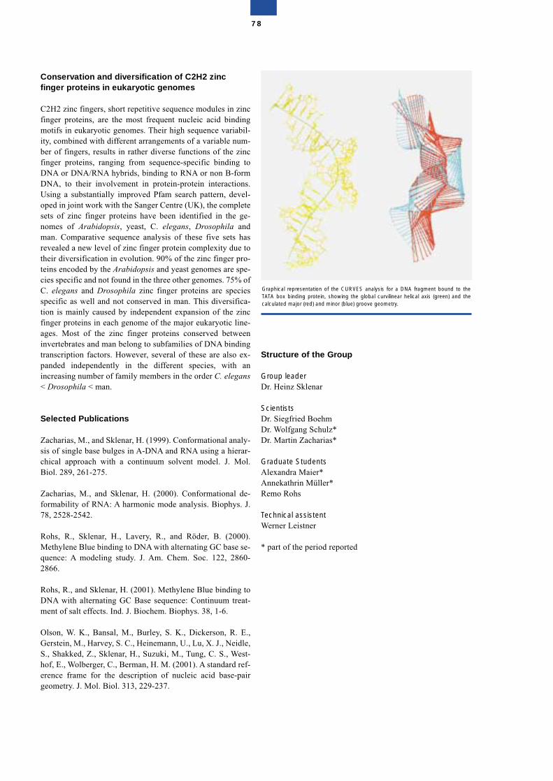
Conservation and diversification of C2H2 zincfinger proteins in eukaryotic genomes
C2H2 zinc fingers, short repetitive sequence modules in zincfinger proteins, are the most frequent nucleic acid bindingmotifs in eukaryotic genomes. Their high sequence variabil-ity, combined with different arrangements of a variable num-ber of fingers, results in rather diverse functions of the zincfinger proteins, ranging from sequence-specific binding toDNA or DNA/RNA hybrids, binding to RNA or non B-formDNA, to their involvement in protein-protein interactions.Using a substantially improved Pfam search pattern, devel-oped in joint work with the Sanger Centre (UK), the completesets of zinc finger proteins have been identified in the ge-nomes of Arabidopsis, yeast, C. elegans, Drosophila andman. Comparative sequence analysis of these five sets hasrevealed a new level of zinc finger protein complexity due totheir diversification in evolution. 90% of the zinc finger pro-teins encoded by the Arabidopsis and yeast genomes are spe-cies specific and not found in the three other genomes. 75% ofC. elegans and Drosophila zinc finger proteins are speciesspecific as well and not conserved in man. This diversifica-tion is mainly caused by independent expansion of the zincfinger proteins in each genome of the major eukaryotic line-ages. Most of the zinc finger proteins conserved betweeninvertebrates and man belong to subfamilies of DNA bindingtranscription factors. However, several of these are also ex-panded independently in the different species, with anincreasing number of family members in the order C. elegans< Drosophila < man.
Selected Publications
Zacharias, M., and Sklenar, H. (1999). Conformational analy-sis of single base bulges in A-DNA and RNA using a hierar-chical approach with a continuum solvent model. J. Mol.Biol. 289, 261-275.
Zacharias, M., and Sklenar, H. (2000). Conformational de-formability of RNA: A harmonic mode analysis. Biophys. J.78, 2528-2542.
Rohs, R., Sklenar, H., Lavery, R., and Röder, B. (2000).Methylene Blue binding to DNA with alternating GC base se-quence: A modeling study. J. Am. Chem. Soc. 122, 2860-2866.
Rohs, R., and Sklenar, H. (2001). Methylene Blue binding toDNA with alternating GC Base sequence: Continuum treat-ment of salt effects. Ind. J. Biochem. Biophys. 38, 1-6.
Olson, W. K., Bansal, M., Burley, S. K., Dickerson, R. E.,Gerstein, M., Harvey, S. C., Heinemann, U., Lu, X. J., Neidle,S., Shakked, Z., Sklenar, H., Suzuki, M., Tung, C. S., West-hof, E., Wolberger, C., Berman, H. M. (2001). A standard ref-erence frame for the description of nucleic acid base-pairgeometry. J. Mol. Biol. 313, 229-237.
Structure of the Group
Group leaderDr. Heinz Sklenar
ScientistsDr. Siegfried BoehmDr. Wolfgang Schulz*Dr. Martin Zacharias*
Graduate StudentsAlexandra Maier*Annekathrin Müller*Remo Rohs
Technical assistentWerner Leistner
* part of the period reported
78
Graphical representation of the CURVES analysis for a DNA fragment bound to theTATA box binding protein, showing the global curvilinear helical axis (green) and thecalculated major (red) and minor (blue) groove geometry.
Conformation, Stabilityand Interaction of BiologicalMacromolecules
Heinz Welfle
The main areas of our research involve studies of the physic-ochemical properties of nucleic acid-binding proteins andtheir complexes with RNA or DNA, and of antibodies andantibody-peptide complexes. The main tools used are circulardichroism, fluorescence, infrared and Raman spectroscopy,and calorimetric methods.
Antibody-peptide interactions
Essentially the same features determine the recognition ofproteins and peptides by antibodies. However, the usually un-folded and flexible structure of peptide ligands in solutioncomplicates the first steps of recognition; large changes in theconformational entropy contribute to the energetics of thebinding reaction. Isothermal titration calorimetry provides anexperimental tool for the investigation of the free energy ofbinding, �G°, separating it into enthalpic and entropic com-ponents. In collaboration with Wolfgang Höhne and JensSchneider-Mergener, Humboldt-Universität Berlin, we havestudied in detail the binding of peptides by high affinity anti-bodies CB/RS/1 (anti-interleukin 10 monoclonal antibody),CB4-1 (anti-p24 (HIV-1) monoclonal antibody), and tAB2(anti-TGF� monoclonal antibody). Although monospecificinteraction of an endogeneous epitope is expected for thephysiological function of antibodies, antibodies can bind tohomologous and non-homologous peptides; high affinitybinders were identified by screening of large combinatorialpeptide libraries. For example, the murine anti-p24 (HIV-1)antibody CB4-1, binds to a linear peptide epitope of the cap-sid protein and also to several unrelated peptides. X-ray crys-tal structure analyses show that these peptides assume differ-ent backbone conformations and utilize different interactionsin CB4-1 Fab fragment complexes with epitope-homologousand non-homologous peptides (T. Keitel, A. Kramer, H.Wessner, C. Scholz, J. Schneider-Mergener, Cell 91, 1997,811-820). Binding constants, binding stoichiometry and bind-ing enthalpies were obtained experimentally to allow the cal-culation of binding free energies and binding entropies. Forthe antibody-peptide interactions studied so far, enthalpy and
entropy contributions to the free energy differ significantly,but in each system the complex formation is enthalpicallydriven.
Translational initiation factor IF2 from Bacillusstearothermophilus
Initiation factor IF2 is involved in the initiation step of eubac-terial translation, and its main known function is the correctpositioning of initiator fMet-tRNAfMet in the ribosomal P site.In collaboration with C.O. Gualerzi, University of Camerino,Italy, and U. Heinemann, MDC, we have provided clear evi-dence for the localization of the entire fMet-tRNAfMet bindingsite in a small C-terminal subdomain, IF2 C-2, from the Ba-cillus stearothermophilus factor. Our earlier protein unfoldingstudies had suggested that IF2 C, the 24.5-kDa fMet-tRNAbinding domain of IF2, may consist of two subdomains. Re-placement of the four Phe residues of IF2 C yielded four var-iant proteins having intrinsic fluorescence markers in differ-ent positions of the molecule. Comparison of the circulardichroism and Trp fluorescence changes induced by increas-ing concentrations of guanidine hydrochloride demonstratedthat IF2 C indeed consists of two subdomains: the more stableN-terminal (IF2 C-1) subdomain and the less stable C-termi-nal (IF2 C-2) subdomain. Isolated subdomain C-2, whichconsists of just 110 amino acids, was found to bind fMet-tRNAfMet with the same specificity and affinity as native IF2 orIF2 C-domain. Characterization of IF2 C-2 by circular dich-roism, by urea-, guanidine hydrochloride-, and temperature-induced unfolding, and by differential scanning calorimetryindicates the properties of a globular molecule containingpredominantly � structures (33%) and turns (19%). Complexformation between fMet-tRNAfMet and IF2 C or IF2 C-2 is ac-companied by barely detectable spectral changes as demon-strated by a comparison of the Raman spectra of the com-plexes with the calculated sum of the spectra of the individualcomponents. These results and the temperature-dependenceof the Kd of the protein-RNA complexes, determined by ana-lytical ultracentrifugation studies, indicate that complex for-mation is not accompanied by obvious conformationalchanges in the components and, possibly, depends on a rathersmall binding site comprising only a few interacting residuesof the two components.
Plasmid pSM19035-encoded proteins
Because of the central role of the pSM19035-encoded �-pro-tein in regulating the maintenance of low-copy-number plas-mids, we initiated a detailed analysis of this protein. Thelongterm goal of this project is to elucidate, in collaborationwith W. Saenger, FU Berlin, and J. Alonso, C.S.I.C. Madrid,the cellular systems that ensure stable inheritance of low-copy-number plasmids within bacterial cells by an accuratecontrol of DNA replication and an ordered partition at cell di-vision. pSM19035-encoded products that avoid the appear-ance of plasmid-free cells, map within the SegA and SegB re-gions. The SegB region, that makes plasmids ~1000 timesmore stable than expected for random segregation, includesthe , �, and � genes. At the transcriptional level, � protein,the product of the � gene, coordinates the expression of pro-
79
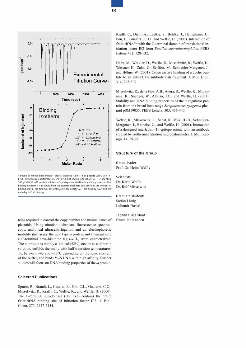
teins required to control the copy number and maintainance ofplasmids. Using circular dichroism, fluorescence spectros-copy, analytical ultracentrifugation and an electrophoreticmobility shift assay, the wild type � protein and a variant witha C-terminal hexa-histidine tag (�-H6) were characterized.The �-protein is mainly �-helical (42%), occurs as a dimer insolution, unfolds thermally with half transition temperatures,Tm, between ~43 and ~78°C depending on the ionic strengthof the buffer, and binds PcopS DNA with high affinity. Furtherstudies will focus on DNA binding properties of the � protein.
Selected Publications
Spurio, R., Brandi, L., Caserta, E., Pon, C.L., Gualerzi, C.O.,Misselwitz, R., Krafft, C., Welfle, K., and Welfle, H. (2000).The C-terminal sub-domain (IF2 C-2) contains the entirefMet-tRNA binding site of initiation factor IF2. J. Biol.Chem. 275, 2447-2454.
Krafft, C., Diehl, A., Laettig, S., Behlke, J., Heinemann, U.,Pon, C., Gualerzi, C.O., and Welfle, H. (2000). Interaction offMet-tRNAfMet with the C-terminal domain of translational in-itiation factor IF2 from Bacillus stearothermophilus. FEBSLetters 471, 128-132.
Hahn, M., Winkler, D., Welfle, K., Misselwitz, R., Welfle, H.,Wessner, H., Zahn, G., Seiffert, M., Schneider-Mergener, J.,and Höhne, W. (2001). Crossreactive binding of a cyclic pep-tide to an anti-TGF� antibody Fab fragment. J. Mol. Biol.,314, 293-309
Misselwitz, R., de la Hoz, A.B., Ayora, S., Welfle, K., Muray-ama, K., Saenger, W., Alonso, J.C., and Welfle, H. (2001).Stability and DNA binding properties of the � regulator pro-tein from the broad-host range Streptococcus pyogenes plas-mid pSM19035. FEBS Letters, 505, 436-440.
Welfle, K., Misselwitz, R., Sabat, R., Volk, H.-D., Schneider-Mergener, J., Reineke, U., and Welfle, H. (2001). Interactionof a designed interleukin-10 epitope mimic with an antibodystudied by isothermal titration microcalorimetry. J. Mol. Rec-ogn. 14, 89-98.
Structure of the Group
Group leaderProf. Dr. Heinz Welfle
ScientistsDr. Karin WelfleDr. Rolf Misselwitz
Graduate studentsStefan LättigLubomir Dostal
Technical assistantBrunhilde Kannen
80
Titration of monoclonal anti-p24 (HIV-1) antibody CB4-1 with peptide ATPQDLNTn-LeuL. Titration was performed at 25°C in 50 mM sodium phosphate, pH 7.2, injecting100 µl of 0.53 mM peptide solution in 5 µl steps into 0.016 mM antibody solution. Thebinding isotherm is calculated from the experimental data and provides the number ofbinding sites n, the binding constant Ka, the free energy �G°, the entropy T�S° and theenthalpy �H° of binding.
Bioethics and ScienceCommunication
Christof Tannert
At the beginning of 2002 a new research unit was establishedat MDC to collaborate closely with a sociological researchgroup from the Research Center Juelich (ForschungszentrumJülich, FZJ) on the project “Discourse on the Ethical Ques-tions of Biomedicine”. The project is funded by the FederalMinistry for Education and Research.
Brief description of the project
• A public initiative designed to serve as a model of a bio-medical research centre in close collaboration with a soci-ological unit of the FZJ on matters involving public infor-mation, dialog and consensus processes concerningethically relevant biomedical questions of today
• Testing and exploitation of a variety of structured dis-course options to assist in the formation of public opin-ion/consensus building over a wide range of issues and atvarious levels
• The testing of new communication media for discussions(Internet discussion)
• A one-year scientific and interdisciplinary discussion pro-cess accompanied and moderated by experts and “advis-ers” from specific social groups on the subject of “Ethicalquestions regarding research on human stem cells andtheir medical exploitation”
• Implementation of a conference of laymen on the samesubject
• Scientific evaluation and further development of the ap-plied procedures to help resolve value conflicts in society
Target discussion groups
• The general public throughout the Federal Republic• Decision-makers from politics and business• Scientists and other experts
Principles
The guiding principle of the project is a permanent, informeddiscourse between researchers and the public which is struc-tured towards creating consensus processes. In this connec-tion, the group aims to develop an interface for German bio-medicine that will serve as an example in the future. Thiscomplex and long-term project will take place in close asso-ciation with the MDC and its staff and take direct advantageof their expertise. However, it will not be thematically limitedto research matters of interest to the MDC, but will extend be-yond this and deal with the issues in a complex and inter-linked manner and in close collaboration with the social sci-ences.
It is of fundamental importance for achieving a consensusopinion of experts and the general public that the discourse isnot designed to be simply an instrument for gaining accep-tance, but to be more a form for identifying common socialconcerns. Declarations of ethical dissent based on technicallyargumentative discussion must, therefore, be considered asbeing completely valid.
Finally, the essential objectives of the project areas follows:
• gathering experience by means of an interdisciplinary dis-course in a controlled environment, which will be record-ed in a report and scientifically evaluated
• introducing scientific and methodological evaluation andfurther development of interactive media forms for thepublic discussion of ethical matters (Internet discussion)
• evaluating and further developing all the discussion op-tions used with regard to their suitability for resolvingvalue conflicts in society
Structure of the Interdisciplinary Group
Group leaderDr. Christof Tannert (Biologist)
Sub-group leader FZJDr. Peter Wiedemann (Psychologist)*
ScientistsDr. Susanne Reif (Biologist)Dr. Silke Schicktanz (Philosopher)*
Graduate studentSilke Domasch (Linguist)*
Management assistantAli ben Salem
* Guest Scientists (FZJ)
81

82
83
Cell Growth and Differentation
Cell Growth and Differentiation
Approximately 35,000 genes make up the human genome.Although we know the DNA sequence of all these genes, wehave only a basic idea of how they are regulated and the func-tions they exhibit, in particular those that can induce disease.Genes encode structural and regulatory proteins that balancecell growth, differentiation and homeostasis through the con-trol of cell-cell interactions, signal transduction, cell cycleprogression, and gene regulation. In many diseases, criticalgenes are mutated or misexpressed so that cell growth anddifferentiation are distorted. Thus, it is a formidable task toidentify the critical disease genes and to find out how theycommunicate with each other and with the rest of the ge-nome. This is essential for developing future rational thera-pies for almost all diseases, including cancer and cardiovas-cular disease, Alzheimer disease, cystic fibrosis, or diseasesaffecting bone and muscle development.
In the Cell Growth and Differentiation Programme of theMDC, we study how genes and their products interact to con-trol normal tissue development and function, and how disrup-tion of their interactions leads to disease. A major focus of theProgramme is to unravel all the features of cancer and cardi-ovascular disease at the molecular level using methods ofmolecular genetics, biochemistry and cell biology.
A reductionistic approach is often required to track the func-tion of a gene. Accordingly, we use model systems, such asDrosophila or baker’s yeast, to analyze basic gene functions.However, highly sophisticated tissue functions developedlater during evolution and, so, we also need to study higherorganisms that are amenable to genetic analysis such as thezebrafish or the laboratory mouse, to understand complex ge-netic functions. Such a combination of approaches has helpedus to uncover several important mechanisms of disease devel-opment and major advances have been made in our Pro-gramme during the last two years.
For example, an important role is played by chemokines andtheir receptors in the process of dissemination and metastasisof solid and of haematopoietic tumors. It was found that the
Zellwachstum und Differenzierung
Das menschliche Genom umfaßt rund 35.000 Gene. Obwohluns die DNA Sequenz aller Gene bekannt ist, wissen wir nurwenig über ihre Funktion und Regulation, insbesondere auchihre Rolle bei der Entstehung von Krankheiten. Gene kodie-ren für Strukturproteine und Regulatorproteine, die Zell-wachstum, Differenzierung und Homöostase durch Kontrollevon Zell-Zell-Interaktionen, Signaltransduktionen und Zell-teilung in eine Balance bringen. In vielen Krankheiten sindkritische Gene mutiert oder fehlerhaft exprimiert, so dassZellwachstum und Differenzierung gestört ablaufen. UnsereAufgabe besteht somit darin, kritische Gene mit Krankheits-wert zu ermitteln und herauszufinden, wie sie untereinanderund mit dem Rest des Genoms kommunizieren. Dies ist dieprimäre Aufgabe, die es zu lösen gilt, um in Zukunft rationaleTherapien für alle Krankheiten zu entwickeln, einschließlichKrebs, kardiovaskulären und neurodegenerativen Krankhei-ten, für die Zystische Fibrose oder für Krankheiten, die dieEntwicklung der Knochen und Muskeln betreffen.
Im MDC-Programm für Zellwachstum und Differenzierunguntersuchen wir, wie Gene und ihre Produkte miteinander in-teragieren, um die Entwicklung und die Funktion von norma-lem Gewebe zu kontrollieren, und wie Veränderungen dieserWechselwirkung zu Krankheiten führen. Ein wesentlicherProgrammpunkt besteht darin, die molekulare Basis vonKrebs und kardiovaskulären Krankheiten mit den Methodender Molekulargenetik, der Biochemie und der Zellbiologieaufzudecken.
Um der Funktion eines Gens auf die Spur zu kommen, mußman sich oft reduktionistischer Methoden bedienen. Dem-gemäß setzen wir einfache Modellsysteme wie z. B. dieFruchtfliege oder die Bäckerhefe ein, um grundlegende Gen-funktionen zu analysieren. Jedoch haben sich komplexereGewebefunktionen erst sehr spät im Verlauf der Evolutiongebildet. Wir müssen deshalb auch höhere Organismen wieden Zebrafisch oder die Labormaus genetisch analysieren,wenn wir komplexe genetische Funktionen verstehen wollen.Durch die Kombination dieser Ansätze ist es in unserem Pro-gramm in den letzten zwei Jahren gelungen, mehrere wich-
84
chemokine receptors CCR7 and CXCR4 are expressed onReed-Sternberg cells of classical Hodgkin’s disease. Upregu-lation of CCR7 correlates with the dissemination of neoplas-tic cells to the interfollicular T cell zone of lymph nodeswhere chemokine ligands are expressed. In Hodgkin-derivedcell lines, CCR7 upregulation depends on constitutive NF�Bactivity (Höpken et al., Blood 99, 1109-1116, 2002).
The cell adhesion and signalling protein, �-catenin, is impor-tant in embryonic development and in the formation of carci-nomas. Using Cre/loxP technology, a conditional deletion ofthe �-catenin gene was introduced in the epidermis and hairfollicles. Hair is completely lost in mutants after the first haircycle, and regressing hair follicles are transformed into epi-dermal cysts. It has been demonstrated that �-catenin is es-sential for the ultimate fate of stem cells in the skin: in the ab-sence of �-catenin, stem cells do not differentiate intofollicular keratinocytes, but instead have an epidermal fate(Huelsken et al., Cell 105, 533-545, 2001).
Using a gene targeting approach in mice, NF-�B was shownto be required for the early morphogenesis of hair follicles,exocrine glands and teeth. The phenotype of NF-�B inhibi-tion closely resembles hypohydrotic ectodermal dysplasia(HED) in humans and is identical to that in mice deficient insignalling molecules, called EDA or EDAR. Our data showthat defective signalling through the EDA/NF-�B pathwaycauses HED (Schmidt-Ullrich et al., Development 128, 3843-3853, 2001).
Novel genes that are associated with metastatic tumor pro-gression have been identified. The expression of sialyl trans-ferase ST6Gal-II is related to poor survival of patients withcolorectal carcinomas. ST6Gal-I also plays a crucial role inthe adhesion and metastasis of carcinoma cells. Breast carci-noma cells that express sialyltransferase display reduced cell-cell adhesion and an enhanced invasion capacity, whereascell clones that express an antisense-construct exhibit stronghomotypic cell-cell adhesion (Schneider et al., Cancer Res.61, 4605-4611, 2001).
C/EBP transcription factors are involved in many develop-mental processes that regulate proliferation and terminaldifferentiation in a wide variety of cell types. C/EBP� andC/EBP� proteins occur in different isoforms that display en-tirely different biological activities. It has been found thatregulation of translational initiation is responsible for thegeneration of C/EBP isoforms. Dysregulation of translationinitiation pathways or misexpression of C/EBP isoforms dis-rupting the coupling between proliferation and differentiationprograms has direct implications for cancer research, haemat-opoiesis, adipogenesis, lung function and female reproduc-tive biology (Calkhoven et al., Genes & Development 14,1920-1932, 2000).
Ubiquitin-conjugation during endoplasmic reticulum-asso-ciated degradation (ERAD) is essential for the elimination ofmisfolded proteins from the secretory pathway. It has beenshown that the basic ERAD activity is sufficient to eliminateunfolded proteins under normal growth conditions. However,under stress, ERAD activity is increased by the UPR (Un-folded Protein Response) through transcriptional induction of
tige Mechanismen der Krankheitsentstehung aufzuklären unddabei die folgenden, spürbaren Fortschritte zu erzielen.
Eine wichtige Rolle bei der Verbreitung und Metastasierungvon soliden und hämatopoietischen Tumoren spielen die Che-mokine und ihre Rezeptoren. Es konnte gezeigt werden, dassdie Chemokinrezeptoren CCR7 und CXCR4 von Reed-Stern-berg-Zellen der klassischen Hodgkin-Krankheit exprimiertwerden. Die Erhöhung von CCR7 korreliert mit der Verbrei-tung neoplastischer Zellen in der interfollikulären T-Zell-Zone der Lymphknoten, in denen Chemokin-Liganden expri-miert werden. In Zelllinien aus Hodgkin-Tumoren wiederumhängt die CCR7-Hochregulierung an der konstitutiven Akti-vität von NF-�B (Höpken et al., Blood 99, 1109-1116, 2002).
Das Zelladhäsions- und Signalprotein �-Catenin spielt einewichtige Rolle in der Embryonalentwicklung und bei der Bil-dung von Karzinomen. Mit Hilfe der Cre/lox-Technologiekonnte eine konditionale Deletion des �-Catenin-Gens in dieEpidermis und in die Haarfollikel der Maus eingefügt wer-den. Die Mutanten verlieren sämtliche Haare, und die sichrückbildenden Haarfollikel werden in epidermale Zystentransformiert. Es konnte gezeigt werden, dass �-Catenin ent-scheidend für das Schicksal der Stammzellen in der Haut ist:In der Abwesenheit von �-Catenin diffenzieren sich dieStammzellen nicht zu follikulären Keratinozyten und nehmenstatt dessen epidermale Eigenschaften an (Huelsken et al.,Cell 105, 533-545, 2001).
Mit Hilfe der Methode des „Gene-Targeting“ konnte in Mäu-sen gezeigt werden, dass NF-�B für die frühe Morphogeneseder Haarfollikel, der exokrinen Drüsen und der Zähnebenötigt wird. Der Phänotyp der Inhibition von NF-�B ähneltsehr stark der hypohydrotischen ektodermalen Dysplasie(HED) beim Menschen. Außerdem wird der gleiche Phänotypin Mäusen beobachtet, denen die als EDA und EDAR be-kannten Signalproteine fehlen. Die Daten machen deutlich,dass Signaldefekte in der Synthese von EDA/NF-�B die Ur-sache von HED sind (Schmidt-Ullrich et al., Development128, 3843-3853, 2001).
Neue Gene konnten identifiziert werden, die mit Tumorpro-gression und Metastasierung assoziiert sind. Die Expressionder Sialyl-Transferase STGal-II steht in Beziehung zu gerin-ger Überlebensrate von Patienten mit kolorektalen Tumoren.STGal-I spielt auch eine wichtige Rolle bei der Adhäsion undMetastasierung von Karzinomzellen. Brustkarzinomzellen,die Sialyltransferase exprimieren, zeigten eine verminderteZell-Zell-Adhäsion und eine erhöhte Invasivität, währendZellklone, die ein Antisense-Konstrukt enthielten, starkehomotypische Zell-Zell-Adhäsion zeigen (Schneider et al.,Cancer Res, 61, 4605-4611, 2001).
C/EBP-Transkriptionsfaktoren spielen in vielen Entwick-lungsprozessen und Zelltypen bei der Regulation der Prolife-ration und der terminalen Differenzierung eine ausschlag-gebende Rolle. C/EBP-Proteine existieren in verschiedenenIsoformen, die vollkommen unterschiedliche biologischeAktivitäten aufweisen. Es konnte gezeigt werden, dass dieRegulation des Translationsbeginns für die Generierung derC/EBP-Isoformen verantwortlich ist. Fehlregulation desTranslationsbeginns oder fehlerhafte Expression der C/EBP-
85
the involved genes. This demonstrates for the first time that aregulatory loop between ERAD and UPR is essential for thenormal growth of eukaryotic cells (Friedlander et al., NatureCell Biol. 2, 379-384, 2000).Through large-scale screening of embryonic lethal mutationsin zebrafish, the genes heart and soul have been identified,based on a defective heart tube morphology and a disruptedpigmented epithelium surrounding the neural retina. It hasbeen shown that heart and soul encode a component of theapical Par-protein complex, termed aPKC�. The role ofaPKC� in epithelial formation is best understood in Drosoph-ila where Par-3, Par-6, and aPKC localize to the apico-lateralmembrane of embryonic epithelia. During the early stages ofepithelial differentiation, heart and soul appears to regulatethe apical clustering and maintenance of adherens junctions.In addition to the epithelial defects, heart and soul affects themorphogenesis of the heart tube and the gut with its asso-ciated organs (Horne-Badovinac et al., Current Biol. 11,1492-1502, 2001).Smooth muscle cells (SMC) express different isoforms of themolecular motor type-II myosin. The expression of smoothmuscle MyHC (SM-MyHC) can be eliminated by gene tar-geting technology. Smooth muscle from knock-out neonatalmice lacked phasic contraction while tonic contraction re-mained normal. Accordingly, sustained force generation inthe absence of SM-MyHC was sufficient for normal fetal de-velopment. However, the strong phasic contraction becomesindispensable for survival and normal growth soon afterbirth. In addition, both contractile systems are associatedwith different second messenger pathways in smooth muscle:the SM-MyHC and NM-MyHC systems appear to be in-volved in electromechanical and pharmacomechanicalcoupling, respectively (Morano et al., Nature Cell Biol. 2,371-375, 2000).A fundamental process in every cell division cycle is faithfulduplication of genetic and epigenetic information. Construc-tion of a chimeric green fluorescent protein fused to a centralcomponent of the DNA replication machinery (proliferatingcell nuclear antigen, PCNA) permitted visualization and dy-namic studies of replication sites in living cells using real-time microscopy. DNA replication in the nucleus occurs atdiscrete sites (replication foci) that change throughout theS-phase of the cell cycle. These changes are not due to a di-rectional movement of replication foci but rather to asynchro-nous disassembly, reassembly and activation of replication atnew subnuclear sites (Leonhardt et al., J. Cell Biol. 149, 271-280, 2000).
Members of our Programme have been invited speakers atmany international scientific meetings during the last twoyears, among them Keystone Symposia, FASEB Confer-ences, EMBO Meetings and Workshops, ELSO Meeting,American Association of Cancer Res. Conferences, and theBanbury Meeting (Cold Spring Harbor).
The revolution in molecular biology has begun to permeatecancer and cardiovascular medicine, and molecular methodsare already being used in clinical diagnosis and disease prog-nosis. Moreover, better understanding of cell-type specificregulatory mechanisms may eventually allow us to selec-tively control proliferation and differentiation processes andto find new therapeutic interventions. Potential new com-
Isoformen unterbricht die Kopplung zwischen den Prolifera-tions- und Differenzierungsprogrammen und hat unmittelba-re Implikationen für die Krebsentstehung, die Hämatopoese,die Adipogenese, die Lungenfunktion und die Ovulation(Calkhoven et al., Genes & Development 14, 1920-1932,2000).
Für die Eliminierung falsch gefalteter sekretorischer Proteineist die Ubiquitin-Konjugation im endoplasmatischen Reticu-lum (ERAD) wesentlich. Es konnte gezeigt werden, dass diegrundlegende ERAD-Aktivität ausreicht, um fehlgefalteteProteine unter normalen Wachstumsbedingungen zu elimi-nieren. Unter Stressbedingungen nimmt die ERAD-Aktivitätdurch die UPR („Unfolded Protein Response“) zu, und zwardurch Transkriptionsinduktion der betroffenen Gene. DieseBeobachtung zeigt zum ersten Mal eine regulatorische Kopp-lung zwischen ERAD und UPD, die für das normale Wachs-tum von eukaryontischen Zellen essentiell ist (Friedlander etal., Nature Cell Biol. 2, 379-384, 2000).
Durch „Screenen“ auf lethale Mutationen im Zebrafischem-bryo konnten die Gene heart and soul identifiziert werden,die mit einer defekten Morphologie des Herzens und einemzerstörten pigmentierten Epithel im Umkreis der neuralenRetina verbunden sind. Es konnte gezeigt werden, dass heartand soul eine Komponente des apikalen Par-Protein-Komplexes darstellt, der aPKC� genannt wird. Eine Rolle vonaPKC� bei der Epithelbildung wird am besten in Drosophilaverstanden, wo Par-3, Par-6 und aPKC an der apico-lateralenMembran embryonaler Epithelien sitzt. Zusätzlich zu denEpitheldefekten beeinflußt heart and soul die Morphogenesedes Herzens und des Darms mit seinen assoziierten Organen(Horne-Badovinac et al., Current Biol. 11, 1492-1502, 2001).
Glatte Muskelzellen (SMC) exprimieren unterschiedlicheIsoformen des molekularen Typ-II-Myosins. Die Expressionvon glattem Muskel-MyHC (SM-MyHC) konnte durch dieTechnik des „gene targeting“ ausgeschaltet werden. GlatteMuskeln in neonatalen Knock-out-Mäusen ließen die pha-sische Kontraktion vermissen, während die tonische Kontrak-tion normal blieb. Entsprechend war die nachhaltige Kraftge-nerierung in Abwesenheit von SM-MyHC für die normalefetale Entwicklung ausreichend. Die starke phasische Kon-traktion wird jedoch unentbehrlich für das Überleben und dasnormale Wachstum nach der Geburt. Zusätzlich sind beidekontraktilen Systeme mit verschiedenen „second messengerpathways“ im glatten Muskel assoziiert: Während das SM-MyHC-System mit der elektromechanischen Ankopplungverbunden ist, hat das NM-MyHC-System mit der pharmako-mechanischen Ankopplung zu tun (Morano et al., Nature CellBiol. 2, 371-375, 2000).
Ein grundlegender Vorgang bei jeder Zellteilung ist die ge-naue Duplikation der genetischen und der epigenetischenInformation. Die Konstruktion eines chimärischen Grünflu-oreszierenden Proteins, das mit einer zentralen Komponenteder DNA Replikationsmaschinerie fusioniert war (dem proli-ferating cell nuclear antigen PCNA), erlaubte neben der di-rekten Beobachtung auch dynamische Studien der Replika-tionsorte in lebenden Zellen durch Echtzeitmikroskopie. DieReplikation der DNA beginnt in den Zellen an umgrenztenReplikationsstellen, die sich im Verlauf der S-Phase des
86
pounds for the treatment of diseases are presently being iden-tified worldwide through so-called molecular screening.These procedures use molecular targets which operate in thesignalling cascades of growth and differentiation processes ofcells. Several groups in our Programme are involved in suchresearch, mostly in collaboration with pharmaceutical com-panies. Moreover, together with the Molecular Therapy Pro-gram and the MDC clinics, gene therapy procedures are beingdeveloped based on an understanding of how genes influencegrowth differentiation of cells.
Walter BirchmeierAchim Leutz
Cited ReferencesCalkhoven, C.F., Müller, C., and Leutz, A. (2000) Transla-tional control of C/EBP alpha and C/EBP beta isoform ex-pression. Genes Dev., 1920-1932.
Friedlander, R., Jarosch, E., Urban, J., Volkwein, C., andSommer, T. (2000). A regulatory link between ER-associatedprotein degradation and the unfolded protein response. Na-ture Cell Biology 2, 379-384.
Höpken, U.E., H.D. Foss, H.D., Meyer, D., Hinze, M., Leder,K., Stein, H., and Lipp, M. (2002). Upregulation of the chem-okine receptor CCR7 in classical but not in lymphocyte pre-dominant Hodgkin disease correlates with distinct dissemina-tion of neoplastic cells in lymphoid organs. Blood 99,1109-1116
Horne-Badovinac, S., Lin, D., Waldron, S., Schwarz, M.,Mbamalu, G., Pawson, T., Jan, Y.N., Stainier, D.Y.R., and Ab-delilah-Seyfried, S. (2001). Positional cloning of heart andsoul reveals multiple roles for PKC� in zebrafish organogen-esis. Current Biology 11, 1492-1502.
Huelsken, J., Vogel, R., Erdmann, B., Cotsarelis, G., andBirchmeier, W. (2001). Beta-catenin controls hair folliclemorphogenesis and stem cell differentiation in the skin. Cell105, 533-545.
Leonhardt, H., Rahn, H.-P., Weinzierl, P., Sporbert, A.,Cremer, T., Zink, D., and Cardoso, M. C. (2000). Dynamicsof DNA replication factories in living cells. J. Cell Biol. 149,271-280.
Morano, I., Chai, G.X., Baltas, L.G., Lamounier-Zepter, V.,Kott, M., Haase, E.H., Walther, T., and Bader, M. (2000).Smooth muscle contraction without smooth muscle myosin.Nature Cell Biology, 2, 371-375.
Schmidt-Ullrich, R., Aebischer, T, Hülsken, J., Birchmeier,W., Klemm, U., and Scheidereit, C. (2001). Requirement ofNF-kB/Rel for the development of hair follicles and otherepidermal appendices. Development 128, 3843-3853.
Schneider F, Kemmner W, Haensch W, Franke G, GretschelS, Karsten U, and Schlag P.M. (2001). Overexpression of sia-lyltransferase ST6GalNAc-II is related to poor patient survivalin human colorectal carcinomas. Cancer Res, 61, 4605-4611.
Zellzyklus verändern. Diese Änderungen kommen nichtdurch eine gerichtete Bewegung der Replikationsorte zustan-de, sondern durch asynchrones Zerlegen, Zusammensetzenund Aktivieren der Replikation an neuen subnuklearen Stel-len (Leonhardt et al., J. Cell Biol. 149, 271-280, 2000).
In den vergangenen zwei Jahren sind Mitglieder unseres Pro-gramms zu Vorträgen auf zahlreichen internationalen wissen-schaftlichen Tagungen eingeladen worden, unter anderem aufdie Keystone Symposia, die FASEB-Konferenzen, EMBO-Tagungen und Workshops, ELSO-Meetings, Konferenzen derAmerican Association of Cancer Research und zu Banbury-Meetings (in Cold Spring Harbor).
Die Revolution der Molekularbiologie durchdringt inzwi-schen die Krebsmedizin und die Medizin kardiovaskulärerErkrankungen. Längst sind molekularbiologische Methodenin der klinischen Diagnostik und für prognostische Einschät-zungen im Einsatz. Darüber hinaus wird uns das Verständnisder Zelltyp-spezifischen Regulationsmechanismen helfen,schließlich Prozesse der Proliferation und Differenzierungselektiv zu kontrollieren und somit neue therapeutische Ein-griffsmöglichkeiten zu finden. Neue Substanzen für die Be-handlung von Krankheiten werden derzeit weltweit mit Hilfedes sogenannten molekularen Screenings identifiziert. DieseVerfahren beruhen auf molekularen Zielstrukturen, die inSignalkaskaden auftreten, welche Wachstums- und Differen-zierungsprozesse der Zellen steuern. Mehrere Gruppen unse-res Programms sind an solchen Untersuchungen beteiligt, zu-meist in Kooperation mit pharmazeutischen Unternehmen.Darüber hinaus werden mit dem Molekulare-Therapie-Pro-gramm und den Kliniken des MDC Verfahren für die Genthe-rapie entwickelt, die auf dem Verständnis des Einflusses derGene auf Wachstum und Differenzierung von Zellen basie-ren.
Walter BirchmeierAchim Leutz
87
Growth Control and Gene Regula-tion in the Hematopoietic System
Achim Leutz
Cells of the blood originate from stem cells that are located inthe bone marrow. Stem cells give rise to proliferating pro-genitors that differentiate into mature, growth arrested celltypes such as, erythrocytes, granulocytes or macrophages.Dysregulation of pathways that control proliferation and dif-ferentiation may cause various diseases, such as immune de-fects and leukemia. Thus, hematopoiesis provides striking ex-amples to address fundamental biological and clinicallyrelevant questions such as: How is cell identity achieved dur-ing lineage commitment? How are cell proliferation and dif-ferentiation regulated and how are they intertwined?
Hematopoiesis and leukemogenesis are regulated by a num-ber of key transcription factors that control developmentalprograms and lineage specific gene expression. Therefore,understanding the molecular basis of growth control, lineagecommitment, differentiation and leukemogenesis requires theidentification of regulated genes and elucidation of how thesegenes are regulated. Several years ago, we identified the firstmolecular switch that controls myeloid differentiation. Theswitch consists of two types of transcription factors both ofwhich may turn into oncogenes by mutation. Transcriptionfactors of the CCAAT/Enhancer Binding Protein family(C/EBP) regulate myeloid differentiation and proliferationarrest. The other transcription factor is derived from the Mybprotooncogene and is essential for development of all hemat-opoietic lineages. Both types of transcription factors collabo-rate in a concerted action to induce myeloid gene expression.Mutations in either group of transcription factors that abro-gate their collaboration may induce leukemia. This concepthas now been confirmed by many research groups and hasbeen extended to other co-operating hematopoietic transcrip-tion factors in different cell lineages. Combinatorial geneswitches permit plasticity of regulation and limit the numberof regulators and pathways required for cell type specifica-tion.
Chromatin remodeling and lineage specific geneexpression
A prerequisite for activation of silent genes is to overcome therepressive effects of chromatin. Using an assay that monitorsactivation of endogenous, chromatin embedded genes, wehave unraveled the mechanism of the collaboration betweenMyb and C/EBP. C/EBP� interacts with the chromatin re-modeling SWI/SNF complex through an N-terminal domain.This domain is contained only in one particular isoform ofC/EBP� that is generated by selective translation initiationcontrol (see below). The interaction between SWI/SNF andC/EBP is required to activate several myeloid genes togetherwith Myb. Grafting the N-terminus of C/EBP� onto Mybgenerates a chimeric transcription factor that no longer re-quires C/EBP� because it may recruit SWI/SNF all on itsown. This was the first demonstration that transcription fac-tors recruit the SWI/SNF complex to remodel chromatin dur-ing cell type specific gene expression in vertebrates. AsC/EBPs participate in many cell specification events recruit-ment of SWI/SNF may represent a major determinant of celllineage commitment.
Translational regulation of C/EBP
Individual C/EBP isoforms arise by differential initiation oftranslation. Individual isoforms mediate different functions.Full length C/EBP proteins are transactivators and inhibit pro-liferation while internally initiated, truncated proteins are re-pressors of gene activation that permit or even induce growth.Hence, regulation of translation initiation that determines theC/EBP isoform ratio plays a crucial role in the control of pro-liferation and differentiation.
Site directed mutagenesis has shown that a small upstreamopen reading frame (uORF) mediates translation control ofC/EBPs. Translation regulation through the uORF is down-stream of the signal transduction pathways that modulate theactivity of translation initiation factors and thus the genera-tion of C/EBP isoforms. We found that differentially initiatedC/EBP isoforms display striking differences in the recruit-ment of chromatin remodeling complexes and in transactiva-tion. In addition, truncated C/EBP isoforms abrogated growtharrest and uncouple proliferation and differentiation.
Differential selection of translation initiation sites is a con-served mechanism of gene expression control that allowscells to rapidly adjust to environmental, nutritional or hormo-nal changes. Proteins involved in translational regulation,such as PI3 kinase, AKT-kinase, PTEN-phosphatase or the in-itiation factors eIF-2� or eIF-4E, can also exhibit oncogenicproperties. This suggests an important role of translation initi-ation control in cell proliferation, cell type specification andtumorigenesis. It is anticipated that pathways and factors in-volved in the control of translational initiation are importantregulators of hematopoiesis and may represent novel targetsfor innovative drug therapies. Accordingly, we are searchingfor connections between dysregulated translation initiationcontrol and tumorigenesis and we are developing screeningsystems to discover interfering drugs.
88
Activation of the notch pathway by E1A oncoprotein
Signaling through the Notch pathway controls cell prolifera-tion and differentiation in many metazoan cell lineages. Fol-lowing binding of its ligands, the intracellular part of the cellsurface Notch1 receptor (Notch1-IC) is released and translo-cates to the nucleus where it alters the function of the DNAbinding transcription factor CBF1/RBP-J�. As a result,CBF1/RBP-J� is converted from a repressor to an activator ofgene transcription that controls expression of cell fate genes.
The Notch-CBF1 growth control pathway is exploited by theEpstein-Barr viral EBNA2 oncoprotein for viral replication.EBNA2 activates cellular and viral genes through CBF1/RBP-J� sites. The fact that functional CBF1/RBP-J� bindingsites have been identified in various adenoviral promoters letus explore whether the adenoviral E1A proteins may also tar-get CBF1/RBP-J�. We found that the adenoviral onco-protein13SE1A binds to CBF1/RBP-J�, displaces associated co-re-pressor complexes, and activates CBF1/RBP-J�-dependentgene expression. Our results suggest that the central role ofthe Notch-CBF1/RBP-J� signaling pathway in cell fate deci-sions renders it susceptible to various pathways of viral repli-cation and oncogenic conversion. Moreover, our results sug-gest a central role of the transcription factor CBF1/RBP-J�that, like the Rb and p53 tumor-suppressors, is modulated byvarious oncoproteins of DNA tumor viruses.
Selected Publications
Calkhoven, C.F., Müller, C., and Leutz, A. (2000). Transla-tional control of C/EBP alpha and C/EBP beta isoform ex-pression. Genes & Development 14, 1920-1932.
Ansieau, S., Strobl, L., and Leutz, A. (2001). Activation of theNotch regulated transcription factor CBF1/RBP-Jk throughthe 13SE1A oncoprotein. Genes & Development 15. 380-380.
Müller, C., and Leutz, A. (2001). Development and Chroma-tin remodeling. Curr. Op. Gen. & Dev. 11, 167-174.
Oelgeschläger, M., Kowenz-Leutz, E., Schreek, S., Leutz, A.,and Lüscher, B. (2001). Tumorigenic N-terminal deletions ofc-Myb modulate DNA binding, transactivation, and coopera-tivity with C/EBP. Oncogene, 20, 7420-7424.
Pedersen, T.A.; Kowenz-Leutz, E.; Leutz, A. and Nerlov, C.(2001) Cooperation between C/EBP� TBP/TFIIB and SWI/SNF recruiting domains is required for adipocyte differentia-tion. Genes & Development, 15, 3208-3216.
Structure of the Research Group
Group LeaderDr. Achim Leutz
ScientistsDr. Stephane AnsieauDr. Valerie Begay-MüllerDr. Cor CalkhovenDr. Christine MüllerDr. Elisabeth Kowenz-LeutzDr. Yves LaumonnierDr. Hubertus Pietsch
Graduate StudentsSimone JoschkoVolker Wiesenthal
Technical AssistantsChristiane Calließ Kerstin Klingner
SecretariatHenriette Mödig
89
Signal Transduction in Tumor Cells
Claus Scheidereit
Cell growth and differentiation largely depends on the expres-sion of sets of genes which are controlled essentially at thelevel of transcription. Gene expression patterns are pro-grammed by transcription factors, whose activity in turn ismodulated by complex networks of signal transducing mole-cules. The main aim of our laboratory is to understand howsignal transduction processes are coupled to transcription. Amodel system with wide physiological and medical relevanceis nuclear factor kappaB (NF-�B) and its co-regulators andaccessory proteins. A major goal of our research is to under-stand the structures and mechanisms underlying gene regula-tion by this complex system and its implications in diseasedevelopment.
Pathways and structures that regulate NF-�B activity
The pleiotropic transcription regulator nuclear factor kappaB(NF-�B) plays a central role in the inducible expression of alarge number of genes which encode cytokines, surface recep-tors, adhesion molecules, transcription factors and othermolecules controlling various immune functions as well ascell proliferation and programmed cell death. In its inactivelatent form, NF-�B is retained in the cytoplasm by associa-tion with I�B molecules, which inhibit nuclear translocationand DNA binding activity of NF-�B. Stimulation of cells witha variety of agents, such as bacterial lipopolysaccharides,tumor necrosis factor � (TNF�), or interleukin-1� (IL-1�) re-sults in the proteolysis of I�B molecules and liberation of ac-tive NF-�B into the nucleus. Induced I�B proteolysis is trig-gered by I�B phosphorylation mediated by an I�B kinase(IKK) complex, which is activated by many NF-�B-stimulat-ing pathways. The composition and regulation of the IKKcomplex is under investigation.
Differential regulation of NF-�B activity by I�B�,I�B�, p105 and Bcl-3
The mammalian NF-�B family consists of five members,p50, p65, p52, c-Rel and RelB. These proteins form hetero-and homodimers and are bound by I�B molecules I�B�, �and , the precursor proteins for p50 and p52, p105 and p100,respectively, or by the nuclear I�B homologue Bcl-3.
The IKK complex phosphorylates I�B�, � and at a con-served signal response domain and this sequence, containingalso lysines for phosphorylation-dependent ubiquitin-conju-gation, is sufficient to confer inducible degradation. The sig-nal response domain of I�B�, when fused to other proteins,triggers degradation of these proteins when cells are activatedby TNF� or other agents which activate IKKs. We have alsofound that the NF-�B precursor proteins, p105 and p100,which on processing give rise to p50 and p52, sequester otherNF-�B subunits including their processing products in the cy-toplasm and so act like I�B molecules. On stimulation withNF-�B activating agents, cellular p105 is phosphorylated byIKKs at two serines close to the carboxyterminal end of p105.To bind the IKK complex, p105 contains an IKK docking sitelocated in a death domain, which is separate from the sub-strate site. Upon phosphorylation by IKK, p105 attracts theSCF E3 ubiquitin ligase substrate recognition molecule�TrCP, resulting in polyubiquitination and complete degrada-tion by the proteasome. p105-associated NF-�B subunits,such as p50, which is formed by processing of p105, are lib-erated and transported to the nucleus. Thus, in parallel withthe release of NF-�B dimers by IKK-induced degradation ofI�B�, � or , other NF-�B subunits, including p50 homodim-ers, are released by p105 degradation. In the nucleus, p50 ho-modimers then associate with Bcl-3 and act as transcriptionalactivators.
Requirement of NF-�B for embryonic developmentof epidermal appendices and for secondarylymphoid organ formation
Gene ablation studies of NF-�B transcription factors and I�Bkinases (IKK) have shown essential functions of these pro-teins for immune response and inflammation, but also forbone-morphogenesis and keratinocyte differentiation. Otherfunctions were inaccessible due to the embryonic lethality ofNF-�B or IKK-deficient animals. With a conditional gene tar-geting approach, we have ubiquitously expressed an NF-�Bsuperrepressor I�B��N to investigate NF-�B functions in theadult. Mice with suppressed NF-�B survive to adulthood anddisplay macrophage dysfunction and alymphoplasia, the lackof secondary lymphoid organs. NF-�B inhibition causes se-vere defects in the early developmental steps of epidermal ap-pendages, including hair follicles, tear and sweat glands. Nor-mally, these structures display strong NF-�B transcriptionalactivity, as we demonstrated with �-galactosidase reportermice. This includes the stem cell-containing bulge region inhair follicles, which responds to morphogenic signals for hairfollicle generation. The epidermal phenotype is analogous tohypohidrotic (anhydrotic) ectodermal dysplasia (HED) in hu-mans, and identical to phenotypes of eda, edar or crinkledmice. The eda and edar genes belong to the TNF family of
90
ligands and receptors, respectively. Therefore, our data indi-cate that NF-�B is required in epidermal development foredar to transmit eda signals.
Requirement of NF-�B for cell proliferation and sur-vival of lymphoma and leukemia cells
In collaboration with the research group of B. Dörken, wehave discovered an essential role of aberrant constitutive NF-�B activity in the viability of Hodgkin's disease (HD) tumorcells. The NF-�B/I�B system is dysregulated in a cell-auton-omous manner, involving both mutations of I�B genes andpersistent activation of the IKK complex. Constitutive NF-�Bblocks apoptosis and promotes proliferation and tumorigenic-ity of the malignant cells. A similar constitutive NF-�B andIKK activation has been found in acute lymphoblastic leuke-mia, and also occurs in subsets of various cancers. We haveinvestigated NF-�B target genes in HD cells by using highdensity cDNA membranes and DNA chip technology. NF-�Baccounts for the high expression of a gene network encodingthe cell-cycle regulatory protein cyclin D2, the antiapoptoticproteins Bfl-1/A1, c-IAP2, TRAF1, and Bcl-x(L), and otherpathogenetically significant molecules. Many of the identi-fied NF-�B-dependent genes are known to exhibit frequent,marker-like expression in primary HD cells, which under-scores the crucial role of NF-�B in lymphoma etiology. Fu-ture studies aim at identifying the mechanism of constitutiveNF-�B and IKK activation.
Selected publications
Hatada, E.N., Krappmann, D., and Scheidereit, C. (2000).NF-�B and the innate immune response. Curr. Opin. Immu-nol. 12, 52-8.
Krappmann, D., Hatada, E.N., Tegethoff, S., Li, J., Klippel,A., Giese, K., Baeuerle, P.A., and Scheidereit, C. (2000). TheI�B kinase complex is tripartite and contains IKK� but notIKAP as a regular component. J. Biol. Chem. 275, 29779-29787.
Heissmeyer, V., Krappmann, D., Hatada, E.N., and Scheider-eit, C. (2001). Shared pathways of IKK induced SCF�TrCP me-diated ubiquitination and degradation for the NF-�B precur-sor p105 and I�B�. Mol. Cell. Biol. 21, 1024-1035.
Hinz, M., Löser, P., Mathas, S., Krappmann, D., Dörken, B.,and Scheidereit, C. (2001). Constitutive NF-�B maintainshigh expression of a characteristic gene network, includingCD40, CD86 and a set of anti-apoptotic genes in Hodgkin/Reed-Sternberg cells. Blood 97, 2798-2807.
Krappmann, D., Patke, A., Heissmeyer, V., and Scheidereit,C. (2001). B cell receptor and phorbolester-induced NF-�Band JNK activation in B cells requires novel PKCs. Mol. Cell.Biol. 21, 6640-6650.
Schmidt-Ullrich, R., Aebischer, T., Hülsken, J., Birchmeier,W., Klemm, U., and Scheidereit, C. (2001). Requirement ofNF-�B/Rel for the development of hair follicles and otherepidermal appendices. Development 128, 128, 3843-3853.
Structure of the group
Group leader Dr. Claus Scheidereit
ScientistsDr. Annette AhlersDr. Michael HinzDr. Vigo Heißmeyer*Dr. Daniel KrappmannDr. Stefan MathasDr. Felix Mehrhof*Dr. Benjamin MordmüllerDr. Ruth Schmidt-Ullrich
Graduate and undergraduate studentsMeike Bröhmer*Christian Freund*Petra Lemke*Alina Patke*Sebastian TegethoffElmar Wegener*
Technical AssistentsErika ScharschmidtSabine JungmannRudolf DettmerKarin Ganzel
SecretariatDaniela Keyner
* part of the time reported
91
Differentiation and Growth Control in Lymphocyte Development andImmunopathogenesis
Martin Lipp
The identification and functional analysis of differentiationand growth control genes in lymphocyte development willimprove our understanding how these genes are involved inthe multistep process of tumorigenesis and immunopathogen-esis. Many of these genes may also represent potential targetsfor novel therapeutical strategies. In this context, our investi-gations are focussing on the following research topics: i) roleof chemokines and chemokine receptors in lymphocytemigration, lymphoid organogenesis and systemic immune re-sponses; ii) immune modulatory and growth-inducing func-tions of chemokine receptors encoded by human herpesvi-ruses; iii) role of lysosphingophospholipid receptors in theimmune system; iv) regulation and function of CD155/poliovirus receptor; v) cell cycle-dependent control of transcrip-tion.
Organization of secondary lymphoid organs byCCR7 and CXCR5 - Lessons from knock-out mice
The adaptive immune response relies on a precise temporaland spatial positioning of lymphocytes within lymphoid andnonlymphoid tissues. Chemokines, either constitutively ex-pressed or induced during inflammation, provide a flexiblenavigation system directing lymphocytes into specific micro-environments. Chemokines are small basic proteins, whichexert their chemoattractive activities via binding to seven-transmembrane-domain receptors signalling via heterotri-meric G proteins. Precision and specificity in this process areachieved by varying the patterns of chemokine receptors ex-pressed on the cell surface of lymphocytes in the course ofcell differentiation. The chemokine receptors CXCR5 andCCR7 are principal regulators for targeting T cells, B cellsand dendritic cells into secondary lymphoid organs, therebypromoting the encounter of cells that need to interact to gen-erate an effective immune response. The analyses of knock-out mice have been instrumental in exploring the crucial roleof these receptors for the compartmentalization of secondarylymphoid organs into functionally separated T and B cellzones.
The impaired migration of lymphocytes and dendritic cells inCCR7 knockout mice causes a profound disorganization ofsecondary lymphoid tissues and a significant delay in mount-ing antibody responses and primary T cell responses. Interest-ingly, the recently recognized function of the lymphotoxin(LT)/TNF family and their receptors on regular lymphoid or-gan architecture can be attributed, at least in part, to their abil-ity to induce expression of chemokines, CXCL13 (BLC) aswell as CCL21 (SLC) and CCL19 (ELC), the ligands forchemokine receptors CXCR5 and CCR7, respectively. Incooperation with J. Cyster, UCSF, we have found that BLC-deficient mice exhibit a similar phenotype to the previouslydescribed CXCR5-deficient mice, and both molecules are re-quired for the development of most lymph nodes and Peyer`spatches. In addition, BLC induces B cells to upregulate mem-brane lymphotoxin LT�1�2, which itself promotes BLC ex-pression in follicular dendritic cells, establishing a positivefeedback loop that is likely to be important in follicle devel-opment and homeostasis. This observation suggests the in-volvement of homeostatic chemokines in the formation of theorganized ectopic lymphoid structures observed in cases ofgastric lymphoma, rheumatoid arthritis, and autoimmune dis-eases.
CCR7 in Hodgkin's disease (in cooperation with H.D. Foss and H. Stein, FU; M. Hinz,MDC)
An important role has recently emerged for chemokines andtheir receptors involving their participation in the process ofdissemination and metastasis of solid and haematopoietic tu-mor cells. Hodgkin's disease (HD) consists of a group of lym-phomas characterized by the presence of neoplastic Reed-Sternberg cells and their variant forms most commonlylocated within lymphoid tissue. We have shown that chemo-kine receptors CCR7 and CXCR4 are expressed consistentlyon Reed-Sternberg cells of classic Hodgkin disease (cHD)and upregulation of CCR7 correlates with the disseminationof neoplastic cells to the interfollicular T cell zone of lymphnodes where both chemokine ligands for CCR7, namelyCCL19 and CCL21, have been shown to be expressed. Inter-estingly, in Hodgkin-derived cell lines, CCR7 upregulation ismaintained and depends on constitutive NF�B activity. Incontrast to classic HD, in the so called lymphocyte predomi-nant form of Hodgkin disease located within the B cell richfollicular structures of lymph nodes, the Hodgkin cells lackCCR7 expression.
Systemic immunoregulatory functions of homeo-static chemokine receptors
Based on the expression of human chemokine receptors it hasbeen possible to define functionally separable memory/effec-tor T cell populations. Previously, we have shown that thememory response is mediated by two distinct CD4+ T cellsubsets: CCR7-negative effector memory T cells (TEM) able tomigrate to inflamed tissue, and CCR7-positive central mem-ory T cells (TCM) with the potential to home into lymphoidogans. Recently, we have identified a novel subpopulationwithin the CCR7-positive central memory T cell pool that co-
92
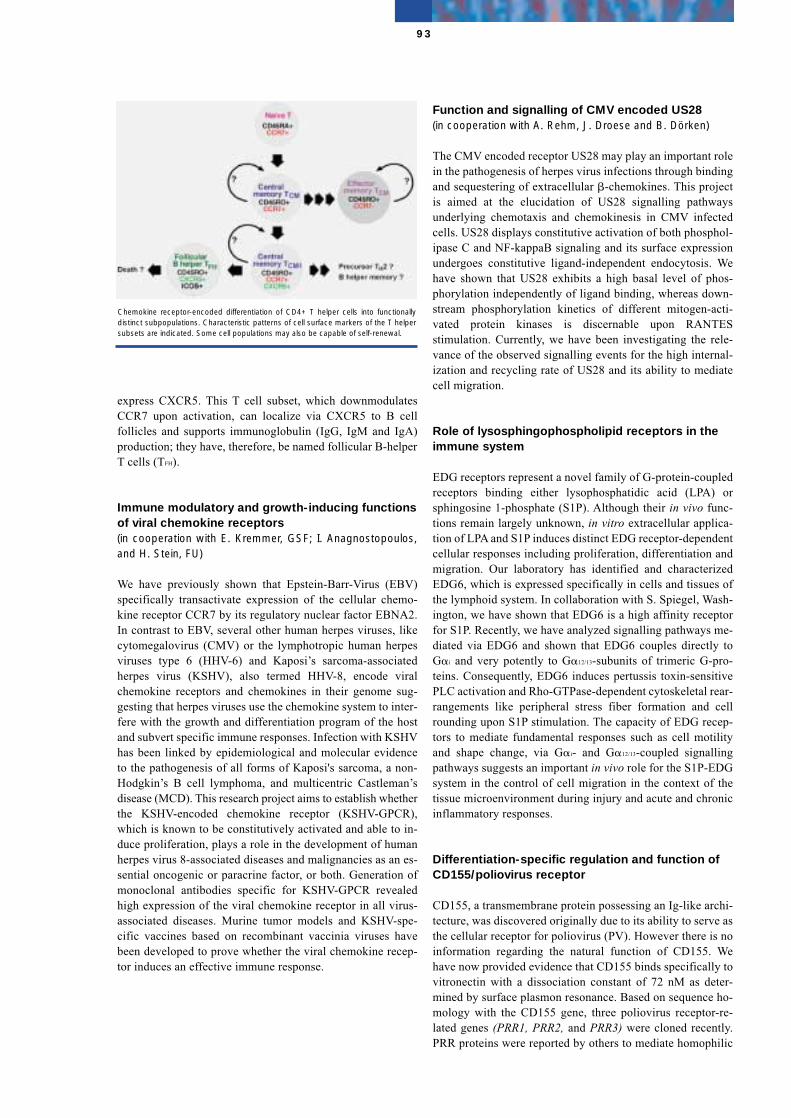
express CXCR5. This T cell subset, which downmodulatesCCR7 upon activation, can localize via CXCR5 to B cellfollicles and supports immunoglobulin (IgG, IgM and IgA)production; they have, therefore, be named follicular B-helperT cells (TFH).
Immune modulatory and growth-inducing functionsof viral chemokine receptors(in cooperation with E. Kremmer, GSF; I. Anagnostopoulos,and H. Stein, FU)
We have previously shown that Epstein-Barr-Virus (EBV)specifically transactivate expression of the cellular chemo-kine receptor CCR7 by its regulatory nuclear factor EBNA2.In contrast to EBV, several other human herpes viruses, likecytomegalovirus (CMV) or the lymphotropic human herpesviruses type 6 (HHV-6) and Kaposi’s sarcoma-associatedherpes virus (KSHV), also termed HHV-8, encode viralchemokine receptors and chemokines in their genome sug-gesting that herpes viruses use the chemokine system to inter-fere with the growth and differentiation program of the hostand subvert specific immune responses. Infection with KSHVhas been linked by epidemiological and molecular evidenceto the pathogenesis of all forms of Kaposi's sarcoma, a non-Hodgkin’s B cell lymphoma, and multicentric Castleman’sdisease (MCD). This research project aims to establish whetherthe KSHV-encoded chemokine receptor (KSHV-GPCR),which is known to be constitutively activated and able to in-duce proliferation, plays a role in the development of humanherpes virus 8-associated diseases and malignancies as an es-sential oncogenic or paracrine factor, or both. Generation ofmonoclonal antibodies specific for KSHV-GPCR revealedhigh expression of the viral chemokine receptor in all virus-associated diseases. Murine tumor models and KSHV-spe-cific vaccines based on recombinant vaccinia viruses havebeen developed to prove whether the viral chemokine recep-tor induces an effective immune response.
Function and signalling of CMV encoded US28(in cooperation with A. Rehm, J. Droese and B. Dörken)
The CMV encoded receptor US28 may play an important rolein the pathogenesis of herpes virus infections through bindingand sequestering of extracellular �-chemokines. This projectis aimed at the elucidation of US28 signalling pathwaysunderlying chemotaxis and chemokinesis in CMV infectedcells. US28 displays constitutive activation of both phosphol-ipase C and NF-kappaB signaling and its surface expressionundergoes constitutive ligand-independent endocytosis. Wehave shown that US28 exhibits a high basal level of phos-phorylation independently of ligand binding, whereas down-stream phosphorylation kinetics of different mitogen-acti-vated protein kinases is discernable upon RANTESstimulation. Currently, we have been investigating the rele-vance of the observed signalling events for the high internal-ization and recycling rate of US28 and its ability to mediatecell migration.
Role of lysosphingophospholipid receptors in theimmune system
EDG receptors represent a novel family of G-protein-coupledreceptors binding either lysophosphatidic acid (LPA) orsphingosine 1-phosphate (S1P). Although their in vivo func-tions remain largely unknown, in vitro extracellular applica-tion of LPA and S1P induces distinct EDG receptor-dependentcellular responses including proliferation, differentiation andmigration. Our laboratory has identified and characterizedEDG6, which is expressed specifically in cells and tissues ofthe lymphoid system. In collaboration with S. Spiegel, Wash-ington, we have shown that EDG6 is a high affinity receptorfor S1P. Recently, we have analyzed signalling pathways me-diated via EDG6 and shown that EDG6 couples directly toG�i and very potently to G�12/13-subunits of trimeric G-pro-teins. Consequently, EDG6 induces pertussis toxin-sensitivePLC activation and Rho-GTPase-dependent cytoskeletal rear-rangements like peripheral stress fiber formation and cellrounding upon S1P stimulation. The capacity of EDG recep-tors to mediate fundamental responses such as cell motilityand shape change, via G�i- and G�12/13-coupled signallingpathways suggests an important in vivo role for the S1P-EDGsystem in the control of cell migration in the context of thetissue microenvironment during injury and acute and chronicinflammatory responses.
Differentiation-specific regulation and function ofCD155/poliovirus receptor
CD155, a transmembrane protein possessing an Ig-like archi-tecture, was discovered originally due to its ability to serve asthe cellular receptor for poliovirus (PV). However there is noinformation regarding the natural function of CD155. Wehave now provided evidence that CD155 binds specifically tovitronectin with a dissociation constant of 72 nM as deter-mined by surface plasmon resonance. Based on sequence ho-mology with the CD155 gene, three poliovirus receptor-re-lated genes (PRR1, PRR2, and PRR3) were cloned recently.PRR proteins were reported by others to mediate homophilic
93
Chemokine receptor-encoded differentiation of CD4+ T helper cells into functionallydistinct subpopulations. Characteristic patterns of cell surface markers of the T helpersubsets are indicated. Some cell populations may also be capable of self-renewal.
cell adhesion. Neither PRR1 nor PRR2 binds poliovirus and itis assumed that their physiological functions differ from thatof CD155. Indeed, we found that mPRR2 binds to vitronectinonly weakly, but exhibits significant self-adhesion activity,whereas there is no evidence for CD155 self-adhesion. BothCD155 and vitronectin colocalize to follicular dendritic cellsand B cells inside the germinal centers of secondary lymphoidtissue (tonsils). This novel observation suggests that theCD155/vitronectin interaction might be required for the es-tablishment of a proper immune response in this particularcontext.
Cell cycle-dependent transcriptional control via E2F
Cell proliferation is controlled by a network of extracellularand intracellular signalling pathways leading either to initia-tion and maintenance, or arrest of cell cycle progression.Transitions between certain cell cycle stages are regulated atcheckpoints monitored by coordinately regulated kinase cas-cades turning genes on and off. Recent evidence suggests thattranscription factors of the E2F-family and the tumor sup-pressor protein RB do not only control genes necessary forcell cycle progression, but also induce growth arrest andapoptosis upon oncogenic and hyperproliferative signals byactivating p53, a tumor suppressor protein known to be phos-phorylated and govern checkpoint arrest in response to DNA-damaging agents. It is further supposed that phosphorylationof p53 occurs through a DNA-dependent kinase (DNA-PK)composed of a large catalytic subunit and two DNA-targetingproteins, Ku70 and Ku80. DNA-PK is also involved in DNAdouble-strand break repair and recombination of immuno-globulin genes. Based on our recent finding that E2F factorsphysically interact via a conserved domain with Ku70 and canbe phosphorylated by the DNA-PK holoenzyme, this researchproject proposes that functional interaction of E2F and DNA-PK abrogates E2F-dependent transcription, thereby congre-gating the antiproliferative and apoptotic signals induced byDNA-damaging agents.
Selected Publications
Ansel, K. M., V. N. Ngo, P. L. Hyman, S. A . Luther, R.Förster, J. D. Sedgwick, J. L. Browning, M. Lipp, and J. G.Cyster (2000) A chemokine-driven positive feedback loop or-ganizes lymphoid follicles. Nature, 406, 309-314
Breitfeld, D., L. Ohl, E. Kremmer J. Ellwart, F. Sallusto, M.Lipp, and R. Förster (2000) Follicular B helper T cells expressCXC chemokine receptor 5, localize to B cell follicles andsupport immunoglobulin production, J. Exp. Med., 192,1545-1552
Müller, G., and M. Lipp (2001) Signal transduction by thechemokine receptor CXCR5: Structural requirements for Gprotein activation analyzed by chimeric CXCR1/CXCR5molecules. Biol. Chem. 382,1387-1397.
Höpken, U. E., H.-D. Foss, D. Meyer, M. Hinz, K. Leder, H.Stein, and M. Lipp (2002) Upregulation of the chemokine re-ceptor CCR7 in classical but not in lymphocyte predominantHodgkin disease correlates with distinct dissemination ofneoplastic cells in lymphoid organs, Blood 99, 1109-1116
Van Brocklyn#, J. R., M. H. Gräler#, G. Bernhardt, M. Lipp,and S. Spiegel (2000). Sphingosine-1-phosphate is a ligandfor the G protein-coupled receptor EDG6, Blood 95, 2624-2629 (# Equal first authorship)
Lange, R., E. Wimmer, M. Lipp, and G. Bernhardt (2001) Thepoliovirus receptor CD155 displays cell to matrix interactionby binding to vitronectin. Virology 285, 218-227.
Structure of the Group
Group leaderDr. Martin Lipp
ScientistsDr. Günter BernhardtDr. Felix CifireDr. Uta HöpkenDr. Gerd Müller Dr. Christian Ried Dr. Reinhold Förster*Dr. Angelika Kusch*
Graduate and undergraduate studentsJemina BengaSven GolfierMarkus Gräler*Frank JeblonskiHeiko JohnenRudolf Gabor Kaba*Robert LangeThilo MokrosLars Ohl* Ata-Ur RasheedSteffen Posner*Philipp ReitererNanthakumar ThirunarayananFrank Wilde
Technical AssistantsDagmar BreitfeldPeter Graßhoff Dagmar Meyer
SecretariatDaniela Keyner
* Part of the period reported.
94
Initiation of DNA Replication
Manfred Gossen
The research group is interested in the mechanisms control-ling the initiation of DNA replication in multicellular eukar-yotes. Apart from bacteria, viral systems or yeast, in meta-zoans both the cis and trans acting elements contributing tothe initation at replication are poorly characterized. This is,however, a prerequisite for a detailed understanding of thoseprocesses controlling cellular proliferation. It would also al-low new insights in the way the cell safeguards one aspect ofits genomic integrity. To this end, we are investigating the ar-chitecture of chromosomal replication origins as well as ana-lyzing the binding proteins involved. This work is being con-ducted with mammalian tissue cultures as well as withDrosophila embryos or cultured cells.
Analyzing the functional architecture of a Drosophila replication origin
The chorion gene region of Drosophila melanogaster encom-passes an origin of DNA replication, which by now is prob-ably the best characterized of all metazoa. Its activity hasbeen demonstrated in specialized cells of the ovary (the folli-cle cells), where it directs the developmentally controlled am-plification of its neighbouring chromosomal regions. In re-cent years we have identified and analysed the originrecognition complex (ORC) of Drosophila. It is expected tointeract with origins in concert with other replication initia-tion factors. Recent data show this interaction also occurs inchorion origins. However, it remains unclear if this origin isactive in cells other than follicle cells and, if so, which of itssequences directs the tissue-specific amplification program.Alternatively, the chromatin structure might play a decisiverole in determining origin specificity. We plan to investigatethese questions in Drosophila embryos as well as in insect tis-sue cultures. Such studies are needed to decide if the chorionreplication origin can serve as a model system for origin ar-chitecture in metazoans.
Expression profiling of human replication genes
The proteins participating in the initiation of DNA replicationare only poorly characterized at the biochemical level, whichis largely due to the lack of a suitable in vitro assay for repli-cation. As an alternative approach to gain insight into theirfunction and regulation, we are undertaking quantitative ex-pression profiling both at the RNA as well as the protein level.This analysis is performed by comparing cycling vs. restingand/or differentiated cells. We hope to learn which of the rep-lication initiation factors are downregulated in nonproliferat-ing cells and if this is actually one of the mechanisms bywhich unscheduled replication in these cells is prevented.
Initiation proteins as diagnostic and prognosticmarkers for cancer cellsin collaboration with R. C. Bargou, K. Bommert and B.Dörken, MDC
Unlike the downregulation of at least some of the replicationinitiation genes in differentiated cells, a reactivation of thesegenes has to take place in cells which reenter the cell cycle.Thus, one has to postulate that tumor cells express the com-plete set of DNA replication initiation genes described above,We are now analyzing quantitative and qualitative parametersof this reactivation and hope to learn if the expression of thesegenes can also serve as a diagnostic and prognostic marker forvarious malignant diseases. This part of our work relies on theimmunohistochemical detection of the replication proteins.We plan to test the feasibilty of such an approach by compar-ing expression levels in biopsies from undiseased tissue withthose from tissue representing various stages of MultipleMyeloma.
Selected Publications
Chesnokov, I., Gossen, M., Remus, D., and Botchan, M.(1999). Assembly of functionally active Drosophila originrecognition complex from recombinant proteins. Genes &Development 13, 1289-96.
Parker, L., Harris, S., Gossen, M., and Botchan, M. (2000).The bovine papillomavirus E2 transactivator is stimulated bythe E1 initiator through the E2 activation domain. Virology270, 430-43.
Santarelli, L., Gobbi, G., Debs, P., Sibille, E., Blier, P., Hen,R., and Heath, M. (2001). Genetic and pharmacological dis-ruption of neurokinin 1 receptor function decreases anxiety-related behaviors and increases serotonergic function. Proc.Natl. Acad. Sci. USA 98, 1912-7.
95

Structure of the Group
Group leaderDr. Manfred Gossen
ScientistsDr. Katrin Engel
Graduate and undergraduate studentsTina BaldingerPierre DebsAnand Ranjan
Technical AssistantsDr. Maren MiethMarion Papst
96
Epithelial Differentiation, Invasion,and Metastasis
Walter Birchmeier
Our laboratory concentrates on the molecular analysis of epi-thelial morphogenesis and differentiation. In previous years,we have defined the adhesion and signaling capacities of theE-cadherin/catenin/Wnt system. Moreover, we have investi-gated the role of scatter factor/hepatocyte growth factor (SF/HGF) and its receptor, the c-met tyrosine kinase, in morpho-genesis of epithelial cells. Components of the Wnt and c-metpathways are mutated in a variety of human tumors.
Epithelial cells can loose expression of E-cadherin during tu-mor progression and this loss correlates with the appearanceof highly invasive carcinoma cells. The function of cadherinsdepends directly on cytoplasmic linkage molecules, �-cate-nin, plakoglobin, p120, which mediate interaction of cadhe-rins with the cytoskeleton. We have shown that �-catenin alsobinds to the transcription factors LEF-1/TCF and that thisinteraction translocates �-catenin to the cell nucleus and reg-ulates gene expression (Behrens et al., 1996). This provides amolecular mechanism for transmission of signals from celladhesion components and the Wnt signalling pathway to thecell nucleus.
The scatter factor/c-met system transduces various signals inepithelial cells, such as scattering, differentiation and prolife-ration. A unique activity of SF/HGF and c-met on epithelialcells in culture is the ability to induce branching or other mor-phogenic events. We have recently identified a new substrateof c-met, Gab1, which mediates the signal responsible forbranching morphogenesis (Weidner et al., 1996). Gab1 is amember of the family of membrane-bound multiadapter pro-teins which transmits signaling of tyrosine kinase receptors.
Requirement of �-catenin in anterior-posterior axisformation and hair development in miceJörg Hülsken, Markus Morkel, and Regina Vogel. In coopera-tion with Carmen Birchmeier (MDC) and Bettina Erdmann(MDC)
We recently developed �-catenin-deficient mice and observeda defect in anterior-posterior axis formation at embryonic day5.5, as visualized by the absence of the markers Hex andHesx1 and the mislocation of Cerberus-like and Lim1 expres-sion. Subsequently, no mesoderm and head structures are gen-erated. Intercellular adhesion is maintained since plakoglobinsubstitutes for �-catenin. Our data demonstrate that �-cateninfunction is essential in anterior-posterior axis formation in themouse, and experiments with chimeric embryos show thatthis function is required in the embryonic ectoderm (Huelskenet al., 2000).
To study the role of �-catenin in development of the skin andits appendages, we introduced a conditional mutation of thegene in the epidermis and hair follicles using Cre/loxP tech-nology. When �-catenin is mutated in the ectoderm duringembryogenesis, formation of the epithelial placodes that gen-erate hair follicles is blocked. We have shown that �-cateninis required genetically downstream of tabby/downless andupstream of bmp and shh for the formation of hair follicles. If�-catenin is mutated after hair follicles have formed, hair iscompletely lost after the first hair cycle, and the regressinghair follicles are transformed into epidermal cysts (see Fig-ure). Further analysis demonstrates that �-catenin is essentialfor fate decisions of stem cells in the skin: in the absence of�-catenin, stem cells do not differentiate into follicular kerat-inocytes but, instead, adopt an epidermal fate (see Figure andHuelsken et al., 2001).
A new member of the Wnt pathway, Diversin, has been dis-covered and is being studied by Thomas Schwarz-Romondand Christian Asbrand. A new binding partner of tyrosine-phosphorylated E-cadherin, Hakai, has been identified byYasuyuki Fujita. Hakai is a c-cbl-like E3 enzyme involved inturnover of E-cadherin. A gene ablation of the catenin p120has been performed by Michael Tönjes. This results in embry-onic death and defects in the axial skeleton.
Coupling of Gab1 to c-Met and downstream effectors mediate biological responsesMartin Sachs, Ute Schaeper, Dietmar Zechner, Renate Frankeand Ingrid Walther. In collaboration with Henning Brohmann,Thomas Müller and Carmen Birchmeier (MDC)
The docking protein Gab1 binds phosphorylated c-Met recep-tor tyrosine kinase directly and mediates signals of c-Met incell culture. Gab1 is phosphorylated by c-Met and by other re-ceptor and non-receptor tyrosine kinases (Schaeper et al.,2000). We have performed the functional analysis of Gab1 bytargeted mutagenesis in the mouse and compared the pheno-types of the Gab1 and c-Met mutations. Gab1 is essential forseveral steps in development: migration of myogenic precur-sor cells into the limb is impaired in Gab1 -/- embryos. As aconsequence, extensor muscle groups of the forelimbs are vir-tually absent, and the flexor muscles are shorter. Fewer hind-
97
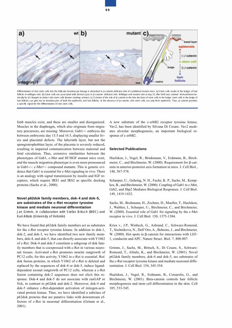
limb muscles exist, and these are smaller and disorganized.Muscles in the diaphragm, which also originate from migra-tory precursors, are missing. Moreover, Gab1-/- embryos diebetween embryonic day 13.5 and 16.5, displaying smaller liv-ers and placental defects. The labyrinth layer, but not thespongiotrophoblast layer, of the placenta is severely reduced,resulting in impaired communication between maternal andfetal circulation. Thus, extensive similarities between thephenotypes of Gab1, c-Met and SF/HGF mutant mice exist,and the muscle migration phenotype is even more pronouncedin Gab1-/-: c-Met+/- compound mutants. This is genetic evi-dence that Gab1 is essential for c-Met signaling in vivo. Thereis an analogy with signal transmission by insulin and IGF re-ceptors, which require IRS1 and IRS2 as specific dockingproteins (Sachs et al., 2000).
Novel p62dok family members, dok-4 and dok-5,are substrates of the c-Ret receptor tyrosinekinase and mediate neuronal differentiationJan Grimm, in collaboration with Stefan Britsch (MDC) andKari Alitalo (University of Helsinki)
We have found that p62dok family members act as substratesfor the c-Ret receptor tyrosine kinase. In addition to dok-1,dok-2, and dok-3, we have identified two new family mem-bers, dok-4, and dok-5, that can directly associate with Y1062of c-Ret. Dok-4 and dok-5 constitute a subgroup of dok fam-ily members that is coexpressed with c-Ret in various neuro-nal tissues. Activated c-Ret promotes neurite outgrowth ofPC12 cells; for this activity, Y1062 in c-Ret is essential. Ret/dok fusion proteins, in which Y1062 of c-Ret is deleted andreplaced by the sequences of dok-4 or dok-5, induce ligand-dependent axonal outgrowth of PC12 cells, whereas a c-Retfusion containing dok-2 sequences does not elicit this re-sponse. Dok-4 and dok-5 do not associate with rasGAP orNck, in contrast to p62dok and dok-2. Moreover, dok-4 anddok-5 enhance c-Ret-dependent activation of mitogen-acti-vated protein kinase. Thus, we have identified a subclass ofp62dok proteins that are putative links with downstream ef-fectors of c-Ret in neuronal differentiation (Grimm et al.,2001).
A new substrate of the c-erbB2 receptor tyrosine kinase,Vav2, has been identified by Silvana Di Cesare. Vav2 medi-ates alveolar morphogenesis, an important biological re-sponse of c-erbB2.
Selected Publications
Huelsken, J., Vogel, R., Brinkmann, V., Erdmann, B., Birch-meier, C., and Birchmeier, W. (2000). Requirement for �-cat-enin in anterior-posterior axis formation in mice. J. Cell Biol.,148, 567-578.
Schaeper, U., Gehring, N. H., Fuchs, K. P., Sachs, M., Kemp-kes, B., and Birchmeier, W. (2000). Coupling of Gab1 to c-Met,Grb2, and Shp2 Mediates Biological Responses. J. Cell Biol.149, 1419-1432.
Sachs, M., Brohmann, H., Zechner, D., Mueller, T., Huelsken,J., Walther, I., Schaeper, U., Birchmeier, C., and Birchmeier,W. (2000). Essential role of Gab1 for signaling by the c-Metreceptor in vivo. J. Cell Biol. 150, 1375-1384.
Kries v., J.P., Winbeck, G., Asbrand, C., Schwarz-Romond,T., Sochnikova, N., Dell’Oro, A., Behrens, J., and Birchmeier,W. (2000). Hot spots in �-catenin for interactions with LEF-1, conductin and APC. Nature Struct. Biol. 7, 800-807.
Grimm, J., Sachs, M., Britsch, S., Di Cesare, S., Schwarz-Romond, T., Alitalo, K., and Birchmeier, W. (2001). Novelp62dok family members, dok-4 and dok-5, are substrates ofthe c-Ret receptor tyrosine kinase and mediate neuronal diffe-rentiation. J. Cell Biol. 154, 345-354.
Huelsken, J., Vogel, R., Erdmann, B., Cotsarelis, G., andBirchmeier, W. (2001). Beta-catenin controls hair folliclemorphogenesis and stem cell differentiation in the skin. Cell105, 533-545.
98
Differentiation of skin stem cells into the follicular keratinocyte lineage is disturbed in �-catenin-deficient skin of conditional mutant mice. (a) Stem cells reside in the bulges of hairfollicles in wildtype skin. (b) Stem cells are associated with dermal cysts in �-catenin- deficient skin. Wildtype and mutant skin at day 32 after birth was stained immunohistoche-mically for �1-Integrin to detect skin stem cells (brown staining; arrows). (c) Scheme of the role of �-catenin in the fate decision of stem cells in the bulge: stem cells in the bulge ofhair follicles can give rise to keratinocytes of both the epidermis and hair follicles. In the absence of �-catenin, skin stem cells can only form epidermis. Thus, �-catenin providesa specific signal for the differentiation of skin stem cells.
Structure of the Group
Group leaderProf. Dr. Walter Birchmeier
ScientistsDr. Felix Brembeck*Dr. Yasuyuki FujitaDr. Jörg HülskenDr. Jens-Peter von Kries*Dr. Markus MorkelDr. Marta Rosário*Dr. Martin Sachs*Dr. Hans-Jörg Schaeffer*Dr. Ute SchaeperDr. Martin SeidenstickerDr. Dietmar Zechner
Graduate studentsChristian AsbrandJolanta Chmielowiec*Silvana Di CesareJan Grimm*Boris JerchowGunnar SchützThomas Schwarz-RomondNatalia SochnikovaMichael Tönjes*
Technical assistantsAndrea Dell’OroKatharina FellerRenate FrankeRegina VogelIngrid Walther
SecretariatIrmgard WiznerowiczGerhild Richter
* part of the time reported
99
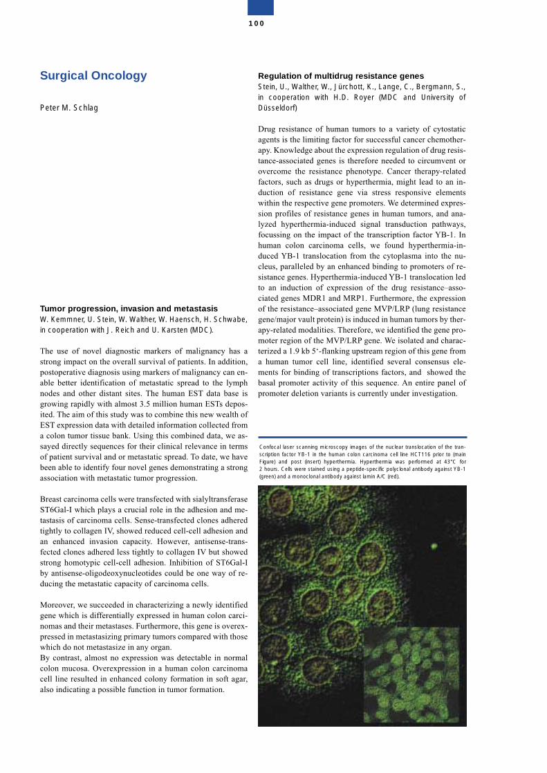
Surgical Oncology
Peter M. Schlag
Tumor progression, invasion and metastasisW. Kemmner, U. Stein, W. Walther, W. Haensch, H. Schwabe,in cooperation with J. Reich and U. Karsten (MDC).
The use of novel diagnostic markers of malignancy has astrong impact on the overall survival of patients. In addition,postoperative diagnosis using markers of malignancy can en-able better identification of metastatic spread to the lymphnodes and other distant sites. The human EST data base isgrowing rapidly with almost 3.5 million human ESTs depos-ited. The aim of this study was to combine this new wealth ofEST expression data with detailed information collected froma colon tumor tissue bank. Using this combined data, we as-sayed directly sequences for their clinical relevance in termsof patient survival and or metastatic spread. To date, we havebeen able to identify four novel genes demonstrating a strongassociation with metastatic tumor progression.
Breast carcinoma cells were transfected with sialyltransferaseST6Gal-I which plays a crucial role in the adhesion and me-tastasis of carcinoma cells. Sense-transfected clones adheredtightly to collagen IV, showed reduced cell-cell adhesion andan enhanced invasion capacity. However, antisense-trans-fected clones adhered less tightly to collagen IV but showedstrong homotypic cell-cell adhesion. Inhibition of ST6Gal-Iby antisense-oligodeoxynucleotides could be one way of re-ducing the metastatic capacity of carcinoma cells.
Moreover, we succeeded in characterizing a newly identifiedgene which is differentially expressed in human colon carci-nomas and their metastases. Furthermore, this gene is overex-pressed in metastasizing primary tumors compared with thosewhich do not metastasize in any organ.By contrast, almost no expression was detectable in normalcolon mucosa. Overexpression in a human colon carcinomacell line resulted in enhanced colony formation in soft agar,also indicating a possible function in tumor formation.
Regulation of multidrug resistance genesStein, U., Walther, W., Jürchott, K., Lange, C., Bergmann, S.,in cooperation with H.D. Royer (MDC and University ofDüsseldorf)
Drug resistance of human tumors to a variety of cytostaticagents is the limiting factor for successful cancer chemother-apy. Knowledge about the expression regulation of drug resis-tance-associated genes is therefore needed to circumvent orovercome the resistance phenotype. Cancer therapy-relatedfactors, such as drugs or hyperthermia, might lead to an in-duction of resistance gene via stress responsive elementswithin the respective gene promoters. We determined expres-sion profiles of resistance genes in human tumors, and ana-lyzed hyperthermia-induced signal transduction pathways,focussing on the impact of the transcription factor YB-1. Inhuman colon carcinoma cells, we found hyperthermia-in-duced YB-1 translocation from the cytoplasma into the nu-cleus, paralleled by an enhanced binding to promoters of re-sistance genes. Hyperthermia-induced YB-1 translocation ledto an induction of expression of the drug resistance–asso-ciated genes MDR1 and MRP1. Furthermore, the expressionof the resistance–associated gene MVP/LRP (lung resistancegene/major vault protein) is induced in human tumors by ther-apy-related modalities. Therefore, we identified the gene pro-moter region of the MVP/LRP gene. We isolated and charac-terized a 1.9 kb 5‘-flanking upstream region of this gene froma human tumor cell line, identified several consensus ele-ments for binding of transcriptions factors, and showed thebasal promoter activity of this sequence. An entire panel ofpromoter deletion variants is currently under investigation.
100
Confocal laser scanning microscopy images of the nuclear translocation of the tran-scription factor YB-1 in the human colon carcinoma cell line HCT116 prior to (mainFigure) and post (insert) hyperthermia. Hyperthermia was performed at 43°C for2 hours. Cells were stained using a peptide-specific polyclonal antibody against YB-1(green) and a monoclonal antibody against lamin A/C (red).
Jet-Injection for nonviral gene therapy of cancerW. Walther, U. Stein, in cooperation with I. Fichtner (MDC)
The novel technology of jet-injection can be applied to nonvi-ral in vivo gene transfer. Jet-injection is efficient in genetransfer of LacZ and GFP gene constructs into differentmouse tumor models and xenotransplanted human tumormodels. Qualitative and quantitative expression analysis ofjet-injected tumor tissues revealed efficient expression ofthese genes associated with a broad distribution of the trans-genes in the targeted tissues. The experiments demonstratedthat two or more gene constructs can be simultaneously trans-duced into one tissue. This nonviral gene transfer technologywill be used for the transduction of “therapy-inducible” vec-tor constructs. These vectors utilize conditionally active pro-moters, which can be induced by cytostatic drugs or by hyper-thermia – therapeutic modalities that are used for thetreatment of cancer. We have used the mdr1 promoter for suchinducible expression and showed the drug- and heat-indu-cibility of this promoter. Further studies will show, whethernonviral gene transfer of “therapy-inducible” vectors are ef-fective.
Pathobiology of colorectal cancers with microsatellite instabilityK. Kölble, B. Barthel in cooperation with S. Scherneck (MDC)
In contrast to the majority of human colorectal cancers (CRC)which mostly derive from polyploid adenomas, approxi-mately 10% of CRC characteristically carry somatic muta-tions in simple repetitive sequences. This microsatellite in-stability (MSI) is indicative of germline and/or somaticalterations in various DNA mismatch repair genes which, inhereditary non-polyposis colorectal cancer (HNPCC) lead tocolorectal carcinogenesis in patients under the age of 50. Inorder to elucidate the pathogenesis of this cancer susceptibil-ity syndrome and improve its clinical management, solid tu-mors of potential HNPCC cases have been registered and in-vestigated by a combination of clinical and pedigree studies,histopathology, immunohistology, MSI and sequence analy-sis. Using this integrated approach, it was shown that immu-nohistology alone allows one to identify patients with germ-line mutations in the DNA mismatch repair gene MSH2 andmany cases with germline mutations in the DNA mismatchrepair gene MLH1. However, as some missense and truncat-ing mutations are missed, immunohistology cannot replacetesting for MSI to predict HNPCC carrier status or identifyMSI-positive sporadic colorectal cancer. In addition, muta-tional targets specific for high grade MSI have been found inthe beta-catenin (CTNNB1) and AXIN2 genes, suggestingthat somatic genomic alterations in WNT-pathway genes arecommon to both subsets of CRC.
Mechanisms of endogenous porphyrin accumula-tion in colorectal carcinomas and metastasesT. Moesta, T. Handke, W. Kemmner, in cooperation with H.Rinneberg (Physikalisch-Technische Bundesanstalt Berlin)
Colorectal cancers and their metastases endogenously accu-mulate Protoporphyrin IX (PpIX), the immediate heme-pre-cursor substance and a natural fluorophore. We have investi-gated tumor-specific alterations in the heme synthesispathway on mRNA- and protein levels to elucidate the mech-anism of PpIX accumulation in tissue samples of gastrointes-tinal cancers and corresponding normal tissues. Currently,surgical specimens are spectroscopically characterized by atime-delayed fluorescence spectroscopy system developed byour collaborator. To investigate subcellular localization ofPpIX, a time-delayed laser-induced fluorescence microscopyprocedure has been developed.
Selected Publications
Moesta, K. T., Ebert, B., Nowack, C., Nolte, D., Handke, T.,Haensch, W. E., Pandey, R., Dougherty, T.J., Rinneberg, H.,and Schlag, P. M. (2001). Protoporphyrin IX occurs naturallyin colorectal cancers and their metastases. Cancer Res. 61,991-999.
Savelyeva, L., Claas, A., Matzner, I., Schlag, P., Hofmann,W., Scherneck, S., Weber, B., and Schwab, M. (2001). Consti-tutional genomic instability with inversions, duplications, andamplifications in 9p23-24 in BRCA2 mutation carriers. Can-cer Res. 61, 5179-85.
Schneider, F., Kemmner, W., Haensch, W., Franke, G., Grets-chel, S., Karsten, U., and Schlag, P.M. (2001). Overexpres-sion of sialyltransferase ST6GalNAc-II is related to poor pa-tient survival in human colorectal carcinomas. Cancer Res.61, 4605-4611.
Schumacher, K., Haensch, W., Roefzaad, C., Schlag, P.M.(2001). Prognostic significance of activated CD8(+) T cell in-filtrations within esophageal carcinomas. Cancer Res. 61,3932-3936.
Stein, U., Jürchott, K., Walther, W., Bergmann, S., Schlag,P.M., and Royer, H.-D. (2001). Hyperthermia-induced nu-clear translocation of transcription factor YB-1 leads to en-hanced expression of multidrug resistance-related ABC trans-porters. J Biol Chem. 276, 28562-28569.
Walther, W., Stein, U., Fichtner, I., Malcherek, L., Lemm, M.,and Schlag, P.M. (2001). Non-viral in vivo gene delivery intotumors using a novel low volume jet-injection technology.Gene Ther. 8, 173-180.
101

Structure of the group
Group LeaderProf. Dr. Peter M. Schlag
ScientistsDr. Georgi GraschewDr. Wolfgang KemmnerDr. Konrad KölbleDr. Barbara LustigDr. Karin SchumacherDr. Ulrike SteinDr. Wolfgang Walther
Graduate and undergraduate studentsHolger SchwabeKarsten JürchottStephan BergmannFranziska ArltAnke KopacekBeatrix BarthelHeike FischerMarcel SimonDienje SchindlerVahid HosseiniTobias PetrettiAlexandra ZuskeSabine Haase
Technical assistantsLieselotte MalcherekLisa BauerSabine GrigullGudrun KochClaudia RöefzaadStefanie Balz
102
Molecular Muscle Physiology
A European Marie Curie Training Site
Ingo L. Morano
Contractility of cardiac and smooth muscle is regulated bycalcium ions (Ca2+), which enter the cells through voltage-gated L-type Ca2+ channels and subsequently induce the re-lease of high amounts of Ca2+ from the sarcoplasmic reticuluminto the myoplasm through Calcium Release Channels (Rya-nodin Receptors). Ca2+ activate intracellular signalling path-ways and contraction. In cardiomyocytes, Ca2+ activate thecontractile apparatus by binding to troponin C, which turnsthe thin filament from an “off” to an “on” state, allowing themolecular motor myosin to interact with the thin filament toproduce force and shortening. In smooth muscle cells, Ca2+
form a complex with calmodulin which activate the myosinlight chain kinase, an enzyme which phosphorylates a 20kDaregulatory light chain of myosin, thus allowing the smoothmuscle molecular motors to generate contraction on interact-ing with the thin filaments. Because of their key-roles in mus-cle, we are studying the expression regulation, post-transla-tional modifications, and functional roles of the subunits ofL-type Ca2+ channel, Ry-anodine Receptor, Proteins of theCa2+ signalling pathways, and Type II myosin in cardiac andsmooth muscle. Any change in these key proteins, by muta-tion, differential gene expression, alternative splicing of thetranscripts, or post-translational modification modulates car-diac and smooth muscle function. Understanding muscle con-traction regulation at the molecular and functional level pro-vides an opportunity to develop new therapies for thetreatment of cardiac and smooth muscle dysfunction.
Understanding the molecular motor
Essential myosin light chain isoforms regulate human heartcontractilityType II myosin isoenzymes are hexamers of about 500 kDacomposed of two heavy chains (MyHC) and 4 light chains(MLC), designated essential and regulatory MLC. Two genescoding for MyHC are expressed in the heart, �-MyHC and �-MyHC, located in tandem on chromosome 14. Atrium- andventricle-specific essential (ALC-1 and VLC-1, respectively)and regulatory (ALC-2 and VLC-2, respectively) MLC exist.
Cardiomyocytes of hypertrophied ventricles of patients withcongenital heart diseases and hypertrophic cardiomyopathyreexpressed ALC-1, while MyHC isoenzymes did not change.This is in sharp contrast to the hypertrophied rodent ventriclewhich changes MyHC isoforms rather than MLC expression.Expression of the cardiac-specific basic-helix-loop-helixtranscription factors eHAND and dHAND which bind toE-box elements in the ALC-1 promoter was increased in thehypertro-phied human ventricle. Ventricular cross-bridges as-sociated with ALC-1 revealed a higher shortening velocityand rate of force development than normal cross-bridgeswithout ALC-1. Maximal isometric force production percross-sectional area as well as Ca2+ sensitivity of the force-Ca2+ ratio were enhanced. The failing ventricles of patientswith dilated cardiomypathy, however, hardly expressed ALC-1. Therefore, an adenoviral vector containing the humanALC-1 (hALC-1) expression cassete has been developed forthe upregulation of the hALC-1 in the cardiomyocytes of thefailing human heart as a novel gene therapeutic approach.
Regulation of smooth muscle contractility by recruitment ofnon-muscle myosin in an SM-MyHC knock-out modelProlonged smooth muscle activation produces an initialphasic contraction with high sometric force development andmaximal shortening velocity (Vmax), which is followed by atonic contraction with low force generation and Vmax. Weeliminated the expression of smooth muscle MyHC (SM-MyHC) by gene targeting technology. Smooth muscle fromknock-out neonatal mice did not exhibit phasic contractionwhile tonic contraction remained normal. In both wild-typeand knock-out animals there was a similar intracellular Ca2+
transient, i.e. an initial transient Ca2+ peak which subsequentlydeclined to almost resting levels while tonic force develop-ment remained. Thus, the phasic contraction is generated bySM-MyHC recruitment while the sustained tonic contractionstate can produce NM-MyHC activation. Non-muscle-MyHC-dependent sustained force generation was sufficientfor normal fetal development. However, the strong phasiccontraction caused by SM-MyHC, becomes indispensable forsurvival and normal growth soon after birth. In addition, bothcontractile systems in smooth muscle are associated with dif-ferent second messenger pathways: both the SM-MyHC andNM-MyHC systems seem to be involved in electromechani-cal and pharmacomechanical coupling, respectively.
Understanding calcium-handling proteins
Ca2+ channels are multisubunit complexes composed of thepore-forming �1 subunit along with regulatory � and �2/subunits. Coordinated upregulation of Ca2+ channel subunitexpression is observed in patients with hypertrophic, but notdilated, cardiomyopathy. Furthermore, we have identified fe-tal isoforms of both �1 - and �-subunits. Ahnak, a 700-kDaprotein, was detected in mammalian cardiomyocytes. Itundergoes substantial protein kinase A phosphorylation and ispreferentially localized in the plasma membrane of cardiom-yocytes. Most of the C-terminus of ahnak strongly binds tothe �2-subunit of the Ca2+ channels as well as to F-actin. Ah-nak, therefore, may provide a structural basis for the subsar-colemmal cytoarchitecture and signal transduction in the car-
103
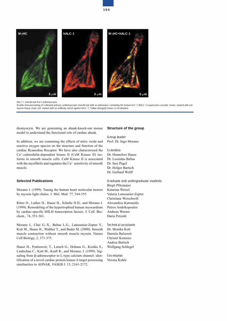
diomyocyte. We are generating an ahnak-knock-out mousemodel to understand the functional role of cardiac ahnak.
In addition, we are examining the effects of nitric oxide andreactive oxygen species on the structure and function of thecardiac Ryanodine Receptor. We have also characterized theCa2+-calmodulin-dependent kinase II (CaM Kinase II) iso-forms in smooth muscle cells. CaM Kinase II is associatedwith the myofibrils and regulates the Ca2+ sensitivity of smoothmuscle.
Selected Publications
Morano I. (1999). Tuning the human heart molecular motorsby myosin light chains. J. Mol. Med. 77, 544-555.
Ritter O., Luther H., Haase H., Schulte H.D., and Morano I.(1999). Remodeling of the hypertrophied human myocardiumby cardiac-specific bHLH transcription factors. J. Cell. Bio-chem., 74, 551-561.
Morano I., Chai G.-X., Baltas L.G., Lamounier-Zepter V.,Kott M., Haase H., Walther T., and Bader M. (2000). Smoothmuscle contraction without smooth muscle myosin. NatureCell Biology, 2, 371-375.
Haase H., Podzuweit, T., Lutsch G., Hohaus G., Kostka S.,Lindschau C., Kott M., Kraft R., and Morano, I. (1999). Sig-naling from �-adrenoceptor to L-type calcium channel: iden-tification of a novel cardiac protein kinase A target possessingsimiliarities to AHNAK. FASEB J. 13, 2161-2172.
Structure of the group
Group leaderProf. Dr. Ingo Morano
ScientistsDr. Hannelore HaaseDr. Leonidas BaltasDr. Ines PagelDr. Holger BartschDr. Gerhard Wolff
Graduate and undergraduate studentsBirgit PfitzmaierKatarina WetzelValeria Lamounier-ZepterChristiane WoischwillAlexandros KatranidisPetros AndrikopoulosAndreas WernerDaria Petzold
Technical assistantsDr. Monika KottDaniela BalzereitChristel KemsiesAndrea BartschWolfgang Schlegel
SecretariatVerona Kuhle
104
hALC-1 transfected Rat CardiomyocytesDouble-immunostaining of cultivated primary cardiomyocytes transfected with an adenovirus containing the human ALC-1 (hALC-1) expression cassette. Green. stained with ant-myosin heavy chain; red: stained with an antibody raised against hALC-1; Yellow (merged) shows co-localisation.
M�HC hALC-1 M�HC+hALC-1
5 m 5 m 5 m
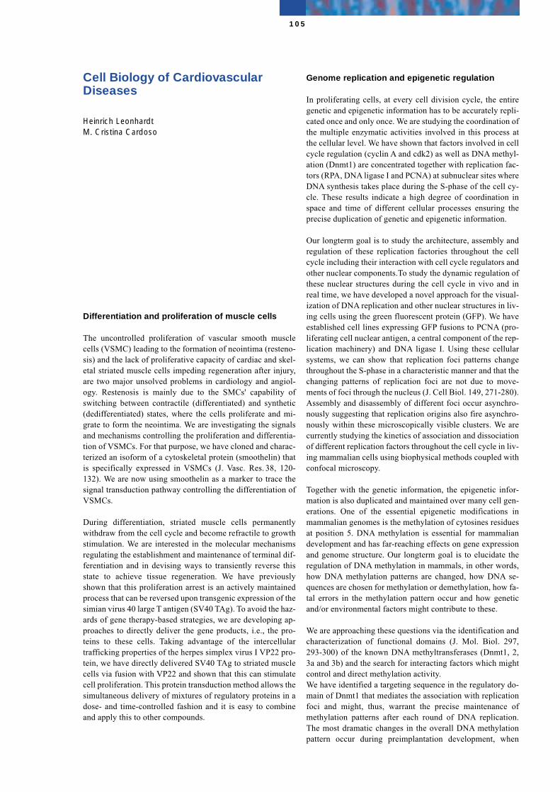
Cell Biology of CardiovascularDiseases
Heinrich LeonhardtM. Cristina Cardoso
Differentiation and proliferation of muscle cells
The uncontrolled proliferation of vascular smooth musclecells (VSMC) leading to the formation of neointima (resteno-sis) and the lack of proliferative capacity of cardiac and skel-etal striated muscle cells impeding regeneration after injury,are two major unsolved problems in cardiology and angiol-ogy. Restenosis is mainly due to the SMCs' capability ofswitching between contractile (differentiated) and synthetic(dedifferentiated) states, where the cells proliferate and mi-grate to form the neointima. We are investigating the signalsand mechanisms controlling the proliferation and differentia-tion of VSMCs. For that purpose, we have cloned and charac-terized an isoform of a cytoskeletal protein (smoothelin) thatis specifically expressed in VSMCs (J. Vasc. Res. 38, 120-132). We are now using smoothelin as a marker to trace thesignal transduction pathway controlling the differentiation ofVSMCs.
During differentiation, striated muscle cells permanentlywithdraw from the cell cycle and become refractile to growthstimulation. We are interested in the molecular mechanismsregulating the establishment and maintenance of terminal dif-ferentiation and in devising ways to transiently reverse thisstate to achieve tissue regeneration. We have previouslyshown that this proliferation arrest is an actively maintainedprocess that can be reversed upon transgenic expression of thesimian virus 40 large T antigen (SV40 TAg). To avoid the haz-ards of gene therapy-based strategies, we are developing ap-proaches to directly deliver the gene products, i.e., the pro-teins to these cells. Taking advantage of the intercellulartrafficking properties of the herpes simplex virus I VP22 pro-tein, we have directly delivered SV40 TAg to striated musclecells via fusion with VP22 and shown that this can stimulatecell proliferation. This protein transduction method allows thesimultaneous delivery of mixtures of regulatory proteins in adose- and time-controlled fashion and it is easy to combineand apply this to other compounds.
Genome replication and epigenetic regulation
In proliferating cells, at every cell division cycle, the entiregenetic and epigenetic information has to be accurately repli-cated once and only once. We are studying the coordination ofthe multiple enzymatic activities involved in this process atthe cellular level. We have shown that factors involved in cellcycle regulation (cyclin A and cdk2) as well as DNA methyl-ation (Dnmt1) are concentrated together with replication fac-tors (RPA, DNA ligase I and PCNA) at subnuclear sites whereDNA synthesis takes place during the S-phase of the cell cy-cle. These results indicate a high degree of coordination inspace and time of different cellular processes ensuring theprecise duplication of genetic and epigenetic information.
Our longterm goal is to study the architecture, assembly andregulation of these replication factories throughout the cellcycle including their interaction with cell cycle regulators andother nuclear components.To study the dynamic regulation ofthese nuclear structures during the cell cycle in vivo and inreal time, we have developed a novel approach for the visual-ization of DNA replication and other nuclear structures in liv-ing cells using the green fluorescent protein (GFP). We haveestablished cell lines expressing GFP fusions to PCNA (pro-liferating cell nuclear antigen, a central component of the rep-lication machinery) and DNA ligase I. Using these cellularsystems, we can show that replication foci patterns changethroughout the S-phase in a characteristic manner and that thechanging patterns of replication foci are not due to move-ments of foci through the nucleus (J. Cell Biol. 149, 271-280).Assembly and disassembly of different foci occur asynchro-nously suggesting that replication origins also fire asynchro-nously within these microscopically visible clusters. We arecurrently studying the kinetics of association and dissociationof different replication factors throughout the cell cycle in liv-ing mammalian cells using biophysical methods coupled withconfocal microscopy.
Together with the genetic information, the epigenetic infor-mation is also duplicated and maintained over many cell gen-erations. One of the essential epigenetic modifications inmammalian genomes is the methylation of cytosines residuesat position 5. DNA methylation is essential for mammaliandevelopment and has far-reaching effects on gene expressionand genome structure. Our longterm goal is to elucidate theregulation of DNA methylation in mammals, in other words,how DNA methylation patterns are changed, how DNA se-quences are chosen for methylation or demethylation, how fa-tal errors in the methylation pattern occur and how geneticand/or environmental factors might contribute to these.
We are approaching these questions via the identification andcharacterization of functional domains (J. Mol. Biol. 297,293-300) of the known DNA methyltransferases (Dnmt1, 2,3a and 3b) and the search for interacting factors which mightcontrol and direct methylation activity.We have identified a targeting sequence in the regulatory do-main of Dnmt1 that mediates the association with replicationfoci and might, thus, warrant the precise maintenance ofmethylation patterns after each round of DNA replication.The most dramatic changes in the overall DNA methylationpattern occur during preimplantation development, when
105
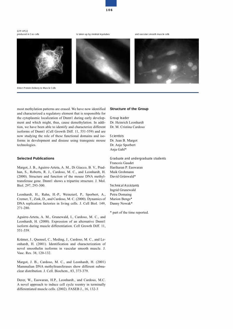
most methylation patterns are erased. We have now identifiedand characterized a regulatory element that is responsible forthe cytoplasmic localization of Dnmt1 during early develop-ment and which might, thus, cause demethylation. In addi-tion, we have been able to identify and characterize differentisoforms of Dnmt1 (Cell Growth Diff. 11, 551-559) and arenow studying the role of these functional domains and iso-forms in development and disease using transgenic mousetechnologies.
Selected Publications
Margot, J. B., Aguirre-Arteta, A. M., Di Giacco, B. V., Prad-han, S., Roberts, R. J., Cardoso, M. C., and Leonhardt, H.(2000). Structure and function of the mouse DNA methyl-transferase gene. Dnmt1 shows a tripartite structure. J. Mol.Biol. 297, 293-300.
Leonhardt, H., Rahn, H.-P., Weinzierl, P., Sporbert, A.,Cremer, T., Zink, D., and Cardoso, M. C. (2000). Dynamics ofDNA replication factories in living cells. J. Cell Biol. 149,271-280.
Aguirre-Arteta, A. M., Grunewald, I., Cardoso, M. C., andLeonhardt, H. (2000). Expression of an alternative Dnmt1isoform during muscle differentiation. Cell Growth Diff. 11,551-559.
Krämer, J., Quensel, C., Meding, J., Cardoso, M. C., and Le-onhardt, H. (2001). Identification and characterization ofnovel smoothelin isoforms in vascular smooth muscle. J.Vasc. Res. 38, 120-132.
Margot, J. B., Cardoso, M. C., and Leonhardt, H. (2001)Mammalian DNA methyltransferases show different subnu-clear distribution. J. Cell. Biochem., 83, 373-379.
Derer, W., Easwaran, H.P., Leonhardt., and Cardoso, M.C.A novel approach to induce cell cycle reentry in terminallydifferentiated muscle cells. (2002). FASEB J., 16, 132-3
Structure of the Group
Group leaderDr. Heinrich LeonhardtDr. M. Cristina Cardoso
ScientistsDr. Jean B. MargotDr. Anje SporbertAnja Gahl*
Graduate and undergraduate studentsFrancois GaudetHariharan P. EaswaranMaik GrohmannDavid Grünwald*
Technical AssistantsIngrid GrunewaldPetra DomaingMarion Bengs* Danny Nowak*
* part of the time reported.
106
Direct Protein Delivery to Muscle Cells
GFP-VP22produced in Cos cells is taken up by skeletal myotubes and vascular smooth muscle cells
Intracellular Proteolysis
Thomas Sommer
The research of our group focusses on two highly conservedand essential cell biological processes: ubiquitin-proteasome-dependent protein degradation and intracellular protein trans-port. A link between both processes is provided by the obser-vation that the secretory pathway of eukaryotic cells harboursan elaborate protein quality control system, which preventsdeployment of the secretory pathway of misfolded or unas-sembled proteins. Endoplasmic Reticulum associated proteindegradation (ERAD) is an important component of this qual-ity assurance system and directs misfolded proteins for de-struction by the cytoplasmic ubiquitin-proteasome pathway.Compartment-specific functions of the ubiqitin-proteasomepathway also confer specificity on the turnover of nuclearregulators. We have been able to demonstrate that the cellularlocalization of a protein determines its half-life, and that nu-cleus-restricted degradation pathways exist. Thus, selectivenuclear transport provides a tool to regulate both the activityand turnover rates of nuclear regulators.
To study these processes, we are using the yeast Saccharo-myces cerevisiae as an experimental system. Yeast is one ofthe favorite model organisms in cell biology because it canconveniently be used in both genetic and biochemical experi-ments.
Protein degradation at the Endoplasmic Reticulum
The basic principles of Endoplasmic Reticulum associatedprotein degradation have been investigated by our group. It isa process common to most eukaryotic organisms and of greatmedical importance. The genesis of some serious human dis-eases is closely linked to the ERAD system, the most promi-nent example being cystic fibrosis. Moreover, some humanviruses use ERAD or related systems to destroy specific hostproteins and manifest themselves in the infected cell.
Protein quality control requires both chaperones of the ER-lu-men as well as the proteolytic ERAD pathway. The content ofmolecular chaperones in the ER lumen is controlled by a sig-
naling cascade known as the unfolded protein response(UPR). We were able to obtain evidence that a coordinatedregulation of UPR and ERAD system is pivotal to cell viabil-ity. This demonstrates that protein quality control in the secre-tory pathway is an essential cell biological process.
Based on our results we can distinguish four steps in ERAD:firstly, misfolded proteins are detected in the ER-lumen; sec-ondly, the proteolytic substrates are transported back into thecytosol (dislocation). This requires an aqueous channelformed by the Sec61p-complex. Thirdly, dislocated substratesare marked with the polypeptide ubiquitin by the ubiquitin-conjugating enzymes Ubc1, Ubc6 and Cue1/Ubc7 and theubiquitin ligase Hrd1/Hrd3. Fourthly, the cytosolic 26S-pro-teasome complex digests the misfolded proteins.
The major question is if and how transport and proteolysis arelinked. We have been able to demonstrate that high-molecularweight ubiquitination of the transport substrate is a prerequi-site for its dislocation. What is the function of polyubiquitina-tion in substrate export? Our results show that it does not re-cruit the proteasome which, in principle, could provide thedriving force for dislocation, since it also contains AAA-AT-Pases. So, either long polyubiquitin chains are sufficient toprevent movement back into the ER lumen, or alternatively,reverse movement may be prevented by other factors that se-lectively bind such chains. We favour the latter hypothesis,since we demonstrated a function of the AAA-ATPaseCdc48p in ERAD. Our data indicate that the Cdc48-complex,consisting of Cdc48, Ufd1 and Npl4, is not required for ubi-quitination of substrates and does not act at the earliest stageof the pathway, but functions prior to the proteasome. It hasbeen shown that the Cdc48-complex undergoes major structu-ral changes following ATP-binding, which could provide amechanical force. Moreover, it has been demonstrated thatCdc48p binds polyubiquitinated substrates. Thus, we postu-late that both high molecular weight ubiquitination and theCdc48 complex contribute to the driving force for dislocation.
The data above provide an important framework to clarify thecomposition of the transporter and its interplay with the ubi-quitin ligation machinery.
Degradation of nuclear proteins
Recently, we could demonstrate that a nuclear substrate, theyeast transcriptional repressor Mat�2, is also degraded via lo-calized functions of the ubiquitin-proteasome system. In addi-tion to components of the ubiquitin system, Mat�2 turnoverdepends on signals within the protein itself. One of them is thedegradation signal DEG1. We were able to distinguish twoDEG1 dependent pathways: A rapid and nucleus specific onefrom a slow one that takes place at the cytosolic surface of theER-membrane. Thus, a degradation signal like DEG1 is onlyfunctional in conjunction with its respective cellular localiza-tion signal, which in the case of Mat�2p is the nuclear local-ization sequence. In cell biological terms this represents amechanism to regulate a proteins half-life. By transporting aprotein into a different cellular compartment, the turnovermight be up- or down regulated, because the ubiquitinationcascades are restricted to certain areas within an eukaryotic
107
cell. Especially for regulatory factors of the nucleus, thismechanism has been shown to be an important tool for regula-tion.
Selected Publications
Friedlander, R., Jarosch, E., Urban, J., Volkwein, C., andSommer, T. (2000) A regulatory link between ER-associatedprotein degradation and the unfolded protein response. NatureCell Biology 2, 379-384.
Lenk, U., and Sommer, T. (2000) Ubiquitin-mediated proteol-ysis of a short-lived regulatory protein depends on its cellularlocalization. J. Biol. Chem.275, 39403-39410.
Walter, J., Urban, J., and Sommer, T. (2001) Sec61p indepen-dent degradation of the tail-anchored ER-membrane proteinUbc6p. EMBO J. 20, 3124-3131
Jarosch, E., Taxis, C., Volkwein, C., Bordallo, J., Finley, D.,Wolf, D. H., and Sommer, T. (2002). Protein Dislocation fromthe ER requires Polyubiquitination and the AAA-ATPaseCDC 48. Nature Cell Biol, 4, 134-139
Structure of the Group
Group leaderDr. Thomas Sommer
ScientistsDr. Ernst JaroschDr. Uwe Lenk
Graduate and undergraduate studentsRuth FriedlanderBirgit MeußerJörg UrbanJan Walter
Technical AssistantsCorinna VolkweinAngelika Wittstruck
SecretariatSylvia Klahn
108
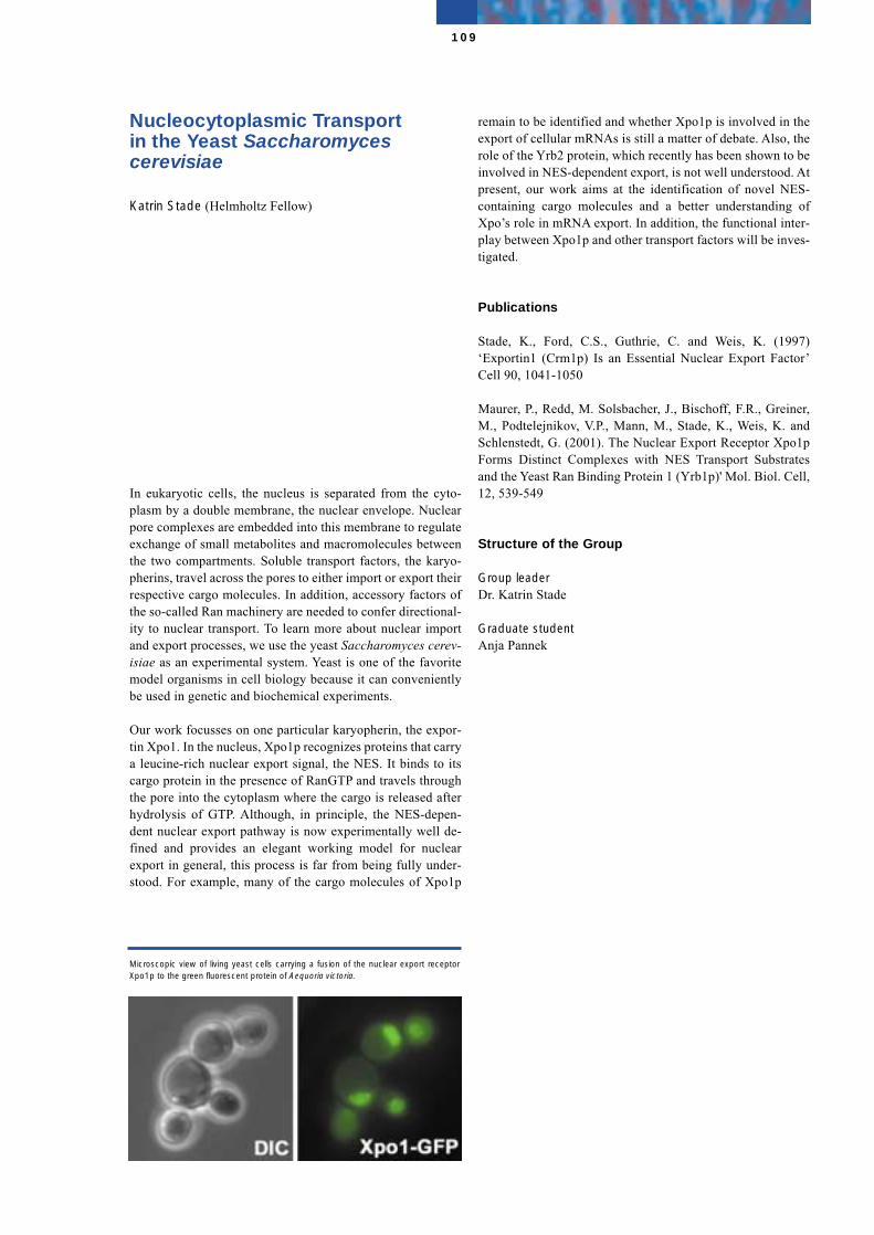
Nucleocytoplasmic Transport in the Yeast Saccharomycescerevisiae
Katrin Stade (Helmholtz Fellow)
In eukaryotic cells, the nucleus is separated from the cyto-plasm by a double membrane, the nuclear envelope. Nuclearpore complexes are embedded into this membrane to regulateexchange of small metabolites and macromolecules betweenthe two compartments. Soluble transport factors, the karyo-pherins, travel across the pores to either import or export theirrespective cargo molecules. In addition, accessory factors ofthe so-called Ran machinery are needed to confer directional-ity to nuclear transport. To learn more about nuclear importand export processes, we use the yeast Saccharomyces cerev-isiae as an experimental system. Yeast is one of the favoritemodel organisms in cell biology because it can convenientlybe used in genetic and biochemical experiments.
Our work focusses on one particular karyopherin, the expor-tin Xpo1. In the nucleus, Xpo1p recognizes proteins that carrya leucine-rich nuclear export signal, the NES. It binds to itscargo protein in the presence of RanGTP and travels throughthe pore into the cytoplasm where the cargo is released afterhydrolysis of GTP. Although, in principle, the NES-depen-dent nuclear export pathway is now experimentally well de-fined and provides an elegant working model for nuclearexport in general, this process is far from being fully under-stood. For example, many of the cargo molecules of Xpo1p
remain to be identified and whether Xpo1p is involved in theexport of cellular mRNAs is still a matter of debate. Also, therole of the Yrb2 protein, which recently has been shown to beinvolved in NES-dependent export, is not well understood. Atpresent, our work aims at the identification of novel NES-containing cargo molecules and a better understanding ofXpo’s role in mRNA export. In addition, the functional inter-play between Xpo1p and other transport factors will be inves-tigated.
Publications
Stade, K., Ford, C.S., Guthrie, C. and Weis, K. (1997)‘Exportin1 (Crm1p) Is an Essential Nuclear Export Factor’Cell 90, 1041-1050
Maurer, P., Redd, M. Solsbacher, J., Bischoff, F.R., Greiner,M., Podtelejnikov, V.P., Mann, M., Stade, K., Weis, K. andSchlenstedt, G. (2001). The Nuclear Export Receptor Xpo1pForms Distinct Complexes with NES Transport Substratesand the Yeast Ran Binding Protein 1 (Yrb1p)' Mol. Biol. Cell,12, 539-549
Structure of the Group
Group leaderDr. Katrin Stade
Graduate studentAnja Pannek
109
Microscopic view of living yeast cells carrying a fusion of the nuclear export receptorXpo1p to the green fluorescent protein of Aequoria victoria.
Cytochromes P450 and Endoplasmic Reticulum
Wolf-Hagen Schunck
This group is interested in the role of arachidonic acid (AA)metabolizing P450 enzymes in the regulation of vasculartone, renal function, and the control of inflammation. In col-laboration with other groups at the MDC and the Franz Vol-hard Clinic, we have examined pathophysiological changes inrenal P450-dependent AA metabolism using mice and ratmodels of hypertension and end-organ damage. Moreover, wehave analyzed P450-dependent epoxygenation of eicosapen-taenoic acid (EPA) as a source of novel vasoactive metab-olites.
P450-dependent arachidonic acid metabolism inangiotensin II-induced hypertension and end-organdamage in collaboration with D.N. Müller, A. Mullally and F.C. Luft,Franz Volhard Clinic
These studies were performed using double transgenic rats(dTGR) which overexpress both the human renin and angio-tensinogen genes. Previous studies at the Franz Volhard Clinicdemonstrated that dTGR develop severe hypertension asso-ciated with impaired renal hemodynamics and tubular sodiumreabsorption and die of cardiac and renal damage. We havenow found that these pathologic features are accompanied bya kidney-specific down-regulation of P450-dependent AAmetabolism. HPLC analysis of the metabolites produced byrenal microsomal P450 enzymes revealed that both the AAepoxygenase and hydroxylase activities were significantlydecreased in dTGR, compared with control rats. Moreover,the levels of P450 proteins functioning as AA epoxygenases(P450 2C and 2J isoforms) were markedly reduced. These al-terations indicate an impaired capacity to produce metaboliteslike 20-HETE (20-hydroxyarachidonic acid) and 11,12-EET(11,12-epoxyeicosatrienoic acid). 11,12 EET and 20-HETEserve as mediators in the regulation of renal blood flow andsalt excretion. Moreover, since 11,12-EET exhibits anti-in-flammatory properties, the low levels of this metabolite maycontribute to the onset and maintenance of angiotensin II-in-duced inflammation and renal damage in this rat model.
Recent studies have been aimed at elucidating the mecha-nisms involved in the cross-talk between angiotensin II-in-duced pro-inflammatory and P450-mediated anti-inflamma-tory pathways.
P450-dependent eicosapentaenoic acid metabolitesare novel activators of calcium-activated potassium (BK) channels in vascular smooth muscle cells in collaboration with B. Lauterbach, M. Gollasch and F.C.Luft, Franz Volhard Clinic
Dietary supplementation with EPA exerts a wide range ofbeneficial effects on vascular function. EPA (20:5, n-3 fattyacid) is structurally closely related to AA (20:4, n-6 fatty acid)which is a source of P450-dependent vasoactive metabolites.Therefore, we first examined the question whether or not EPAcan serve as an alternative substrate of AA metabolizing P450isoforms and, second, whether or not the EPA metabolitesproduced have vasoactive properties.
To address the first question, we selected P450 isoforms ex-pressed in endothelial cells (P450s 2C8 and 2J2) and in vas-cular smooth muscle cells (P450s 4A1, 4A2 and 4A3). Usingrecombinant P450 enzymes, we found that all the P450 iso-forms tested are able to convert AA and EPA with similar ef-ficiencies. Converting AA, P450s 2C8 and 2J2 produced a se-ries of regio-isomeric EETs that are known to activate BKchannels in vascular smooth muscle cells and to cause vaso-relaxation. EPA was converted by these P450 isoforms to onemajor metabolite. We identified the metabolite as 17,18-epox-yeicosatetraenoic acid (17,18-EETeTr). Further analysis bychiral-phase HPLC revealed predominant formation of the17(R),18(S)-enantiomer. P450s 4A1, 4A2, and 4A3 hydroxy-lated AA to 19- and 20-HETE, which is known to act as a vas-oconstrictor by inhibiting BK channels. With EPA as sub-strate, the analogous hydroxylation products, 19- and20-OH-EPA, were formed. However, P450 4A1 was also ableto epoxygenate EPA and produced 17,18-EETeTr with a ster-eoselectivity favouring the R,S-enantiomer.
To gain insight into the potential vasoactive nature of theP450-dependent EPA metabolites, we synthesized these me-tabolites by chemical means and tested them for their effectson the BK channel activity. Patch-clamp studies demon-strated that 17(R),18(S)-EETeTr is a highly potent activatorof BK channels in rat cerebral arterial vascular smooth mus-cle cells. 17(R),18(S)-EETeTr shared this property with 11,12-EET, the compound that is proposed to function as the pri-mary endothelium derived hyperpolarizing factor in a numberof vascular beds. Direct comparison showed that EPA epoxideeven exceeded the effect of the AA epoxide. Indicating a highdegree of stereo- and regioselectivity, only the 17(R),18(S)-EETeTr was active but not the respective S,R-enantiomer orother regioisomeric EPA epoxides. Taken together with ourresults on P450 expression and activities, we propose thatcompetition between AA and EPA for conversion by P450 en-zymes and the resulting formation of alternative metabolites,such as 17(R),18(S)-EETeTr, may contribute to the beneficialeffects attributed to diets rich in EPA e.g. fish oil.
110
Selected Publications
Gross, V., Schunck, W.-H., Honeck, H., Milia, A.F, Kärgel,E., Walther, T., Bader, M., Inagami, T., Schneider, W., Luft,F.C. (2000). Inhibition of pressure natriuresis in mice lackingthe AT2 receptor. Kidney Int. 57, 191-202.
Honeck, H., Gross, V., Erdmann, B., Kärgel, E., Neunaber, R.,Milia, A.F., Schneider, W., Luft, F.C., Schunck, W.-H. (2000).Cytochrome P450-dependent renal arachidonic acid metab-olism in desoxycorticosterone acetate-salt hypertensive mice.Hypertension 36, 610-616.
Schwarz, D., Kisselev, P., Honeck, H., Cascorbi, I., Schunck,W.-H., Roots, I. (2001). Co-expression of human cytochromeP4501A1 (CYP1A1) variants and human NADPH-cyto-chrome P450 reductase in the baculovirus/insect cell system.Xenobiotica 31, 345-56.
Schwarz, D., Kisselev, P., Schunck, W.-H., Chernogolov, A.,Boidol, W., Cascorbi, I., Roots, I. (2000). Allelic variants ofhuman cytochrome P450 1A1 (CYP1A1): effect of T461Nand I462V substitutions on steroid hydroxylase specificity.Pharmacogenetics 10, 519-30.
Schwarz, D., Kisselev, P., Cascorbi, I., Schunck, W.-H.,Roots, I. (2001). Differential metabolism of benzo[a]pyreneand benzo[a]pyrene-7,8-dihydrodiol by human CYP1A1 var-iants. Carcinogenesis 22, 453-9.
Structure of the Group
Group leaderDr. Wolf-Hagen Schunck
ScientistsDr. Horst HoneckDr. Eva KärgelDr. Ralf NeunaberDr. Alexej Chernogolov*Dr. Jürgen Theuer*
Graduate and undergraduate studentsEduardo-Barbosa-SicardManuela Wirsching*
Technical assistantsChristel AndreéRamona Zummach
Associated GroupsDr. Solveigh Krusekopf Dr. Dieter Schwarz/Anne Sternke
SecretariatPetra Haink
* part of the time reported
111
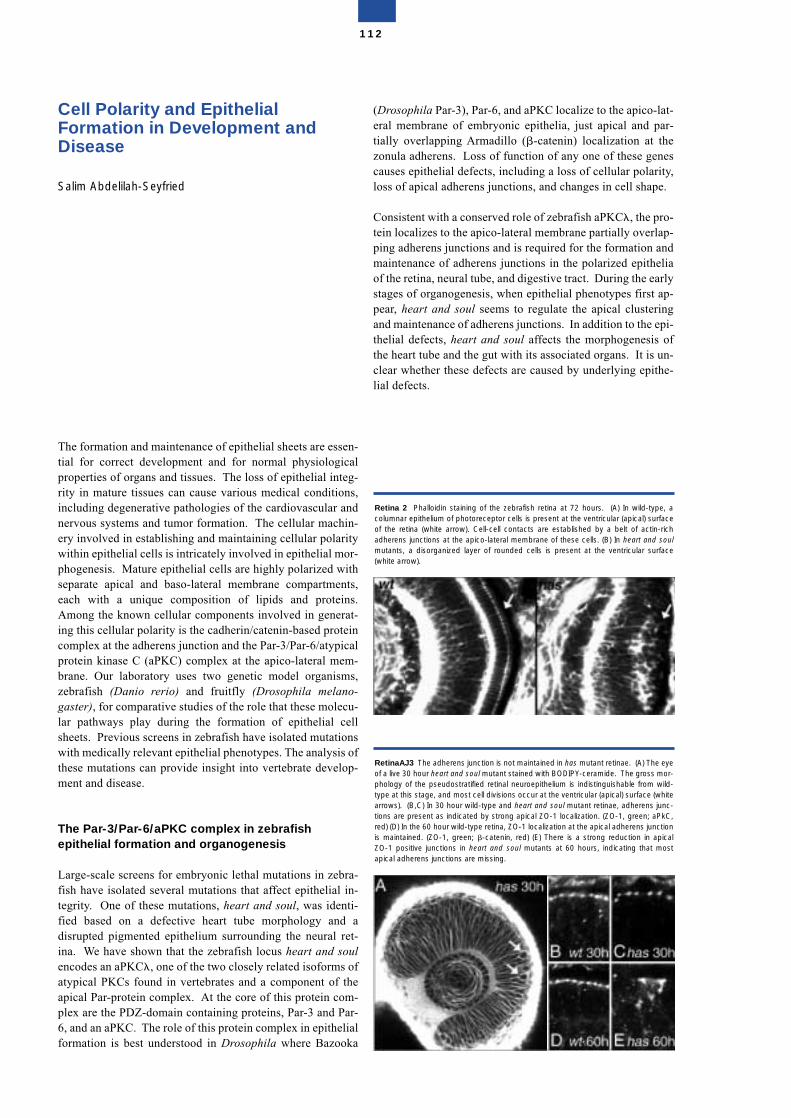
Cell Polarity and Epithelial Formation in Development and Disease
Salim Abdelilah-Seyfried
The formation and maintenance of epithelial sheets are essen-tial for correct development and for normal physiologicalproperties of organs and tissues. The loss of epithelial integ-rity in mature tissues can cause various medical conditions,including degenerative pathologies of the cardiovascular andnervous systems and tumor formation. The cellular machin-ery involved in establishing and maintaining cellular polaritywithin epithelial cells is intricately involved in epithelial mor-phogenesis. Mature epithelial cells are highly polarized withseparate apical and baso-lateral membrane compartments,each with a unique composition of lipids and proteins.Among the known cellular components involved in generat-ing this cellular polarity is the cadherin/catenin-based proteincomplex at the adherens junction and the Par-3/Par-6/atypicalprotein kinase C (aPKC) complex at the apico-lateral mem-brane. Our laboratory uses two genetic model organisms,zebrafish (Danio rerio) and fruitfly (Drosophila melano-gaster), for comparative studies of the role that these molecu-lar pathways play during the formation of epithelial cellsheets. Previous screens in zebrafish have isolated mutationswith medically relevant epithelial phenotypes. The analysis ofthese mutations can provide insight into vertebrate develop-ment and disease.
The Par-3/Par-6/aPKC complex in zebrafish epithelial formation and organogenesis
Large-scale screens for embryonic lethal mutations in zebra-fish have isolated several mutations that affect epithelial in-tegrity. One of these mutations, heart and soul, was identi-fied based on a defective heart tube morphology and adisrupted pigmented epithelium surrounding the neural ret-ina. We have shown that the zebrafish locus heart and soulencodes an aPKC�, one of the two closely related isoforms ofatypical PKCs found in vertebrates and a component of theapical Par-protein complex. At the core of this protein com-plex are the PDZ-domain containing proteins, Par-3 and Par-6, and an aPKC. The role of this protein complex in epithelialformation is best understood in Drosophila where Bazooka
(Drosophila Par-3), Par-6, and aPKC localize to the apico-lat-eral membrane of embryonic epithelia, just apical and par-tially overlapping Armadillo (�-catenin) localization at thezonula adherens. Loss of function of any one of these genescauses epithelial defects, including a loss of cellular polarity,loss of apical adherens junctions, and changes in cell shape.
Consistent with a conserved role of zebrafish aPKC�, the pro-tein localizes to the apico-lateral membrane partially overlap-ping adherens junctions and is required for the formation andmaintenance of adherens junctions in the polarized epitheliaof the retina, neural tube, and digestive tract. During the earlystages of organogenesis, when epithelial phenotypes first ap-pear, heart and soul seems to regulate the apical clusteringand maintenance of adherens junctions. In addition to the epi-thelial defects, heart and soul affects the morphogenesis ofthe heart tube and the gut with its associated organs. It is un-clear whether these defects are caused by underlying epithe-lial defects.
112
RetinaAJ3 The adherens junction is not maintained in has mutant retinae. (A) The eyeof a live 30 hour heart and soul mutant stained with BODIPY-ceramide. The gross mor-phology of the pseudostratified retinal neuroepithelium is indistinguishable from wild-type at this stage, and most cell divisions occur at the ventricular (apical) surface (whitearrows). (B,C) In 30 hour wild-type and heart and soul mutant retinae, adherens junc-tions are present as indicated by strong apical ZO-1 localization. (ZO-1, green; aPkC,red) (D) In the 60 hour wild-type retina, ZO-1 localization at the apical adherens junctionis maintained. (ZO-1, green; �-catenin, red) (E) There is a strong reduction in apicalZO-1 positive junctions in heart and soul mutants at 60 hours, indicating that mostapical adherens junctions are missing.
Retina 2 Phalloidin staining of the zebrafish retina at 72 hours. (A) In wild-type, acolumnar epithelium of photoreceptor cells is present at the ventricular (apical) surfaceof the retina (white arrow). Cell-cell contacts are established by a belt of actin-richadherens junctions at the apico-lateral membrane of these cells. (B) In heart and soulmutants, a disorganized layer of rounded cells is present at the ventricular surface(white arrow).
Research in our laboratory is currently directed towards thecloning and characterization of other zebrafish mutations thataffect cellular polarity and epithelial integrity. The identifica-tion of the molecular pathways involved in vertebrate epithe-lial morphogenesis may lead to relevant animal models forhuman epithelial pathologies and allow novel therapeutic ap-proaches to be investigated.
Selected Publications
Abdelilah-Seyfried, S., Chan, Y.M., Zeng, C., Justice, N.,Younger-Shepherd, S., Sharp, L., Barbel, S., Meadows, S.,Jan, L. and Jan, Y.N. (2000). A gain-of-function screen forgenes that affect the development of the Drosophila adult ex-ternal sensory organ. Genetics, 155, 733-752.
Horne-Badovinac, S., Lin, D., Waldron, S., Schwarz, M.,Mbamalu, G., Pawson, T., Jan, Y.N., Stainier, D.Y.R. and Ab-delilah-Seyfried, S. (2001). Positional cloning of heart andsoul reveals multiple roles for PKC� in zebrafish organogen-esis. Current Biol. 11, 1492-1502.
Cox, D., Abdelilah-Seyfried, S., Jan, L., and Jan, Y.N. (2001).Bazooka and atypical protein kinase C are required to regu-late oocyte differentiation in the Drosophila ovary. Proc. Nat.Acad. Sci. USA 98, 14475-14480.
Structure of the Group
Group leaderDr. Salim Abdelilah-Seyfried
Graduate studentStefan Rohr
Technical assistantJacqueline Klewer
113
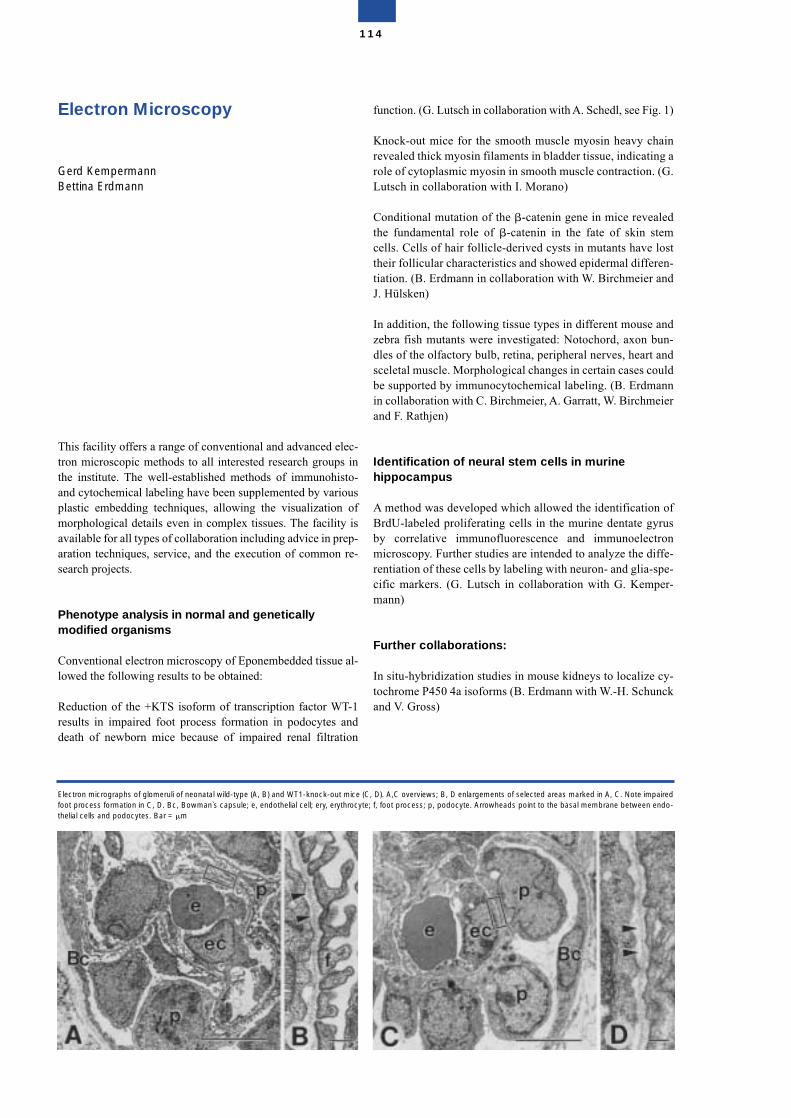
Electron Microscopy
Gerd KempermannBettina Erdmann
This facility offers a range of conventional and advanced elec-tron microscopic methods to all interested research groups inthe institute. The well-established methods of immunohisto-and cytochemical labeling have been supplemented by variousplastic embedding techniques, allowing the visualization ofmorphological details even in complex tissues. The facility isavailable for all types of collaboration including advice in prep-aration techniques, service, and the execution of common re-search projects.
Phenotype analysis in normal and geneticallymodified organisms
Conventional electron microscopy of Eponembedded tissue al-lowed the following results to be obtained:
Reduction of the +KTS isoform of transcription factor WT-1results in impaired foot process formation in podocytes anddeath of newborn mice because of impaired renal filtration
function. (G. Lutsch in collaboration with A. Schedl, see Fig. 1)
Knock-out mice for the smooth muscle myosin heavy chainrevealed thick myosin filaments in bladder tissue, indicating arole of cytoplasmic myosin in smooth muscle contraction. (G.Lutsch in collaboration with I. Morano)
Conditional mutation of the �-catenin gene in mice revealedthe fundamental role of �-catenin in the fate of skin stemcells. Cells of hair follicle-derived cysts in mutants have losttheir follicular characteristics and showed epidermal differen-tiation. (B. Erdmann in collaboration with W. Birchmeier andJ. Hülsken)
In addition, the following tissue types in different mouse andzebra fish mutants were investigated: Notochord, axon bun-dles of the olfactory bulb, retina, peripheral nerves, heart andsceletal muscle. Morphological changes in certain cases couldbe supported by immunocytochemical labeling. (B. Erdmannin collaboration with C. Birchmeier, A. Garratt, W. Birchmeierand F. Rathjen)
Identification of neural stem cells in murinehippocampus
A method was developed which allowed the identification ofBrdU-labeled proliferating cells in the murine dentate gyrusby correlative immunofluorescence and immunoelectronmicroscopy. Further studies are intended to analyze the diffe-rentiation of these cells by labeling with neuron- and glia-spe-cific markers. (G. Lutsch in collaboration with G. Kemper-mann)
Further collaborations:
In situ-hybridization studies in mouse kidneys to localize cy-tochrome P450 4a isoforms (B. Erdmann with W.-H. Schunckand V. Gross)
114
Electron micrographs of glomeruli of neonatal wild-type (A, B) and WT1-knock-out mice (C, D). A,C overviews; B, D enlargements of selected areas marked in A, C. Note impairedfoot process formation in C, D. Bc, Bowman`s capsule; e, endothelial cell; ery, erythrocyte; f, foot process; p, podocyte. Arrowheads point to the basal membrane between endo-thelial cells and podocytes. Bar = m
Electron microscopic investigations of
• Vav2-induced alveolar morphogenesis of mammary glandepithelial cells (B. Erdmann with W. Birchmeier and S. DiCesare)
• localization of CALEB in the superior colliculus and theretina in mouse and chicken (B. Erdmann with F. Rathjenand M. Moré)
• neuron-glia interaction in the cerebellum of EGFP/GFAtransgenic mice (B. Erdmann with H. Kettenmann and C.Nolte)
• detection of foreign DNA in endothelial cells followingnon-viral gene transfer (B. Erdmann with M. Boettger)
• location of the small heat shock proteins, Hsp 25 and_B-crystallin, in normal and ischemic rat kidney(G. Lutsch with W. Smoyer and R. Benndorf, Ann Arbor)
• identification of the sequence segments of Hsp 25 that areresponsible for inhibition of actin polymerization(G. Lutsch with R. Benndorf, Ann Arbor)
• characterization of folding pathways of model proteins(G. Lutsch with G. Damaschun)
Selected Publications
Hammes, A., Guo, J.K., Lutsch, G., Leheste, J.R., Landrock,D., Ziegler, U., Gubler, M.C., and Schedl, A. (2001) Twosplice variants of the Wilms`tumor 1 gene have distinct func-tion during sex determination and nephron formation. Cell106, 319-29
Huelsken, J., Vogel, R., Erdmann, B., Cotsarelis, G., andBirchmeier, W. (2001) �-catenin controls hair follicle mor-phogenesis and stem cell differentiation in the skin. Cell 105,533-545
Morano,I., Chai, G.X., Baltas, L.G., Lamounier-Zepter, V.,Lutsch, G., Kott, M., Haase, H., and Bader, M. (2000)Smooth-muscle contraction without smooth-muscle myosin.Nat Cell Biol. 2, 371-375
Huelsken, J., Vogel, R., Brinkmann, V., Erdmann, B., Birch-meier, C., and Birchmeier, W. (2000) Requirement for �-cat-enin in anterior-posterior axis formation in mice. J. Cell Biol.148, 567-578
Honeck,H., Gross, V., Erdmann, B., Kärgel, E., Neunaber, R.,Milia, A.F., Schneider, W., Luft, F.C. and Schunck, W.-H.(2000) Cytochrome P450-dependent renal arachidonic acidmetabolism in desoxycorticosterone acetate-salt hypertensivemice. Hypertension 36, 610-616
Smoyer, W.E., Ransom, R., Harris, R.C., Welsh, M.J., Lutsch,G., and Benndorf, R. (2000) Ischemic acute renal failure in-duces differential expression of small heat shock proteins. J.Am. Soc. Nephrol. 11, 211-221
Structure of the group
Group leaderDr. Gerd KempermannDr. Bettina Erdmann
ScientistDr. Gudrun Lutsch*
Techn. AssistantsErika Kotitschke*Helga Rietzke (see also group Dr. T. Sommer)
* part of the period reported
115

116
117
Molecular Therapy
Molecular Therapy
The aim of this program is to develop new therapeutic strate-gies for those diseases, such as cancer or cardiovascular dis-ease, which often remain resistant to treatment. Our effortsare based on a wide variety of experimental strategies that ex-ploit the latest knowledge emerging from the fast-growingfields of cell biology, cancer biology, immunology and mam-malian genetics. The molecular mechanisms that underliemalignant transformation have been unravelled to a consider-able degree, and a great deal is now understood about howtumors become resistant to standard therapies and escape im-mune recognition and destruction. For instance, it is recog-nized that tumors often express potentially immunogenicantigens that, nevertheless, fail to elicit an effective immuneresponse from the host. It is also know that T cells mustundergo an elaborate activation process in order to reject ma-lignant tissues. An arsenal of cloned genes is now availablewhose products are involved in cell-cycle arrest, apoptosis,selective killing of tumor cells, and the induction of immuneresponses. Combining this knowledge and these reagents, anumber of gene transfer technologies now allow the develop-ment of very precise and, hopefully, more effective and lesstoxic therapeutic modalities.
Molekulare Therapie
Das Ziel dieses Programms besteht darin, neue therapeuti-sche Strategien für Erkrankungen zu entwickeln, die häufignicht auf traditionelle Behandlungsmethoden ansprechen,wie dies bei Krebs oder Herzkreislaufkrankheiten der Fall ist.Unsere Bemühungen basieren auf einer großen Vielfalt vonexperimentellen Strategien, die den neuesten Stand des Wis-sens nutzen, wie er sich aus den rasch wachsenden Bereichender Zellbiologie, Krebsforschung, Immunologie und der Säu-getiergenetik ergibt. Die molekularen Mechanismen, die bös-artigen Transformationen zugrunde liegen, konnten zu einemGroßteil aufgeklärt werden, und sehr gut verstanden ist auch,wie Tumoren gegenüber Standardtherapien resistent werdenund der Erkennung und Zerstörung durch das Immunsystementgehen. So ist zum Beispiel bekannt, dass Tumoren zwaroft potentielle immunogene Antigene exprimieren, ohne dasses diesen Molekülen aber gelingt, beim Wirt eine Immunre-aktion auszulösen. Es ist auch bekannt, dass T-Zellen einenausgedehnten Prozeß der Aktivierung durchlaufen müssen,um bösartig verändertes Gewebe abzustoßen. Ein Arsenalvon klonierten Genen steht heute zur Verfügung, deren Pro-dukte zu der Arretierung des Zellzyklus, zur Apoptose, zumselektiven Absterben von Tumorzellen und der Induktion derImmunreaktion beitragen. Die Kombination dieses Wissensmit den genannten Agenzien liefert eine Vielzahl von Gen-transfertechnologien, mit deren Hilfe die Entwicklung vonsehr präzisen und hoffentlich auch wesentlich effektiverenund weniger toxischen therapeutischen Modalitäten ent-wickelt werden kann.
118
Myocardial Regeneration
Rainer Dietz
When fully differentiated, the mammalian heart is composedof cardiomyocytes which have withdrawn from the cell cycle.Thus, the heart is not able to compensate for cell loss render-ing it biologically inert as far as regeneration is concerned. Sofar, conventional therapy given to patients with heart failureaims at reducing the hemodynamic load in order to alleviatecardiac function. The establishment of a molecular approachto reinstall cardiomyocyte cell division would revolutionizestandard treatment regimens for heart failure patients.
In general, there are two possibilities to prevent loss of cardiaccontractile tissue after myocardial damage: 1. prevention ofcell death, and 2. reinduction of cell cycle activity in surround-ing healthy cardiomyocytes. Using mainly cell culture modellsof primary cardiomyocytes, our group is trying to identify pro-as well as anti-apoptotic signalling pathways in differentforms of cardiomyocyte apoptosis. As a consequence of bothacute and chronic myocardial damage, in most instances, det-rimental cardiomyocyte hypertrophy develops. However, theintracellular pathways responsible for this myocardial mala-daptive growth remain unknown. Therefore, the intercalationbetween pro- and anti-apoptotic pathways on one side andclassical growth cascades including cell cycle pathways on theother is another focus of our research interest.
Regulation of E2F1-dependent transcription and apopto-sis by the ETS-related transcription factor GABP�1
In this regard, our observation is of interest that both cyclin-dependent kinase inhibitors, p21CIP1 and p27KIP1, need tobe downregulated in order to trigger apoptosis in cardiomyo-cytes. Also, yeast two hybrid screening of a human heartlibrary revealed that the transcription factor E2F1, which pre-viously has been shown by us and other groups to act in a pro-apoptotic fashion in primary cardiomyocytes, interacts withthe ETS-related transcription factor GABP�1. This interac-tion links growth and cell death related pathways in cardiom-yocytes. More importantly, this is the first observation of apRb-independent mechanism regulating E2F1-dependenttranscription and apoptosis.
Phosphorylation by protein kinase CK2: A signallingswitch for the caspase-inhibiting protein ARC.
ARC, a recently discovered anti-apoptotic factor, the expres-sion of which appears to be restricted to cardiac and skeletalmuscle tissue, has been found by our group to be a substrateof the casein kinase II (CK2). Constitutive phosphorylation ofARC by CK2 is required for ARC to act in an anti-apoptoticfashion.
p21CIP1 controls proliferating cell nuclear antigen pro-tein levels in adult cardiomyocytes.
While trying to understand how cell death of cardiomyocytesis triggered, much of the effort of our group is devoted to de-ciphering the regulation of cardiomyocyte cell cycle with-drawal. Employing different models we have found that p21-CIP1 plays a critical role in the prevention of cardiomyocytecell cycle activation. Importantly, p21CIP appears to act as asuppressor of the cardiomyocyte cell cycle by regulating thedegradation of proliferating cell nuclear antigen (PCNA)rather than by its inhibitory effect on cyclin-dependentkinases.
Selected Publications
Bergmann M, Loser P, Dietz R, von Harsdorf R (2001). Effectof NF-�B inhibition on TNF-alpha-induced apoptosis anddownstream pathways in cardiomyocytes. J Mol Cell Cardiol33, 1223-1232.
Mehrhof FB, Müller FU, Bergmann MW, Wang Y, Li P,Schmitz W, Dietz R, von Harsdorf R (2001). In cardiomyo-cyte hypoxia insulin-like growth factor-I-induced anti-apop-totic signalling requires phosphatidylinositol-3-OH-kinase-and MAP-kinase-dependent activation of the transcriptionfactor CREB. Circulation 104, 2088-2094.
von Harsdorf R (2001). Can cardiomyocytes divide? Heart86, 481-482
Katchanov J, Harms C, Gertz K, Hauck L, Waeber C, Hiert L,Priller J, von Harsdorf R, Bruck W, Hortnagl H, Dirnagl U,Bhide PG, Endres M (2001). Mild cerebral ischemia inducesloss of cyclin-dependent kinase inhibitors and activation ofcell cycle machinery before delayed neuronal cell death.J Neurosci 21, 5045-5053
119
Structure of the Group
Group leaderProf. Dr. Rainer Dietz
ScientistsDr. Rüdiger v. HarsdorfDr. Ludger HauckDr. Peifeng LiDr. Martin BergmannDr. Felix MehrhofDr. Stefan DonathDr. Jincheng LiDr. Mikhail Skatchkov
Graduate and undergraduate studentsFelix EngelJens RohneNinette PustkowskiAlan PunnooseChristian Freund
Technical AssistantsMarlies GriebenDaniela GrotheGabi Welsch
120
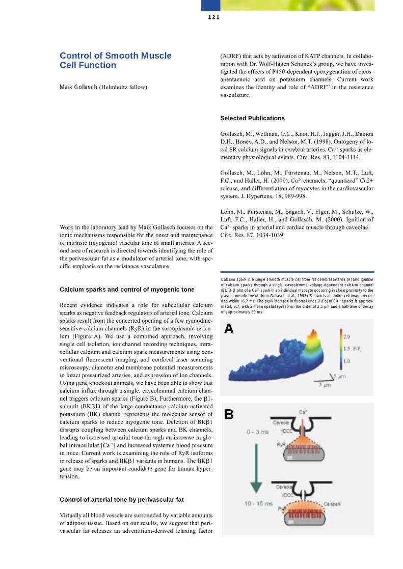
Control of Smooth Muscle Cell Function
Maik Gollasch (Helmholtz fellow)
Work in the laboratory lead by Maik Gollasch focuses on theionic mechanisms responsible for the onset and maintenanceof intrinsic (myogenic) vascular tone of small arteries. A sec-ond area of research is directed towards identifying the role ofthe perivascular fat as a modulator of arterial tone, with spe-cific emphasis on the resistance vasculature.
Calcium sparks and control of myogenic tone
Recent evidence indicates a role for subcellular calciumsparks as negative feedback regulators of arterial tone. Calciumsparks result from the concerted opening of a few ryanodine-sensitive calcium channels (RyR) in the sarcoplasmic reticu-lum (Figure A). We use a combined approach, involvingsingle cell isolation, ion channel recording techniques, intra-cellular calcium and calcium spark measurements using con-ventional fluorescent imaging, and confocal laser scanningmicroscopy, diameter and membrane potential measurementsin intact pressurized arteries, and expression of ion channels.Using gene knockout animals, we have been able to show thatcalcium influx through a single, caveolemmal calcium chan-nel triggers calcium sparks (Figure B), Furthermore, the �1-subunit (BK�1) of the large-conductance calcium-activatedpotassium (BK) channel represents the molecular sensor ofcalcium sparks to reduce myogenic tone. Deletion of BK�1disrupts coupling between calcium sparks and BK channels,leading to increased arterial tone through an increase in glo-bal intracellular [Ca2+] and increased systemic blood pressurein mice. Current work is examining the role of RyR isoformsin release of sparks and BK�1 variants in humans. The BK�1gene may be an important candidate gene for human hyper-tension.
Control of arterial tone by perivascular fat
Virtually all blood vessels are surrounded by variable amountsof adipose tissue. Based on our results, we suggest that peri-vascular fat releases an adventitium-derived relaxing factor
(ADRF) that acts by activation of KATP channels. In collabo-ration with Dr. Wolf-Hagen Schunck’s group, we have inves-tigated the effects of P450-dependent epoxygenation of eicos-apentaenoic acid on potassium channels. Current workexamines the identity and role of “ADRF” in the resistancevasculature.
Selected Publications
Gollasch, M., Wellman, G.C., Knot, H.J., Jaggar, J.H., DamonD.H., Bonev, A.D., and Nelson, M.T. (1998). Ontogeny of lo-cal SR calcium signals in cerebral arteries. Ca2+ sparks as ele-mentary physiological events. Circ. Res. 83, 1104-1114.
Gollasch, M., Löhn, M., Fürstenau, M., Nelson, M.T., Luft,F.C., and Haller, H. (2000). Ca2+ channels, “quantized” Ca2+release, and differentiation of myocytes in the cardiovascularsystem. J. Hypertens. 18, 989-998.
Löhn, M., Fürstenau, M., Sagach, V., Elger, M., Schulze, W.,Luft, F.C., Haller, H., and Gollasch, M. (2000). Ignition ofCa2+ sparks in arterial and cardiac muscle through caveolae. Circ. Res. 87, 1034-1039.
121
Calcium spark in a single smooth muscle cell from rat cerebral arteries (A) and ignitionof calcium sparks through a single, caveolemmal voltage-dependent calcium channel(B). 3-D plot of a Ca2+ spark in an individual myocyte occurring in close proximity to theplasma membrane (A, from Gollasch et al., 1998). Shown is an entire-cell image recor-ded within 16.7 ms. The peak increase in fluorescence (F/Fo) of Ca2+ sparks is approxi-mately 2.7, with a mean spatial spread on the order of 2.5 µm and a half-time of decayof approximately 50 ms.
A
B

Plüger, S., Faulhaber, J., Fürstenau, M., Löhn, M.,Waldschütz, R., Gollasch, M., Haller, H., Luft, F.C., Ehmke,H., and Pongs, O. (2000). Mice with disrupted BK channel b1subunit gene feature abnormal Ca2+ spark/STOC coupling andelevated blood pressure. Circ. Res. 87, E53-E60.
Structure of the group
Group leaderDr. Dr. Maik Gollasch
ScientistsDr. Matthias LöhnBirgit Lauterbach Dr. Galina DubrovskaKirill Essine
122
Immunology of CardiovascularDiseases
Gerd Wallukat
The interest of our group is focussed on immunological pro-cesses in cardiovascular diseases. In several cardiovasculardiseases, we have identified functional autoantibodies againstextracellular structures of G-protein coupled receptors.
We have observed autoantibodies against adrenergic recep-tors and AT1-receptors in the sera of patients with myocar-ditis, dilated cardiomyopathy, and hypertension. These auto-antibodies recognize epitopes on the first or secondextracellular loop of the receptors and act as the correspond-ing pharmacological agonists. In patients with myocarditisand dilated cardiomyopathy, as well as in Chagas´disease, theautoantibodies recognize �1-adrenoceptors. In patients withhypertension we observed autoantibodies against the �1-adr-energic and the angiotensin II AT1-receptor.
In recent years we have investigated in more detail the effectsof these autoantibodies, and have been able to show that theantibodies stabilize the dimeric agonistic conformation of thereceptors resulting in an agonist-like effect on the autoanti-bodies.
Autoantibodies in myocarditis and dilatedcardiomyopathy
The suggestion that the anti-�1-adrenoceptor autoantibodymight play a role in the pathogenesis of DCM is supported bysimilar findings in patients with myocarditis, a disease widelyheld to be a precursor of DCM. It is, therefore, also of interestin the present context that, in a patient with acute myocarditis,the healing process, as reflected by a normalization of theejection fraction and the heart rate, correlates with a disap-pearance of anti-�1-adrenoceptor autoantibodies from theblood.
Based on our autoimmune hypothesis, we have proposed newtherapeutic options to treat patients with endstage dilated car-diomyopathy. One of them is immunoadsorption using Thera-sorb columns to remove immunoglobulins from the patient’s
plasma. After this treatment, a marked improvement in car-diac function and a normalization of the heart size was ob-served. The strong correlation observed between the reduction in theamount of circulating autoantibodies against the �1-adreno-ceptors and improvement in heart function just described sup-port the hypothesis that the anti-�1-adrenoceptor antibodiesmay play a role in the pathophysiology of myocarditis andDCM.
To confirm this hypothesis, we have developed a specific im-munoadsorption column. Based on our epitope analysis, apeptide column was developed that selectively removes anti-�1-adrenoceptor autoantibodies. This column is presently be-ing used in a clinical pilot study.
Autoantibodies in hypertension
We have also investigated the role of autoantibodies in essen-tial and malignant hypertension. In some sera of patients withthis disease we have detected autoantibodies directed againstthe �1-adrenergic receptor. These autoantibodies recognizeepitopes of the first or second extracellular loop of the �1-ad-renergic receptors and acting like �1-adrenergic agonists.
In patients with malignant hypertension and in patients withpreeclampsia we have observed autoantibodies against theangiotensin II AT1-receptor. In preeclamptic patients, thisantibody is detectable after the 20th week of pregnancy anddisappears after delivery. The anti-AT1-receptor antibodiesact like the agonist angiotensin II and induce the formation ofthe transcription factor AP-1. These functional autoantibodiesare found in all preeclamptic women investigated so far andmay play a role in elevating vascular resistance and promot-ing hypertension and cardiac hypertrophy in these patients.
Role of mast cells in the heart
Another topic of our research involves cardiac mast cells. Be-cause the heart of patients with dilated cardiomyopathy con-tain four times more mast cells and more histamine than con-trols, we wanted to identify the role of these cells. Using amonoclonal antibody against surface determinants of rat con-nective tissue mast cells, we have been able to identify a greatnumber of, mostly undifferentiated, mast cells in the neonatalrat heart and in cell cultures prepared from this organ. In cellculture, we were able to differentiate the mast cells. Thesedifferentiated mast cells, mostly in intimate contact with car-diomyocytes, synthesize mediators histamine, serotonin andtumor necrosis factor � (TNF�).
In heart tissue of DCM patients, we have investigated the de-granulation of the mediators TNF� and tryptase from mastcells. Both mediators may be involved in the development offibrosis and remodelling in the failing heart.
123
Selected publications
Wallukat, G., Homuth, V., Fischer, T., Lindschau, C., Horst-kamp, B., Jüpner, A., Baur, E., Nissen, E., Vetter, K., Neichel,D., Dudenhausen, J.W., Haller, H., and Luft, F.C. (1999)Patients with preeclampsia develop agonistic autoantibodiesagainst the angiotensin AT1 receptor. J.Clin. Invest. 103, 945-952.
Müller, J., Wallukat, G., Dandel, M., Bieda, H., Brandes, K.,Spiegelsberger, S., Nissen, E., Kunze, R., and Hetzer, R.(2000)Immunoglobulin adsorption in patients with idiopathic di-lated cardiomyopathy. Circulation 101, 385-391.
Dechend, R., Homuth, V., Wallukat, G., Kreuzer, J., Park,J.U., Theuer, J., Juepner A., Gulba, D.C., Machmann, N.,Haller, H., and Luft, F.C. (2000)AT(1) agonistic antibodies from preeclamptic patients causevascular cells to express tissue factor. Circulation 101, 2382-2387.
Schimke, I., Müller, J., Priem, F., Kruse, I., Schon, B., Stein,J., Kunze, R., Wallukat, G., and Hetzer, R. (2001)Decreased oxidative stress in patients with idiopathic dilatedcardiomyopathy one year after immunoglobulin adsorptions.J. Am. Coll. Cardiol. 38, 178-183.
Structure of the Group
Group leaderDr. Gerd Wallukat
ScientistsDr. Rosemarie MorwinskiDr. Eberhard NissenDr. Wolfgang Schulze
Graduate and undergraduate studentsDajana NeichelAlexander Goihl
Technical assistantsKarin KarczewskiHolle SchmidtMonika Wegener
SecretariatDana Lafuente
124
Hematology, Oncology and TumorImmunology
Bernd Dörken
Identification of genetic risk profiles and genetherapy of malignant tumorsPeter Daniel, Bernhard Gillissen, Philipp Hemmati, IsridSturm, Thomas Wieder
Cell cycle deregulation and apoptosis is the underlying causeof tumorigenesis and tumor progression. The inactivation ofkey regulators of apoptosis contributes to the development ofresistance to therapy. We have shown the relevance of suchevents in the p53 signaling pathway. Multigene analysis ofupstream regulators and downstream effectors of p53 in pa-tients with cancer of the upper and lower gastrointestinaltract, breast cancer and acute and chronic lymphocytic leuke-mias shows that the loss of Bax, apart from mutation of p53,is a central event in the development of resistance. Patientswith an intact Bax show, in most cases, a better response totherapy and improved survival. There are, however, excep-tions to this rule, such as in gastric cancer where separate ge-netic events appear to be responsible for the poor prognosis inthis disease. We have shown that the inclusion of additionalgenes such as downstream regulators of apoptosis, i.e. APAF-1, IAPs, and the caspases as well as components of the Rbpathway of cell cycle regulation improves the identificationof patients with a good or poor prognosis. Apart from provid-ing a better understanding of resistance to conventional can-cer therapy, we have also laid the foundation for novel,molecular therapies. The aim is to transfer cell cycle-inhibi-tory and apoptosis-inducing genes into tumor cells. To thisend, we have demonstrated that overexpression of procas-pase-3, Bak, Bik/Nbk, Bax and p14ARF may sensitize cancercells to conventional cytotoxic therapies or induce apoptoticcell death by itself.
Biology of Hodgkin’s diseaseStephan Mathas, Martin Janz, Franziska Jundt, KristinaSchulze-Pröbsting in cooperation with Claus Scheidereit(MDC) and Harald Stein (UKBF)
Constitutive NF-�B activity is a characteristic of Hodgkin-/Reed-Sternberg (HRS) cells and is required for proliferationand survival of these cells. We have identified different de-fects in the signaling cascade leading to the constitutiveNF-�B activity. Thus, mutated IkB proteins, detected in celllines and lymph node biopsies, are unable to bind to NF-�B.Furthermore, the I�B kinase (IKK) complex is constitutivelyactive in HRS cells. The central role of NF-�B in the biologyof HRS cells was confirmed by the identification of NF-�B-dependent anti-apoptotic genes in HRS cells. Currently, weare investigating the possibility of establishing the inhibitionof constitutive NF–�B activity as a specific treatment optionfor Hodgkin’s disease. Different pharmacological inhibitorsof transient NF-�B activation, including aspirin or arsenicals,have been described. For some of these drugs, we have beenable to show strong in vitro and in vivo anti-tumor activity.Further studies have examined the role of NF-�B-related tran-scription factors and signaling pathways and their role in HRScells. In addition, mechanisms leading to constitutive NF-�Bactivity are being investigated by a variety of methods, in-cluding high density DNA micro arrays.
Identification of molecular regulators involved inB cell apoptosisBarbara Tiedt, Kurt Bommert in cooperation with BrigitteWittmann-Liebold (MDC), Manfred Gossen (MDC)
In order to identify the regulators involved in B cell apopto-sis, we have compared apoptotic and non apoptotic cells ofthe BL-60 cell line using two-dimensional gel electrophoresisand DNA micro arrays. We have identified an early responsegene that is strongly upregulated shortly after anti-IgM addi-tion. The high degree of homology with members of theCCCH family of tandem zinc finger proteins suggests thatthey promote the turnover of mRNAs containing class IIAREs. We are currently investigating the link betweenmRNA turnover and apoptosis.
ORC (origin recognition complex) and MCM (minichromo-some maintenance) proteins are essential for the replicationof chromosomes. At least some of the genes mentioned aboveare turned off in quiescent or differentiated cells. Therefore,new synthesis is a prerequisite for proliferation. We have an-alyzed the expression of MCM and ORC proteins duringapoptosis. After anti-IgM treatment, 70% of the BL-60 cellsare apoptotic. Using H-3 thymidine assays, we have been ableto show that the cells terminate proliferation. Western blotanalysis revealed cleavage of MCM-3 protein into 80 kDaand 55 kDa fragments. Preincubation of the cells with cas-pase-3 inhibitor blocks MCM-3 cleavage as well as anti-IgM-induced apoptosis. In addition, we are analyzing the expres-sion pattern of MCM and ORC mRNAs in samples derivedfrom different disease stages of B-CLL patients.
125
Signaling and survival pathways in multiplemyelomaDirk Hönemann, Manik Chatterjee, Suzanne Lentzsch, TanjaLehmann, Ralf Bargou in cooperation with Axel Greiner(Würzburg) and Maingen GmbH (Frankfurt)
The bone marrow microenvironment produces a number ofdifferent survival factors that are important for the malignantgrowth and drug resistance of multiple myeloma cells. One ofthe main factors which is important for the survival andgrowth of myeloma cells is IL-6. We have been able to showthat the IL-6-gp130-STAT-3 pathway is not essential for thesurvival of human myeloma cells, but might play a crucialrole in the development of drug resistance. In addition, wehave experimental evidence that the MAPK- and PI3K-path-ways may be essential for the survival of malignant plasmacells. Therefore, we are currently investigating the activationmechanisms of these pathways to identify target structures forpharmacologic intervention. Based on these studies we planto develop novel molecular therapy strategies.
Cytotoxic T-cell targeting by bispecific antibodiesMichael Grün, Anja Löffler, Ralf Bargou in cooperation withGerd Riethmüller (München) and Micromet GmbH (München)
We have shown that a novel recombinant bispecific singlechain antibody, bscCD19xCD3, induces rapid and highlylymphoma specific cytotoxicity mediated by unstimulated T-lymphocytes. By redirecting primary human T cells derivedfrom the peripheral blood against CD19-positive lymphomacell lines, the bscCD19xCD3 antibody showed significant cy-totoxic activity at very low concentrations, even in experi-ments without T cell prestimulation. In addition the bscCD19-xCD3, bispecific antibody is able to induce nearly completedepletion of primary lymphoma cells mediated by autologousT-cells of patients with chronic lymphatic leukemia in the ma-jority of cases analyzed. We plan to start a phase-I clinicaltrial of the treatment of patients with refractory B-cell lym-phomas at the end of 2001. Furthermore, we are trying to es-tablish a similar strategy for the treatment of multiple mye-loma patients using a novel plasma cell-specific surfaceantigen as the target structure.
Apoptotic pathways in childhood T-ALL in the con-text of the maturation stage and clinical outcomeLeonid Karawajew, Christian Wuchter, Richard Ratei, Wolf-Dieter Ludwig
Within childhood T-ALL, cortical (CD1a positive) T-ALL hasbeen identified as a subgroup having a favorable clinical out-come. We have investigated whether the different in vivotherapy response could be linked to a differential in vitro sus-ceptibility to apoptotic cell death, and examined a large seriesof T-ALL leukemic cells (n=100) with respect to apoptosis-related features (expression levels of Bax, Bcl-2, CD95; sus-ceptibility to spontaneous, CD95- and drug-induced apopto-sis) as well as to their specific cytokine responsiveness interms of apoptosis inhibition in vitro. Of the parameters in-vestigated, only IL-7 rescue and dexamethasone-inducedapoptosis exhibit a positive correlation with the cortical im-
munophenotype and a better early cytoreduction in vivo. IL-7was also highly effective in inhibiting dexamethasone-in-duced apoptosis in vitro. Therefore, apoptosis-modulatingpathways triggered by IL-7 and glucocorticoids are closelyassociated in T-ALL cells and may share common check-points responsible for the better in vivo-treatment response ofcortical T-ALL.
Selected Publications
Friedrich, K., Wieder, T., Von Haefen, C., Radetzki, S., Ja-nicke, R., Schulze-Osthoff, K., Dörken, B., and Daniel, P. T.(2001). Overexpression of caspase-3 restores sensitivity fordrug-induced apoptosis in breast cancer cell lines with ac-quired drug resistance, Oncogene 20, 2749-60.
Hinz, M., Löser, P., Mathas, S., Krappmann, D., Dörken, B.,and Scheidereit, C. (2001). Constitutive NF-kappaB main-tains high expression of a characteristic gene network, includ-ing CD40, CD86, and a set of antiapoptotic genes in Hodg-kin/Reed-Sternberg cells, Blood 97, 2798-807.
Hönemann, D., Chatterjee, M., Savino, R., Bommert, K., Bur-ger, R., Gramatzki, M., Dörken, B., and Bargou, R. C. (2001).The IL-6 receptor antagonist SANT-7 overcomes bone mar-row stromal cell-mediated drug resistance of multiple mye-loma cells, Int J Cancer 93, 674-80.
Karawajew, L., Ruppert, V., Wuchter, C., Kosser, A.,Schrappe, M., Dörken, B., and Ludwig, W. D. (2000). Inhibi-tion of in vitro spontaneous apoptosis by IL-7 correlates withbcl-2 up-regulation, cortical/mature immunophenotype, andbetter early cytoreduction of childhood T-cell acute lympho-blastic leukemia, Blood 96, 297-306.
Löffler, A., Kufer, P., Lutterbüse, R., Zettl, F., Daniel, P. T.,Schwenkenbecher, J. M., Riethmüller, G., Dörken, B., andBargou, R. C. (2000). A recombinant bispecific single-chainantibody, CD19 x CD3, induces rapid and high lymphoma-di-rected cytotoxicity by unstimulated T lymphocytes, Blood 95,2098-103.
Mathas, S., Rickers, A., Bommert, K., Dörken, B., and Ma-para, M. Y. (2000). Anti-CD20- and B-cell receptor-mediatedapoptosis: evidence for shared intracellular signaling path-ways, Cancer Res 60, 7170-6.
Sturm, I., Petrowsky, H., Volz, R., Lorenz, M., Radetzki, S.,Hillebrand, T., Wolff, G., Hauptmann, S., Dörken, B., andDaniel, P. T. (2001). Analysis of p53/BAX/p16(ink4a/CDKN2) in esophageal squamous cell carcinoma: high BAXand p16 (ink4a/CDKN2) identifies patients with good prog-nosis, J Clin Oncol 19, 2272-81.
126
Structure of the Group
Group leaderProf. Dr. Bernd Dörken
ScientistsDr. Ralf BargouDr. Kurt BommertDr. Sophie CayeuxDr. Peter DanielDr. Florian Emmerich*Dr. Bernd GillissenDr. Martin Janz*Dr. Franziska JundtDr. Leonid KarawajewDr. Ida-Johanna KörnerDr. Suzanne Lentzsch*Prof. Dr. Wolf-Dieter LudwigDr. Markus Mapara*Dr. Stephan MathasDr. Richard RateiDr. Isrid SturmDr. Thomas WiederDr. Christian Wuchter*
Graduate and undergraduate studentsManik Chatterjee*Michael GrünDirk HöhnemannTanja Lehmann*Andreas Lietz*Kristina Schulze-Pröbsting*Barbara Tiedt
Technical AssistantsKati Hildebrandt*Andrea KatzerKatarina KleyKerstin KrügerKatrin LangnerUte Nitschke
* part of the period reported
127
Molecular Immunotherapy
Antonio Pezzutto
Our group is working on the development of immunotherapystrategies for the treatment of renal cell carcinoma, colorectalcarcinoma and chronic myeloid leukemia. In our GMP labor-atory in the Robert Rössle Klinik we have completed thepreparation of a cellular vaccine consisting of a gene-modi-fied tumor cell line that expresses a shared renal carcinomaantigen recognized by T cells in context of HLA-A*0201 (de-veloped in cooperation with Th. Blankenstein (MDC) and D.Schendel (GSF, Munich). We have also established GMP-cer-tified production protocols for the generation of cellular vac-cines consisting of peptide-loaded, tumor-lysate pulsed, orgene-modified dendritic cells.
Induction of T-cell immunity against EpCam (Epithelial Cell adhesion molecule) Oliver Schmetzer, Christian Koch
Some patients with colorectal cancer develop an immune re-sponse to peptides of the epithelial adhesion molecule Ep-Cam, which is overexpressed in human adenocarcinomas. Asensitive ELISA assay for the screening of naturally occur-ring autoantibodies against EpCam has been established.Transgenic mice expressing human EpCam have been gener-ated for use in preclinical vaccination experiments. Dendriticcells (DC) pulsed with recombinant EpCam protein or se-lected MHC-I and MHC-II peptides are being evaluated fortheir ability to induce rejection of EpCam-positive tumors.Multimeres of MHC-II binding EpCam epitopes are beingprepared and evaluated in cooperation with K. Falk and O.Rötzschke (MDC). GMP procedures for the production oflarge quantities of recombinant EpCam protein are being op-timized. The feasibility of a clinical vaccination study in pa-tients with EpCam-positive adenocarcinomas is also beingevaluated.
Use of dendritic cells for the induction ofantileukemic immune responseMonika Schwarz, Beate Wittmann
Peptides derived from the bcr-abl fusion protein that is pro-duced as a consequence of the t(9:22) chromosomal translo-cation in patients with chronic myeloid leukemia (CML) canbind to HLA-A3, -A11, or -B8. We have started a clinical trialusing in vitro-generated, bcr-abl positive DC in CML-patientswith the aim of inducing a CML-directed immune response.Therapy is safe and feasible and evaluation of immune re-sponses is ongoing. In cooperation with B. Wittmann-Lie-bold, E. Müller and A. Otto, (MDC) we are analyzing natu-rally processed peptides from CML-cells in order to detectother potential candidate peptides for vaccination. We haveestablished a bcr-abl specific ELISPOT assay for detection ofbcr-abl specific T cells, an assay based on the use of HLA-Class I tetrameres is being developed. We have already de-tected bcr-abl specific T cells in some patients in clinical re-mission following interferon treatment. These assays willallow us to monitor anti-leukemic immunity in CML patients.Using 2-D gel electrophoresis (cooperation with A. Otto andE. Müller), we are evaluating CML cells treated with the tyro-sine-kinase inhibitor STI571, in order to identify proteins thatare expressed selectively in CML cells and that could be po-tential target for an immunotherapy approach.
Gene modification of dendritic cellsJörg Westermann, Tam Nguyeng-Hoay
Both human and murine DC can be gene-modified using ret-roviral vectors and receptor mediated endocytosis (targetingthe mannose receptor). Complexes of DNA, polyethiyleni-mine (PEI) and mannose are efficiently internalized, resultingin gene expression. A particular advantage of this method isthe possibility of transferring several genes with the sameconstruct, allowing expression of both tumor-antigens andgenes that can modulate DC function, such as superantigens,chemokine receptors, and adhesion molecules. A strongerstimulation of the immune response or a change in the im-mune response with predominance of TH1 immunity can beachieved with this method. These studies are being performedin cooperation with M. Zenke (MDC). The use of cytokinesthat can modify the number and function of DC, such as Flt-3Ligand and GM-CSF, is being investigated in gene transfermodels in tumor vaccination experiments.
Role of ICOS Ligand in the regulation of theimmune responseGünther Richter, Antonio Pezzutto,
The inducible co-stimulator receptor (ICOS) is a third mem-ber of the CD28 receptor family that regulates T cell activa-tion and function. We have used soluble ICOSIg to furthercharacterize the ICOS ligand (ICOSL) on APCs. We have alsogenerated a new monoclonal antibody to ICOSL. ICOSL isexpressed on monocytes, dendritic cells, and B cells. OnCD34+ hematopoietic precursor cells, ICOSL is induced priorto CD80/CD86 induction suggesting that ICOSL is an earlydifferentiation marker along the monocytic/dendritic matura-
128
tion pathway. Induction of ICOSL is dependent on TNF-�and is regulated via NF-�B. TNF-�-induced ICOSL expres-sion seems to be functionally important for the co-stimulatorycapacity of early hematopoietic precursors. The co-stimula-tory capacity of leukemic cells at different maturation stagesis being analyzed and correlated with ICOSL expression.
Selected Publications
Aicher A., M. Hayden-Ledbetter, W.A. Brady, A. Pezzutto,G. Richter, D. Magaletti, S. Buckwalter, J. Ledbetter, E.A.Clark. (2000). Characterization of Human ICOS Ligand Ex-pression and Function J. Immunol. 164: 4689-4696.
Westermann J., J. Kopp, I. Körner, G. Richter, Z. Qin, T.Blankenstein, B. Dörken, A. Pezzutto. 2000. Bcr/Abl+ autol-ogous dendritic cells for vaccination in chronic myeloid leu-kemia. Bone Marrow Transplant. 25: 46-49
Westermann J., G. Reich, J. Kopp, U. Haus, B. Dörken, A.Pezzutto. (2001). Granulocyte / macrophage-colony stimulat-ing factor plus interleukin-2 plus interferon-a in the treatmentof metastatic renal cell carcinoma: a pilot study. J. Immunol.Immunother.49: 613-620
Richter G., M. Hayden-Ledbetter, M. Irgang, J.A. Ledbetter,J. Westermann, I. Körner, K. Daemen, E.A. Clark, A. Aicher,A. Pezzutto. (2001), Tumor necrosis factor-alpha regulatesthe expression of inducible costimulator receptor ligand onCD34+ progenitor cells during differentiation into antigenpresenting cells. J. Biol. Chem. 276: 45686-45693
Structure of the Group
Group leader Prof. Dr. Antonio Pezzutto
Scientists Dr. med. Jörg WestermannDr. med. Kang Hun LeeChristian KochBeate Wittmann
Graduate and undergraduate studentsTam Nguyen-HoayOliver SchmetzerMark SchnitzlerMonika SchwarzAchim Wiefelspütz
129
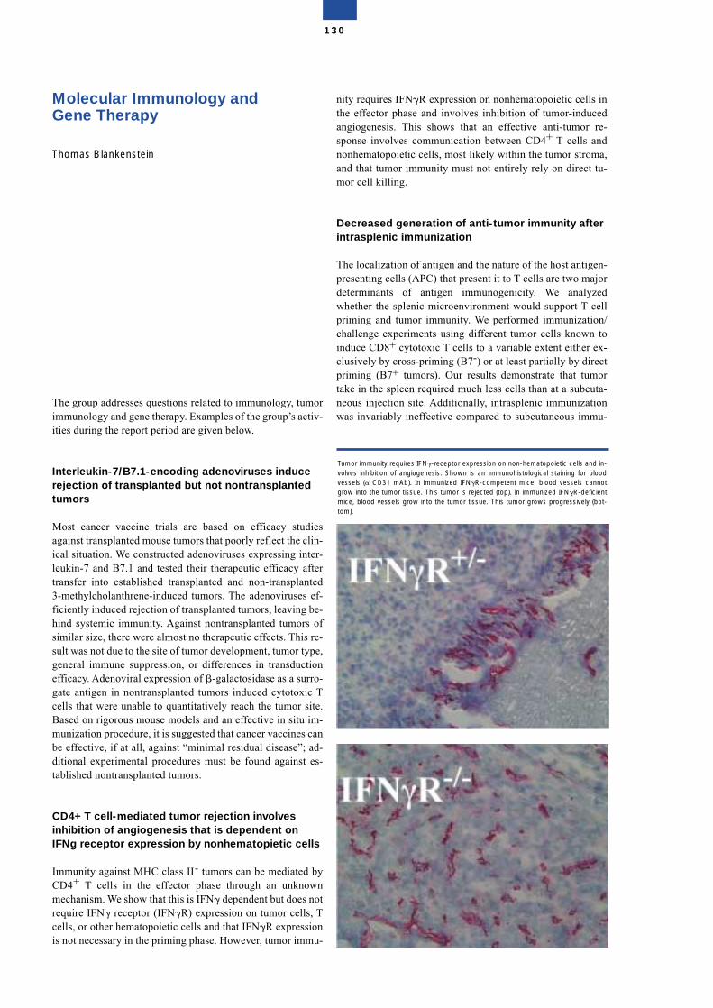
Molecular Immunology andGene Therapy
Thomas Blankenstein
The group addresses questions related to immunology, tumorimmunology and gene therapy. Examples of the group’s activ-ities during the report period are given below.
Interleukin-7/B7.1-encoding adenoviruses inducerejection of transplanted but not nontransplantedtumors
Most cancer vaccine trials are based on efficacy studiesagainst transplanted mouse tumors that poorly reflect the clin-ical situation. We constructed adenoviruses expressing inter-leukin-7 and B7.1 and tested their therapeutic efficacy aftertransfer into established transplanted and non-transplanted3-methylcholanthrene-induced tumors. The adenoviruses ef-ficiently induced rejection of transplanted tumors, leaving be-hind systemic immunity. Against nontransplanted tumors ofsimilar size, there were almost no therapeutic effects. This re-sult was not due to the site of tumor development, tumor type,general immune suppression, or differences in transductionefficacy. Adenoviral expression of �-galactosidase as a surro-gate antigen in nontransplanted tumors induced cytotoxic Tcells that were unable to quantitatively reach the tumor site.Based on rigorous mouse models and an effective in situ im-munization procedure, it is suggested that cancer vaccines canbe effective, if at all, against “minimal residual disease”; ad-ditional experimental procedures must be found against es-tablished nontransplanted tumors.
CD4+ T cell-mediated tumor rejection involvesinhibition of angiogenesis that is dependent onIFNg receptor expression by nonhematopietic cells
Immunity against MHC class II- tumors can be mediated byCD4+ T cells in the effector phase through an unknownmechanism. We show that this is IFN� dependent but does notrequire IFN� receptor (IFN�R) expression on tumor cells, Tcells, or other hematopoietic cells and that IFN�R expressionis not necessary in the priming phase. However, tumor immu-
nity requires IFN�R expression on nonhematopoietic cells inthe effector phase and involves inhibition of tumor-inducedangiogenesis. This shows that an effective anti-tumor re-sponse involves communication between CD4+ T cells andnonhematopoietic cells, most likely within the tumor stroma,and that tumor immunity must not entirely rely on direct tu-mor cell killing.
Decreased generation of anti-tumor immunity afterintrasplenic immunization
The localization of antigen and the nature of the host antigen-presenting cells (APC) that present it to T cells are two majordeterminants of antigen immunogenicity. We analyzedwhether the splenic microenvironment would support T cellpriming and tumor immunity. We performed immunization/challenge experiments using different tumor cells known toinduce CD8+ cytotoxic T cells to a variable extent either ex-clusively by cross-priming (B7-) or at least partially by directpriming (B7+ tumors). Our results demonstrate that tumortake in the spleen required much less cells than at a subcuta-neous injection site. Additionally, intrasplenic immunizationwas invariably ineffective compared to subcutaneous immu-
130
Tumor immunity requires IFN�-receptor expression on non-hematopoietic cells and in-volves inhibition of angiogenesis. Shown is an immunohistological staining for bloodvessels (� CD31 mAb). In immunized IFN�R-competent mice, blood vessels cannotgrow into the tumor tissue. This tumor is rejected (top). In immunized IFN�R-deficientmice, blood vessels grow into the tumor tissue. This tumor grows progressively (bot-tom).
nization. We further showed that B cells were not responsiblefor the inefficient intrasplenic immunization. Therefore, de-livering the tumor cell antigens inside the spleen by intra-splenic immunization did not improve but rather decreasedthe efficacy of tumor cell vaccines.
Efficient gene transfer into primary human CD8+ Tlymphocytes by MuLV-10A1 retrovirus pseudotype
Efficient and stable gene transfer into primary human T lym-phocytes would greatly improve their use for adoptive trans-fer to treat acquired disorders, viral diseases, and cancer. Wehave constructed retroviral vector pseudotypes of ampho-tropic murine leukemia viruses (A-MuLV, MuLV-10A1), gib-bon ape leukemia virus (GaLV), and feline endogenous virus(RD114) containing the enhanced green fluorescent protein(GFP) as a marker gene. Transduction of primary humanCD8+ T lymphocytes by the different GFP-retrovirus pseudo-types revealed the superiority of MuLV-10A1 in comparisonwith A-MuLV, GaLV, and RD114, respectively. The superiortransduction efficacy of CD8+ T cells by MuLV-10A1 corre-lates with its higher stability and, probably, the utilization ofboth the A-MuLV receptor (Pit2) and the GaLV receptor(Pit1) for cell entry.
Adoptive tumor therapy with T lymphocytesenriched through an IFN� capture assay
Successful adoptive T cell therapy has been demonstrated inviral disease and selected forms of cancer. However, it is lim-ited by the difficulty to efficiently isolate and amplify autolo-gous tumor-reactive T cell clones. MHC class I-tetramers andpeptide have greatly facilitated the characterization of CD8+
T cells specific for tumor-associated antigens; yet for adop-tive T cell therapy, MHC-tetramers have limitations: i) theyrequire knowledge of tumor antigens which is often not avail-able; ii) they select T cells with a single specificity, thereby,posing risk for selection of tumor escape variants; iii) they donot select for function, so that T cells may be anergic whenisolated from cancer patients; and iv) they do not allow theisolation of CD4+ T cells that can be essential for tumor rejec-tion. Because IFN� is essential for tumor rejection, we used amethod to isolate live T cells based on their IFNg production.IFN� secreted by previously activated T cells is retained onthe cell surface, allowing their specific isolation and expan-sion. We showed that IFN�+ but not IFN�- T cells fromtumor-immunized mice are cytolytic and mediate tumorrejection upon adoptive transfer. Importantly, tumor-specificT cells can be enriched from lymphocytes infiltrating humanrenal cell carcinoma by the IFN� capture assay.
Selected publications
Willimsky, G., and Blankenstein, Th. (2000). IL-7/B7.1-en-coding adenoviruses induce rejection of transplanted but notnon-transplanted tumors. Cancer Res. 60, 685-692.
Qin, Z., and Blankenstein, Th. (2000). CD4+ T cell-mediatedtumor rejection involves inhibition of angiogenesis that is de-pendent on IFN� receptor expression by nonhematopieticcells. Immunity 12, 677-686.
Uckert, W., Becker, C., Gladow, M., Klein, D., Kammertöns, T.,Pedersen, L., and Blankenstein, Th. (2000). Efficient gene trans-fer into primary human CD8+ T lymphocytes by MuLV-10A1retrovirus pseudotype. Hum. Gene Therapy 11, 1005-1014.
Cayeux, S., Qin, Z., Dörken, B., and Blankenstein, Th. (2001).Decreased generation of tumor immunity after intrasplenicimmunization. Eur. J. Immunol. 31, 1392-1399.
Becker, C., Pohla, H., Frankenberger, B., Assenmacher, M.,Schendel, D. J., and Blankenstein, Th. (2001). Adoptive tumortherapy with T lymphocytes enriched through an IFN� cap-ture assay. Nat. Med. 7, 1159-1162.
Structure of the group
Group leaderProf. Dr. Thomas Blankenstein
ScientistsDr. Thomas KammertönsDr. Hye-Jung KimDr. Liang-Ping LiDr. Mariette MohauptDr. Zhihai QinDr. Thomas SchülerDr. Wolfgang UckertDr. Gerald Willimsky
Graduate and undergraduate studentsChristian BuschowBoris EngelsMonika GladowHakan CamSandra KörnigChristoph LampertSusanne Preiß
Technical assistants SecretariatDenise Barthel Sylvia Klahn Christiane BeckerAngelika GärtnerMartina GrabbertIrmgard KüttnerBeate RothTanja SpecowiakRalf WillebrandChristel Westen
131
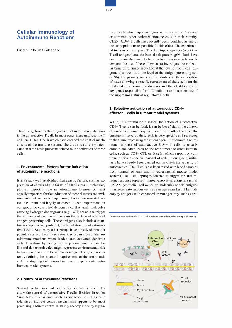
Cellular Immunology of Autoimmune Reactions
Kirsten Falk/Olaf Rötzschke
The driving force in the progression of autoimmune diseasesis the autoreactive T cell. In most cases these autoreactive Tcells are CD4+ T cells which have escaped the control mech-anisms of the immune system. The group is currently inter-ested in three basic problems related to the activation of thesecells:
1. Environmental factors for the induction of autoimmune reactions
It is already well established that genetic factors, such as ex-pression of certain allelic forms of MHC class II molecules,play an important role in autoimmune diseases. At leastequally important for the induction of these diseases are envi-ronmental influences but, up to now, these environmental fac-tors have remained largely unknown. Recent experiments inour group, however, had demonstrated that small moleculescarrying hydrogen donor groups (e.g. –OH) are able to triggerthe exchange of peptide antigens on the surface of activatedantigen-presenting cells. These antigens also include autoan-tigens (peptides and proteins), the target structure of autoreac-tive T cells. Studies by other groups have already shown thatpeptides derived from these autoantigens can induce fatal au-toimmune reactions when loaded onto activated dendriticcells. Therefore, by catalysing this process, small molecularH-bond donor molecules might represent environmental riskfactors which have not been considered yet. The group is cur-rently defining the structural requirements of the compoundsand investigating their impact in several experimental auto-immune model systems.
2. Control of autoimmune reactions
Several mechanisms had been described which potentiallyallow the control of autoreactive T cells. Besides direct (or“suicidal”) mechanisms, such as induction of ‘high-zonetolerance’, indirect control mechanisms appear to be mostpromising. Indirect control is mainly accomplished by regula-
tory T cells which, upon antigen-specific activation, ‘silence’or eliminate other activated immune cells in their vicinity.CD25+ CD4+ T cells have recently been identified as one ofthe subpopulations responsible for this effect. The experimen-tal tools in our group are T cell epitope oligomers (repetitiveT cell antigens) and the heat shock protein gp96. Both havebeen previously found to be effective tolerance inducers invivo and the use of these allows us to investigate the molecu-lar basis of tolerance induction at the level of the T cell (oli-gomers) as well as at the level of the antigen presenting cell(gp96). The primary goals of these studies are the explorationof ways allowing a specific recruitment of these cells for thetreatment of autoimmune diseases and the identification ofkey genes responsible for differentiation and maintenance ofthe suppressor status of regulatory T cells.
3. Selective activation of autoreactive CD4+effector T cells in tumour model systems
While, in autoimmune diseases, the action of autoreactiveCD4+ T cells can be fatal, it can be beneficial in the contextof tumour-immunotherapies. In contrast to other therapies thedamage inflicted by these cells is very specific and restrictedto the tissue expressing the autoantigen. Furthermore, the im-mune response of autoreactive CD4+ T cells is usuallychronic and often leads to the recruitment of other immunecells, such as CD8+ CTL or B cells, which support or con-tinue the tissue-specific removal of cells. In our group, initialtests have already been carried out in which the capacity ofautoreactive CD4+ T cells has been tested with blood samplesfrom tumour patients and in experimental mouse modelsystems. The T cell epitopes selected to trigger the autoim-mune response represent tumour-associated antigens such asEPCAM (epithelial cell adhesion molecule) or self-antigenstransfected into tumour cells as surrogate markers. The trialsemploy antigens with enhanced immunogenicity, such as epi-
132
Schematic mechanism of CD4+ T cell mediated tissue distruction (Multiple Sklerosis)
ACP
B Cell
AutoreactiveCD4+T cell
cytokines
LyticFactors
T cellreceptor
MHC class IImolecule
Axon
Myelin
Myelinprotein
T cellautoantigen
tope oligomers or lipo-peptides, and include the constructionof inducible animal model systems (TET system). The goal ofthese studies is to investigate whether autoimmune responsesdriven by CD4+ T cells can in fact be used for tumour rejec-tion.
Selected Publications
Falk, K., Rötzschke, O., Santambrogio, L., Dorf, M.E., Bros-nan, C., and Strominger, J.L. (2000). Induction and suppres-sion of an autoimmune disease by oligomerized T cell epi-topes: enhanced in vivo potency of encephalitogenic peptides.J. Exp. Med. 191, 717-730.
Falk, K., Rötzschke O., and Strominger, J.L. (2000). Antigen-specific elimination of T cells induced by oligomerized he-magglutinin (HA) 306-318. Eur. J. Immunol. 30, 3012-3020.
Steinekemeier, M., Falk, K., Rötzschke, O., Weishaupt, A.,Schneider, C., Toyka, K.V., Gold, R., and Strominger, J.L.(2001). Vaccination, prevention and treatment of experimen-tal autoimmune neuritis (EAN) by an oligomerized T cell epi-tope. Proc. Natl. Acad. Sci. USA, 98, 13872-13877.
Falk, K., Lau, J.M., Santambrogio, L., Marin Esteban, V.,Puentes, F., Strominger, J.L., and Rötzschke, O. (2002).Ligand-exchange of MHC class II proteins is triggered byH-bond donor groups of small molecules. J. Biol. Chem. 277,2709-2715
Structure of the Group
Group leaderDr. Kirsten FalkDr. Olaf Rötzschke
ScientistsDr. Mirelle StarkeDr. Anja Mehl-LautschamDr. Georg Lautscham
Graduate and undergraduate studentsEva GoldscheidViviana Marin EstebanMarkus KleinewietfeldAnita OerenFabiola Puentes
Technical AssistantsMaria HofstätterRobert Niestroj
SecretariatSylvia Olbrich
133
Molecular and Cell Biology of Hematopoietic Cells
Martin Zenke
The research focus of this group is the molecular and cellbiology of hematopoietic cells using human cells and, in ex-perimental model systems, hematopoietic cells from mouseand chicken. Two hematopoietic cell types are being studiedin detail: red blood cells and antigen presenting dendriticcells. Stem cells and early hematopoietic progenitor cells andtheir comittment and differentiation are also being investi-gated.
Determining the gene expression repertoire of redblood cellsHacker, C., Lemke, B., Koh, K.-R., Anzinger, B., Blendinger,G , Knespel, S., and Bartunek, P.
Red blood cells represent one of the most abundant special-ized cell types in vertebrate organisms. Their developmentfrom hematopoietic stem/progenitor cells is tightly controlledto ensure that they are produced in sufficient numbers to servethe needs of the organism both in the physiological and path-ological states. We have now determined the gene expressionrepertoire of human red blood cells by DNA chip technology(in collaboration with Pfizer Inc., Groton, CT, USA) by em-ploying an in vitro differentiation system of SCF/Epo-depen-dent erythroid progenitor cells (Panzenböck et al., Blood 92,3658, 1998). The expression profile of more than 6000 anno-tated genes during erythroid cell differentiation has been de-termined. This analysis identified several molecules with adetermining function in erythroid cell development that arecurrently being further analysed.
Thyroid hormone regulates the obesity gene, tubbyKoritschoner, N. P., Kurz, S. M., Heikenwälder, M. F., Bartunek, P., Blendinger, G.
The c-erbA protooncogene encodes a high affinity receptorfor thyroid hormone (T3/T4; thyroid hormone receptor, TR).Our previous work established that TR/c-erbA has a decisiverole in red blood cell differentiation and acts as a binary
switch in determining erythroid cell fate: unligandedTR/c-erbA supports the growth of erythroid progenitor cellswhile ligand-activated TR/c-erbA induces their differentia-tion (Zenke et al., Cell 61, 1035, 1990; Bartunek and Zenke,Mol. Endo. 12, 1269, 1998). TR/c-erbA exerts its activity byactivating and/or repressing gene expression, and severalTR/c-erbA target genes have been identified. We have nowfocused on the analysis of the TR/c-erbA target gene, tubby.Tubby has been implicated in intracellular signalling down-stream of insulin and G-protein coupled receptors and, whenmutated in the tubby mouse, causes obesity and sensory defi-ciencies. We demonstrated that thyroid hormone regulatestubby in vivo and in vitro and, thus, revealed a molecular linkbetween thyroid status and obesity (Koritschoner et al., 2001;in collaboration with A. Munoz, CSIC, Madrid, Spain; seealso Heikenwälder et al., 2001). This is particularly interest-ing given the fact that thyroid hormone increases energy ex-penditure and thyroid hormone deficiency predisposes toweight gain and eventually obesity in humans.
Gene expression in antigen presenting dendritic cellsJu, X.-S., Hieronymus, T., Hacker, C., Madruga, J., Kurz, S. M., Jorgas, T., Knespel, S., Euler, U.
Dendritic cells (DC) are professional antigen presenting cellsthat are unique in that they can initiate primary immune re-sponses. However, many functional and molecular propertiesof DC remain poorly understood. We previously described anin vitro differentiation system for DC based on the condi-tional, hormone inducible v-rel estrogen receptor fusion genev-relER (Boehmelt et al., Cell 80, 341, 1995). In this systemDC differentiation and ontogeny were investigated, andv-relER DC were found to express lymphoid marker genesand, thus, appear to be related to the lymphoid subset of DC(Madruga et al., Immunobiology 202, 394, 2000).
To gain further insight into the underlying mechanisms thatdetermine DC development, in vitro systems for differentia-tion of human and mouse DC from hematopoietic stem/pro-genitor cells have been developed. Cells are grown with astem cell factor cytokine cocktail that maintains the progeni-tor phenotype and are induced to undergo synchronous diffe-rentiation into DC by administration of GM-CSF and IL-4.Differentiated cells express all the hallmarks of DC as judgedby morphology, surface marker expression, functional activ-ities and their gene expression profile, and can be induced tomature by TNF�. The genetic program that determines DCdevelopment from hematopoietic stem/progenitor cells isbeing determined by employing DNA chip technology (incollaboration with J. Reich et al. and C. Scheidereit et al.,MDC, Berlin). Several genes, that have not been implicatedin DC development and function, and novel genes have beenidentified and are being studied using knockout mice strains.
For example, DC express c-met/scatter factor (SF) receptorand activation of c-met/SF receptor by cognate ligand wasdemonstrated to induce exit of Langerhans cells (LC), the cu-taneous contingent of DC from skin (Kurz et al., Eur. J. Im-munol., in press; in collaboration with W. Birchmeier et al.,MDC, Berlin).
134
Gene transfer into antigen presenting dendriticcells (DC)Gust, T. C., Hieronymus, T., Gontscharenko, M. and Diebold, S. S.
Given their unique properties in antigen-specific T cell acti-vation, DC represent a particularly attractive cell type for usein the immunotherapy of disease. In peripheral organs (forexample in skin) DC are exposed to a variety of pathogens,such as viruses and bacteria, which they capture through spe-cific cell surface receptors. Accordingly, gene-modified DCwere generated that capitalize on using such surface receptorsfor gene delivery into DC by receptor-mediated endocytosis(in collaboration with M. Cotton, IMP, Vienna, Austria; E.Wagner, Boehringer Ingelheim Austria R&D, Vienna, Aus-tria; J. Westermann and A. Pezzutto, Charite, Robert-Rössle-Klinik, Berlin).
Mannose polyethylenimine (ManPEI) and adenovirus poly-ethylenimine (Ad/PEI) DNA transfer complexes were effec-tive in delivering DNA and RNA into human and mouse DC,and eliciting specific MHC class I restricted T cell responses(Diebold et al., Hum. Gene Ther. 10,775, 1999; Diebold et al.,J. Biol. Chem. 274, 19087, 1999; T. Gust and M. Zenke, un-published). We have now also generated chimeric transferrinreceptor (TfR) and invariant chain (Ii) fusion genes with oval-bumin (OVA) antigen to target the MHC class II processingpathway. Such TfR-OVA and Ii-OVA fusion genes were foundto effectively produce both CD8 cytotoxic T cell responsesand CD4 T cell help (Diebold et al., 2001; in collaborationwith N. Koch, University of Bonn, Bonn, Germany). Thus,modification of the antigen encoding cDNA represents a con-venient way to direct endogenously expressed antigens toMHC class II presentation and, thus, generate T cell help.
Stem cells and cell fate in hematopoiesisBartunek, P., Kirsch, R. D., Hacker, C. , Hieronymus, T. andBlendinger, G.
All mature cell types of the hematopoietic system developfrom hematopoietic stem cells through successive steps ofcommitment and differentiation. To study the underlyingmechanisms that determine cell fate and lineage choice, he-matopoietic stem/progenitor cells were isolated from 2 dayold chicken embryos and mouse bone marrow (in collabora-tion with M. Dvorak, IMG, Prague, Czech Republic and A.Müller, University of Würzburg, Würzburg, Germany). In thechicken system, early bFGF-dependent progenitors were iso-lated with a differentiation potential confined to both theerythroid and myeloid lineage (Karafiat et al., 2001; Bartuneket al., 2002). Also, in the mouse system, we are currentlyscreening for and analysing genes that are important for theestablishment and maintenance of the stem cell phenotypeand for lineage choice (Hacker et al., manuscript in prepara-tion; in collaboration with G. Kempermann, MDC, Berlin; A.M. Wobus, IPK, Gatersleben, Germany and W. Huttner, MPI,Dresden, Germany).
Selected Publications
Bartunek, P., Pajer, P., Karafiat, V., Blindinger, G., Dvorak,M. and Zenke, M. (2002)bFGF signaling and v-Myb cooperate in sustained growth ofprimitive erythroid progenitors. Oncogene 21, 400-410.
Diebold, S. S., Cotten, M., Koch, N. and Zenke, M. (2001)MHC class II presentation of endogenously expressed anti-gens by transfected dendritic cells. Gene Ther. 8, 487-493.
Heikenwälder. M. F., Koritschoner, N. P., Pajer, P., Chabois-sier, M.-C., Kurz, S. M., Briegel, K. S., Bartunek, P. andZenke, M. (2001)Molecular cloning, expression and regulation of the aviantubby-like protein 1 (tulp1) gene. Gene 273, 131-139.
Karafiat, V., Dvorakova, M., Pajer, P., Kralova, J., Horejsi, Z.,Aermak, V., Bartunek, P., Zenke, M. and Dvorak, M. (2001)The leucine zipper region of Myb oncoprotein regulates com-mitment of hematopoietic progenitors. Blood 98, 3668-3676.
Koritschoner, N. P, Madruga, M., Knespel, S., Blendinger, B.,Anzinger, B., Otto, A., Zenke, M. and Bartunek, P. (2001).The nuclear orphan receptor TR4 promotes proliferation ofmyeloid progenitor cells. Cell Growth & Diff. 12, 563-572.
Koritschoner, N. P., Alvarez-Dolado, M., Kurz, S. M.,Heikenwälder, M. F., Hacker, C., Vogel, F., Munoz, A. andZenke, M. (2001)Thyroid hormone regulates the obesity gene tub. EMBO Re-ports 2, 499-504.
Structure of the group
Group LeaderDr. Martin Zenke
ScientistsDr. Christine HackerDr. Thomas HieronymusDr. Xin-Sheng JuDr. Ralf D. Kirsch*Dr. Ki-Ryang Koh
Graduate StudentsBirgit Anzinger*Sandra S. Diebold*Tatjana GustSteffen M. Kurz*Britt LemkeMykola Gontscharenko*
Diploma StudentsMathias F. Heikenwälder*Thorsten Jorgas
Visiting ScientistDr. Petr Bartunek,Uta Euler*
135
Technical AssistantsGitta BlendingerSiegne Knespel
SecretariatIrene Gallagher**Petra Haink
*part of the time reported,** part time
Cell Cycle Regulation and GeneTherapy
Research Group of the Humboldt University Berlin at the MDC
The role of the tumor suppressor DPC4 in TGF-� signaling and its implications for cancerprogressionSusanne Boeckh
TGF-�s act as potent growth suppressors in the majority ofmammalian cells, whereas many cancers are resistant to TGF-�. Especially in tumors derived from pancreatic and colonepithelial cells, impairment of the TGF-� pathway, as mani-fested by genetic alterations of TGF-�s, their receptors, orelements of the associated signaling cascade such as the Smadproteins, have been observed. In order to understand the ac-quisition of a malignant phenotype in pancreatic carcinomawe are aiming to identify alterations in gene expression in-duced in pancreatic and colon cancer cells by TGF-�. For thispurpose we use cDNA microarray technologies to comparethe transcript pool of cells harboring a mutant Smad4 protein(also called DPC4 for deleted in pancreatic carcinoma) withtheir reconstituted counterparts. Moreover, our goal is to ana-lyse the temporal program of transcription in response toTGF-� by comparing expression profiles at different time-points of induction. Using this approach we intend to draw acomprehensive picture of the signaling network of the majorTGF-� responsive genes and, in particular, try to understandthe role of DPC4 in the TGF-signaling cascade. We hope toidentify target genes suitable for a pharmacological or genetherapeutic approach to cancer therapy. We are currently fo-cussing on a prominent cluster of serine protease inhibitors(serpins) involved in suppression of invasion and inhibitionof angiogenesis whose expression is solely dependent on thepresence of DPC4 and, moreover, localized on the same chro-mosome downstream of this tumor suppressor. The activationof this cluster is likely to be one of the major mechanisms bywhich DPC4 prevents tumor growth and metastasis.
Gene therapy of malignant tumorsKarsten Brand, Sefer Eleskurtaj, Martina Geheeb, ChristinaMontag, Ansiah Shakeri-Garakani, Michael Sachariat, Mari-anne Wedde
This group deals mainly with the development of new meth-ods of virus-based gene therapy of tumors, especially colorec-tal liver metastases. We have basically adopted threeapproaches:
1. Gene therapeutic chemotherapy by intratumoral genetransfer of the Herpes Simplex Virus thymidine kinase genewhich renders intravenously applied Ganciclovir toxic.In previous studies, we have described the toxicitiy of this ap-proach and the possible underlying mechanisms. We thendemonstrated how to overcome this toxicity with a markeddegree of anti-tumor efficacy by using the tumor tissue-spe-cific CEA promoter instead of the ubiquitously expressedCMV promoter. Currently, we are constructing gutless ade-noviral vectors which are less toxic than first-generationadenoviruses. We are also examining a variety of amelioratedtumor tissue-specific promoters in the context of this newvector generation aimed at clinically relevant therapy withhigh efficacy but low toxicity.
2. Transfer of cell cycle inhibitory and apoptosis-inducinggenes.We were able to show the therapeutic relevance of the simul-taneous expression of more than one gene of these classes ofproteins. We have examined the interactions of the apoptosisinducer p53 and the cell cycle modulators pRb and E2F. Thenature of these interactions can in turn be exploited for genetherapeutic applications.
3. Inhibition of the invasion of micrometastases by the trans-fer of protease inhibitors into the unaffected tissue of hostorgans to confer a defensive function.We have recently shown that the adenoviral gene transfer ofinhibitors of tumor cell asssociated proteases leads to a dra-matically decreased growth of metastatic deposits in the liverof mice after injection of highly metastatic cells into thespleens of these animals. We are currently trying to constructmodern viral vectors (AAV, gutless Ads) with protease inhib-itor genes aimed at combining this very efficient approachwith the lowest possible toxicity.
Gene therapy of monogenetic liver diseasesG. Cichon in collaboration with T. Benhidjeb and P.M. Schlag
The goal of this project is the development of viral gene trans-fer systems suitable for long-term expression of therapeuticgenes in the liver of mammals. This project includes threedifferent topics:
In the frame of the first topic, possible adverse side-effects ofviral gene therapy (especially adenoviral gene therapy) areexplored. The effect of systemic high dose applications oncellular and humoral components of the blood and inflamma-tory changes of the organs (in rabbits, rats and mice) are be-ing analysed and prophylactic measures are being exploredregarding their protective potential.
136
A second topic is the development of viral hybrid vectorswhich combine the excellent in vivo gene transfer efficiencyof recombinant adenoviruses and the single property of len-tiviral vectors to integrate their genes into chromosomes ofresting cells. During this part of the project, new techniquesfor large scale production of lentiviral vectors are also beingexplored.
The third topic of the project is the development of an animalmodel suitable for long-term expression studies of therapeuticgenes. Long-term gene expression of therapeutic genes is of-ten hindered by a combination of factors including immuno-logical defense mechanisms of the host and physiologicaloverload of transgenic cells. To differentiate between the im-pact of immunological reactions and cellular changes whichcould be attributed to the permanent expression of the trans-gene two mice strains were bred which combined a monoge-netic defect (low density receptor deficiency; LDL-R k.o.mice) and a severe combined immunodeficiency (SCID mice)in one animal. In this model long-term expression studies af-ter transfer of an LDL-receptor gene are being performed.
Selected publications:
Cichon, G., Schmidt, H., Benhidjeb. T., Löser, P., Haas, R.,Grewe, N., Ziemer, S., Schnieders, F., Heeren, J., Manns, M.,Schlag, P., and Strauss, M. (1999) Intravenous application ofrecombinant adenoviruses causes thrombocytopenia, anemiaand erythroblastosis in rabbits. J Gene Med. 1: 360-371
Brand, K., Baker, A.H., Perez-Cantó, A. Stein, W., Poßling,A., Arnold, W. (2000) Treatment of colorectal liver metasta-ses by adenoviral transfer of tissue inhibitor of metallopro-teinases-2 (TIMP-2) into the liver tissue. Cancer Res. 60:5723-5730.
Nylandsted J., Rohde M., Brand K., Bastholm L., Elling F.,Jäättelä M. (2000) Selective depletion of heat shock protein70 (Hsp70) activates a tumor-specific death program that isindependent of caspases and bypasses Bcl-2. Proc Natl AcadSci USA 97: 7871-7876.
Nylandsted J, Brand K, Jäättelä M. (2000) Heat shock protein70 is required for the survival of cancer cells. Ann N Y AcadSci. 926:122-125.
Buttgereit, P., Weineck, S., Röpke, G., Märten, A., Brand, K.,Heinicke, T., Caselmann, W.H., Huhn, D., Schmidt-Wolf,I.G.H. (2000) Efficient gene transfer into lymphoma cells us-ing adenoviral vectors combined with lipofectin. CancerGene Ther. 7: 1145-1155.
Cichon, G., Boeckh-Herwig, S., Schmidt, HH., Wehnes, E.,Müller, T., Pring-Akerblom, P. and Burger, R. (2001) Com-plement activation by recombinant adenoviruses. Gene Ther.8: 1794-1800
Structure of the group
Group leader N. N.(formerly Prof. Michael Strauss, deceased in 1999)
Project leadersDr. Susanne BoeckhDr. Karsten BrandDr. Günter Cichon
Postdoctoral fellowsDr. Ullrich EckhardtDr. Ansiah ShakeriDr. Marianne WeddeDr. Andrea Zobel
Graduate and undergraduate studentsCorinna HofmannSefer ElezkurtajMartina Geheeb Xiaoqing Wu
Technical assistantsUta FischerBeate GolbrichKordelia HummelHeidrun PeterInes RebschlägerHeidemarie RiedelMichael Sacharjat
TraineesJan ClemensJenny SchlesingerFüssun Usku
137
Evolution, Regulation and GeneticApplications of TransposableElements in Vertebrates
Zoltán Ivics
Work in the “Transposition” group involves transposableDNA elements. In the past years we have laid the foundationsfor the use of Sleeping Beauty (SB), a synthetic transposableelement, as a molecular tool for vertebrate genetics. We arenow concentrating our efforts on the following research areas.
1. SB has a number of advantages as a gene vector whencompared with current viral and non-viral gene transfertechnologies. Our goal is to evaluate, develop, and mod-ify the SB vector system so that it will become a usefulvector for human gene therapy. Specifically, we are inthe process of determining the rate at which transposonvectors integrate into chromosomes of non-dividing cells.We are evaluating various viral and non-viral gene deliv-ery agents in combination with transposon vectors toderive high-efficiency gene transfer and long-term geneexpression in vivo.
2. We propose to exploit transposons to determine the iden-tity, function and biological relevance of genes that areassociated with vertebrate embryonic development andhuman disease by isolating their counterparts frommodel organisms such as fish, frogs, and mice. Specifi-cally, we are in the process of a) introducing both directed and random mutations into the
transposase gene in the hope that we can derive hyper-active versions of the transposon system. With such hy-peractive vectors we hope to be able to efficiently knockout genes in vertebrate model organisms;
b) initiating a transposon-based insertional mutagenesisscreen in the zebrafish, using gene-trap transposon vec-tors in which the expression is dependent on transposi-tion into transcribed genes. Spatial and temporal pat-terns of reporter expression can be colocalized withphenotypic changes in developing zebrafish embryos;
c) conducting a transposon-based misexpression screen inmammalian cells in order to identify novel genes in-volved in tumorigenesis.
3. Transposons have learned how to coexist peacefully formillions of years with their host cells. We are investigat-ing how transposition is regulated in vertebrates, andthe molecular interactions through which that peacefulrelationship has been achieved during evolution. a) We are following a “candidate” approach, by looking
for interactions with cellular factors that are involved inother recombination systems. We have established thathigh mobility group proteins as well as proteins that areinvolved in double-strand DNA break repair are hostfactors for transposition. We are in the process of inves-tigating the involvement of other repair and/or cell cyclecheckpoint proteins in transposition.
b) We are also following a “blind” approach, by perform-ing a yeast two-hybrid screen of a human gene library.With this screen, we have already identified two humanproteins that specifically interact with the SleepingBeauty transposase.
In our search for gene regulatory networks that are acti-vated in response to transposition, we are in the process ofidentifying relevant transcriptional changes in gene ex-pression by using Affymetrix gene chips. This approachallows us to gain insight into the complex regulation oftransposition in vertebrate cells.
Selected publications
Ivics, Z., Izsvák, Zs. and Hackett, P.B. (1999). Genetic appli-cations of transposons and other repetitive elements in zebra-fish. IN (Detrich III, H., Westerfield, M. and Zon, L. I., eds).The zebrafish: Genetics and Genomics. Methods Cell Biol.60, 99-131.
Plasterk, R.H., Izsvák, Zs. and Ivics, Z. (1999). Resident Ali-ens: The Tc1/mariner superfamily of transposable elements.Trends Genet. 15, 326-332.
Yant, S.R., Meuse, L., Chiu, W., Ivics, Z., Izsvák, Z. and Kay,M.A. (2000). Somatic integration and long-term transgeneexpression in normal and haemophilic mice using a DNAtransposon system. Nat. Genet. 25, 35-41.
Izsvák, Zs., Ivics, Z. and Plasterk, R.H. (2000). SleepingBeauty, a wide host-range transposon vector for genetic trans-formation in vertebrates. J. Mol. Biol. 302, 93-102.
Structure of the group
Group leaderDr. Zoltán Ivics
ScientistsDr. Zsuzsanna IzsvákDr. Chris Kaufman
Graduate studentsHatem ZayedOliver WaliskoCsaba Miskey
138
Guest scientistKornélia Szabó
Technical assistantsDora FiedlerEva Stüwe
SecretariatVerona Kuhle
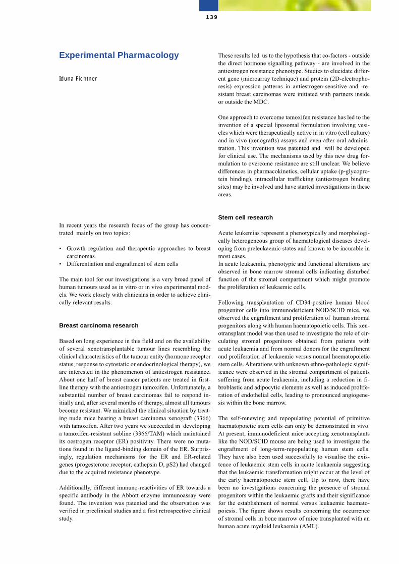
Experimental Pharmacology
Iduna Fichtner
In recent years the research focus of the group has concen-trated mainly on two topics:
• Growth regulation and therapeutic approaches to breastcarcinomas
• Differentiation and engraftment of stem cells
The main tool for our investigations is a very broad panel ofhuman tumours used as in vitro or in vivo experimental mod-els. We work closely with clinicians in order to achieve clini-cally relevant results.
Breast carcinoma research
Based on long experience in this field and on the availabilityof several xenotransplantable tumour lines resembling theclinical characteristics of the tumour entity (hormone receptorstatus, response to cytostatic or endocrinological therapy), weare interested in the phenomenon of antiestrogen resistance.About one half of breast cancer patients are treated in first-line therapy with the antiestrogen tamoxifen. Unfortunately, asubstantial number of breast carcinomas fail to respond in-itially and, after several months of therapy, almost all tumoursbecome resistant. We mimicked the clinical situation by treat-ing nude mice bearing a breast carcinoma xenograft (3366)with tamoxifen. After two years we succeeded in developinga tamoxifen-resistant subline (3366/TAM) which maintainedits oestrogen receptor (ER) positivity. There were no muta-tions found in the ligand-binding domain of the ER. Surpris-ingly, regulation mechanisms for the ER and ER-relatedgenes (progesterone receptor, cathepsin D, pS2) had changeddue to the acquired resistance phenotype.
Additionally, different immuno-reactivities of ER towards aspecific antibody in the Abbott enzyme immunoassay werefound. The invention was patented and the observation wasverified in preclinical studies and a first retrospective clinicalstudy.
These results led us to the hypothesis that co-factors - outsidethe direct hormone signalling pathway - are involved in theantiestrogen resistance phenotype. Studies to elucidate differ-ent gene (microarray technique) and protein (2D-electropho-resis) expression patterns in antiestrogen-sensitive and -re-sistant breast carcinomas were initiated with partners insideor outside the MDC.
One approach to overcome tamoxifen resistance has led to theinvention of a special liposomal formulation involving vesi-cles which were therapeutically active in in vitro (cell culture)and in vivo (xenografts) assays and even after oral adminis-tration. This invention was patented and will be developedfor clinical use. The mechanisms used by this new drug for-mulation to overcome resistance are still unclear. We believedifferences in pharmacokinetics, cellular uptake (p-glycopro-tein binding), intracellular trafficking (antiestrogen bindingsites) may be involved and have started investigations in theseareas.
Stem cell research
Acute leukemias represent a phenotypically and morphologi-cally heterogeneous group of haematological diseases devel-oping from preleukaemic states and known to be incurable inmost cases. In acute leukaemia, phenotypic and functional alterations areobserved in bone marrow stromal cells indicating disturbedfunction of the stromal compartment which might promotethe proliferation of leukaemic cells.
Following transplantation of CD34-positive human bloodprogenitor cells into immunodeficient NOD/SCID mice, weobserved the engraftment and proliferation of human stromalprogenitors along with human haematopoietic cells. This xen-otransplant model was then used to investigate the role of cir-culating stromal progenitors obtained from patients withacute leukaemia and from normal donors for the engraftmentand proliferation of leukaemic versus normal haematopoieticstem cells. Alterations with unknown ethno-pathologic signif-icance were observed in the stromal compartment of patientssuffering from acute leukaemia, including a reduction in fi-broblastic and adipocytic elements as well as induced prolife-ration of endothelial cells, leading to pronounced angiogene-sis within the bone marrow.
The self-renewing and repopulating potential of primitivehaematopoietic stem cells can only be demonstrated in vivo.At present, immunodeficient mice accepting xenotransplantslike the NOD/SCID mouse are being used to investigate theengraftment of long-term-repopulating human stem cells.They have also been used successfully to visualise the exis-tence of leukaemic stem cells in acute leukaemia suggestingthat the leukaemic transformation might occur at the level ofthe early haematopoietic stem cell. Up to now, there havebeen no investigations concerning the presence of stromalprogenitors within the leukaemic grafts and their significancefor the establishment of normal versus leukaemic haemato-poiesis. The figure shows results concerning the occurrenceof stromal cells in bone marrow of mice transplanted with anhuman acute myeloid leukaemia (AML).
139
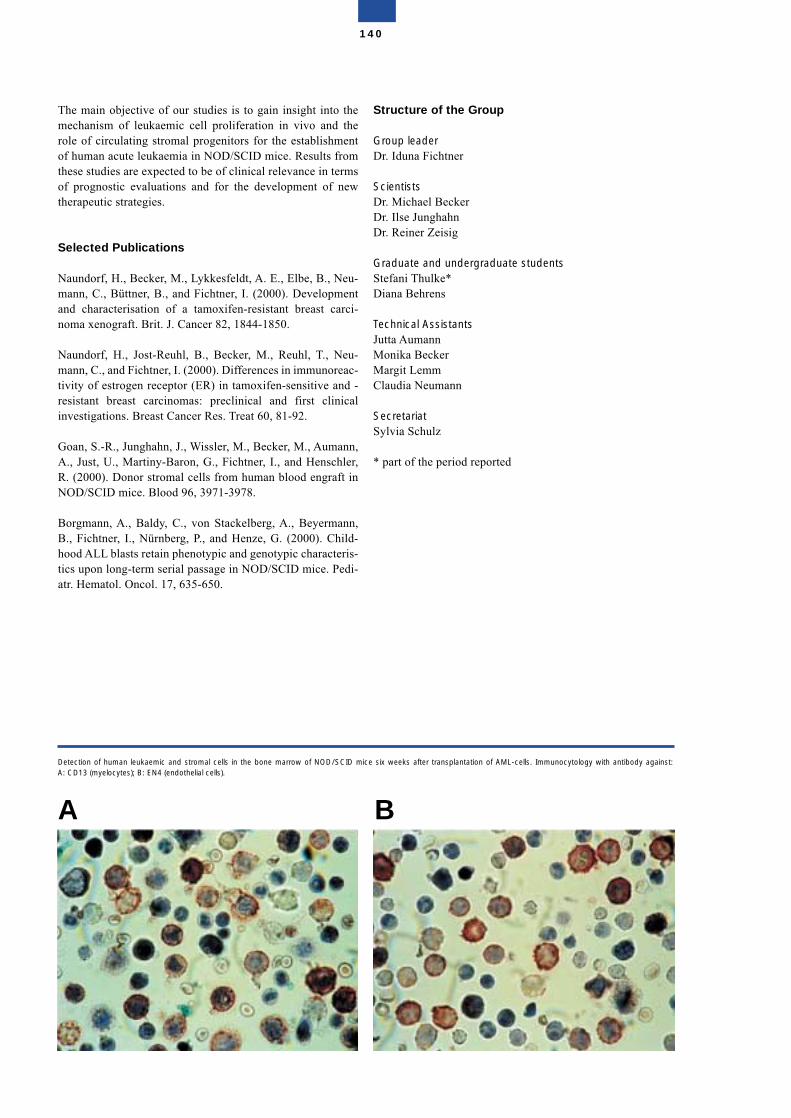
The main objective of our studies is to gain insight into themechanism of leukaemic cell proliferation in vivo and therole of circulating stromal progenitors for the establishmentof human acute leukaemia in NOD/SCID mice. Results fromthese studies are expected to be of clinical relevance in termsof prognostic evaluations and for the development of newtherapeutic strategies.
Selected Publications
Naundorf, H., Becker, M., Lykkesfeldt, A. E., Elbe, B., Neu-mann, C., Büttner, B., and Fichtner, I. (2000). Developmentand characterisation of a tamoxifen-resistant breast carci-noma xenograft. Brit. J. Cancer 82, 1844-1850.
Naundorf, H., Jost-Reuhl, B., Becker, M., Reuhl, T., Neu-mann, C., and Fichtner, I. (2000). Differences in immunoreac-tivity of estrogen receptor (ER) in tamoxifen-sensitive and -resistant breast carcinomas: preclinical and first clinicalinvestigations. Breast Cancer Res. Treat 60, 81-92.
Goan, S.-R., Junghahn, J., Wissler, M., Becker, M., Aumann,A., Just, U., Martiny-Baron, G., Fichtner, I., and Henschler,R. (2000). Donor stromal cells from human blood engraft inNOD/SCID mice. Blood 96, 3971-3978.
Borgmann, A., Baldy, C., von Stackelberg, A., Beyermann,B., Fichtner, I., Nürnberg, P., and Henze, G. (2000). Child-hood ALL blasts retain phenotypic and genotypic characteris-tics upon long-term serial passage in NOD/SCID mice. Pedi-atr. Hematol. Oncol. 17, 635-650.
Structure of the Group
Group leaderDr. Iduna Fichtner
ScientistsDr. Michael BeckerDr. Ilse JunghahnDr. Reiner Zeisig
Graduate and undergraduate studentsStefani Thulke*Diana Behrens
Technical AssistantsJutta AumannMonika BeckerMargit LemmClaudia Neumann
SecretariatSylvia Schulz
* part of the period reported
140
Detection of human leukaemic and stromal cells in the bone marrow of NOD/SCID mice six weeks after transplantation of AML-cells. Immunocytology with antibody against:A: CD13 (myelocytes); B: EN4 (endothelial cells).
A B
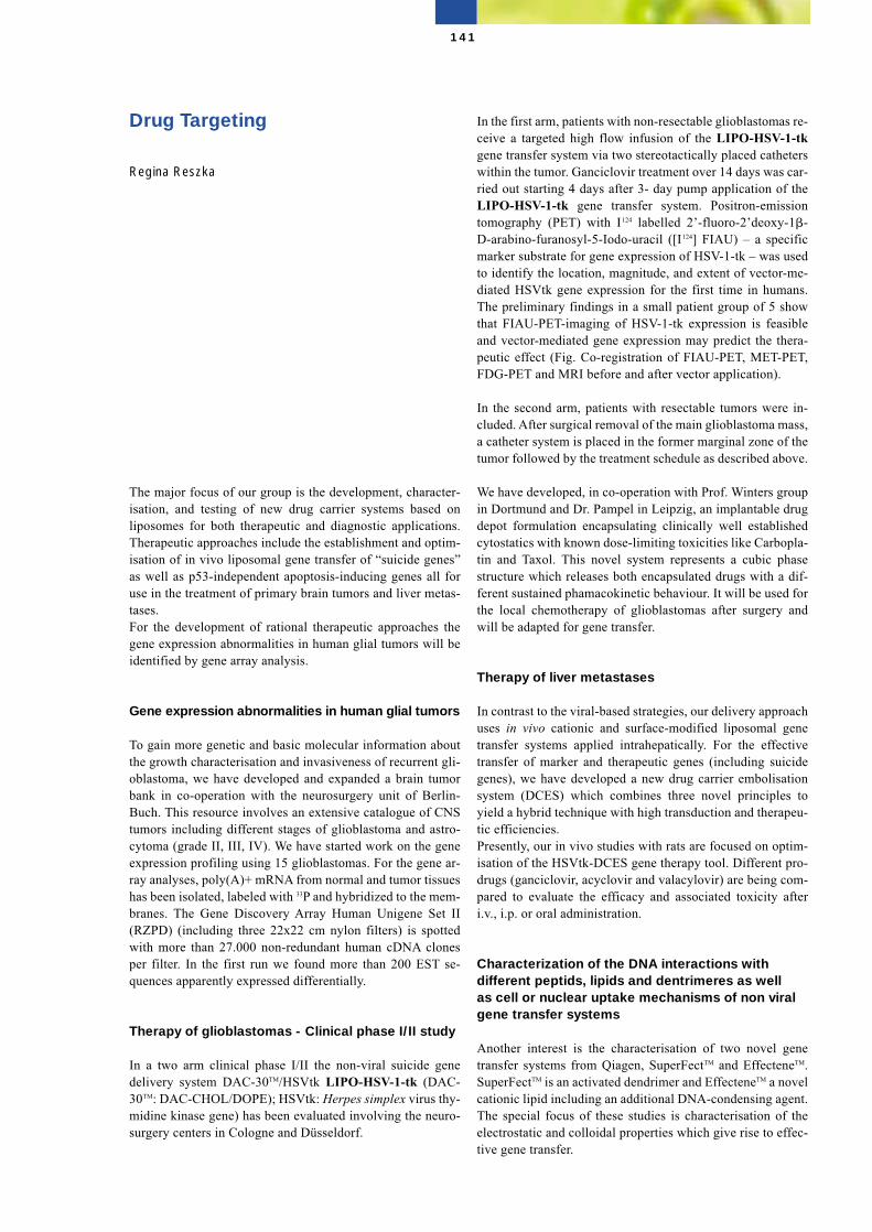
Drug Targeting
Regina Reszka
The major focus of our group is the development, character-isation, and testing of new drug carrier systems based onliposomes for both therapeutic and diagnostic applications.Therapeutic approaches include the establishment and optim-isation of in vivo liposomal gene transfer of “suicide genes”as well as p53-independent apoptosis-inducing genes all foruse in the treatment of primary brain tumors and liver metas-tases.For the development of rational therapeutic approaches thegene expression abnormalities in human glial tumors will beidentified by gene array analysis.
Gene expression abnormalities in human glial tumors
To gain more genetic and basic molecular information aboutthe growth characterisation and invasiveness of recurrent gli-oblastoma, we have developed and expanded a brain tumorbank in co-operation with the neurosurgery unit of Berlin-Buch. This resource involves an extensive catalogue of CNStumors including different stages of glioblastoma and astro-cytoma (grade II, III, IV). We have started work on the geneexpression profiling using 15 glioblastomas. For the gene ar-ray analyses, poly(A)+ mRNA from normal and tumor tissueshas been isolated, labeled with 33P and hybridized to the mem-branes. The Gene Discovery Array Human Unigene Set II(RZPD) (including three 22x22 cm nylon filters) is spottedwith more than 27.000 non-redundant human cDNA clonesper filter. In the first run we found more than 200 EST se-quences apparently expressed differentially.
Therapy of glioblastomas - Clinical phase I/II study
In a two arm clinical phase I/II the non-viral suicide genedelivery system DAC-30TM/HSVtk LIPO-HSV-1-tk (DAC-30TM: DAC-CHOL/DOPE); HSVtk: Herpes simplex virus thy-midine kinase gene) has been evaluated involving the neuro-surgery centers in Cologne and Düsseldorf.
In the first arm, patients with non-resectable glioblastomas re-ceive a targeted high flow infusion of the LIPO-HSV-1-tkgene transfer system via two stereotactically placed catheterswithin the tumor. Ganciclovir treatment over 14 days was car-ried out starting 4 days after 3- day pump application of theLIPO-HSV-1-tk gene transfer system. Positron-emissiontomography (PET) with I124 labelled 2’-fluoro-2’deoxy-1�-D-arabino-furanosyl-5-Iodo-uracil ([I124] FIAU) – a specificmarker substrate for gene expression of HSV-1-tk – was usedto identify the location, magnitude, and extent of vector-me-diated HSVtk gene expression for the first time in humans.The preliminary findings in a small patient group of 5 showthat FIAU-PET-imaging of HSV-1-tk expression is feasibleand vector-mediated gene expression may predict the thera-peutic effect (Fig. Co-registration of FIAU-PET, MET-PET,FDG-PET and MRI before and after vector application).
In the second arm, patients with resectable tumors were in-cluded. After surgical removal of the main glioblastoma mass,a catheter system is placed in the former marginal zone of thetumor followed by the treatment schedule as described above.
We have developed, in co-operation with Prof. Winters groupin Dortmund and Dr. Pampel in Leipzig, an implantable drugdepot formulation encapsulating clinically well establishedcytostatics with known dose-limiting toxicities like Carbopla-tin and Taxol. This novel system represents a cubic phasestructure which releases both encapsulated drugs with a dif-ferent sustained phamacokinetic behaviour. It will be used forthe local chemotherapy of glioblastomas after surgery andwill be adapted for gene transfer.
Therapy of liver metastases
In contrast to the viral-based strategies, our delivery approachuses in vivo cationic and surface-modified liposomal genetransfer systems applied intrahepatically. For the effectivetransfer of marker and therapeutic genes (including suicidegenes), we have developed a new drug carrier embolisationsystem (DCES) which combines three novel principles toyield a hybrid technique with high transduction and therapeu-tic efficiencies.Presently, our in vivo studies with rats are focused on optim-isation of the HSVtk-DCES gene therapy tool. Different pro-drugs (ganciclovir, acyclovir and valacylovir) are being com-pared to evaluate the efficacy and associated toxicity afteri.v., i.p. or oral administration.
Characterization of the DNA interactions withdifferent peptids, lipids and dentrimeres as wellas cell or nuclear uptake mechanisms of non viralgene transfer systems
Another interest is the characterisation of two novel genetransfer systems from Qiagen, SuperFectTM and EffecteneTM.SuperFectTM is an activated dendrimer and EffecteneTM a novelcationic lipid including an additional DNA-condensing agent.The special focus of these studies is characterisation of theelectrostatic and colloidal properties which give rise to effec-tive gene transfer.
141
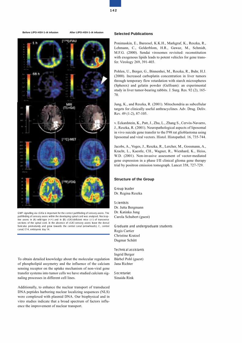
To obtain detailed knowledge about the molecular regulationof phospholipid assymetry and the influence of the calciumsensing receptor on the uptake mechanism of non-viral genetransfer systems into tumor cells we have studied calcium sig-naling processes in different cell lines.
Additionally, to enhance the nuclear transport of transducedDNA peptides harboring nuclear localizing sequences (NLS)were complexed with plasmid DNA. Our biophysical and invitro studies indicate that a broad spectrum of factors influ-ence the improvement of nuclear transport.
Selected Publications
Ponimaskin, E., Bareesel, K.K.H., Markgraf, K., Reszka, R.,Lehmann, C., Gelderblom, H.R., Gawaz, M., Schmidt,M.F.G. (2000). Sendai virosomes revisited: reconstitutionwith exogenous lipids leads to potent vehicles for gene trans-fer. Virology 269, 391-403.
Pohlen, U., Berger, G., Binnenhei, M., Reszka, R., Buhr, H.J.(2000). Increased carboplatin concentration in liver tumorsthrough temporary flow retardation with starch microspheres(Spherex) and gelatin powder (Gelfoam): an experimentalstudy in liver tumor-bearing rabbits. J. Surg. Res. 92 (2), 165-70.
Jung, K., and Reszka, R. (2001). Mitochondria as subcellulartargets for clinically useful anthracyclines. Adv. Drug. Deliv.Rev. 49 (1-2), 87-105.
v. Eckardstein, K., Patt, J., Zhu, L., Zhang S., Cervòs-Navarro,J., Reszka, R. (2001). Neuropathological aspects of liposomalin vivo suicide gene transfer to the F98 rat glioblastoma usingliposomal and viral vectors. Histol. Histopathol. 16, 735-744.
Jacobs, A., Voges, J., Reszka, R., Lercher, M., Gossmann, A.,Kracht, L., Kaestle, CH., Wagner, R., Wienhard, K., Heiss,W.D. (2001). Non-invasive assessment of vector-mediatedgene expression in a phase I/II clinical glioma gene therapytrial by positron emission tomograph. Lancet 358, 727-729.
Structure of the Group
Group leaderDr. Regina Reszka
ScientistsDr. Jutta BergmannDr. Katinka JungCarola Schubert (guest)
Graduate and undergraduate studentsRegis CartierChristine KratzelDagmar Schütt
Technical assistantsIngrid BergerBärbel Pohl (guest)Jana Richter
SecretariatSinaida Rink
142
GMP signalling via cGKIa is important for the correct pathfinding of sensory axons. Thepathfinding of sensory axons within the developing spinal cord was analyzed. Nocicep-tive axons in (A) wild-type (+/+) and in (B) cGKI-deficient mice (-/-) of transversesections of the spinal cord. In the absence of cGKI sensory axons leave the dorsalfuniculus prematurely and grow towards the central canal (arrowheads). C, centralcanal; E14, embryonic day 14.
Before LIPO-HSV-1-tk infusion After LIPO-HSV-1-tk infusion
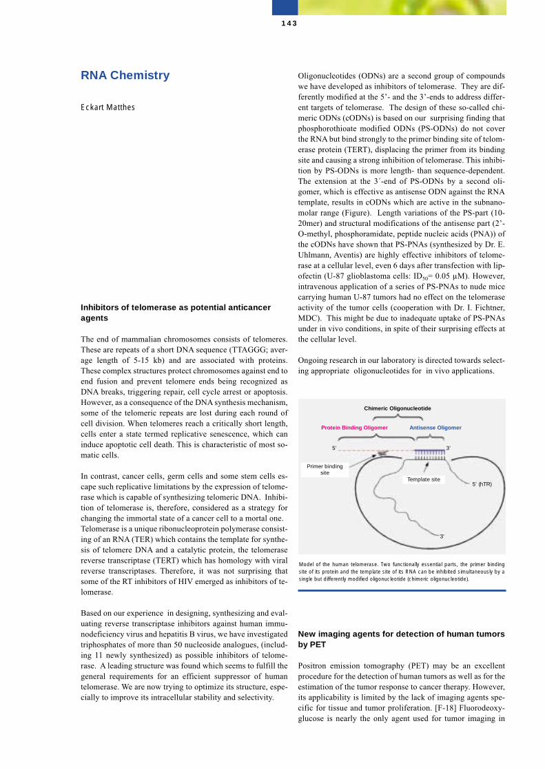
RNA Chemistry
Eckart Matthes
Inhibitors of telomerase as potential anticanceragents
The end of mammalian chromosomes consists of telomeres.These are repeats of a short DNA sequence (TTAGGG; aver-age length of 5-15 kb) and are associated with proteins.These complex structures protect chromosomes against end toend fusion and prevent telomere ends being recognized asDNA breaks, triggering repair, cell cycle arrest or apoptosis.However, as a consequence of the DNA synthesis mechanism,some of the telomeric repeats are lost during each round ofcell division. When telomeres reach a critically short length,cells enter a state termed replicative senescence, which caninduce apoptotic cell death. This is characteristic of most so-matic cells.
In contrast, cancer cells, germ cells and some stem cells es-cape such replicative limitations by the expression of telome-rase which is capable of synthesizing telomeric DNA. Inhibi-tion of telomerase is, therefore, considered as a strategy forchanging the immortal state of a cancer cell to a mortal one.Telomerase is a unique ribonucleoprotein polymerase consist-ing of an RNA (TER) which contains the template for synthe-sis of telomere DNA and a catalytic protein, the telomerasereverse transcriptase (TERT) which has homology with viralreverse transcriptases. Therefore, it was not surprising thatsome of the RT inhibitors of HIV emerged as inhibitors of te-lomerase.
Based on our experience in designing, synthesizing and eval-uating reverse transcriptase inhibitors against human immu-nodeficiency virus and hepatitis B virus, we have investigatedtriphosphates of more than 50 nucleoside analogues, (includ-ing 11 newly synthesized) as possible inhibitors of telome-rase. A leading structure was found which seems to fulfill thegeneral requirements for an efficient suppressor of humantelomerase. We are now trying to optimize its structure, espe-cially to improve its intracellular stability and selectivity.
Oligonucleotides (ODNs) are a second group of compoundswe have developed as inhibitors of telomerase. They are dif-ferently modified at the 5’- and the 3’-ends to address differ-ent targets of telomerase. The design of these so-called chi-meric ODNs (cODNs) is based on our surprising finding thatphosphorothioate modified ODNs (PS-ODNs) do not coverthe RNA but bind strongly to the primer binding site of telom-erase protein (TERT), displacing the primer from its bindingsite and causing a strong inhibition of telomerase. This inhibi-tion by PS-ODNs is more length- than sequence-dependent.The extension at the 3´-end of PS-ODNs by a second oli-gomer, which is effective as antisense ODN against the RNAtemplate, results in cODNs which are active in the subnano-molar range (Figure). Length variations of the PS-part (10-20mer) and structural modifications of the antisense part (2’-O-methyl, phosphoramidate, peptide nucleic acids (PNA)) ofthe cODNs have shown that PS-PNAs (synthesized by Dr. E.Uhlmann, Aventis) are highly effective inhibitors of telome-rase at a cellular level, even 6 days after transfection with lip-ofectin (U-87 glioblastoma cells: ID50= 0.05 µM). However,intravenous application of a series of PS-PNAs to nude micecarrying human U-87 tumors had no effect on the telomeraseactivity of the tumor cells (cooperation with Dr. I. Fichtner,MDC). This might be due to inadequate uptake of PS-PNAsunder in vivo conditions, in spite of their surprising effects atthe cellular level.
Ongoing research in our laboratory is directed towards select-ing appropriate oligonucleotides for in vivo applications.
New imaging agents for detection of human tumorsby PET
Positron emission tomography (PET) may be an excellentprocedure for the detection of human tumors as well as for theestimation of the tumor response to cancer therapy. However,its applicability is limited by the lack of imaging agents spe-cific for tissue and tumor proliferation. [F-18] Fluorodeoxy-glucose is nearly the only agent used for tumor imaging in
143
Model of the human telomerase. Two functionally essential parts, the primer bindingsite of its protein and the template site of its RNA can be inhibited simultaneously by asingle but differently modified oligonucleotide (chimeric oligonucleotide).
Chimeric Oligonucleotide
Template site
Primer bindingsite
Protein Binding Oligomer Antisense Oligomer
5’ (hTR)
3’ 5’
3’
clinical practice but it is more an indicator of glucose metab-olism than of tumor proliferation. Therefore there is an urgentneed for specific tumor imaging agents.
We have designed, synthesized and investigated a series ofmodified thymidine analogues to meet the following biologi-cal and chemical requirements we consider essential for suc-cessive tumor imaging with thymidine analogues.
1. They must be taken up by tumor cells and phosphorylatedwell by thymidine kinase, an enzyme which increasedabout 10-20 fold when cells synthesize DNA. The phos-phorylated products are trapped and accumulate inside ofproliferating cells.
2. The modified thymidine analogues must be resistant to py-rimidine nucleoside degrading enzymes, thereby, avoidingpositron-emitting cleavage products distributing nonspe-cifically in the body.
3. The chemical labeling and purification procedure of traceramounts of the products must be simple and rapid.
We have selected some compounds which seem to be highlyattractive for our aims. One of these analogues is 5–monoflu-oro-methyl-3´-fluorodeoxyuridine, in which the imaging[F-18] fluorine group is introduced into the methyl group. Thenext step will be to adapt the [F-18] labeling procedure to anautomated synthesis. These labeling- and all tomograph-de-pendent preclinical experiments in animal tumor models anda possible pilot study with lung cancer patients will be carriedout in cooperation with the Clinic of Nuclear Medicine ofHumboldt University (Charité), Berlin.
Selected Publications
von Janta-Lipinski, M., Gaertner, K., Lehmann, C., Scherr,H., Schildt, J., and Matthes, E. (1999). Protein and RNA ofhuman telomerase as targets for modified oligonucleotides.Nucleosides & Nucleotides 18, 1719-1720.
Mentel, R., Kurek, S., Wegner, U., von Janta-Lipinski, M.,Gürtler, L., Matthes, E. (2000) Inhibition of adenovirus DNApolymerase by modified nucleoside triphosphate analogs cor-relate with their antiviral effects on cellular level. Med. Mi-crobiol. Immunol. 189, 91-95.
Harnack, U., Lehmann, C., Matthes, E., Pecher, G. (2001)Upregulation of telomerase activity on Herpesvirus saimiriimmortalized human T-lymphocytes. Anticancer Res. 21,3969-3972.
P. M. Schmidt, E. Matthes, F. W. Scheller, F. F. Bier (2001)Nachweis der Telomeraseaktivität in Zellkulturen mittels ei-nes faseroptischen Sensors. Sensorik, Suppl., 47-51.
Structure of the group
Group leaderDr. Eckart Matthes
ScientistDr. Martin von Janta-LipinskiDr. Dieter Bärwolff
Graduate and undergraduate studentsYong WuPeter Lahnert
Technical assistantsKlaus GaertnerChristine LehmannInge KrahnHelga ScheerJürgen SchildtMarianne Stulich
144
145
Molecular and DevelopmentalNeurosciences
Molecular and DevelopmentalNeurosciences
As the average life-expectancy continues to increase in theWestern hemisphere, it is anticipated that the incidence ofage-related disorders, such as Alzheimer's disease, will alsorise. This expectation has stimulated renewed interest in theneurosciences. In the past few decades, significant advanceshave been made in our understanding of the functional basisof the nervous system. Nevertheless, despite the rapid growthin neuroscience research at the international level, consider-able progress remains to be made in the elucidation of thosemolecular events that underly brain disorders.
Due to the immense complexity of the brain, progress inunderstanding the molecular processes of the brain’s functionhas been slow. However, two different approaches havehelped combat this problem. On one hand, Positron EmissionTomography (PET) and Nuclear Magnetic Resonance Imag-ing (NMR) enable neuroscientists to visualize and map activecenters of the brain in relation to specific functions. On theother hand, molecular and cell biology tools are employed tostudy the properties and behavior of single brain cells. Thecombination of these two approaches should lead to an im-proved understanding of higher brain function, and will helpin the design new treatments for the specific degenerativemechanisms which lie at the root of many brain diseases. Theneuroscience program at the MDC is focused on a molecularapproach. This perfectly complements the brain imaging ac-tivities at the Jülich Research Center since both centers con-stitute the neuroscience program of the Helmholtz Society.
The research groups of the MDC neuroscience program focuson both molecular and cellular approaches. Helmut Ketten-mann´s group investigates the role of glial cells in health anddisease, while the Developmental Neurobiology group, underthe direction of Fritz G. Rathjen, analyzes molecular aspectsof axonal growth and synaptic plasticity of the central ner-vous system. Gary R. Lewin and his coworkers are identify-ing novel genes responsible for mechano-transduction andtheir regulation by neurotrophins. Christiane Alexander isidentifying the genes involved in neurodegeneration. GerdKempermann´s group studies stem cells in the adult nervous
Molekulare Neurowissenschaft undEntwicklungsneurobiologie
Da die durchschnittliche Lebenserwartung in der westlichenHemisphäre weiter zunimmt, ist auch damit zu rechnen, dassdie Zahl an altersbedingten Krankheiten wie etwa MorbusAlzheimer weiter steigen wird. Diese Erwartung hat zu einemneuerlich wachsenden Interesse an den Neurowissenschaftengeführt. In den vergangenen Jahrzehnten konnte das Verste-hen der funktionalen Basis des Nervensystems deutlich ver-bessert werden. Nichtsdestoweniger werden unabhängig vondem raschen Wachstum in den Neurowissenschaften auf in-ternationaler Ebene noch viele weitere Fortschritte benötigt,um die molekularen Ereignisse aufzuklären, die den Erkran-kungen des Gehirns zugrunde liegen.
Wegen der ungeheuren Komplexität des Gehirns kommt derFortschritt beim Verständnis der zu den Gehirnvorgängengehörenden molekularen Vorgänge nur langsam voran. ZweiAnsätze konnten inzwischen genutzt werden, um gegen die-ses Problem anzukommen. Da sind auf der einen Seite diePositronen Emissions Tomographie (PET) und die Kernmag-netresonanz-Bildgebung (NMR Imaging, NMRI), mit derenHilfe die Neurowissenschaftler aktive Zentren des Gehirns inRelation zu spezifischen Funktionen sichtbar machen und ak-tiv kartieren. Auf der anderen Seite werden die Werkzeugeder Molekular- und der Zellbiologie benutzt, um die Eigen-schaften und das Verhalten einzelner Nervenzellen zu analy-sieren. Die Kombination beider Ansätze sollte zum einen zueinem verbesserten Verständnis der höheren Hirnfunktionenführen, und sie wird zum zweiten helfen, neue Behandlungs-methoden für die spezifischen degenerativen Mechanismenzu entwickeln, die an der Wurzel vieler Hirnerkrankungenliegen. Die Forschungen am MDC komplettieren auf perfekteWeise die Bildgebungsaktivitäten am Forschungszentrum inJülich, da beide Zentren das neurowissenschaftliche Pro-gramm der Helmholtz Gemeinschaft ausmachen.
Die Forschungsgruppen des MDC-Programms für die Neuro-wissenschaften konzentrieren sich sowohl auf den molekula-ren als auch auf den zellulären Ansatz. Die Gruppe von Hel-mut Kettenmann untersucht die Rolle der Gliazellen ingesundem und kranken Gewebe, während die Gruppe Ent-
146
system in order to understand the role of these cells in brainplasticity. Erich Wanker´s group will use high-throughputfunctional genomics approaches for the identification andcharacterization of novel drug targets involved in neurode-generative disorders, such as Alzheimer’s, Parkinson’s, andHuntington’s diseases. Furthermore, screening assays for theidentification of small molecules preventing protein aggrega-tion in late-onset neurodegenerative diseases will be devel-oped.
The central theme of the MDC is to link basic and clinical re-search. We have, therefore, established collaborations withthe Neurosurgery Department in Berlin-Buch and the Charitéwith a focus on brain tumors and with the Department ofNeurology at the Charité with a focus on brain inflammationand neural stem cell research. These interactions are encour-aged since we are part of the Collaborative Research Center(Sonderforschungsbereich) and the Graduiertenkolleg at theCharité established to study the role of brain cells in the path-ogenesis of CNS diseases.
wicklungsneurobiologie unter der Leitung von Fritz G. Rathjendie molekularen Aspekte des axonalen Wachstums und dersynaptischen Plastizität des Zentralen Nervensystems ana-lysiert. Gary R. Lewin und seine Mitarbeiter identifizierenneuartige Gene, die für die mechanische Übertragung undihre Regulierung durch Neurotrophine verantwortlich sind.Christiane Alexander befaßt sich mit der Identifizierung vonGenen, die an der Neurodegeneration beteiligt sind. GerdKempermanns Gruppe studiert Stammzellen im erwachsenenNervensystem, um die Rolle dieser Zellen in der Plastizitätdes Gehirns zu verstehen. Erich Wankers Gruppe wird dieneuen Ansätze der funktionalen Genomik mit hohen Durch-laufraten einsetzen, um neuartige Ziele für solche Arzneimit-tel zu identifizieren und zu charakterisieren, die Einfluß aufneurodegenerative Erkrankungen wie Alzheimer, Parkinsonoder Huntington haben. Darüber hinaus werden Verfahren fürReihenuntersuchungen entwickelt, mit denen kleine Mole-küle identifiziert werden können, die die Proteinaggregationverhindern, die bei spät einsetzenden neurodegenerativen Er-krankungen beobachtet werden.
Das zentrale Thema des MDC ist die Verbindung von grund-legender und klinischer Forschung. Wir haben daher Kolla-borationen zum einen mit der neurochirurgischen Abteilungin Berlin-Buch und der Charité begonnen, in deren FokusHirntumoren stehen, und wir haben zum zweiten eine Kolla-boration mit der Abteilung für Neurobiologie an der Charitébegonnen, in deren Fokus die Erforschung von Hirnentzün-dungen und neuronalen Stammzellen steht. Diese Wechsel-wirkungen werden durch die Tatsache ermutigt, dass wir zueinem Sonderforschungsbereich und dem Graduiertenkolleggehören, die beide von der Charité eingerichtet worden sind,um die Rolle von Hirnzellen in der Pathogenese der Erkran-kungen des Zentralen Nervensystems zu erkunden.
147
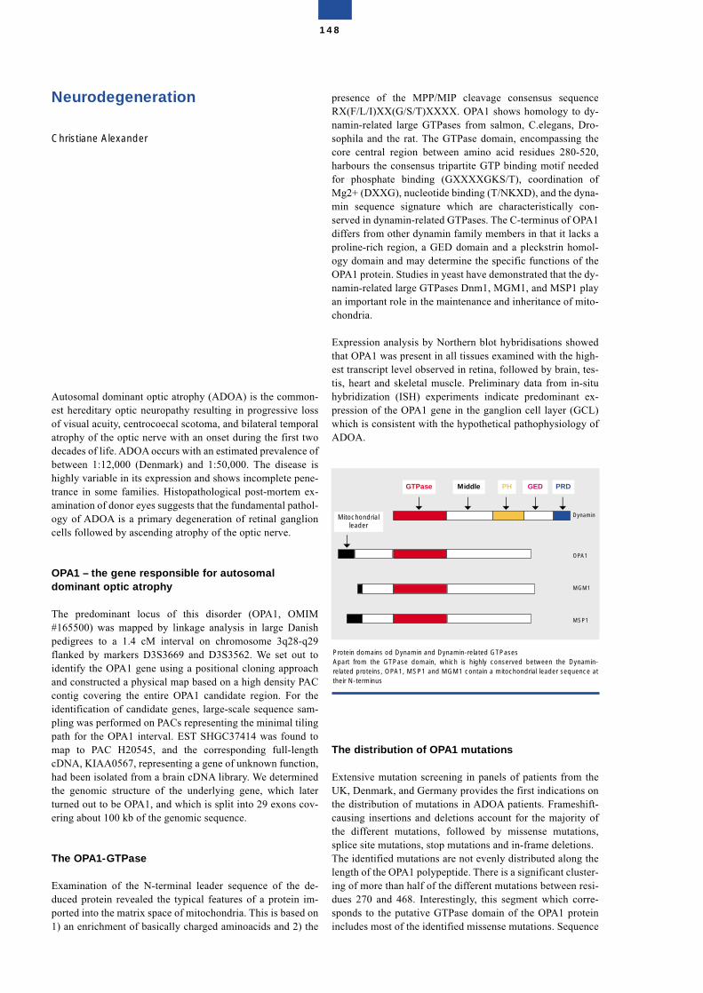
Neurodegeneration
Christiane Alexander
Autosomal dominant optic atrophy (ADOA) is the common-est hereditary optic neuropathy resulting in progressive lossof visual acuity, centrocoecal scotoma, and bilateral temporalatrophy of the optic nerve with an onset during the first twodecades of life. ADOA occurs with an estimated prevalence ofbetween 1:12,000 (Denmark) and 1:50,000. The disease ishighly variable in its expression and shows incomplete pene-trance in some families. Histopathological post-mortem ex-amination of donor eyes suggests that the fundamental pathol-ogy of ADOA is a primary degeneration of retinal ganglioncells followed by ascending atrophy of the optic nerve.
OPA1 – the gene responsible for autosomal dominant optic atrophy
The predominant locus of this disorder (OPA1, OMIM#165500) was mapped by linkage analysis in large Danishpedigrees to a 1.4 cM interval on chromosome 3q28-q29flanked by markers D3S3669 and D3S3562. We set out toidentify the OPA1 gene using a positional cloning approachand constructed a physical map based on a high density PACcontig covering the entire OPA1 candidate region. For theidentification of candidate genes, large-scale sequence sam-pling was performed on PACs representing the minimal tilingpath for the OPA1 interval. EST SHGC37414 was found tomap to PAC H20545, and the corresponding full-lengthcDNA, KIAA0567, representing a gene of unknown function,had been isolated from a brain cDNA library. We determinedthe genomic structure of the underlying gene, which laterturned out to be OPA1, and which is split into 29 exons cov-ering about 100 kb of the genomic sequence.
The OPA1-GTPase
Examination of the N-terminal leader sequence of the de-duced protein revealed the typical features of a protein im-ported into the matrix space of mitochondria. This is based on1) an enrichment of basically charged aminoacids and 2) the
presence of the MPP/MIP cleavage consensus sequenceRX(F/L/I)XX(G/S/T)XXXX. OPA1 shows homology to dy-namin-related large GTPases from salmon, C.elegans, Dro-sophila and the rat. The GTPase domain, encompassing thecore central region between amino acid residues 280-520,harbours the consensus tripartite GTP binding motif neededfor phosphate binding (GXXXXGKS/T), coordination ofMg2+ (DXXG), nucleotide binding (T/NKXD), and the dyna-min sequence signature which are characteristically con-served in dynamin-related GTPases. The C-terminus of OPA1differs from other dynamin family members in that it lacks aproline-rich region, a GED domain and a pleckstrin homol-ogy domain and may determine the specific functions of theOPA1 protein. Studies in yeast have demonstrated that the dy-namin-related large GTPases Dnm1, MGM1, and MSP1 playan important role in the maintenance and inheritance of mito-chondria.
Expression analysis by Northern blot hybridisations showedthat OPA1 was present in all tissues examined with the high-est transcript level observed in retina, followed by brain, tes-tis, heart and skeletal muscle. Preliminary data from in-situhybridization (ISH) experiments indicate predominant ex-pression of the OPA1 gene in the ganglion cell layer (GCL)which is consistent with the hypothetical pathophysiology ofADOA.
The distribution of OPA1 mutations
Extensive mutation screening in panels of patients from theUK, Denmark, and Germany provides the first indications onthe distribution of mutations in ADOA patients. Frameshift-causing insertions and deletions account for the majority ofthe different mutations, followed by missense mutations,splice site mutations, stop mutations and in-frame deletions. The identified mutations are not evenly distributed along thelength of the OPA1 polypeptide. There is a significant cluster-ing of more than half of the different mutations between resi-dues 270 and 468. Interestingly, this segment which corre-sponds to the putative GTPase domain of the OPA1 proteinincludes most of the identified missense mutations. Sequence
148
Protein domains od Dynamin and Dynamin-related GTPasesApart from the GTPase domain, which is highly conserved between the Dynamin-related proteins, OPA1, MSP1 and MGM1 contain a mitochondrial leader sequence attheir N-terminus
Mitochondrialleader
Dynamin
OPA1
MGM1
MSP1
GTPase Middle PH GED PRD
➝ ➝ ➝ ➝ ➝
➝
variations affecting the binding of GTP or GTP hydrolysisseem not to be tolerated and result in disease symptoms.Moreover, minor mutation clusters have been observed in theN-terminal leader sequence, as well as in the C-terminus ofthe OPA1 protein.
Identification of a founder effect in the Danish population
An important discovery was the identification of a foundermutation in about 50% of the Danish ADOA patients, whichcould explain the higher incidence of the disease in Denmarkcompared with other countries. The founder mutation is lo-cated in the last coding exon of the OPA1 gene, exon 28.
Selected Publications
Alexander, C., Votruba, M., Pesch U.E.A., Thiselton, D.L.,Mayer, S., Moore, A., Rodriguez, M., Kellner, U., Leo-Kot-tler, B., Auburger, G., Bhattacharya, S.S., Wissinger, B.(2000). OPA1, encoding a dynamin-related GTPase, is mu-tated in autosomal dominant optic atrophy linked to chromo-some 3q28. Nature Genet. 26, 211-215.
Alexander, C., Bernstein, S.L., Rocchi, M., Auburger, G.(2001). Saturating density of STSs (1/6 kb) in a 1.1 Mb regionon 3q28-q29: a valuable resource for cloning of diseasegenes. Eur. J. Hum. Genet. 9 (4), 307-10.
Pesch, U.E., Leo-Kottler, B., Mayer, S., Jurklies, B., Kellner,U., Apfelstedt-Sylla, E., Zrenner, E., Alexander, C., Wis-singer, B. (2001). OPA1 mutations in patients with autosomaldominant optic atrophy and evidence for semi-dominant in-heritance. Hum. Mol. Genet.10 (13), 1359-68.
Thiselton, D.L., Alexander, C., Morris, A., Brooks, S., Rosen-berg, T., Eiberg, H., Kjer, B., Kjer, P., Bhattacharya, S.S., Vo-truba, M. (2001). A frameshift mutation in exon 28 of theOPA1 gene explains the high prevalence of dominant opticatrophy in the Danish population: evidence for a founder ef-fect. Hum. Genet. 109 (5): 498-502
Structure of the Group
Group leaderDr. Christiane Alexander
Technical AssistantIska Liebner
Graduate and undergraduate studentsMaja FiketVasudheva Akepati
149
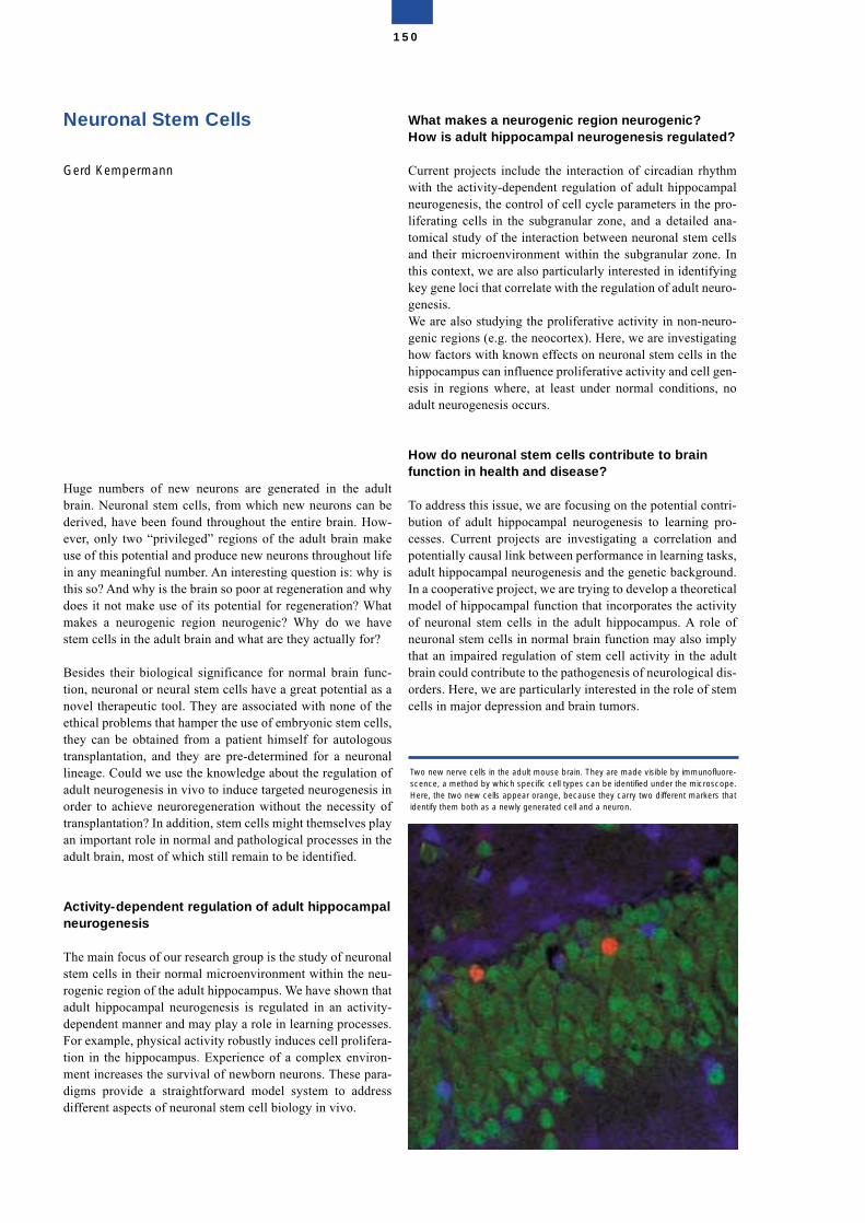
Neuronal Stem Cells
Gerd Kempermann
Huge numbers of new neurons are generated in the adultbrain. Neuronal stem cells, from which new neurons can bederived, have been found throughout the entire brain. How-ever, only two “privileged” regions of the adult brain makeuse of this potential and produce new neurons throughout lifein any meaningful number. An interesting question is: why isthis so? And why is the brain so poor at regeneration and whydoes it not make use of its potential for regeneration? Whatmakes a neurogenic region neurogenic? Why do we havestem cells in the adult brain and what are they actually for?
Besides their biological significance for normal brain func-tion, neuronal or neural stem cells have a great potential as anovel therapeutic tool. They are associated with none of theethical problems that hamper the use of embryonic stem cells,they can be obtained from a patient himself for autologoustransplantation, and they are pre-determined for a neuronallineage. Could we use the knowledge about the regulation ofadult neurogenesis in vivo to induce targeted neurogenesis inorder to achieve neuroregeneration without the necessity oftransplantation? In addition, stem cells might themselves playan important role in normal and pathological processes in theadult brain, most of which still remain to be identified.
Activity-dependent regulation of adult hippocampalneurogenesis
The main focus of our research group is the study of neuronalstem cells in their normal microenvironment within the neu-rogenic region of the adult hippocampus. We have shown thatadult hippocampal neurogenesis is regulated in an activity-dependent manner and may play a role in learning processes.For example, physical activity robustly induces cell prolifera-tion in the hippocampus. Experience of a complex environ-ment increases the survival of newborn neurons. These para-digms provide a straightforward model system to addressdifferent aspects of neuronal stem cell biology in vivo.
What makes a neurogenic region neurogenic? How is adult hippocampal neurogenesis regulated?
Current projects include the interaction of circadian rhythmwith the activity-dependent regulation of adult hippocampalneurogenesis, the control of cell cycle parameters in the pro-liferating cells in the subgranular zone, and a detailed ana-tomical study of the interaction between neuronal stem cellsand their microenvironment within the subgranular zone. Inthis context, we are also particularly interested in identifyingkey gene loci that correlate with the regulation of adult neuro-genesis. We are also studying the proliferative activity in non-neuro-genic regions (e.g. the neocortex). Here, we are investigatinghow factors with known effects on neuronal stem cells in thehippocampus can influence proliferative activity and cell gen-esis in regions where, at least under normal conditions, noadult neurogenesis occurs.
How do neuronal stem cells contribute to brainfunction in health and disease?
To address this issue, we are focusing on the potential contri-bution of adult hippocampal neurogenesis to learning pro-cesses. Current projects are investigating a correlation andpotentially causal link between performance in learning tasks,adult hippocampal neurogenesis and the genetic background.In a cooperative project, we are trying to develop a theoreticalmodel of hippocampal function that incorporates the activityof neuronal stem cells in the adult hippocampus. A role ofneuronal stem cells in normal brain function may also implythat an impaired regulation of stem cell activity in the adultbrain could contribute to the pathogenesis of neurological dis-orders. Here, we are particularly interested in the role of stemcells in major depression and brain tumors.
150
Two new nerve cells in the adult mouse brain. They are made visible by immunofluore-scence, a method by which specific cell types can be identified under the microscope.Here, the two new cells appear orange, because they carry two different markers thatidentify them both as a newly generated cell and a neuron.
The research group “Neuronal stem cells” works in closeinteraction with the independent group “Neurogenic permis-siveness”, also headed by Gerd Kempermann, funded byVolkswagen Stiftung and located at the Dept. of Neurology,Charité University-Hospital, Berlin. The goal of this interac-tion is to allow clinicians to pursue stem cell research involv-ing close interaction with the clinic, but within the scientificenvironment provided by the MDC.
Selected Publications
Kempermann, G. (2002) Regulation of adult hippocampalneurogenesis — implications for novel theories of major de-pression. Bipolar Disorders, in press.
Kohl, Z., Kuhn, H.G., Cooper-Kuhn, C.M., Winkler, J.,Aigner, L., and Kempermann, G. (2002) Preweaning enrich-ment has no lasting effect on adult hippocampal neurogenesisin four-month old mice. Genes, Brain and Behavior 1, 46-54.
Horner, P.J., Power, A.E., Kempermann, G., Kuhn, H.G.,Palmer, T.D., Winkler, J., Thal, L.J., and Gage, F.H. (2000)Proliferation and differentiation of progenitor cells through-out the intact adult rat spinal cord. Journal of Neuroscience20, 2218-1128.
van Praag, H., Kempermann, G., and Gage, F.H. (2000) Neu-ral consequences of environmental enrichment. Nature Re-views Neuroscience 1, 191-198.
van Praag, H., Kempermann, G., and Gage, F.H. (1999) Run-ning increases cell proliferation and neurogenesis in the adultmouse dentate gyrus. Nature Neuroscience 2, 266-270.
Structure of the Group
Group leaderDr. Gerd Kempermann
ScientistsDr. Gudrun LutschDr. Golo KronenbergBarbara SteinerAnika Bick-SanderSebastian JessbergerDr. Gerold Brüning (visiting scientist)
Graduate and undergraduate studentsDan EhningerMoritz BrandtMelissa HolmesZacharias Kohl
Technical AssistantsDaniela GastErika KotitschkeRuth SegnerIrene Thun
151
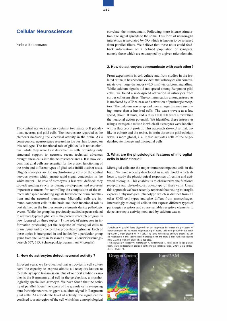
Cellular Neurosciences
Helmut Kettenmann
The central nervous system contains two major cell popula-tions, neurons and glial cells. The neurons are regarded as theelements mediating the electrical activity in the brain. As aconsequence, neuroscience research in the past has focused onthis cell type. The functional role of glial cells is not as obvi-ous: while they were first described as cells providing onlystructural support to neurons, recent technical advancesbrought these cells into the neuroscience arena. It is now evi-dent that glial cells are essential for the proper functioning ofthe brain and different types of glial cells fulfill distinct tasks.Oligodendrocytes are the myelin-forming cells of the centralnervous system which ensure rapid signal conduction in thewhite matter. The role of astrocytes is less well defined; theyprovide guiding structures during development and representimportant elements for controlling the composition of the ex-tracellular space mediating signals between the brain endothe-lium and the neuronal membrane. Microglial cells are im-muno-competent cells in the brain and their functional role isbest defined as the first responsive elements during pathologicevents. While the group has previously studied aspects relatedto all three types of glial cells, the present research program isnow focussed on three topics: (1) the role of astrocytes in in-formation processing (2) the response of microglial cells tobrain injury and (3) the cellular properties of gliomas. Each ofthese topics is intergrated in and funded by a particular groupgrant from the German Research Council (Sonderforschungs-bereich 507, 515, Schwerpunktprogramm on Microglia).
1. How do astrocytes detect neuronal activity ?
In recent years, we have learned that astrocytes in cell culturehave the capacity to express almost all receptors known tomediate synaptic transmission. One of our best studied exam-ples is the Bergmann glial cell in the cerebellum, a morpho-logically specialized astrocyte. We have found that the activ-ity of parallel fibers, the axons of the granule cells synapsingonto Purkinje neurons, triggers a calcium signal in Bergmannglial cells. At a moderate level of activity, the signal can beconfined to a subregion of the cell which has a morphological
correlate, the microdomain. Following more intense stimula-tion, the signal spreads to the soma. This form of neuron-gliainteraction is mediated by NO which is known to be releasedfrom parallel fibers. We believe that these units could feed-back information on a defined population of synapses,namely those which are enwrapped by a given microdomain.
2. How do astrocytes communicate with each other?
From experiments in cell culture and from studies in the iso-lated retina, it has become evident that astrocytes can commu-nicate over large distances (<0.5 mm) via calcium signalling.While calcium signals did not spread among Bergmann glialcells., we found a wide-spread activation in astrocytes fromcorpus callosum slices. The communication among astrocytesis mediated by ATP release and activation of purinergic recep-tors. The calcium waves spread over a large distance involv-ing more than a hundred cells. The wave travels at a lowspeed, about 10 mm/s, and is thus 1 000 000 times slower thanthe neuronal action potential. We identified these astrocytesusing a transgenic mouse in which all astrocytes were labelledwith a fluorescent protein. This approach showed us that, un-like in culture and the retina, in brain tissue the glial calciumwave is more global, i. e. it also activates cells of the oligo-dendrocyte lineage and microglial cells.
3. What are the physiological features of microglialcells in brain tissue?
Microglial cells are the major immunocompetent cells in thebrain. We have recently developed an in situ model which al-lows to study the physiological responses of resting and acti-vated microglia. This enables us to characterize the funtionalreceptors and physiological phenotype of these cells. Usingthis approach we have recently reported that resting microgliaexpress a physiological phenotype which is distinct from allother CNS cell types and also differs from macrophages.Interestingly microglial cells in situ express different types ofpurinergic receptors and so are suitable receptive elements todetect astrocyte activity mediated by calcium waves.
152
Stimulation of parallel fibers triggered calcium responses in somata and processes ofBergmann glia cells. To record responses in processes, cells were perfused via a patchpipet with Oregon green BAPTA-1 (left). The soma (white spot) and two processes canbe recognized in this color-coded micrograph. On the right, a slice with bulk-loaded(Fura-2/AM) Bergmann glial cells is depicted.From Matyash V, Filippov V, Mohrhagen K, Kettenmann H. Nitric oxide signals parallelfiber activity to Bergmann glial cells in the mouse cerebellar slice. (2001) Mol Cell Neu-rosci. 18:664-70.
4. What are the factors controlling the properties ofastrocytes in pathological conditions?
Astrocytes respond with reactive gliosis to brain injury indi-cated by an increase in proliferation and migration to the le-sion site. To study factors controlling astrocyte migration, wehave examined how cultured astrocytes migrate into a cell-free zone produced by a scratch within a cell monolayer.Blocking ryanodine receptors, intracellular calcium-releasechannels, strongly attenuated the migratory activity of astro-cytes. RT-PCR analysis, Western blotting and immunocyto-chemistry revealed that cultured and acutely isolated astrocy-tes express exclusively the ryanodine receptor 3 isotype(RyR3). In cultures prepared from RyR3-deficient mice, as-trocyte motility was significantly impaired and ryanodine didnot induce any further decrease in the rate of cell migration.These results indicate that calcium signaling via RyR3 is animportant factor for the control of astrocyte migration.
5. What are the factors controlling the properties ofmicroglial cells in pathological conditions?
A candidate for signalling neuronal injury to microglial cellsis the CC chemokine CCL21 since damaged neurons expressCCL21. Investigating microglia in acute slices and in culture,we have demonstrated that CCL21 triggers an increase inCl-conductance. Moreover, CCL21 triggers a chemotactic re-sponse which is sensitive to Cl-channel blockers. Both typesof response are mediated by activation of CXCR3 and notCCR7 receptors indicating that in brain, CCL21 acts via a dif-ferent receptor system to that in lymphoid tissue.
6. What are the physiological properties of gliomasand how do they compare with normal glia?
The majority of tumors of the central nervous system arethought to originate from glial cells. These include astrocyto-mas, oligodendrogliomas and the most malignant (and un-treatable) brain tumor, the glioblastoma multiforme. We arestudying the cellular properties of these tumor cells and com-paring them with normal glial cells with respect to their phys-iological properties, their ability to proliferate and to migrate.Recently, we focused on the expression of GABA receptorsby glioma cells since we have found a very strong correlationbetween the expression pattern and the malignancy of the tu-mor. Only cells from tumors with low malignancy expressGABA receptors, and all tumor cell lines which are selectedfor high proliferative activity lack this receptor. We have evi-dence that contact with neurons triggers the induction of thisreceptor and the expression is correlated with an decrease inmetabolic activity.
Selected Publications
Boucsein C., Kettenmann, H., and Nolte, C. (2000). Electro-physiological properties of microglial cells in normal andpathologic rat brain slices. Eur. J. Neurosci., 12, 2049-2058.
Lyons, S. A., Pastor, A., Kann, O., Ohlemeyer, C., Wiegand,F., Prass, K., Knapp, F., Kettenmann, H. and Dirnagl U.(2000) Distinct physiological properties of microglia andblood-borne cells in rat brain slices after permanent middlecerebral artery occlusion. J. Cer. Blood Flow Met., 20:1537-1549.
Matyash, M., Matyash, V,. Nolte, C., Sorrentino, V. and Ket-tenmann H. Requirement of functional ryanodine receptortype 3 for astrocyte migration, (2002). FASEB J., 16, 84-86.
Nolte, C., Matyash, M., Pivneva, P., Schipke, C., Ohlemeyer,C., Hanisch, U.-K., Kirchhoff, F., and H. Kettenmann (2001).GFAP promotor controlled EGFP expressing transgenic mice:a tool to visualize astrocytes and astrogliosis in living braintissue, GLIA 33, 72–86.
Schipke C., Ohlemeyer C., Matyash M., Nolte C., Ketten-mann H., and Kirchhoff F. (2001). Astrocytes of the mousecortex express functional N-methyl-D-aspartate receptors,FASEB J. 15, 1270-1272
Structure of the Group
Group leaderProf. Dr. Helmut Kettenmann
ScientistsDr. Clemens BoucseinDr. Vitali FilipovDr. Uwe-Karsten HanischDr. Anja HoffmannSusanne KuhnDr. Christiane NolteDr. Christopher RoseDr. Susanne ArnoldKatrin FärberDr. Oliver Peters
Graduate and undergraduate studentsAgnes EllinghausGeorg HäuslerWolfgang KresseSanja PavlovicAngelika RappertCarola SchipkeRobert Zacharias
Technical AssistantsSilke FleischhauerChristiane Gras/Regina HehlBrigitte GerlachGerda MüllerMichaela Seeger (part time)Horst Kagelmaker
SecretariatBirgit Jarchow
153
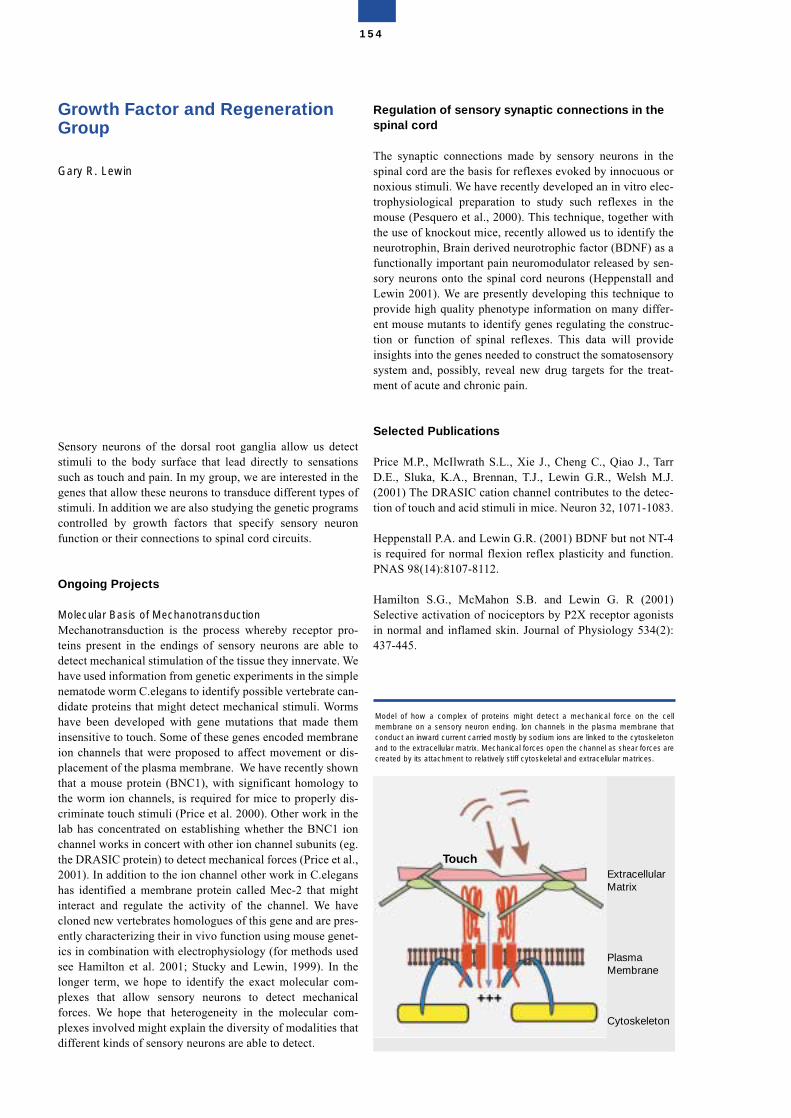
Growth Factor and RegenerationGroup
Gary R. Lewin
Sensory neurons of the dorsal root ganglia allow us detectstimuli to the body surface that lead directly to sensationssuch as touch and pain. In my group, we are interested in thegenes that allow these neurons to transduce different types ofstimuli. In addition we are also studying the genetic programscontrolled by growth factors that specify sensory neuronfunction or their connections to spinal cord circuits.
Ongoing Projects
Molecular Basis of MechanotransductionMechanotransduction is the process whereby receptor pro-teins present in the endings of sensory neurons are able todetect mechanical stimulation of the tissue they innervate. Wehave used information from genetic experiments in the simplenematode worm C.elegans to identify possible vertebrate can-didate proteins that might detect mechanical stimuli. Wormshave been developed with gene mutations that made theminsensitive to touch. Some of these genes encoded membraneion channels that were proposed to affect movement or dis-placement of the plasma membrane. We have recently shownthat a mouse protein (BNC1), with significant homology tothe worm ion channels, is required for mice to properly dis-criminate touch stimuli (Price et al. 2000). Other work in thelab has concentrated on establishing whether the BNC1 ionchannel works in concert with other ion channel subunits (eg.the DRASIC protein) to detect mechanical forces (Price et al.,2001). In addition to the ion channel other work in C.eleganshas identified a membrane protein called Mec-2 that mightinteract and regulate the activity of the channel. We havecloned new vertebrates homologues of this gene and are pres-ently characterizing their in vivo function using mouse genet-ics in combination with electrophysiology (for methods usedsee Hamilton et al. 2001; Stucky and Lewin, 1999). In thelonger term, we hope to identify the exact molecular com-plexes that allow sensory neurons to detect mechanicalforces. We hope that heterogeneity in the molecular com-plexes involved might explain the diversity of modalities thatdifferent kinds of sensory neurons are able to detect.
Regulation of sensory synaptic connections in thespinal cord
The synaptic connections made by sensory neurons in thespinal cord are the basis for reflexes evoked by innocuous ornoxious stimuli. We have recently developed an in vitro elec-trophysiological preparation to study such reflexes in themouse (Pesquero et al., 2000). This technique, together withthe use of knockout mice, recently allowed us to identify theneurotrophin, Brain derived neurotrophic factor (BDNF) as afunctionally important pain neuromodulator released by sen-sory neurons onto the spinal cord neurons (Heppenstall andLewin 2001). We are presently developing this technique toprovide high quality phenotype information on many differ-ent mouse mutants to identify genes regulating the construc-tion or function of spinal reflexes. This data will provideinsights into the genes needed to construct the somatosensorysystem and, possibly, reveal new drug targets for the treat-ment of acute and chronic pain.
Selected Publications
Price M.P., McIlwrath S.L., Xie J., Cheng C., Qiao J., TarrD.E., Sluka, K.A., Brennan, T.J., Lewin G.R., Welsh M.J.(2001) The DRASIC cation channel contributes to the detec-tion of touch and acid stimuli in mice. Neuron 32, 1071-1083.
Heppenstall P.A. and Lewin G.R. (2001) BDNF but not NT-4is required for normal flexion reflex plasticity and function.PNAS 98(14):8107-8112.
Hamilton S.G., McMahon S.B. and Lewin G. R (2001)Selective activation of nociceptors by P2X receptor agonistsin normal and inflamed skin. Journal of Physiology 534(2):437-445.
154
TouchExtracellularMatrix
PlasmaMembrane
Cytoskeleton
Model of how a complex of proteins might detect a mechanical force on the cellmembrane on a sensory neuron ending. Ion channels in the plasma membrane thatconduct an inward current carried mostly by sodium ions are linked to the cytoskeletonand to the extracellular matrix. Mechanical forces open the channel as shear forces arecreated by its attachment to relatively stiff cytoskeletal and extracellular matrices.
Price M.P., Lewin G.R., McIlwrath S.L., Cheng C., Xie J.,Heppenstall P.A., Stucky C.L., Mannsfeldt A.G., BrennanT.J., Drummond H.A., Qiao J., Benson C.J., Tarr D.E., HrstkaR.F., Yang B. Williamson R.A., Welsh M.J. (2000) The Mam-malian Na+ Channel BNC1 Is Required for Normal TouchSensation. Nature 407:1007-11
Pesquero J.B., Araujo, R.C, Heppenstall, P.A., Stucky, C.L.,Silva Jr.A., Walther, T., Kettritz, R., Oliveira, S. M., Pes-quero, J.L., Paiva, A.C., Calixto,, J.B., Lewin, G.R., Bader,M. (2000) Hypoalgesia and altered inflammatory responses inmice lacking kinin B1 receptors. PNAS 97: 8140-8145.
Structure of the Group
Group LeaderDr. Gary R. Lewin
ScientistsDr. Paul HeppenstallDr. Andreas EilersDr. Jing HuDr. Carlos Martinez-Salgado
Graduate StudentsAnne Mannsfeldt*Sabrina McIlwrath*Jung-Bum ShinChristiane WetzelGireesh Anirudhan*
Technical AssistantsAnke KanehlHeike ThränhardtJacqueline Klewer*
* part of time reported
155
Synapse Formation and Function
Frank W. Pfrieger
A major aim of neurobiological research is to understand theformation and function of chemical synapses, highly special-ized intercellular connections that mediate the exchange ofelectrical signals between neurons. Our group focuses on theidentification of the signals and mechanisms that control thedevelopment of synapses in the mammalian central nervoussystem (CNS).
Identification of cholesterol as a glia-derived factorpromoting synapse development in CNS neurons.
The top priority of our group has been to identify a glia-de-rived factor that promotes synapse development in retinalganglion cells (RGCs) and to analyse its mode of action. Theidentification of this factor has been long-awaited, ever sincethe publication by Pfrieger & Barres (1997) and there hasbeen increased interest due to recent publications (Nägler etal., 2001; Ullian et al. 2001). Recently, we succeeded in iden-tifying the factor by the use of two parallel experimental ap-proaches. Firstly, we partially purified the component fromglia-conditioned medium by a series of chromatographic sep-aration steps using electrophysiological recordings of synap-tic activity as a bioassay. Secondly, we detected glia-inducedchanges in the protein composition of neuronal membranesby two-dimensional gel electrophoresis and mass spectrome-try. Surprisingly, we found that cholesterol released by glialcells in apolipoprotein E-containing lipoproteins completelymimicked the effects of glia-conditioned medium on synapsedevelopment (Mauch et al., 2001).
Since cholesterol homeostasis in the brain is independent ofblood levels, our results suggest the following scenario:RGCs appear to produce enough cholesterol to support theirvital cellular functions and form axons and dendrites, butrequire large additional amounts to develop numerous andmature synaptic connections. Consequently, they depend oncholesterol synthesis by glial cells and its delivery via apolip-oprotein E-containing lipoproteins. These findings have twoimportant implications: firstly, they suggest a new role for
glial cells that may explain, why massive synaptogenesis inthe CNS occurs just after differentiation of macroglial cells,and why glial cells secrete cholesterol-rich lipoproteins at all;secondly, they provide the first evidence for an exciting linkbetween cholesterol homeostasis and the development andplasticity of synaptic connections, which may help to explainsome of the neurologic symptoms resulting from genetic de-fects in cholesterol or lipoprotein metabolism.
A new method to isolate CNS neurons
The development of neurons is controlled by extrinsic signalsfrom non-neuronal cells, particularly from glia. One possibleway to analyse the underlying signaling pathways is to separ-ate neurons from these cells and to monitor their developmentin culture under strictly defined conditions. However, this el-egant approach has been hampered by the lack of suitablemethods to isolate CNS neurons. So far, only RGCs havebeen separated from other cells in sufficiently high numbers.In this project, we aimed to establish a new method to obtainhighly purified neurons from postnatal mouse hippocampusand to culture these under strictly defined conditions in theabsence of non-neuronal cells (Nägler & Pfrieger, submitted).We found that mouse hippocampal neurons can be isolated in99.9% purity by a modified version of the immunopanningprotocol that can be used to isolate RGCs. This new cell cul-ture preparation will allow us, for the first time, to define thefundamental survival and differentiation requirements ofthese key CNS neurons.
Selected Publications
Mauch DH, Nägler K, Schumacher S, Göritz C, Müller EC,Otto A, Pfrieger FW (2001) CNS synaptogenesis promoted byglia-derived cholesterol. Science 294: 1354-1357.
Nägler K, Mauch DH, Pfrieger FW (2001) Glia-derived sig-nals induce synapse formation in neurones of the rat centralnervous system. J Physiol 533: 665-679.
Structure of the Group
Group LeaderDr. Frank W. Pfrieger
Graduate and Undergraduate StudentsChristian GöritzDaniela MauchKarl Nägler
Technical AssistantsIrene HauptJacqueline Klewer
Present addressDr. Frank W. Pfrieger, MPG-CNRS Group, Centre de Neuro-chimie, F-67084 Strasbourg cedex, 5, rue Blaise Pascal,e-mail: [email protected]
156
Developmental Neurobiology
Fritz G. Rathjen
Activity-dependent and –independent processesregulate the pattern of neuronal connectivity
The formation of precise and selective synaptic connectionsbetween neurons is essential for the proper functioning of thenervous system. The establishment of these synaptic connec-tions throughout the nervous system involves a complex se-ries of events. During embryonic development growth cones,the tips of extending axons, respond to an array of molecularsignals that are present in their microenvironment. These dis-tinct signals are mediated and integrated by so-called guid-ance receptors on the surface of growth cones to activateintracellular signal transduction pathways that modulate theactin-myosin cytoskeleton. Several classes of guidance recep-tors have been shown to be implicated in these processes in-cluding neural members of the immunoglobulin superfamily(IgSF), semaphorins, netrins, ephrins, neuropilins, plexins,Eph-kinases and several extracellular matrix proteins.
Once the initial pattern of neuronal connectivity has been es-tablished by axonal guidance receptors, these synaptic con-nections are subsequently refined by processes dependent onthe electric activity of neurons. It is likely that simple behavi-ours such as reflexes are mediated by neuronal circuits thatare completely formed in animals without any need of neuro-nal activity and will not be re-shaped. In contrast, circuits thatare responsible for more complex behaviours, including thosethat are essentially human, might be influenced to a substan-tial degree by experience-dependent activity. Experience, assoon as it is transduced into neuronal action potentials, ap-pears to modulate the wiring of the nervous system to suit theunique needs of its owner. It is, therefore, a fascinating ques-tion of how epigenetic influences, such as neuronal activity,interact with genetic instructions to form and modify circuitswithin the nervous system.
Axonal IgSF members
The most diverse class of proteins that is implicated in con-tact-dependent regulation of axon guidance are the neuralmembers of the IgSF which can be categorized into severalstructural subclasses including the L1- and F11-subgroups.Functional in vitro studies and intriguing in vivo observationsin chicks, mice and in humans have indicated that these pro-teins are indeed important for the correct wiring of the ner-vous system. Our research in the past grant period on thisclass of proteins has focussed on their intracellular sorting,disease models and the identification of novel members.
The L1 protein, which is the first member of a subfamily ofthe IgSF (L1, neurofascin, NrCAM and CHL1), has beendemonstrated to be involved in hereditary brain disorder witha relatively broad spectrum of symptoms. It has been termedX-linked hydrocephalus, MASA syndrome or spastic paraple-gia type I and includes mental retardation, hydrocephalus, hy-poplasia of the corticalspinal tract and an underdevelopedcorpus callosum. As a first step towards an understanding ofthe molecular mechanisms of these diseases, we investigatedhow mutations in the L1 gene influence binding of differentligands and intracellular trafficking of the L1 protein (in col-laboration with S. Kenwrick, Cambridge). These studiesshowed that different disease-associated mutations have dis-tinct effects on heterophilic ligand binding as well as on theexpression of L1 on the cell surface. Our results suggest thatsome aspects of patient pathology are due to disturbances ininterneuronal interactions.
To study the function of NrCAM, a member of the L1 sub-family, in an in vivo situation we generated and analyzed amouse strain deficient for this gene. We found that the ab-sence of NrCAM causes the formation of mature cataracts inthe mouse. Cataracts, the most common cause of visual im-pairment, are generated in NrCAM-deficient mice by a disor-ganization of lens fibers, followed by cellular disintegrationand accumulation of cellular debris. The disorganization oflens fiber cells includes abnormalities of the cytoskeleton andconnexin50-containing gap junctions.
To identify proteins that interact with L1 subfamily membersintracellularly and therefore might link L1 members to traf-ficking, spatial targeting or signalling pathways we used yeasttwo-hybrid screening. We have demonstrated that syntenin-1,an intracellular protein containing two PDZ domains, interactsspecifically with the cytoplasmic segment of neurofascin butnot with other L1-subfamily members. Although the biologi-cal function of this interaction remains to be defined it is con-ceivable that syntenin-1 might function by linking bound neu-rofascin to trafficking or recycling pathways in neural cells.
By a PCR-based screen we identified FAR-2, a novel F11/F3/contactin-related protein of the IgSF that is expressed bysubsets of cerebellar Purkinje cells, predominantly in the cau-dal part of the chick cerebellum. FAR-2-positive clusters ofPurkinje cells alternate with FAR-2-negative clusters in bothtangential dimensions of the cerebellar cortex, along the ros-trocaudal axis and along the mediolateral axis. The patch-work-like expression pattern of FAR-2 in the cerebellar cor-tex, in combination with its structural similarity to
157
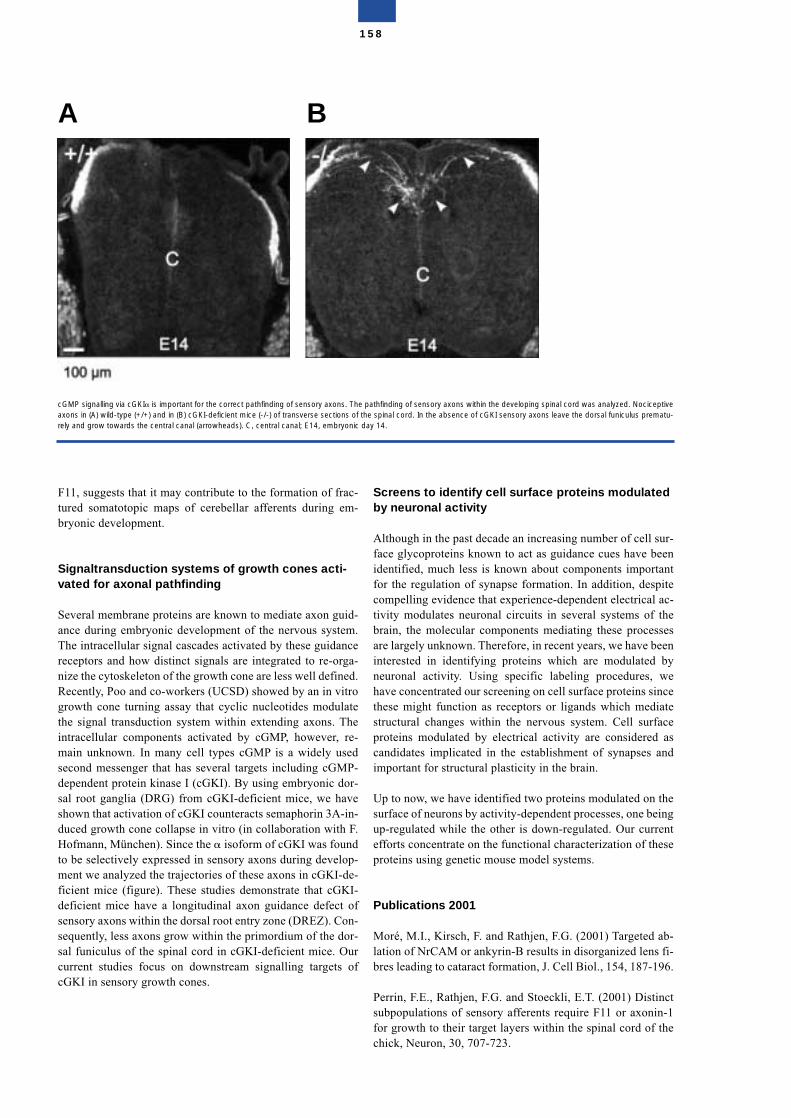
F11, suggests that it may contribute to the formation of frac-tured somatotopic maps of cerebellar afferents during em-bryonic development.
Signaltransduction systems of growth cones acti-vated for axonal pathfinding
Several membrane proteins are known to mediate axon guid-ance during embryonic development of the nervous system.The intracellular signal cascades activated by these guidancereceptors and how distinct signals are integrated to re-orga-nize the cytoskeleton of the growth cone are less well defined.Recently, Poo and co-workers (UCSD) showed by an in vitrogrowth cone turning assay that cyclic nucleotides modulatethe signal transduction system within extending axons. Theintracellular components activated by cGMP, however, re-main unknown. In many cell types cGMP is a widely usedsecond messenger that has several targets including cGMP-dependent protein kinase I (cGKI). By using embryonic dor-sal root ganglia (DRG) from cGKI-deficient mice, we haveshown that activation of cGKI counteracts semaphorin 3A-in-duced growth cone collapse in vitro (in collaboration with F.Hofmann, München). Since the � isoform of cGKI was foundto be selectively expressed in sensory axons during develop-ment we analyzed the trajectories of these axons in cGKI-de-ficient mice (figure). These studies demonstrate that cGKI-deficient mice have a longitudinal axon guidance defect ofsensory axons within the dorsal root entry zone (DREZ). Con-sequently, less axons grow within the primordium of the dor-sal funiculus of the spinal cord in cGKI-deficient mice. Ourcurrent studies focus on downstream signalling targets ofcGKI in sensory growth cones.
Screens to identify cell surface proteins modulatedby neuronal activity
Although in the past decade an increasing number of cell sur-face glycoproteins known to act as guidance cues have beenidentified, much less is known about components importantfor the regulation of synapse formation. In addition, despitecompelling evidence that experience-dependent electrical ac-tivity modulates neuronal circuits in several systems of thebrain, the molecular components mediating these processesare largely unknown. Therefore, in recent years, we have beeninterested in identifying proteins which are modulated byneuronal activity. Using specific labeling procedures, wehave concentrated our screening on cell surface proteins sincethese might function as receptors or ligands which mediatestructural changes within the nervous system. Cell surfaceproteins modulated by electrical activity are considered ascandidates implicated in the establishment of synapses andimportant for structural plasticity in the brain.
Up to now, we have identified two proteins modulated on thesurface of neurons by activity-dependent processes, one beingup-regulated while the other is down-regulated. Our currentefforts concentrate on the functional characterization of theseproteins using genetic mouse model systems.
Publications 2001
Moré, M.I., Kirsch, F. and Rathjen, F.G. (2001) Targeted ab-lation of NrCAM or ankyrin-B results in disorganized lens fi-bres leading to cataract formation, J. Cell Biol., 154, 187-196.
Perrin, F.E., Rathjen, F.G. and Stoeckli, E.T. (2001) Distinctsubpopulations of sensory afferents require F11 or axonin-1for growth to their target layers within the spinal cord of thechick, Neuron, 30, 707-723.
158
cGMP signalling via cGKI� is important for the correct pathfinding of sensory axons. The pathfinding of sensory axons within the developing spinal cord was analyzed. Nociceptiveaxons in (A) wild-type (+/+) and in (B) cGKI-deficient mice (-/-) of transverse sections of the spinal cord. In the absence of cGKI sensory axons leave the dorsal funiculus prematu-rely and grow towards the central canal (arrowheads). C, central canal; E14, embryonic day 14.
A B
Schumacher, S., Jung, M.,Nörenberg, U., Dorner, A., Chi-quet-Ehrismann, R., Stuermer, C.A.O., and Rathjen, F.G.(2001) CALEB Binds via its Acidic Stretch to the Fibrinogen-like Domain of Tenascin-C or Tenascin-R and its ExpressionIs Dynamically Regulated after Optic Nerve Lesion. J. Biol.Chem., 276, 7337-7345.
Koroll, M., Rathjen, F.G. and Volkmer, H (2001) The neuralcell recognition molecule neurofascin interacts with syntenin-1 but not with syntenin-2, both of which reveal self-associat-ing activity, J. Biol. Chem., 276, 10646-10654.
Brümmendorf, T. and Lemmon, V. (2001) IgSF receptors:cis-interactions, intracellular adapters and alternative splic-ing. Curr.Opin. Cell Biol., 13, 611-618.
De Angelis, E., Brümmendorf, T., Cheng, L., Lemmon, V. andKenwrick, S. (2001) Alternative use of a mini exon of the L1gene affects L1 binding to neural ligands. J. Biol. Chem., 276,32738-32742.
De Angelis, E., Watkins, A., Schäfer, M., Brümmendorf, T.and Kenwrick, S. (2002) Disease-associated mutations in L1-CAM interfere with ligand interactions and cell surface ex-pression. Hum.Mol.Genet., 11, 1-12.
Plagge, A., Sendtner-Voelderndorff, L., Sirim, P., Rader, C.,Freigang, J., Sonderegger, P. and Brümmendorf, T. (2001)The contactin-related protein FAR-2 defines Purkinje cellclusters and labels subpopulations of climbing fibers in thedeveloping cerebellum. Mol.Cell.Neurosci., 18, 91-107.
Structure of the Group
Group leaderProf. Dr. Fritz G. Rathjen
ScientistsDr. Armin DornerDr. René JüttnerDr. Michael KorollDr. Margret Moré2)
Dr. Ursel Nörenberg1)
Dr. Ute Zacharias
1) deceased2) maternity leave
Graduate studentsDebashish DasSusanne SchäfferKamal Sharma
Technical assistantsHannelore DrechslerMechthild HenningFrank-Peter Kirsch
SecretariatBirgit Cloos (part time)
Associated Research GroupGroup leaderDr. Thomas Brümmendorf*
Graduate studentMichael Schäfer
Technicial assistantDieter Jobsky
* until 11. 2001
159
Proteomics and Molecular Mechanisms of NeurodegenerativeDisorders
Erich Wanker
The accumulation of highly insoluble intra- and extracellularprotein aggregates is a common feature of progressive late-onset neurodegenerative disorders including Alzheimer’s,Parkinson’s, and Huntington’s disease. Although the causalrelationship between protein aggregation and neurodegenera-tion has not been proven, within recent years several lines ofbiochemical and genetic evidence have suggested that theprocess of aggregate formation in patient brains is linked toneuronal dysfunction and disease progression. The main ob-jective of our work is to understand the pathophysiologicalmechanisms of late-onset neurodegenerative disorders and todevelop causal therapies. Another goal of our research is tounderstand the normal function of proteins involved in neuro-degenerative disorders. We use high-throughput functionalgenomics approaches for the identification of large protein-protein networks and novel drug targets. Furthermore, in vitroand in vivo functional assays are being developed for high-throughput drug screening.
Polyglutamine aggregation in Huntington’s disease
Huntington’s disease (HD) is caused by an elongated polyglu-tamine (polyQ) sequence within a large protein, huntingtin, ofunknown function. We have found that N-terminal huntingtinfragments with polyQ tracts in the pathological range (>37glutamines), but not with polyQ tracts in the normal range(20-30 glutamines) form high molecular weight protein ag-gregates with a fibrillar morphology. The self-assembly ofhuntingtin fibrils in vitro depends highly on the polyQ repeatlength, protein concentration and time. Furthermore, aggre-gate formation can be seeded by preformed fibrils, suggestingthat huntingtin protein aggregates in neuronal cells areformed by a nucleation-dependent process. To study hunting-tin aggregation in vivo, a cell culture model system of HD hasbeen developed. In mammalian cells, N-terminal huntingtinfragments with polyQ tracts in the pathological range (51 or83 glutamines) accumulated in aggresome-like perinuclearinclusion bodies. Interestingly, inhibition of proteasome ac-tivity with lactacystin resulted in a twofold increase in the
amount of ubiquitinated huntingtin aggregates, indicating thatthe activity of the ubiquitin-proteasome system is critical forthe accumulation of mutant huntingtin protein in vivo. More-over, immunofluorescence and electron microscopy revealedthat the 20S, 19S and 11S subunits of the 26S proteasome co-localize with the perinuclear inclusions. Thus, our findingssupport the hypothesis that the ubiquitin-proteasome systemis a potential target for therapeutic interventions in HD andrelated glutamine repeat disorders.
Identification of polyglutamine aggregation inhibitors by high-throughput screening
We suggest that the process of protein aggregation is a keystep in the development of neurodegenerative disorders.Thus, preventing aggregate formation must slow down dis-ease progression. In order to identify huntingtin aggregationinhibitors, we have developed a sensitive filter retardation as-say which is suitable for high-throughput drug screening. Us-ing this assay we first tested a number of known inhibitors of�-amyloid, PrPscr, and microtubule fibril formation for theireffect on huntingtin aggregation. We found that Congo red,thioflavine S, chrysamine G and Direct fast yellow are potentinhibitors of polyQ aggregation and suppress the self-assem-bly of HD exon 1 protein in a dose-dependent manner. Theseresults were confirmed in cell culture models of HD and wealso showed that the monoclonal antibody 1C2, which specif-ically recognizes the elongated polyQ stretch, is a potent in-hibitor of huntingtin aggregation. Using an automated filterretardation assay, we tested ~184,000 chemical compoundsand identified about 700 small molecules that prevent hun-tingtin aggregation in vitro. Currently, these compounds arebeing tested in cell culture model systems of HD.
Chaperones are potent inhibitors of protein aggre-gation in neurodegenerative disorders
It is well known that molecular chaperones prevent misfold-ing and aggregation of disease proteins. We have found that acombination of the heat shock proteins Hsp70 and Hsp40 ismost effective in inhibiting huntingtin aggregation in vitroand in mammalian cell culture model systems. In addition,treatment of mammalian cells with geldanamycin, a naturallyoccuring antitumor drug, induced the expression of Hsp70and Hsp40 and blocked huntingtin aggregation in a dose-de-pendent manner. This was the first demonstration that deposi-tion of mutant huntingtin in cells can be suppressed by chem-ical compounds activating a specific heat shock response. Ourfindings may provide the basis for the development of a novelpharmacotherapy for HD and related glutamine repeat disor-ders.
Analysis of huntingtin function and dysfunction
In order to understand huntingtin function we have searchedfor interacting proteins using the yeast two-hybrid system. Wehave found that the proteins HIP1 and SH3GL3 specificallyinteract with huntingtin. HIP1 is homologous to the yeast pro-tein Sla2p, which is associated with the membrane cytoskele-
160
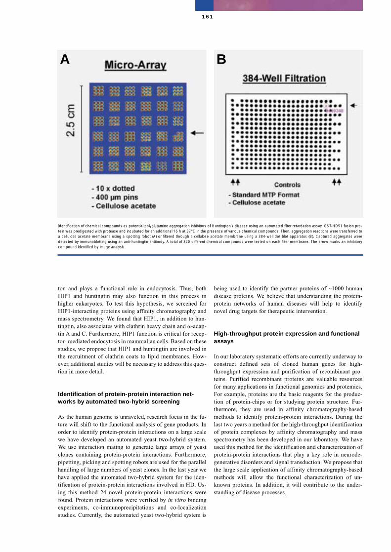
ton and plays a functional role in endocytosis. Thus, bothHIP1 and huntingtin may also function in this process inhigher eukaryotes. To test this hypothesis, we screened forHIP1-interacting proteins using affinity chromatography andmass spectrometry. We found that HIP1, in addition to hun-tingtin, also associates with clathrin heavy chain and �-adap-tin A and C. Furthermore, HIP1 function is critical for recep-tor- mediated endocytosis in mammalian cells. Based on thesestudies, we propose that HIP1 and huntingtin are involved inthe recruitment of clathrin coats to lipid membranes. How-ever, additional studies will be necessary to address this ques-tion in more detail.
Identification of protein-protein interaction net-works by automated two-hybrid screening
As the human genome is unraveled, research focus in the fu-ture will shift to the functional analysis of gene products. Inorder to identify protein-protein interactions on a large scalewe have developed an automated yeast two-hybrid system.We use interaction mating to generate large arrays of yeastclones containing protein-protein interactions. Furthermore,pipetting, picking and spotting robots are used for the parallelhandling of large numbers of yeast clones. In the last year wehave applied the automated two-hybrid system for the iden-tification of protein-protein interactions involved in HD. Us-ing this method 24 novel protein-protein interactions werefound. Protein interactions were verified by in vitro bindingexperiments, co-immunoprecipitations and co-localizationstudies. Currently, the automated yeast two-hybrid system is
being used to identify the partner proteins of ~1000 humandisease proteins. We believe that understanding the protein-protein networks of human diseases will help to identifynovel drug targets for therapeutic intervention.
High-throughput protein expression and functionalassays
In our laboratory systematic efforts are currently underway toconstruct defined sets of cloned human genes for high-throughput expression and purification of recombinant pro-teins. Purified recombinant proteins are valuable resourcesfor many applications in functional genomics and protemics.For example, proteins are the basic reagents for the produc-tion of protein-chips or for studying protein structure. Fur-thermore, they are used in affinity chromatography-basedmethods to identify protein-protein interactions. During thelast two years a method for the high-throughput identificationof protein complexes by affinity chromatography and massspectrometry has been developed in our laboratory. We haveused this method for the identification and characterization ofprotein-protein interactions that play a key role in neurode-generative disorders and signal transduction. We propose thatthe large scale application of affinity chromatography-basedmethods will allow the functional characterization of un-known proteins. In addition, it will contribute to the under-standing of disease processes.
161
Identification of chemical compounds as potential polyglutamine aggregation inhibitors of Huntington’s disease using an automated filter retardation assay. GST-HD51 fusion pro-tein was predigested with protease and incubated for an additional 16 h at 37°C in the presence of various chemical compounds. Then, aggregation reactions were transferred toa cellulose acetate membrane using a spotting robot (A) or filtered through a cellulose acetate membrane using a 384-well dot blot apparatus (B). Captured aggregates weredetected by immunoblotting using an anti-huntingtin antibody. A total of 320 different chemical compounds were tested on each filter membrane. The arrow marks an inhibitorycompound identified by image analysis.
A B
Selected Publications
Heiser, V., Scherzinger, E., Boeddrich, A., Nordhoff, E., Lurz,R., Schugardt, N., Lehrach, H., and Wanker, E. E. (2000). In-hibition of huntingtin fibrillogenesis by specific antibodiesand small molecules: implications for Huntington's diseasetherapy. Proc Natl Acad Sci U S A 97, 6739-6744.
Muchowski, P. J., Schaffar, G., Sittler, A., Wanker, E. E.,Hayer-Hartl, M. K., and Hartl, F. U. (2000). Hsp70 and hsp40chaperones can inhibit self-assembly of polyglutamine pro-teins into amyloid-like fibrils. Proc Natl Acad Sci U S A 97,7841-7846.
Sittler, A., Lurz, R., Lueder, G., Priller, J., Hayer-Hartl, M. K.,Lehrach, H., Hartl, F. U., and Wanker, E. E. (2001). Gelda-namycin activates a heat shock response and inhibits hunting-tin aggregation in a cell culture model of Huntington's dis-ease. Hum Mol Genet 10, 1307-1315.
Waelter, S., Boeddrich, A., Lurz, R., Scherzinger, E., Lueder,G., Lehrach, H., and Wanker, E. E. (2001). Accumulation ofmutant huntingtin fragments in aggresome-like inclusionbodies as a result of insufficient protein degradation. MolBiol Cell 12, 1393-1407.
Waelter, S., Scherzinger, E., Hasenbank, R., Nordhoff, E.,Lurz, R., Goehler, H., Gauss, C., Sathasivam, K., Bates, G. P.,Lehrach, H., and Wanker, E. E. (2001). The huntingtin inter-acting protein HIP1 is a clathrin and alpha-adaptin- bindingprotein involved in receptor-mediated endocytosis. Hum MolGenet 10, 1807-1817.
Structure of the Group
Group leaderProf. Dr. Erich Wanker
ScientistsDr. Annett BöddrichDr. Anja DrögeDr. Sabine EngemannDr. Klaus GenserDr. Eva-Christine MüllerSusanne KostkaDr. Albrecht OttoDr. Martin StrödickeUwe Worm
Graduate and undergraduate studentsMansur AssadiBianca BauerAnne BuschFigen ErtasHeike GöhlerChihab KloseStephanie Wälter
Technical AssistantsClaudia AbrahamGerlinde GrelleRenate HasenbankMaria KnoblichNancy SchugardtChristina Steffens
SecretariatErika Pisch
162

163
Structure and Organization
Organizational Structure
As provided by §7 of its charter the organizational structureof the Max Delbrück Center for Molecular Medicine (MDC)are the following:
• the Board of Trustees with the Scientific Committee,• the Management Board.
The Board of Trustees
As provided by § 8 of the Charter of the Foundation, the Bo-ard of Trustees ensures that the transactions of the Foundationare conducted in a lawful, expedient and financially responsi-ble manner. The Board determines within the framework ofthe law the broad research objectives and the main researchpolicy and financial matters of the Foundation, lays downprinciples of management and the principles for evaluatingresults, intervenes appropriately within the decisions of theBoard of Management, and directs the Board of Managementin special matters of research policy and finances.
Furthermore, the Board of Trustees approves annual and ex-tended budgets (including expansion and investment pro-grams), draws up the Charter and decides upon amendmentsto it, decides upon the dissolution of the Foundation, and ta-kes decisions in other cases provided for in the Law and theCharter.
Members of the Board of Trustees
Parliamentary State Secretary Wolf-Michael Catenhusen(Chair)Federal Ministry of Education and Research, Berlin/Bonn
State Secretary Prof. Dr. Ingolf Hertel (Vice-Chair)Senate Administration for Science, Research and Culture,Berlin (until March 2000)
State Secretary Dr. Josef Lange (Vice-Chair)Senate Administration for Science, Research and Culture,Berlin (April 2000 – June 2001)
State Secretary Dr. Bernd Köppl (Vice-Chair)Senate Administration for Science, Research and Culture,Berlin (August 2001 – February 2002)
State Secretary Dr. Peer Pasternack (Vice-Chair)Senate Administration for Science, Research and Culture,Berlin (since March 2002)
Dr. Jürgens BehrensMax Delbrück Center for Molecular Medicine (MDC) Berlin-Buch, Berlin (until March 2001)
Prof. Dr. Günter Breithardt Medical Clinic, University of Münster, Münster (since October 2001)
Prof. Dr. Hans R. Brunner C.H.U. Vaudois, Division of Hypertension, Lausanne, Swit-zerland
Dietmar BürgenerFederal Ministry of Finances, Bonn/Berlin (until April 2000)
Prof. Dr. Manfred DietelDirector of the Clinical Board of the Charité, Medical Facultyof the Humboldt University of Berlin, Berlin (since January2001)
Dr. Reinhold FörsterMax Delbrück Center for Molecular Medicine (MDC) Berlin-Buch, Berlin (until March 2001)
Prof. Dr. Bärbel FriedrichInstitute for Biology and Microbiology, Humboldt Universityof Berlin, BerlinVice President Deutsche Forschungsgemeinschaft (since October 2001)
Prof. Dr. Peter GaehtgensPresident of the Free University of Berlin, Berlin
Dr. Michael HackenbrochFederal Ministry of Education and Research, Bonn/Berlin
Prof. Dr. Dr. Falko Herrmann Institute for Human Genetics, University of Greifswald,Greifswald
Senator Beate HübnerSenate Administration of Health, Berlin (March 2000 –February 2002)
Senator Dr. Heidi Knake-WernerSenate Administration of Health, Berlin (since March 2002)
164
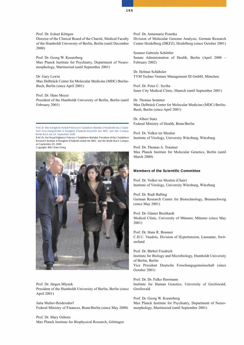
Prof. Dr. Eckart KöttgenDirector of the Clinical Board of the Charité, Medical Facultyof the Humboldt University of Berlin, Berlin (until December2000)
Prof. Dr. Georg W. KreutzbergMax Planck Institute for Psychiatry, Department of Neuro-morphology, Martinsried (until September 2001)
Dr. Gary LewinMax Delbrück Center for Molecular Medicine (MDC) Berlin-Buch, Berlin (since April 2001)
Prof. Dr. Hans Meyer President of the Humboldt University of Berlin, Berlin (untilFebruary 2001)
Prof. Dr. Jürgen MlynekPresident of the Humboldt University of Berlin, Berlin (sinceApril 2001)
Jutta Multer-Heidersdorf Federal Ministry of Finances, Bonn/Berlin (since May 2000)
Prof. Dr. Mary OsbornMax Planck Institute for Biophysical Research, Göttingen
Prof. Dr. Annemarie PoustkaDivision of Molecular Genome Analysis, German ResearchCenter Heidelberg (DKFZ), Heidelberg (since October 2001)
Senator Gabriele SchöttlerSenate Administration of Health, Berlin (April 2000 –February 2002)
Dr. Helmut SchühslerTVM Techno Venture Management III GmbH, München
Prof. Dr. Peter C. ScribaInner City Medical Clinic, Munich (until September 2001)
Dr. Thomas SommerMax Delbrück Center for Molecular Medicine (MDC) Berlin-Buch, Berlin (since April 2001)
Dr. Albert Statz Federal Ministry of Health, Bonn/Berlin
Prof. Dr. Volker ter MeulenInstitute of Virology, University Würzburg, Würzburg
Prof. Dr. Thomas A. TrautnerMax Planck Institute for Molecular Genetics, Berlin (untilMarch 2000)
Members of the Scientific Committee
Prof. Dr. Volker ter Meulen (Chair)Institute of Virology, University Würzburg, Würzburg
Prof. Dr. Rudi BallingGerman Research Centre for Biotechnology, Braunschweig(since May 2001)
Prof. Dr. Günter Breithardt Medical Clinic, University of Münster, Münster (since May2001)
Prof. Dr. Hans R. Brunner C.H.U. Vaudois, Division of Hypertension, Lausanne, Swit-zerland
Prof. Dr. Bärbel FriedrichInstitute for Biology and Microbiology, Humboldt Universityof Berlin, BerlinVice President Deutsche Forschungsgemeinschaft (sinceOctober 2001)
Prof. Dr. Dr. Falko Herrmann Institute for Human Genetics, University of Greifswald,Greifswald
Prof. Dr. Georg W. KreutzbergMax Planck Institute for Psychiatry, Department of Neuro-morphology, Martinsried (until September 2001)
165
Prof. Dr. Ihre Königliche Hoheit Prinzessin Chulabhorn Mahidol, Präsidentin des Chulab-horn Forschungsinstitut in Bangkok (Thailand) besuchte das MDC und den CampusBerlin-Buch am 29. September 2000Prof. Dr. Her Royal Highness Princess Chulabhorn Mahidol, President of the ChulabhornResearch Institute in Bangkok (Thailand) visited the MDC and the Berlin-Buch Campuson September 29, 2000Copyright: MDC/Uwe Eising
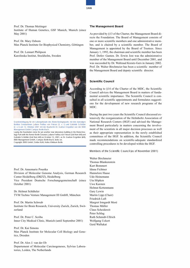
Prof. Dr. Thomas MeitingerInstitute of Human Genetics, GSF Munich, Munich (sinceMay 2001)
Prof. Dr. Mary OsbornMax Planck Institute for Biophysical Chemistry, Göttingen
Prof. Dr. Lennart PhilipsonKarolinska Institut, Stockholm, Sweden
Prof. Dr. Annemarie PoustkaDivision of Molecular Genome Analysis, German ResearchCenter Heidelberg (DKFZ), HeidelbergVice President Deutsche Forschungsgemeinschaft (sinceOctober 2001)
Dr. Helmut SchühslerTVM Techno Venture Management III GmbH, München
Prof. Dr. Martin SchwabInstitute for Brain Research, University Zurich, Zurich, Swit-zerland
Prof. Dr. Peter C. ScribaInner City Medical Clinic, Munich (until September 2001)
Prof. Dr. Kai SimonsMax Planck Institute for Molecular Cell Biology and Gene-tics, Dresden
Prof. Dr. Alex J. van der EbDepartement of Molecular Carcinogeneses, Sylvius Labora-tories, Leiden, The Netherlands
The Management Board
As provided by §13 of the Charter, the Management Board di-rects the Foundation. The Board of Management consists ofone or more scientific members and one administrative mem-ber, and is chaired by a scientific member. The Board ofManagement is appointed by the Board of Trustees. SinceJanuary 1, 1992, the chairman and scientific member has beenProf. Detlev Ganten. Dr. Erwin Jost was the administrativemember of the Management Board until December 2001, andwas succeeded by Dr. Waltraud Kreutz-Gers in January 2002.Prof. Dr. Walter Birchmeier has been a scientific member ofthe Management Board and deputy scientific director.
Scientific Council
According to §14 of the Charter of the MDC, the ScientificCouncil advices the Management Board in matters of funda-mental scientific importance. The Scientific Council is con-sulted in all scientific appointments and formulates suggesti-ons for the development of new research programs of theMDC.
During the past two years the Scientific Council discussed ex-tensively the reorganization of the Helmholtz Association ofNational Research Centers (HGF) and advised the Manage-ment Board particularly in matters concerning the involve-ment of the scientists in all major decision processes as wellas their appropriate representation in the newly establishedcommittees of the HGF. In addition, the Scientific Councilmade recommendations on scientific-adequate standardizedcontrolling procedures to be developed within the HGF.
Members of the Scientific Council (as of November 2001)
Walter BirchmeierThomas BlankensteinKurt BommertIduna FichtnerHannelore HaaseUdo HeinemannUta HöpkenUwe KarstenHelmut KettenmannGary LewinMartin Lipp (Chair)Friedrich LuftMargret Irmgardt MoréThomas MüllerClaus ScheidereitPeter SchlagRuth Schmidt-UllrichWolfgang UckertGerd Wallukat
166
Grundsteinlegung für ein Laborgebäude des Biotechnologieparks mit den damaligenBerliner Senatorinnen Juliane Freifrau von Friesen (3. v. l.) und Gabriele Schöttler(2. v. l.) am 12. Oktober 2001 mit der Bauherrin Dr. Gudrun Erzgräber von der BBBManagement GmbH Campus Berlin-Buch.Laying the foundation stone for yet another new laboratory building in the Biotechno-logy Park with the former Berlin Senators Juliane Freifrau von Friesen (3rd from left) andGabriele Schöttler (2nd from left) on October 12, 2001, as Dr. Gudrun Erzgräber (left),BBB Management GmbH, Campus Berlin-Buch, looks on. Copyright: BBB GmbH; Stefan Kühl, Helios Klinikum Berlin
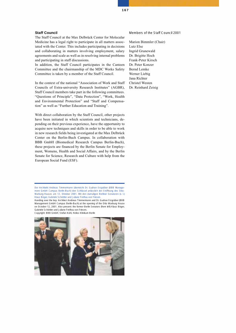
Staff CouncilThe Staff Council at the Max Delbrück Center for MolecularMedicine has a legal right to participate in all matters assoc-iated with the Center. This includes participating in decisionsand collaborating in matters involving employment, salaryagreements and scale as well as in resolving internal problemsand participating in staff discussions.In addition, the Staff Council participates in the CanteenCommittee and the chairmanship of the MDC Works SafetyCommittee is taken by a member of the Staff Council.
In the context of the national “Association of Work and StaffCouncils of Extra-university Research Institutes” (AGBR),Staff Council members take part in the following committees.“Questions of Principle”, “Data Protection”, “Work, Healthand Environmental Protection” and “Staff and Compensa-tion” as well as “Further Education and Training”.
With direct collaboration by the Staff Council, other projectshave been initiated in which scientists and technicians, de-pending on their previous experience, have the opportunity toacquire new techniques and skills in order to be able to workin new research fields being investigated at the Max DelbrückCenter on the Berlin-Buch Campus. In collaboration withBBB GmbH (Biomedical Research Campus Berlin-Buch),these projects are financed by the Berlin Senate for Employ-ment, Womens, Health and Social Affairs, and by the BerlinSenate for Science, Research and Culture with help from theEuropean Social Fund (ESF).
Members of the Staff Council 2001
Marion Bimmler (Chair)Lutz ElseIngrid GrunewaldDr. Brigitte HochFrank-Peter KirschDr. Peter KonzerBernd LemkeWerner LiebigJana RichterChristel WestenDr. Reinhard Zeisig
167
Der Architekt Andreas Timmermann überreicht Dr. Gudrun Erzgräber (BBB Manage-ment GmbH Campus Berlin-Buch) den Schlüssel anlässlich der Eröffnung des Otto-Warburg-Hauses am 12. Oktober 2001. Mit den damaligen Berliner Senatoren (v. l.)Klaus Böger, Gabriele Schöttler und Juliane Freifrau von Friesen. Handing over the key: Architect Andreas Timmermann and Dr. Gudrun Erzgräber (BBBManagement GmbH Campus Berlin-Buch) at the opening of the Otto Warburg Houseon October 12, 2001. Also present: the former Berlin Senators (from left) Klaus Böger,Gabriele Schöttler and Juliane Freifrau von Friesen. Copyright: BBB GmbH; Stefan Kühl, Helios Klinikum Berlin
Supporting Divisions
Safety
The diversity and quality of scientific research carried out atthe MDC requires adherence to a wide range of laws, regula-tions, guidelines, and standards. Measures to ensure not onlythe quality of the science, but also the safety of researchers in-clude the new Working Protection Law, the Genetic Engineer-ing Law, the Radiation Protection Decree, the Chemicals Lawand the Waste Law. The Safety Group regularly inspects facil-ities and compiles internal operating instructions to preventworking accidents, work-related illness, and emergencies.Safety instructions for working groups, identification of pos-sible sources of danger, as well as equipment testing and thetraining in safety procedures are all important functions of thesafety engineers. The Committee for Working Safety at theMDC consists of scientically trained safety officers and spe-cialists in technical matters and radiation protection. TheSafety Group regularly discusses topical problems concern-ing health and safety at work in close cooperation with themedical officer and the staff council.
Head: Dr. Peter Stolley / Dr. Regina Möhring
Building CoordinationEngineering and Reconstruction
Early in 2000, the foundation stone was laid for the Commu-nications Center on the Berlin Buch campus. The buildingwork was carried out as planned and it was opened on29.11.2001 by the President of the Federal Republic ofGermany, Johannes Rau, at an award ceremony for theGerman Future Prize, a prize presented for Technology andInnovation.
In 2000, approval was given for setting up new animal faci-lities and a Building for Theoretical Studies. Extensive pre-paration of the site was needed before building could start in2000.
Construction is going ahead as planned and it is expected tobe able to complete the work and open it to staff in 2003.
Renovation of the facade of the Max Delbrück Haus has beendelayed because of poor weather during the fourth quarter of2001. However, the MDC has ensured that work will continuethrough the winter months and it is hoped to complete thework in the first quarter of 2002.
In June 2000, thanks to the efforts of donors, the Max Del-brück Center saw the first discussions of the building plansfor “Medical Genome Research”. This meeting covered as-pects such as staffing levels, number of research groups, andopinions about the infrastructure. The discussion were sofruitful that preliminary preparations can be started in January2002 to establish the budgeting and accounting building. Thebuilding plans were prepared in collaboration with the FMP.
Head: Grit Kuhlmann
Auditing and Legal Affairs
The duties of Auditing and Legal Affairs are to plan and im-plement the inspection of and adherence to legal issues. TheAuditing Office bears the responsibility for overseeing thatlaws, practices, regulations and the directives of the Manage-ment Board are properly observed and that allocated publicresources are appropriately, economically and productivelyused. For these purposes, examination plans, approved by theManagement Board, are instituted yearly to check regulatorycompliance with organizational activities, so that individualactions are inspected on a case-by-case basis for full compli-ance with regard to both form and content.
All checks of regulatory compliance are undertaken with res-pect to economic feasibility and efficiency so as to ensure thatproper judgment is exercised in establishing regulations andin the execution of all business matters. These checks resultin the establishment of recommendations as to how detectedoversights might best be remedied and avoided in the future.
In the area of legal affairs, special attention is given to the ad-ministration of cooperative research contracts with close sup-port from the Finance Department. The majority of such con-tracts are established with industrial partners. In addition,matters involving jurisdiction aspects need to be preparedand, in many cases, legal opinions must be sought.
The Legal Department also deals with the commercialisationof all MDC patents. Contracts are negotiated and administe-red in close cooperation with both the patent lawyer and thetechnology transfer committee.
Head: Anja Ammar
168
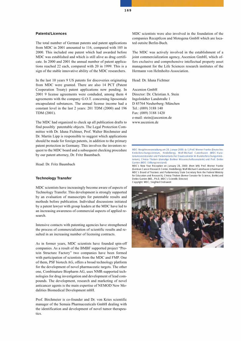
Patents/Licences
The total number of German patents and patent applicationsfrom MDC in 2001 amounted to 116, compared with 105 in2000. This included one patent which had awarded beforeMDC was established and which is still alive as drug certifi-cate. In 2000 and 2001 the annual number of patent applica-tions reached 22 each, compared with 20 in 1999. This is asign of the stable innovative ability of the MDC researchers.
In the last 10 years 9 US patents for discoveries originatingfrom MDC were granted. There are also 14 PCT (PatentCooperation Treaty) patent applications now pending. In2001 9 license agreements were conduded, among them 4agreements with the company G.O.T. concerning liposomaleencapsulated substances. The annual license income had aconstant level in the last 2 years: 201 TDM (2000) and 196TDM (2001).
The MDC had organized to check up all publication drafts tofind possibly patentable objects. The Legal Protection Com-mittee with Dr. Iduna Fichtner, Prof. Walter Birchmeier andDr. Martin Lipp is responsible to suggest which applicationsshould be made for foreign patents, in addition to the primarypatent protection in Germany. This involves the inventors re-quest to the MDC board and a subsequent checking procedureby our patent attorney, Dr. Fritz Baumbach.
Head: Dr. Fritz Baumbach
Technology Transfer
MDC scientists have increasingly become aware of aspects ofTechnology Transfer. This development is strongly supportedby an evaluation of manuscripts for patentable results andmethods before publication. Individual discussions initiatedby a patent lawyer with group leaders at the MDC have led toan increasing awareness of commercial aspects of applied re-search.
Intensive contacts with patenting agencies have strengthenedthe process of commercialization of scientific results and re-sulted in an increasing number of licensing contracts.
As in former years, MDC scientists have founded spin-offcompanies. As a result of the BMBF supported project “Pro-tein Structure Factory” two companies have been formedwith participation of scientists from the MDC and FMP. Oneof them, PSF biotech AG, offers a broad technology platformfor the development of novel pharmaceutic targets. The otherone, Combinature Biopharm AG, uses NMR-supported tech-nologies for drug investigation and development of lead com-pounds. The development, research and marketing of novelanticancer agents is the main expertise of NEMOD New Mo-dalities Biomedical Development mbH.
Prof. Birchmeier is co-founder and Dr. von Kries scientificmanager of the Semaia Pharmaceuticals GmbH dealing withthe identification and development of novel tumor therapeu-tics.
MDC scientists were also involved in the foundation of thecompanies ReceptIcon and Motogena GmbH which are loca-ted outsite Berlin-Buch.
The MDC was actively involved in the establishment of ajoint commercialization agency, Ascenion GmbH, which of-fers exclusive and comprehensive intellectual property assetmanagement for the Life Sciences research institutes of theHermann von Helmholtz-Association.
Head: Dr. Iduna Fichtner
Ascenion GmbHDirector: Dr. Christian A. SteinIngolstädter Landstraße 1D 85764 Neuherberg /MünchenTel.: (089) 3188 140Fax: (089) 3188 1420e-mail: [email protected]
169
MDC-Neujahrsveranstaltung am 28. Januar 2000: (v. l.) Prof. Werner Franke (DeutschesKrebsforschungszentrum, Heidelberg), Wolf-Michael Catenhusen (MDC-Kura-toriumsvorsitzender und Parlamentarischer Staatssekretär im Bundesforschungsminis-terium), Christa Thoben (damalige Berliner Wissenschaftssenatorin) und Prof. DetlevGanten (MDC-Stiftungsvorstand)MDC`s New Year Reception on January 28, 2000: (from left): Prof. Werner Franke(German Cancer Research Center, Heidelberg), Wolf-Michael Catenhusen (chairman ofMDC`s Board of Trustees and Parliamentary State Secretary from the Federal Ministryfor Education and Research), Christa Thoben (former Senator for Science, Berlin) andDetlev Ganten (MD., Ph.D, MDC`s Scientific Director) Copyright: MDC; Siegfried Endruweit
Press and Public Relations
Research at the MDC is conducted at the frontiers of biomed-ical science - in the field of molecular medicine. The MDC´sresearch activities need to be communicated to the generalpublic in a way that can be readily understood. In addition,the cost of running the MDC must be justified to the Germantaxpayer. It is supported by public funds, like all other 15 in-stitutions in the Helmholtz-Association of National ResarchCenters. The Press Office carries out a range of activities toserve this need which are outlined below.
In 2000 and 2001 the MDC Press Office organized more than30 guided tours for almost 500 visitors - university students,high school students, international delegations, and the gen-eral public. These tours included lectures and visits to the la-boratories of the MDC, the cooperating university affiliatedclinics, the Robert Rössle Cancer Center and the Franz Vol-hard Clinic for Cardiovascular Diseases, as well as the LifeScience Learning Laboratory at the Biomedical ResearchPark.
In 2000 and 2001 the MDC Press Office also continued theseries of popular scientific lectures in the City Hall of BerlinPankow, initiated in 1992, the MDC´s first year of existence,with 18 “Sunday Lectures” given by scientists from the MDCand other scientific institutions in Germany. It also presentedthe MDC`s research activities at various exhibitions e.g. inChina (Deutsche Forschungsgemeinschaft, DFG), at theWorld Fair Expo 2000 in Hannover including two EXPO pro-jects on the Berlin-Buch Campus, an exhibition of the Helm-holtz-Association at the Deutsche Museum in Munich, theBiotechnology Fair “Bio 2001” in San Diego, USA, the Sci-ence Fair in Berlin and Warsaw (Poland) and various activ-ities as part of the “Year of the Life Sciences” and “Wissens-chaft im Dialog”, including the first “Long Night of theSciences”.
A total of 97 news releases published in both German andEnglish by the MDC Press Office in 2000 and 2001 formedthe basis for many reports in the media. In addition, the MDCPress Office was also in overall charge of press relations for
Presse- und Öffentlichkeitsarbeit
Als Forschungsinstitut, das molekularbiologische Grund-lagenforschung und klinische Forschung miteinander ver-knüpft, richtet sich das Augenmerk der Presse und der Öffent-lichkeit bei der Biomedizin und der Genforschung in denvergangenen Jahren verstärkt auf das Max-Delbrück-Centrum für Molekulare Medizin (MDC) Berlin-Buch. DasMDC wird wie alle weiteren 15 Einrichtungen der Helm-holtz-Gemeinschaft Deutscher Forschungszentren (HGF) mitöffentlichen Geldern finanziert. Es ist deshalb bemüht, einerbreiten Öffentlichkeit und damit dem Steuerzahler in leichtverständlicher Form zu vermitteln, für welche Forschungenes diese Mittel einsetzt. Die MDC-Pressestelle hat dazu fol-gende verschiedene Aktivitäten initiiert:
Im Jahre 2000 und 2001 betreute die MDC-Pressestelle inüber 30 Führungen nahezu 500 Besucher – darunter Studen-ten, Schüler und internationale Delegationen. Die Presse-stelle bereitete die Besuchsprogramme inhaltlich und orga-nisatorisch vor und betreute mit den Wissenschaftlerinnenund Wissenschaftlern die Gäste. Die Führungen beinhaltetenVorträge, Laborbesuche im MDC, in den Kliniken sowie inFirmen des Biotechnologieparks. Eingeschlossen in dieseBesuchsprogramme ist auch das Gläserne Labor der BBBManagement GmbH.
Die MDC-Pressestelle führte im Berichtszeitraum die Seriepopulärwissenschaftlicher Sonntagsvorlesungen im RathausBerlin-Pankow fort, die das MDC 1992, dem Jahr seinerGründung, initiiert hatte. Referenten sind Wissenschaftler desMDC, Kliniker der Robert-Rössle- und Franz-Volhard-Kliniksowie anderer wissenschaftlicher Einrichtungen im Bundes-gebiet.
Weiter präsentierte sich das MDC in verschiedenen Ausstel-lungen im In- und Ausland, so in China bei einer Ausstellungder Deutschen Forschungsgemeinschaft (DFG), in Hannovermit der Helmholtz-Gemeinschaft auf der EXPO 2000 sowiemit zwei Expo-Projekten auf dem Campus Berlin-Buch. Esbeteiligte sich des weiteren an der Helmholtz-Ausstellung„Lebendige Wissenschaft“ im Deutschen Museum in Mün-
170
the first joint Memorial of German Science for the Victims ofthe Nazi Regime, which was erected jointly by the MaxPlanck Gesellschaft, the Deutsche Forschungsgemeinschaftand the MDC. About 6 000 newspaper articles, with a circu-lation of well over 600 million copies, were published on as-pects of research carried out at the MDC, the Robert Rössleand Franz Volhard Clinics, and the Biotechnology Park. Inaddition, 20 television and 30 radio productions, includingthe French/German TV channel Arte, the national TV chan-nels ZDF and ARD as well as the Deutschlandfunk, were de-voted to the research conducted at the MDC, its clinics andthe Biotechnology Park.
In the time-span reported, three press conferences wereplanned, organized and run by the MDC Press Office. One ofthese press conferences, for example, was held at the “2ndInternational Symposium on Obesity and Hypertension” inBerlin-Buch in 2001.
In 2000 and 2001 the MDC Press Office prepared and orga-nized more than 600 interviews for the media in Germany andabroad, including newspapers, magazines, television andbroadcasting stations and scientific journals such as Nature,Science and The Lancet. In addition, the Press Office pre-pared many contributions about the MDC and its research forbrochures such as the Helmholtz Association of GermanResearch Centers. It also published four press reports sum-marizing the coverage of the MDC, its clinics, the Bio-technology Park, and related topics in the printed press aswell as two issues of the MDC Report, an in-house magazine.
Head: Barbara Bachtler
chen. Zusammen mit der BBB Management GmbH nahm dasMDC an der Bio 2001 in San Diego, USA teil. Mit dem Glä-sernen Labor beteiligte es sich auf der Science Fair in Berlinund in Warschau (Polen) sowie an verschiedenen Aktivitätenim Rahmen des „Jahrs der Lebenswissenschaften“ und von„Wissenschaft im Dialog“ in Berlin. Dazu gehörte auch dieerstmals durchgeführte „Lange Nacht der Wissenschaften“.
Die MDC-Pressestelle recherchierte, verfasste und veröffent-lichte im Berichtszeitraum 97 Pressemitteilungen in Deutschund Englisch, die den Medien Anlass zu einer breiten Be-richterstattung boten. Die MDC-Pressestelle war darüber hin-aus federführend bei der Pressearbeit für das erste gemein-same Mahnmal der Deutschen Wissenschaft für die Opfer desNazi-Regimes, das die Max-Planck-Gesellschaft und dieDeutsche Forschungsgemeinschaft mit dem MDC errichtethaben. In den Jahren 2000 und 2001 erschienen über 6.000Zeitungsartikel, die einer Auflage von mehr als 600 Millio-nen entsprechen, in denen über die Forschungsarbeiten desMDC mit dem Campus Berlin-Buch berichtet wurde. Hinzukamen mehr als 20 Fernseh- und 30 Hörfunkbeiträge, darun-ter auch Berichte in den Hauptnachrichtensendungen vonARD und ZDF sowie im Deutschlandfunk und im deutsch-französchen Kanal Arte.
Die MDC-Pressestelle führte im Berichtszeitraum drei Pres-sekonferenzen durch, die sie inhaltlich und organisatorischvorbereitet hatte. Dazu zählte zum Beispiel eine Presseveran-staltung im Rahmen des „2. Internationalen Symposiumsüber Adipositas und Bluthochdruck“ in Berlin-Buch im Jahre2001.
Die MDC-Pressestelle vermittelte in den vergangenen zweiJahren über 600 Interviews für Tageszeitungen, Magazine,Wochenzeitungen, Fernsehen, Hörfunk sowie für wissen-schaftliche Magazine wie Nature, Science und The Lancet,die sie inhaltlich und organisatorisch vorbereitete. Weiterverfasste die Pressestelle mehrere Beiträge für verschiedeneBroschüren, darunter auch der Helmholtz-Gemeinschaft.Weiter gab das MDC vier Pressespiegel sowie zwei Ausgabendes MDC-Reports, einer Zeitschrift des MDC für Mitarbeite-rinnen und Mitarbeiter, heraus.
Leiterin: Barbara Bachtler
171
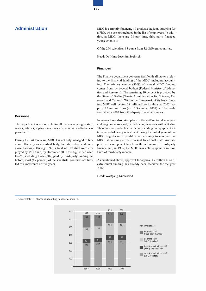
Administration
Personnel
The department is responsible for all matters relating to staff,wages, salaries, separation allowances, removal and travel ex-penses etc..
During the last ten years, MDC has not only managed to fun-ction efficently as a unified body, but stuff also work in aclose harmony. During 1992, a total of 382 stuff were em-ployed by MDC and, by December 2001 this figure had risento 692, including those (207) paid by third-party funding. Asbefore, most (89 percent) of the scientists’ contracts are limi-ted to a maximum of five years.
MDC is currently financing 17 graduate students studying fora PhD, who are not included in the list of employees. In addi-tion, at MDC, there are 78 part-time, third-party financedyoung scientists.
Of the 294 scientists, 83 come from 32 different countries.
Head: Dr. Hans-Joachim Seehrich
Finances
The Finance department concerns itself with all matters relat-ing to the financial funding of the MDC, including account-ing. The primary source (90%) of annual MDC fundingcomes from the Federal budget (Federal Ministry of Educa-tion and Research). The remaining 10 percent is provided bythe State of Berlin (Senate Administration for Science, Re-search and Culture). Within the framework of its basic fund-ing, MDC will receive 55 million Euro for the year 2002; ap-prox. 15 million Euro (as of December 2001) will be madeavailable in 2002 from third-party financial sources.
Increases have also taken place in the staff sector, due to gen-eral wage increases and, in particular, increases within Berlin.There has been a decline in recent spending on equipment af-ter a period of heavy investment during the initial years of theMDC. Significant expenditure is necessary to maintain theMDC laboratories in their present functional state. Anotherpositive development has been the attraction of third-partyfinance and, in 1996, the MDC was able to spend 9 millionEuro of third-party income.
As mentioned above, approval for approx. 15 million Euro ofextra-mural funding has already been received for the year2002.
Head: Wolfgang Kühlewind
172
Personnel status. Distinctions according to financial sources.
Personnel status
1998 1999 2000 2001
296 307330 331
52 5465 67
145 148
154 154
159 144
144 140652 653
693 692
0
100
200
300
400
500
600
700
Scientific staff(Third-party founded)
Scientific staff(MDC founded)
technical and admin. staff(third-party founded)
technical and admin. staff(MDC founded)
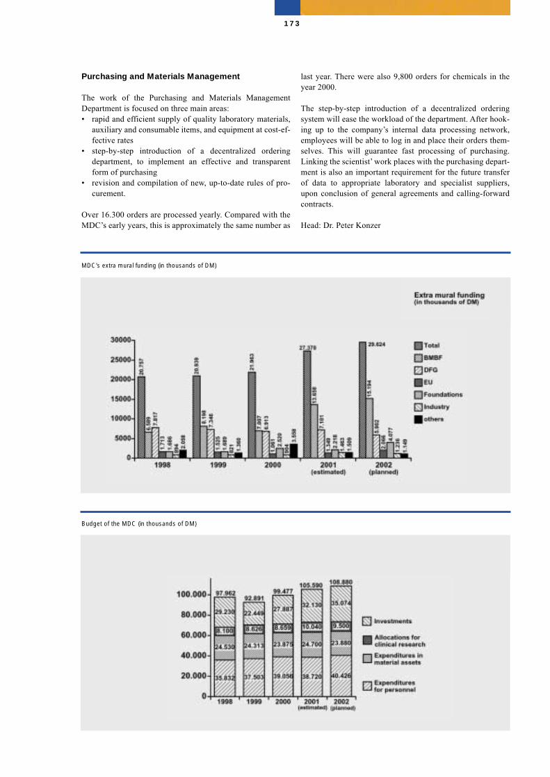
Purchasing and Materials Management
The work of the Purchasing and Materials ManagementDepartment is focused on three main areas:• rapid and efficient supply of quality laboratory materials,
auxiliary and consumable items, and equipment at cost-ef-fective rates
• step-by-step introduction of a decentralized orderingdepartment, to implement an effective and transparentform of purchasing
• revision and compilation of new, up-to-date rules of pro-curement.
Over 16.300 orders are processed yearly. Compared with theMDC’s early years, this is approximately the same number as
last year. There were also 9,800 orders for chemicals in theyear 2000.
The step-by-step introduction of a decentralized orderingsystem will ease the workload of the department. After hook-ing up to the company’s internal data processing network,employees will be able to log in and place their orders them-selves. This will guarantee fast processing of purchasing.Linking the scientist’ work places with the purchasing depart-ment is also an important requirement for the future transferof data to appropriate laboratory and specialist suppliers,upon conclusion of general agreements and calling-forwardcontracts.
Head: Dr. Peter Konzer
173
Budget of the MDC (in thousands of DM)
MDC’s extra mural funding (in thousands of DM)
Central Facilities
Library
The MDC library is a specialized scientific library involved inproviding and supplying information in all research areas ofthe MDC and its associated clinics. The library acts as a mo-dern information center using advanced network structuresand allowing access to information sources around the world.Modern information networks offer increased opportunitiesto supply users with specific literature and information re-sources at their work place. The collection contains more than43 000 media items and 250 print periodicals as well as diffe-rent kinds of non-print materials. In addition to the print sub-scriptions, the library offers more than 900 electronic fulltextjournals. Campus-wide provision of major local databases in-clude Medline (starting from 1966), Current Contents Connect(5 Science Editions) as well as Web of Science (starting from1980) and CD-ROM databases via an internal MDC-networkwith a range of scientific options.
The open area provides 26 reading desks and 5 computerworkstations with internet access. The library operates withthe local library computer system SISIS. The OPAC (OnlinePublic Access Catalogue) lists all collections, starting from1994, and is also available via the internet. Printed cataloguesdescribing older holdings with alphabetical and classified in-dices are also available while the library and its services areall available via the internet. The client-server architectureprovides fast access, regardless of the user’s operatingsystem. The library offers a selection of main links related toresearch on the campus.
Head: Dr. Dorothea Busjahn
Animal Facilities
Animal experiments make a major contribution to biomedicalresearch, particularly in such complex areas as cardiovascu-lar, cancer and neurological diseases. Animals experimentsare especially concerned with the development of methods forimproved diagnosis and treatment of human tumors and car-diovascular diseases. They are essential in establishing genetherapy strategies. With the recent development of powerfulnew technologies for manipulating genes, scientists world-wide have produced thousands of transgenic animals andknock-out models. Both approaches have become invaluablein studies of gene function in disease. In 1993 a transgenicunit was set up, which allows the production of transgenicrats and mice and mouse chimeras. Since then, more than 90transgenic rat strains have been developed to model hyperten-sion. In addition, mice were reconstructed by ES-cell injec-tion and have been successfully bred since 1995. More than300 strains of knock out mice are now available as experi-mental models in cancer research, cardiovascular and neuro-logical diseases. The animal facility of the MDC posesseswithin the research institutes of Berlin the most geneticallyengeneered rat strains and mice stocks.
The four animal houses support transgenic and animal exper-imentation at the MDC. Mice, rats and rabbits are bred. Lo-cated on a 1260 square-meter area, the facilities include ani-mal rooms (636 m2), surgery rooms, storage rooms and cagewashing facilities. The animals are bred in a disease-free en-vironment. Nevertheless, the number of available animalrooms and surgical facilities in and around the MDC does notmeets present requirements.
The rapidly growing number of genetically engeneered mod-els of severe human illness means, that further animal facil-ities for breeding and experimentation are needed. The MDCtherefore builds up a new central animal house. This new fa-cility possesses substancial more capacity for animals (29 an-imal rooms on a 1015 square- meter area) and laboratories (19labs on 665 square meters). This animal house will be openedby the year 2003.
Head: Dr. Karin Jacobi
Campus Net Management
The wide area computer network at the Berlin-Buch ResearchCampus is managed by BBB Management GmbH. In parti-cular, its Campus-IT team is responsible for the operation ofthe high speed connection with the Internet via the recentlyopened G-Win (scientific network, Internet-II) connection ofthe DFN-Verein. The campus network is supplied with allnecessary internet services, such as as E-Mail, World WideWeb, FTP, and a Phone Directory. A dedicated Web site toprovide information about the Campus in general is underdevelopment and will be launched at the beginning of 2002.Further services for the Campus, like video-conferencing andIP-traffic billing/controlling, have been established during thelast three years.
Head: Hans Mitulla
174
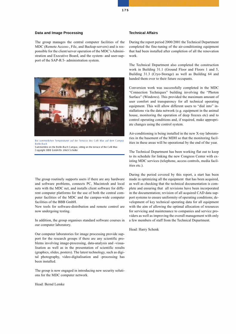
Data and Image Processing
The group manages the central computer facilities of theMDC (Remote Access-, File, and Backup-servers) and is res-ponsible for the client/server operation of the MDC’s Admini-stration and Executive Board, and the system- and user-sup-port of the SAP-R/3- administration system.
The group routinely supports users if there are any hardwareand software problems, connects PC, Macintosh and localnets with the MDC net, and installs client software for diffe-rent computer platforms for the use of both the central com-puter facilities of the MDC and the campus-wide computerfacilities of the BBB GmbH. New tools for software-distribution and remote control arenow undergoing testing.
In addition, the group organises standard software courses inour computer laboratory.
Our computer laboratories for image processing provide sup-port for the research groups if there are any scientific pro-blems involving image-processing, data-analysis and -visua-lisation as well as in the presentation of scientific results(graphics, slides, posters). The latest technology, such as digi-tal photography, video-digitalisation and -processing hasbeen installed.
The group is now engaged in introducing new security soluti-ons for the MDC computer network.
Head: Bernd Lemke
Technical Affairs
During the report period 2000/2001 the Technical Departmentcompleted the fine-tuning of the air-conditioning equipmentthat had been installed after completion of all the renovationwork.
The Technical Department also completed the constructionwork in Building 31.1 (Ground Floor and Floors 1 and 5,Building 31.3 (Cryo-Storage) as well as Building 64 andhanded them over to their future occupants.
Conversion work was successfully completed in the MDC“Connection Techniques” building involving the “PhotonSurface” (Windows). This provided the maximum amount ofuser comfort and transparency for all technical operatingequipment. This will allow different users to “dial into” in-stallations via the data network (e.g. equipment in the animalhouse, monitoring the operation of deep freezes etc) and tocontrol operating conditions and, if required, make appropri-ate changes using the control system.
Air-conditioning is being installed in the new X-ray laborato-ries in the basement of the MDH so that the monitoring facil-ities in these areas will be operational by the end of the year.
The Technical Department has been working flat out to keepto its schedule for linking the new Congress Center with ex-isting MDC services (telephone, access controls, media facil-ities etc.).
During the period covered by this report, a start has beenmade in optimizing all the equipment that has been acquired,as well as checking that the technical documentation is com-plete and ensuring that all revisions have been incorporatedin the documentation; revision of all acquired CAD data sup-port systems to ensure uniformity of operating conditions; de-velopment of key technical operating data for all equipmentwith the aim of allowing the optimal allocation of resourcesfor servicing and maintenance to companies and service pro-viders as well as improving the overall management with onlya few members of staff from the Technical Department.
Head: Harry Schenk
175
Bei sommerlichen Temperaturen auf der Terrasse des Café Max auf dem CampusBerlin-Buch Summertime on the Berlin-Buch Campus, sitting on the terrace of the Café MaxCopyright: BBB GmbH/Dr. Ulrich Scheller

Meetings, Workshops andSymposia
The following events organized under the auspices of theMDC and its clinical partners took place in 2000 and 2001
2000
4th Workshop Interventionelle Kardiologie, Franz VolhardClinic for Cardiovascular Diseases (January 28, 2000)
BIO 2000 International Meeting & Exhibition(March 26-30, 2000, Boston, MA, Hynes Convention Center)
Coping with Sarcoma –Functional, Psychological and SocialDimensionsRobert Rössle Cancer Center, Charité and Freie UniversitätBerlin(April 13, 2000)
MDC-Neuro-MeetingMax Delbrück Center for Molecular Medicine (MDC) Berlin-Buch (May 4-5, 2000)
6th MDC Graduate Students’ Symposium (May 17, 2000)
“Neue Therapiestrategien in der Hämatologie: Was ist gesi-chert?” im Rahmen des 49. Deutschen Ärztekongresses Berlin(May 30, 2000)
Experten Meeting “Medical Genomics”Max Delbrück Center for Molecular Medicine (MDC) Berlin-Buch (June 5 and June 23, 2000)
Symposium Brain TumorsMax Delbrück Center for Molecular Medicine (MDC) Berlin-Buch (June 15-16, 2000)
6th Bucher Symposium “Science Meets Clinic”Franz Volhard Clinic for Cardiovascular Diseases, CampusBerlin Buch (July 1, 2000)
29th European Muscle ConferenceMax Delbrück Center for Molecular Medicine (MDC) BerlinBuch and Charité, Humboldt University, Berlin(September 8-13, 2000)
Symposium zum 100. Geburtstag von Nikolai WladimirovichTimoféeff-Ressovsky “Genetik in Russland und Deutschland”Max-Delbrück-Centrum für Molekulare Medizin (MDC) Ber-lin-Buch(September 21-22, 2000)
176
Impressionen von der ersten „Langen Nacht der Wissenschaften“ auf dem CampusBerlin-Buch am 15. September 2001 Impressions of the first “Long Night of the Sciences” on the Berlin-Buch Campus, Sep-tember 15, 2001 Copyright: BBB GmbH; Thomas Oberländer/Helios-Kliniken
Neue Wege in der Diagnostik von Herzerkrankungen: Interessierte Besucher lassen sichwährend der „Langen Nacht der Wissenschaften“ am 15. September 2001 in der Franz-Volhard-Klinik der Charité /Helios Kliniken GmbH die Magnetresonanz-Tomographieerklären. New methods for diagnosing heart diseases – Interested visitors are told about magne-tic resonance tomography during the “Long Night of the Sciences” on September 15,2001, at the Franz Volhard Clinic of the Charité /Helios Kliniken GmbH. Copyright: BBB GmbH; Thomas Oberländer /Helios-Kliniken
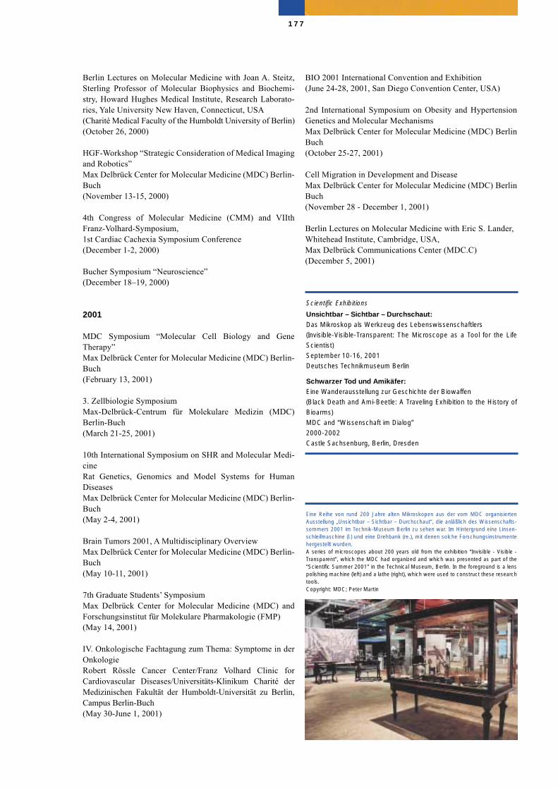
Berlin Lectures on Molecular Medicine with Joan A. Steitz,Sterling Professor of Molecular Biophysics and Biochemi-stry, Howard Hughes Medical Institute, Research Laborato-ries, Yale University New Haven, Connecticut, USA(Charité Medical Faculty of the Humboldt University of Berlin)(October 26, 2000)
HGF-Workshop “Strategic Consideration of Medical Imagingand Robotics”Max Delbrück Center for Molecular Medicine (MDC) Berlin-Buch(November 13-15, 2000)
4th Congress of Molecular Medicine (CMM) and VIIthFranz-Volhard-Symposium, 1st Cardiac Cachexia Symposium Conference(December 1-2, 2000)
Bucher Symposium “Neuroscience”(December 18–19, 2000)
2001
MDC Symposium “Molecular Cell Biology and GeneTherapy”Max Delbrück Center for Molecular Medicine (MDC) Berlin-Buch(February 13, 2001)
3. Zellbiologie Symposium Max-Delbrück-Centrum für Molekulare Medizin (MDC)Berlin-Buch(March 21-25, 2001)
10th International Symposium on SHR and Molecular Medi-cineRat Genetics, Genomics and Model Systems for HumanDiseasesMax Delbrück Center for Molecular Medicine (MDC) Berlin-Buch(May 2-4, 2001)
Brain Tumors 2001, A Multidisciplinary OverviewMax Delbrück Center for Molecular Medicine (MDC) Berlin-Buch(May 10-11, 2001)
7th Graduate Students’ SymposiumMax Delbrück Center for Molecular Medicine (MDC) andForschungsinstitut für Molekulare Pharmakologie (FMP) (May 14, 2001)
IV. Onkologische Fachtagung zum Thema: Symptome in derOnkologieRobert Rössle Cancer Center/Franz Volhard Clinic forCardiovascular Diseases/Universitäts-Klinikum Charité derMedizinischen Fakultät der Humboldt-Universität zu Berlin,Campus Berlin-Buch(May 30-June 1, 2001)
BIO 2001 International Convention and Exhibition(June 24-28, 2001, San Diego Convention Center, USA)
2nd International Symposium on Obesity and HypertensionGenetics and Molecular MechanismsMax Delbrück Center for Molecular Medicine (MDC) BerlinBuch(October 25-27, 2001)
Cell Migration in Development and DiseaseMax Delbrück Center for Molecular Medicine (MDC) BerlinBuch(November 28 - December 1, 2001)
Berlin Lectures on Molecular Medicine with Eric S. Lander, Whitehead Institute, Cambridge, USA,Max Delbrück Communications Center (MDC.C)(December 5, 2001)
177
Eine Reihe von rund 200 Jahre alten Mikroskopen aus der vom MDC organisiertenAusstellung „Unsichtbar – Sichtbar – Durchschaut“, die anläßlich des Wissenschafts-sommers 2001 im Technik-Museum Berlin zu sehen war. Im Hintergrund eine Linsen-schleifmaschine (l.) und eine Drehbank (re.), mit denen solche Forschungsinstrumentehergestellt wurden. A series of microscopes about 200 years old from the exhibition “Invisible - Visible -Transparent”, which the MDC had organized and which was presented as part of the“Scientific Summer 2001” in the Technical Museum, Berlin. In the foreground is a lenspolishing machine (left) and a lathe (right), which were used to construct these researchtools.Copyright: MDC; Peter Martin
Scientific Exhibitions
Unsichtbar – Sichtbar – Durchschaut:Das Mikroskop als Werkzeug des Lebenswissenschaftlers(Invisible-Visible-Transparent: The Microscope as a Tool for the LifeScientist)September 10-16, 2001Deutsches Technikmuseum Berlin
Schwarzer Tod und Amikäfer:Eine Wanderausstellung zur Geschichte der Biowaffen(Black Death and Ami-Beetle: A Traveling Exhibition to the History ofBioarms)MDC and “Wissenschaft im Dialog”2000-2002Castle Sachsenburg, Berlin, Dresden
Awards
2000
MDC and Forschungszentrum KarlsruhePreis des Bundesministeriums für Bildung und Forschungfür Gründungsinitiativen, vergeben durch die Karl-Heinz-Beckurts-Stiftung
Jürgen BehrensGerhard-Domagk-Preis für Experimentelle Krebsforschung.
Peer BorkPreis der Berlin-Brandenburgischen Akademie der Wissen-schaften, sponsored by the Gottlieb Daimler- und Karl-Benz-Stiftung
Reinhold Förster, Martin Lipp, Elisabeth Kremmer, EckardWolfErwin-Schrödinger-Preis 2000 des Stifterverbands für dieDeutsche Wissenschaft und der Helmholtz-GemeinschaftDeutscher Forschungszentren
Detlev GantenBundesverdienstkreuz
Friedrich LuftArthur Corcoran Award. American Heart Association.
Thomas WillnowPreis für „Medizinische Grundlagenforschung“ der Smith-Kline Beecham Stiftung
Thomas WillnowButenandt-Habilitationspreis der Ernst Schering ResearchFoundation
2001
Carmen Birchmeier-KohlerGottfried Wilhelm Leibniz Preis für 2002
Ralf DechendPrize of the American Heart Association
Michael GotthardtSofja Kovalevskaya Preis
Peter Langen Karl-Heinrich-Bauer-Medaille, Deutsche Krebsgesellschaft e.V.
Young-Ae LeePreis für klinische Forschung der SmithKline BeechamStiftung
Friedrich Luft Preis der Helmut und Ruth-Lingen Stiftung
Friedrich Luft Dr. honoris causa, University Pecs (Hungary)
Max Delbrück MedalSince 1992, outstanding scientists are being awarded the MaxDelbrück Medal by Berlin research institutions and theSchering Research Foundation.
Recipients in the past years were
1992Günter BlobelRockefeller University New York, USA;Nobel Laureate in 1999
1994Sydney BrennerUniversity of Cambridge, UK
1995Jean-Pierre ChangeuxInstitut Pasteur, Paris, France
1996Robert A. WeinbergWhitehead Institute, Massachusetts Institute of Technology,Cambridge/USA
1996Nihat BilginturanUniversity of Hacettepe, Ankara, Turkey
1997Charles WeissmannUniversity of Zürich, Switzerland
178
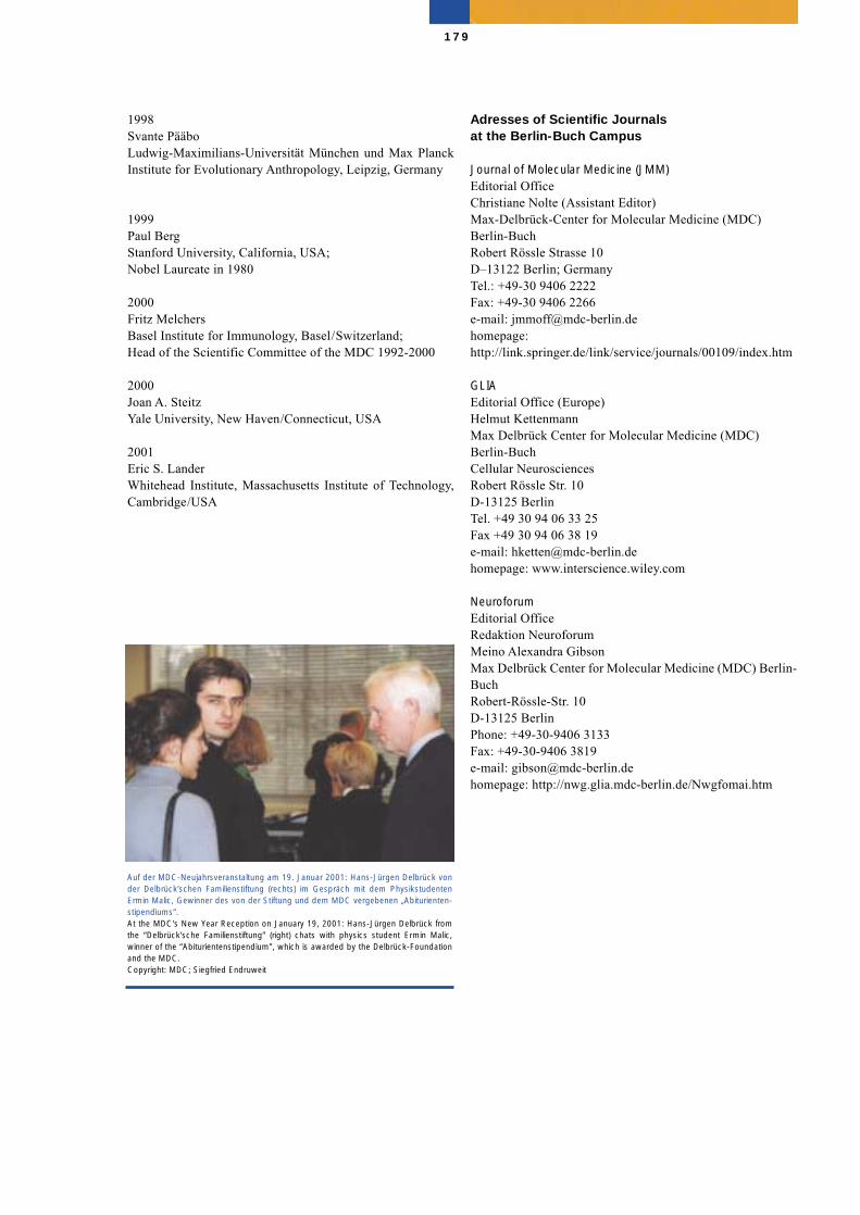
1998Svante PääboLudwig-Maximilians-Universität München und Max PlanckInstitute for Evolutionary Anthropology, Leipzig, Germany
1999Paul BergStanford University, California, USA;Nobel Laureate in 1980
2000 Fritz MelchersBasel Institute for Immunology, Basel/Switzerland;Head of the Scientific Committee of the MDC 1992-2000
2000 Joan A. Steitz Yale University, New Haven/Connecticut, USA
2001 Eric S. LanderWhitehead Institute, Massachusetts Institute of Technology,Cambridge/USA
Adresses of Scientific Journals at the Berlin-Buch Campus
Journal of Molecular Medicine (JMM)Editorial OfficeChristiane Nolte (Assistant Editor)Max-Delbrück-Center for Molecular Medicine (MDC) Berlin-BuchRobert Rössle Strasse 10D–13122 Berlin; GermanyTel.: +49-30 9406 2222Fax: +49-30 9406 2266e-mail: [email protected]: http://link.springer.de/link/service/journals/00109/index.htm
GLIAEditorial Office (Europe)Helmut KettenmannMax Delbrück Center for Molecular Medicine (MDC) Berlin-BuchCellular NeurosciencesRobert Rössle Str. 10D-13125 BerlinTel. +49 30 94 06 33 25Fax +49 30 94 06 38 19e-mail: [email protected]: www.interscience.wiley.com
NeuroforumEditorial OfficeRedaktion NeuroforumMeino Alexandra GibsonMax Delbrück Center for Molecular Medicine (MDC) Berlin-Buch Robert-Rössle-Str. 10D-13125 BerlinPhone: +49-30-9406 3133Fax: +49-30-9406 3819e-mail: [email protected]: http://nwg.glia.mdc-berlin.de/Nwgfomai.htm
179
Auf der MDC-Neujahrsveranstaltung am 19. Januar 2001: Hans-Jürgen Delbrück vonder Delbrück’schen Familienstiftung (rechts) im Gespräch mit dem PhysikstudentenErmin Malic, Gewinner des von der Stiftung und dem MDC vergebenen „Abiturienten-stipendiums“. At the MDC’s New Year Reception on January 19, 2001: Hans-Jürgen Delbrück fromthe “Delbrück’sche Familienstiftung” (right) chats with physics student Ermin Malic,winner of the “Abiturientenstipendium”, which is awarded by the Delbrück-Foundationand the MDC.Copyright: MDC; Siegfried Endruweit
Index
AAbdelilah-Seyfried, Salim ................................ 33, 112, 113Abraham, Claudia ........................................................... 162Adams, Frauke ................................................................. 56Ahlers, Annette ................................................................. 91Akepati, Vasudheva ........................................................ 149Alenina, Natalia ............................................................... 46Alexander, Christiane ....................... 32, 146, 147, 148, 149Alitalo, Kari ..................................................................... 98Al-Kateb, Hussam ............................................................ 48Allegro, Heloisa Baptista ................................................. 46Ammar, Anja .................................................................. 168Anagnostopoulos, I. ......................................................... 93Andrade, Miguel .............................................................. 69André, Francoise .............................................................. 51Andreé, Christel .............................................................. 111Andrikopoulos, Petros .................................................... 104Anirudhan, Gireesh ........................................................ 155Ansieau, Stephane ............................................................ 89Anzinger, Birgit ...................................................... 134, 135Appel, Silke ...................................................................... 51Arlt, Franziska ................................................................ 102Arndt, Dietrich ................................................................. 35Arnold, Susanne ............................................................. 153Asbrand, Christian ............................................................ 99Assadi, Mansur................................................................ 162Aumann, Jutta ................................................................ 140Aydin, Atakan ................................................................... 48
BBachtler, Barbara ...................................... 171, Inside CoverBader, Michael ..................................................... 41, 45, 46Bähring, Sylvia ................................................................. 48Bärwolff, Dieter ............................................................. 144Bäßmann, Ingelore ........................................................... 51Baldinger, Tina ................................................................. 96Balling, Rudi .................................................................. 165Baltas, Leonidas ............................................................. 104Baltatu, Ovidiu ................................................................. 46Balz, Stefanie ................................................................. 102Balzereit, Daniela ........................................................... 104Barbosa-Sicard, Eduardo ................................................ 111Bargou, Ralf ..................................................... 95, 126, 127Barthel, Denise ............................................................... 131Barthel, Beatrix ................................................ 60, 101, 102Bartsch, Andrea .............................................................. 104Bartsch, Holger .............................................................. 104Bartunek, Petr ......................................................... 134, 135Bashamekh, Saleh ............................................................ 46Bauer, Bianca ................................................................. 162Bauer, Lisa ..................................................................... 102Bauerfeind, Anja .............................................................. 69Baumbach, Fritz ............................................................. 169Becker, Christian .............................................................. 51Becker, Christiane .......................................................... 131Becker, Michael .............................................................. 140Becker, Monika .............................................................. 140Begay-Müller, Valerie ...................................................... 89
Behrens, Diana ............................................................... 140Behrens, Jürgen .......................................... 34, 36, 164, 178Benga, Jemina .................................................................. 94Bengs, Marion ................................................................ 106Benhidjeb, T. .................................................................. 136Berg, Paul ....................................................................... 179Berger, Ingrid ................................................................. 142Bergmann, Jutta .............................................................. 142Bergmann, Martin .......................................................... 120Bergmann, Stephan ................................................. 100, 102Bernhardt, Günter ............................................................. 94Bick-Sander, Anika ........................................................ 151Bienert, Ralf ............................................................... 72, 74Bieringer, Markus ............................................................. 48Bilginturan, Nihat ........................................................... 178Bimmler, Marion ............................................................ 167Birchmeier-Kohler, Carmen ......... 32, 35, 36, 40, 42, 61, 63, ............................................................ 97, 178, Inside CoverBirchmeier, Walter ................... 16, 32, 87, 97, 99, 166, 169, .......................................................................... Inside CoverBirkenfeld, Andreas .......................................................... 56Blachut, Susanne .............................................................. 44Blankenstein, Thomas .............. 130, 131, 166, Inside CoverBlendinger, Gitta .................................................... 134, 135Blobel, Günter ................................................................ 178Boeckh, Susanne .................................................... 136, 137 Böddrich, Annett ............................................................ 162Böger, Klaus ................................................................... 167Boehm, Siegfried .............................................................. 78Bönsch, Christian ............................................................. 67Böhnke, Jana .................................................................... 55Böttger, Adelheid ............................................................. 46Bommert, Kurt .......................................... 95, 125, 127, 166Bork, Peer ........................................................ 42, 68, 69, 178Borowiak, Malgorzata ...................................................... 63Boucsein, Clemens ......................................................... 153Bräuner, Corinna .............................................................. 44Brand, Karsten ........................................................ 136, 137Brandt, Moritz ................................................................ 151Branoner, Wolfgang .......................................................... 25Breitfeld, Dagmar ............................................................. 94Breithardt, Günter ................................................... 164, 165Brembeck, Felix ............................................................... 99Brenner, Sydney ............................................................. 178Brett, David ...................................................................... 69Brinckmann, Anja ............................................................. 51Brinsuk, Marina ................................................................ 56Britsch, Stefan ...................................................... 61, 63, 98Bröhmer, Meike ................................................................ 91Brohmann, Henning ............................................. 62, 63, 97Brümmendorf, Thomas ................................................... 159Brüning, Gerold .............................................................. 151Brunner, Hans R. .................................................... 164, 165Buchert, Sven ................................................................... 63Bürgener, Dietmar .......................................................... 164Burger, Reinhard .............................................................. 15Burghardt-Göttges, Elke ................................................... 59Burmeister, Regina ........................................................... 67Busch, Anne ................................................................... 162Buschow, Christian ......................................................... 131Busjahn, Dorothea .......................................................... 174
180
CCalkhoven, Cor ................................................................ 89Calließ, Christiane ............................................................ 89Cam, Hakan .................................................................... 131Campos, Luciana Aparecida ............................................. 46Cardoso, Cibele Campos .................................................. 46Cardoso, M. Cristina .............................................. 105, 106Cartier, Regis .................................................................. 142Catenhusen, Wolf-Michael ......................... 13, 37, 164, 169Cayeux, Sophie .............................................................. 127Cayla, Cécile .................................................................... 46Cesare, Silvana Di ............................................................ 99Chaboissier, Marie-Christine ............................................ 65Changeux, Jean-Pierre .................................................... 178Chatterjee, Manik ................................................... 126, 127Chernogolov, Alexej ....................................................... 111Chmielowiec, Jolanta ....................................................... 99Chulabhorn, Mahidol, HRH Princess ............................. 165 Cichon, Günter ................................................. 15, 136, 137 Cifire, Felix ...................................................................... 94Claßen, Elvira ................................................................... 59Clemens, Jan .................................................................. 137Cloos, Birgit ............................................................. 74, 159Contzen, Jörg .................................................................... 76
DDamaschun, Gregor .............................................. 42, 70, 71Damaschun, Hilde ............................................................ 71Damm, Henning ............................................................... 55Dandekar, Thomas ............................................................ 69Daniel, Peter ........................................................... 125, 127Das, Debashish ............................................................... 159Debs, Pierre ...................................................................... 96Dechend, Ralf ................................................................. 178Delbrück, Hans-Jürgen .....................................................179Delbrück, Heinrich ..................................................... 72, 74Dell’Oro, Andrea .............................................................. 99Dettmer, Rudolf ................................................................ 91Diebold, Sandra S. .......................................................... 135Dietel, Manfred .............................................................. 164Dietz, Rainer ..................................................... 24, 119, 120Dörken, Bernd ...................................... 24, 93, 95, 125, 127Doerks, Tobias .................................................................. 69Dokup, Kornelia ............................................................... 51Domaing, Petra ............................................................... 106Domasch, Silke ................................................................ 81Donath, Stefan ................................................................ 120Dorner, Armin ................................................................ 159Dostal, Lubomir ............................................................... 80Drab, Marek ..................................................................... 48Drechsler, Hannelore ...................................................... 159Dröge, Anja .................................................................... 162Droese, J. .......................................................................... 93Dubrovska, Galina .................................................... 55, 122
EEaswaran, Hariharan P. ................................................... 106Eckhardt, Ullrich ............................................................ 137Eckl, Katja-Martina .......................................................... 51Ehninger, Dan ................................................................. 151
Eicher, Eva ....................................................................... 69Eigen, Marc ...................................................................... 67Eilers, Andreas ............................................................... 155Eisenhaber, Birgit ............................................................. 69Ekici, Arif Bülent ............................................................. 51Elezkurtaj, Sefer ..................................................... 136, 137 Ellinghaus, Agnes ........................................................... 153Else, Lutz ....................................................................... 167Emmerich, Florian .......................................................... 127Engel, Felix .................................................................... 120Engel, Katrin .................................................................... 96Engeli, Stefan ................................................................... 55Engels, Boris .................................................................. 131Engemann, Sabine .......................................................... 162Erdmann, Bettina .............................................. 97, 114, 115Erhardt, Manfred ................................................................ 9Ertas, Figen .................................................................... 162Erzgräber, Gudrun ........................................24, 26, 166, 167Essine, Kirill ................................................................... 122Esteban, Viviana Marin .................................................. 133Estevez-Schwarz, Lope .................................................... 60Euler, Uta ............................................................... 134, 135Ey, Stefan ......................................................................... 51
FFabian, H .......................................................................... 70Faelber, Katja ............................................................. 73, 74Färber, Katrin ................................................................. 153Falk, Kirsten ........................................................... 132, 133Feldner, Anja .................................................................... 44Feldpausch, Mareike ........................................................ 55Feller, Katharina ............................................................... 99Feske, Anette .................................................................... 74Fichtner, Iduna ................................ 101, 139, 140, 166, 169Fiedler, Dora ................................................................... 138Fiket, Maja ..................................................................... 149Filipov, Vitali .................................................................. 153Fischer, Heike ..................................................... 51, 60, 102Fischer, Uta .................................................................... 137Fleischhauer, Silke ......................................................... 153Förster, Reinhold .................................. 34, 36, 94, 164, 178Foss, H. D. ....................................................................... 92Franke, Gabriele ............................................................... 56Franke, Jacqueline ............................................................ 57Franke, Renate ............................................................ 97, 99Franke, Werner .................................................................169Frege, Renate ................................................................... 60Freund, Christian ...................................................... 91, 120Friedlander, Ruth ............................................................ 108Friedrich, Bärbel ..................................................... 164, 165Fujita, Yasuyuki ................................................................ 99
GGärtner, Angelika ........................................................... 131Gaertner, Klaus ............................................................... 144Gaehtgens, Peter ............................................................. 164Gahl, Anja ...................................................................... 106Gaio, Ursula ..................................................................... 63Gaiser, Olaf ...................................................................... 74Gallagher, Irene .............................................................. 135
181
Galle, Renate .................................................................... 60Ganten, Detlev ............................. 10, 11, 13, 20, 24, 30, 43,............................................................44, 165, 169, 166, 178Ganzel, Karin ................................................................... 91Garratt, Alistair ........................................................... 61, 63Gast, Klaus ....................................................................... 71Gast, Daniela .................................................................. 151Gaudet, Francois ............................................................ 106Geheeb, Martina ..................................................... 136, 137 Genser, Klaus ................................................................. 162Gerhardt, Ute .................................................................... 55Gerlach, Brigitte ............................................................. 153Gerull, Brenda .................................................................. 53Geupel, Anke .................................................................... 53Gibson, Meino Alexandra ............................................... 179Gillissen, Bernd ...................................................... 125, 127Girresch, Bärbel ............................................................... 55Gladow, Monika ............................................................. 131Göhler, Heike ................................................................. 162Göritz, Christian ............................................................. 156Gösele, Claudia ................................................................ 44Gössel, Claudia ................................................................ 55Goihl, Alexander ............................................................ 124Golbrich, Beate .............................................................. 137Goldscheid, Eva ............................................................. 133Golfier, Sven .................................................................... 94Gollasch, Maik .......................................... 41, 110, 121, 122Gong, Maolian .................................................................. 44Gontscharenko, Mykola ................................................. 135Gorzelniak, Kerstin .......................................................... 55Gossen, Manfred ................................................ 95, 96, 125Gotthardt, Michael ................................................... 37, 178Gottschalk, Iris ................................................................. 55Gottschling, Karin ............................................................ 63Grabbert, Martina ........................................................... 131Gräler, Markus ................................................................. 94Gramlich, Michael ............................................................ 53Gras, Christiane .............................................................. 153Graschew, Georgi ........................................................... 102Grasmo-Wendler, Unn-Hilde ............................................ 59Graßhoff, Peter ................................................................. 94Greiner, Axel .................................................................. 126Grelle, Gerlinde .............................................................. 162Grieben, Marlies ............................................................. 120Grigull, Sabine ............................................................... 102Grimm, Jan ................................................................. 98, 99Grishkovskaya, Irina .................................................. 73, 74Grohmann, Maik ............................................................ 106Gross, Volkmar ................................................................. 48Grothe, Daniela .............................................................. 120Grüger, Sabine .................................................................. 48Grün, Michael ......................................................... 126, 127Grünwald, David ............................................................ 106Grunewald, Ingrid .................................................. 106, 167Grzimek, Sabina .................................................................26Gunia, Wilfried ................................................................. 69Guo, Jiankan ..................................................................... 65Gust, Tatjana .................................................................. 135
HHaase, Hannelore .................................................... 104, 166Haase, Sabine ................................................................. 102Hackenbroch, Michael .................................................... 164Hacker, Christine .................................................... 134, 135Haensch, W. .................................................................... 100Häusler, Georg ................................................................ 153Haink, Petra ............................................................ 111, 135Hammes, Annette ............................................................. 65Hampig, Franziska ........................................................... 48Handke, T. ...................................................................... 101Hanisch, Uwe-Karsten .................................................... 153Hartwig, Frauke ................................................................ 55Hasenbank, Renate ......................................................... 162Hauck, Ludger ................................................................ 120Haucke, Lisa ..................................................................... 51Haupt, Irene .................................................................... 156Hehl, Regina ................................................................... 153Heikenwälder, Mathias F. ....................................... 134, 135Heinemann, Udo ..................... 42, 72, 74, 166, Inside CoverHeiring, C ......................................................................... 73Heißmeyer, Vigo .............................................................. 91Hemmati, Philipp ........................................................... 125Hennies, Hans Christian ................................................... 51Henning, Mechthild ........................................................ 159Heppenstall, Paul ............................................................ 155Herrmann, Alexander ....................................................... 69Herrmann, Falko ..................................................... 164, 165Hertel, Ingolf .................................................................. 164Hieronymus, Thomas .............................................. 134, 135Hildebrandt, Kati ............................................................ 127Hilpert, Jan ....................................................................... 67Hinz, Michael ............................................................. 91, 92Hoch, Brigitte ................................................................. 167Höhnemann, Dirk ................................................... 126, 127Höpken, Uta ............................................................. 94, 166Hoffmann, Anja .............................................................. 153Hoffmann, Katrin ............................................................. 51Hofmann, Corinna .......................................................... 137Hofmann, Wera ................................................................ 59Hofstätter, Maria ............................................................ 133Holmes, Melissa ............................................................. 151Homuth, Volker ................................................................ 48Honeck, Horst ................................................................. 111Horn, Denise .................................................................... 59Hosseini, Vahid .............................................................. 102Hu, Jing .......................................................................... 155Hübner, Beate ................................................................. 164Hübner, Norbert .................................................... 31, 43, 44Hülsken, Jörg ........................................................ 16, 97, 99Huijnen, Martijn ............................................................... 69Hummel, Kordelia .......................................................... 137
IIliescu, Radu ..................................................................... 46Ivics, Zoltán ................................................................... 138Izsvák, Zsuzsanna ........................................................... 138
182
JJacobi, Karin .................................................................. 174Jandrig, Burkhard ............................................................. 59Janke, Jürgen .................................................................... 55Janz, Martin ............................................................ 125, 127Jarchow, Birgit ............................................................... 153Jarosch, Ernst ................................................................. 108Jeblonski, Frank ............................................................... 94Jerchow, Boris .................................................................. 99Jessberger, Sebastian ...................................................... 151Jobsky, Dieter ................................................................. 159Johnen, Heiko ................................................................... 94Jordan, Jens ................................................................ 41, 56Jorgas, Thorsten ...................................................... 134, 135Joschko, Simone ............................................................... 89Jost, Erwin ................................................................ 10, 166Ju, Xin-Sheng ......................................................... 134, 135Jürchott, Karsten ..................................................... 100, 102Jüttner, René ................................................................... 159Jung, Christiane .......................................................... 75, 76Jung, Katinka ................................................................. 142Junghahn, Ilse ................................................................. 140Junghans, Christine .......................................................... 48Jungmann, Sabine ............................................................ 91Junker, Bettina .................................................................. 55Jundt, Franziska ...................................................... 125, 127
KKaba, Rudolf Gabor ......................................................... 94Kärgel, Eva ..................................................................... 111Kagelmaker, Horst .......................................................... 153Kammertöns, Thomas ..................................................... 131Kanehl, Anke .................................................................. 155Kannen, Brunhilde ........................................................... 80Karawajew, Leonid ................................................. 126, 127Karczewski, Karin .......................................................... 124Kariakine, Andrei ............................................................. 76Karsten, Uwe .................................................... 35, 100, 166Katranidis, Alexandros ................................................... 104Katzer, Andrea ................................................................ 127Kaufman, Chris .............................................................. 138Kemmner, Wolfgang ....................................... 100, 101, 102Kempermann, Gerd ..... 32, 33, 114, 115, 146, 147, 150, 151Kemsies, Christel ........................................................... 104Kettenmann, Helmut ........................ 30, 146, 152, 153, 166, ...................................................................179, Inside CoverKettritz, Ralph .................................................................. 48Keyner, Daniela .......................................................... 91, 94Khare, Dheeraj ........................................................... 72, 74Kim, Hye-Jung ............................................................... 131Kirsch, Frank-Peter ................................................ 159, 167Kirsch, Ralf D. ............................................................... 135Kirst, Elisabeth ................................................................. 51Kistel, Heide ..................................................................... 44Klahn, Sylvia .......................................................... 108, 131Klamm, Ursula ................................................................. 76Klein, Eireen .................................................................... 48Kleinewietfeld, Markus .................................................. 133Klewer, Jacqueline .......................................... 113, 155, 156Kley, Katarina ................................................................ 127Klingner, Kerstin .............................................................. 89
Klose, Chihab ................................................................. 162Knake-Werner, Heidi ...................................................... 164Knespel, Andreas .............................................................. 74Knespel, Siegne ...................................................... 134, 135Knoblauch, Margit ........................................................... 44Knoblich, Maria ............................................................. 162Koch, Christian ....................................................... 128, 129Koch, Gudrun ................................................................. 102Koch, Ina .......................................................................... 69Kohl, Zacharias .............................................................. 151Köhler, Matthias ......................................................... 41, 57Kölble, Konrad ................................................. 60, 101, 102Köppl, Bernd .................................................................. 164Körner, Ida-Johanna ....................................................... 127Körnig, Sandra ............................................................... 131Köttgen, Eckart .............................................................. 165Koh, Ki-Ryang ....................................................... 134, 135Kolbe, Michael ................................................................. 74Konzer, Peter .......................................................... 167, 173Kopacek, Anke ............................................................... 102Korbel, Jan ....................................................................... 69Koritschoner, N. P. ......................................................... 134Koroll, Michael .............................................................. 159Kostka, Susanne ............................................................. 162Kotitschke, Erika .................................................... 115, 151Kott, Monika .................................................................. 104Kowenz-Leutz, Elisabeth ................................................. 89Krahn, Inge ..................................................................... 144Krappmann, Daniel .......................................................... 91Kratzel, Christine ........................................................... 142Krause, E. ......................................................................... 70Krause, Ernst-Georg ................................................... 34, 35Krause, Julia ..................................................................... 51Krause, Kerstin ................................................................. 60Kremmer Elisabeth ............................................. 36, 93, 178Kreutzberg, Georg W. ..................................................... 165Kresse, Carmen ................................................................ 55Kresse, Wolfgang ........................................................... 153Kreutz-Gers, Waltraud ................................................ 9, 166Krivokharchenko, Alexander ............................................ 46Kröber, Reinhard .............................................................. 71Kronenberg, Golo ........................................................... 151Krüger, Kerstin ............................................................... 127Krüger, Nadine ................................................................. 56Krupp, Götz ...................................................................... 56Krusekopf, Solveigh ....................................................... 111Kühlewind, Wolfgang ..................................................... 172Kühn, Renate .................................................................... 53Küttner, Irmgard ............................................................. 131Kuhle, Verona ................................................... 67, 104, 138Kuhlmann, Grit .............................................................. 168Kuhn, Susanne ................................................................ 153Kupprion, Christine .......................................................... 53Kurz, Steffen M. ..................................................... 134, 135Kusch, Angelika ............................................................... 94
LLättig, Stefan .................................................................... 80Lafuente, Dana ......................................................... 46, 124Lahnert, Peter ................................................................. 144Lamounier-Zepter, Valeria .............................................. 104
183
Lampert, Christoph ......................................................... 131Lander, Eric S. .......................................................... 20, 179Landrock, Danilo .............................................................. 65Langen, Peter ................................................................. 178Lange, C. ......................................................................... 100Lange, Josef ................................................................... 164Lange, Robert ................................................................... 94Langenickel, Thomas ....................................................... 46Langner, Katrin .............................................................. 127Lapidous, Irina ................................................................. 59Lathe, Warren ................................................................... 69Laumonnier, Yves ............................................................. 89Lauterbach, Birgit ................................................... 110, 122Lautscham, Georg .......................................................... 133Lee, Kang Hun ............................................................... 129Lee, Young-Ae ........................................ 34, 36, 37, 51, 178Leheste, Jörg-Robert ........................................................ 67Lehmann, Christine ........................................................ 144Lehmann, Gerrit ............................................................... 69Lehmann, Tanja ...................................................... 126, 127Leistner, Werner ............................................................... 78Lemke, Bernd ......................................................... 167, 175Lemke, Britt ........................................................... 134, 135Lemke, Petra .................................................................... 91Lemm, Margit ................................................................. 140Lenk, Uwe ...................................................................... 108Lentzsch, Suzanne .................................................. 126, 127Leonhardt, Heinrich .......................................... 35, 105, 106Leschke, Andrea ............................................................... 63Leutz, Achim ............................................ 14, 32, 87, 88, 89Lewin, Gary R. .................. 12, 146, 147, 154, 155, 165, 166Li Li .................................................................................. 63Li, Jincheng .................................................................... 120Li, Liang-Ping ................................................................ 131Li, Peifeng ...................................................................... 120Liebig, Werner ................................................................ 167Liebner, Iska ................................................................... 149Lietz, Andreas ................................................................ 127Linding, Raun Rune ......................................................... 69Linkens, Hans-Josef ......................................... Inside CoverLipp, Martin ............................ 32, 36, 92, 94, 166, 169, 178Lisheng, Liu ..................................................................... 31Löffler, Anja ................................................................... 126Löhn, Matthias ............................................................... 122Lucke, Barbara ................................................................. 51Ludwig, Wolf-Dieter .............................................. 126, 127Lützkendorf, Susanne ....................................................... 51Luft, Friedrich C. ............................... 32, 36, 41, 42, 47, 48,................................................... 110, 166, 178, Inside CoverLustig, Barbara ............................................................... 102Lutsch, Gudrun ....................................................... 115, 151
MMácha, Inés ...................................................................... 51Madruga, J. ..................................................................... 134Maier, Alexandra .............................................................. 78Malcherek, Lieselotte ..................................................... 102Malic, Ermin ....................................................................179Mannsfeldt, Anne ........................................................... 155Mapara, Markus ............................................................. 127Margot, Jean B. .............................................................. 106
Martinez-Salgado, Carlos ............................................... 155Mathas, Stefan .................................................. 91, 125, 127Matthes, Eckart ....................................................... 143, 144Mauch, Daniela .............................................................. 156McIlwrath, Sabrina ......................................................... 155Meitinger, Thomas ......................................................... 166Mehl-Lautscham, Anja ................................................... 133Mehling, Heidrun ............................................................. 55Mehrhof, Felix .......................................................... 91, 120Meissner, Kathrin ............................................................. 44Melchers, Fritz ......................................................... 13, 179Meußer, Birgit ................................................................ 108Meyer, Birgit .................................................................... 51Meyer, Dagmar ................................................................. 94Meyer, Hans ................................................................... 165Micheel, Burkhard ............................................................ 60Mieth, Maren .................................................................... 96Milan, Sigrid .................................................................... 53Miskey, Csaba ................................................................ 138Misselwitz, Rolf ............................................................... 80Mitulla, Hans .................................................................. 174Mlynek, Jürgen ............................................................... 165Modler, Andreas ............................................................... 71Mödig, Henriette .............................................................. 89Möhring, Regina ............................................................ 168Moesta, T. ....................................................................... 101Mohaupt, Mariette .......................................................... 131Mokros, Thilo ................................................................... 94Moldenhauer, Ferdinand ................................................... 69Montag, Christina ........................................................... 136Monti, Jan ........................................................................ 44Morano, Ingo .......................................................... 103, 104Mordmüller, Benjamin ..................................................... 91Moré, Margret ........................................................ 159, 166Morkel, Markus .......................................................... 97, 99Morwinski, Rosemarie ................................................... 124Mrowka, Christian ............................................................ 65Mühl, Astrid ..................................................................... 48Müller, Annekathrin ......................................................... 78Müller, Anita .................................................................... 44Müller, D. N. .................................................................. 110Müller, Eva-Christine ..................................................... 162Müller, Christine .............................................................. 89Müller, Dominik ............................................................... 48Müller, Gerd ..................................................................... 94Müller, Gerda ................................................................. 153Müller, Jürgen J. ......................................................... 72, 74Müller, Thomas .................................................. 63, 97, 166Mueller, Uwe .............................................................. 72, 74Mullally, A. .................................................................... 101Muller, Yves A. ........................................................... 73, 74Multer-Heidersdorf, Jutta ............................................... 165
NNägler, Karl .................................................................... 156Neichel, Dajana .............................................................. 124Nentwig, Brigitte .............................................................. 57Neuhaus, Yvette ................................................................ 48Neumann, Claudia .......................................................... 140Neunaber, Ralf ................................................................ 111Nguyen-Hoay, Tam ................................................. 128, 129
184
Niedenzu, Timo .......................................................... 73, 74Niestroj, Robert .............................................................. 133Nissen, Eberhard ............................................................ 124Nitschke, Ute .................................................................. 127Nitz, Monika .................................................................... 46Nörenberg, Ursel ............................................................ 159Nolte, Christiane ..................................................... 153, 179Nothnagel, Anita .............................................................. 69Nowak, Danny ................................................................ 106Nürnberg, Gudrun ...................................................... 51, 69Nürnberg, Peter ................................... 31, 33, 34, 41, 49, 51
OOeren, Anita ................................................................... 133Oetker, Arend .....................................................................10Özcelik, Cemil ............................................................ 61, 63Ohl, Lars .......................................................................... 94Olbrich, Sylvia ............................................................... 133Osborn, Mary ......................................................... 165, 166Otto, Albrecht ................................................................. 162
PPachale, Dietlind .............................................................. 51Pääbo, Svante ......................................................... 178, 179Pagel, Ines ...................................................................... 104Pankow, Harald ................................................................ 69Pannek, Anja .................................................................. 109Papst, Marion ................................................................... 96Pasternack, Peer ............................................................. 164Patke, Alina ...................................................................... 91Pavlovic, Sanja ............................................................... 153Pech, Jenny ....................................................................... 51Perez-Iracheta, Carolina ................................................... 69Peter, Heidrun ................................................................. 137Peter, Jens-Uwe ................................................................ 46Peters, Oliver .................................................................. 153Petersen, Helle Heibroch .................................................. 67Petretti, Tobias ................................................................ 102Petzold, Daria ................................................................. 104Pezzutto, Antonio ................................................... 128, 129Pfitzmaier, Birgit ............................................................ 104Pfrieger, Frank W. ............................................... 17, 34, 156Philipson, Lennart .......................................................... 166Pietsch, Hubertus .............................................................. 89Pisch, Erika .................................................................... 162Plaumann, Marlies ............................................................ 59Pohl, Bärbel .................................................................... 142Popowa, Elena .................................................................. 46Poppe, Brunhilde .............................................................. 69Poppe, Konstanze ............................................................. 60Posner, Steffen ................................................................. 94Pospiech, Regina .............................................................. 51Pospisil, Heike ................................................................. 69Poustka, Annemarie ................................................ 166, 166Preiß, Susanne ................................................................ 131Prokoph, Helmuth ............................................................ 59Puentes, Fabiola ............................................................. 133Punnoose, Alan ............................................................... 120Pustkowski, Ninette ........................................................ 120
QQuensel, Christina ............................................................ 57Qin, Zhihai ..................................................................... 131
RRäder, Charlotte ............................................................... 67Ranjan, Anand .................................................................. 96Ransom, Bruce ...................................................................30Rappert, Angelika ........................................................... 153Rasheed, Ata-Ur ............................................................... 94Ratei, Richard ......................................................... 126, 127Rathjen, Fritz G. ..................................... 146, 147, 157, 159Rau, Johannes.........................................................9, 19, 168Rebschläger, Ines ............................................................ 137Reich, Jens G. ............................................... 42, 68, 69, 100Reif, Susanne ................................................................... 81Reis, André Wisemann Da Silva.........................................34Reiterer, Philipp ............................................................... 94Rehm, A. .......................................................................... 93Rehm, Sigrid .................................................................... 55Reszka, Regina ....................................................... 141, 142Richter, Gerhild ................................................................ 99Richter, Günther ............................................................. 128Richter, Jana ........................................................... 142, 167Rickert, Mathias ............................................................... 44Ried, Christian ................................................................. 94Riedel, Heidemarie ......................................................... 137Riethmüller, Gerd ........................................................... 126Rietzke, Helga ................................................................ 115Rink, Sinaida .................................................................. 142Rinneberg, H. ................................................................. 101Röefzaad, Claudia .......................................................... 102Rötzschke, Olaf ...................................................... 132, 133Rohde, Klaus .................................................................... 69Rohne, Jens .................................................................... 120Rohr, Stefan .................................................................... 113Rohs, Remo ...................................................................... 78Rosário, Marta .................................................................. 99Rose, Christopher ........................................................... 153Roske, Yvette .............................................................. 72, 74Roth, Beate ..................................................................... 131Royer, Hans-Dieter ............................................. 34, 35, 100Rudolph, Cathrin .............................................................. 63Rücker, Karin ................................................................... 60Rüschendorf, Franz .......................................................... 51Rutai, Hui ......................................................................... 31
SSaar, Kathrin .................................................................... 51Sacharjat, Michael .................................................. 136, 137Sachs, Martin .............................................................. 97, 99Salem, Ali ben .................................................................. 81Santibanez-Koref, Mauro ................................................. 69Sasse, Verena .................................................................... 63Sasse-Klaassen, Sabine .................................................... 53Schäfer, Michael ............................................................. 159Schaeffer, Hans-Jörg ........................................................ 99Schäffer, Susanne ........................................................... 159Schaeper, Ute ............................................................. 97, 99Schalow, Tanja ................................................................. 63
185
Scharschmidt, Erika ......................................................... 91Scharte, Gudrun ................................................................ 60Schedl, Andreas .............................................. 34, 35, 64, 65Scheer, Helga ................................................................. 144Scheidereit, Claus ................................. 22, 90, 91, 125, 166Scheller, Ulrich ................................................................. 27Schenk, Harry ................................................................. 175Schenk, Jörg ..................................................................... 60Scherneck, Siegfried ..................................... 41, 58, 59, 101Schicktanz, Silke .............................................................. 81Schildt, Jürgen ................................................................ 144Schindler, Dienje ............................................................ 102Schipke, Carola .............................................................. 153Schlag, Peter M. .............. 100, 102, 136, 166, Inside CoverSchlegel, Wolfgang ........................................................ 104Schlesinger, Jenny .......................................................... 137Schmetzer, Oliver ................................................... 128, 129Schmidt, Holle ................................................................ 124Schmidt, Mathilde ............................................................ 55Schmidt, Sabine .......................................................... 44, 65Schmidt, Steffen ............................................................... 69Schmidt, Willy ................................................................. 69Schmidt, Ulla .....................................................................24Schmidt-Ullrich, Ruth .............................................. 91, 166Schmitz, Christian ............................................................ 67Schnitzer, Jutta ...................................................................11Schnitzler, Mark ............................................................. 129Schober, Daniel ................................................................ 69Schöttler, Gabriele .......................................... 165, 166, 167Schröder, Christoph .......................................................... 56Schubert, Carola ............................................................. 142Schühsler, Helmut .................................................. 165, 166Schüler, Thomas ............................................................. 131Schütt, Dagmar ............................................................... 142Schütz, Gunnar ................................................................. 99Schugardt, Nancy ........................................................... 162Schultz, Joerg ................................................................... 69Schulz, Hannelore ............................................................ 67Schulz, Herbert ................................................................. 44Schulz, Sylvia ................................................................. 140Schulz, Wolfgang ............................................................. 78Schulze, Wolfgang .......................................................... 124Schulze-Pröbsting, Kristina .................................... 125, 127Schumacher, Karin ......................................................... 102Schunck, Wolf-Hagen ............................................. 110, 111Schuster, Stefan ................................................................ 69Schwab, Martin ........................................................ 58, 166Schwabe, Holger .................................................... 100, 102Schwartz, Arnfried ........................................................... 59Schwartz, Thomas ...................................................... 72, 74Schwarz, Dieter .............................................................. 111Schwarz, Monika .............................................. 51, 128, 129Schwarzer, Rolf ................................................................ 76Schwarz-Romond, Thomas .............................................. 99Scriba, Peter C. ....................................................... 165, 166Seeger, Michaela ............................................................ 153Seehrich, Hans-Joachim ................................................. 172Segner, Ruth ................................................................... 151Seidensticker, Martin ........................................................ 99Seitz, Susanne .................................................................. 59Shagdarsuren, Erdenechimeg ........................................... 48Shakeri-Garakani, Ansiah ....................................... 136, 137
Sharma, Arya M. ............................................ 32, 41, 54, 55Sharma, Kamal ............................................................... 159Shin, Jung-Bum .............................................................. 155Sieber, Martin ............................................................. 61, 63Siemer, Ansgar ................................................................. 71Simon, Bernd ................................................................... 69Simon, Marcel ................................................................ 102Simons, Kai .................................................................... 166Siouniaev, Chamil ............................................................ 69Skatchkov, Mikhail ......................................................... 120Sklenar, Gisela ........................................................... 73, 74Sklenar, Heinz ...................................................... 42, 77, 78Skorna, Madeleine ............................................................ 51Sochnikova, Natalia ......................................................... 99Sommer, Thomas ............................................ 107, 108, 165Specowiak, Tanja ............................................................ 131Spoelgen, Robert .............................................................. 67Sporbert, Anje ................................................................ 106Stade, Katrin ................................................................... 109Starke, Mirelle ................................................................ 133Statz, Albert .................................................................... 165Steffens, Christina .......................................................... 162Stein, Christian A. .......................................................... 169Stein, H. ..................................................................... 92, 93Stein, Harald ................................................................... 125Stein, Ulrike ................................................... 100, 101, 102Steiner, Barbara .............................................................. 151Steitz, Joan A. ................................................................. 179Sternke, Anne ................................................................. 111Stölzl, Christoph ......................................................... 24, 25Stoeter, Michael ............................................................... 55Stoffels, Grit ..................................................................... 55Stoffels, Mandy ................................................................ 56Stolley, Peter .................................................................. 168Strauß, Anke ..................................................................... 55Strauss, Michael ............................................... 37, 136, 137Strehle, Michael ......................................................... 62, 63Strödicke, Martin ............................................................ 162Struk, Bertold ................................................................... 53Stüwe, Eva ..................................................................... 138Stulich, Marianne ........................................................... 144Sturm, Isrid ............................................................. 125, 127Sümnich, Ingeborg ........................................................... 60Sun. Yinyan ...................................................................... 44Szabó, Kornélia .............................................................. 138Szangolies, Inka ............................................................... 51
TTank, Jens ......................................................................... 56Tannert, Christof ......................................................... 33, 81Tegethoff, Sebastian ......................................................... 91ter Meulen, Volker .......................................................... 165Theuer, Jürgen ................................................................ 111Tiedt, Barbara ......................................................... 125, 127Thiele, Holger .................................................................. 51Thiele, Verena .................................................................. 69Thierfelder, Ludwig ........................................ 41, 42, 52, 53Thirunarayanan, Nanthakumar ......................................... 94Thoben, Christa ................................................................169Thränhardt, Heike ........................................................... 155Thulke, Stefani ............................................................... 140
186
Thun, Irene ..................................................................... 151Timmermann, Andreas ................................................... 167Todiras, Mihail ................................................................. 46Tönjes, Michael ................................................................ 99Törö, Imre ........................................................................ 69Toliat, Mohammad Reza .................................................. 51Trautner, Thomas A. ....................................................... 165Trippmacher, Ilona ........................................................... 53Tschapek, Alexej .............................................................. 69Tünnemann, Gisela ........................................................... 74
UUckert, Wolfgang .............................................. 34, 131, 166Uhlmann, Karen ............................................................... 51Uhlmann, Regina .............................................................. 48Urban, Jörg ..................................................................... 108Usku, Füssun .................................................................. 137
Vvan der Eb, Alex J. ......................................................... 165Vassioutina, Elena ...................................................... 62, 63Vetter, Donate ................................................................... 67Vidal, Valerie .................................................................... 65Vogel, Regina ............................................................. 97, 99Volkwein, Corinna .......................................................... 108von Friesen, Juliane Freifrau .................................. 166, 167von Harsdorf, Rüdiger .................................................... 120von Janta-Lipinski, Martin ............................................. 144von Kries, Jens-Peter ................................................ 99, 169
WWälter, Stephanie ........................................................... 162Walisko, Oliver ............................................................... 138Wallukat, Gerd .................................................123. 124, 166Walter, Jan ...................................................................... 108Walther, Diego .................................................................. 46Walther, Ingrid ............................................................ 97, 99Walther, Wolfgang .......................................... 100, 101, 102Wanker, Erich ..................................... 33, 35, 147, 160, 162Waßmuth, Peter ................................................................ 59Wedde, Marianne .................................................... 136, 137 Wedekind, Brigitta ........................................................... 63Wedlich, Doris D................................................................32Wegener, Elmar ................................................................ 91Wegener, Monika ........................................................... 124Weinberg, Robert A. ....................................................... 178Weissmann, Charles ....................................................... 178Welfle, Heinz ........................................................ 42, 79, 80Welfle, Karin .................................................................... 80Wellner, Margaret ............................................................. 55Welsch, Gabi .................................................................. 120Wenzel, Katrin .................................................................. 59Werner, Andreas ............................................................. 104Werner, Erik ............................................................... 72, 74Werner, Sabine ................................................................. 60Wernicke, Dirk ................................................................. 53Westen, Christel ...................................................... 131, 167Westermann, Jörg ................................................... 128, 129Wetzel, Christiane .......................................................... 155
Wetzel, Katarina ............................................................. 104Wiedemann, Friedrich ...................................................... 59Wiedemann, Peter ............................................................ 81Wieder, Thomas ...................................................... 125, 127Wiefelspütz, Achim ........................................................ 129Wiesenthal, Volker ........................................................... 89Wilde, Frank ..................................................................... 94Wildner, Hendrik .............................................................. 63Wilhelm, Claudia .............................................................. 46Willebrand, Ralf ............................................................. 131Willimsky, Gerald .......................................................... 131Willnow, Thomas E. .................... 34, 35, 37, 42, 66, 67, 178Winkler, Liselotte ............................................................. 46Wirsching, Manuela ........................................................ 111Wittmann, Beate ..................................................... 128, 129Wittmann-Liebold, Brigitte ...................................... 35, 125Wittstruck, Angelika ....................................................... 108Wiznerowicz, Irmgard ...................................................... 99Woischwill, Christiane ................................................... 104Woldeyesus, Masresha ............................................... 61, 63Wolf, Eckhard ........................................................... 36, 178Wolf, Edelgard ................................................................. 69Wolff, Gerhard ............................................................... 104Wollstein, Andreas ........................................................... 51Wong, Frances .................................................................. 65Worm, Uwe .................................................................... 162Wu, Xiaoqing ................................................................. 137Wu, Yong ........................................................................ 144Wuchter, Christian .................................................. 126, 127Wunderlich, Volker .......................................... Inside Cover
XXing, Yiming .................................................................... 65Xu, Yaxin ......................................................................... 44
ZZacharias, Martin ............................................................. 78Zacharias, Robert ........................................................... 153Zacharias, Ute ................................................................ 159Zaitseva, Ioulia ................................................................. 46Zastrow, Inga .................................................................... 69Zayed, Hatem ................................................................. 138Zechner, Dietmar ........................................................ 97, 99Zeidler, Helga ................................................................... 60Zeisig, Reinhard ..................................................... 140, 167Zeller, Constanze .............................................................. 59Zenke, Martin ......................................................... 134, 135Ziegler, Ulrike .................................................................. 65Zimdahl, Heike ................................................................. 44Zirwer, Dietrich ................................................................ 71Zobel, Andrea ................................................................. 137Zummach, Ramona ......................................................... 111Zuske, Alexandra ............................................................ 102
187
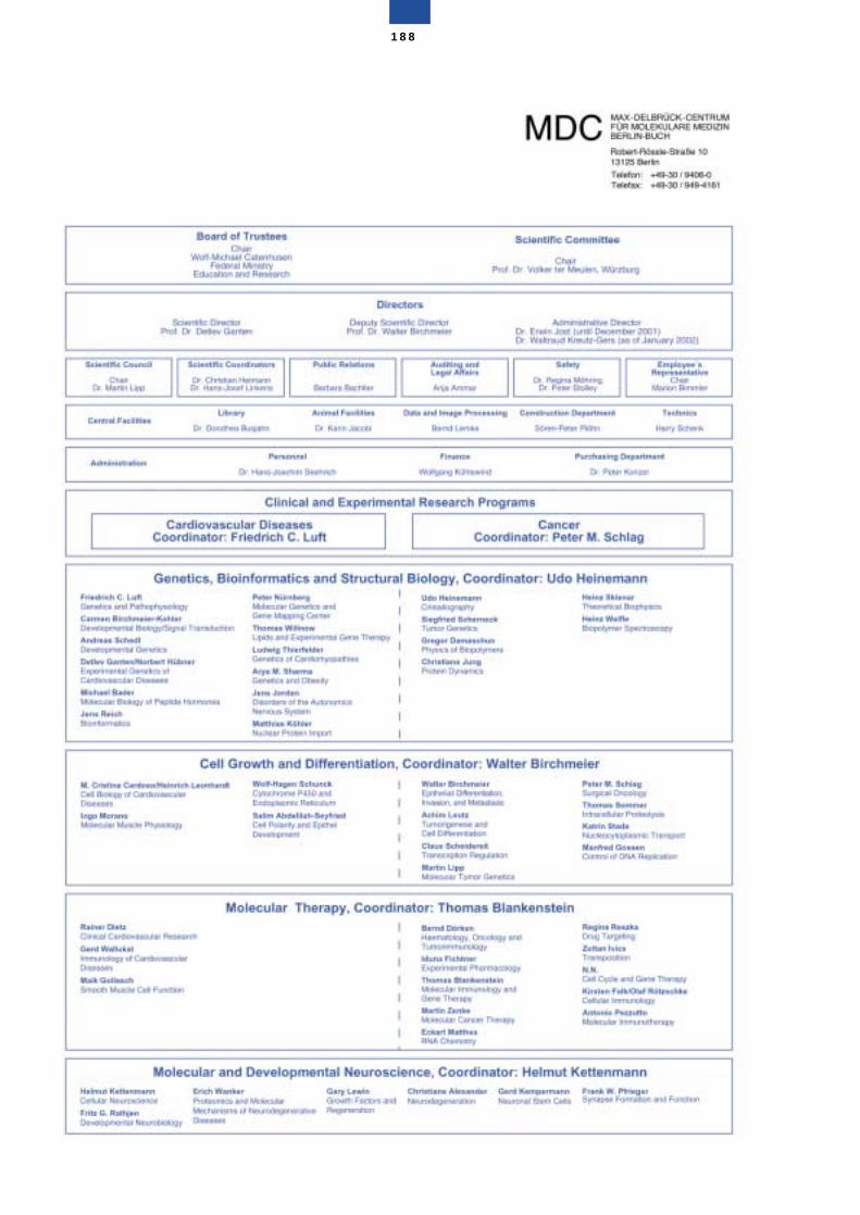
188
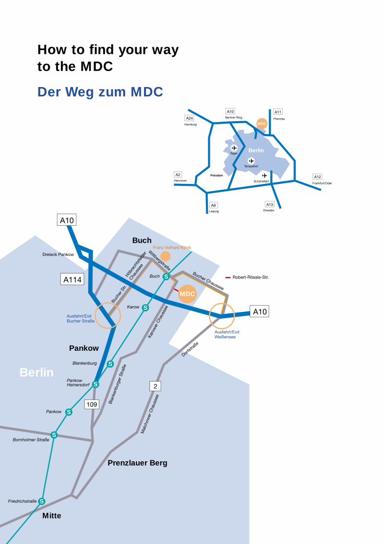
How to find your wayto the MDC
Berlin
Prenzlauer Berg
Pankow
Bornholmer Straße
Friedrichstraße
Pankow
PankowHeinersdorf
Blankenburg
Ausfahrt/ExitBucher Straße
Franz Volhard Klinik
Dreieck Pankow
Robert-Rössle-Str.
Ausfahrt/ExitWeißensee
Karow
Buch
Mitte
Kar
ower
Cha
usse
e
Mal
chow
er C
haus
see
Dorfstra
ße
Bla
nken
burg
er S
traß
e
Buche
r Str.
Höbre
chts
feld
er
Chaus
see
Bucher Chaussee
A114
A10
A10
109
2
MDC
BuchW
iltbergstraße
S
S
S
S
S
S
S
Hamburg
Berliner Ring
A10
Dresden
A13
Frankfurt/Oder
A12
Prenzlau
A11
A24
Leipzig
A9
Hannover
A2
MDC
Berlin
Potsdam
Schönefeld
Tempelhof
Tegel
Der Weg zum MDC
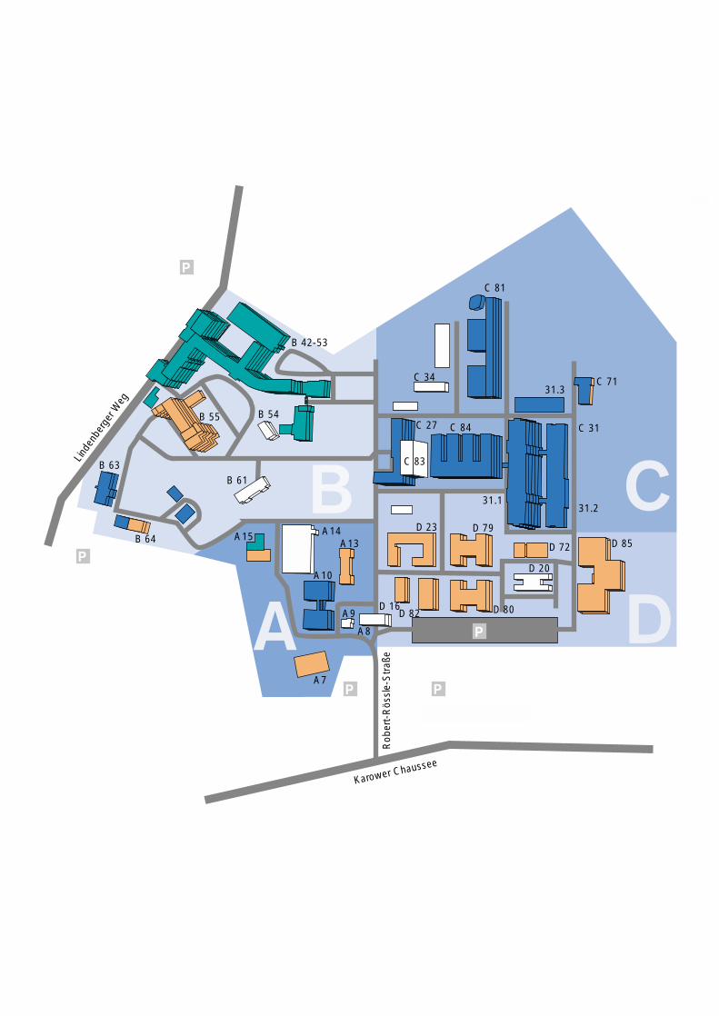
Linde
nber
ger W
eg
Karower Chaussee
Rob
ert-
Rös
sle-
Str
aße
31.3
31.231.1
C 71
C 84
C 34
C 83
D 79
D 72 D 85
D 20
D 80D 82
A8
A9
A7
A10
A13A14A15B 64
B 63B 61
B 54B 55
B 42-53
D 16
D 23
C 27 C 31
C 81
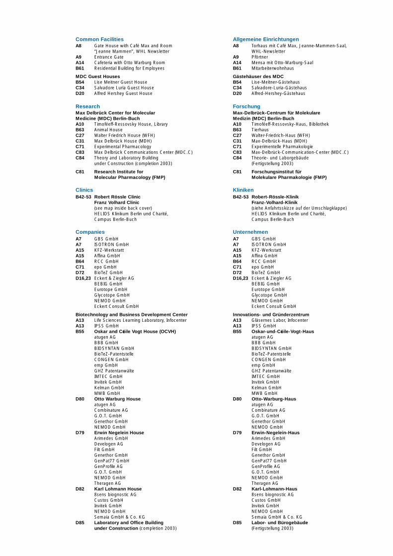
Common FacilitiesA8 Gate House with Café Max and Room
“Jeanne Mammen”, WHL NewsletterA9 Entrance GateA14 Cafeteria with Otto Warburg RoomB61 Residential Building for Employees
MDC Guest HousesB54 Lise Meitner Guest HouseC34 Salvadore Luria Guest HouseD20 Alfred Hershey Guest House
ResearchMax Delbrück Center for Molecular Medicine (MDC) Berlin-BuchA10 Timoféeff-Ressovsky House, LibraryB63 Animal HouseC27 Walter Friedrich House (WFH)C31 Max Delbrück House (MDH)C71 Experimental PharmacologyC83 Max Delbrück Communications Center (MDC.C)C84 Theory and Laboratory Building
under Construction (completion 2003)
C81 Research Institute for Molecular Pharmacology (FMP)
ClinicsB42-53 Robert Rössle Clinic
Franz Volhard Clinic (see map inside back cover)HELIOS Klinikum Berlin und Charité, Campus Berlin-Buch
CompaniesA7 GBS GmbHA7 ISOTRON GmbHA15 KFZ-WerkstattA15 Affina GmbHB64 RCC GmbHC71 epo GmbHD72 BioTeZ GmbHD16,23 Eckert & Ziegler AG
BEBIG GmbHEurotope GmbHGlycotope GmbHNEMOD GmbHEckert Consult GmbH
Biotechnology and Business Development Center A13 Life Sciences Learning Laboratory, InfocenterA13 IPSS GmbHB55 Oskar and Cécile Vogt House (OCVH)
atugen AGBBB GmbHBIOSYNTAN GmbHBioTeZ-PatentstelleCONGEN GmbHemp GmbHGHZ PatentanwälteIMTEC GmbHInvitek GmbHKelman GmbHMWB GmbH
D80 Otto Warburg Houseatugen AGCombinature AGG.O.T. GmbHGenethor GmbHNEMOD GmbH
D79 Erwin Negelein HouseArimedes GmbHDevelogen AGFilt GmbHGenethor GmbHGenPat77 GmbHGenProfile AGG.O.T. GmbHNEMOD GmbHTheragen AG
D82 Karl Lohmann House8sens biognostic AGCustos GmbHInvitek GmbHNEMOD GmbHSemaia GmbH & Co. KG
D85 Laboratory and Office Building under Construction (completion 2003)
Allgemeine EinrichtungenA8 Torhaus mit Café Max, Jeanne-Mammen-Saal,
WHL-NewsletterA9 PförtnerA14 Mensa mit Otto-Warburg-SaalB61 Mitarbeiterwohnhaus
Gästehäuser des MDC B54 Lise-Meitner-GästehausC34 Salvadore-Luria-GästehausD20 Alfred-Hershey-Gästehaus
ForschungMax-Delbrück-Centrum für Molekulare Medizin (MDC) Berlin-BuchA10 Timoféeff-Ressovsky-Haus, BibliothekB63 TierhausC27 Walter-Friedrich-Haus (WFH)C31 Max-Delbrück-Haus (MDH)C71 Experimentelle PharmakologieC83 Max-Delbrück-Communication-Center (MDC.C)C84 Theorie- und Laborgebäude
(Fertigstellung 2003)
C81 Forschungsinstitut für Molekulare Pharmakologie (FMP)
KlinikenB42-53 Robert-Rössle-Klinik
Franz-Volhard-Klinik (siehe Anfahrtsskizze auf der Umschlagklappe)HELIOS Klinikum Berlin und Charité, Campus Berlin-Buch
UnternehmenA7 GBS GmbHA7 ISOTRON GmbHA15 KFZ-WerkstattA15 Affina GmbHB64 RCC GmbHC71 epo GmbHD72 BioTeZ GmbHD16,23 Eckert & Ziegler AG
BEBIG GmbHEurotope GmbHGlycotope GmbHNEMOD GmbHEckert Consult GmbH
Innovations- und Gründerzentrum A13 Gläsernes Labor, InfocenterA13 IPSS GmbHB55 Oskar-und-Cécile-Vogt-Haus
atugen AGBBB GmbHBIOSYNTAN GmbHBioTeZ-PatentstelleCONGEN GmbHemp GmbHGHZ PatentanwälteIMTEC GmbHInvitek GmbHKelman GmbHMWB GmbH
D80 Otto-Warburg-Hausatugen AGCombinature AGG.O.T. GmbHGenethor GmbHNEMOD GmbH
D79 Erwin-Negelein-HausArimedes GmbHDevelogen AGFilt GmbHGenethor GmbHGenPat77 GmbHGenProfile AGG.O.T. GmbHNEMOD GmbHTheragen AG
D82 Karl-Lohmann-Haus8sens biognostic AGCustos GmbHInvitek GmbHNEMOD GmbHSemaia GmbH & Co. KG
D85 Labor- und Bürogebäude (Fertigstellung 2003)
Max Delbrück Center for Moleculare Medicine (MDC) Berlin-Buch
Robert-Rössle-Strasse 10D-13122 Berlin
Tel.: +49-30-94 06-0Fax: +49-30-949-41 61e-mail: [email protected]://www.mdc-berlin.de

![Compact and Discreet - yaesu.ru · Battery Life [5-5-90 duty with battery saver]MDC-1200® Features: - MDC-1200® ANI - MDC-1200® Call Alert - MDC-1200® Sel Call - MDC-1200® Radio](https://img.pdfslide.us/doc/110x75/5e80d3de6005c20fb639820d/compact-and-discreet-yaesuru-battery-life-5-5-90-duty-with-battery-savermdc-1200.jpg)