Embed Size (px)
Citation preview
Funduscopic InterpretationUnderstanding the Fundus: is that normal?
Gillian McLellanBVMS PhD DVOphthalDECVO DACVO MRCVS
With thanks to Christine Heinrich and all who contributed images

Fundus ≠ Retina
working knowledge of fundus anatomy
how to evaluate the fundus systematically
normal fundus variants
hallmarks of fundus pathology
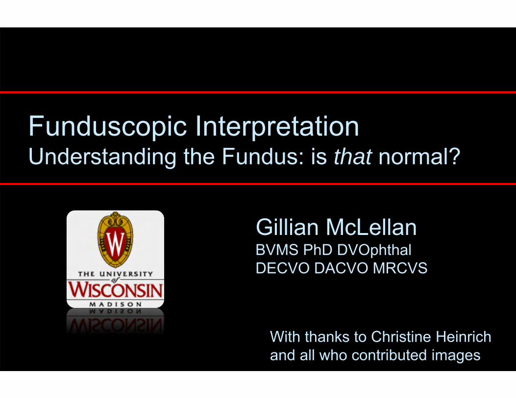
Fundus anatomy
Vitreous normally optically clear (almost)
Only retinal vessels / pigmented RPE +/- inner choroid (tapetum) make a major contribution to the typical appearance of a normal fundus
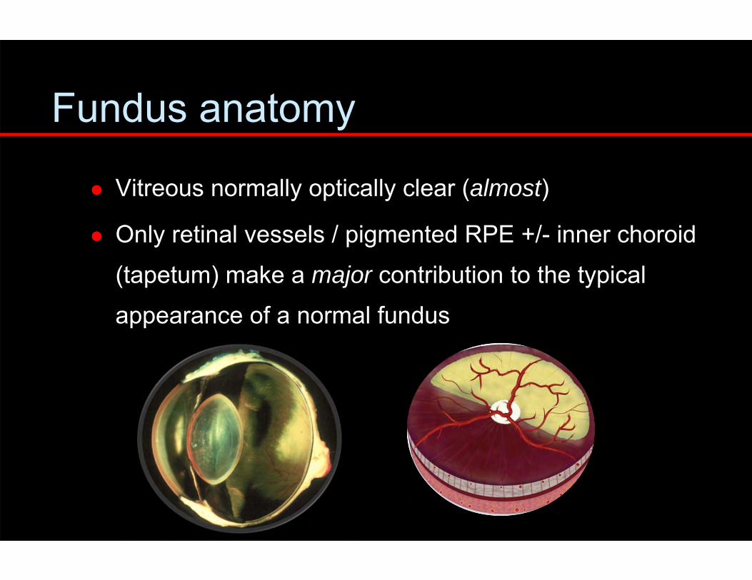
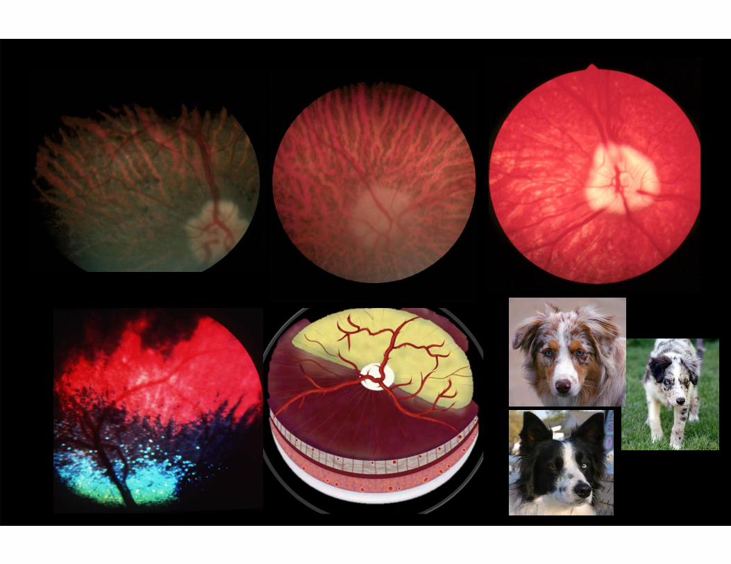
Retinal Vascular Patternsholangiotic
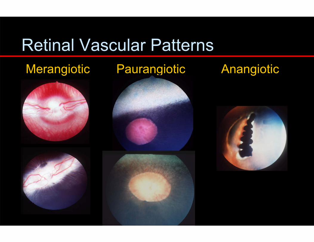
Retinal Vascular PatternsMerangiotic Paurangiotic Anangiotic
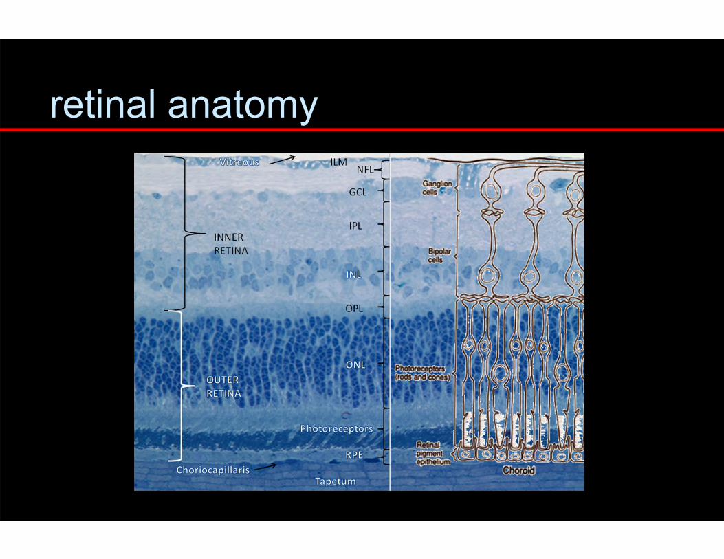
retinal anatomy
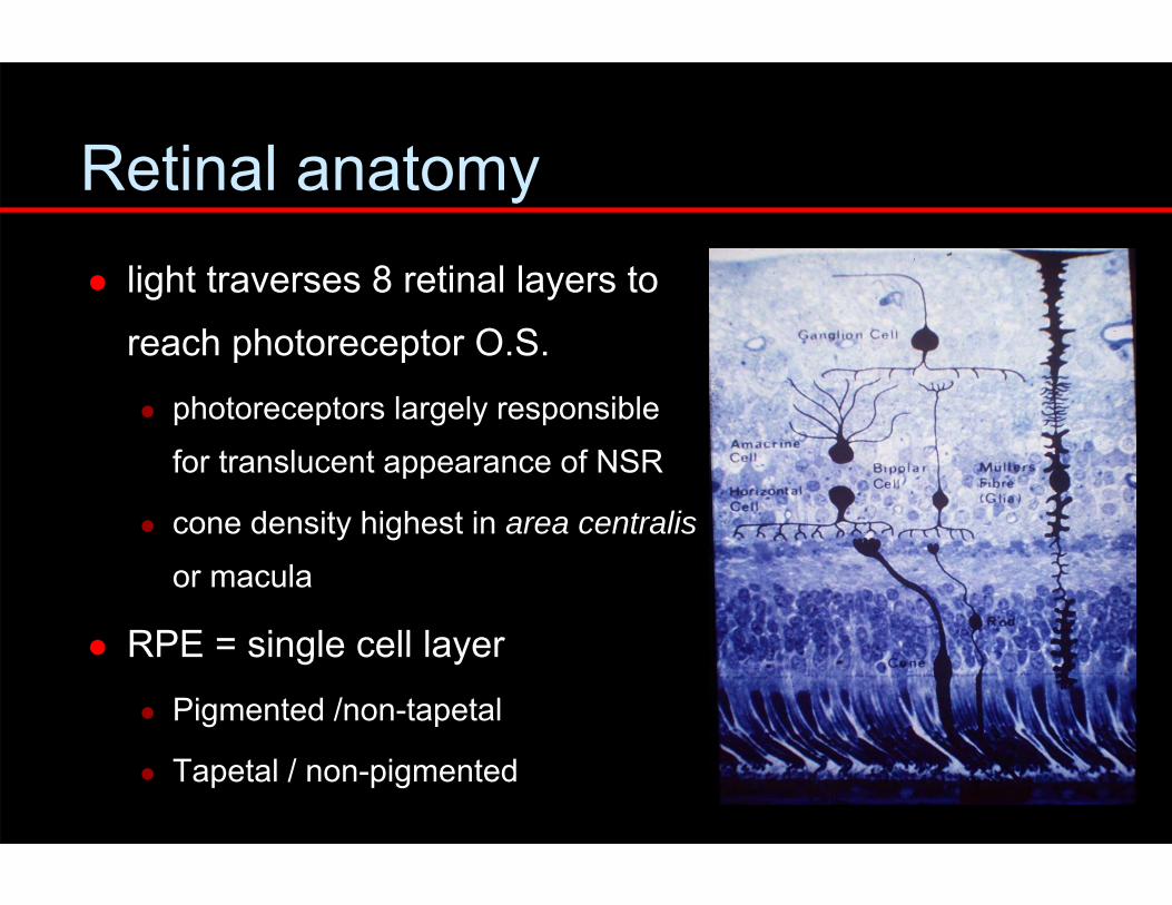
Retinal anatomy light traverses 8 retinal layers to
reach photoreceptor O.S. photoreceptors largely responsible
for translucent appearance of NSR
cone density highest in area centralis
or macula
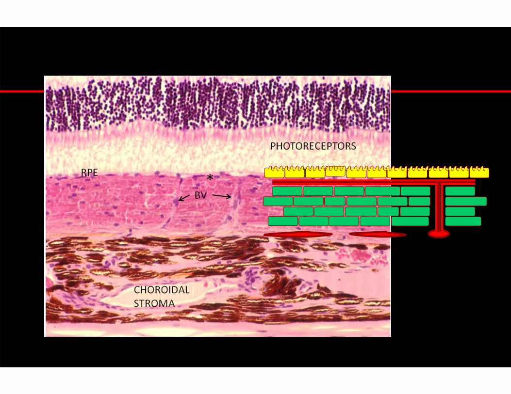
RPE = single cell layer Pigmented /non-tapetal
Tapetal / non-pigmented
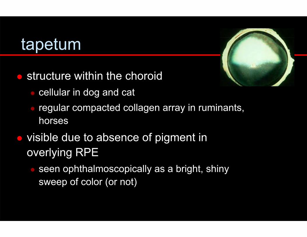
tapetum
structure within the choroid cellular in dog and cat regular compacted collagen array in ruminants,
horses
visible due to absence of pigment in overlying RPE seen ophthalmoscopically as a bright, shiny
sweep of color (or not)
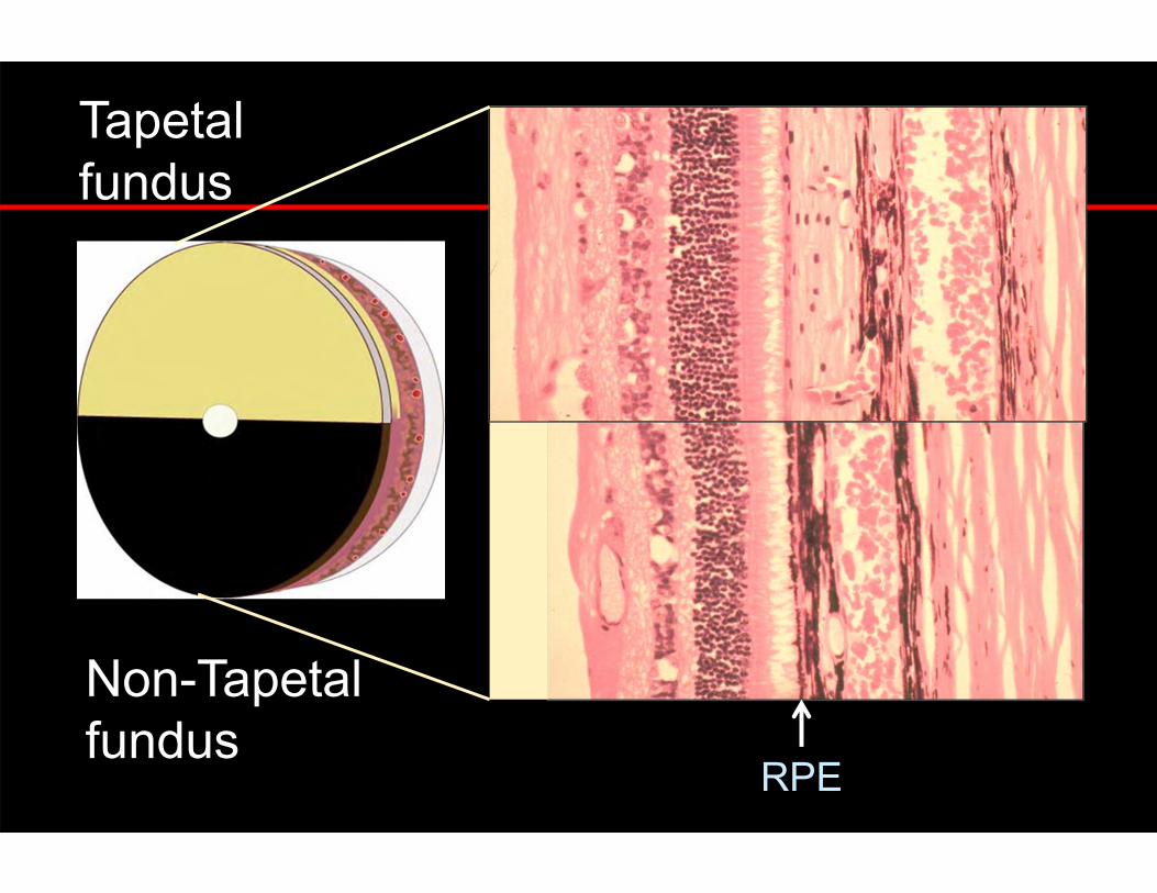
RPE
Tapetalfundus
Non-Tapetalfundus
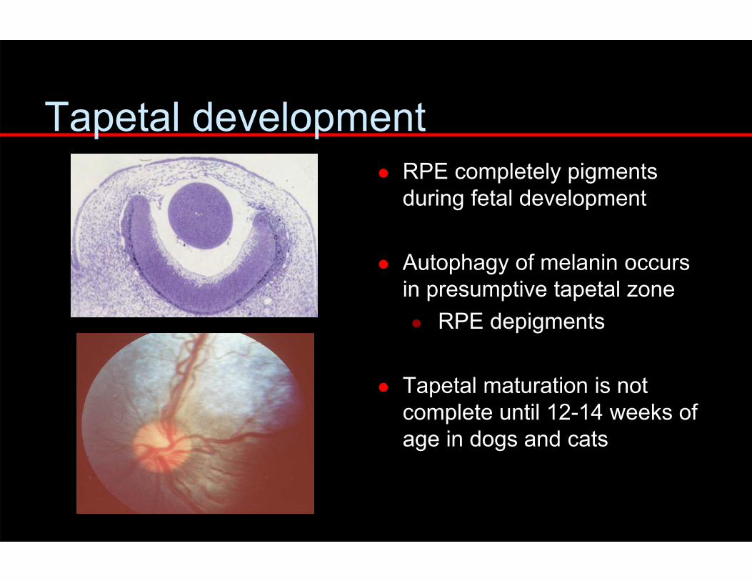
Tapetal development RPE completely pigments
during fetal development
Autophagy of melanin occurs in presumptive tapetal zone RPE depigments
Tapetal maturation is not complete until 12-14 weeks of age in dogs and cats
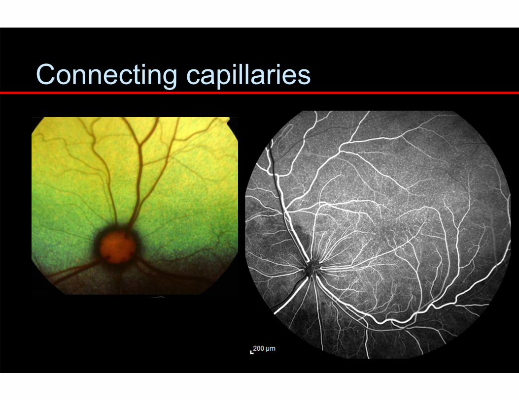
Connecting capillaries
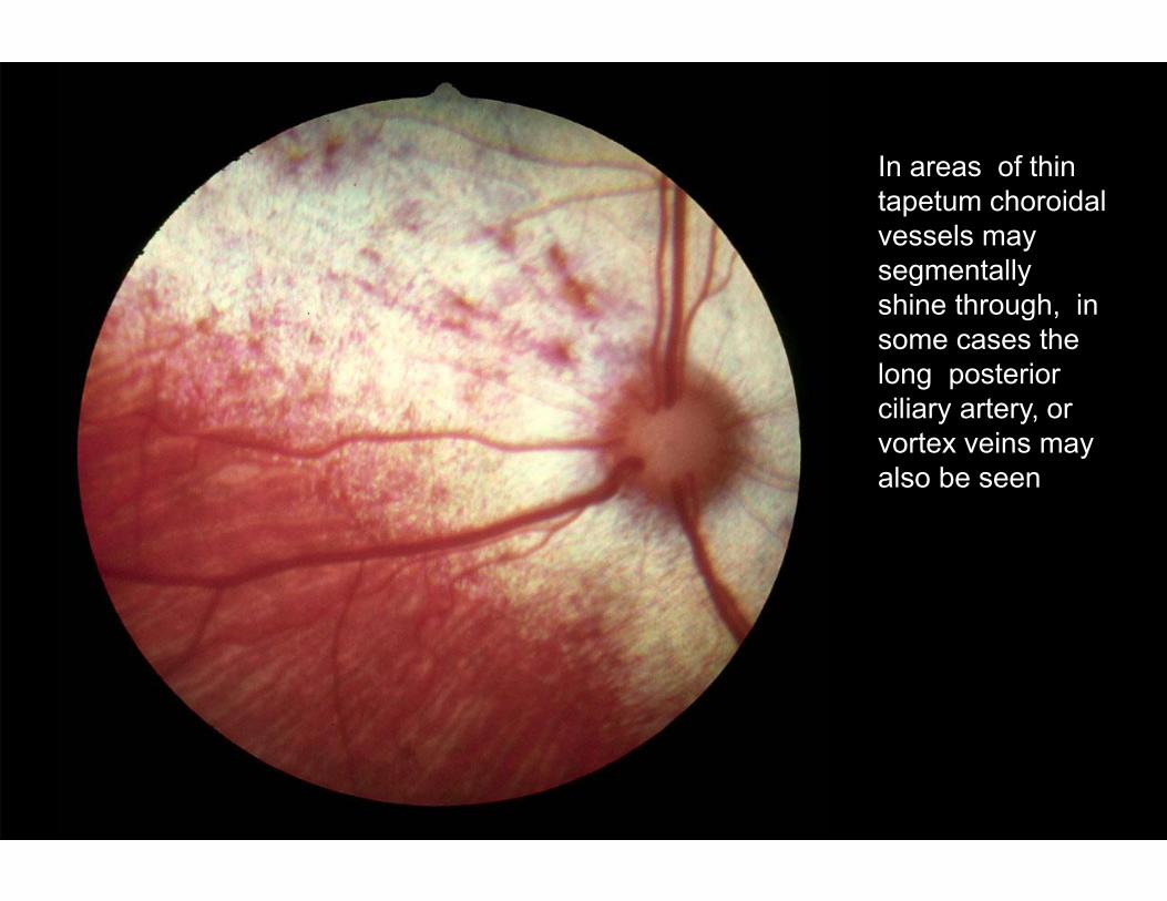
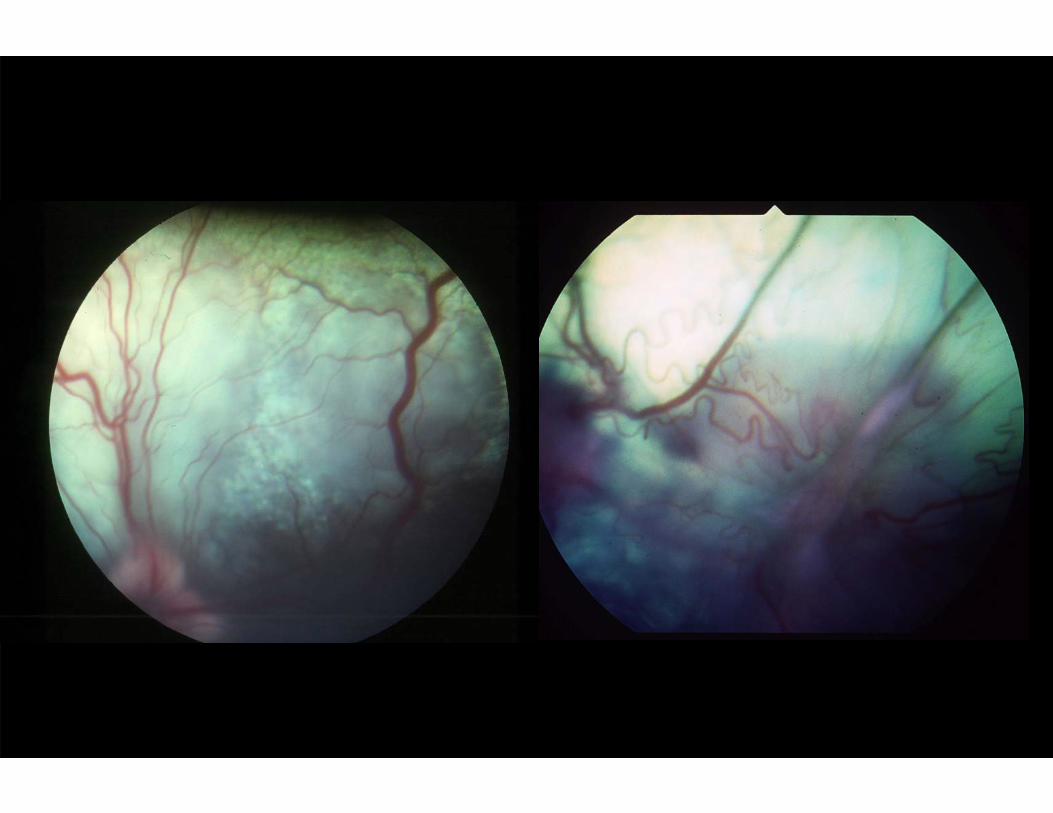
In areas of thin tapetum choroidalvessels may segmentallyshine through, in some cases the long posterior ciliary artery, or vortex veins may also be seen
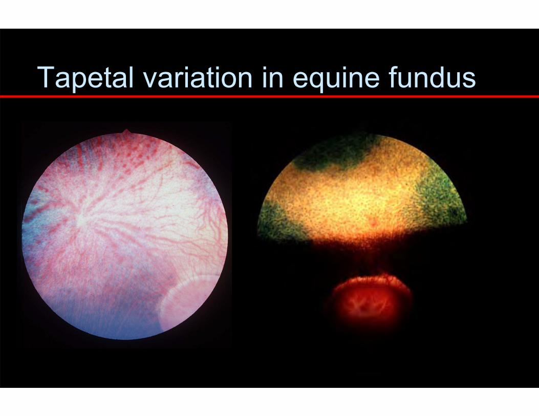
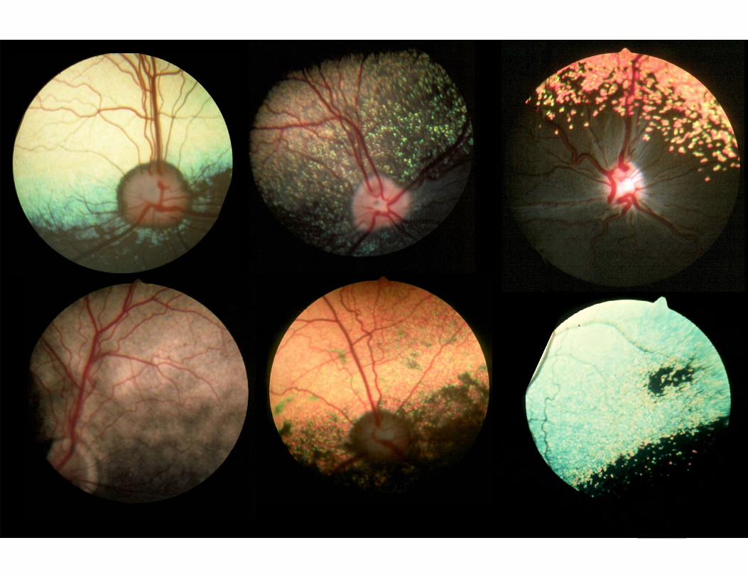
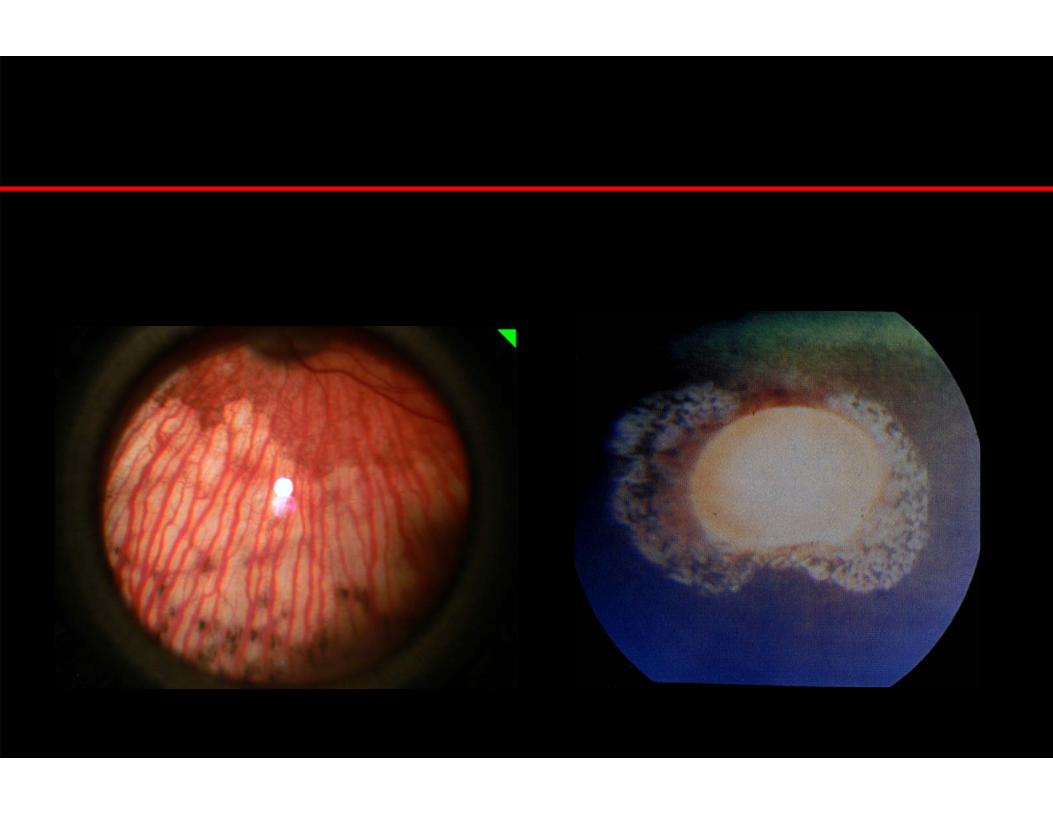
Tapetal variation in equine fundus
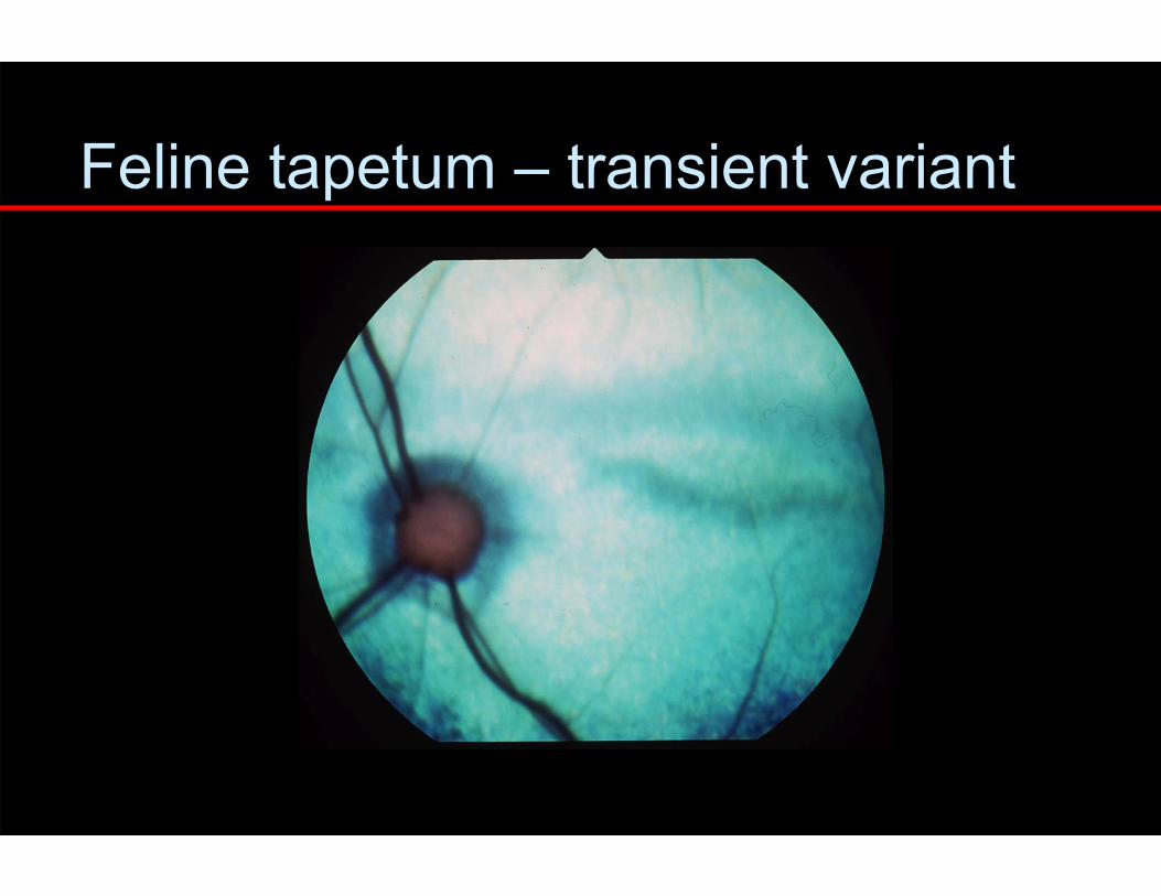
Feline tapetum – transient variant
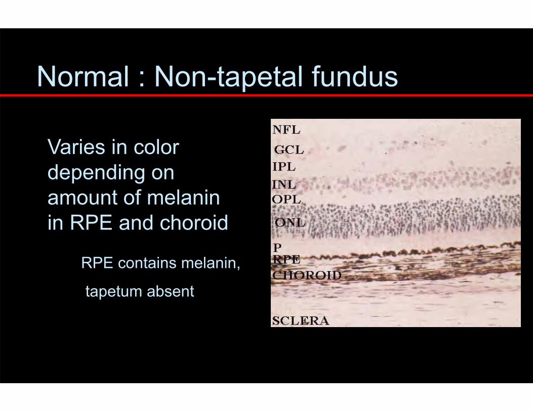
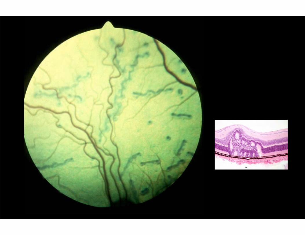
Normal : Non-tapetal fundus
RPE contains melanin,
tapetum absent
Varies in color depending on amount of melanin in RPE and choroid
Normal : Non-tapetal fundus
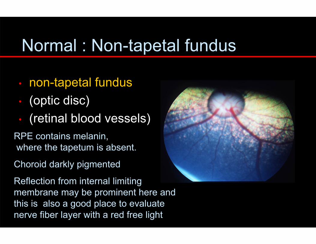
• non-tapetal fundus• (optic disc)• (retinal blood vessels)
RPE contains melanin,where the tapetum is absent.
Choroid darkly pigmented
Reflection from internal limiting membrane may be prominent here and this is also a good place to evaluate nerve fiber layer with a red free light
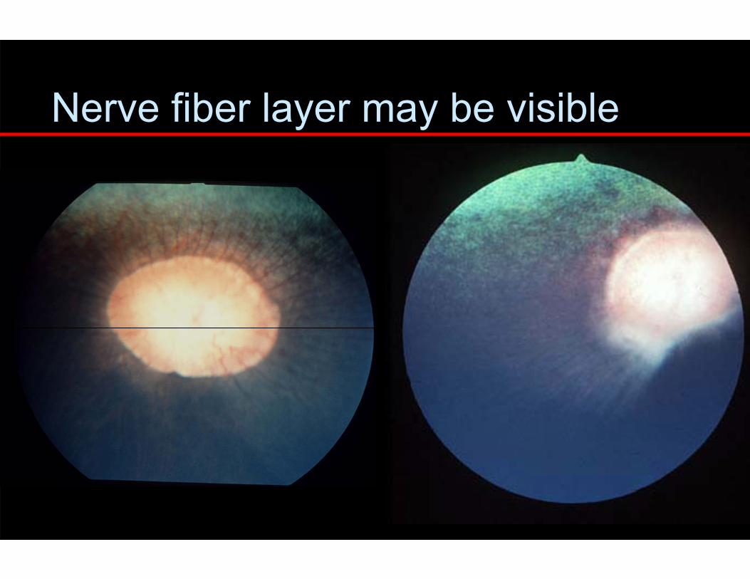
Nerve fiber layer may be visible
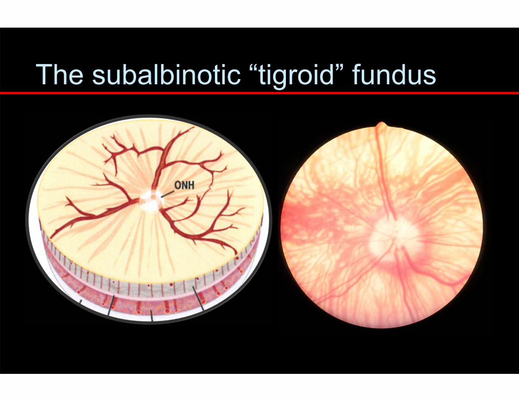
The subalbinotic “tigroid” fundus
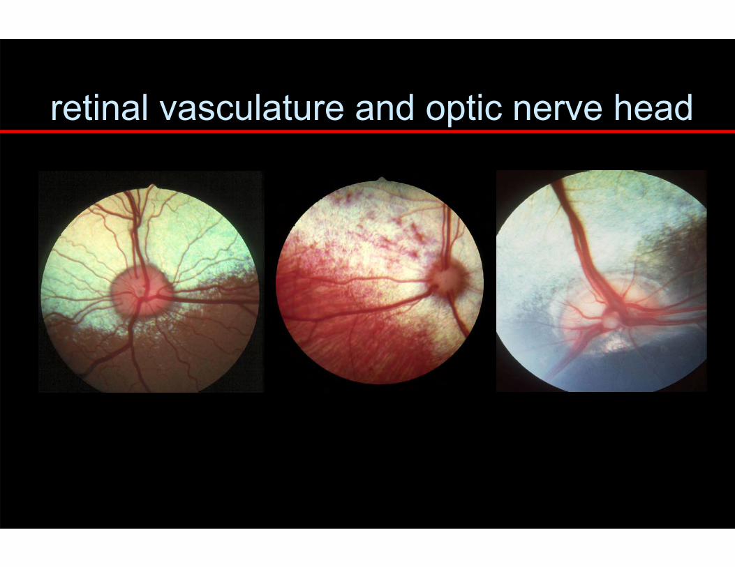
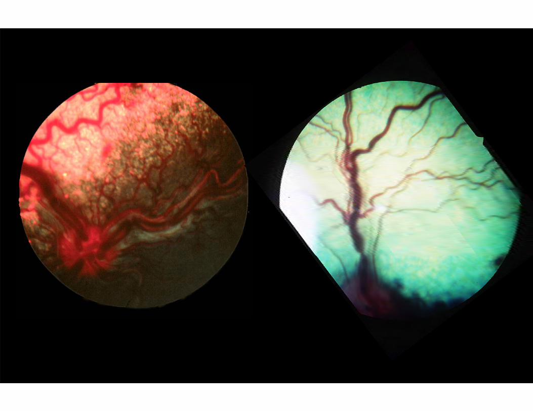
retinal vasculature and optic nerve head
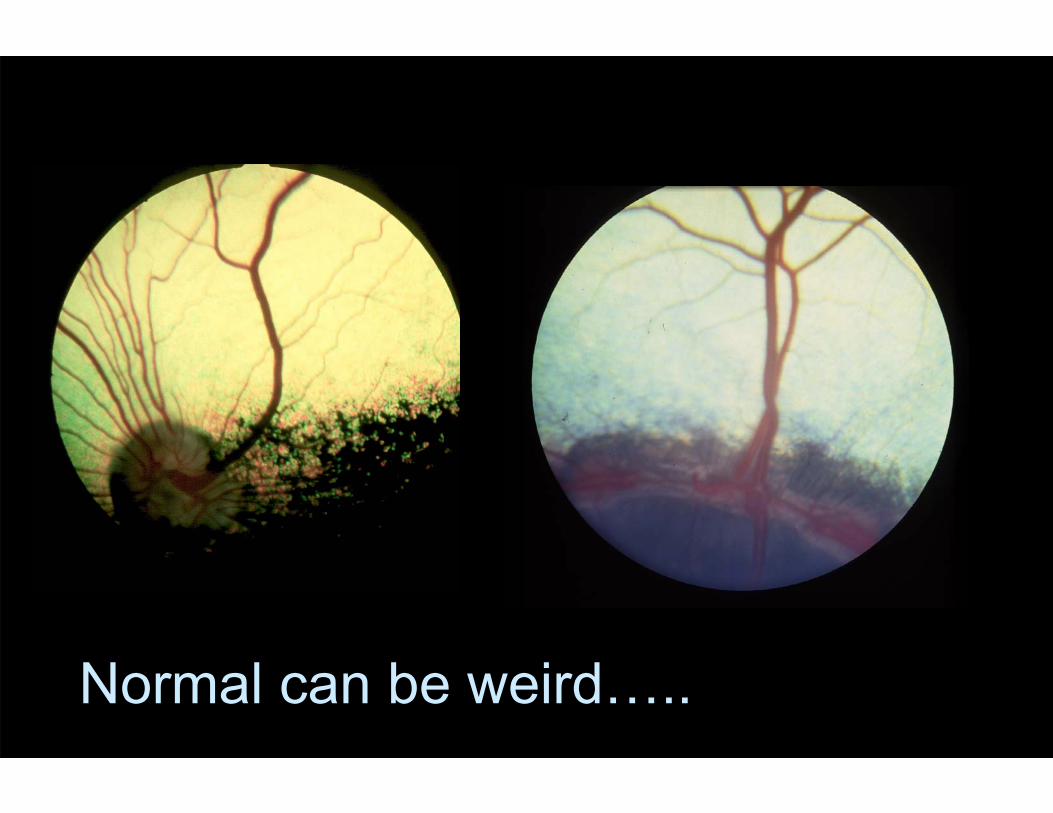
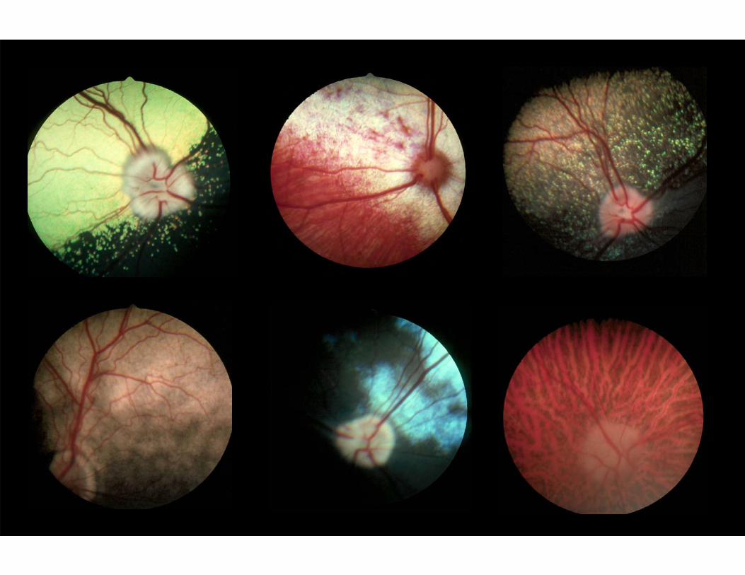
Normal can be weird…..
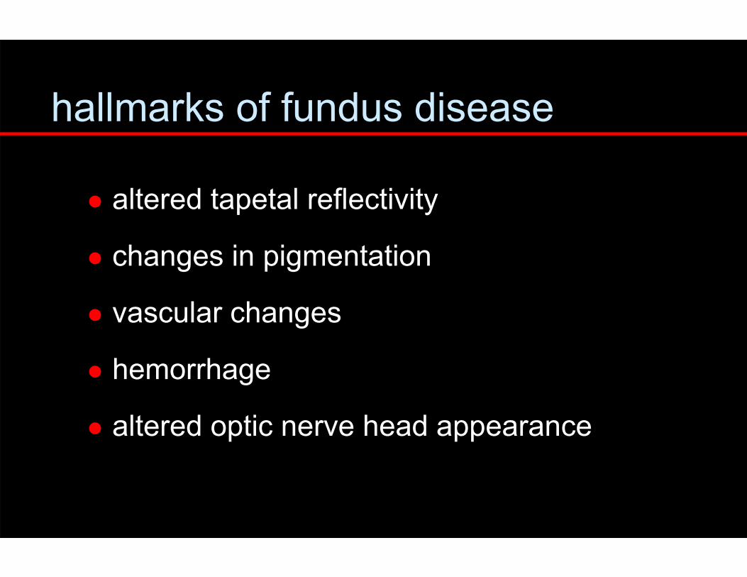
hallmarks of fundus disease
altered tapetal reflectivity
changes in pigmentation
vascular changes
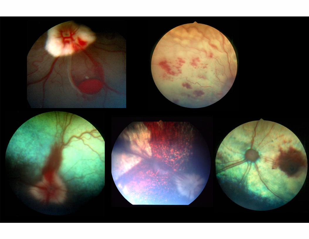
hemorrhage
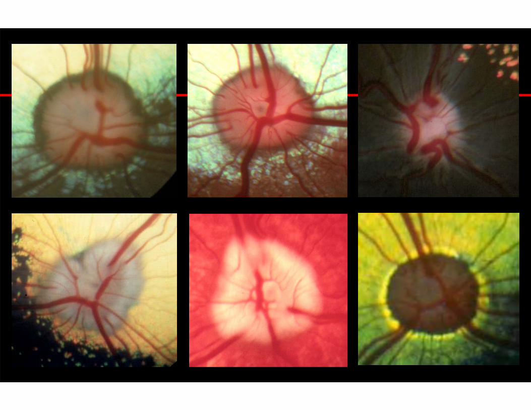
altered optic nerve head appearance
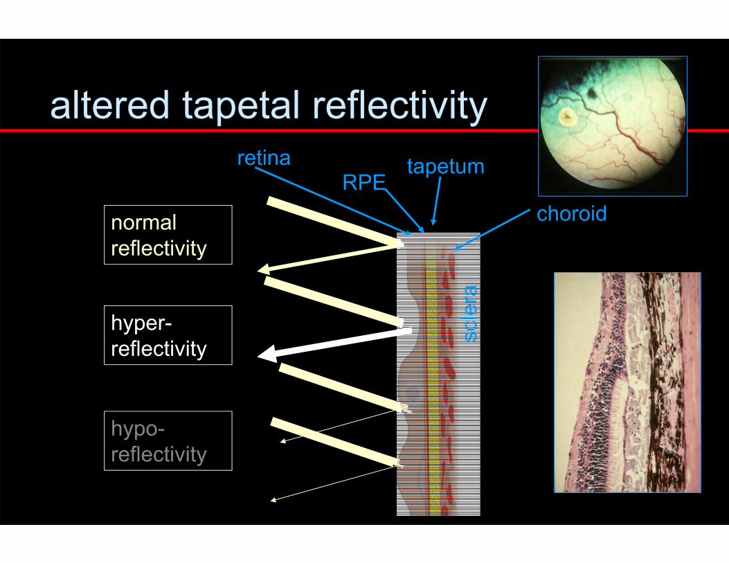
choroid
tapetumRPE
retina
normal reflectivity
hyper-reflectivity
hypo-reflectivity
altered tapetal reflectivity
scle
ra
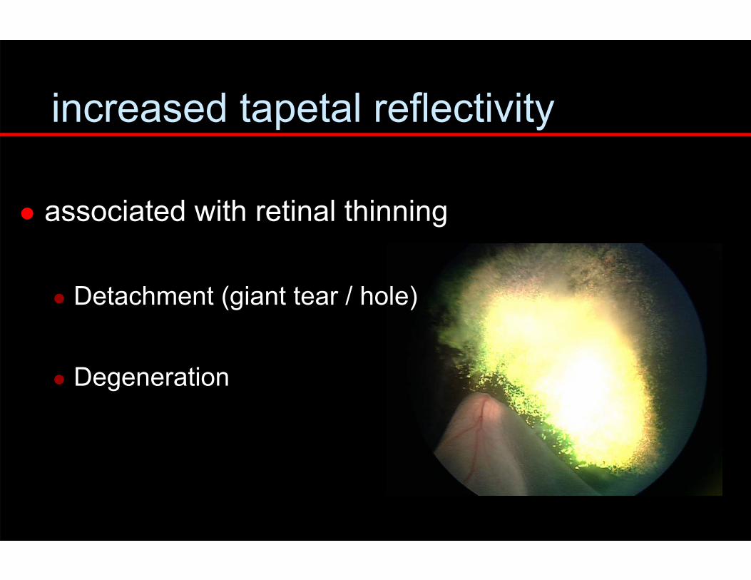
increased tapetal reflectivity
associated with retinal thinning
Detachment (giant tear / hole)
Degeneration
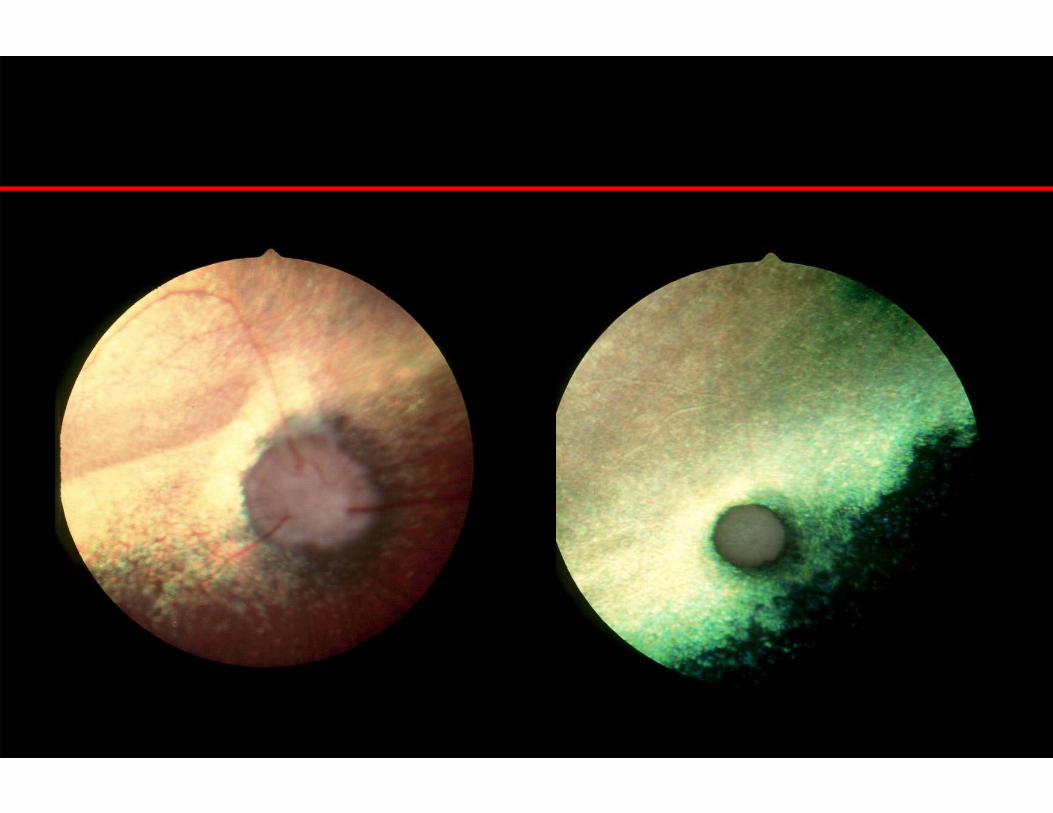
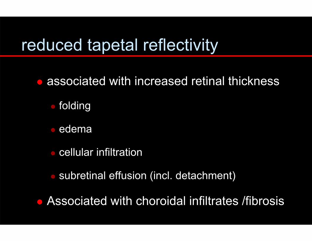
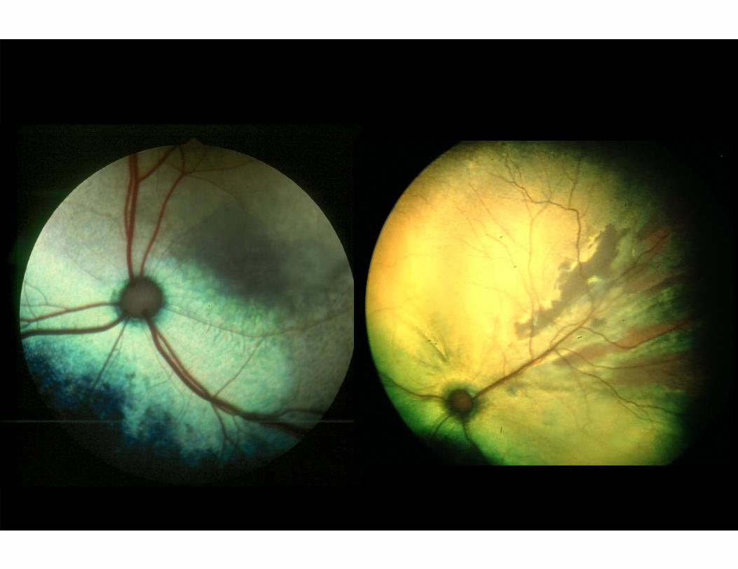
reduced tapetal reflectivity
associated with increased retinal thickness
folding
edema
cellular infiltration
subretinal effusion (incl. detachment)
Associated with choroidal infiltrates /fibrosis
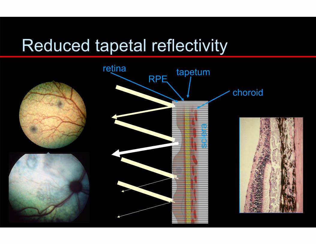
choroid
tapetumRPE
retina
Reduced tapetal reflectivity
scle
ra
altered fundus pigmentation
appearance of pigment in tapetal fundus
post-inflammatory
aberrant metabolites
lipopigment (RPED)
loss of pigment in non-tapetal fundus
post-inflammatory
degenerative retinal disease (PRA)
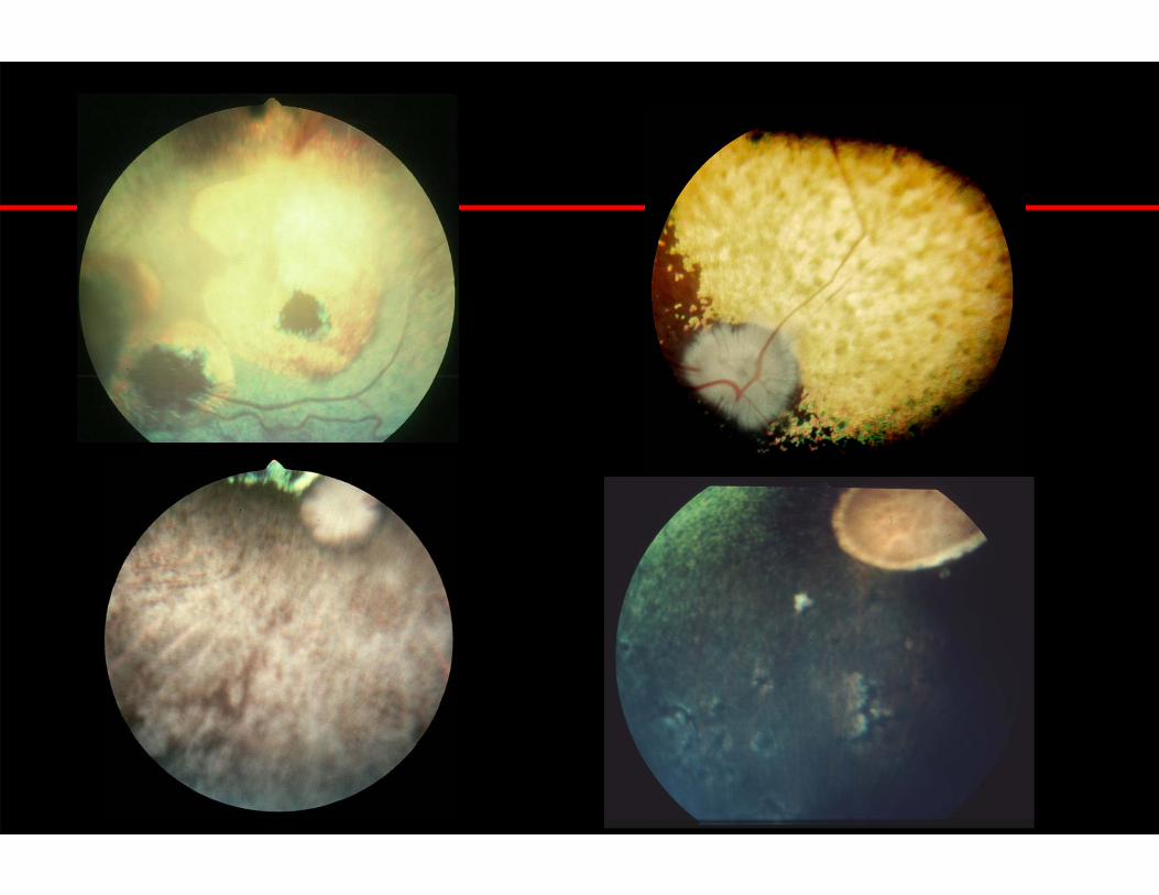
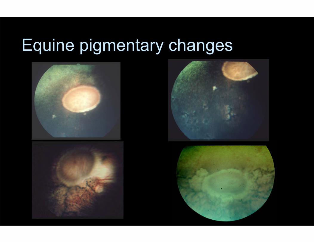
Equine pigmentary changes
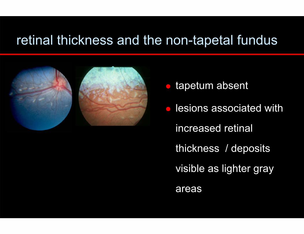
retinal thickness and the non-tapetal fundus
tapetum absent
lesions associated with
increased retinal
thickness / deposits
visible as lighter gray
areas
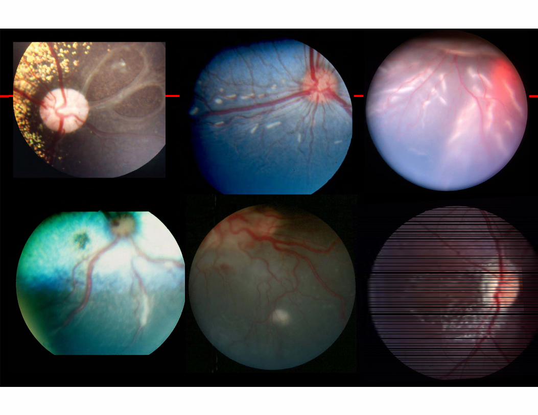
retinal vasculature
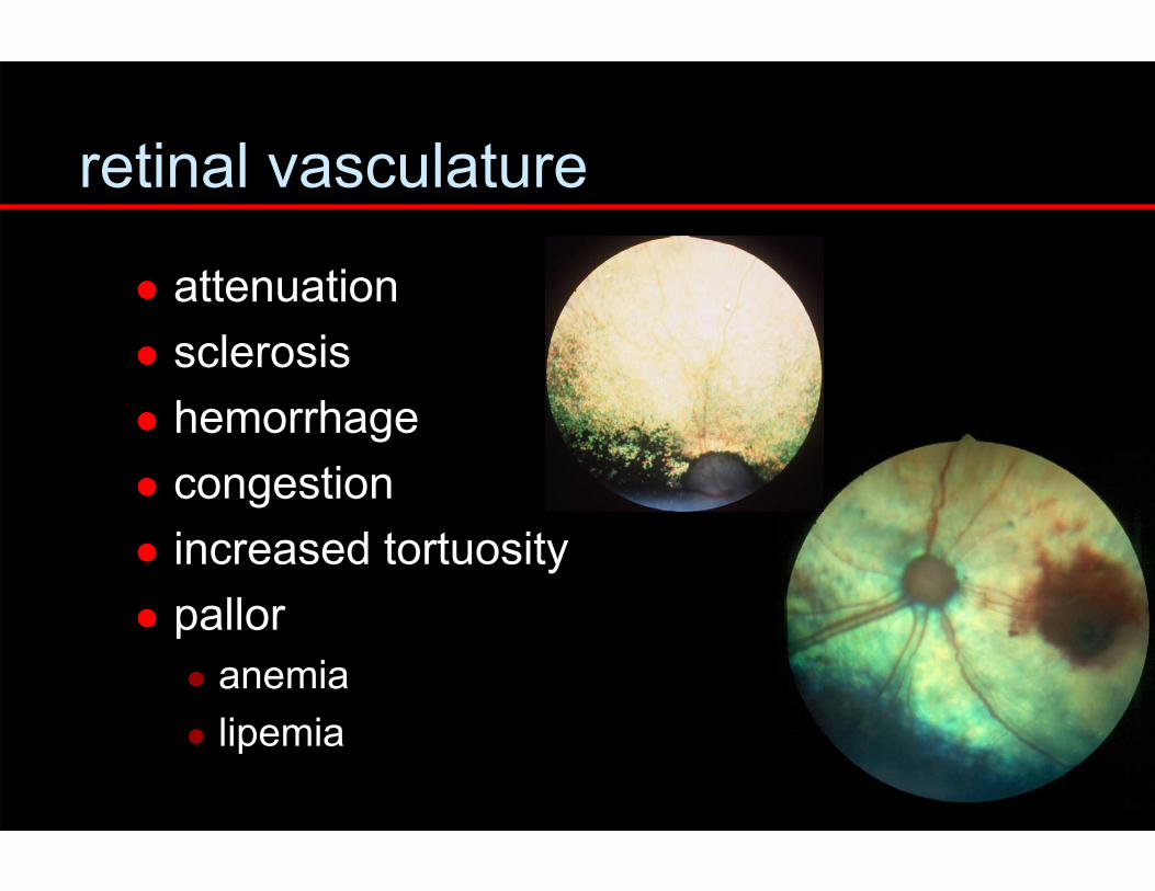
attenuation sclerosis hemorrhage congestion increased tortuosity pallor
anemia lipemia
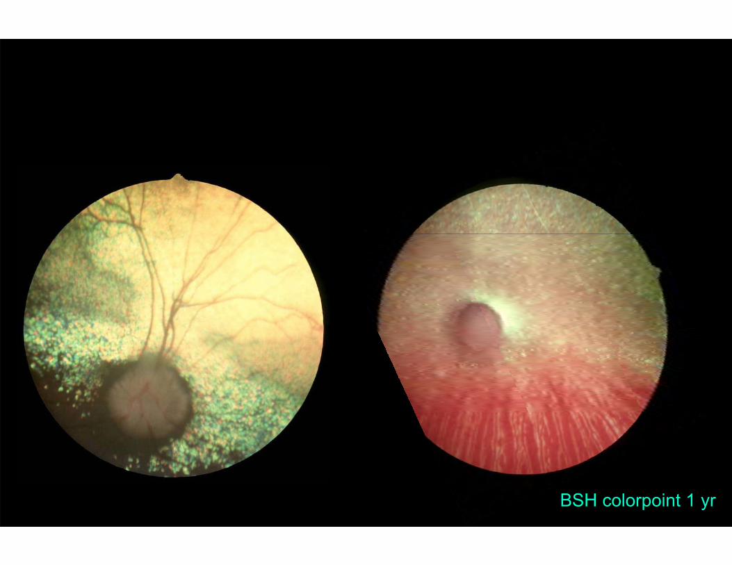
BSH colorpoint 1 yr
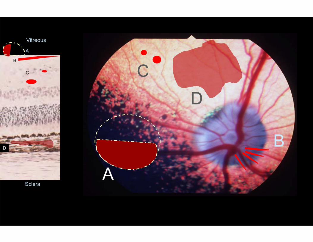
C
A
B
D
A
B
C
D
Vitreous
Sclera
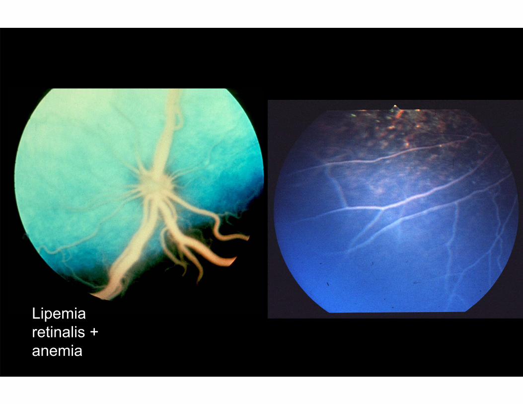
Lipemiaretinalis + anemia
optic nerve head changes reduction in size or enlargement inflammation
associated with hemorrhages, infiltrates depression / cupping / coloboma darkening
associated with loss of neural tissue (atrophy)? Congestion?
pallor associated with loss of vasculature Associated with gliosis
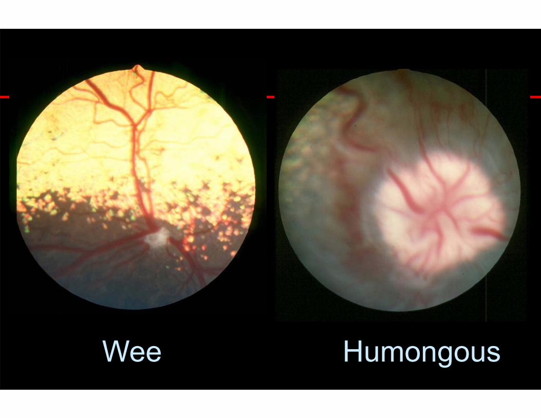
Wee Humongous
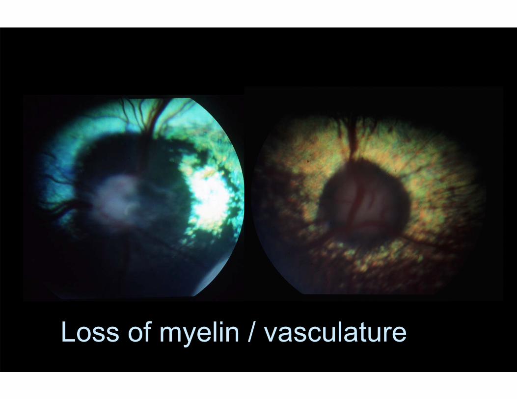
Loss of myelin / vasculature

UC Davis R. Bellhorn

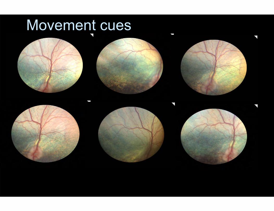
Movement cues
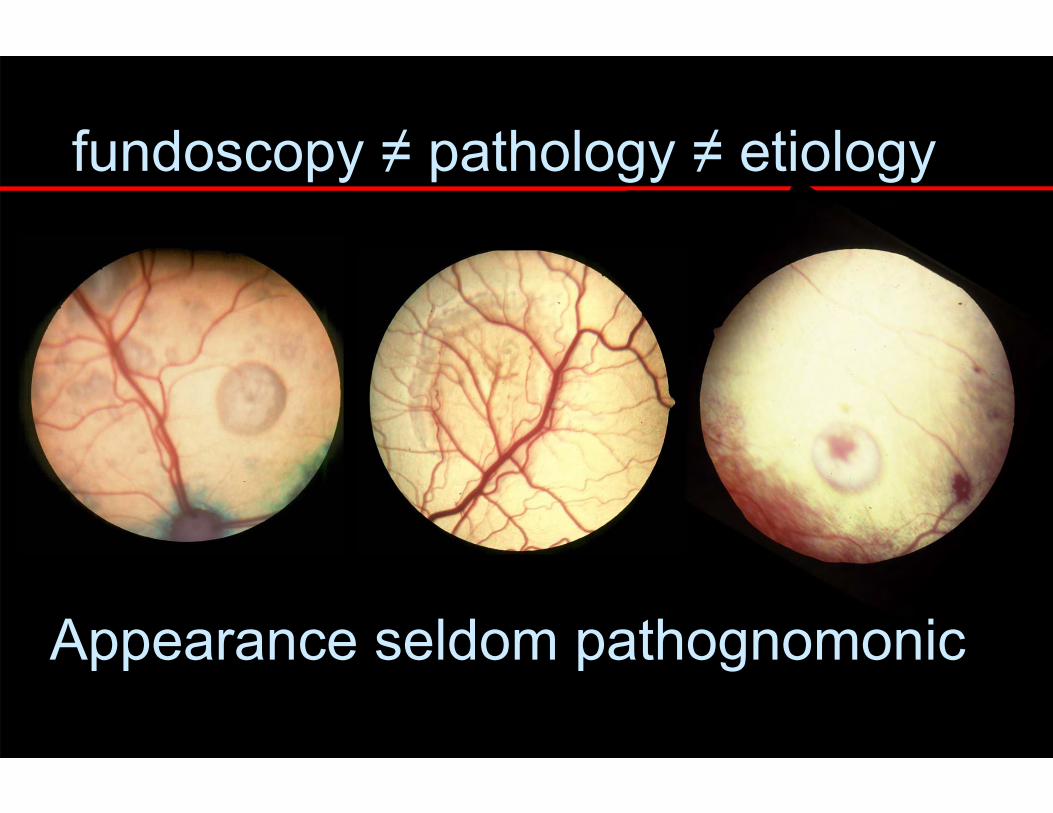
fundoscopy ≠ pathology ≠ etiology
Appearance seldom pathognomonic
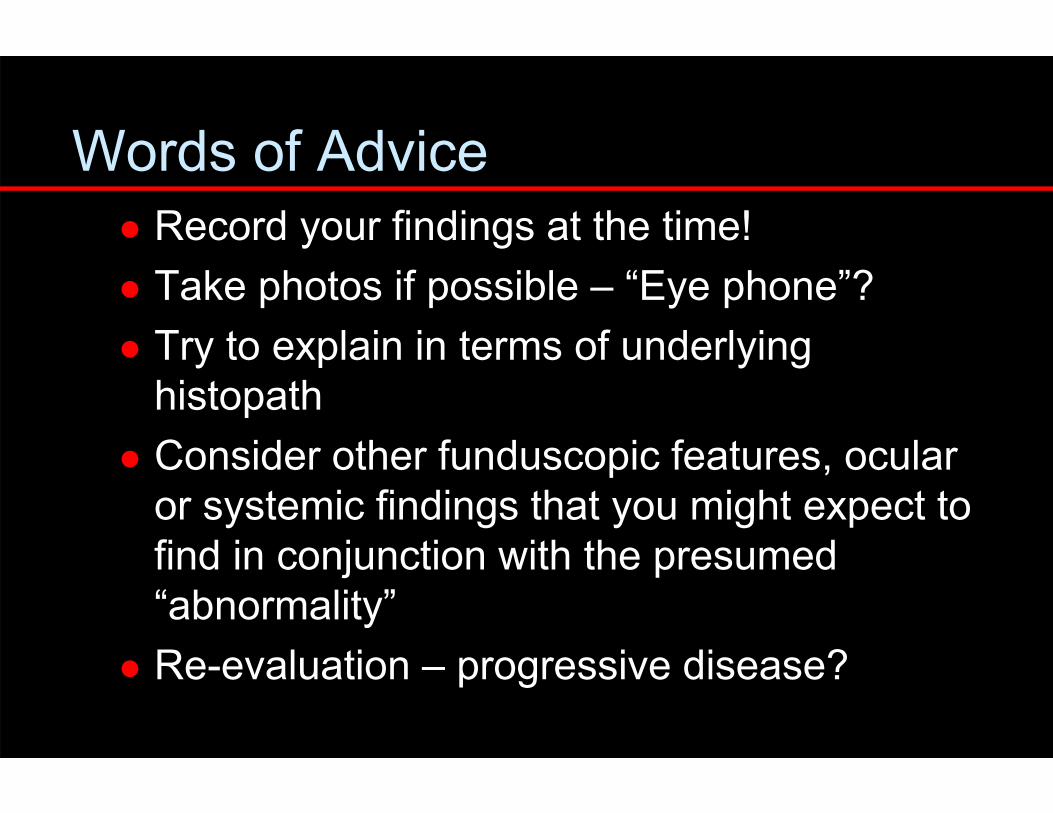
Words of Advice Record your findings at the time! Take photos if possible – “Eye phone”? Try to explain in terms of underlying
histopath Consider other funduscopic features, ocular
or systemic findings that you might expect to find in conjunction with the presumed “abnormality”
Re-evaluation – progressive disease?
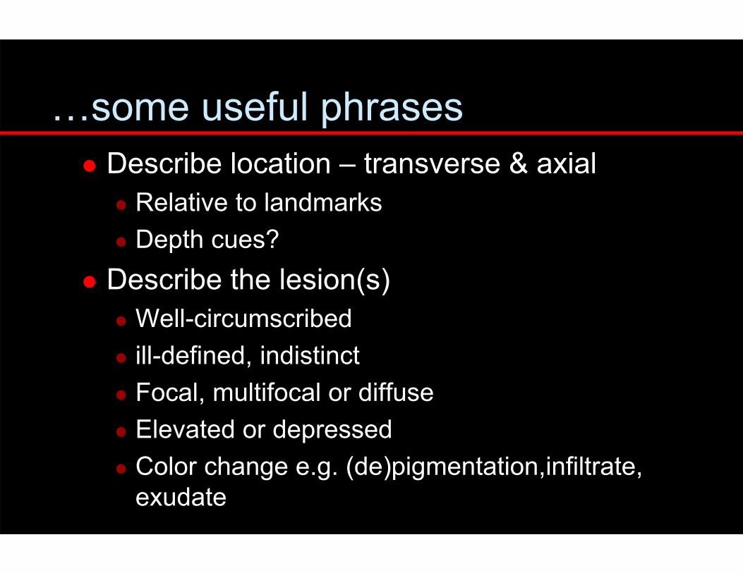
…some useful phrases Describe location – transverse & axial
Relative to landmarks Depth cues?
Describe the lesion(s) Well-circumscribed ill-defined, indistinct Focal, multifocal or diffuse Elevated or depressed Color change e.g. (de)pigmentation,infiltrate,
exudate
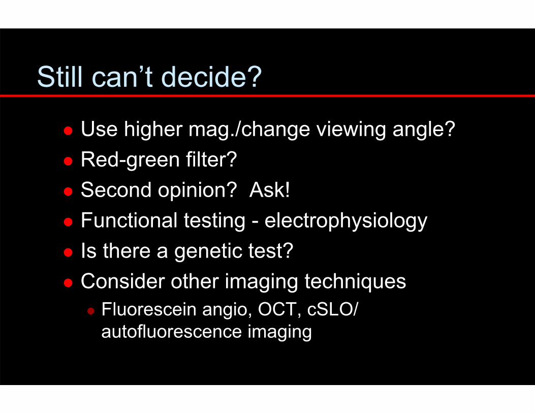
Still can’t decide? Use higher mag./change viewing angle? Red-green filter? Second opinion? Ask! Functional testing - electrophysiology Is there a genetic test? Consider other imaging techniques
Fluorescein angio, OCT, cSLO/ autofluorescence imaging
Maybe we are not as invincible as we think we are?
Appearances can be deceptive


![[David McLellan] Marx Before Marxism(Bookos.org)](https://img.pdfslide.us/doc/110x75/55cf9c4c550346d033a95702/david-mclellan-marx-before-marxismbookosorg.jpg)