Embed Size (px)
Citation preview
1
Interpretation of Chest Radiographs
Reynard McDonald, MD
Medical Director NJMS Global Tuberculosis Institute
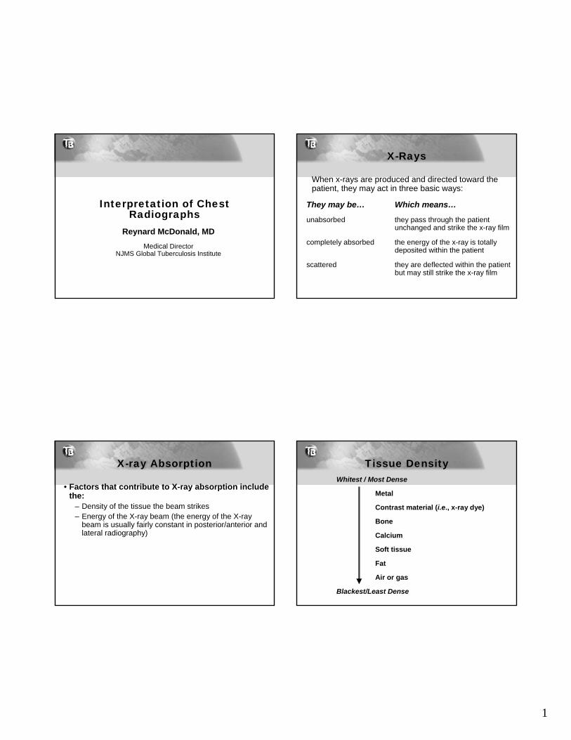
X-Rays
When x-rays are produced and directed toward the patient, they may act in three basic ways:
They may be… Which means…
unabsorbed they pass through the patient unchanged and strike the x-ray film
completely absorbed the energy of the x-ray is totally deposited within the patient
scattered they are deflected within the patient but may still strike the x-ray film
X-ray Absorption
• Factors that contribute to X-ray absorption include the:
– Density of the tissue the beam strikes– Energy of the X-ray beam (the energy of the X-ray
beam is usually fairly constant in posterior/anterior and lateral radiography)
Tissue DensityWhitest / Most Dense
Metal
Contrast material (i.e., x-ray dye)
Bone
Calcium
Soft tissue
Fat
Air or gas
Blackest/Least Dense
2
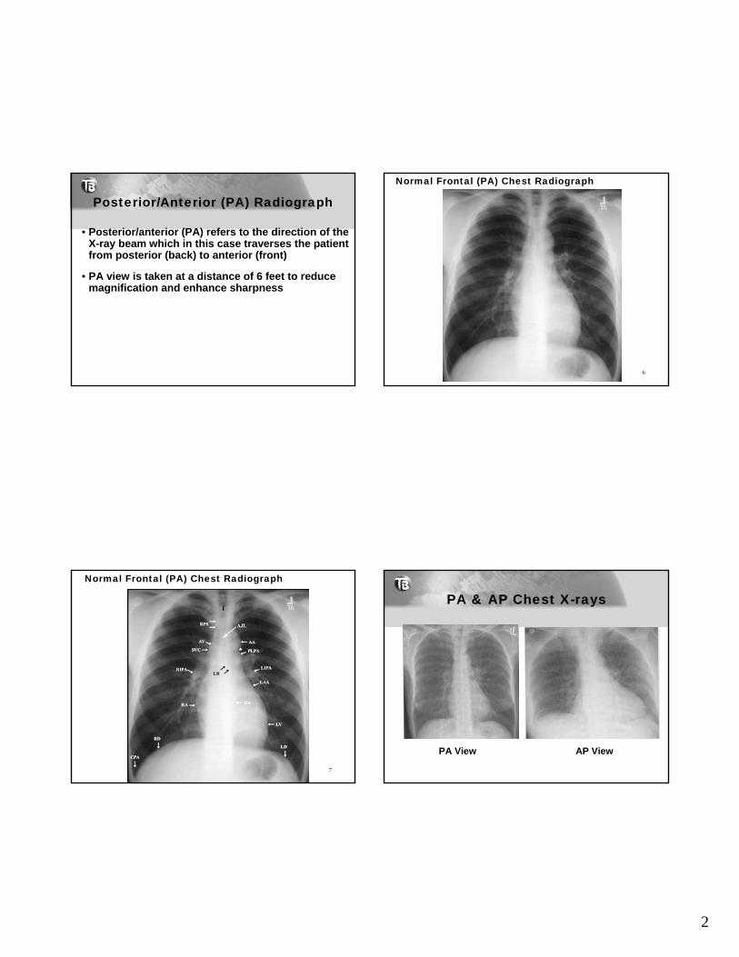
Posterior/Anterior (PA) Radiograph
• Posterior/anterior (PA) refers to the direction of the X-ray beam which in this case traverses the patient from posterior (back) to anterior (front)
• PA view is taken at a distance of 6 feet to reduce magnification and enhance sharpness
Normal Frontal (PA) Chest Radiograph
6
Normal Frontal (PA) Chest Radiograph
7
PA & AP Chest X-rays
PA View AP View
3
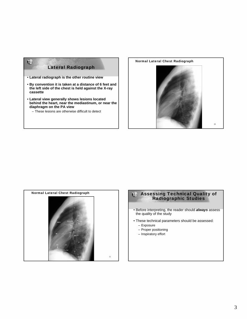
Lateral Radiograph
• Lateral radiograph is the other routine view
• By convention it is taken at a distance of 6 feet and the left side of the chest is held against the X-ray cassette
• Lateral view generally shows lesions located behind the heart, near the mediastinum, or near the diaphragm on the PA view
– These lesions are otherwise difficult to detect
Normal Lateral Chest Radiograph
10
Normal Lateral Chest Radiograph
11
Assessing Technical Quality of Radiographic Studies
• Before interpreting, the reader should always assess the quality of the study
• These technical parameters should be assessed:– Exposure– Proper positioning– Inspiratory effort
4
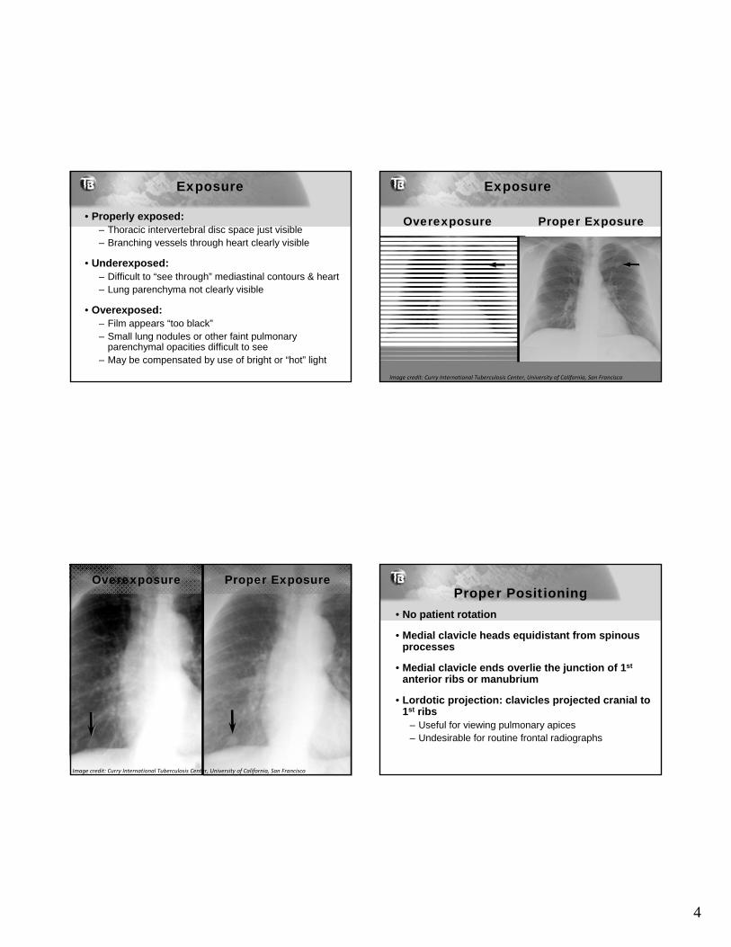
Exposure
• Properly exposed:– Thoracic intervertebral disc space just visible– Branching vessels through heart clearly visible
• Underexposed:– Difficult to “see through” mediastinal contours & heart – Lung parenchyma not clearly visible
• Overexposed:– Film appears “too black”– Small lung nodules or other faint pulmonary
parenchymal opacities difficult to see– May be compensated by use of bright or “hot” light
Overexposure Proper Exposure
Exposure
Image credit: Curry International Tuberculosis Center, University of California, San Francisco
Overexposure Proper Exposure
Image credit: Curry International Tuberculosis Center, University of California, San Francisco
Proper Positioning• No patient rotation
• Medial clavicle heads equidistant from spinousprocesses
• Medial clavicle ends overlie the junction of 1st
anterior ribs or manubrium
• Lordotic projection: clavicles projected cranial to 1st ribs
– Useful for viewing pulmonary apices – Undesirable for routine frontal radiographs
5
Rotated (Oblique)Image credit: Curry International Tuberculosis Center, University of California, San Francisco
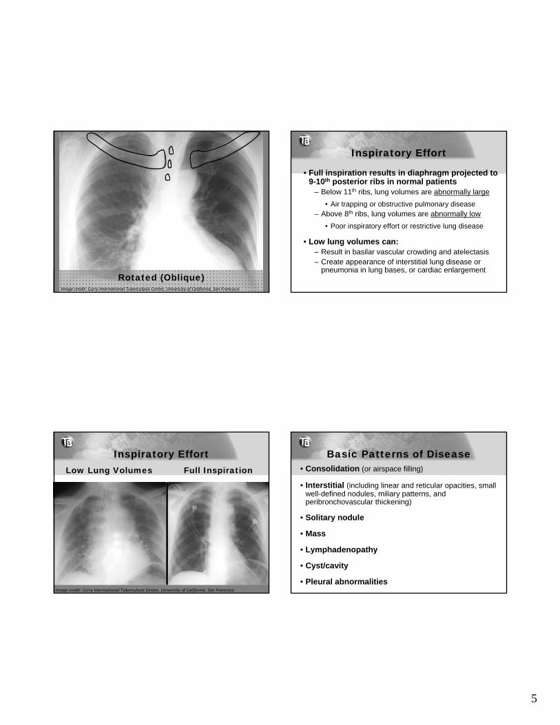
Inspiratory Effort
• Full inspiration results in diaphragm projected to 9-10th posterior ribs in normal patients
– Below 11th ribs, lung volumes are abnormally large
• Air trapping or obstructive pulmonary disease– Above 8th ribs, lung volumes are abnormally low
• Poor inspiratory effort or restrictive lung disease
• Low lung volumes can: – Result in basilar vascular crowding and atelectasis– Create appearance of interstitial lung disease or
pneumonia in lung bases, or cardiac enlargement
Inspiratory EffortLow Lung Volumes Full Inspiration
Image credit: Curry International Tuberculosis Center, University of California, San Francisco
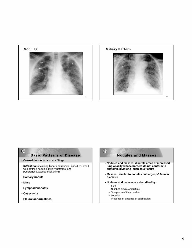
Basic Patterns of Disease• Consolidation (or airspace filling)
• Interstitial (including linear and reticular opacities, small well-defined nodules, miliary patterns, and peribronchovascular thickening)
• Solitary nodule
• Mass
• Lymphadenopathy
• Cyst/cavity
• Pleural abnormalities
6
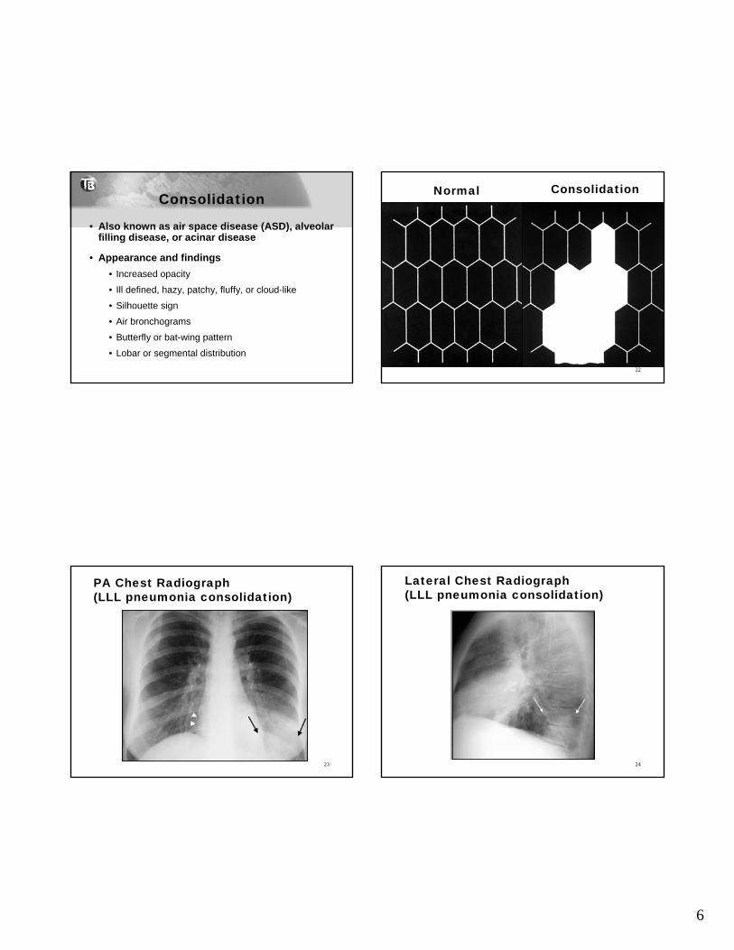
Consolidation
• Also known as air space disease (ASD), alveolar filling disease, or acinar disease
• Appearance and findings
• Increased opacity
• Ill defined, hazy, patchy, fluffy, or cloud-like
• Silhouette sign
• Air bronchograms
• Butterfly or bat-wing pattern
• Lobar or segmental distribution
Normal Consolidation
22
PA Chest Radiograph (LLL pneumonia consolidation)
23
Lateral Chest Radiograph(LLL pneumonia consolidation)
24
7
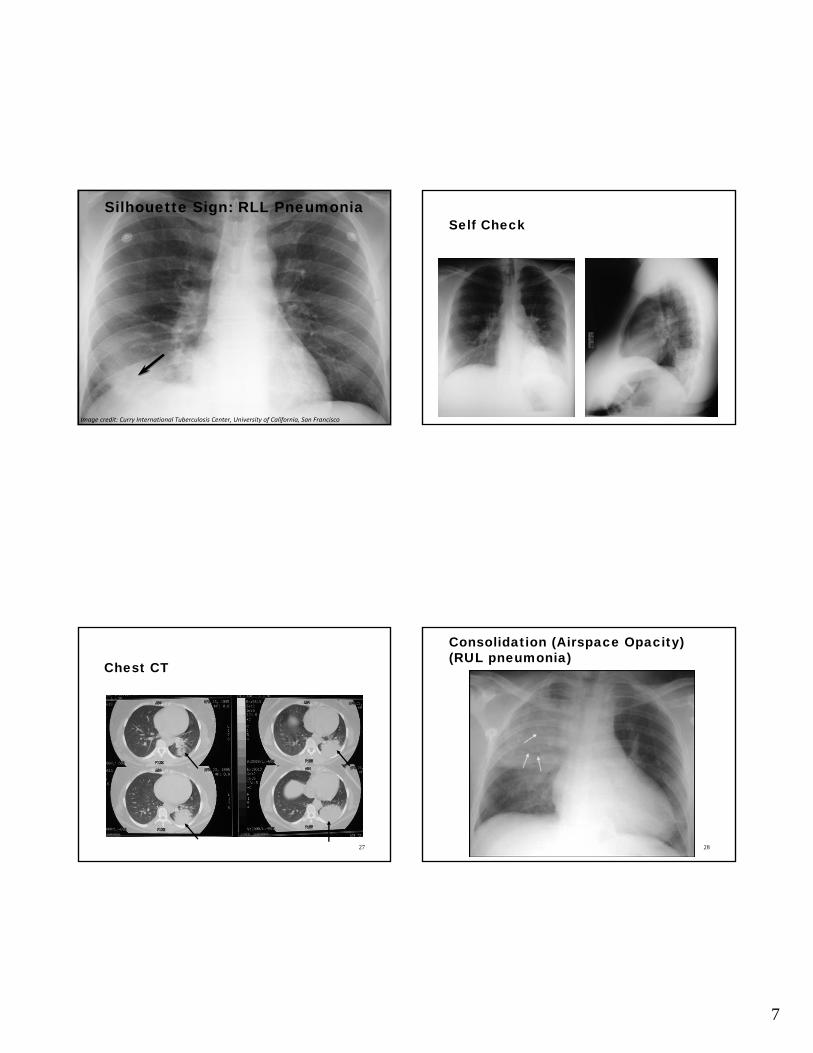
Silhouette Sign: RLL PneumoniaSilhouette Sign: RLL Pneumonia
Image credit: Curry International Tuberculosis Center, University of California, San Francisco
Self Check
26
Chest CT
27
Consolidation (Airspace Opacity)(RUL pneumonia)
28
8
Basic Patterns of Disease• Consolidation (or airspace filling)
• Interstitial (including linear and reticular opacities, small well-defined nodules, miliary patterns, and peribronchovascular thickening)
• Solitary nodule
• Mass
• Lymphadenopathy
• Cyst/cavity
• Pleural abnormalities
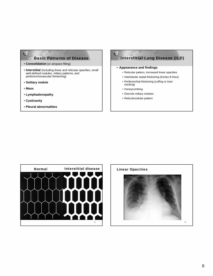
Interstitial Lung Disease (ILD)
• Appearance and findings
• Reticular pattern, increased linear opacities
• Interlobular septal thickening (Kerley B lines)
• Peribronchial thickening (cuffing or tram tracking)
• Honeycombing
• Discrete miliary nodules
• Reticulonodular pattern
Normal Interstitial disease
31
Linear Opacities
32
9
Nodules
33
Miliary Pattern
34
Basic Patterns of Disease• Consolidation (or airspace filling)
• Interstitial (including linear and reticular opacities, small well-defined nodules, miliary patterns, and peribronchovascular thickening)
• Solitary nodule
• Mass
• Lymphadenopathy
• Cyst/cavity
• Pleural abnormalities
Nodules and Masses
• Nodules and masses: discrete areas of increased lung opacity whose borders do not conform to anatomic divisions (such as a fissure)
• Masses: similar to nodules but larger, >30mm in diameter
• Nodules and masses are described by:– Size– Number, single or multiple – Sharpness of their borders– Location– Presence or absence of calcification
10
Lung Mass
37
Basic Patterns of Disease• Consolidation (or airspace filling)
• Interstitial (including linear and reticular opacities, small well-defined nodules, miliary patterns, and peribronchovascular thickening)
• Solitary nodule
• Mass
• Lymphadenopathy
• Cyst/cavity
• Pleural abnormalities
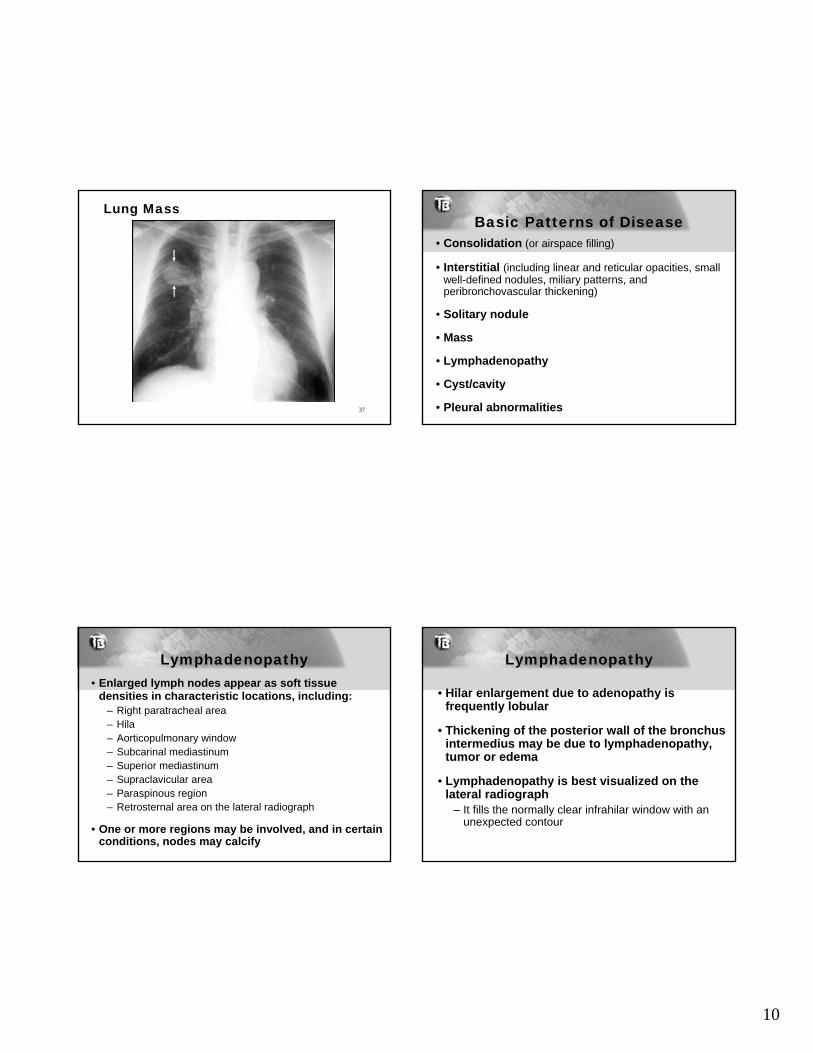
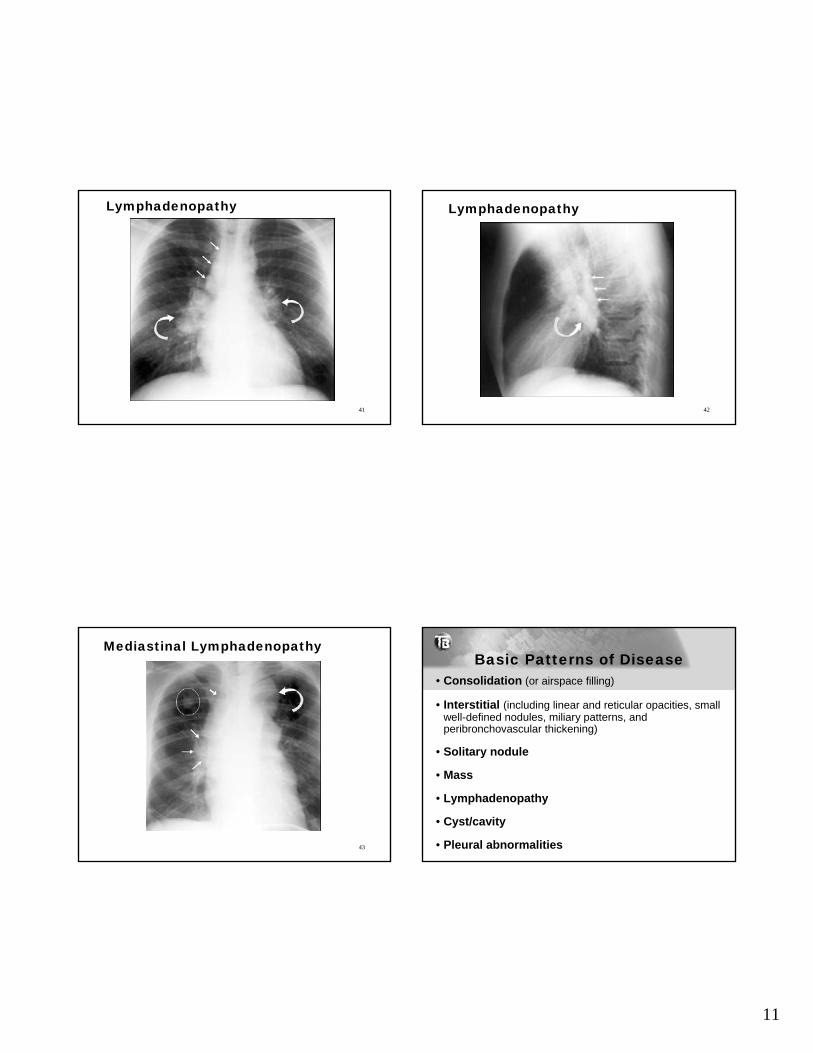
Lymphadenopathy• Enlarged lymph nodes appear as soft tissue
densities in characteristic locations, including:– Right paratracheal area– Hila– Aorticopulmonary window – Subcarinal mediastinum– Superior mediastinum– Supraclavicular area– Paraspinous region– Retrosternal area on the lateral radiograph
• One or more regions may be involved, and in certain conditions, nodes may calcify
Lymphadenopathy
• Hilar enlargement due to adenopathy is frequently lobular
• Thickening of the posterior wall of the bronchus intermedius may be due to lymphadenopathy, tumor or edema
• Lymphadenopathy is best visualized on the lateral radiograph
– It fills the normally clear infrahilar window with an unexpected contour
11
Lymphadenopathy
41
Lymphadenopathy
42
Mediastinal Lymphadenopathy
43
Basic Patterns of Disease• Consolidation (or airspace filling)
• Interstitial (including linear and reticular opacities, small well-defined nodules, miliary patterns, and peribronchovascular thickening)
• Solitary nodule
• Mass
• Lymphadenopathy
• Cyst/cavity
• Pleural abnormalities
12
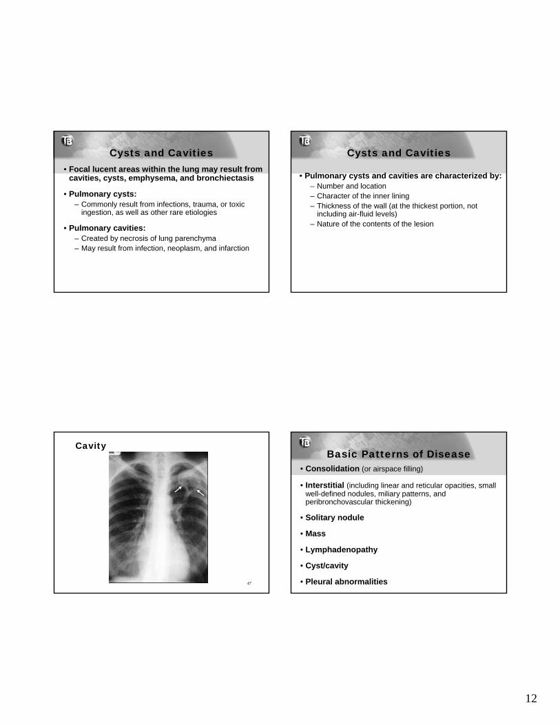
Cysts and Cavities• Focal lucent areas within the lung may result from
cavities, cysts, emphysema, and bronchiectasis
• Pulmonary cysts:– Commonly result from infections, trauma, or toxic
ingestion, as well as other rare etiologies
• Pulmonary cavities: – Created by necrosis of lung parenchyma– May result from infection, neoplasm, and infarction
Cysts and Cavities
• Pulmonary cysts and cavities are characterized by:– Number and location– Character of the inner lining– Thickness of the wall (at the thickest portion, not
including air-fluid levels) – Nature of the contents of the lesion
Cavity
47
Basic Patterns of Disease• Consolidation (or airspace filling)
• Interstitial (including linear and reticular opacities, small well-defined nodules, miliary patterns, and peribronchovascular thickening)
• Solitary nodule
• Mass
• Lymphadenopathy
• Cyst/cavity
• Pleural abnormalities
13
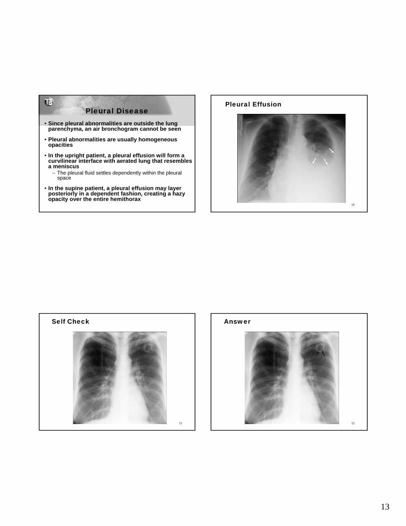
Pleural Disease• Since pleural abnormalities are outside the lung
parenchyma, an air bronchogram cannot be seen
• Pleural abnormalities are usually homogeneous opacities
• In the upright patient, a pleural effusion will form a curvilinear interface with aerated lung that resembles a meniscus
– The pleural fluid settles dependently within the pleural space
• In the supine patient, a pleural effusion may layer posteriorly in a dependent fashion, creating a hazy opacity over the entire hemithorax
Pleural Effusion
50
Self Check
51
Answer
52
14
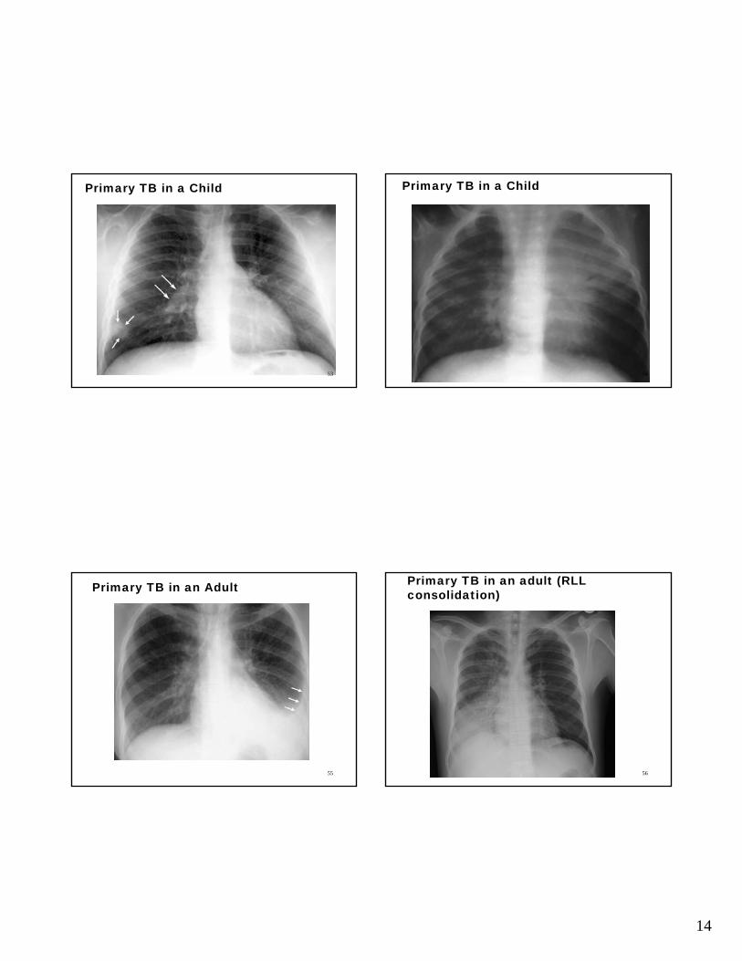
Primary TB in a Child
53
Primary TB in a Child
54
Primary TB in an Adult
55
Primary TB in an adult (RLL consolidation)
56
15
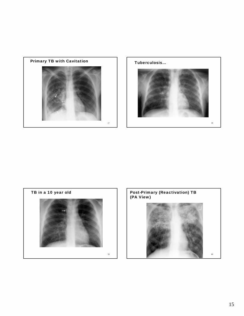
Primary TB with Cavitation
57
Tuberculosis…
58
TB in a 10 year old
59
Post-Primary (Reactivation) TB (PA View)
60
16
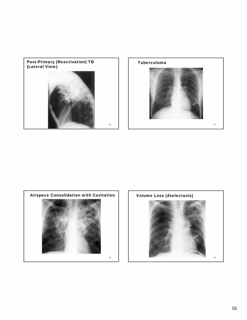
Post-Primary (Reactivation) TB (Lateral View)
61
Tuberculoma
62
Airspace Consolidation with Cavitation
63
Volume Loss (Atelectasis)
64
17
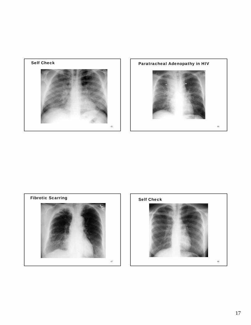
Self Check
65
Paratracheal Adenopathy in HIV
66
Fibrotic Scarring
67
Self Check
68
18
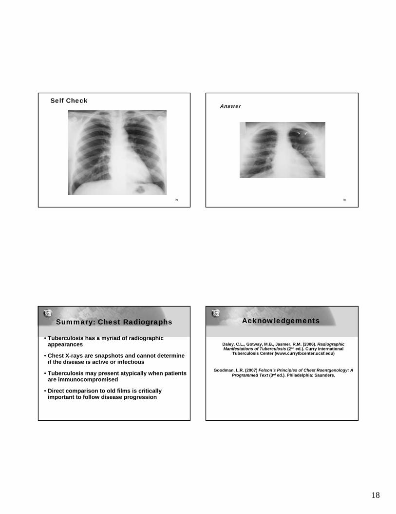
Self Check
69
Answer
70
Summary: Chest Radiographs
• Tuberculosis has a myriad of radiographic appearances
• Chest X-rays are snapshots and cannot determine if the disease is active or infectious
• Tuberculosis may present atypically when patients are immunocompromised
• Direct comparison to old films is critically important to follow disease progression
Acknowledgements
Daley, C.L., Gotway, M.B., Jasmer, R.M. (2006). Radiographic Manifestations of Tuberculosis (2nd ed.). Curry International
Tuberculosis Center (www.currytbcenter.ucsf.edu)
Goodman, L.R. (2007) Felson’s Principles of Chest Roentgenology: A Programmed Text (3rd ed.). Philadelphia: Saunders.