Embed Size (px)
Citation preview
1
Interpretation of Chest Radiographs
Interpretation of Chest Radiographs
Reynard McDonald, MDMedical Director
NJMS Global Tuberculosis Institute
X-RaysX-Rays
When x-rays are produced and directed toward the patient, they may act in three basic ways:
They may be… Which means…
unabsorbed they pass through the patient unchanged and strike the x-ray film
completely absorbed the energy of the x-ray is totally deposited within the patient
scattered they are deflected within the patient but may still strike the x-ray film
X-ray AbsorptionX-ray Absorption
• Factors that contribute to X-ray absorption include:
– The density of the tissue the beam strikes– The energy of the X-ray beam (the enery of the X-ray
bean is usually fairly constant in posterior/anterior and lateral radiography)
Tissue DensityTissue DensityWhitest/Most Dense
Metal
Contrast material (i.e., x-ray dye)
Bone
Calcium
Soft tissue
Fat
Air or gas
Blackest/Least Dense

2
Posterior/Anterior (PA) Radiograph Posterior/Anterior (PA) Radiograph
• The term posterior/anterior (PA) refers to the direction of the X-ray beam which in this case traverses the patient from posterior (back) to anterior (front)
• The PA view taken at a distance of 6 feet to reduce magnification and enhance sharpness
Normal Frontal (PA) Chest Radiograph
Normal Frontal (PA) Chest Radiograph
PA & AP Chest X-raysPA & AP Chest X-rays
PA View AP View
3
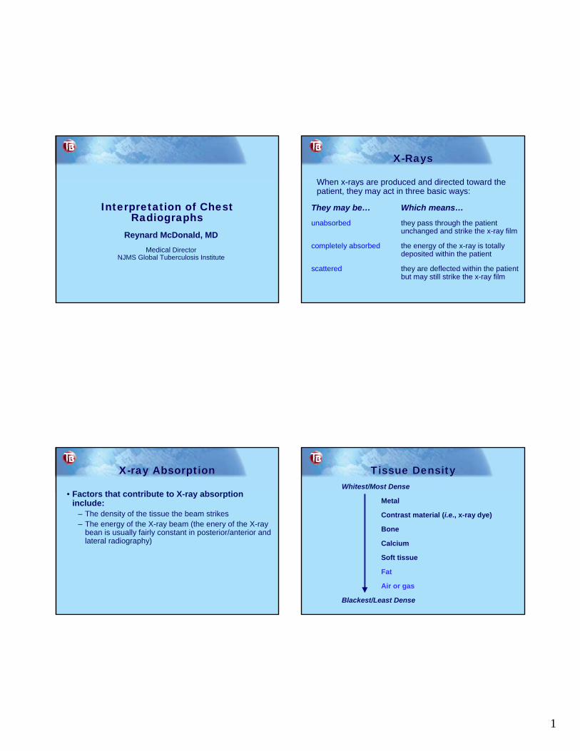
Lateral Radiograph Lateral Radiograph
• The other routine view is the lateral radiograph
• By convention it is taken at a distance of 6 feet and the left side of the chest is held against the X-ray cassette
• Often it is difficult to detect lesions located behind the heart, near the mediastinum, or near the diaphragm on the PA view
• The lateral view generally shows such lesions, so we use it routinely
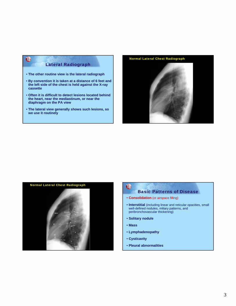
Normal Lateral Chest Radiograph
Normal Lateral Chest Radiograph
Basic Patterns of DiseaseBasic Patterns of Disease• Consolidation (or airspace filling)
• Interstitial (including linear and reticular opacities, small well-defined nodules, miliary patterns, and peribronchovascular thickening)
• Solitary nodule
• Mass
• Lymphadenopathy
• Cyst/cavity
• Pleural abnormalities
4
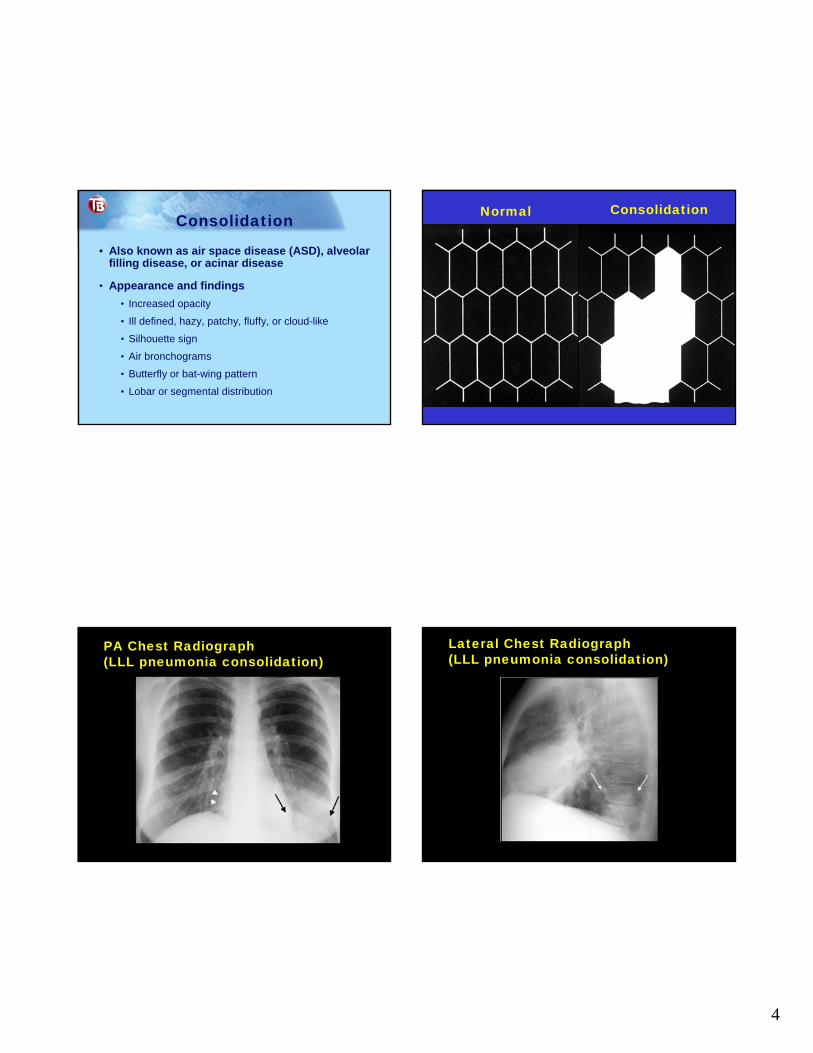
Consolidation
• Also known as air space disease (ASD), alveolar filling disease, or acinar disease
• Appearance and findings• Increased opacity
• Ill defined, hazy, patchy, fluffy, or cloud-like
• Silhouette sign
• Air bronchograms
• Butterfly or bat-wing pattern
• Lobar or segmental distribution
Normal Consolidation
PA Chest Radiograph (LLL pneumonia consolidation)
Lateral Chest Radiograph(LLL pneumonia consolidation)
5
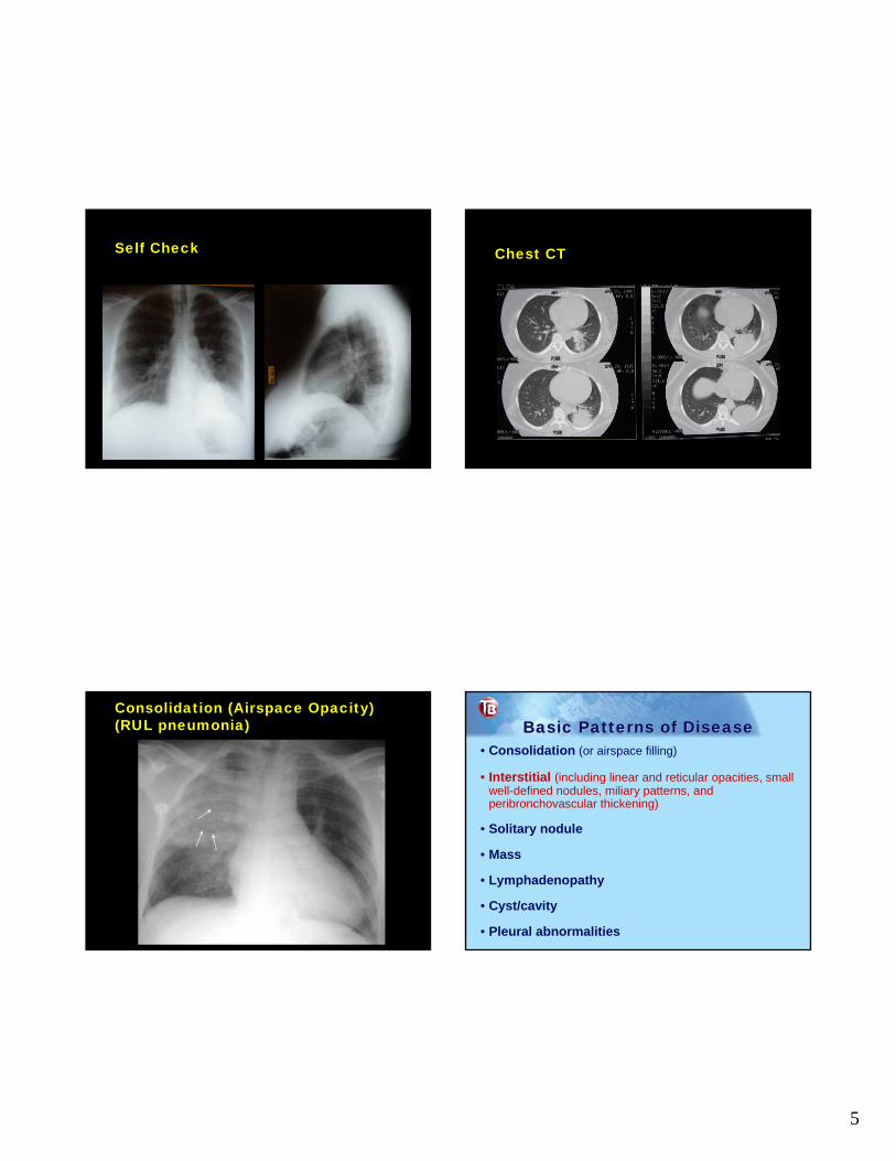
Self Check Chest CT
Consolidation (Airspace Opacity)(RUL pneumonia) Basic Patterns of DiseaseBasic Patterns of Disease
• Consolidation (or airspace filling)
• Interstitial (including linear and reticular opacities, small well-defined nodules, miliary patterns, and peribronchovascular thickening)
• Solitary nodule
• Mass
• Lymphadenopathy
• Cyst/cavity
• Pleural abnormalities
6
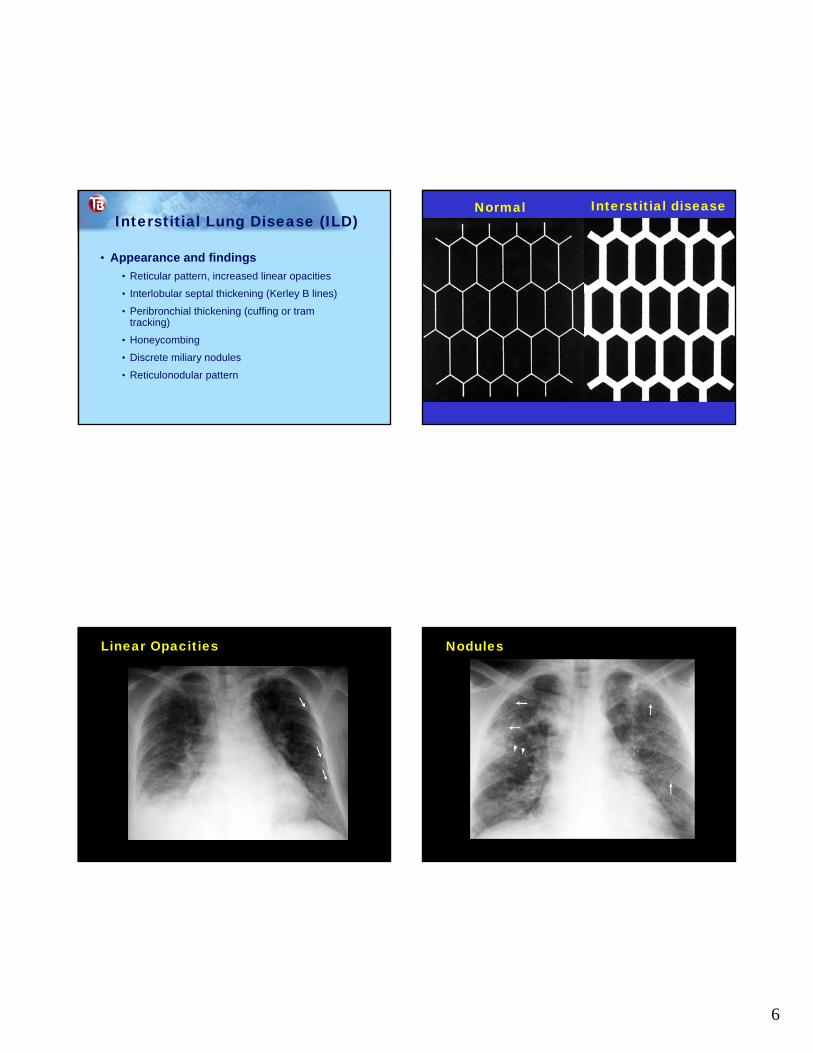
Interstitial Lung Disease (ILD)
• Appearance and findings• Reticular pattern, increased linear opacities
• Interlobular septal thickening (Kerley B lines)
• Peribronchial thickening (cuffing or tram tracking)
• Honeycombing
• Discrete miliary nodules
• Reticulonodular pattern
Normal Interstitial disease
Linear Opacities Nodules
7
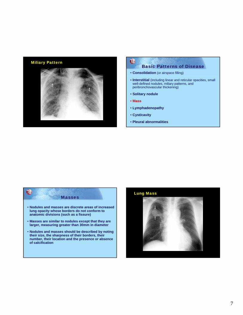
Miliary PatternBasic Patterns of DiseaseBasic Patterns of Disease
• Consolidation (or airspace filling)
• Interstitial (including linear and reticular opacities, small well-defined nodules, miliary patterns, and peribronchovascular thickening)
• Solitary nodule
• Mass
• Lymphadenopathy
• Cyst/cavity
• Pleural abnormalities
MassesMasses
• Nodules and masses are discrete areas of increased lung opacity whose borders do not conform to anatomic divisions (such as a fissure)
• Masses are similar to nodules except that they are larger, measuring greater than 30mm in diameter
• Nodules and masses should be described by noting their size, the sharpness of their borders, their number, their location and the presence or absence of calcification
Lung Mass
8
Basic Patterns of DiseaseBasic Patterns of Disease• Consolidation (or airspace filling)
• Interstitial (including linear and reticular opacities, small well-defined nodules, miliary patterns, and peribronchovascular thickening)
• Solitary nodule
• Mass
• Lymphadenopathy
• Cyst/cavity
• Pleural abnormalities
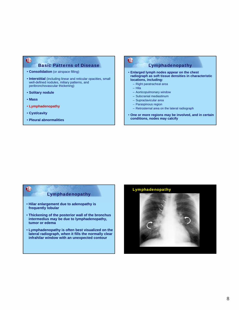
LymphadenopathyLymphadenopathy• Enlarged lymph nodes appear on the chest
radiograph as soft tissue densities in characteristic locations, including:
– Right paratracheal area– Hila– Aorticopulmonary window – Subcranial mediastinum– Supraclavicular area– Paraspinous region– Retrosternal area on the lateral radiograph
• One or more regions may be involved, and in certain conditions, nodes may calcify
LymphadenopathyLymphadenopathy
• Hilar enlargement due to adenopathy is frequently lobular
• Thickening of the posterior wall of the bronchus intermedius may be due to lymphadenopathy, tumor or edema
• Lymphadenopathy is often best visualized on the lateral radiograph, when it fills the normally clear infrahilar window with an unexpected contour
Lymphadenopathy

9
Lymphadenopathy Mediastinal Lymphadenopathy
Basic Patterns of DiseaseBasic Patterns of Disease• Consolidation (or airspace filling)
• Interstitial (including linear and reticular opacities, small well-defined nodules, miliary patterns, and peribronchovascular thickening)
• Solitary nodule
• Mass
• Lymphadenopathy
• Cyst/cavity
• Pleural abnormalities
Cysts and CavitiesCysts and Cavities• Focal lucent areas within the lung may result from
cavities, cysts, emphysema, and bronchiectasis
• Pulmonary cysts differ from cavities in that cavities are created by necrosis of lung parenchyma, whereas true cysts are formed by other means
• Pulmonary cavities may result from infection, neoplasm, and infarction
• Pulmonary cysts commonly result from infections, trauma, or toxic ingestion, as well as other rare etiologies
10
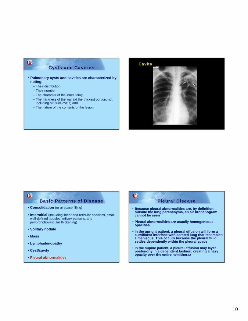
Cysts and CavitiesCysts and Cavities
• Pulmonary cysts and cavities are characterized by noting:
– Their distribution– Their number– The character of the inner lining– The thickness of the wall (at the thickest portion, not
including air-fluid levels) and – The nature of the contents of the lesion
Cavity
Basic Patterns of DiseaseBasic Patterns of Disease• Consolidation (or airspace filling)
• Interstitial (including linear and reticular opacities, small well-defined nodules, miliary patterns, and peribronchovascular thickening)
• Solitary nodule
• Mass
• Lymphadenopathy
• Cyst/cavity
• Pleural abnormalities
Pleural DiseasePleural Disease• Because pleural abnormalities are, by definition,
outside the lung parenchyma, an air bronchogramcannot be seen
• Pleural abnormalities are usually homogeneous opacities
• In the upright patient, a pleural effusion will form a curvilinear interface with aerated lung that resembles a meniscus. This occurs because the pleural fluid settles dependently within the pleural space
• In the supine patient, a pleural effusion may layer posteriorly in a dependent fashion, creating a hazy opacity over the entire hemithorax
11
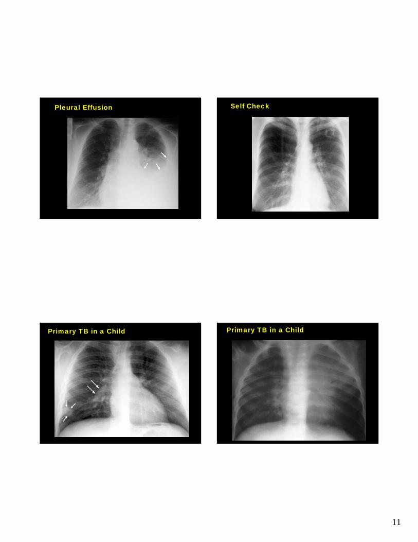
Pleural Effusion Self Check
Primary TB in a Child Primary TB in a Child

12
Primary TB in an Adult Primary TB with Cavitation
Tuberculosis… TB in a 10 year old

13
Post-Primary (Reactivation) TB (PA View)
Post-Primary (Reactivation) TB (Lateral View)
Tuberculoma Airspace Consolidation with Cavitation
14
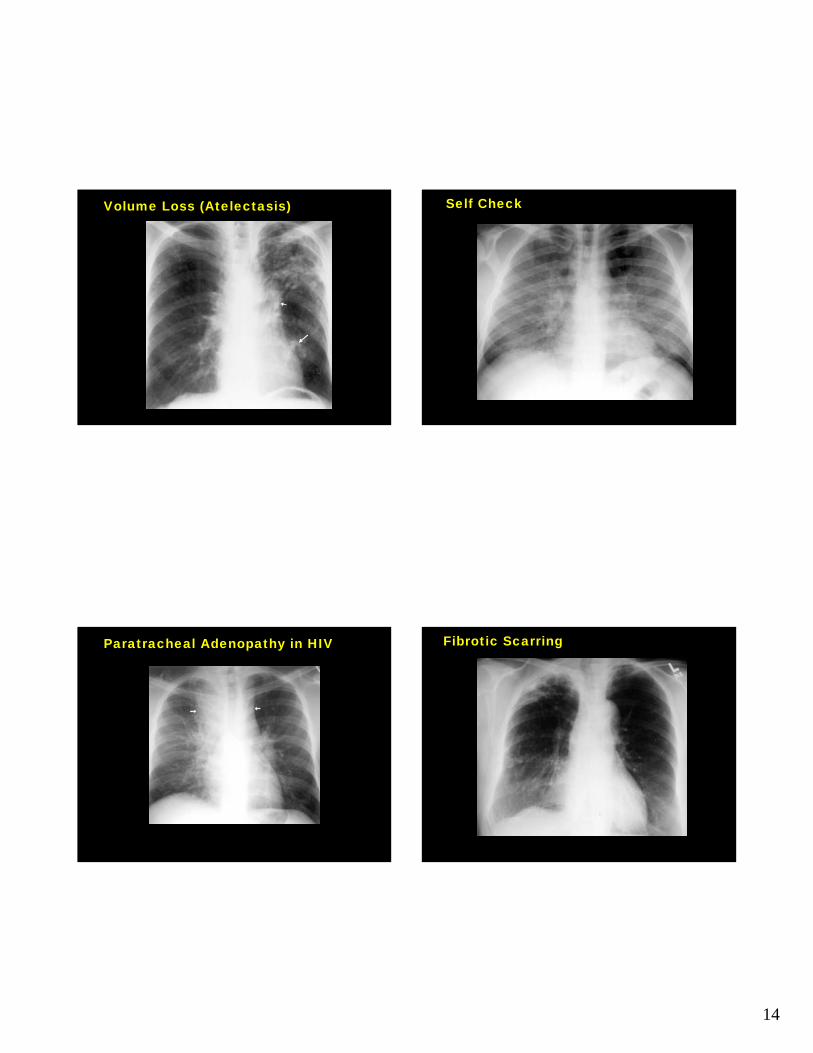
Volume Loss (Atelectasis) Self Check
Paratracheal Adenopathy in HIV Fibrotic Scarring

15
Self Check Self Check
Answer Summary: Chest RadiographsSummary: Chest Radiographs
• Tuberculosis has a myriad of radiographic appearances
• Chest X-rays are snapshots and cannot determine if the disease is active or infectious
• Tuberculosis may present atypically when patients are immune compromised
• Direct comparison to old films is critically important to follow disease progression
16
AcknowledgementsAcknowledgements
Daley, C.L., Gotway, M.B., Jasmer, R.M. (2006). Radiographic Manifestations of Tuberculosis (2nd ed.). Francis J. Curry National TB
Center (www.nationaltbcenter.edu)
Goodman, L.R. (2007) Felson’s Principles of Chest Roentgenology: A Programmed Text (3rd ed.). Philadelphia: Saunders.


![Multi‑scale Morphological Image Enhancement of Chest ...research.iaun.ac.ir/pd/mahdavinasab/pdfs/PaperM_6402.pdf · chest radiographs, mammograms, and chest CT images).[34] In this](https://img.pdfslide.us/doc/110x75/5f3dd7277ba40343a062efad/multiascale-morphological-image-enhancement-of-chest-chest-radiographs-mammograms.jpg)


![A chest radiograph scoring system in ... - BMC Medical Imagingdata collected in epidemiological studies of acute respira-tory infections [7]. For example, chest radiographs are a component](https://img.pdfslide.us/doc/110x75/611d2ab1b132b339a353139f/a-chest-radiograph-scoring-system-in-bmc-medical-imaging-data-collected-in.jpg)


![NJMS IT Overview.pptx [Read-Only]](https://img.pdfslide.us/doc/110x75/6169f9d711a7b741a34d73fc/njms-it-read-only.jpg)