Embed Size (px)
Citation preview
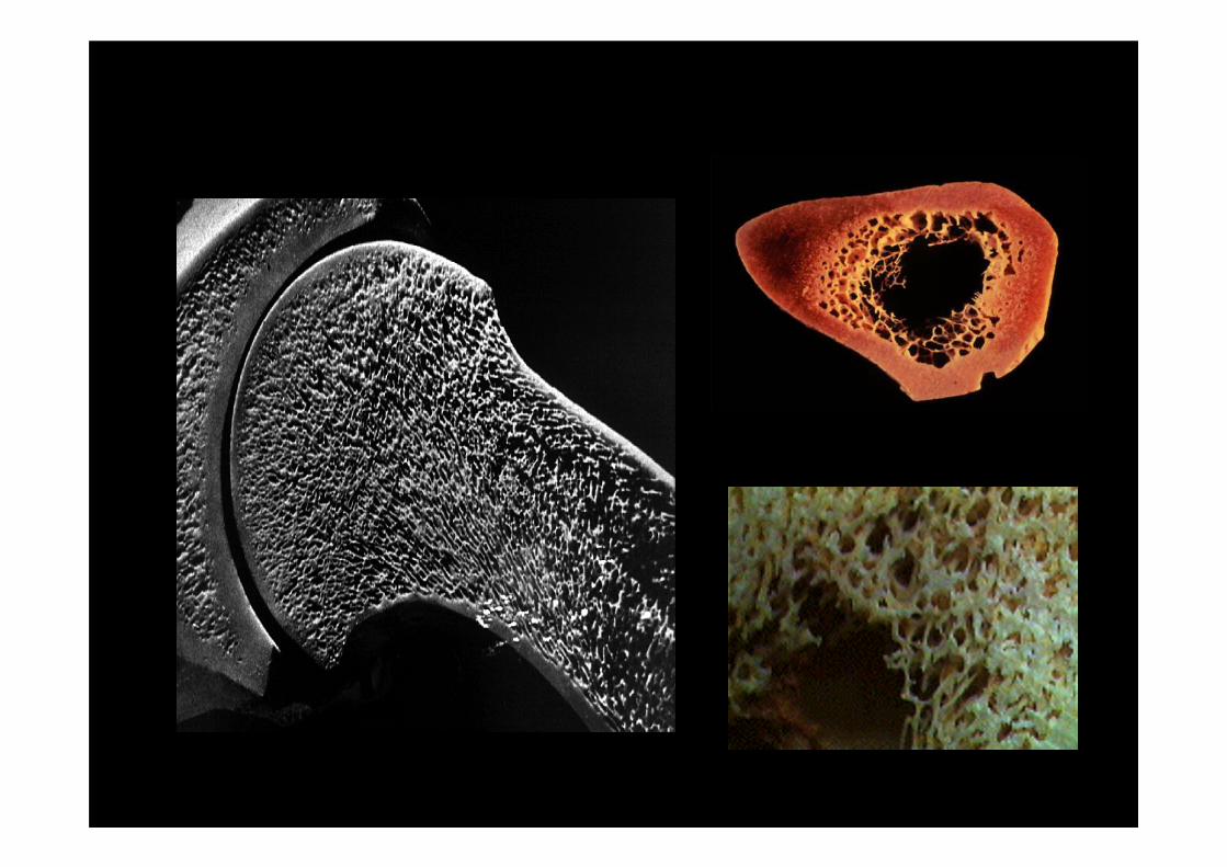
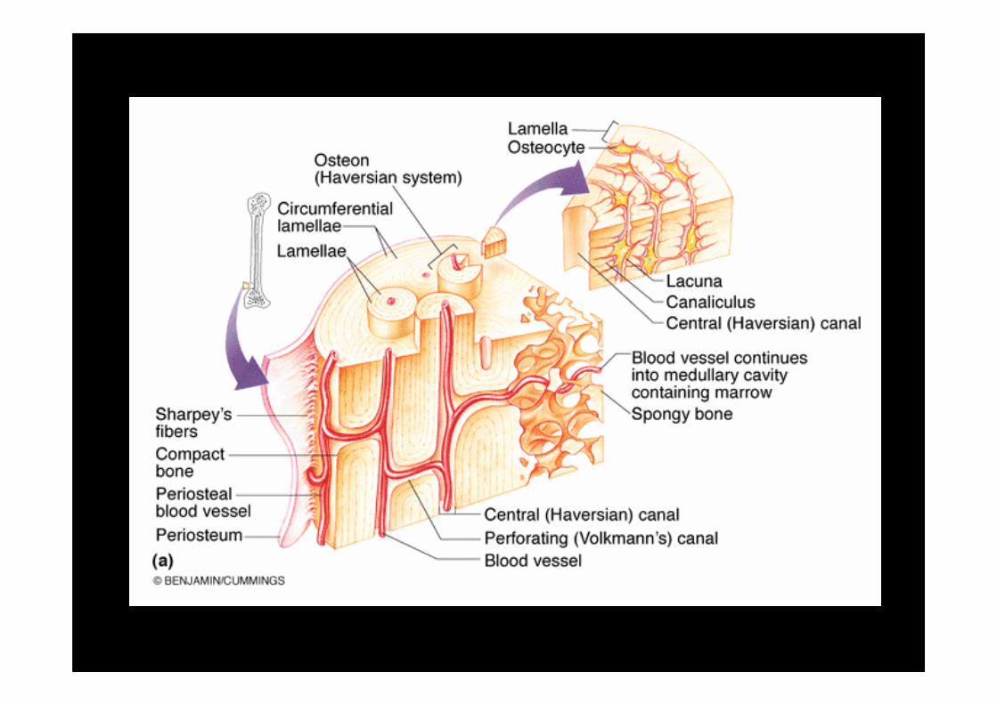
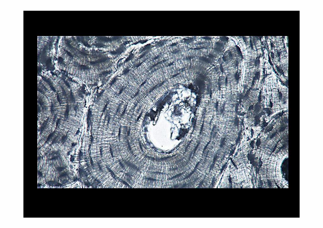
Biology of Bone
Dr Mark A. BirchMusculoskeletal Research Group
The Medical School
Newcastle University
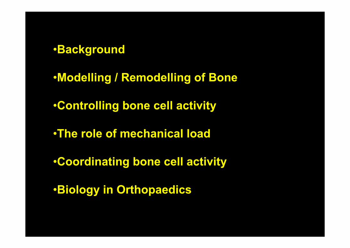
•Background
•Modelling / Remodelling of Bone
•Controlling bone cell activity
•The role of mechanical load
•Coordinating bone cell activity
•Biology in Orthopaedics
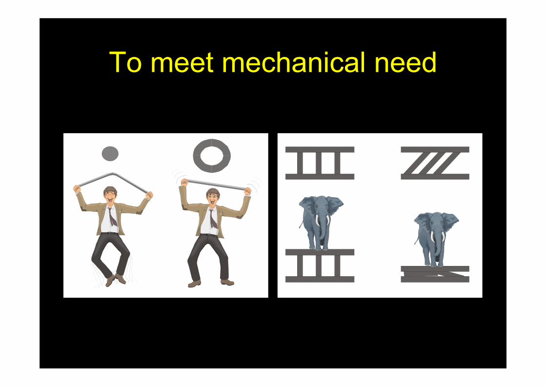
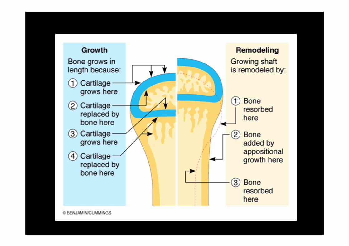
To meet mechanical need
Bone formation
• Bone formation is termed “ossification” or “osteogenesis”
• The “early” skeleton in an embryo is composed of fibrous membranes and hyaline cartilage
• During the process of ossification (around the 6-7th week of embryonic development):
– Chondroblasts form cartilage
– Osteoblasts form bone (mineralization)
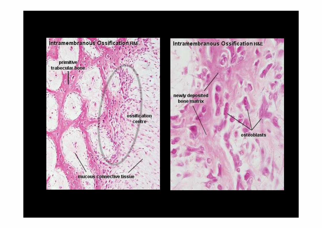
: Intramembranous Ossification
• Bone formation of the surface skull bones and clavicles
• Osteoblasts cluster around the centre of ossification– Here, osteoblasts secrete a collagenous matrix to form a
framework for mineralization
– The collagenous matrix is then calcified by the deposition of hydroxyapatite
– The osteoblasts and their surrounding calcified matrix are now referred to as a trabecula
• Most of the trabeculae will be eventually destroyed and reformed to give a bone its final adult size and shape
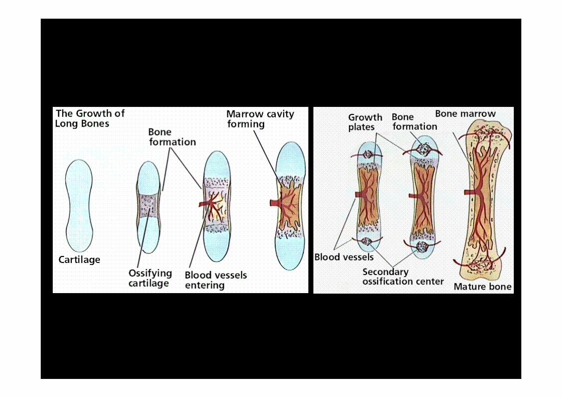
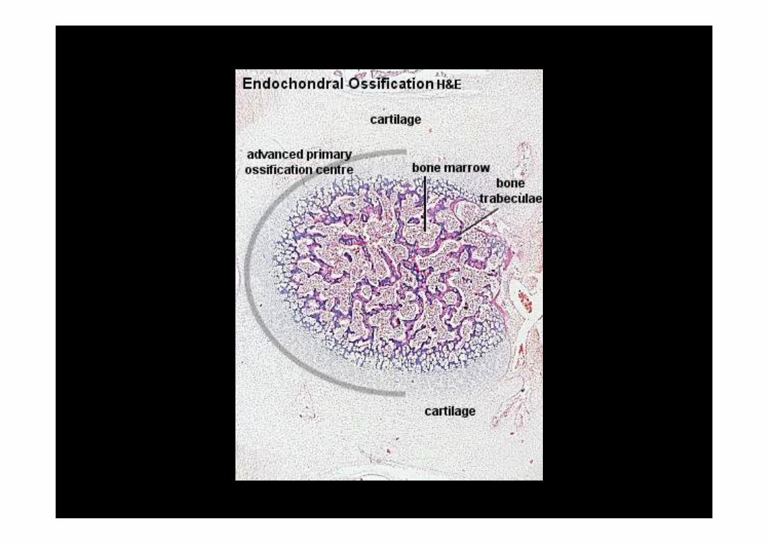
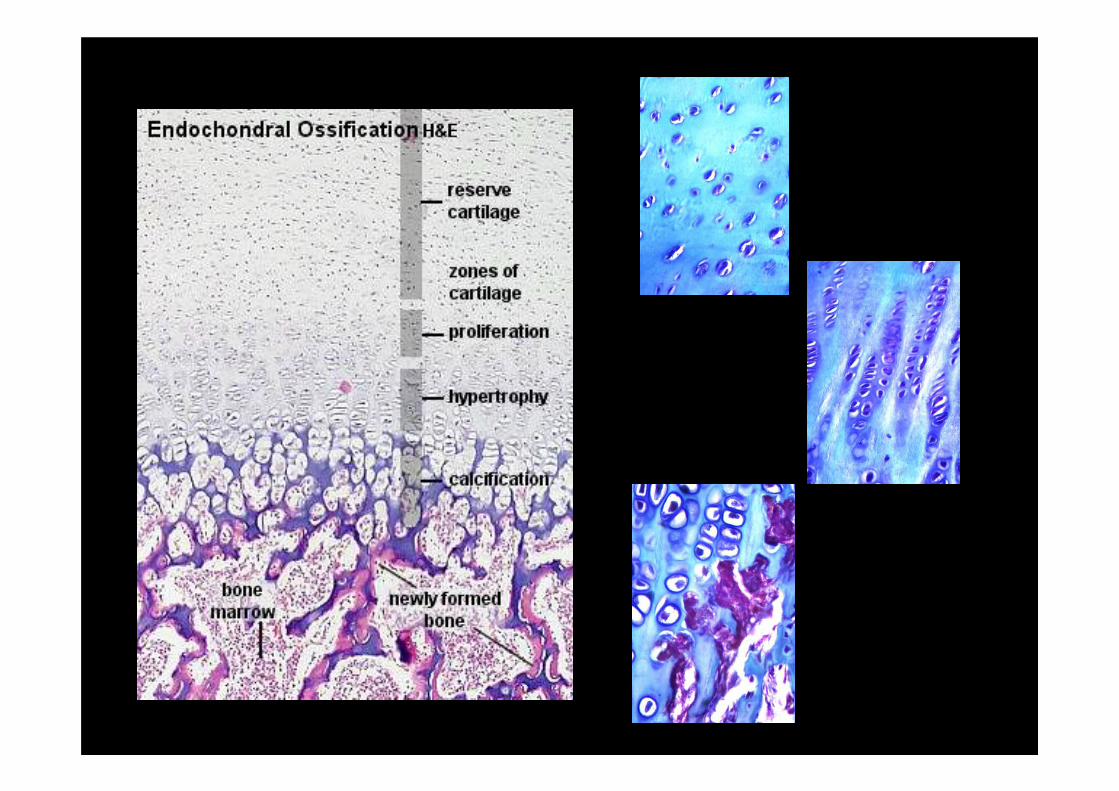
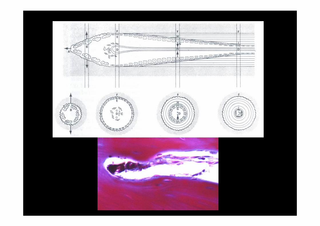
Endochondral Ossification
• Replacement of cartilage with bone
• Primary ossification process for most bones of the body– Best exemplified in long bones
• During embryonic development, a cartilage model, or perichondrium, is laid down– Compact bone then forms around this area and is called the
periosteum
• Periosteal collar
• Cartilage grows outward from it’s center and is gradually calcified into bone tissue– Primary ossification centre: diaphysis
– Secondary ossification centre: epihysis
– Two areas remain uncalcified cartilage: articulations, growth plate
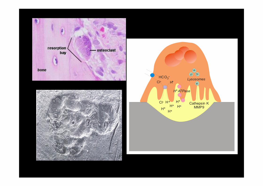
HCO -3
Cl-
Cl- H+
H+H+
H+
H+
H ATPase+
H+
H+
Lysosomes
Cathepsin KMMP9
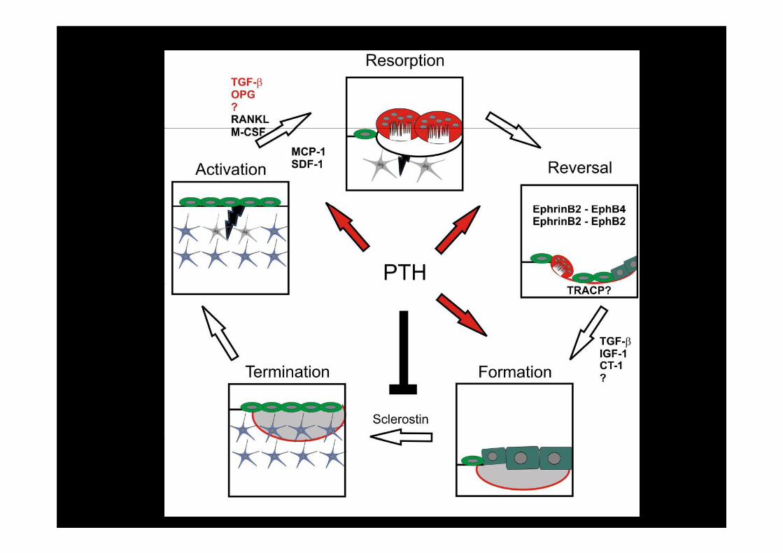
Modelling / Remodelling of Bone
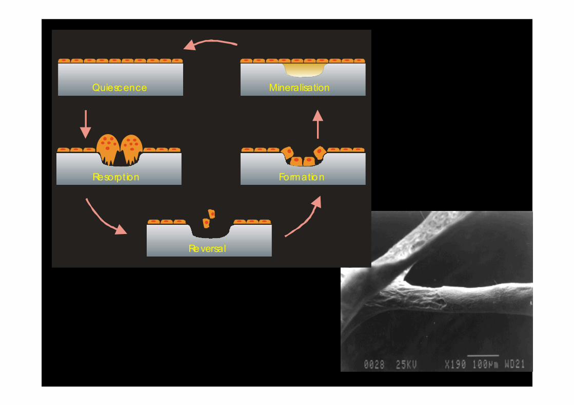
Quiescence
Resorption
Reversal
Formation
Mineralisation
Controlling bone cell activity
- Osteoclast
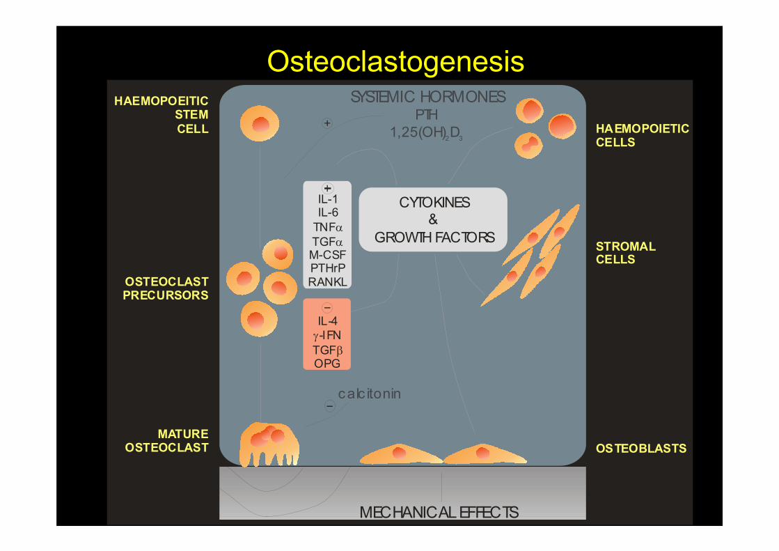
Osteoclastogenesis
HAEMOPOIETICCELLS
STROMALCELLS
OSTEOBLASTS
CYTOKINES&
GROWTH FACTORS
IL-1IL-6
TNF
TGFM-CSFPTHrPRANKL
α
α
HAEMOPOEITICSTEM
CELL
OSTEOCLASTPRECURSORS
MATUREOSTEOCLAST
IL-4
-IFN
TGFOPG
γ
β
SYSTEMIC HORMONESPTH
1,25(OH) D2 3
MECHANICAL EFFECTS
calc itonin
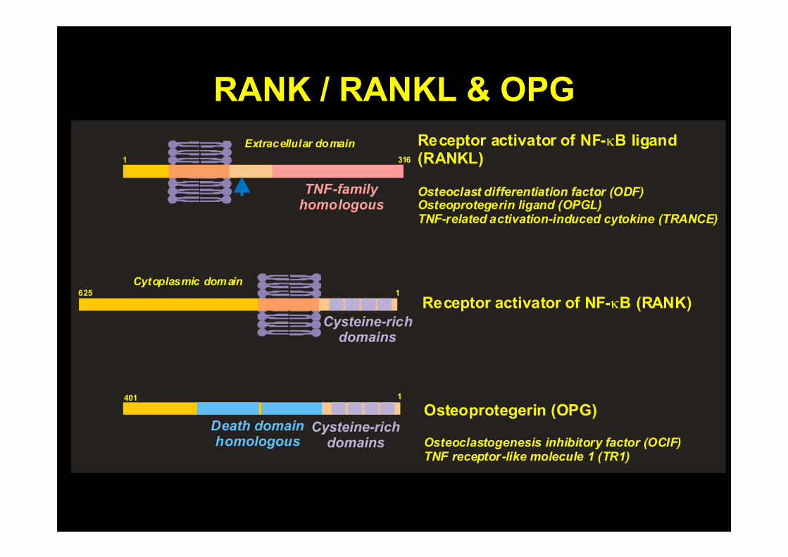
RANK / RANKL & OPG
Extracellular domain
TNF-family homologous
Cysteine-rich domains
Cysteine-rich domains
Death domain homologous
1 316
1625
1401
Cytoplasmic domain
Receptor activator of NF- B ligand (RANKL)
κ
Osteoclast differentiation factor (ODF)Osteoprotegerin ligand (OPGL)TNF-related activation-induced cytokine (TRANCE)
Receptor activator of NF- B (RANK)κ
Osteoprotegerin (OPG)
Osteoclastogenesis inhibitory factor (OCIF)TNF receptor-like molecule 1 (TR1)
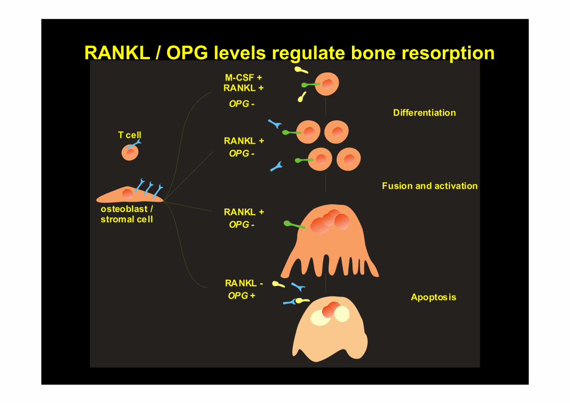
Differentiation
Fusion and activation
Apoptosis
M-CSF +RANKL +
RANKL +
RANKL +
RANKL -
OPG +
OPG -
OPG -
OPG -
osteoblast /stromal cell
T cell
RANKL / OPG levels regulate bone resorption
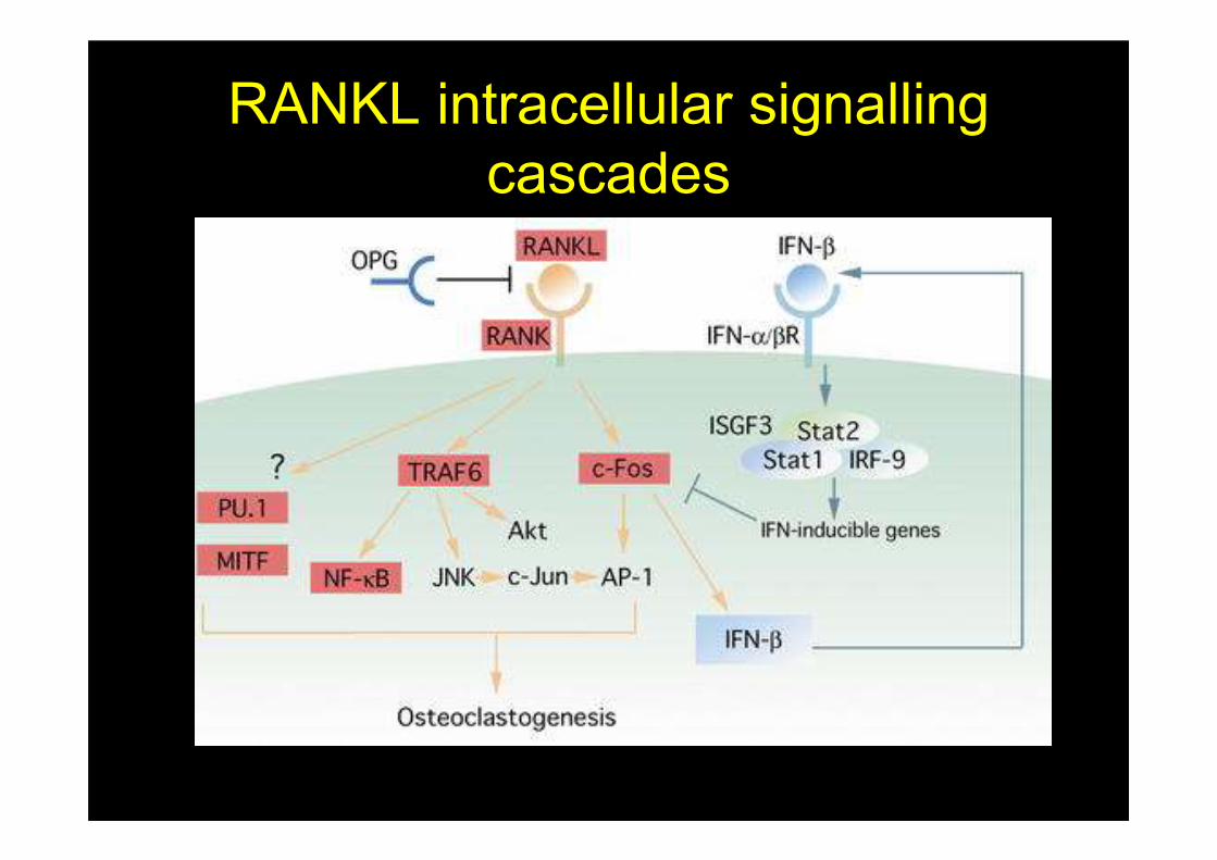
RANKL intracellular signalling
cascades
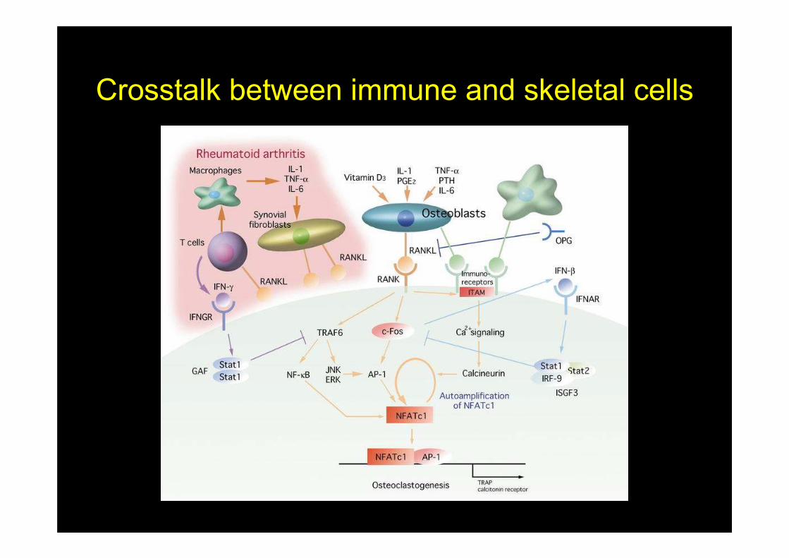
Crosstalk between immune and skeletal cells
Controlling bone cell activity
- Osteoblast
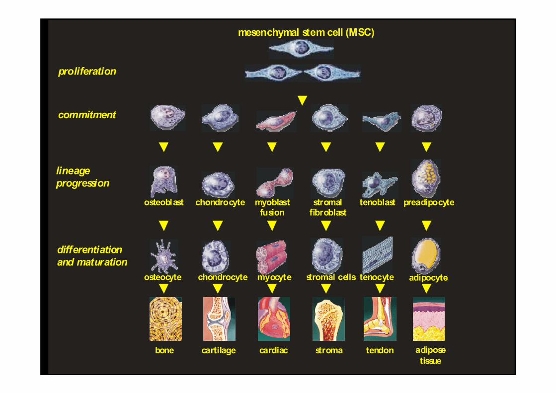
mesenchymal stem cell (MSC)
proliferation
commitment
lineage
progression
differentiation
and maturation
osteoblast
osteocyte
chondrocyte
chondrocyte
myoblastfusion
myocyte
stromalfibroblast
stromal cells
tenoblast
tenocyte
preadipocyte
adipocyte
bone cartilage cardiac stroma tendon adipose
tissue
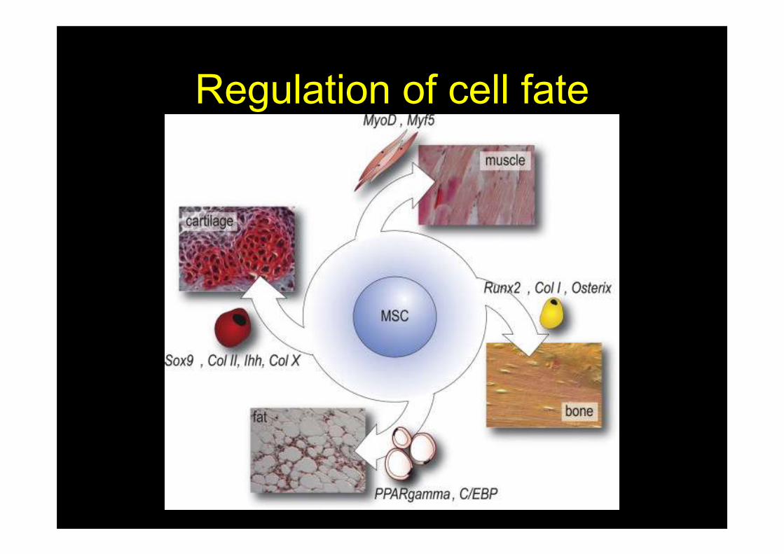
Regulation of cell fate
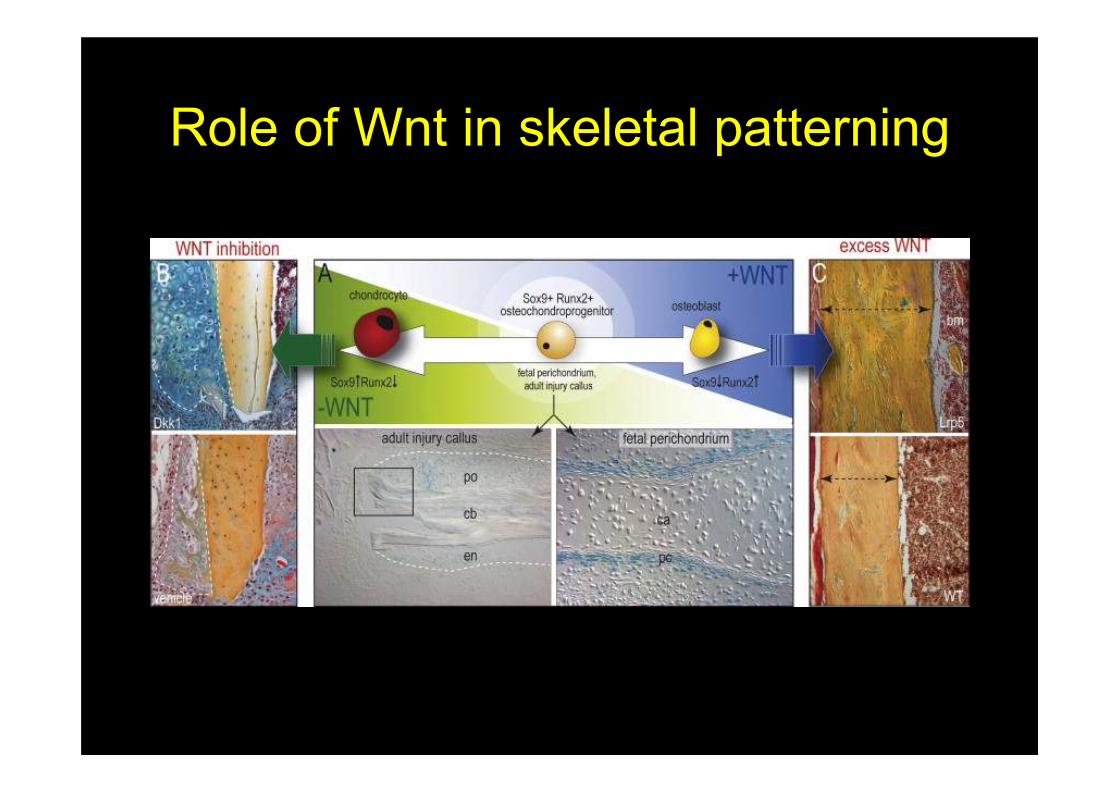
Role of Wnt in skeletal patterning
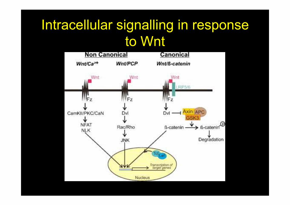
Intracellular signalling in response
to Wnt
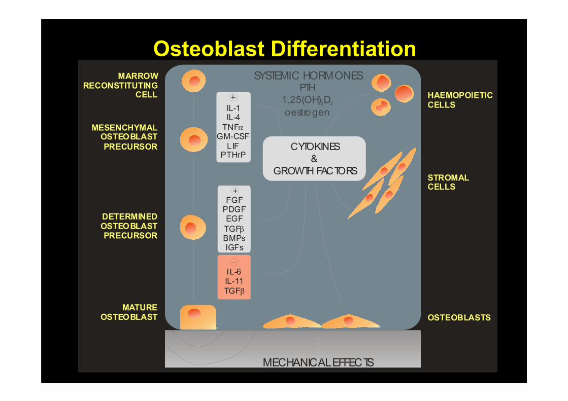
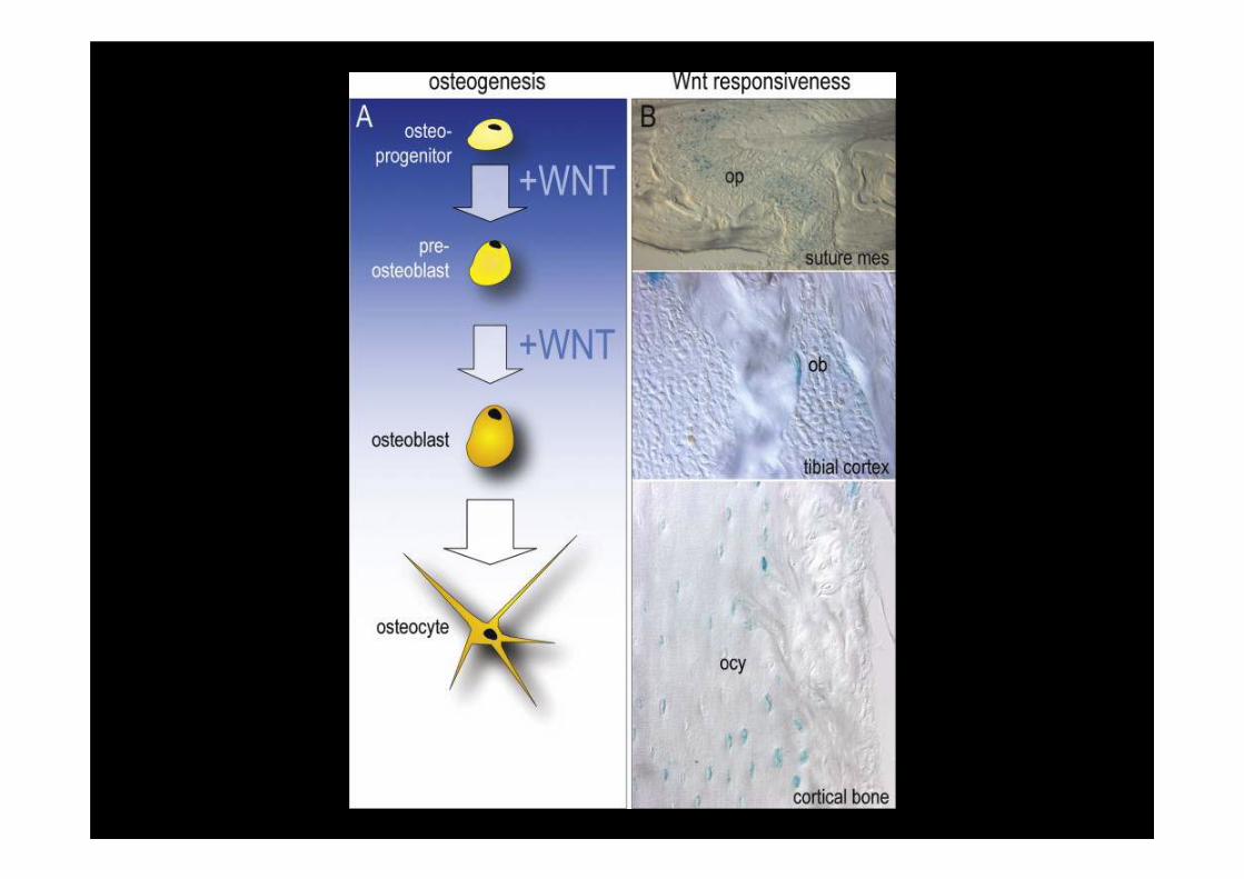
Osteoblast Differentiation
HAEMOPOIETICCELLS
STROMALCELLS
OSTEOBLASTS
CYTOKINES
&
GROWTH FACTORS
IL-1IL-4
TNFGM-CSF
LIFPTHrP
α
MARROWRECONSTITUTING
CELL
MESENCHYMALOSTEOBLAST
PRECURSOR
DETERMINEDOSTEOBLASTPRECURSOR
MATUREOSTEOBLAST
FGFPDGFEGF
TGFBMPsIGFs
β
IL-6IL-11
TGFβ
SYSTEMIC HORMONESPTH
1,25(OH) D
oestrogen2 3
MECHANICAL EFFECTS
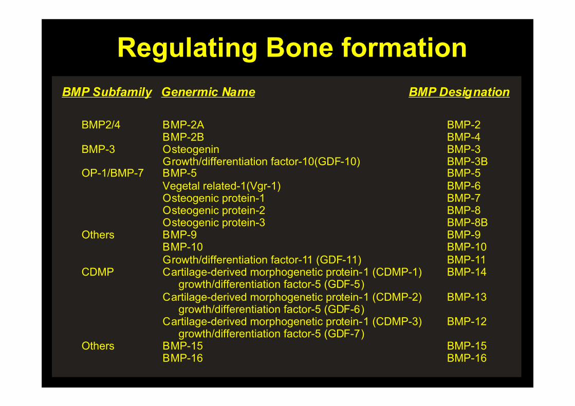
Regulating Bone formation
BMP2/4
BMP-3
OP-1/BMP-7
Others
CDMP
Others
BMP-2ABMP-2BOsteogeninGrowth/differentiation factor-10(GDF-10)BMP-5Vegetal related-1(Vgr-1)Osteogenic protein-1Osteogenic protein-2Osteogenic protein-3BMP-9BMP-10Growth/differentiation factor-11 (GDF-11)Cartilage-derived morphogenetic protein-1 (CDMP-1) growth/differentiation factor-5 (GDF-5)Cartilage-derived morphogenetic protein-1 (CDMP-2) growth/differentiation factor-5 (GDF-6)Cartilage-derived morphogenetic protein-1 (CDMP-3) growth/differentiation factor-5 (GDF-7)BMP-15BMP-16
BMP-2BMP-4BMP-3BMP-3BBMP-5BMP-6BMP-7BMP-8BMP-8BBMP-9BMP-10BMP-11BMP-14
BMP-13
BMP-12
BMP-15BMP-16
BMP Subfamily Genermic Name BMP Designation
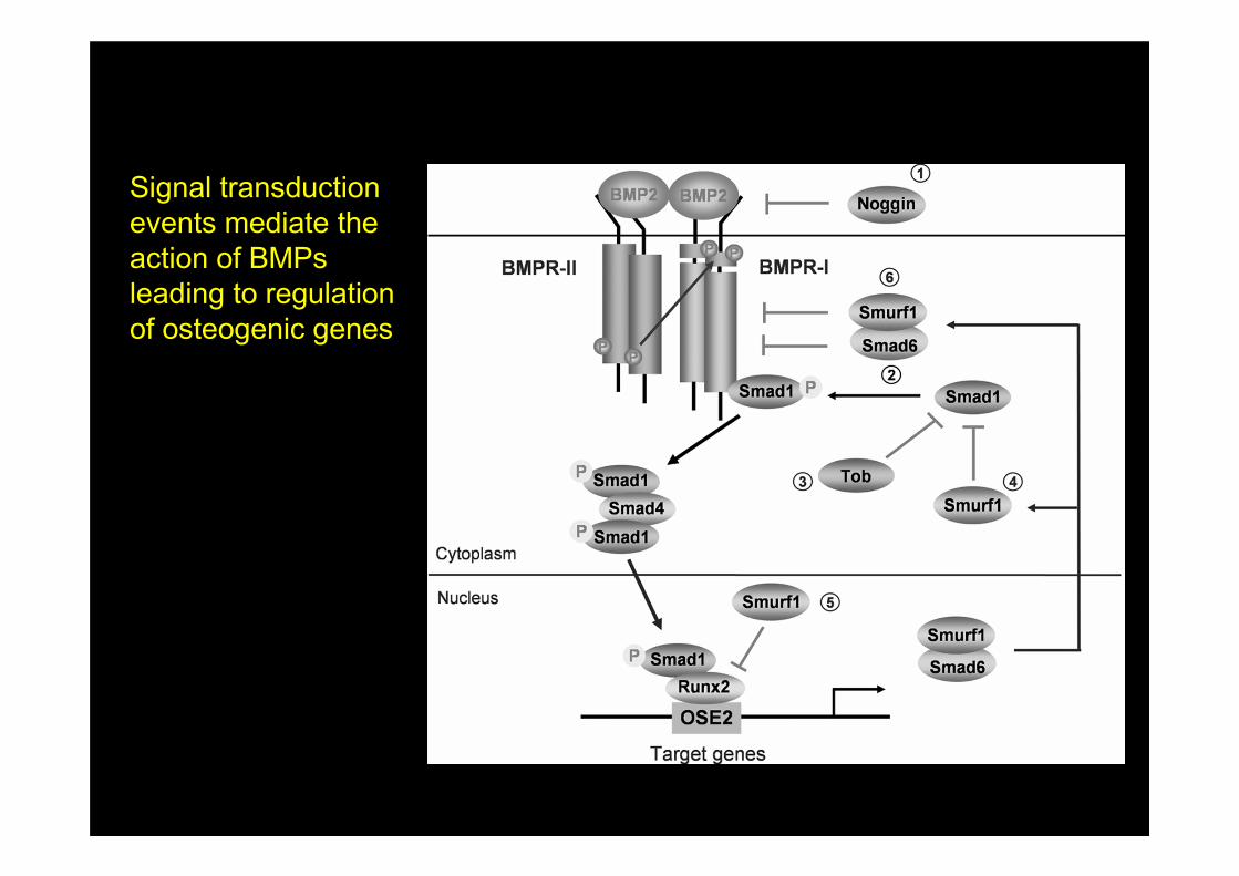
Signal transduction
events mediate the
action of BMPs
leading to regulation
of osteogenic genes
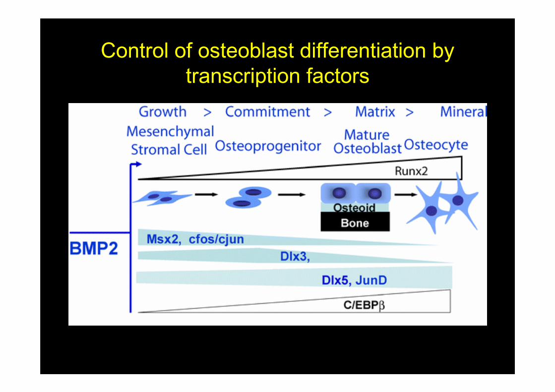
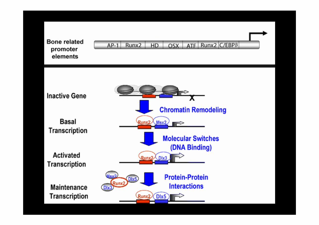
Control of osteoblast differentiation by
transcription factors

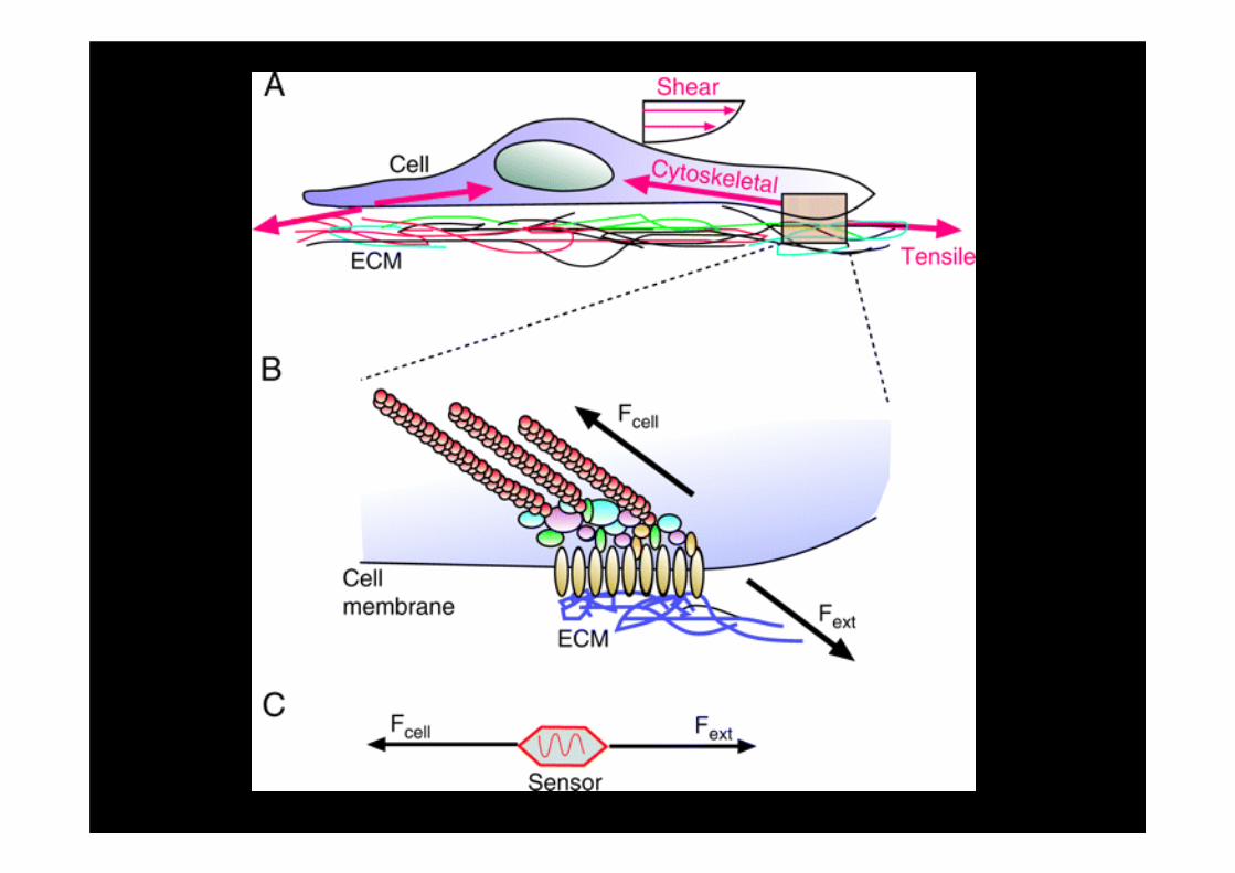
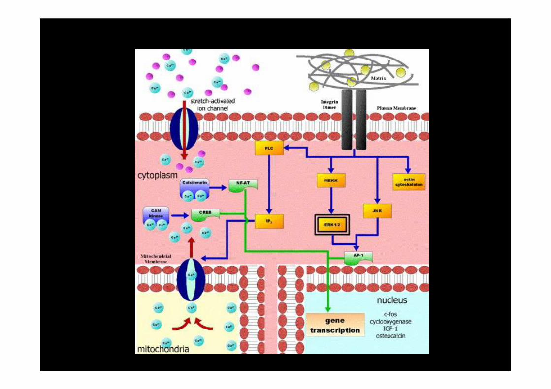
Mechanical load regulates
bone cell activity and bone
mass
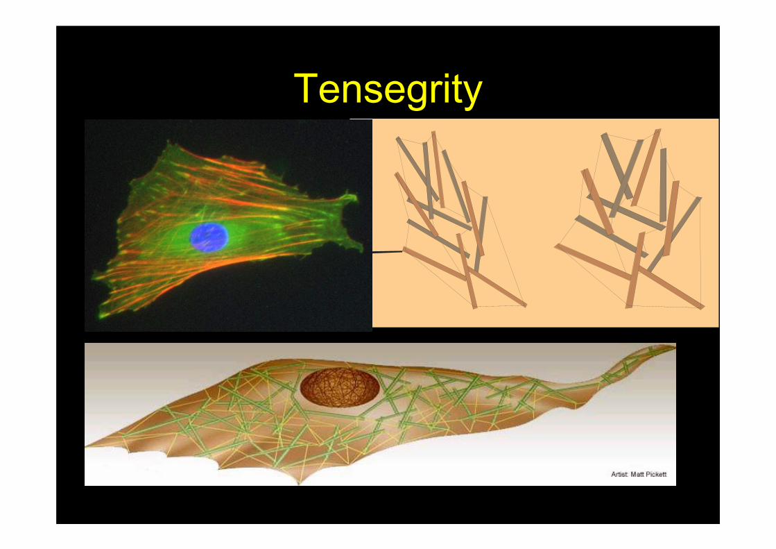
Tensegrity
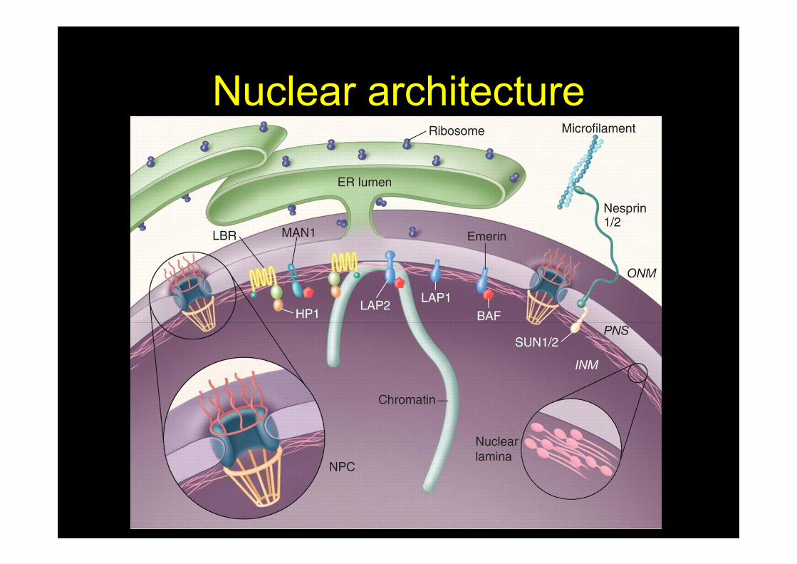
Nuclear architecture
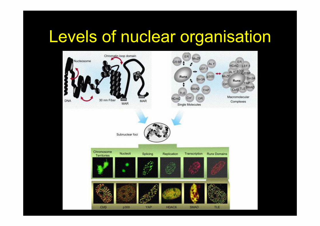
Levels of nuclear organisation
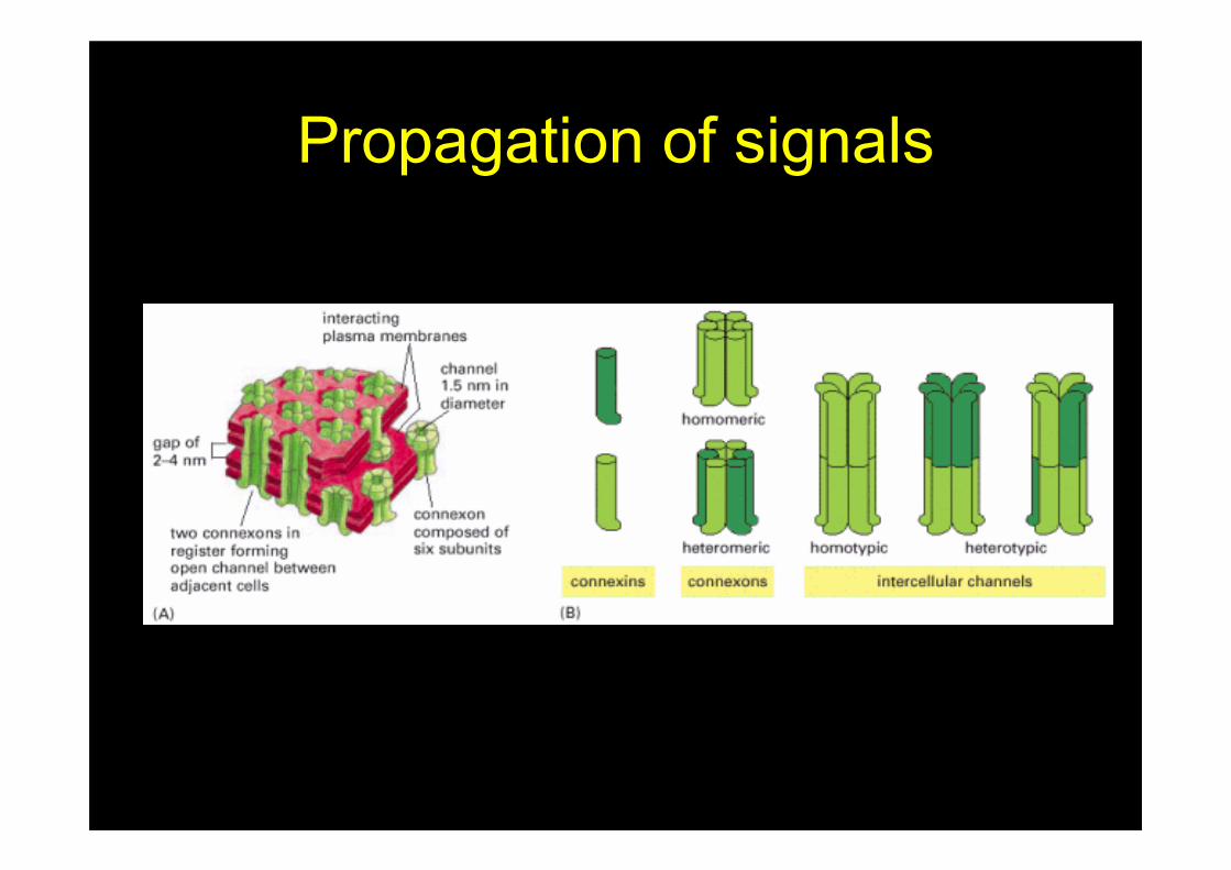
Propagation of signals
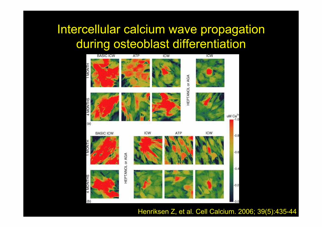
Intercellular calcium wave propagation
during osteoblast differentiation
Henriksen Z, et al. Cell Calcium. 2006; 39(5):435-44
Coordinating bone cell
behaviour
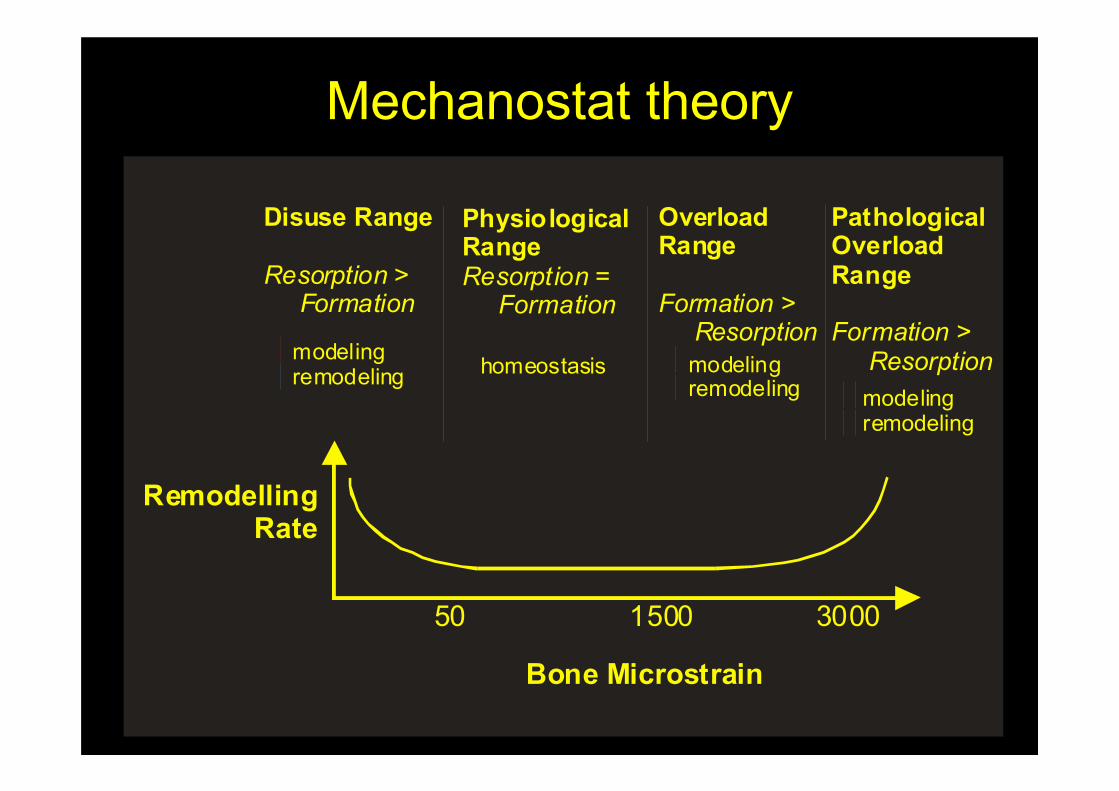
Mechanostat theory
50 1500 3000
Bone Microstrain
RemodellingRate
Disuse Range
Resorption > Formation
modelingremodeling
PhysiologicalRangeResorption = Formation
homeostasis
OverloadRange
FormationResorption
>
modelingremodeling
PathologicalOverloadRange
FormationResorption
>
modelingremodeling
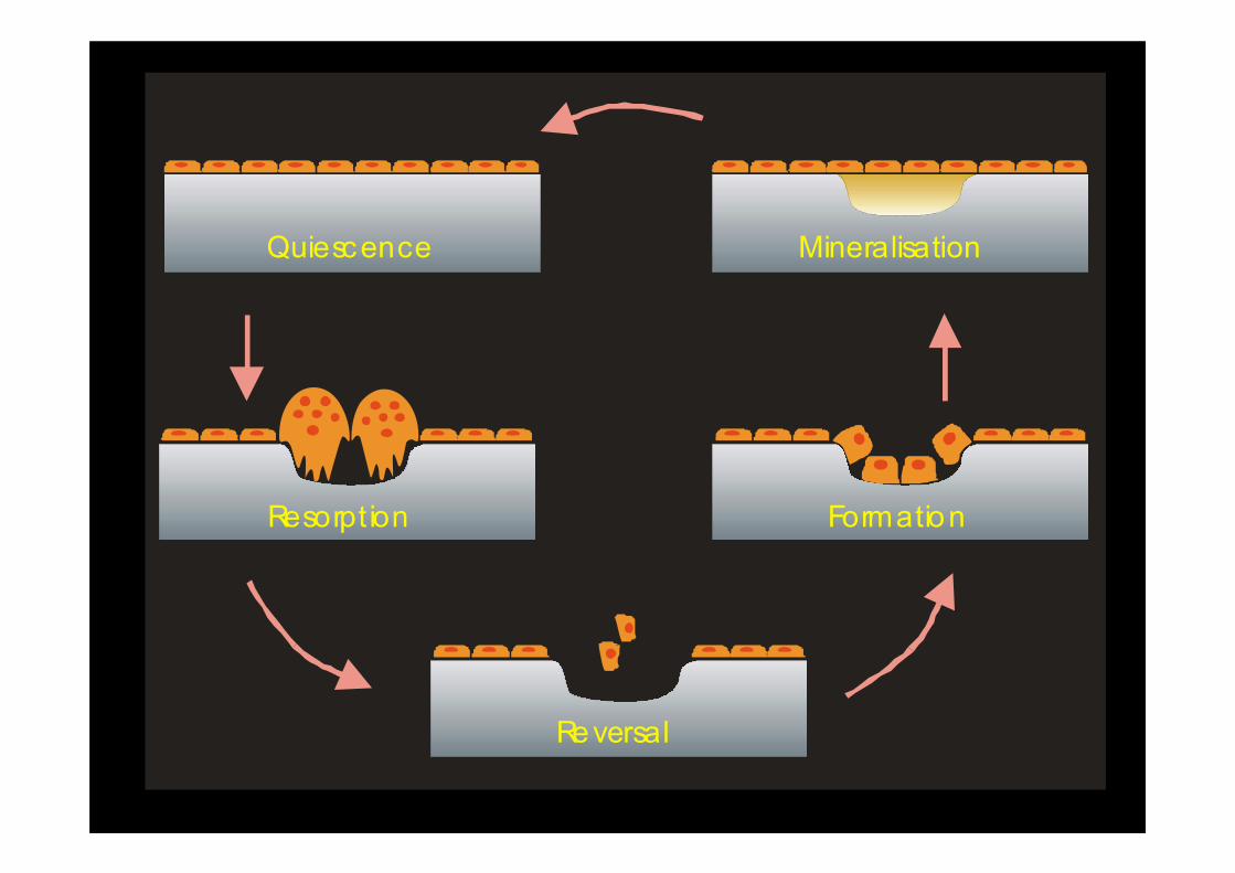
Quiescence
Resorption
Reversal
Formation
Mineralisation
Time & Space
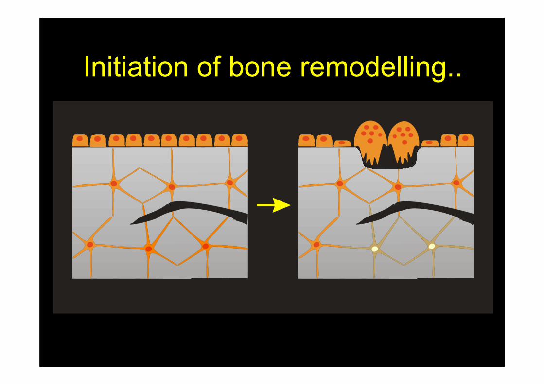
Initiation of bone remodelling..
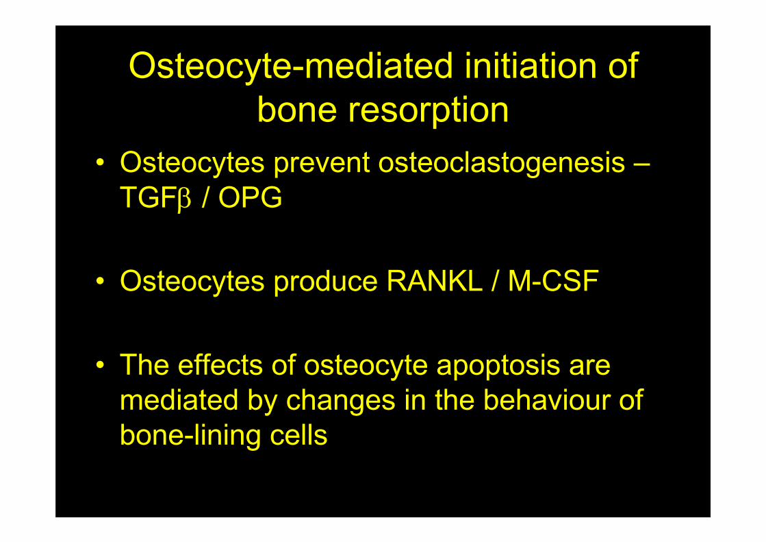
Osteocyte-mediated initiation of
bone resorption
• Osteocytes prevent osteoclastogenesis –
TGFβ / OPG
• Osteocytes produce RANKL / M-CSF
• The effects of osteocyte apoptosis are
mediated by changes in the behaviour of
bone-lining cells
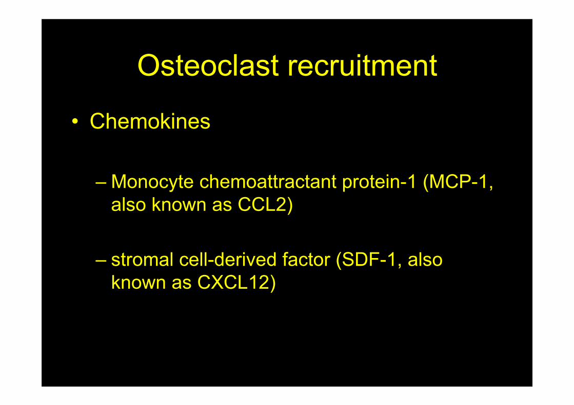
Osteoclast recruitment
• Chemokines
– Monocyte chemoattractant protein-1 (MCP-1,
also known as CCL2)
– stromal cell-derived factor (SDF-1, also
known as CXCL12)
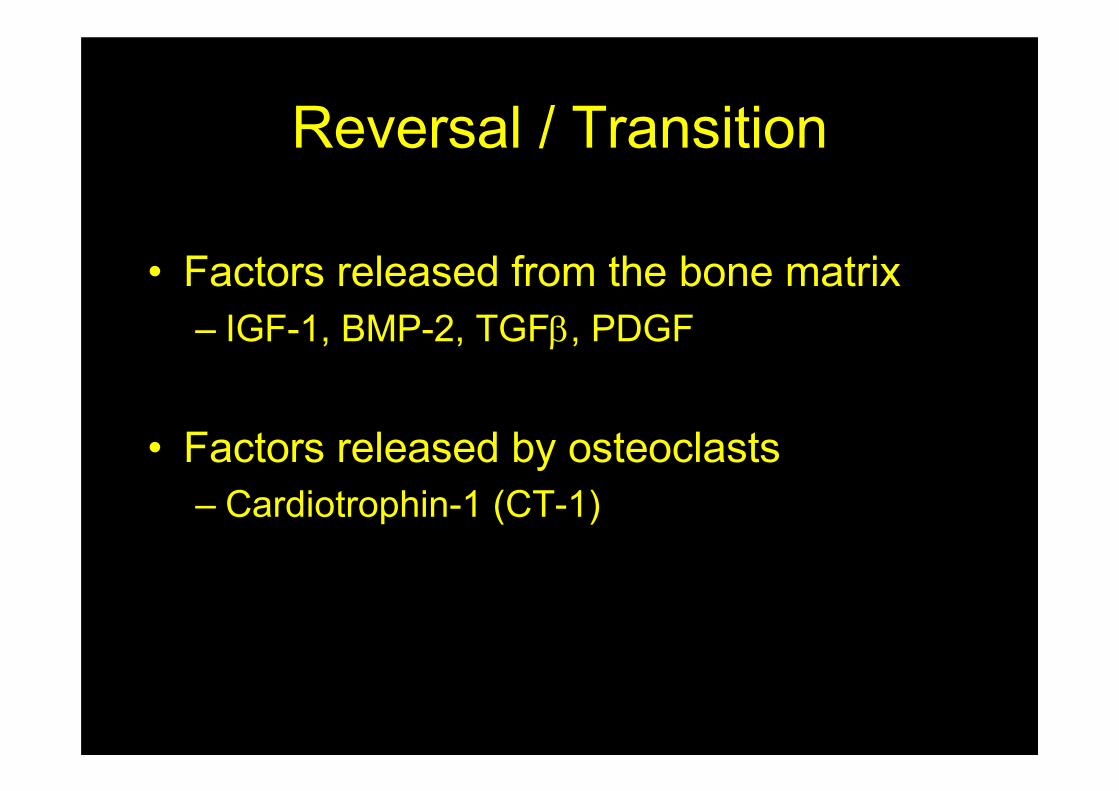
Reversal / Transition
• Factors released from the bone matrix
– IGF-1, BMP-2, TGFβ, PDGF
• Factors released by osteoclasts
– Cardiotrophin-1 (CT-1)
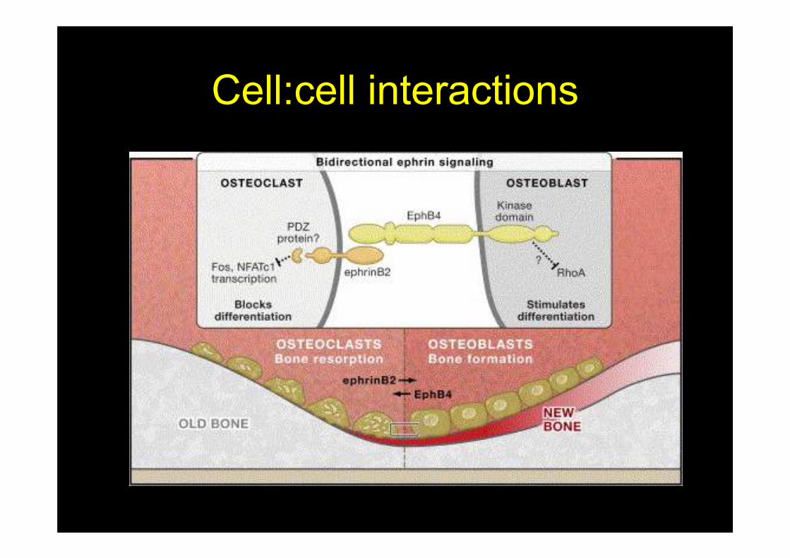
Cell:cell interactions
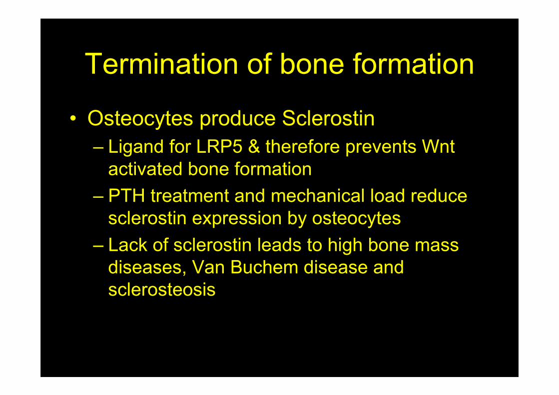
Termination of bone formation
• Osteocytes produce Sclerostin
– Ligand for LRP5 & therefore prevents Wnt
activated bone formation
– PTH treatment and mechanical load reduce
sclerostin expression by osteocytes
– Lack of sclerostin leads to high bone mass
diseases, Van Buchem disease and
sclerosteosis
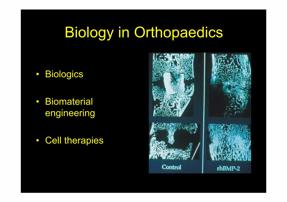
Biology in Orthopaedics
• Biologics
• Biomaterial
engineering
• Cell therapies
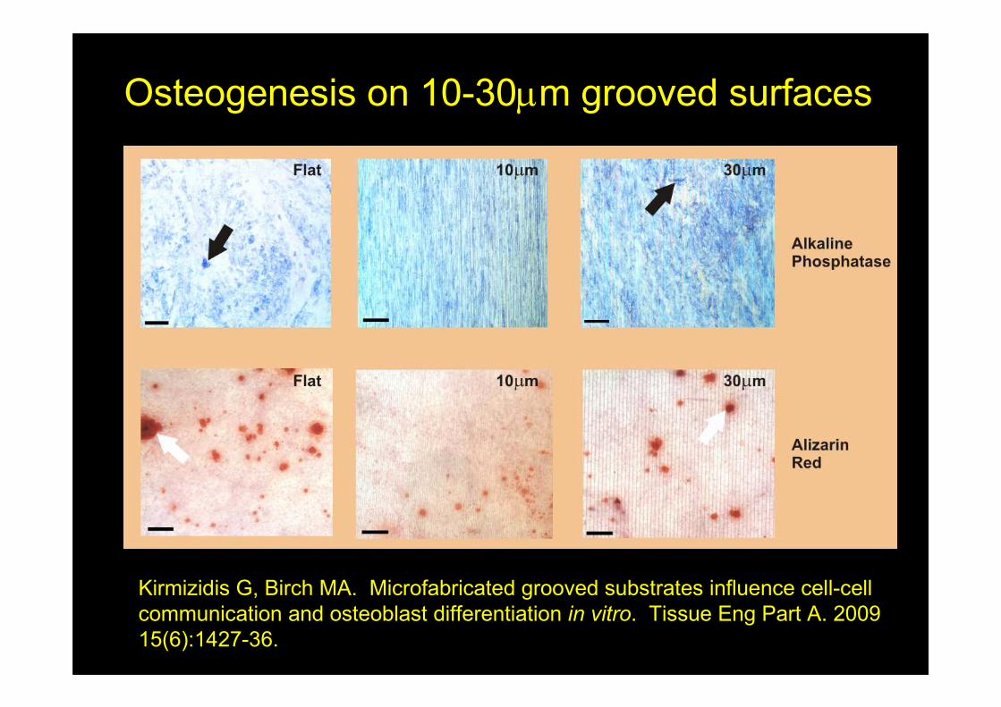
Osteogenesis on 10-30µm grooved surfaces
Kirmizidis G, Birch MA. Microfabricated grooved substrates influence cell-cell
communication and osteoblast differentiation in vitro. Tissue Eng Part A. 2009
15(6):1427-36.
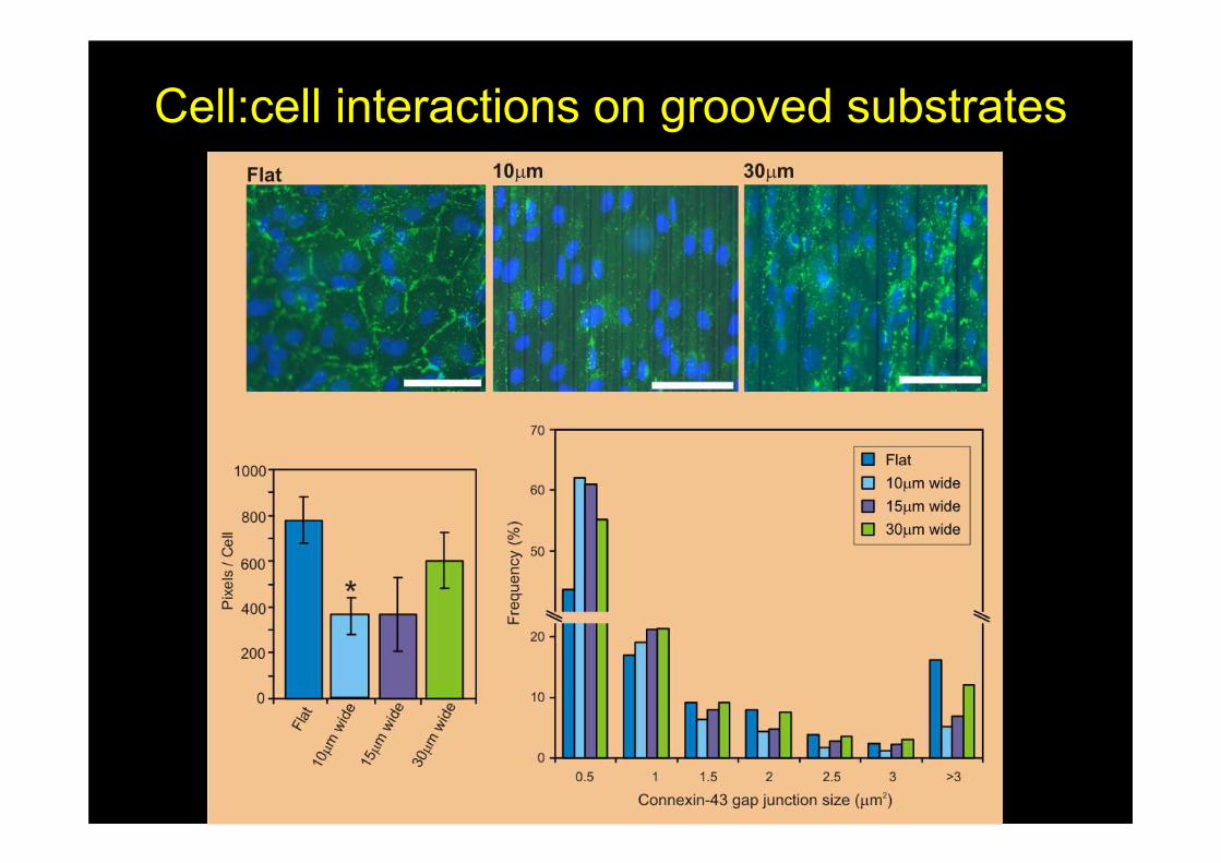
Cell:cell interactions on grooved substrates
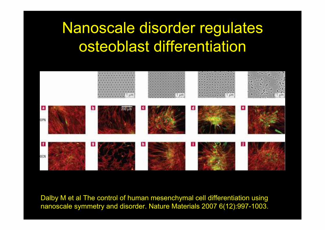
Nanoscale disorder regulates
osteoblast differentiation
Dalby M et al The control of human mesenchymal cell differentiation using
nanoscale symmetry and disorder. Nature Materials 2007 6(12):997-1003.
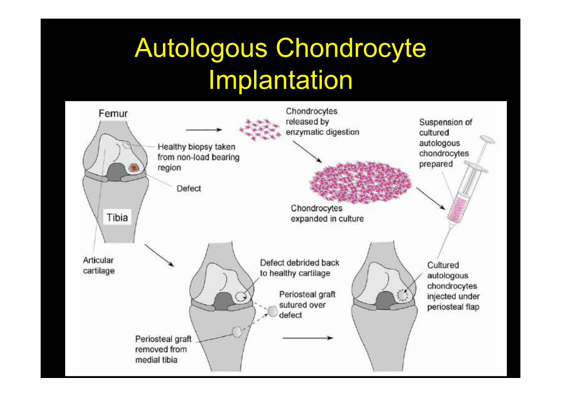
Autologous Chondrocyte
Implantation

Any questions?