Embed Size (px)
Citation preview

Chapter 9Skeletal health

Chapter overview
• Introduction
• Biology of bone
• Osteoporosis: definition, prevalence and consequences
• Physical activity and bone strength
• Physical activity and fracture risk
• Physical activity in prevention and management of osteoporosis
• Physical activity and osteoarthritis
• Summary

Structure of a long bone
• an average adult has 10–12 kg of bone;
• bone offers ‘strength with lightness’;
• cortical bone – dense, ivory-like;
• trabecular bone – lattice of thin, calcified struts
• membrane, the periosteum, covers surface of cortical bone.
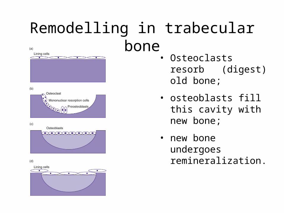
Remodelling in trabecular bone
• Osteoclasts resorb (digest) old bone;
• osteoblasts fill this cavity with new bone;
• new bone undergoes remineralization.

Measuring the structural properties of bone
• Dual energy X-ray absorptiometry (DXA) – measures bone mineral content and bone mineral density (BMD).
• Quantitative ultrasound – measurements reflect bone architecture as well as bone mineral.
• Quantitative computed tomography – measures bone mineral content, BMD and axial cross-sectional area.

Changes in bone mineral density over the life-span

Adaptation to load bearing
• Bone is deposited according to the load it must bear;
• strains produced during loading stimulate an adaptive, osteogenic response;
• response is determined by the magnitude, rate and distribution of strains, as well as the number of repetitions (strain cycles);
• immobilization and space flight both lead to net bone loss.

Osteoporosis
A skeletal disorder characterized by low bone mass and microarchitectural deterioration of bone tissue, with a consequent increase in fragility and susceptibility to fracture.

Osteoporotic changes in lumbar vertebrae
Normal, good weight-bearing Osteoporotic, loss of weight-bearing competence

What determines likelihood of osteoporosis or osteopenia?
• Peak bone mass as a young adult
– childhood and adolescence therefore a ‘window of opportunity’;
• rate of bone loss experienced with ageing
– dietary factors
– physical activity level;
• genetic variability.

Side-to-side differences in humerus of female tennis and squash players

Effect of 18 months of high-impact training in women aged 35–45
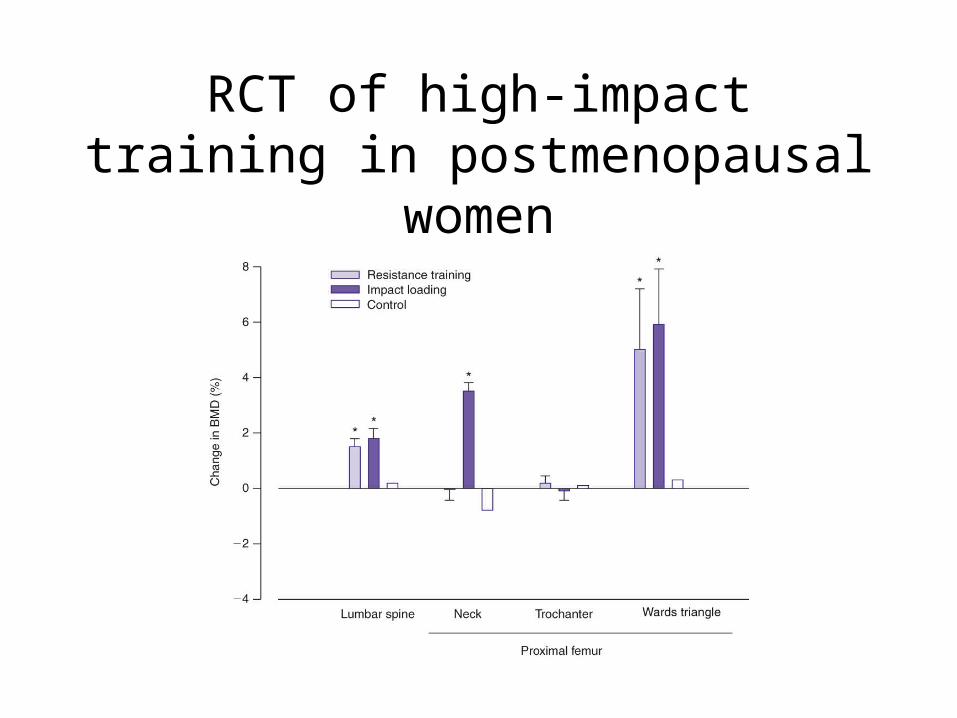
RCT of high-impact training in postmenopausal women

Clinical endpoint, hip fracture – Nurses’ Health Study (2002)
• 55% lower risk in postmenopausal women reporting > 24 MET-h per week, compared with < 3 MET-h per week;
• risk was 6% less for each increase in activity of 3 MET-h per week;
• in women who did no other exercise, walking for at least four hours per week was associated with a 41% lower risk than among those who walked for less than one hour per week.

Summary I
• Functional loading is the most important influence on bone remodelling.
• Strain rate and an unusual strain distribution largely determine its osteogenic effects.
• Bone mass increases during growth and reaches a peak towards the end of the second decade.
• Age related loss of bone can lead to osteopenia and osteoporosis, compromising strength and increasing the risk of fracture.

Summary II
• In premenopausal women, the effect of exercise is mainly conservation of bone. In older women it is to reduce the rate of loss.
• Physically active women have a lower risk of osteoporotic fracture of the hip and maybe of the spine.
• Regular exercise may decrease the risk of fall-related fractures.
• Moderate amounts and intensities of exercise have a favourable effect on pain and function in osteoarthritis of the knee, but sports involving high-intensity impacts or torsional types of stress increase risk.


![CHAPTER 11 BONE MARROW ADIPOGENESIS IN OSTEOPOROSIS chapters... · with conditions that lead to bone loss or osteoporosis, such as aging [1, 2], disuse [3, 4], long-term glucocorticoid](https://img.pdfslide.us/doc/110x75/601eb5077a3fcb54d13dccf1/chapter-11-bone-marrow-adipogenesis-in-chapters-with-conditions-that-lead-to.jpg)