Embed Size (px)
Citation preview
1/18/2017
1
©2016 MFMER | slide-1
Mayo Clinic Department of Cardiovascular Diseases
Mayo Clinic Echocardiography Review Course for Boards and Recertification
Diastolic Function AssessmentNew Guideline Update
Practical Approach
Jae K. Oh, MDEcho Hawaii 2017January 26th, 2017
©2016 MFMER | slide-2
Learning Objectives for Diastology
• Apply the new 2016 ASE/EACVI Guideline
• Classify and grade diastolic function
• Estimate filling pressure in most patients
After this talk, you should be able to
1/18/2017
2
©2016 MFMER | slide-3
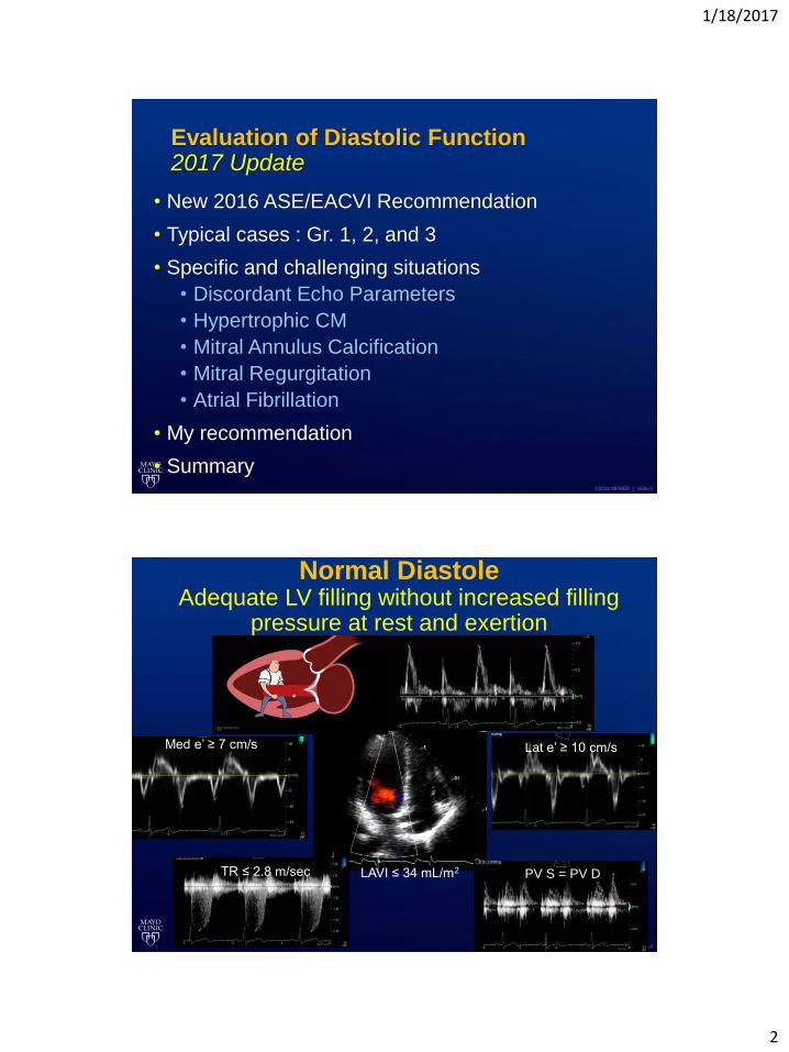
Evaluation of Diastolic Function 2017 Update
• New 2016 ASE/EACVI Recommendation
• Typical cases : Gr. 1, 2, and 3
• Specific and challenging situations
• Discordant Echo Parameters
• Hypertrophic CM
• Mitral Annulus Calcification
• Mitral Regurgitation
• Atrial Fibrillation
• My recommendation
• Summary
©2016 MFMER | slide-4
Normal DiastoleAdequate LV filling without increased filling
pressure at rest and exertion
Med e’ ≥ 7 cm/s Lat e’ ≥ 10 cm/s
TR ≤ 2.8 m/sec LAVI ≤ 34 mL/m2 PV S = PV D
1/18/2017
3
©2016 MFMER | slide-5
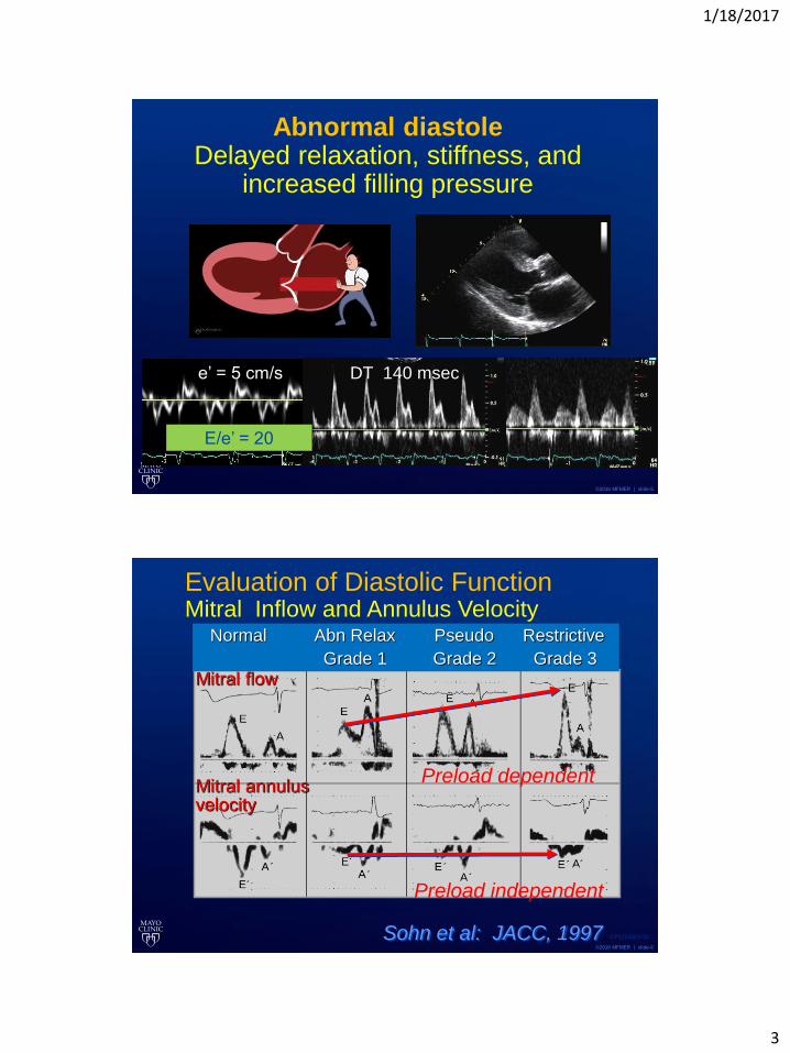
Abnormal diastoleDelayed relaxation, stiffness, and
increased filling pressure
DT 140 msece’ = 5 cm/s
E/e’ = 20
©2016 MFMER | slide-6
Mitral flow
Mitral annulusvelocity
Evaluation of Diastolic FunctionMitral Inflow and Annulus Velocity
Sohn et al: JACC, 1997
Normal Abn Relax Pseudo Restrictive
Grade 1 Grade 2 Grade 3
CP1254003-30
Preload dependent
Preload independent
1/18/2017
4
©2016 MFMER | slide-7
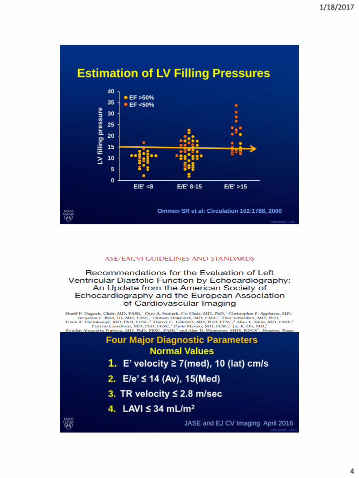
Estimation of LV Filling Pressures
0
5
10
15
20
25
30
35
40
Ommen SR et al: Circulation 102:1788, 2000
LV
fillin
g p
ressu
reEF >50%
EF <50%
E/E <8 E/E 8-15 E/E >15
©2016 MFMER | slide-8
Four Major Parameters in DiastologyNormal values
1. E’ velocity ≥ 7(med), 10 (lat) cm/s
2. E/e’ ≤ 14 (Av), 15(Med)
3. TR velocity ≤ 2.8 m/sec
4. LAVI ≤ 34 mL/m2
Four Major Diagnostic Parameters
Normal Values
JASE and EJ CV Imaging April 2016
1/18/2017
5
©2016 MFMER | slide-9
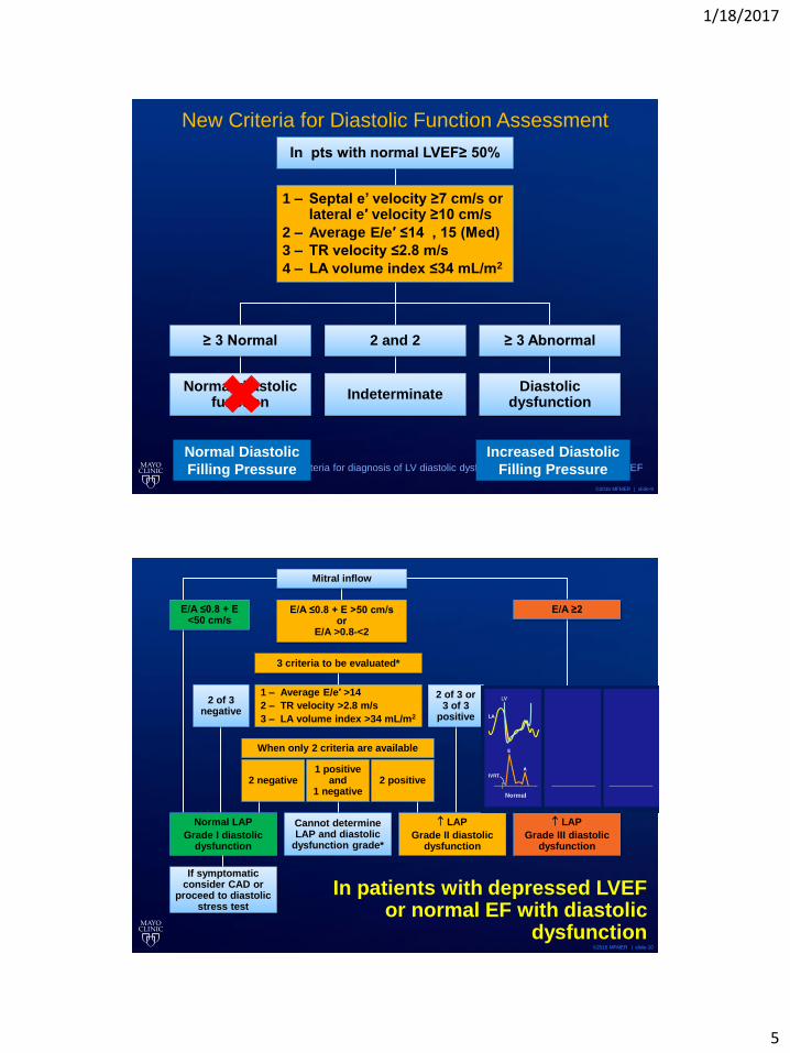
New Criteria for Diastolic Function Assessment
Criteria for diagnosis of LV diastolic dysfunction in patients with normal LVEF
In pts with normal LVEF≥ 50%
1 – Septal e’ velocity ≥7 cm/s orlateral e′ velocity ≥10 cm/s
2 – Average E/e′ ≤14 , 15 (Med)
3 – TR velocity ≤2.8 m/s
4 – LA volume index ≤34 mL/m2
≥ 3 Normal 2 and 2 ≥ 3 Abnormal
Normal diastolic function
IndeterminateDiastolic
dysfunction
Normal Diastolic
Filling Pressure
Increased Diastolic
Filling Pressure
©2016 MFMER | slide-10
In patients with depressed LVEF or normal EF with diastolic
dysfunction
Mitral inflow
E/A ≤0.8 + E <50 cm/s
E/A ≤0.8 + E >50 cm/sor
E/A >0.8-<2
E/A ≥2
Normal LAP
Grade I diastolic dysfunction
3 criteria to be evaluated*
1 – Average E/e′ >14
2 – TR velocity >2.8 m/s
3 – LA volume index >34 mL/m2
2 of 3 negative
2 of 3 or3 of 3
positive
2 negative1 positive
and1 negative
2 positive
When only 2 criteria are available
Cannot determine LAP and diastolic
dysfunction grade*
LAP
Grade II diastolic dysfunction
LAP
Grade III diastolic dysfunction
If symptomatic consider CAD or
proceed to diastolic stress test
1/18/2017
6
©2016 MFMER | slide-11
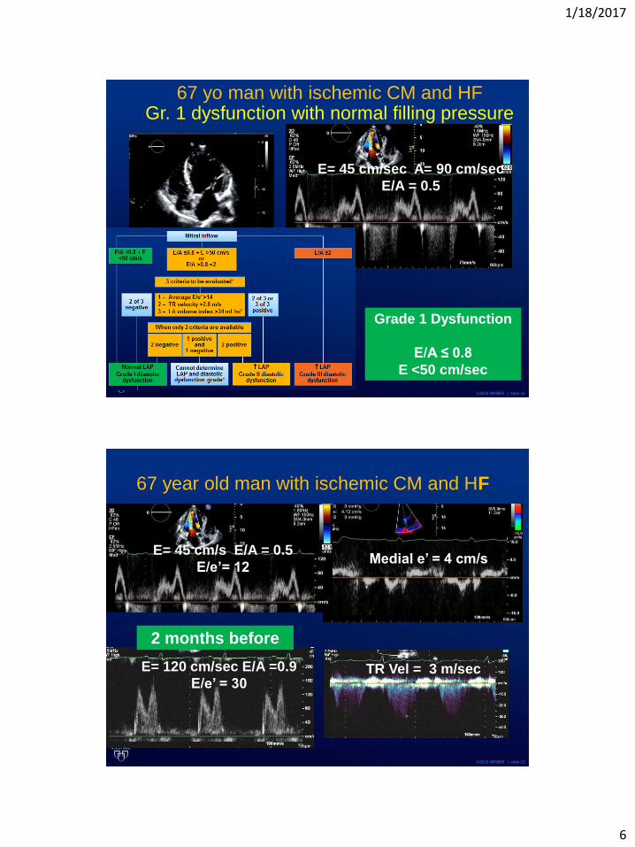
67 yo man with ischemic CM and HFGr. 1 dysfunction with normal filling pressure
E= 45 cm/sec A= 90 cm/sec
E/A = 0.5
Grade 1 Dysfunction
E/A ≤ 0.8
E <50 cm/sec
©2016 MFMER | slide-12
67 year old man with ischemic CM and HF
2 months before
E= 120 cm/sec E/A =0.9
E/e’ = 30TR Vel = 3 m/sec
E= 45 cm/s E/A = 0.5
E/e’= 12Medial e’ = 4 cm/s

1/18/2017
7
©2016 MFMER | slide-13
Ischemic Cardiomyopathy Echo PredictorSTICH Trial (N=1511)
Lin et al. 2014 AHA
Best survival with E/A 0.6-0.8
©2016 MFMER | slide-14
67 year old man with ischemic CM and HF
E= 120 cm/sec E/A =0.9 TR Vel = 3 m/sec
e’= 4 E/e’ =30
1/18/2017
8
©2016 MFMER | slide-15
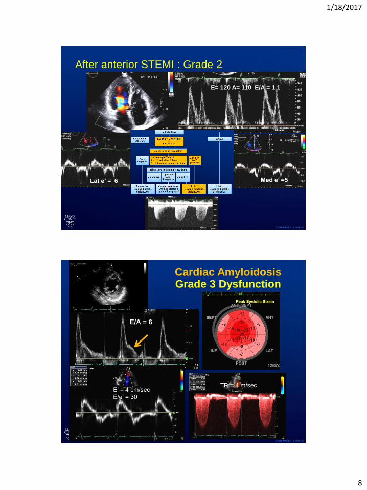
After anterior STEMI : Grade 2
Lat e’ = 6 Med e’ =5
E= 120 A= 110 E/A = 1.1
©2016 MFMER | slide-16
Cardiac AmyloidosisGrade 3 Dysfunction
E’ = 4 cm/sec
E/e’ = 30
TR = 4 m/sec
E/A = 6

1/18/2017
9
©2016 MFMER | slide-17
CP1100934-2Frommelt et al: J Am Soc Echocardiogr 16:176, 2003
Mid-diastolic mitral flow (L)Delayed relaxation
A. Fib
©2016 MFMER | slide-18
What to do in indeterminate cases?
1/18/2017
10
©2016 MFMER | slide-19
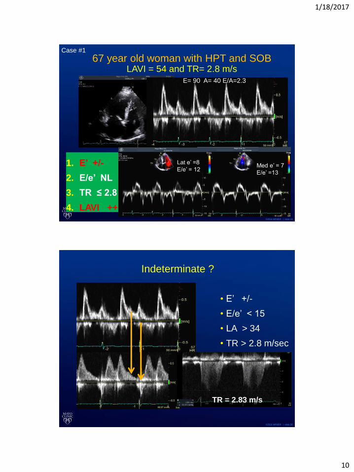
67 year old woman with HPT and SOBLAVI = 54 and TR= 2.8 m/s
1. E’ +/-
2. E/e’ NL
3. TR ≤ 2.8
4. LAVI ++
Med e’ = 7
E/e’ =13
Lat e’ =8
E/e’ = 12
E= 90 A= 40 E/A=2.3
Case #1
©2016 MFMER | slide-20
Indeterminate ?
• E’ +/-
• E/e’ < 15
• LA > 34
• TR > 2.8 m/sec
TR = 2.83 m/s
1/18/2017
11
©2016 MFMER | slide-21
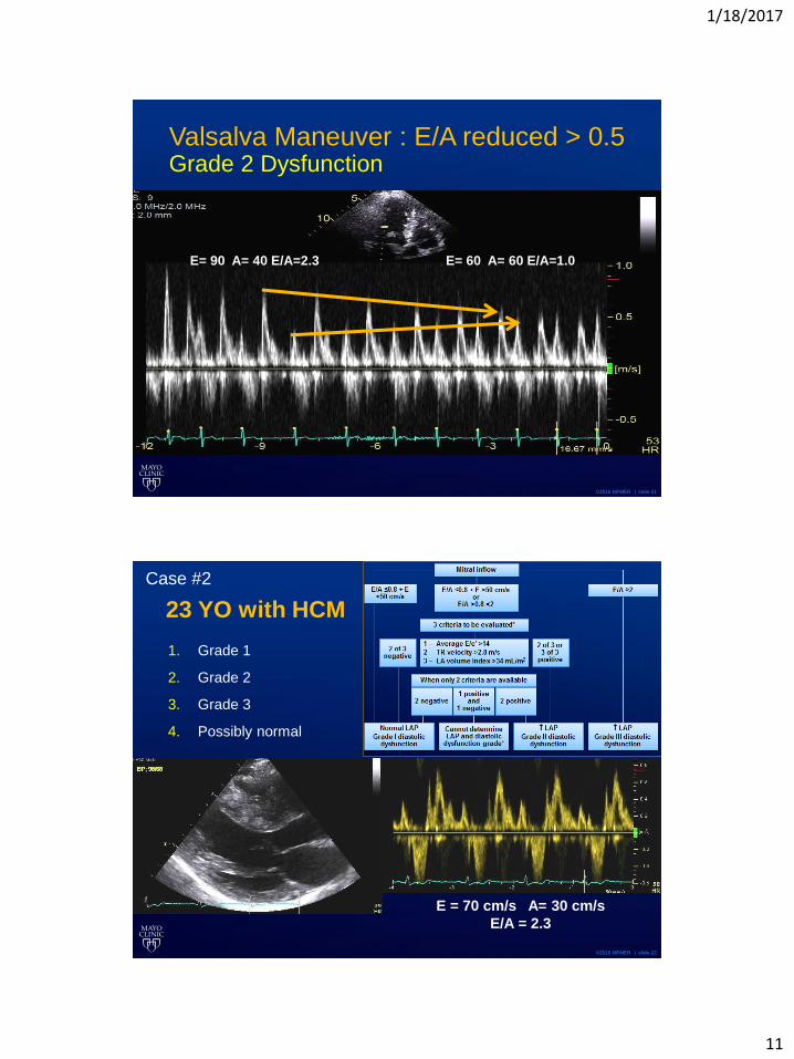
Valsalva Maneuver : E/A reduced > 0.5Grade 2 Dysfunction
E= 90 A= 40 E/A=2.3 E= 60 A= 60 E/A=1.0
©2016 MFMER | slide-22
23 YO with HCM
1. Grade 1
2. Grade 2
3. Grade 3
4. Possibly normal
E = 70 cm/s A= 30 cm/s
E/A = 2.3
Case # 6Case #2
1/18/2017
12
©2016 MFMER | slide-23
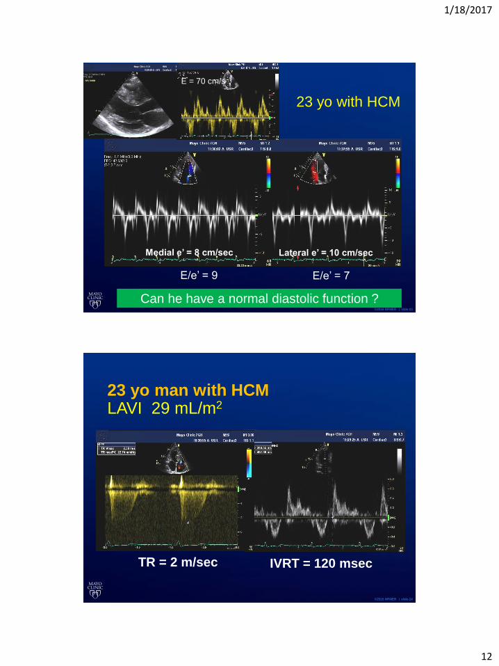
23 yo with HCM
Medial e’ = 8 cm/sec Lateral e’ = 10 cm/sec
E = 70 cm/s
E/e’ = 9 E/e’ = 7
Can he have a normal diastolic function ?
©2016 MFMER | slide-24
23 yo man with HCMLAVI 29 mL/m2
TR = 2 m/sec IVRT = 120 msec
1/18/2017
13
©2016 MFMER | slide-25
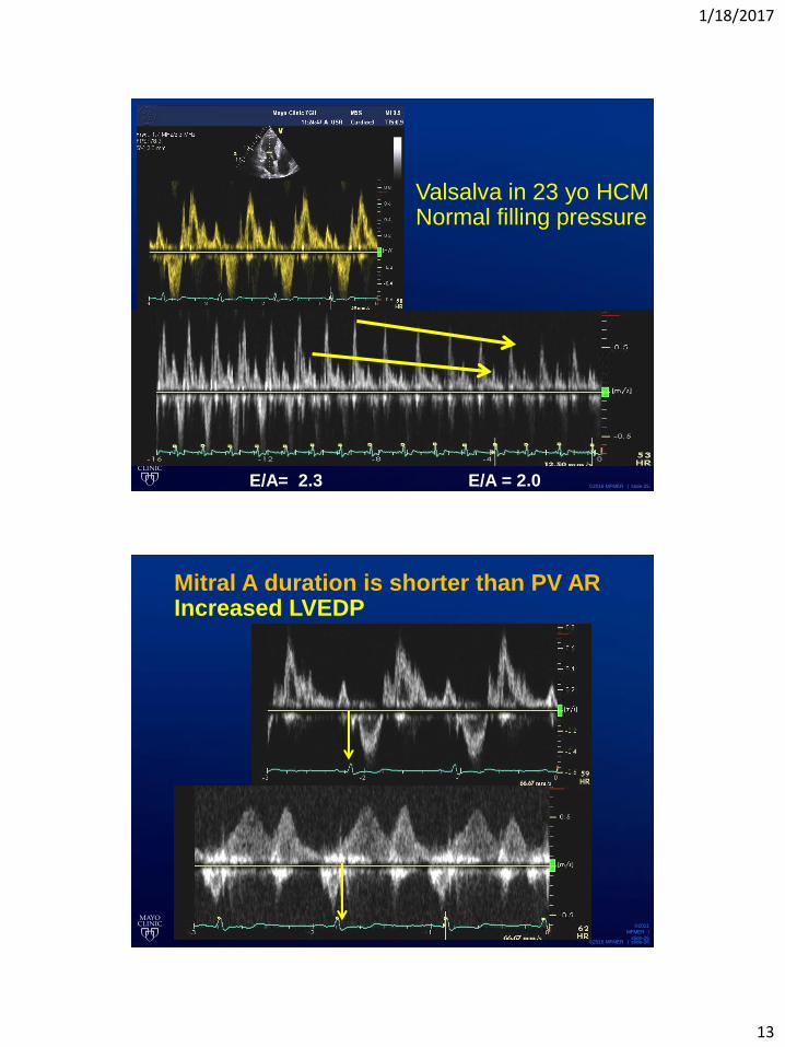
Valsalva in 23 yo HCMNormal filling pressure
E/A= 2.3 E/A = 2.0
©2016 MFMER | slide-26
Mitral A duration is shorter than PV ARIncreased LVEDP
©2011
MFMER |
slide-26

1/18/2017
14
©2016 MFMER | slide-27
Rossvoll and Hatle:
JACC, 1993
PVad-Ad (ms)
EDP(mm Hg)
r=0.68P<0.001
0
5
10
1520
25
3035
40
45
-100 -50 0 50 100
Ma=100 msPVa=165 ms
0.5
0.5
Mitral flow velocity LV pressure
Pulmonary vein
25 mm Hg
DopplerDeterminationof LVEDP
CP1057136-18
LVEDP can be increased with
normal mean LV diastolic pressure
©2016 MFMER | slide-28
In patients with depressed LVEF or normal EF with diastolic dysfunction
Mitral inflow
E/A ≤0.8 + E <50 cm/s
E/A ≤0.8 + E >50 cm/sor
E/A >0.8-<2
E/A ≥2
Normal LAP
Grade I diastolic dysfunction
3 criteria to be evaluated*
1 – Average E/e′ >14
2 – TR velocity >2.8 m/s
3 – LA volume index >34 mL/m2
2 of 3 negative
2 of 3 or3 of 3
positive
2 negative1 positive
and1 negative
2 positive
When only 2 criteria are available
Cannot determine LAP and diastolic
dysfunction grade*
LAP
Grade II diastolic dysfunction
LAP
Grade III diastolic dysfunction
If symptomatic consider CAD or
proceed to diastolic stress test
reduced e’
1/18/2017
15
©2016 MFMER | slide-29
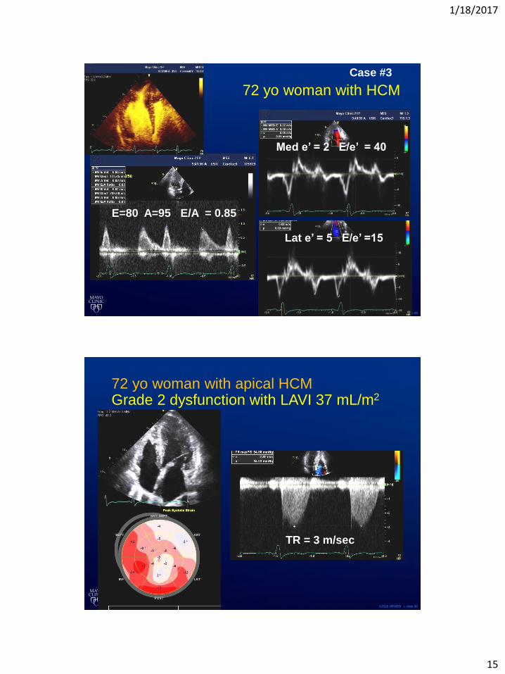
72 yo woman with HCM
E=80 A=95 E/A = 0.85
Med e’ = 2 E/e’ = 40
Lat e’ = 5 E/e’ =15
Case #3
©2016 MFMER | slide-30
72 yo woman with apical HCMGrade 2 dysfunction with LAVI 37 mL/m2
TR = 3 m/sec
1/18/2017
16
©2016 MFMER | slide-31
0
10
20
30
40
50
60
0 5 10 15 20 25 30 35 40 45
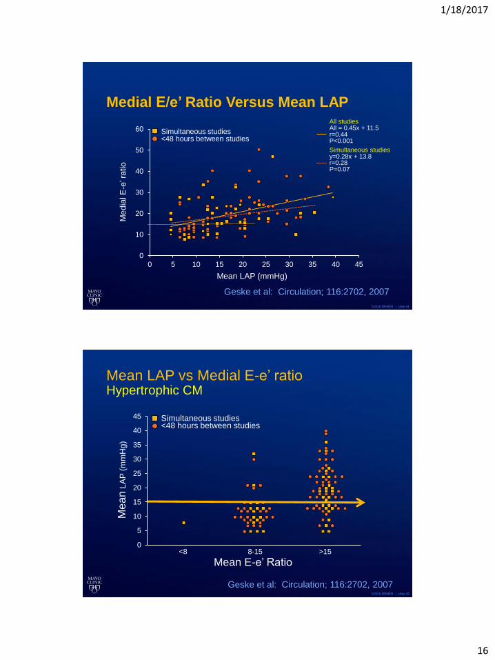
Medial E/e’ Ratio Versus Mean LAP
Me
dia
l E
-e’ r
atio
Mean LAP (mmHg)
Geske et al: Circulation; 116:2702, 2007
All studiesAll = 0.45x + 11.5r=0.44P<0.001
Simultaneous studiesy=0.28x + 13.8r=0.28P=0.07
Simultaneous studies<48 hours between studies
©2016 MFMER | slide-32
0
5
10
15
20
25
30
35
40
45
<8 8-15 >15
Mean LAP vs Medial E-e’ ratioHypertrophic CM
Me
an
LA
P (
mm
Hg)
Mean E-e’ Ratio
Geske et al: Circulation; 116:2702, 2007
Simultaneous studies<48 hours between studies
1/18/2017
17
©2016 MFMER | slide-33
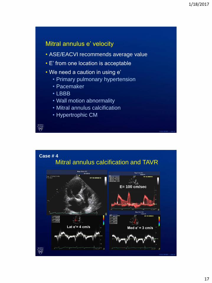
Mitral annulus e’ velocity
• ASE/EACVI recommends average value
• E’ from one location is acceptable
• We need a caution in using e’
• Primary pulmonary hypertension
• Pacemaker
• LBBB
• Wall motion abnormality
• Mitral annulus calcification
• Hypertrophic CM
©2016 MFMER | slide-34
Mitral annulus calcification and TAVR
E= 100 cm/sec
Lat e’= 4 cm/s Med e’ = 3 cm/s
Case # 4
1/18/2017
18
©2016 MFMER | slide-35
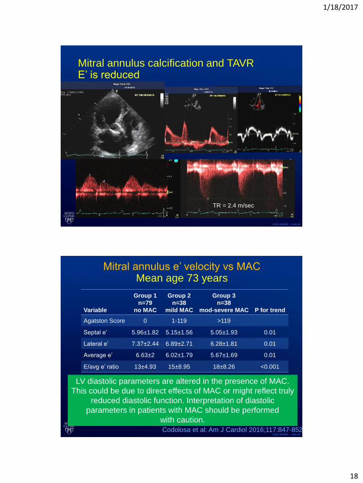
Mitral annulus calcification and TAVRE’ is reduced
TR = 2.4 m/sec
©2016 MFMER | slide-36
Mitral annulus e’ velocity vs MACMean age 73 years
Variable
Group 1
n=79
no MAC
Group 2
n=38
mild MAC
Group 3
n=38
mod-severe MAC P for trend
Agatston Score 0 1-119 >119
Septal e’ 5.96±1.82 5.15±1.56 5.05±1.93 0.01
Lateral e’ 7.37±2.44 6.89±2.71 6.28±1.81 0.01
Average e’ 6.63±2 6.02±1.79 5.67±1.69 0.01
E/avg e’ ratio 13±4.93 15±8.95 18±8.26 <0.001
Codolosa et al: Am J Cardiol 2016;117:847-852
LV diastolic parameters are altered in the presence of MAC.
This could be due to direct effects of MAC or might reflect truly
reduced diastolic function. Interpretation of diastolic
parameters in patients with MAC should be performed
with caution.
1/18/2017
19
©2016 MFMER | slide-37
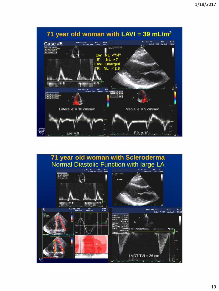
71 year old woman with LAVI = 39 mL/m2
Lateral e’ = 10 cm/sec Medial e’ = 9 cm/sec
E/e’ = 9 E/e’ = 10
E/e’ NL < 14
E’ NL > 7
LAVI Enlarged
TR NL < 2.8
Case #5
©2016 MFMER | slide-38
71 year old woman with Scleroderma Normal Diastolic Function with large LA
LVOT TVI = 26 cm
1/18/2017
20
©2016 MFMER | slide-39
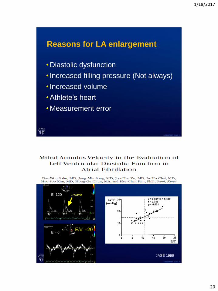
Reasons for LA enlargement
• Diastolic dysfunction
• Increased filling pressure (Not always)
• Increased volume
• Athlete’s heart
• Measurement error
©2016 MFMER | slide-40
E/e’ =20
L waveE=120
E’= 6
JASE 1999
1/18/2017
21
©2016 MFMER | slide-41
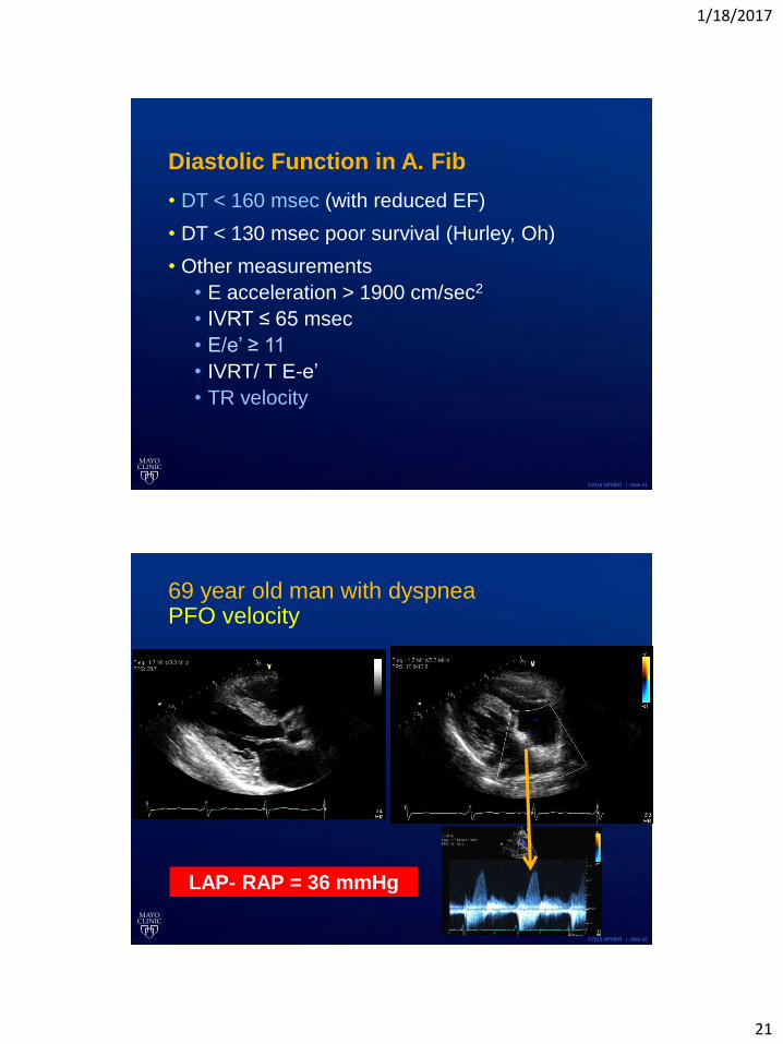
Diastolic Function in A. Fib
• DT < 160 msec (with reduced EF)
• DT < 130 msec poor survival (Hurley, Oh)
• Other measurements
• E acceleration > 1900 cm/sec2
• IVRT ≤ 65 msec
• E/e’ ≥ 11
• IVRT/ T E-e’
• TR velocity
©2016 MFMER | slide-42
69 year old man with dyspneaPFO velocity
LAP- RAP = 36 mmHg
1/18/2017
22
©2016 MFMER | slide-43
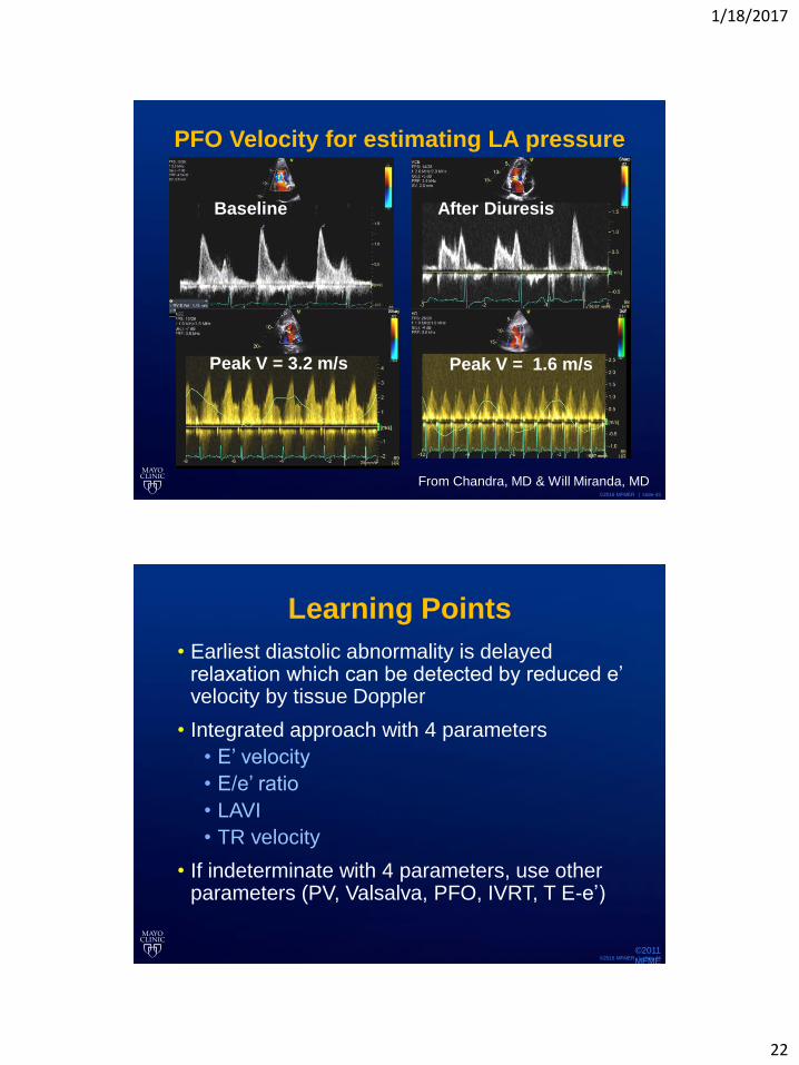
PFO Velocity for estimating LA pressure
Baseline After Diuresis
Peak V = 3.2 m/s Peak V = 1.6 m/s
From Chandra, MD & Will Miranda, MD
©2016 MFMER | slide-44
Learning Points
• Earliest diastolic abnormality is delayed relaxation which can be detected by reduced e’ velocity by tissue Doppler
• Integrated approach with 4 parameters
• E’ velocity
• E/e’ ratio
• LAVI
• TR velocity
• If indeterminate with 4 parameters, use other parameters (PV, Valsalva, PFO, IVRT, T E-e’)
©2011
MFME
R |
slide-
44