Embed Size (px)
Citation preview

Maxillary Osteotomies and Bone Grafts for
Correction of Contoural and Occlusal
Deformities*®
M. L. LEWIN, M.D.
RAVELO V. ARGAMASO, M.D.
ABRAHAM I. FINGEROTH, D.D.S.Bronx, New York
One of the major aims of modern management of the cleft palate
patient is the prevention of skeletal deformity. With present day surgical
management and orthodontic treatment, pronounced maxillary hypoplasia
is rarely encountered. Severe hypoplasia of the entire maxillary compound
is seen in other congenital malformations, as in Crouzon's disease.
For some patients who have a striking disproportion between the max-
illa and the mandible with pseudo prognathism, mandibular osteotomy
with recession of the mandible will restore the occlusal relationship and
correct the contoural deformity (Figure 1).
Skeletal deformities in cleft palate patients are usually limited to the
alveolar portion of the maxilla. The recession of the maxilla in such
patients may often be improved by prosthetic restoration. When the upper
jaw is edentulous or nearly so, a denture will establish satisfactory contour
and occlusion. Occasionally, the upper suleus is deepened to accom-
modate a larger labial component. A denture will not correct the deform-
ity when there is hypoplasia of the entire maxillary compound. If maxil-
lary hypoplasia exists without concomitant malocclusion, onlay bone
grafts to the anterior facial wall will improve the facial contour (7, 8)
(Figure 2). However, maxillary hypoplasia is generally associated with
severe malocclusion and can only be corrected by midface osteotomy.
Facial osteotomies are based on the work of LeFort who studied the
mechanism of fractures of the facial skeleton. Because of the variations in
the strength and fragility of various parts of the facial skeleton, fractures
of the face usually follow a typical pattern.
The most severe fracture leads to separation of the facial skeleton from
the cranial base and is known as LeFort III' (6). The more common is the
LeFort I fracture in which the alveolar process is severed horizontally
from the maxillary wall and from the pterygoid buttresses (Figure 3a).
An elective osteotomy of the entire maxilla was first reported about
* From the Plastic Surgery Division. Department of Surgery, and Cleft Palate Cen-ter, Montefiore Hospital and Medical Center.
' LeFort II is similar to LeFort III, except that the fracture line runs across thepyriform aperture instead of through the naso-frontal junction.
18

MAXILLARY O§STEOTOMIES AND BONE GRAFTS 10
FIGURE la & b. Mild maxillary hypoplasia and mandibular prognathism cor-
rected by ositcotomy of the mandible {and rhinoplasty).
FIGURE 2a & b. Crouzon syndrome of moderate severity without malocclusion,corrected by bone grafts of the infraorbital area (b. ten years after operation}.
twonty years ago by Gillies and Harrison. (3). The main difficulty they
cneountored was in maintaining the maxilla in its advanced position. In
spite of intra and estraoral traction and fixation, the maxilla recessed
during the postoperative period. Although Gillies case was considered a

20 Lewin, Argamaso, Fingeroth
FIGURE 3 a. Lincs of osteotomy according to LeFort: TII (solid black linc), Le-Fort 1 interrupted line. b. Site of osteotomies at the pterygopalatal junction. c. Facialskeleton detached from the base of the skull. d. Locations of bone grafts.
success, his experionce discouraged many surgeons from following this
approach.
A major contribution to this work was made by Paul Tessier, who
combined the osteotomy with bone grafts (18, 14). These bone grafts were
intended to prevent the advanced maxilla from receding and to assure
firm and rapid union. Murray and Swanson, (10) Jabaley and Edgerton,
(4) Obwegeser (11) published case reports of LeFort III osteotomies and
described details of the surgical procedure.
Obwegeser (12) recommended a variety of major and mimor maxillary
osteotomies for the correction of facial deformities, secondary to cleft
palate, or of other etiology.
The operation for midface advancement consists of seven incisions and
seven ostcotomics, the latter being joined together to encircle the maxilla.
Five incisions are made on the face and two intraorally in the upper
buccal suleus in the retromolar area, to gain access to the pterygopalatal
junction.
An incision across the glabella, from one eanthus to the other, exposes
the fronto-nasal junction and the medial orbital wall. The lateral orbital
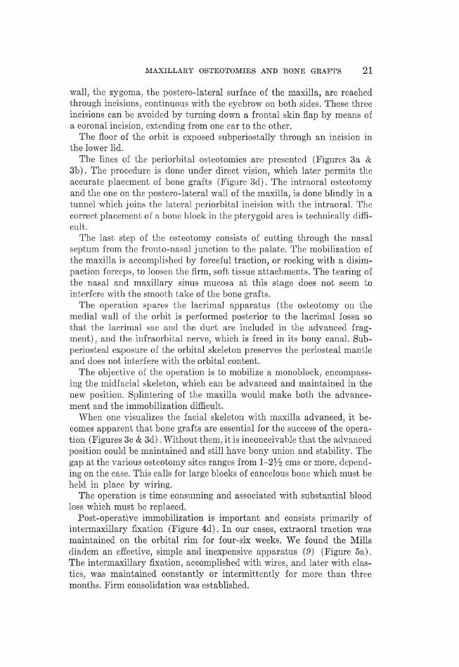
MAXILLARY OSTEOTOMIES AND BONE GRAFTS 21
wall, the zygoma, the postero-lateral surface of the maxilla, are reached
through incisions, continuous with the eyebrow on both sides. These three
incisions can be avoided by turning down a frontal skin flap by means of
a coronal incision, extending from one ear to the other.
The floor of the orbit is exposed subperiostally through an incision in
the lower lid.
The lines of the periorbital osteotomies are presented (Figures 3a &
3b). The procedure is done under direct vision, which later permits the
accurate placement of bone grafts (Figure 83d). The intraoral osteotomy
and the one on the postero-lateral wall of the maxilla, is done blindly in a
tunnel which joins the lateral periorbital incision with the intraoral. The
correct placement of a bone block in the pterygoid area is technically diffi-
cult.
The last step of the osteotomy consists of cutting through the nasal
septum from the fronto-nasal junction to the palate. The mobilization of
the maxilla is accomplished by forceful traction, or rocking with a disim-
paction forceps, to loosen the firm, soft tissue attachments. The tearing of
the nasal and maxillary sinus mucosa at this stage does not seem to
interfere with the smooth take of the bone grafts.
The operation spares the lacrimal apparatus (the osteotomy on the
medial wall of the orbit is performed posterior to the lacrimal fossa so
that the lacrimal sac and the duct are included in the advanced frag-
ment), and the infraorbital nerve, which is freed in its bony canal. Sub-
periosteal exposure of the orbital skeleton preserves the periosteal mantle
and does not interfere with the orbital content.
The objective of the operation is to mobilize a monoblock, encompass-
ing the midfacial skeleton, which can be advanced and maintained in the
new position. Splintering of the maxilla would make both the advance-
ment and the immobilization difficult.
When one visualizes the facial skeleton with maxilla advanced, it be-
comes apparent that bone grafts are essential for the success of the opera-
tion (Figures 3¢ & 3d). Without them, it is inconceivable that the advanced
position could be maintained and still have bony union and stability. The
gap at the various osteotomy sites ranges from 1-24 ems or more, depend-
ing on the case. This calls for large blocks of cancelous bone which must be
held in place by wiring.
The operation is time consuming and associated with substantial blood
loss which must be replaced.
Post-operative immobilization is important and consists primarily of
intermaxillary fixation (Figure 4d). In our cases, extraoral traction was
maintained on the orbital rim for four-six weeks. We found the Mills
diadem an effective, simple and inexpensive apparatus (9) (Figure 5a).
The intermaxillary fixation, accomplished with wires, and later with elas-
tics, was maintained constantly or intermittently for more than three
months. Firm consolidation was established.

FIGURE 4. Sevore facial deformity in Grouzon discase in a 20 year old girl. a, b.
Preoperative and postoperative profile. e, d. Preoperative and postoperative occlusion.
c, {. Preoperative and postoperative radiographs. Note degree of raidface advancement
and postoperative occlusion.

MAXILLARY OSTEOTOMIES AND BONE GRAFTS 23
FIGURE 5. a. Postoperative extraoral tractions using Mills diadem, (same patientas in Fig. 4). b. Preoperative and postoperative dental molds.
In our limited experience, some recession of the advanced maxilla did
occur, but there was no tendency for the deformity to reeur. The new
occlusal relation was maintained (Figures 4 & 5).
LeFort I osteotomy is occasionally indicated in cleft palate rehabilita-
tion. However, much more often there are indications for segmental osteot-
omy of the maxilla (1, 2). The place of this procedure in rehabilitation of
the older cleft palate patient has not been fully appreciated. When there
are teeth in the upper jaw and the occlusal relationship is grossly dis-
turbed, beyond the seope of orthodontic improvement, surgical reposition-
ing should be considered. The lingual displacement of the anterior frag-
ment causes recession of the upper lip and unattractive flatness of the
facial contour. This is an additional indication for osteotomy.
Patient shown Figure 6 had a segmental osteotomy of the maxilla as a
stage in the rehabilitation of his cleft lip and palate. The exact size of the
osteotomized segment is determined by a study of the occlusal models
(Figure 6f). The alveolar fragment is mobilized by osteotomies on its
buccal and palatal surfaces. The soft tissue attachment on the palatal
surface must be freed to expose the predetermined osteotomy site and to
allow for the advancement of the fragment. The osteotomy on the buccal
surface is performed through short vertical incisions and tunnelling, leav-
ing sufficient mucosal attachment to assure the viability of the osteoto-
mized segment. Intermaxillary fixation is an important step in the proce-
dure. Cancelous bone grafts are usually essential to assure firm and rapid
union.
Summary
Maxillary osteotomies of LeFort I or III types and segmental ostcoto-
mies of the maxilla and palate bring us a step forward in the surgical
rehabilitation of patients with severe facial deformities. Partial (cortical)

24 Lewin, Argamaso, Fingeroth
FIGURE 6. 32 year old male with contoural and malocclusal deformity, secondaryto cleft lip and palate. a, b. Preoperative appearance. c. Augmentation of upper lipwith small Abbe flap, corresponding to site of philtrum. d, e. Postoperative appear-ance. f. Preoperative and postoperative dental models and the site of palatal oste-otomy.
or complete osteotomies with bone grafts have an important role, though
not yet fully exploited, in the orthodontic treatment of severe occlusal
deformities.
References
1. J. M., Horowitz, S. L., Guy, C. L., and D., Surgical-ortho-dontic corrections in the bilateral cleft lip. Cleft Palate J., 1, 153-163, 1964.
2. J. M., and Horowitz, S. L., The surgical-orthodontic approach to thetreatment of dentofacial deformities. Amer. J. of Orthodontics, 55, 217-243, 1969.
3. H. and Harrison, S. H., Operative correction by osteotomy of recessedmalar maxillary compound in a case of oxycephaly. Brit. J. Plast. Surg., 8, 123-127, 1951.
4. JaBsueEy, M. E. and Enoerrton, M. T., Surgical correction of congenital midfaceretrusion in the presence of mandibular prognathism. Plast. & Rec. Surg., 44, 1-8,1969.
. Kxowrrs, C. C., The place of mandibular osteotomy in the treaiment plan forcleft lip and palate conditions. Brit. J. Plast. Surg., 23, 365-369, 1969.
6. LeFort, R., Etude experimentale sur les fractures de le machoire superieure. Rev.Chir. Paris, 28, 208, 360, 479, 1901.
Ot

12.
18.
14.
MAXILLARY OSTEOTOMIES AND BONE GRAFTS 25
. Lewin, M. L., Facial and hand deformities in acrocephalosyndactyly. Plastic andReconstructive Surgery, 12, 188-147, 1953.
. Lewin, M. L., Facial deformity in acrocephaly and its surgical co'rection. Arch. ofOphthalmology, 47, 321-327, 1952.
. MILs, R., Personal Communications.
. Murray, J. E. and Swanson, L. T., Mid-face osteotomy and advancement forcraniosynostosis. Plast. & Reconst. Surg., 41, 299-306, 1968.
. OsBwrarsEr, H. L., Surgical correction of small or retrodisplaced maxilla. The"dish face" deformity. Plast. & Reconst. Surg., 48, 351-865, 1969.
H. L., Surgery as an adjunct to orthodontics in normal and cleftpalate patients. Trans. of European Orthodontic Soc., 42, 343-3583, 1966.T'rssier, P., Osteotomies totales de la face syndrome de Crouzon syndrome d'apert,oxycephalies, scaphocephalies, turricephalies. Ann. Chir. Plast. XII, CP 273-286, 1967.T'rssiIERr, P., Private Communications, Seminar, Centre Medico-Chirugical FOCH,Suresnes, France.