Embed Size (px)
Citation preview
Materials Science and Engineering C 68 (2016) 229–240
Contents lists available at ScienceDirect
Materials Science and Engineering C
j ourna l homepage: www.e lsev ie r .com/ locate /msec
Improved bioactivity of selective laser melting titanium: Surfacemodification with micro-/nano-textured hierarchical topography andbone regeneration performance evaluation
Jia-yun Xu a, Xian-shuai Chen b, Chun-yu Zhang b, Yun Liu a, Jing Wang a, Fei-long Deng a,⁎a Department of Oral Implantology, Guanghua School of Stomatology, Hospital of Stomatology, Sun Yat-Sen University, Guangdong Provincial Key Laboratory of Stomatology, Guangzhou 510055,PR Chinab Guangzhou Institute of Advanced Technology, Chinese Academy of Science, Guangzhou 511458, PR China
Abbreviations: SLM, selective laser melting; AM, aextracellular matrix.⁎ Corresponding author at: Department of Oral Impl
Stomatology, Hospital of Stomatology, Sun Yat-sen UniveGuangzhou 510055, PR China.
E-mail address: [email protected] (F. Deng).
http://dx.doi.org/10.1016/j.msec.2016.05.0960928-4931/© 2016 Elsevier B.V. All rights reserved.
a b s t r a c t
a r t i c l e i n f oArticle history:Received 7 March 2016Received in revised form 5 May 2016Accepted 22 May 2016Available online 24 May 2016
Selective laser melting (SLM) titanium requires surface modification to improve its bioactivity. The microroughsurface of it can be utilized as the micro primary substrate to create a micro-/nano-textured topography for im-proved bone regeneration. In this study, the microrough SLM titanium substrate was optimized by sandblasting,and nano-porous features of orderly arranged nanotubes and disorderly arranged nanonetwere produced by an-odization (SAN) and alkali-heat treatment (SAH), respectively. The results were compared with the controlgroup of an untreated surface (native-SLM) and a microtopography only surface treated by acid etching (SLA).The effects of the different topographies on cell functions and bone formation performance were evaluated invitro and in vivo. It was found thatmicro-/nano-textured topographies of SAN and SAH showed enhanced cell be-haviour relative to themicrotopography of SLAwith significantly higher proliferation on the 1st, 3rd, 5th and 7thday (P b 0.05) and higher total protein contents on the 14th day (P b 0.05). In vivo, SAN and SAH formed moresuccessively regenerated bone, which resulted in higher bone-implant contact (BIC%) and bone-bonding forcethan native-SLM and SLA. In addition, the three-dimensional nanonet of SAH was expected to be more similarto native extracellular matrix (ECM) and thus led to better bone formation. The alkaline phosphatase activityof SAH was significantly higher than the other three groups at an earlier stage of the 7th day (P b 0.05) and theBIC% was nearly double that of native-SLM and SLA in the 8th week. In conclusion, the addition of nano-porousfeatures on the microrough SLM titanium surface is effective in improving the bioactivity and bone regenerationperformance, in which the ECM-like nanonet with a disorderly arranged biomimetic feature is suggested to bemore efficient than nanotubes.
© 2016 Elsevier B.V. All rights reserved.
Keywords:Selective laser meltingMicro-/nano-topographyTitaniumNano-porousOsseointegrationSurface modification
1. Introduction
Fabrication of custom-made bone substitutes has always been ap-pealing in dentistry, orthopaedics, and joint arthroplasty clinical prac-tices [1–5]. However, complicated and irregular anatomies makeconventional manufacturing difficult because of complex cast processesand high costs of rawmaterials [6,7]. In addition, multiple process stepsmay result in imprecise products. Recently, the rapid developments inadditive manufacturing (AM) have enabled the production of freeform
dditive manufacturing; ECM,
antology, Guanghua School ofrsity, No.56, Ling Yuan Xi Road,
geometries [5,8,9], allowing for custom-made and precisely controlledbone substitute architectures based on computer-aided design (CAD)data [3,10–13]. AM is more convenient in fabricating complex and cus-tom-made structures than conventional methods based on a one-stepmanufacturing process [14].
Selective lasermelting (SLM), one of the latest types of AM, is widelyused in biomedical implants fabrication. Titaniumand its alloys fabricat-ed by SLM are expected to be promising biomaterials as bone substi-tutes because of their biocompatibility and biomechanical properties[15–17]. Because the bone-implant interface behaviour is important, re-search has focused on the biological property and surface modificationof SLM titanium. Warnke et al. [4] reported that the cell proliferationon the native SLM was only 60% of that on the flat glass. However, inthe research of Tsukanaka [18], no significant differenceswere observedbetween the untreated SLM titanium and flat-rolled surface, but henoted that bioactive treatment had to be applied to the SLM titaniumto improve the effect on osteoblast differentiation. Pattanayak et al.[10] demonstrated that the percentage bone affinity indices of
230 J. Xu et al. / Materials Science and Engineering C 68 (2016) 229–240
chemical- and heat-treated SLM titaniumwere significantly higher thanthose of untreated implants. De Wild et al. [19] compared untreated,sandblasted and sandblasted/acid-etched SLM titanium and foundthat the treated SLM surface provided better osteoconduction. Becausethe early osseointegration plays a crucial role in the long-term successof implants, it is necessary to modify and improve the surface of SLM ti-tanium to achieve rapid and stable osseointegration in clinical settings[15,20,21].
Surface topographymodifications have beenwidely studied in tissueengineering research; they directly interact with cells to modulate theadhesion, migration, proliferation, differentiation, and consequent boneformation [22,23]. Micro-/nano-topographies are reported to be morebiomimetic and effectively increase the bioactivity of implants [24,25].A bone tissue system consists of macrostructures including cancellousand cortical bones, microstructures including lamellae and Haversiansystems, and nanostructures such as proteins, collagenous fibrils and hy-droxyapatite crystals [26,27]. Compared with a simplex micro or nano-structure, a micro-/nano-hierarchical topography better mimics suchcomplex architectures and compositions. A surface with microscaleroughness is believed to increase osteoblast differentiation and inducefaster bone maturation around the implant, offering improved biome-chanical interlocking stability [28]. However, it was reported to reducecell proliferation and accordingly lead to lower bone mass [28]. Withthe addition of nano features, a microscale surface will be altered to amicro-/nano-hierarchical structure, which has been demonstrated to ac-celerate cell functions by synergistically balancing the dilemma betweencell proliferation and differentiation [27,29]. Various micro-/nano-engi-neering methods were reported and utilized on the surface of titaniumand its alloys to achieve better bone regeneration performance [27,30–34]. Kubo et al. [30] reported a micropit-nanonodule topography thatperformed better than micropit-only topography. Zhao and coworkers[27] developed a hybrid topography of the acid-etched microstructurewith the addition of titanate nanotubes and noted a significant enhance-ment in multiple osteoblast behaviour types.
Based on the layer-by-layer manufacturing method, the native SLMproduct shows a layered and rough topography instead of a flat surface.The rough SLMsurface can be used as amicroscale primary substrate forthe establishment of micro-/nano-textured hierarchical topography [7].To the best of our knowledge, there are some bioactive surface treat-ments applied to SLM titanium, but few studies focus on the modifica-tion of micro-/nano-topography and its influence. In the presentstudy, we intended to modify the surface by adding nano-porous fea-tures to the SLMmicrorough substrate to improve its bioactivity for bet-ter bone regeneration. At the same time, we expected to acquire somenew knowledge about the difference between two nano-porous fea-tures of different arrangements.
For this purpose, we used sandblasting to modify native SLM titani-um as a microrough substrate, followed by acid etching, anodizationand alkali-heat treatments to create different micro or nano features,and evaluated their bone regeneration performance in vitro and invivo. The surface treatments were chosen for the following reasons: i)they are commonly used as rapid and easily adopted methods that canbe applied to irregular custom-made structures; ii) sandblasting/acidetching (SLA) is an effectivemodificationmethod that has been adoptedcommercially [15,19]; iii) nano-porous features were reported to bedramatically improve bone performance on titanium implants, inwhich anodization and alkali-heat are two simple and effectivemethods; they can fabricate orderly and disorderly arranged nanopores,respectively [29,31,35].
2. Materials and methods
2.1. Specimen preparation
Specimens were designed by SolidWorks® 12.0 (SolidWorks Corp.,Concord.MA, USA) and manufactured by an SLM machine (SLM125HL,
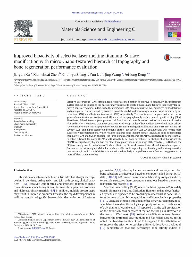
SLM solutions GmbH, Germany) using commercial grade II pure titani-um powders (average 30 μm particle size) as raw materials under99.999 wt% argon to prevent oxidation. A fibre laser and a rectanglescanning strategy were used. The SLM processing parameters were ad-justed to a laser power of 100 W, laser scanning velocity of 275 mm/s,hatch space of 130 μm, layer thickness of 30 μm, laser spot size of83 μm and substrate heat of 140 °C. Specimens for the cell cultureassay were produced as titanium discs (10 × 10 × 1 mm3) (Fig. 1.a). Atitaniumdomewas designed for a rabbitmodel and proved to be simpleand effective in our previous study [31]; in this study, we used a similarspecimen design for the animal experiment (Fig. 1.b, c, d and e). Howev-er, some alterations were made according to this experiment. The po-rous scaffolds were added inside the domes. One of the reasons is thatSLM is usually used for custom-made geometries, especially porousstructures, and the porous scaffolds mimicked the architectures in clin-ical application. The other reason is that the increased superficial area ofporous scaffoldsmake it easier to detect the BIC% difference. Because theinfluence of the forms of the porous structures was not the focus of thisstudy, we used a simple cubic unit design and set the strut size to0.2 mm and the pore size to 0.6 mm, which has been proved to beeffective for bone growth [19,36–38]. After fabrication, the SLM speci-mens were cleaned by compressed air and ultrasonic treatment.
2.2. Surface treatment
Untreated SLM titanium discs and domes were named native-SLMand set as the control group. The other specimenswere first sandblastedwith 250 μm ZrO2 particles and then immersed in an acidic mixtureconsisting of 40% HF and 60% HNO3 (H2O/HF/HNO3, 1:4:5, v:v) for2 min to remove the oxide layer and polish the surface of the SLM tita-nium. For the SLA group, the polished specimens were acid etched in amixture of 98% H2SO4 and 36.5% HCl solution (H2O/HCl/H2SO4, 2:4:3,v:v) at 95 °C for 15 min. Simultaneously, the polished specimens wereused as the electrode anode with a titanium rod as a cathode andwere anodized in a 0.5wt%HF in distilledwater solution acid electrolyteusing a 20 V power supply for 45 min; this group was named the SANgroup. For the SAH group, the sandblasted specimens were well stirredin 5 M NaOH solution at a temperature of 80 °C for 8 h. Afterward, alltreated specimens were thoroughly cleaned ultrasonically in deionizedwater and autoclave sterilized before cell culture studies and animal ex-periments. The specimens were sealed and stored in air in dark place toavoid possible ultraviolet radiation.
2.3. Surface characterization
The surface topography and chemistry were analyzed by field-emis-sion scanning electron microscopy (FE-SEM, Hitachi S-4800, Japan)equipped with an X-ray energy dispersive spectrometry (EDS) and anX-ray photoelectron spectroscopy (XPS, Thermo Scientific ESCALAB250Xi, USA). Surface roughness, including the average height abovethe centreline (Ra) and the rootmean square of Ra (Rq),weremeasuredby a contact profilometer (SJ-210, Japan). Water contact angles weredetermined by the sessile-drop method on an OCA20 drop shape anal-ysis system (DSA100, Germany).
2.4. Cell culture assay
The osteoblast-like cell line MC3T3-E1, Subclone 14, which was ob-tained from the Shanghai Cellular Institute of China Scientific Academy,was used in this study. MC3T3-E1 cells were cultured at 37 °C and 5%CO2 in an α-MEM medium (Gibco, USA) containing 10% foetal bovineserum (FBS, Gibco, USA) and 1% antibiotic/antimycotic solution(Gibco, USA).
Fig. 1.Twodifferent shapes of SLM titanium specimens: the actual sample (a) of thenative SLM titaniumdisc specimens (diameter: 10mm; thickness: 1mm); the actual samples (b, c) anddesign sketches (d, e) of SLM titanium domes (internal diameter: 5mm; outer diameter: 6mm; vertical height: 5mm)with porous scaffolds (strut size: 0.2mm; pore size: 0.6mm) insideand threads (vertical height:1.75 mm; thread depth: 0.3 mm) on the root, as well as four rectangular holes (length of side: 0.7 mm) on the top.
231J. Xu et al. / Materials Science and Engineering C 68 (2016) 229–240
2.5. Cell viability evaluation
For the cell morphology assay, MC3T3-E1 cells were incubated withthe specimen surfaces in 48-well plates at a density of 1 × 10⁴ cellsml−1
for 2 h and fixed in 2.5% glutaraldehyde solution for 6 h. After gradientdehydration in ethanol solutions (50%, 75%, 80%, 90% and 100%) anddrying, the specimens were observed under FE-SEM.
For the cell proliferation assay, MC3T3-E1 cells were seeded ontospecimens in 48-well plates at a density of 1 × 10⁴ cells ml−1 and cul-tured with an α-MEM medium containing 10% FBS for 1, 3, 5 and7 days. At the prescribed time points, the cells were assessed using a
cell counting kit-8 assay (CCK-8, Dojindo, Japan) [31] and measured atthe optical absorbance (OD) of 450 nm.
The alkaline phosphatase (ALP) activity of MC3T3-E1 cells was nor-malized to the total protein content. Cells were seeded onto specimensin 48-well plates at a density of 2 × 10⁴ cells ml−1. After 7 and 14 daysof culturing in mineralized solution, 0.1% Triton X-100 were used to lysecells at 4 °C, and the ALP activity was determined by the p-nitrophenylphosphate (p-NPP) method using an ALP Assay Kit (Jiancheng, Nanjing,China) [31] at an absorbance of 520 nm. The total protein content wasmeasured using a bicinchoninic acid assay kit (BCA, Beyotime, China)[31] at a wavelength of 562 nm.
232 J. Xu et al. / Materials Science and Engineering C 68 (2016) 229–240
2.6. Animal experiment
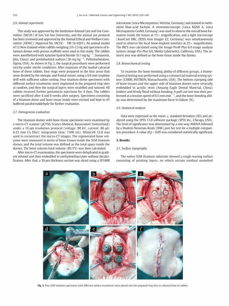
This studywas approved by the Institution Animal Care andUse Com-mittee (IACUC) of Sun Yat-Sen University, and the animal use protocolhas been reviewed and approvedby theAnimal Ethical andWelfare Com-mittee (AEWC) (Approval No. IACUC- DB-16-0201). An animal modelof 12NewZealandwhite rabbitsweighing 2.0–2.5 kg and specimens of ti-tanium domes with porous scaffolds were used in this study. The rabbitswere anesthetizedwith xylazine hydrochloride (0.1mg kg−1, Sumianxin,Jilin, China) and pentobarbital sodium (30 mg kg−1; Pelltobarbitalum,Sigma, USA). As shown in Fig. 2, the surgical procedures were performedstrictly under sterile conditions. After exposure of the medial calvarialbone of these rabbits, four rings were prepared in the four areas thatwere divided by the metopic and frontal suture, using a 6.0 mm trephinedrill with sufficient saline cooling. Four titanium dome specimens withdifferent surface treatments were implanted in the prepared ring sitesat random, and then the surgical layers were stratified and sutured. Allrabbits received further gentamicin injections for 4 days. The rabbitswere sacrificed after 4 and 8 weeks after surgery. Specimens consistingof a titanium dome and bone tissue inside were excised and kept in 4%buffered paraformaldehyde for further evaluation.
2.7. Osteogenesis evaluation
The titanium domes with bone tissue specimens were examined bya micro-CT scanner (μCT50, Scanco Medical, Bassersdorf, Switzerland)under a 10 μm resolution protocol (voltage: 90 kV; current: 88 μA;0.25 mm Cu filter; integration time: 1500 ms). Mimics® 15.0 wasused to reconstruct the micro-CT images. The regenerated bone vol-umes were measured in terms of bone tissues inside the SLM titaniumdomes, and the total volume was defined as the total space inside thedomes. The bone volume/total volume (BV/TV) was then calculated.
Aftermicro-CT examination, the specimenswere dehydrated in gradi-ent ethanol and then embedded in methylmethacrylate without decalci-fication. After that, a 30 μm thickness section was sliced using a SP1600
Fig. 2. Four SLM titanium specimens with different surface treatments w
microtome (LeicaMicrosystems,Wetzlar, Germany) and stained inmeth-ylene blue-acid fuchsin. A stereomicroscope (Leica M205 A, LeicaMicrosystemsGmbH,Germany)wasused to observe the overall bone for-mation inside the tomes at 15× magnification, and a light microscope(AxioCam HRc, ZEISS Axio Imager Z2, Germany) was simultaneouslyused to observe the local bone-implant interface at 25× magnification.The BIC% was calculated using the Image-Pro® Plus 6.0 image analysissystem (Image-Pro Plus 6.0, Media Cybernetics, California, USA). The in-terest area was defined as the bone tissue inside the domes.
2.8. Biomechanical testing
To examine the bone-bonding ability of different groups, a biome-chanical testingwas performedusing a commercialmaterial testing sys-tem (E3000, INSTRON, Massachusetts, USA). The bottom clamping sideof bone tissues and the upper side of titanium domes were severallyembedded in acrylic resin (Anyang Eagle Dental Material, China)holders and firmly fixed without bending. A pull-out test was then per-formed at a tension speed of 0.5mmmin−1, and the bone-bonding abil-ity was determined by the maximum force to failure (N).
2.9. Statistical analysis
Data were expressed as the mean ± standard deviation (SD) and an-alyzed using the SPSS 13.0 software package (SPSS Inc., Chicago, USA).The level of significance was determined by a one-way ANOVA followedby a Student-Newman-Keuls (SNK) post hoc test for a multiple compari-son procedure. A value of p b 0.05 was considered statistically significant.
3. Results
3.1. Surface topography
The native SLM titanium substrate showed a rough waving surfaceconsisting of printing layers, on which certain residual unmelted
ere placed into the prepared ring sites in calvarial bone of rabbits.
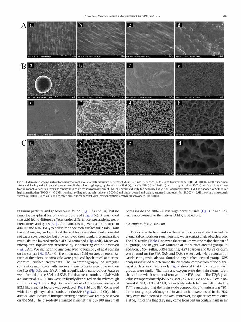
Fig. 3. SEM images showing surface topography of each group: A: natural surface of native-SLM (a, 35×), natural surface (b, 35×) and topography (c, 100×; d, 30,000×) of the specimenafter sandblasting and acid polishing treatment; B: the microrough topographies of native-SLM (a), SLA (b), SAN (c) and SAH (d) at low magnification (5000×); surface without nanofeatures of native-SLM (e), irregular concavities and ridges microtopography of SLA (f), uniformly distributed nanotubes of SAN (g) and hierarchical ECM-like nanonets of SAH (h) athigh magnification (30,000×); C: SAN showing a rolling microrough surface (a, 5000×) and single-layered and orderly arranged nanotubes (b, 120,000×); SAH showing a microroughsurface (c, 10,000×) and an ECM-like three-dimensional nanonet with interpenetrating hierarchical network (d, 100,000×).
233J. Xu et al. / Materials Science and Engineering C 68 (2016) 229–240
titanium particles and spheres were found (Fig. 3.Aa and Ba), but nonano topographical features were observed (Fig. 3.Be). It was notedthat acid led to different effects under different concentrations, treat-ment times and types [39]. After sandblasting, we used a mixture of40% HF and 60% HNO3 to polish the specimen surface for 2 min. Fromthe SEM images, we found that the acid treatment described above didnot cause severe erosion but only removed the irregularities and particleresiduals; the layered surface of SLM remained (Fig. 3.Ab). Moreover,micropitted topography produced by sandblasting can be observed(Fig. 3.Ac). We did not find any concaved topography of acid etchingon the surface (Fig. 3.Ad). On the microrough SLM surface, different fea-tures at the micro- or nanoscale were produced by chemical or electro-chemical surface treatments. The microtopography of irregularconcavities and ridges with macro and micro peaks were engraved onthe SLA (Fig. 3.Bb and Bf). At high magnification, nano-porous featureswere formed on the SAN and SAH. The titanate nanotubes of SAN witha diameter of 50–100 nmwere uniformly distributed on themicroroughsubstrate (Fig. 3.Bc and Bg). On the surface of SAH, a three-dimensionalECM-like nanonet feature was produced (Fig. 3.Bd and Bh). Comparedwith the single-layered nanotubes on the SAN (Fig. 3.Ca and Cb), a hier-archical architecture of interpenetrating nanonet was readily observedon the SAH. The disorderly arranged nanonet has 50–100 nm small
pores inside and 300–500 nm large pores outside (Fig. 3.Cc and Cd),more approximate to the natural ECM grid structure.
3.2. Surface characterization
To examine the basic surface characteristics, we evaluated the surfaceelemental composition, roughness andwater contact angle of each group.The EDS results (Table 1) showed that titaniumwas themajor element ofall groups, and oxygen was found on all the surface-treated groups. Inaddition, 0.95% sulfur, 6.39% fluorine, 4.29% carbon and 0.49% calciumwere found on the SLA, SAN and SAH, respectively. No zirconium ofsandblasting residuals was found on any surface-treated groups. XPSanalysis was used to determine the elemental composition of the outer-most surface more accurately. Fig. 4 showed that the curves of eachgroups were similar. Titanium and oxygen were the main elements onthe surface, which was consistent with the EDS results. The Ti2p3 peakvaluewas approximately 458.5 eV, 459.2 eV, 458.5 eV, and460.3 eV in na-tive-SLM, SLA, SAN and SAH, respectively, which has been attributed toTi4+, suggesting that the main oxide compounds of titanium was TiO2
in the four groups. Although sulfur and calcium were tested in the EDS,they were not detected in the XPS; moreover, the quantities were quitea little, indicating that they may come from certain contaminant in air
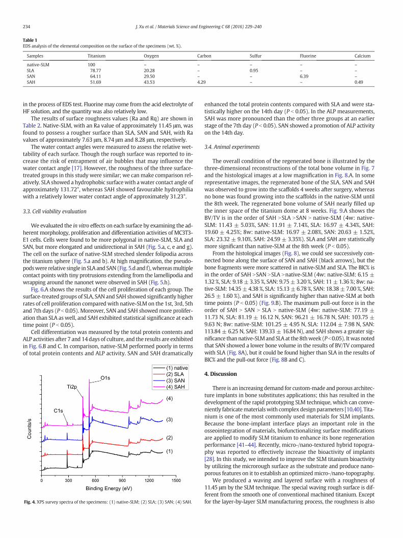
Table 1EDS analysis of the elemental composition on the surface of the specimens (wt. %).
Samples Titanium Oxygen Carbon Sulfur Fluorine Calcium
native-SLM 100 – – – – –SLA 78.77 20.28 – 0.95 – –SAN 64.11 29.50 – – 6.39 –SAH 51.69 43.53 4.29 – – 0.49
234 J. Xu et al. / Materials Science and Engineering C 68 (2016) 229–240
in the process of EDS test. Fluorine may come from the acid electrolyte ofHF solution, and the quantity was also relatively low.
The results of surface roughness values (Ra and Rq) are shown inTable 2. Native-SLM, with an Ra value of approximately 11.45 μm, wasfound to possess a rougher surface than SLA, SAN and SAH, with Ravalues of approximately 7.63 μm, 8.74 μm and 8.28 μm, respectively.
The water contact angles were measured to assess the relative wet-tability of each surface. Though the rough surface was reported to in-crease the risk of entrapment of air bubbles that may influence thewater contact angle [17]. However, the roughness of the three surface-treated groups in this study were similar; we canmake comparison rel-atively. SLA showed a hydrophobic surfacewith awater contact angle ofapproximately 131.72°, whereas SAH showed favourable hydrophiliawith a relatively lower water contact angle of approximately 31.23°.
3.3. Cell viability evaluation
We evaluated the in vitro effects on each surface by examining the ad-herent morphology, proliferation and differentiation activities of MC3T3-E1 cells. Cells were found to be more polygonal in native-SLM, SLA andSAN, but more elongated and unidirectional in SAH (Fig. 5.a, c, e and g).The cell on the surface of native-SLM streched slender folipodia acrossthe titanium sphere (Fig. 5.a and b). At high magnification, the pseudo-podswere relative single in SLAand SAN(Fig. 5.d and f),whereasmultiplecontact points with tiny protrusions extending from the lamellipodia andwrapping around the nanonet were observed in SAH (Fig. 5.h).
Fig. 6.A shows the results of the cell proliferation of each group. Thesurface-treated groups of SLA, SAN and SAH showed significantly higherrates of cell proliferation compared with native-SLM on the 1st, 3rd, 5thand 7th days (P b 0.05). Moreover, SAN and SAH showedmore prolifer-ation than SLA as well, and SAH exhibited statistical significance at eachtime point (P b 0.05).
Cell differentiation was measured by the total protein contents andALP activities after 7 and 14days of culture, and the results are exhibitedin Fig. 6.B and C. In comparison, native-SLM performed poorly in termsof total protein contents and ALP activity. SAN and SAH dramatically
Fig. 4. XPS survey spectra of the specimens: (1) native-SLM; (2) SLA; (3) SAN; (4) SAH.
enhanced the total protein contents compared with SLA and were sta-tistically higher on the 14th day (P b 0.05). In the ALP measurements,SAH was more pronounced than the other three groups at an earlierstage of the 7th day (P b 0.05). SAN showed a promotion of ALP activityon the 14th day.
3.4. Animal experiments
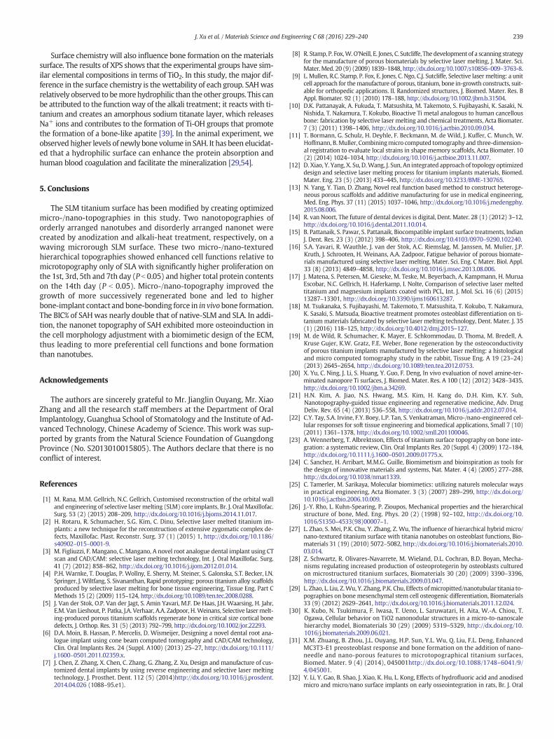
The overall condition of the regenerated bone is illustrated by thethree-dimensional reconstructions of the total bone volume in Fig. 7and the histological images at a low magnification in Fig. 8.A. In somerepresentative images, the regenerated bone of the SLA, SAN and SAHwas observed to grow into the scaffolds 4 weeks after surgery, whereasno bone was found growing into the scaffolds in the native-SLM untilthe 8th week. The regenerated bone volume of SAH nearly filled upthe inner space of the titanium dome at 8 weeks. Fig. 9.A shows theBV/TV is in the order of SAH NSLA NSAN N native-SLM (4w: native-SLM: 11.43 ± 5.03%, SAN: 11.91 ± 7.14%, SLA: 16.97 ± 4.34%, SAH:19.60 ± 4.25%; 8w: native-SLM: 16.97 ± 2.08%, SAN: 20.63 ± 1.52%,SLA: 23.32 ± 9.10%, SAH: 24.59 ± 3.35%). SLA and SAH are statisticallymore significant than native-SLM at the 8th week (P b 0.05).
From the histological images (Fig. 8), we could see successively con-nected bone along the surface of SAN and SAH (black arrows), but thebone fragments were more scattered in native-SLM and SLA. The BIC% isin the order of SAH NSAN NSLA Nnative-SLM (4w: native-SLM: 6.15 ±1.32 %, SLA: 9.18±3.35 %, SAN: 9.75± 3.20 %, SAH: 11±1.36 %; 8w: na-tive-SLM: 14.35± 4.38 %, SLA: 15.13±6.78 %, SAN: 18.38± 7.00 %, SAH:26.5 ± 1.60 %), and SAH is significantly higher than native-SLM at bothtime points (P b 0.05) (Fig. 9.B). The maximum pull-out force is in theorder of SAH N SAN N SLA N native-SLM (4w: native-SLM: 77.19 ±11.73 N, SLA: 81.19 ± 16.12 N, SAN: 96.21 ± 16.78 N, SAH: 103.75 ±9.63 N; 8w: native-SLM: 101.25 ± 4.95 N, SLA: 112.04 ± 7.98 N, SAN:113.84 ± 6.25 N, SAH: 139.33 ± 16.84 N), and SAH shows a greater sig-nificance thannative-SLMand SLAat the 8thweek (Pb0.05). Itwasnotedthat SAN showed a lower bone volume in the results of BV/TV comparedwith SLA (Fig. 8A), but it could be found higher than SLA in the results ofBIC% and the pull-out force (Fig. 8B and C).
4. Discussion
There is an increasing demand for custom-made andporous architec-ture implants in bone substitutes applications; this has resulted in thedevelopment of the rapid prototyping SLM technique, which can conve-niently fabricatematerialswith complex design parameters [10,40]. Tita-nium is one of the most commonly used materials for SLM implants.Because the bone-implant interface plays an important role in theosseointegration of materials, biofunctionalizing surface modificationsare applied to modify SLM titanium to enhance its bone regenerationperformance [41–44]. Recently, micro-/nano-textured hybrid topogra-phy was reported to effectively increase the bioactivity of implants[28]. In this study, we intended to improve the SLM titanium bioactivityby utilizing the microrough surface as the substrate and produce nano-porous features on it to establish an optimizedmicro-/nano-topography.
We produced a waving and layered surface with a roughness of11.45 μm by the SLM technique. The special waving rough surface is dif-ferent from the smooth one of conventional machined titanium. Exceptfor the layer-by-layer SLM manufacturing process, the roughness is also
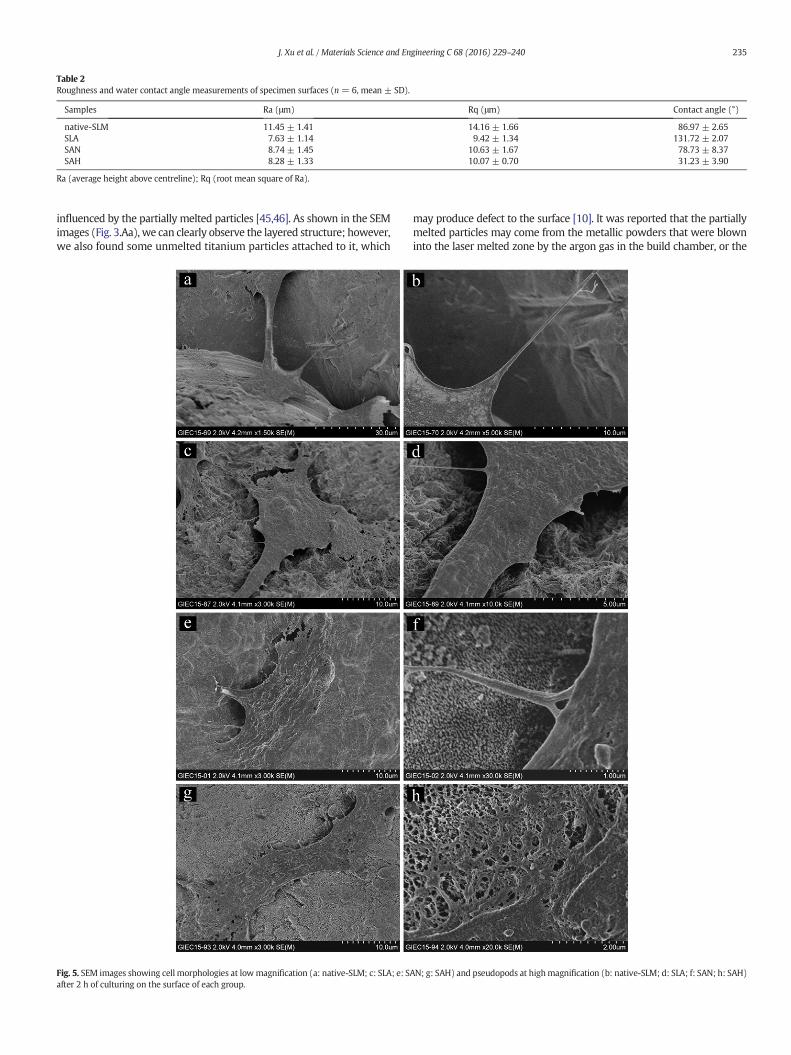
Table 2Roughness and water contact angle measurements of specimen surfaces (n = 6, mean ± SD).
Samples Ra (μm) Rq (μm) Contact angle (°)
native-SLM 11.45 ± 1.41 14.16 ± 1.66 86.97 ± 2.65SLA 7.63 ± 1.14 9.42 ± 1.34 131.72 ± 2.07SAN 8.74 ± 1.45 10.63 ± 1.67 78.73 ± 8.37SAH 8.28 ± 1.33 10.07 ± 0.70 31.23 ± 3.90
Ra (average height above centreline); Rq (root mean square of Ra).
235J. Xu et al. / Materials Science and Engineering C 68 (2016) 229–240
influenced by the partially melted particles [45,46]. As shown in the SEMimages (Fig. 3.Aa), we can clearly observe the layered structure; however,we also found some unmelted titanium particles attached to it, which
Fig. 5. SEM images showing cell morphologies at lowmagnification (a: native-SLM; c: SLA; e: Safter 2 h of culturing on the surface of each group.
may produce defect to the surface [10]. It was reported that the partiallymelted particles may come from the metallic powders that were blowninto the laser melted zone by the argon gas in the build chamber, or the
AN; g: SAH) and pseudopods at highmagnification (b: native-SLM; d: SLA; f: SAN; h: SAH)
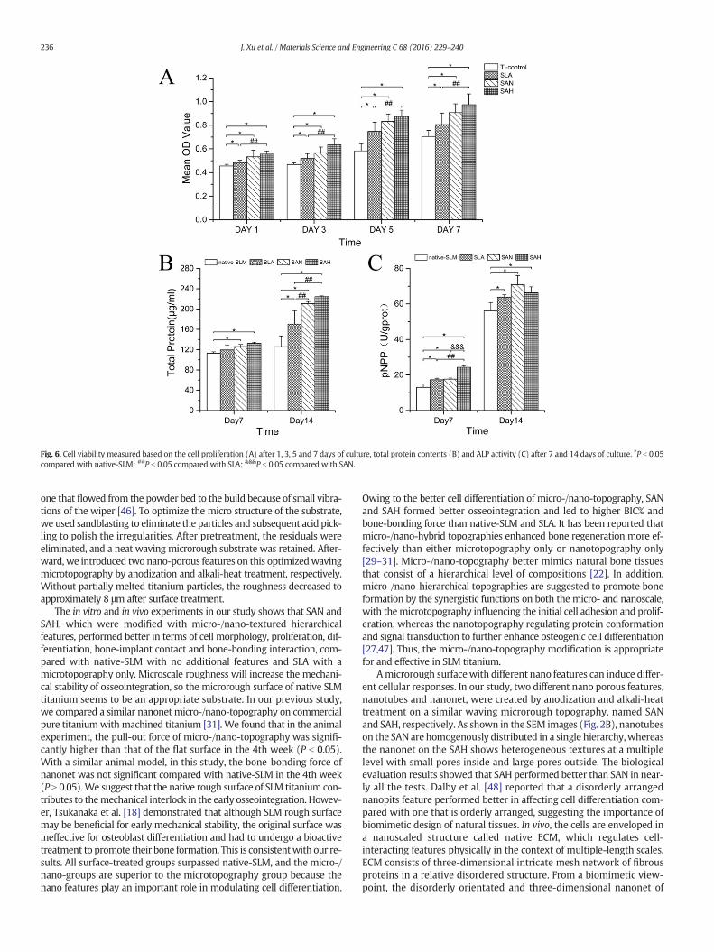
Fig. 6. Cell viability measured based on the cell proliferation (A) after 1, 3, 5 and 7 days of culture, total protein contents (B) and ALP activity (C) after 7 and 14 days of culture. ⁎P b 0.05compared with native-SLM; ##P b 0.05 compared with SLA; &&&P b 0.05 compared with SAN.
236 J. Xu et al. / Materials Science and Engineering C 68 (2016) 229–240
one that flowed from the powder bed to the build because of small vibra-tions of the wiper [46]. To optimize the micro structure of the substrate,we used sandblasting to eliminate the particles and subsequent acid pick-ling to polish the irregularities. After pretreatment, the residuals wereeliminated, and a neat waving microrough substrate was retained. After-ward, we introduced two nano-porous features on this optimizedwavingmicrotopography by anodization and alkali-heat treatment, respectively.Without partially melted titanium particles, the roughness decreased toapproximately 8 μm after surface treatment.
The in vitro and in vivo experiments in our study shows that SAN andSAH, which were modified with micro-/nano-textured hierarchicalfeatures, performed better in terms of cell morphology, proliferation, dif-ferentiation, bone-implant contact and bone-bonding interaction, com-pared with native-SLM with no additional features and SLA with amicrotopography only. Microscale roughness will increase the mechani-cal stability of osseointegration, so the microrough surface of native SLMtitanium seems to be an appropriate substrate. In our previous study,we compared a similar nanonet micro-/nano-topography on commercialpure titaniumwith machined titanium [31]. We found that in the animalexperiment, the pull-out force of micro-/nano-topography was signifi-cantly higher than that of the flat surface in the 4th week (P b 0.05).With a similar animal model, in this study, the bone-bonding force ofnanonet was not significant compared with native-SLM in the 4th week(P N 0.05).We suggest that the native rough surface of SLM titanium con-tributes to themechanical interlock in the early osseointegration. Howev-er, Tsukanaka et al. [18] demonstrated that although SLM rough surfacemay be beneficial for early mechanical stability, the original surface wasineffective for osteoblast differentiation and had to undergo a bioactivetreatment to promote their bone formation. This is consistentwith our re-sults. All surface-treated groups surpassed native-SLM, and the micro-/nano-groups are superior to the microtopography group because thenano features play an important role in modulating cell differentiation.
Owing to the better cell differentiation of micro-/nano-topography, SANand SAH formed better osseointegration and led to higher BIC% andbone-bonding force than native-SLM and SLA. It has been reported thatmicro-/nano-hybrid topographies enhanced bone regeneration more ef-fectively than either microtopography only or nanotopography only[29–31]. Micro-/nano-topography better mimics natural bone tissuesthat consist of a hierarchical level of compositions [22]. In addition,micro-/nano-hierarchical topographies are suggested to promote boneformation by the synergistic functions on both the micro- and nanoscale,with themicrotopography influencing the initial cell adhesion and prolif-eration, whereas the nanotopography regulating protein conformationand signal transduction to further enhance osteogenic cell differentiation[27,47]. Thus, the micro-/nano-topography modification is appropriatefor and effective in SLM titanium.
Amicrorough surfacewith different nano features can induce differ-ent cellular responses. In our study, two different nano porous features,nanotubes and nanonet, were created by anodization and alkali-heattreatment on a similar waving microrough topography, named SANand SAH, respectively. As shown in the SEM images (Fig. 2B), nanotubeson the SAN are homogenously distributed in a single hierarchy,whereasthe nanonet on the SAH shows heterogeneous textures at a multiplelevel with small pores inside and large pores outside. The biologicalevaluation results showed that SAH performed better than SAN in near-ly all the tests. Dalby et al. [48] reported that a disorderly arrangednanopits feature performed better in affecting cell differentiation com-pared with one that is orderly arranged, suggesting the importance ofbiomimetic design of natural tissues. In vivo, the cells are enveloped ina nanoscaled structure called native ECM, which regulates cell-interacting features physically in the context of multiple-length scales.ECM consists of three-dimensional intricate mesh network of fibrousproteins in a relative disordered structure. From a biomimetic view-point, the disorderly orientated and three-dimensional nanonet of
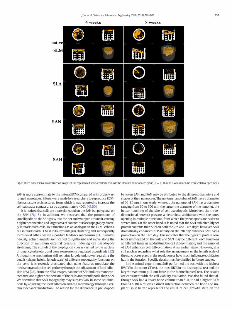
Fig. 7. Three-dimensional reconstruction images of the regenerated bone architecture inside the titanium dome of each group (n=3) at 4 and 8weeks in some representative specimens.
237J. Xu et al. / Materials Science and Engineering C 68 (2016) 229–240
SAH ismore approximate to the natural ECM comparedwith orderly ar-ranged nanotubes. Efforts weremade by researchers to reproduce ECM-like nanoscale architectures, fromwhich it was reported to increase thecell-substrate contact area by approximately 400% [49,50].
It is noticed that cells aremore elongated on the SAH but polygonal onthe SAN (Fig. 5). In addition, we observed that the protrusions oflamellipodia on the SAHgrew into the net andwrapped around it, causinga tighter connection and larger area of contact. Surface topography direct-ly interacts with cells, so it functions as an analogue to the ECM. When acell interacts with ECM, it initializes integrin clustering and subsequentlyforms focal adhesions via a positive feedback mechanism [51]. Simulta-neously, actin filaments are inclined to synthesize and move along thedirection of minimum external pressure, inducing cell pseudopodsstretching. The stimuli of the biophysical cues is carried to the nucleusthrough cytoskeleton, and gene expression is regulated accordingly [52].Although the mechanism still remains largely unknown regarding thedetails (shape, height, length scale) of different topography functions onthe cells, it is recently elucidated that nano features modulate themechanotransduction cell pathway through the adjustment of focal adhe-sion (FA) [22]. From the SEM images, nanonet of SAH induces more con-tact area and tighter connection of the cells and pseudopods than SAN.We speculate that SAH topography may surpass SAN in some cell func-tions by adjusting the focal adhesion and cell morphology through a cer-tain mechanotransduction. The reason for the difference in pseudopods
between SAH and SAN may be attributed to the different diameters andshapes of their nanopores. Theuniformnanotubes of SANhave adiameterof 50–80 nm in our study, whereas the nanonet of SAH has a diameterranging from 50 to 500 nm; the larger the diameter of the nanonet, thebetter matching of the size of cell pseudopods. Moreover, the three-dimensional network presents a hierarchical architecture with the poresopening in multiple directions, from which the pseudopods are easier tostretch into. On the other hand, it is noted that the SAH exhibited higherprotein contents than SAN on both the 7th and 14th days; however, SAHdramatically enhanced ALP activity on the 7th day, whereas SAN had apromotion on the 14th day. This indicates that the types of protein con-tents synthesized on the SAH and SAN may be different; each functionsat different times in modulating the cell differentiation, and the nanonetof SAH enhances cell differentiation at an earlier stage. However, it isstill unclear regarding what role the arrangement or the length scale ofthe nano pores plays in the regulation or howmuch influence each factorhas in the function. Specific details must be clarified in future studies.
In the animal experiment, SAH performed the best with the highestBV/TV in themicro-CT test, themost BIC% in the histological test and thelargest maximum pull-out force in the biomechanical test. The resultsare consistent with the cell viability evaluation. We also found that al-though SAN had a lower bone volume than SLA, it had a higher BIC%than SLA. BIC% reflects a direct interaction between the bone and im-plant, so it better represents the result of cell growth state on the
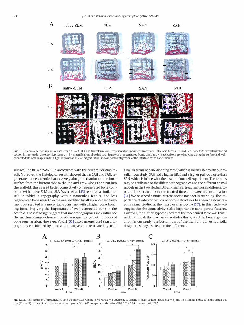
Fig. 8. Histological section images of each group (n = 3) at 4 and 8 weeks in some representative specimens (methylene blue-acid fuchsin stained; red: bone): A: overall histologicalsection images under a stereomicroscope at 15× magnification, showing total ingrowth of regenerated bone, black arrow: successively growing bone along the surface and well-connected; B: local images under a light microscope at 25× magnification, showing osseointegration at the interface of the bone-implant.
238 J. Xu et al. / Materials Science and Engineering C 68 (2016) 229–240
surface. The BIC% of SAN is in accordance with the cell proliferation re-sult. Moreover, the histological results showed that in SAH and SAN, re-generated bone extended successively along the titanium dome innersurface from the bottom side to the top and grew along the strut intothe scaffold; this caused better connectivity of regenerated bone com-pared with native-SLM and SLA. Yavari et al. [53] reported a similar re-sult in which a topography with a nanotubes feature had lessregenerated bonemass than the onemodified by alkali-acid-heat treat-ment but resulted in a more stable construct with a higher bone-bond-ing force, implying the importance of well-connected bone in thescaffold. These findings suggest that nanotopographies may influencethe mechanotransduction and guide a sequential growth process ofbone regeneration. However, Yavari [53] also demonstrated that a to-pography established by anodization surpassed one treated by acid-
Fig. 9. Statistical results of the regenerated bone volume/total volume (BV/TV; A; n=3), percentest (C; n = 3) in the animal experiment of each group. ⁎P b 0.05 compared with native-SLM;
alkali in terms of bone-bonding force, which is inconsistentwith our re-sult. In our study, SAHhad a higher BIC% and a higher pull-out force thanSAN,which is in linewith the results of our cell experiment. The reasonsmay be attributed to the different topographies and the different animalmodels in the two studies. Alkali chemical treatment forms different to-pographies according to the treated time and reagent concentration[31].We observed amore interconnected nanonet in our study. The im-portance of interconnection of porous structures has been demonstrat-ed in many studies at the micro or macroscale [37]; in this study, wesuggest that the connectivity is also important in nano-porous features.However, the author hypothesized that themechanical forcewas trans-mitted through the macroscale scaffolds that guided the bone regener-ation. In our study, the bottom part of the titanium domes is a soliddesign; this may also lead to the difference.
tage of bone-implant contact (BIC%; B; n=6) and themaximum force to failure of pull-out##P b 0.05 compared with SLA.
239J. Xu et al. / Materials Science and Engineering C 68 (2016) 229–240
Surface chemistry will also influence bone formation on the materialssurface. The results of XPS shows that the experimental groups have sim-ilar elemental compositions in terms of TiO2. In this study, the major dif-ference in the surface chemistry is the wettability of each group. SAHwasrelatively observed to bemore hydrophilic than the other groups. This canbe attributed to the functionway of the alkali treatment; it reacts with ti-tanium and creates an amorphous sodium titanate layer, which releasesNa+ ions and contributes to the formation of Ti-OH groups that promotethe formation of a bone-like apatite [39]. In the animal experiment, weobserved higher levels of newly bone volume in SAH. It has been elucidat-ed that a hydrophilic surface can enhance the protein absorption andhuman blood coagulation and facilitate the mineralization [29,54].
5. Conclusions
The SLM titanium surface has been modified by creating optimizedmicro-/nano-topographies in this study. Two nanotopographies oforderly arranged nanotubes and disorderly arranged nanonet werecreated by anodization and alkali-heat treatment, respectively, on awaving microrough SLM surface. These two micro-/nano-texturedhierarchical topographies showed enhanced cell functions relative tomicrotopography only of SLA with significantly higher proliferation onthe 1st, 3rd, 5th and 7th day (P b 0.05) and higher total protein contentson the 14th day (P b 0.05). Micro-/nano-topography improved thegrowth of more successively regenerated bone and led to higherbone-implant contact and bone-bonding force in in vivobone formation.The BIC% of SAHwas nearly double that of native-SLM and SLA. In addi-tion, the nanonet topography of SAH exhibited more osteoinduction inthe cell morphology adjustment with a biomimetic design of the ECM,thus leading to more preferential cell functions and bone formationthan nanotubes.
Acknowledgements
The authors are sincerely grateful to Mr. Jianglin Ouyang, Mr. XiaoZhang and all the research staff members at the Department of OralImplantology, Guanghua School of Stomatology and the Institute of Ad-vanced Technology, Chinese Academy of Science. This work was sup-ported by grants from the Natural Science Foundation of GuangdongProvince (No. S2013010015805). The Authors declare that there is noconflict of interest.
References
[1] M. Rana, M.M. Gellrich, N.C. Gellrich, Customised reconstruction of the orbital walland engineering of selective laser melting (SLM) core implants, Br. J. Oral Maxillofac.Surg. 53 (2) (2015) 208–209, http://dx.doi.org/10.1016/j.bjoms.2014.11.017.
[2] H. Rotaru, R. Schumacher, S.G. Kim, C. Dinu, Selective laser melted titanium im-plants: a new technique for the reconstruction of extensive zygomatic complex de-fects, Maxillofac. Plast. Reconstr. Surg. 37 (1) (2015) 1, http://dx.doi.org/10.1186/s40902–015–0001-9.
[3] M. Figliuzzi, F. Mangano, C. Mangano, A novel root analogue dental implant using CTscan and CAD/CAM: selective laser melting technology, Int. J. Oral Maxillofac. Surg.41 (7) (2012) 858–862, http://dx.doi.org/10.1016/j.ijom.2012.01.014.
[4] P.H. Warnke, T. Douglas, P. Wollny, E. Sherry, M. Steiner, S. Galonska, S.T. Becker, I.N.Springer, J.Wiltfang, S. Sivananthan, Rapid prototyping: porous titanium alloy scaffoldsproduced by selective laser melting for bone tissue engineering, Tissue Eng. Part CMethods 15 (2) (2009) 115–124, http://dx.doi.org/10.1089/ten.tec.2008.0288.
[5] J. Van der Stok, O.P. Van der Jagt, S. Amin Yavari, M.F. De Haas, J.H. Waarsing, H. Jahr,E.M. Van Lieshout, P. Patka, J.A. Verhaar, A.A. Zadpoor, H.Weinans, Selective lasermelt-ing-produced porous titanium scaffolds regenerate bone in critical size cortical bonedefects, J. Orthop. Res. 31 (5) (2013) 792–799, http://dx.doi.org/10.1002/jor.22293.
[6] D.A. Moin, B. Hassan, P. Mercelis, D. Wismeijer, Designing a novel dental root ana-logue implant using cone beam computed tomography and CAD/CAM technology,Clin. Oral Implants Res. 24 (Suppl. A100) (2013) 25–27, http://dx.doi.org/10.1111/j.1600–0501.2011.02359.x.
[7] J. Chen, Z. Zhang, X. Chen, C. Zhang, G. Zhang, Z. Xu, Design and manufacture of cus-tomized dental implants by using reverse engineering and selective laser meltingtechnology, J. Prosthet. Dent. 112 (5) (2014)http://dx.doi.org/10.1016/j.prosdent.2014.04.026 (1088–95.e1).
[8] R. Stamp, P. Fox,W.O'Neill, E. Jones, C. Sutcliffe, The development of a scanning strategyfor the manufacture of porous biomaterials by selective laser melting, J. Mater. Sci.Mater.Med. 20 (9) (2009) 1839–1848, http://dx.doi.org/10.1007/s10856–009–3763-8.
[9] L. Mullen, R.C. Stamp, P. Fox, E. Jones, C. Ngo, C.J. Sutcliffe, Selective laser melting: a unitcell approach for themanufacture of porous, titanium, bone in-growth constructs, suit-able for orthopedic applications. II. Randomized structures, J. Biomed. Mater. Res. BAppl. Biomater. 92 (1) (2010) 178–188, http://dx.doi.org/10.1002/jbm.b.31504.
[10] D.K. Pattanayak, A. Fukuda, T. Matsushita, M. Takemoto, S. Fujibayashi, K. Sasaki, N.Nishida, T. Nakamura, T. Kokubo, Bioactive Ti metal analogous to human cancellousbone: fabrication by selective laser melting and chemical treatments, Acta Biomater.7 (3) (2011) 1398–1406, http://dx.doi.org/10.1016/j.actbio.2010.09.034.
[11] T. Bormann, G. Schulz, H. Deyhle, F. Beckmann, M. de Wild, J. Kuffer, C. Munch, W.Hoffmann, B.Muller, Combiningmicro computed tomography and three-dimension-al registration to evaluate local strains in shape memory scaffolds, Acta Biomater. 10(2) (2014) 1024–1034, http://dx.doi.org/10.1016/j.actbioe.2013.11.007.
[12] D. Xiao, Y. Yang, X. Su, D.Wang, J. Sun, An integrated approach of topology optimizeddesign and selective laser melting process for titanium implants materials, Biomed.Mater. Eng. 23 (5) (2013) 433–445, http://dx.doi.org/10.3233/BME-130765.
[13] N. Yang, Y. Tian, D. Zhang, Novel real function based method to construct heteroge-neous porous scaffolds and additive manufacturing for use in medical engineering,Med. Eng. Phys. 37 (11) (2015) 1037–1046, http://dx.doi.org/10.1016/j.medengphy.2015.08.006.
[14] R. van Noort, The future of dental devices is digital, Dent. Mater. 28 (1) (2012) 3–12,http://dx.doi.org/10.1016/j.dental.2011.10.014.
[15] B. Pattanaik, S. Pawar, S. Pattanaik, Biocompatible implant surface treatments, IndianJ. Dent. Res. 23 (3) (2012) 398–406, http://dx.doi.org/10.4103/0970–9290.102240.
[16] S.A. Yavari, R. Wauthle, J. van der Stok, A.C. Riemslag, M. Janssen, M. Mulier, J.P.Kruth, J. Schrooten, H. Weinans, A.A. Zadpoor, Fatigue behavior of porous biomate-rials manufactured using selective laser melting, Mater. Sci. Eng. C Mater. Biol. Appl.33 (8) (2013) 4849–4858, http://dx.doi.org/10.1016/j.msec.2013.08.006.
[17] J. Matena, S. Petersen, M. Gieseke, M. Teske, M. Beyerbach, A. Kampmann, H. MuruaEscobar, N.C. Gellrich, H. Haferkamp, I. Nolte, Comparison of selective laser meltedtitanium and magnesium implants coated with PCL, Int. J. Mol. Sci. 16 (6) (2015)13287–13301, http://dx.doi.org/10.3390/ijms160613287.
[18] M. Tsukanaka, S. Fujibayashi, M. Takemoto, T. Matsushita, T. Kokubo, T. Nakamura,K. Sasaki, S. Matsuda, Bioactive treatment promotes osteoblast differentiation on ti-taniummaterials fabricated by selective laser melting technology, Dent. Mater. J. 35(1) (2016) 118–125, http://dx.doi.org/10.4012/dmj.2015–127.
[19] M. de Wild, R. Schumacher, K. Mayer, E. Schkommodau, D. Thoma, M. Bredell, A.Kruse Gujer, K.W. Gratz, F.E. Weber, Bone regeneration by the osteoconductivityof porous titanium implants manufactured by selective laser melting: a histologicaland micro computed tomography study in the rabbit, Tissue Eng. A 19 (23–24)(2013) 2645–2654, http://dx.doi.org/10.1089/ten.tea.2012.0753.
[20] X. Yu, C. Ning, J. Li, S. Huang, Y. Guo, F. Deng, In vivo evaluation of novel amine-ter-minated nanopore Ti surfaces, J. Biomed. Mater. Res. A 100 (12) (2012) 3428–3435,http://dx.doi.org/10.1002/jbm.a.34269.
[21] H.N. Kim, A. Jiao, N.S. Hwang, M.S. Kim, H. Kang do, D.H. Kim, K.Y. Suh,Nanotopography-guided tissue engineering and regenerative medicine, Adv. DrugDeliv. Rev. 65 (4) (2013) 536–558, http://dx.doi.org/10.1016/j.addr.2012.07.014.
[22] C.Y. Tay, S.A. Irvine, F.Y. Boey, L.P. Tan, S. Venkatraman, Micro-/nano-engineered cel-lular responses for soft tissue engineering and biomedical applications, Small 7 (10)(2011) 1361–1378, http://dx.doi.org/10.1002/smll.201100046.
[23] A. Wennerberg, T. Albrektsson, Effects of titanium surface topography on bone inte-gration: a systematic review, Clin. Oral Implants Res. 20 (Suppl. 4) (2009) 172–184,http://dx.doi.org/10.1111/j.1600–0501.2009.01775.x.
[24] C. Sanchez, H. Arribart, M.M.G. Guille, Biomimetism and bioinspiration as tools forthe design of innovative materials and systems, Nat. Mater. 4 (4) (2005) 277–288,http://dx.doi.org/10.1038/nmat1339.
[25] C. Tamerler, M. Sarikaya, Molecular biomimetics: utilizing naturels molecular waysin practical engineering, Acta Biomater. 3 (3) (2007) 289–299, http://dx.doi.org/10.1016/j.actbio.2006.10.009.
[26] J.-Y. Rho, L. Kuhn-Spearing, P. Zioupos, Mechanical properties and the hierarchicalstructure of bone, Med. Eng. Phys. 20 (2) (1998) 92–102, http://dx.doi.org/10.1016/S1350–4533(98)00007–1.
[27] L. Zhao, S. Mei, P.K. Chu, Y. Zhang, Z. Wu, The influence of hierarchical hybrid micro/nano-textured titanium surface with titania nanotubes on osteoblast functions, Bio-materials 31 (19) (2010) 5072–5082, http://dx.doi.org/10.1016/j.biomaterials.2010.03.014.
[28] Z. Schwartz, R. Olivares-Navarrete, M. Wieland, D.L. Cochran, B.D. Boyan, Mecha-nisms regulating increased production of osteoprotegerin by osteoblasts culturedon microstructured titanium surfaces, Biomaterials 30 (20) (2009) 3390–3396,http://dx.doi.org/10.1016/j.biomaterials.2009.03.047.
[29] L. Zhao, L. Liu, Z.Wu, Y. Zhang, P.K. Chu, Effects ofmicropitted/nanotubular titania to-pographies on bonemesenchymal stem cell osteogenic differentiation, Biomaterials33 (9) (2012) 2629–2641, http://dx.doi.org/10.1016/j.biomaterials.2011.12.024.
[30] K. Kubo, N. Tsukimura, F. Iwasa, T. Ueno, L. Saruwatari, H. Aita, W.-A. Chiou, T.Ogawa, Cellular behavior on TiO2 nanonodular structures in a micro-to-nanoscalehierarchy model, Biomaterials 30 (29) (2009) 5319–5329, http://dx.doi.org/10.1016/j.biomaterials.2009.06.021.
[31] X.M. Zhuang, B. Zhou, J.L. Ouyang, H.P. Sun, Y.L. Wu, Q. Liu, F.L. Deng, EnhancedMC3T3-E1 preosteoblast response and bone formation on the addition of nano-needle and nano-porous features to microtopographical titanium surfaces,Biomed. Mater. 9 (4) (2014), 045001http://dx.doi.org/10.1088/1748–6041/9/4/045001.
[32] Y. Li, Y. Gao, B. Shao, J. Xiao, K. Hu, L. Kong, Effects of hydrofluoric acid and anodisedmicro and micro/nano surface implants on early osseointegration in rats, Br. J. Oral
240 J. Xu et al. / Materials Science and En
Maxillofac. Surg. 50 (8) (2012) 779–783, http://dx.doi.org/10.1016/j.bjoms.2011.12.008.
[33] L. Gao, B. Feng, J.X. Wang, X. Lu, D.L. Liu, S.X. Qu, J. Weng, Micro/nanostructural po-rous surface on titanium and bioactivity, J. Biomed. Mater. Res. Part B Appl.Biomater. 89B (2) (2009) 335–341, http://dx.doi.org/10.1002/jbm.b.31221.
[34] P.A. George, K. Quinn, J.J. Cooper-White, Hierarchical scaffolds via combined macro-and micro-phase separation, Biomaterials 31 (4) (2010) 641–647, http://dx.doi.org/10.1016/j.biomaterials.2009.09.094.
[35] S. Amin Yavari, Y.C. Chai, A.J. Bottger, R. Wauthle, J. Schrooten, H. Weinans, A.A.Zadpoor, Effects of anodizing parameters and heat treatment on nanotopographicalfeatures, bioactivity, and cell culture response of additively manufactured porous ti-tanium, Mater. Sci. Eng. C Mater. Biol. Appl. 51 (2015) 132–138, http://dx.doi.org/10.1016/j.msec.2015.02.050.
[36] D.A. Hollander, M. von Walter, T. Wirtz, R. Sellei, B. Schmidt-Rohlfing, O. Paar, H.J.Erli, Structural, mechanical and in vitro characterization of individually structuredTi-6Al-4V produced by direct laser forming, Biomaterials 27 (7) (2006) 955–963,http://dx.doi.org/10.1016/j.biomaterials.2005.07.041.
[37] B. Otsuki, M. Takemoto, S. Fujibayashi, M. Neo, T. Kokubo, T. Nakamura, Pore throatsize and connectivity determine bone and tissue ingrowth into porous implants:three-dimensional micro-CT based structural analyses of porous bioactive titaniumimplants, Biomaterials 27 (35) (2006) 5892–5900, http://dx.doi.org/10.1016/j.biomaterials.2006.08.013.
[38] L. Mullen, R.C. Stamp, W.K. Brooks, E. Jones, C.J. Sutcliffe, Selective laser melting: aregular unit cell approach for the manufacture of porous, titanium, bone in-growthconstructs, suitable for orthopedic applications, J. Biomed. Mater. Res. B Appl.Biomater. 89 (2) (2009) 325–334, http://dx.doi.org/10.1002/jbm.b.31504.
[39] S. Amin Yavari, S.M. Ahmadi, J. van der Stok, R. Wauthle, A.C. Riemslag, M. Janssen, J.Schrooten, H. Weinans, A.A. Zadpoor, Effects of bio-functionalizing surface treatmentson the mechanical behavior of open porous titanium biomaterials, J. Mech. Behav.Biomed. Mater. 36 (2014) 109–119, http://dx.doi.org/10.1016/j.jmbbm.2014.04.010.
[40] M. Lindner, S. Hoeges, W. Meiners, K. Wissenbach, R. Smeets, R. Telle, R. Poprawe, H.Fischer, Manufacturing of individual biodegradable bone substitute implants usingselective laser melting technique, J. Biomed. Mater. Res. A 97 (4) (2011) 466–471,http://dx.doi.org/10.1002/jbm.a.33058.
[41] A. Fukuda, M. Takemoto, T. Saito, S. Fujibayashi, M. Neo, D.K. Pattanayak, T.Matsushita, K. Sasaki, N. Nishida, T. Kokubo, T. Nakamura, Osteoinduction of porousTi implants with a channel structure fabricated by selective laser melting, ActaBiomater. 7 (5) (2011) 2327–2336, http://dx.doi.org/10.1016/j.actbio.2011.01.037.
[42] J.E. Biemond, G. Hannink, N. Verdonschot, P. Buma, Bone ingrowth potential of elec-tron beam and selective laser melting produced trabecular-like implant surfaceswith and without a biomimetic coating, J. Mater. Sci. Mater. Med. 24 (3) (2013)745–753, http://dx.doi.org/10.1007/s10856–012–4836-7.
[43] J. Matena, S. Petersen, M. Gieseke, A. Kampmann, M. Teske, M. Beyerbach, H. MuruaEscobar, H. Haferkamp, N.C. Gellrich, I. Nolte, SLM produced porous titanium im-plant improvements for enhanced vascularization and osteoblast seeding, Int. J.Mol. Sci. 16 (4) (2015) 7478–7492, http://dx.doi.org/10.3390/ijms16047478.
[44] J. van der Stok, D. Lozano, Y.C. Chai, S. Amin Yavari, A.P. Bastidas Coral, J.A. Verhaar,E. Gomez-Barrena, J. Schrooten, H. Jahr, A.A. Zadpoor, P. Esbrit, H. Weinans,Osteostatin-coated porous titanium can improve early bone regeneration of corticalbone defects in rats, Tissue Eng. A 21 (9–10) (2015) 1495–1506, http://dx.doi.org/10.1089/ten.tea.2014.0476.
[45] R. Cornock, S. Beirne, B. Thompson, G.G. Wallace, Coaxial additive manufacture ofbiomaterial composite scaffolds for tissue engineering, Biofabrication 6 (2) (2014),025002http://dx.doi.org/10.1088/1758–5082/6/2/025002.
[46] J. Vaithilingam, R.D. Goodridge, R.J.M. Hague, S.D.R. Christie, S. Edmondson, The ef-fect of laser remelting on the surface chemistry of Ti6al4V components fabricatedby selective laser melting, J. Mater. Process. Technol. 232 (2016) 1–8, http://dx.doi.org/10.1016/j.jmatprotec.2016.01.022.
[47] W. Wang, L. Zhao, K. Wu, Q. Ma, S. Mei, P.K. Chu, Q. Wang, Y. Zhang, The role ofintegrin-linked kinase/beta-catenin pathway in the enhanced MG63 differentiationby micro/nano-textured topography, Biomaterials 34 (3) (2013) 631–640, http://dx.doi.org/10.1016/j.biomaterials.2012.10.021.
[48] M.J. Dalby, N. Gadegaard, A.S.G. Curtis, R.O.C. Oreffo, Nanotopographical control ofhuman osteoprogenitor differentiation, Curr. Stem Cell Res. Ther. 2 (2) (2007)129–138, http://dx.doi.org/10.2174/157488807780599220.
[49] G.A. Abrams, S.L. Goodman, P.F. Nealey, M. Franco, C.J. Murphy, Nanoscale topogra-phy of the basement membrane underlying the corneal epithelium of the rhesusmacaque, Cell Tissue Res. 299 (1) (2000) 39–46, http://dx.doi.org/10.1007/s004419900074.
[50] G.A. Silva, C. Czeisler, K.L. Niece, E. Beniash, D.A. Harrington, J.A. Kessler, S.I.Stupp, Selective differentiation of neural progenitor cells by high-epitope densitynanofibers, Science 303 (5662) (2004) 1352–1355, http://dx.doi.org/10.1126/science.1093783.
[51] F.G. Giancotti, E. Ruoslahti, Transduction — integrin signaling, Science 285 (5430)(1999) 1028–1032, http://dx.doi.org/10.1016/j.bjoms.2014.11.017.
[52] F. Guilak, D.M. Cohen, B.T. Estes, J.M. Gimble, W. Liedtke, C.S. Chen, Control of stemcell fate by physical interactions with the extracellular matrix, Cell Stem Cell 5 (1)(2009) 17–26, http://dx.doi.org/10.1016/j.stem.2009.06.016.
[53] S. Amin Yavari, J. van der Stok, Y.C. Chai, R. Wauthle, Z. Tahmasebi Birgani, P.Habibovic, M. Mulier, J. Schrooten, H. Weinans, A.A. Zadpoor, Bone regenerationperformance of surface-treated porous titanium, Biomaterials 35 (24) (2014)6172–6181, http://dx.doi.org/10.1016/j.biomaterials.2014.04.054.
[54] B.S. Kopf, A. Schipanski, M. Rottmar, S. Berner, K. Maniura-Weber, Enhanced differ-entiation of human osteoblasts on Ti surfaces pre-treated with human whole blood,Acta Biomater. 19 (2015) 180–190, http://dx.doi.org/10.1016/j.actbio.2015.03.022.
Jia-yun Xu, MDS, specialize in surface modification of titani-um implant.
gineering C 68 (2016) 229–240
Xian-shuai Chen, PhD, specialize in materials and precisionmachining.
Chun-yu Zhang, M.S., specialize in material processing andSLM technique.
Yun Liu, PhD, specialize in nanometer dental materials andbone regeneration.
Jing Wang, MDS, specialize in clinical oral implantology.
Fei-longDeng, PhD, Prof., have rich experience in dentalma-terials and bone regeneration.

本文献由“学霸图书馆-文献云下载”收集自网络,仅供学习交流使用。
学霸图书馆(www.xuebalib.com)是一个“整合众多图书馆数据库资源,
提供一站式文献检索和下载服务”的24 小时在线不限IP
图书馆。
图书馆致力于便利、促进学习与科研,提供最强文献下载服务。
图书馆导航:
图书馆首页 文献云下载 图书馆入口 外文数据库大全 疑难文献辅助工具


![Bibliography - download.xuebalib.comdownload.xuebalib.com/xuebalib.com.53526.pdf · Applications of the Monte Carlo Method in Statistical Physics. Springer, Berlin, 1984. [37] K](https://img.pdfslide.us/doc/110x75/5b8140337f8b9a466b8bf34e/bibliography-applications-of-the-monte-carlo-method-in-statistical-physics.jpg)
![Biomaterial Substrate-Mediated ... - download.xuebalib.comdownload.xuebalib.com/xuebalib.com.45057.pdf · and tissue engineering.[8–10] In this review, we will provide an overview](https://img.pdfslide.us/doc/110x75/603b249d392d225245386ffe/biomaterial-substrate-mediated-and-tissue-engineering8a10-in-this-review.jpg)
![Free Radical Biology and Medicine - download.xuebalib.comdownload.xuebalib.com/xuebalib.com.46536.pdf · an azaphilonoid structure. ... anti-inflammatory actions [20]. AK has been](https://img.pdfslide.us/doc/110x75/5b0a37227f8b9ac7678c07e1/free-radical-biology-and-medicine-azaphilonoid-structure-anti-inammatory.jpg)