Embed Size (px)
Citation preview
4
Markers for Hematopoietic Stem Cells: Histories and Recent Achievements
Takafumi Yokota1, Kenji Oritani1, Stefan Butz2, Stephan Ewers2, Dietmar Vestweber2 and Yuzuru Kanakura1
1Department of Hematology and Oncology, Osaka University Graduate School of Medicine, Suita
2Department of Vascular Cell Biology, Max-Planck-Institute for Molecular Biomedicine, Münster,
1Japan 2Germany
1. Introduction
Hematopoietic stem cells (HSC) are characterized with the capacity for self-renewal as
well as multi-lineage differentiation, maintaining the immune system and blood cell
formation throughout life. Although studies for the HSC biology have been in the
forefront of the stem cell research field, many questions still remain with regard to the
origin, development, and aging of HSC. Furthermore, needless to say, HSC are very
useful for clinical medicine, particularly in the transplantation and/or regeneration
therapy for hematological malignancies. Success of those therapies depends on how
effectively HSC are purified and transplanted to the patients. In order to address those
important issues in both basic and clinical science, information of cell surface molecules
that selectively mark HSC is essential.
Since the frequency of HSC in bone marrow or peripheral blood is extremely low, many
studies have attempted to identify unique markers associated with those rare cells. As a
result, it is now possible to purify long-term reconstituting HSC from mouse bone
marrow with very high efficiency. However, many of these parameters change
dramatically during ontogeny or inflammation, and what is worse still, they differ
between mouse and man. Efficient HSC-based therapies and the emerging field of tissue-
regenerative medicine will benefit from more precise information about what defines
HSC.
In this chapter, we summarize a large body of information with respect to the HSC-
related markers and introduce Endothelial cell-selective adhesion molecule (ESAM) as a
novel marker for HSC (Yokota et al., 2009). Indeed, ESAM is expressed throughout the
ontogeny in mouse and can be used as a gating parameter for sorting long-term
repopulating HSC. In addition, the marker appears to be useful for the purification of
human HSC.
www.intechopen.com
Advances in Hematopoietic Stem Cell Research
78
2. Development of methodology for HSC purification from mouse bone marrow
In 1988, Spangrude et al tried to find a set of cell surface proteins that were associated
with multi-lineage reconstitution ability, and succeeded to enrich such multipotential
progenitors in the Lineage marker (Lin; generally including TER119, Mac1, Gr1,
CD45R/B220, CD3, CD4, CD8)- Thy-1Low Sca-1+ fraction of mouse bone marrow
(Spangrude et al, 1988). Indeed, they showed that only 30 Lin- Thy-1Low Sca-1+ cells
injected via a tail vein could rescue 50% of lethally irradiated mice. Three years later, in
1991, Ogawa et al reported that hematopoietic progenitor activity of mouse bone marrow
was excusive to the cells expressing c-kit, which is a receptor for stem cell factor (Ogawa
et al., 1991). Since then, Lin- Sca-1+ c-kit+ (LSK) has been generally used as a canonical
marker set for HSC enrichment.
It has been gradually recognized that the LSK fraction is heterogeneous, including long-
term self-renewing HSC, short-term non-self-renewing HSC and lineage-committed
progenitors. In 1996, Osawa et al reported that long-term HSC in adult bone marrow exist
in the CD34 low to negative fraction among LSK cells (Osawa et al., 1996). Injection of a
single CD34-/Low LSK cell resulted in multi-lineage long-term reconstitution in 21% of
lethally irradiated mice whereas CD34+ LSK cells revealed early but only short-term
reconstitution. Transplantation of graded numbers of CD34-/Low LSK cells showed that the
CD34-/Low LSK fraction contains long-term HSC at the frequency of 1 out of 5 cells. In
2001, Christensen and Weissman also showed that the LSK fraction is heterogeneous and
long-term HSC are highly enriched in the Flk2/Flt3 receptor tyrosine kinase negative cells
(Christensen & Weissman, 2001).
In addition to the cell surface markers, another approach has been developed to enrich long-
term HSC activity by focusing on their high efflux activity. Using the fluorescent DNA-
binding dye Hoechst33342, in 1996, Goodell et al found that cells in a small Hoechstlow-
stained population (termed “Side population”) can protect recipients from lethal irradiation
at low cell doses (Goodell et al., 1996). A following study by Matsuzaki et al showed that, in
combination with the CD34-/Low LSK phenotype, the strongest Hoechst33342 efflux activity
(Tip-side population) can purify long-term multi-lineage HSC with almost absolute
efficiency (Matsuzaki et al., 2004).
Recently, Morrison and colleagues reported an alternative method for HSC purification
based on the expression pattern of the signaling lymphocytic activation molecule (SLAM)
family proteins, i.e. CD150, CD244, and CD48 (Kiel et al., 2005). They showed that CD150+
CD48- cells were uniformly CD244- and a simple gating for CD150+ CD48- could enrich long-
term HSC at approximately 1 in 5 cells. Moreover, combined with the canonical HSC marker
LSK, the SLAM code could purify the HSC at 1 in 2 cells (Kiel et al., 2005).
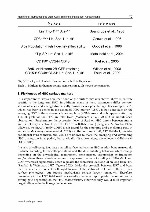
Representative achievements during these 2 decades are summarized in Table 1. With
surface markers, we can now purify the long-term multi-lineage HSC from adult mouse
bone marrow with extremely high efficiency as Lin- Sca-1+ c-kit+ Thy1Low CD34-/low CD150+
CD48- cells. In fact, recent studies have demonstrated that the Lin- Sca-1+ c-kit+ CD34-
CD150+ CD48- fraction in adult mouse bone marrow contains truly dormant HSC, which
divide only 5-6 times during the life span (Wilson et al., 2008; Foudi et al., 2009).
www.intechopen.com
Markers for Hematopoietic Stem Cells: Histories and Recent Achievements
79
*Tip-SP: The highest Hoechst-efflux fraction in the Side Population
Table 1. Markers for hematopoietic stem cells in adult mouse bone marrow
3. Fickleness of HSC surface markers
It is important to stress here that none of the surface markers shown above is entirely
specific to the long-term HSC. In addition, many of these parameters differ between
strains of mice and change dramatically during developmental age. For example, Sca1,
which has been a center in the canonical HSC marker “LSK”, is not detectable on the
emerging HSC in the aorta-gonad-mesonephros (AGM) area and only appears after day
11.5 of gestation on HSC in fetal liver (Matsubara et al., 2005; Our unpublished
observation). Furthermore, the expression level of Sca1 on HSC differs between strains
and is not very effective to enrich HSC from Balb/c mice (Spangrude & Brooks, 1993).
Likewise, the SLAM family CD150 is not useful for the emerging and developing HSC in
embryos (McKinney-Freeman et al., 2009). On the contrary, CD41, CD11b/Mac1, vascular
endothelial (VE)-cadherin, and CD34 are known to mark the emerging and developing
HSC during the fetal period, but gradually disappear along the ontogeny (Mikkola &
Orkin, 2006).
It is also a well-recognized fact that cell surface markers on HSC in adult bone marrow do
fluctuate according to the cell-cycle status and the differentiating behavior, which change
depending on the physiological requirement. Bone marrow suppression by irradiation
and/or chemotherapy revives several disappeared markers including CD11b/Mac1 and
CD34 whereas it significantly down-regulates the expression level of c-kit on long-term HSC
(Randall & Weissman, 1997; Ogawa 2002). Molecular crosstalk between HSC and bone
marrow microenvironment is thought to control the status of HSC and influence their
surface phenotypes, but precise mechanisms remain largely unknown. Therefore,
researchers in the HSC field need to carefully choose an appropriate marker set and a
sorting gate depending on the HSC characteristics, otherwise they would miss important
target cells even in the lineage depletion step.
www.intechopen.com
Advances in Hematopoietic Stem Cell Research
80
4. Difference between mouse and man
Another very critical issue on the topic of the HSC markers is their diversity between
species. Although essential difference has not been observed between mouse and man
regarding either the organs producing HSC or the transcription factors regulating their
differentiation, completely different markers have been used to sort HSC in the two species.
Human HSC do not express Sca1 or the SLAM family CD150 (Larochelle et al., 2011). While
the CD34+ CD38- phenotype has been regarded as the canonical marker set for human HSC,
it has been repeatedly reported that murine adult HSC locate in the CD34- CD38+ fraction
(Randall et al., 1996; Matsuoka et al., 2001; Tajima et al., 2001). There is no reasonable
explanation so far for the change along evolution, and such phenotypic differences between
murine and human HSC have been an obstacle to apply achievement in mouse studies to
human.
Early studies by Berenson et al demonstrated that autologous CD34+ cells enriched from
bone marrow effectively radioprotected baboons and promoted hematopoietic recovery in
human patients after marrow ablative therapy (Berenson et al, 1988, 1991). Over the past 2
decades, the use of CD34 as a marker for hematopoietic stem/progenitor cells has been a
strong tool in the field of clinical hematology. Since the CD34+ fraction of human bone
marrow contains lineage-committed progenitors as well as long-term multi-lineage HSC,
many laboratories have sought additional markers to further enrich the CD34+ population
for long-term HSC. CD90/Thy1, Tie, CD117/c-kit, and CD133/AC133 have been found as
positive markers to enrich long-term-HSC whereas several negative markers including
CD38 have been reported (Baum et al., 1992; Hasiyama et al., 1996; Gunji et al., 1993; Yin et
al., 1997; Terstappen et al., 1991).
Recent advances of xenotransplantation models and techniques have enabled the
assessment of pluripotency as well as self-renewal of human hematopoietic progenitors
in vivo (Shultz et al., 2007). A series of studies by John Dick’s laboratory have
successfully enriched human long-term HSC within the Lin- CD34+ CD38- population
(McKenzie et al., 2007; Doulatov et al., 2010). In a very recent report, they have purified
human HSC from cord blood with a maker set of Lin- CD34+ CD38- CD45RA-
CD90/Thy1+ Rhodamin123Low CD49f+. Indeed, those cells were capable of long-term
multilineage engraftment in NOD/SCID/IL2 receptor common- chain null mice at a
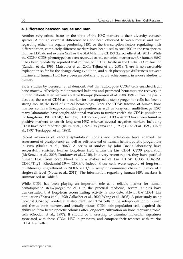
single-cell level (Notta et al., 2011). The information regarding human HSC markers is
summarized in Table 2.
While CD34 has been playing an important role as a reliable marker for human
hematopoietic stem/progenitor cells in the practical medicine, several studies have
demonstrated that long-term reconstituting activity is also detectable in the CD34- Lin-
population (Bhatia et al., 1998; Gallacher et al., 2000; Wang et al., 2003). A prior study using
Hoechst 33342 by Goodell et al also identified CD34- cells in the side-population of human
and rhesus bone marrow, and actually rhesus CD34- side-population cells acquired the
ability to form hematopoietic colonies after long-term cultivation on bone marrow stromal
cells (Goodell et al., 1997). It should be interesting to examine molecular signatures
associated with those CD34- HSC in primates, and compare their features with murine
CD34- LSK cells.
www.intechopen.com
Markers for Hematopoietic Stem Cells: Histories and Recent Achievements
81
Table 2. Markers for human hematopoietic stem cells
5. Endothelial-related markers
Hematopoietic cells are thought to originate from the hemangioblast and/or the hemogenic
endothelium, which can produce hematopoietic cells and endothelial cells. Therefore, it
seems quite natural that HSC share some surface molecules with the endothelial lineage.
CD34, PECAM-1/CD31, endoglin, Tie2 and VE-cadherin are well-known endothelial
antigens that also mark HSC particularly at early developmental stages (Mikkola & Orkin
2006; Takakura et al., 1998; Yokota et al, 2006). In addition, recent studies have identified
endomucin, endothelial protein-C receptor/CD201, and junctional adhesion molecule-A
that are common to HSC and endothelial cells (Matsubara et al., 2005; Balazs et al., 2006;
Sugano et al., 2008). Although, as discussed above, the expression level of some of these
antigens declines or even diminishes at later stages of development (Mikkola & Orkin 2006),
each of these advances offered the promise of learning more about how HSC arise de novo
and function throughout life. It is crucial to define the means to identify the authentic HSC
at all developmental stages so that we can ultimately understand the precise molecular
mechanisms of the HSC development.
www.intechopen.com
Advances in Hematopoietic Stem Cell Research
82
6. Identification of ESAM as a novel HSC marker
We previously reported that Rag1/GFP- Lin- c-kitHigh Sca1+ cells derived from bone marrow
or fetal liver of the Rag1/GFP reporter mice reconstituted lympho-hematopoiesis in lethally
irradiated recipients, while Rag1/GFP+ Lin- c-kitHigh Sca1+ cells only transiently contributed
to T and B lymphopoiesis (Igarashi et al., 2002; Yokota et al., 2003). Those data demonstrated
that Rag1 expression is useful to distinguish early lymphoid progenitors (ELP) from the
long-term HSC. To learn more about the first step of HSC differentiation to the lymphoid
lineage, microarray analyses were conducted to search for genes that characterize the initial
transition of HSC to ELP. The search brought us a large body of information about genes
potentially related to early lymphopoiesis whereas it also identified genes whose expression
seemed to correlate with HSC. Among the HSC-related genes, ESAM strongly drew our
attention because of its conspicuous expression in the HSC fraction and sharp down-
regulation on differentiation to ELP.
ESAM was originally identified as an endothelial cell-specific protein (Hirata et al., 2001;
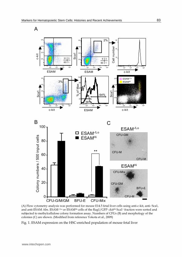
Nasdala et al., 2002). Flow cytometry analyses with anti-ESAM antibodies showed that the
HSC-enriched Rag1- c-kitHigh Sca1+ fraction of E14.5 fetal liver could be subdivided into two
on the basis of ESAM level (Figure 1A). The subpopulation with the high density of ESAM
was enriched for c-kitHigh Sca1High cells, while ones with negative or low levels of ESAM
were found in the c-kitHigh Sca1Low subset. In addition, ESAM expression well correlated
with hematopoietic stem/progenitor activity (Figure 1B). Cells in the ESAMHigh Rag1- c-
kitHigh Sca1+ fraction formed more and larger colonies than those in the ESAM-/Low Rag1- c-
kitHigh Sca1+ fraction. Particularly, majority of CFU-Mix, multi-potent primitive progenitors,
were found in the ESAMHigh fraction (Figure 1B and 1C). In limiting dilution stromal cell co-
cultures, we found that 1 in 2.1 ESAMHigh Rag1- c-kitHigh Sca1+ cells and 1 in 3.5 ESAM-/Low
Rag1- c-kitHigh Sca1+ cells gave rise to blood cells. However, 1 in 8 ESAMHigh Rag1- c-kitHigh
Sca1+ cells produced CD19+ B lineage cells whereas only 1 in 125 ESAM-/Low Rag1- c-kitHigh
Sca1+ cells were lymphopoietic under these conditions. Furthermore, in long-term
reconstituting assays, ESAMHigh Rag1- c-kitHigh Sca1+ cells contributed highly to the multi-
lineage recovery of lympho-hematopoiesis in recipients, but no chimerism was detected in
mice transplanted with ESAM-/Low Rag1- c-kitHigh Sca1+ cells. These results suggested that
the long-term multi-lineage HSC in E14.5 fetal liver are exclusively present in the ESAMHigh
fraction.
7. ESAM marks HSC in different developmental stages and in different species
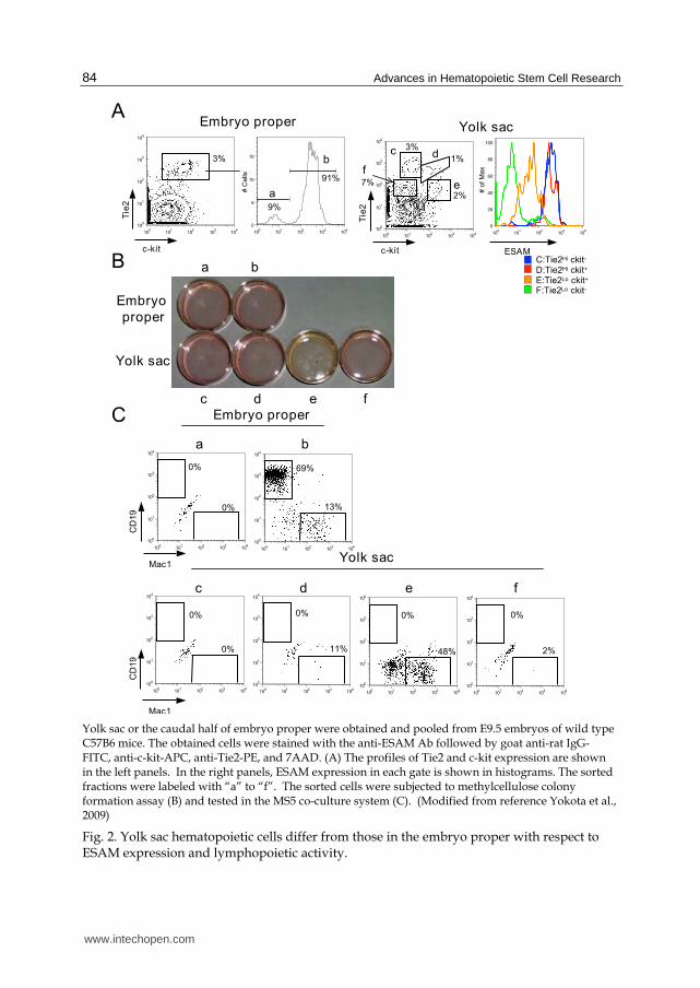
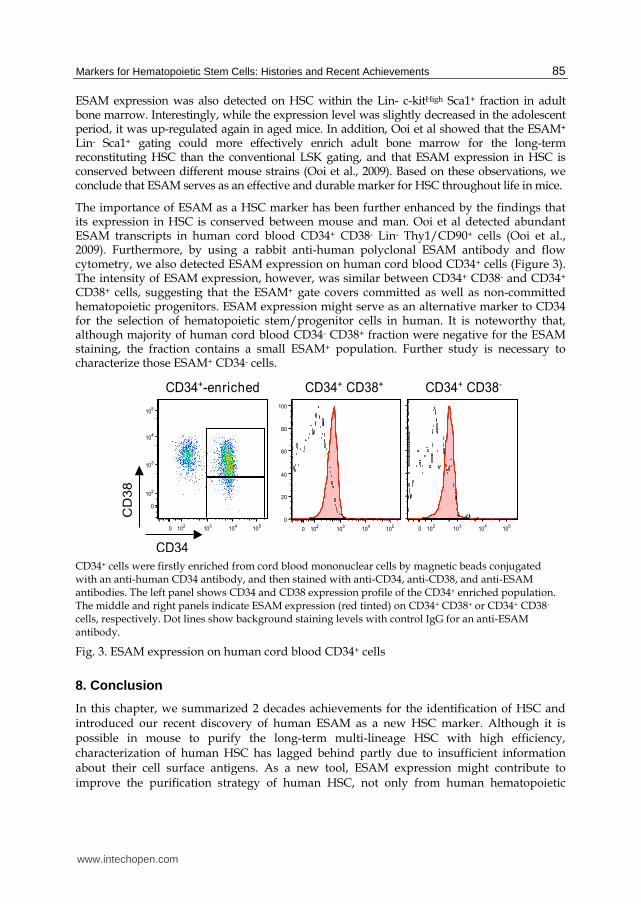
Hematopoietic cells arise from mesoderm precursors at different sites and stages of development (de Bruijn et al., 2000; Oberlin et al., 2002). We previously determined that, while myelo-erythroid progenitors emerge from the yolk sac, hematopoietic progenitors with lymphopoietic potential first develop in the paraaortic splanchnopleura (pSp) /AGM region (Yokota et al., 2006). ESAM+ cells in the AGM were found to co-express c-kit and endothelial antigens, Tie2, CD34 and CD31/PECAM-1 that are known as a marker set for emerging HSC. However, the earlier hematopoietic progenitors in the yolk sac that have limited life span and little lymphopoietic activity were harbored in the ESAMLow Tie2Low c-kitHigh fraction (Figure 2).
www.intechopen.com
Markers for Hematopoietic Stem Cells: Histories and Recent Achievements
83
(A) Flow cytometry analysis was performed for mouse E14.5 fetal liver cells using anti-c-kit, anti- Sca1, and anti-ESAM Abs. ESAM-/Lo or ESAMHi cells of the Rag1/GFP- ckitHi Sca1+ fraction were sorted and subjected to methylcellulose colony formation assay. Numbers of CFUs (B) and morphology of the colonies (C) are shown. (Modified from reference Yokota et al., 2009)
Fig. 1. ESAM expression on the HSC-enriched population of mouse fetal liver
www.intechopen.com
Advances in Hematopoietic Stem Cell Research
84
Yolk sac or the caudal half of embryo proper were obtained and pooled from E9.5 embryos of wild type C57B6 mice. The obtained cells were stained with the anti-ESAM Ab followed by goat anti-rat IgG-FITC, anti-c-kit-APC, anti-Tie2-PE, and 7AAD. (A) The profiles of Tie2 and c-kit expression are shown in the left panels. In the right panels, ESAM expression in each gate is shown in histograms. The sorted fractions were labeled with “a” to “f”. The sorted cells were subjected to methylcellulose colony formation assay (B) and tested in the MS5 co-culture system (C). (Modified from reference Yokota et al., 2009)
Fig. 2. Yolk sac hematopoietic cells differ from those in the embryo proper with respect to ESAM expression and lymphopoietic activity.
www.intechopen.com
Markers for Hematopoietic Stem Cells: Histories and Recent Achievements
85
ESAM expression was also detected on HSC within the Lin- c-kitHigh Sca1+ fraction in adult bone marrow. Interestingly, while the expression level was slightly decreased in the adolescent period, it was up-regulated again in aged mice. In addition, Ooi et al showed that the ESAM+ Lin- Sca1+ gating could more effectively enrich adult bone marrow for the long-term reconstituting HSC than the conventional LSK gating, and that ESAM expression in HSC is conserved between different mouse strains (Ooi et al., 2009). Based on these observations, we conclude that ESAM serves as an effective and durable marker for HSC throughout life in mice.
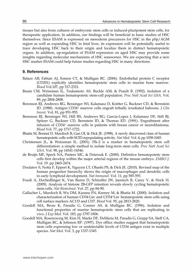
The importance of ESAM as a HSC marker has been further enhanced by the findings that its expression in HSC is conserved between mouse and man. Ooi et al detected abundant ESAM transcripts in human cord blood CD34+ CD38- Lin- Thy1/CD90+ cells (Ooi et al., 2009). Furthermore, by using a rabbit anti-human polyclonal ESAM antibody and flow cytometry, we also detected ESAM expression on human cord blood CD34+ cells (Figure 3). The intensity of ESAM expression, however, was similar between CD34+ CD38- and CD34+ CD38+ cells, suggesting that the ESAM+ gate covers committed as well as non-committed hematopoietic progenitors. ESAM expression might serve as an alternative marker to CD34 for the selection of hematopoietic stem/progenitor cells in human. It is noteworthy that, although majority of human cord blood CD34- CD38+ fraction were negative for the ESAM staining, the fraction contains a small ESAM+ population. Further study is necessary to characterize those ESAM+ CD34- cells.
CD34+ cells were firstly enriched from cord blood mononuclear cells by magnetic beads conjugated with an anti-human CD34 antibody, and then stained with anti-CD34, anti-CD38, and anti-ESAM antibodies. The left panel shows CD34 and CD38 expression profile of the CD34+ enriched population. The middle and right panels indicate ESAM expression (red tinted) on CD34+ CD38+ or CD34+ CD38- cells, respectively. Dot lines show background staining levels with control IgG for an anti-ESAM antibody.
Fig. 3. ESAM expression on human cord blood CD34+ cells
8. Conclusion
In this chapter, we summarized 2 decades achievements for the identification of HSC and introduced our recent discovery of human ESAM as a new HSC marker. Although it is possible in mouse to purify the long-term multi-lineage HSC with high efficiency, characterization of human HSC has lagged behind partly due to insufficient information about their cell surface antigens. As a new tool, ESAM expression might contribute to improve the purification strategy of human HSC, not only from human hematopoietic
www.intechopen.com
Advances in Hematopoietic Stem Cell Research
86
tissues but also from cultures of embryonic stem cells or induced-pluripotent stem cells, for therapeutic application. In addition, our findings will be beneficial to basic studies of HSC themselves. Since ESAM is expressed on mesoderm precursors for HSC in the pSp/AGM region as well as expanding HSC in fetal liver, its expression will be potentially useful to trace developing HSC back to their origin and localize them in distinct hematopoietic organs. In addition, up-regulation of ESAM expression on aged HSC may provide some insights regarding molecular mechanisms of HSC senescence. We are expecting that a new HSC marker ESAM could help future studies regarding HSC in many directions.
9. References
Balazs AB, Fabian AJ, Esmon CT, & Mulligan RC. (2006). Endothelial protein C receptor (CD201) explicitly identifies hematopoietic stem cells in murine bone marrow. Blood Vol.107, pp 317-2321.
Baum CM, Weissman IL, Tsukamoto AS, Buckle AM, & Peault B. (1992). Isolation of a candidate human hematopoietic stem-cell population. Proc Natl Acad Sci USA. Vol. 89, pp 2804-2808.
Berenson RJ, Andrews RG, Bensinger WI, Kalamasz D, Knitter G, Buckner CD, & Bernstein ID. (1988). Antigen CD34+ marrow cells engraft lethally irradiated baboons. J Clin Invest. Vol. 81, pp 951-955.
Berenson RJ, Bensinger WI, Hill RS, Andrews RG, Garcia-Lopez J, Kalamasz DF, Still BJ, Spitzer G, Buckner CD, Bernstein ID, & Thomas ED. (1991). Engraftment after infusion of CD34+ marrow cells in patients with breast cancer or neuroblastoma. Blood Vol. 77, pp 1717-1722.
Bhatia M, Bonnet D, Murdoch B, Gan OI, & Dick JE. (1998). A newly discovered class of human hematopoietic cells with SCID-repopulating activity. Nat Med. Vol. 4, pp 1038-1045.
Christensen JL, & Weissman IL. (2001). Flk-2 is a marker in hematopoietic stem cell differentiation: a simple method to isolate long-term stem cells. Proc Natl Acad Sci USA. Vol. 98, pp 14541-14546.
de Bruijn MF, Speck NA, Peeters MC, & Dzierzak E. (2000). Definitive hematopoietic stem cells first develop within the major arterial regions of the mouse embryo. EMBO J. Vol. 19. pp 2465-2474.
Doulatov S, Notta F, Eppert K, Nguyen LT, Ohashi PS, & Dick JE. (2010). Revised map of the human progenitor hierarchy shows the origin of macrophages and dendritic cells in early lymphoid development. Nat Immunol. Vol. 11, pp 585-593.
Foudi A, Hochedlinger K, Van Buren D, Schindler JW, Jaenisch R, Carey V, & Hock H. (2009). Analysis of histone 2B-GFP retention reveals slowly cycling hematopoietic stem cells. Nat Biotechnol. Vol. 27, pp 84-90.
Gallacher L, Murdoch B, Wu DM, Karanu FN, Keeney M, & Bhatia M. (2000). Isolation and characterization of human CD34-Lin- and CD34+Lin- hematopoietic stem cells using cell surface markers AC133 and CD7. Blood Vol. 95, pp 2813-2820.
Goodell MA, Brose K, Paradis G, Conner AS, & Mulligan RC. (1996). Isolation and functional properties of murine hematopoietic stem cells that are replicating in vivo. J Exp Med. Vol. 183, pp 1797-1806.
Goodell MA, Rosenzweig M, Kim H, Marks DF, DeMaria M, Paradis G, Grupp SA, Sieff CA, Mulligan RC, & Johnson RP. (1997). Dye efflux studies suggest that hematopoietic stem cells expressing low or undetectable levels of CD34 antigen exist in multiple species. Nat Med. Vol. 3, pp 1337-1345.
www.intechopen.com
Markers for Hematopoietic Stem Cells: Histories and Recent Achievements
87
Gunji Y, Nakamura M, Osawa H, Nagayoshi K, Nakauchi H, Miura Y, Yanagisawa M, & Suda T. (1993). Human primitive hematopoietic progenitor cells are more enriched in KITlow cells than in KIThigh cells. Blood Vol. 82, pp 3283-3289.
Hashiyama M, Iwama A, Ohshiro K, Kurozumi K, Yasunaga K, Shimizu Y, Masuho Y, Matsuda I, Yamaguchi N, & Suda T. (1996). Predominant expression of a receptor tyrosine kinase, TIE, in hematopoietic stem cells and B cells. Blood Vol. 87, pp 93-101.
Hirata K, Ishida T, Penta K, Rezaee M, Yang E, Wohlgemuth J, & Quertermous T. (2001). Cloning of an immunoglobulin family adhesion molecule selectively expressed by endothelial cells. J Biol Chem. Vol. 276, pp 16223-16231.
Igarashi H, Gregory SC, Yokota T, Sakaguchi N, & Kincade PW. (2002). Transcription from the RAG1 locus marks the earliest lymphocyte progenitors in bone marrow. Immunity Vol. 17, pp 117-130.
Kiel MJ, Yilmaz OH, Iwashita T, Yilmaz OH, Terhorst C, & Morrison SJ. (2005). SLAM family receptors distinguish hematopoietic stem and progenitor cells and reveal endothelial niches for stem cells. Cell Vol. 121, pp 1109-1121.
Larochelle A, Savona M, Wiggins M, Anderson S, Ichwan B, Keyvanfar K, Morrison SJ, & Dunbar CE. (2011). Human and rhesus macaque hematopoietic stem cells cannot be purified based only on SLAM family markers. Blood Vol. 117, pp1550-1554.
Matsubara A, Iwama A, Yamazaki S, Furuta C, Hirasawa R, Morita Y, Osawa M, Motohashi T, Eto K, Ema H, Kitamura T, Vestweber D, & Nakauchi H. (2005). Endomucin, a CD34-like sialomucin, marks hematopoietic stem cells throughout development. J Exp Med. Vol. 202, pp 1483-1492.
Matsuoka S, Ebihara Y, Xu M, Ishii T, Sugiyama D, Yoshino H, Ueda T, Manabe A, Tanaka R, Ikeda Y, Nakahata T, & Tsuji K. (2001). CD34 expression on long-term repopulating hematopoietic stem cells changes during developmental stages. Blood Vol. 97, pp 419-425.
Matsuzaki Y, Kinjo K, Mulligan RC, & Okano H. (2004). Unexpectedly efficient homing capacity of purified murine hematopoietic stem cells. Immunity Vol. 20, pp 87-93.
McKenzie JL, Takenaka K, Gan OI, Doedens M, & Dick JE. (2007). Low rhodamine 123 retention identifies long-term human hematopoietic stem cells within the Lin-
CD34+CD38- population. Blood Vol. 109, pp 543-545. McKinney-Freeman SL, Naveiras O, Yates F, Loewer S, Philitas M, Curran M, Park PJ, &
Daley GQ. (2009). Surface antigen phenotypes of hematopoietic stem cells from embryos and murine embryonic stem cells. Blood Vol. 114, pp 268-278
Mikkola HKA, & Orkin SH. (2006). The journey of developing hematopoietic stem cells. Development Vol. 133, pp 3733-3744.
Nasdala I, Wolburg-Buchholz K, Wolburg H, Kuhn A, Ebnet K, Brachtendorf G, Samulowitz U, Kuster B, Engelhardt B, Vestweber D, & Butz S. (2002). A transmembrane tight junction protein selectively expressed on endothelial cells and platelets. J Biol Chem. Vol. 277, pp 16294-16303.
Notta F, Doulatov S, Laurenti E, Poeppl A, Jurisica I, & Dick JE. (2011). Isolation of single human hematopoietic stem cells capable of long-term multilineage engraftment. Science Vol. 333, pp 218-221.
Oberlin E., Tavian M., Blazsek I., & Péault B. (2002). Blood-forming potential of vascular endothelium in the human embryo. Development Vol. 129, pp 4147-4157.
Ogawa M, Matsuzaki Y, Nishikawa S, Hayashi S, Kunisada T, Sudo T, Kina T, Nakauchi H, & Nishikawa S-I. (1991). Expression and function of c-kit in hemopoietic progenitor cells. J Exp Med. Vol. 174, pp 63-71.
Ogawa M. (2002). Changing phenotypes of hematopoietic stem cells. Exp Hematol. Vol. 30, pp 3-6.
www.intechopen.com
Advances in Hematopoietic Stem Cell Research
88
Ooi AG, Karsunky H, Majeti R, Butz S, Vestweber D, Ishida T, Quertermous T, Weissman IL, & Forsberg EC. (2009). The adhesion molecule esam1 is a novel hematopoietic stem cell marker. Stem Cells Vol. 27, pp 653-661.
Osawa M, Hanada K, Hamada H, & Nakauchi H. (1996). Long-term lymphohematopoietic reconstitution by a single CD34-low/negative hematopoietic stem cell. Science Vol. 273, pp 242-245.
Randall TD, Lund FE, Howard MC, & Weissman IL. (1996). Expression of murine CD38 defines a population of long-term reconstituting hematopoietic stem cells. Blood Vol. 87, pp 4057-4067.
Randall TD, & Weissman IL. (1997). Phenotypic and functional changes induced at the clonal level in hematopoietic stem cells after 5-fluorouracil treatment. Blood Vol. 89, pp 3596-3606.
Shultz LD, Ishikawa F, & Greiner DL. (2007). Humanized mice in translational biomedical research. Nat Rev Immunol. Vol. 7, pp 118-130.
Spangrude GJ, Heimfeld S, & Weissman IL. (1988). Purification and characterization of mouse hematopoietic stem cells. Science Vol. 241, pp 58-62.
Spangrude GJ, & Brooks DM. (1993). Mouse strain variability in the expression of the hematopoietic stem cell antigen Ly-6A/E by bone marrow cells. Blood Vol. 82, pp 3327-3332.
Sugano Y, Takeuchi M, Hirata A, Matsushita H, Kitamura T, Tanaka M, & Miyajima A. (2008). Junctional adhesion molecule-A, JAM-A, is a novel cell-surface marker for long-term repopulating hematopoietic stem cells. Blood Vol. 111, pp 1167-1172.
Tajima F, Deguchi T, Laver JH, Zeng H, & Ogawa M. (2001). Reciprocal expression of CD38 and CD34 by adult murine hematopoietic stem cells. Blood Vol. 97, pp 2618-2624.
Takakura N, Huang XL, Naruse T, Hamaguchi I, Dumont DJ, Yancopoulos GD, & Suda T. (1998). Critical role of the TIE2 endothelial cell receptor in the development of definitive hematopoiesis. Immunity Vol. 9, pp 677-686.
Terstappen LW, Huang S, Safford M, Lansdorp PM, & Loken MR. (1991). Sequential generations of hematopoietic colonies derived from single nonlineage-committed CD34+CD38- progenitor cells. Blood Vol. 77, pp 1218-1227.
Wang J, Kimura T, Asada R, Harada S, Yokota S, Kawamoto Y, Fujimura Y, Tsuji T, Ikehara S, & Sonoda Y. (2003). SCID-repopulating cell activity of human cord blood-derived CD34- cells assured by intra-bone marrow injection. Blood Vol. 101, pp 2924-2931.
Wilson A, Laurenti E, Oser G, van der Wath RC, Blanco-Bose W, Jaworski M, Offner S, Dunant CF, Eshkind L, Bockamp E, Lió P, Macdonald HR, & Trumpp A. (2008). Hematopoietic stem cells reversibly switch from dormancy to self-renewal during homeostasis and repair. Cell Vol. 135, pp1118-1129.
Yin AH, Miraglia S, Zanjani ED, Almeida-Porada G, Ogawa M, Leary AG, Olweus J, Kearney J, & Buck DW. (1997). AC133, a novel marker for human hematopoietic stem and progenitor cells. Blood Vol. 90, pp 5002-5012.
Yokota T, Kouro T, Hirose J, Igarashi H, Garrett KP, Gregory SC, Sakaguchi N, Owen JJ, & Kincade PW. (2003). Unique properties of fetal lymphoid progenitors identified according to RAG1 gene expression. Immunity Vol. 19, pp 365-375.
Yokota T, Huang J, Tavian M, Nagai Y, Hirose J, Zúñiga-Pflücker JC, Péault B, & Kincade PW. (2006). Tracing the first waves of lymphopoiesis in mice. Development Vol. 133, pp 2041-2051.
Yokota T, Oritani K, Butz S, Kokame K, Kincade PW, Miyata T, Vestweber D, & Kanakura Y. (2009). The endothelial antigen ESAM marks primitive hematopoietic progenitors throughout life in mice. Blood Vol. 113, pp 2914-2923.
www.intechopen.com
Advances in Hematopoietic Stem Cell ResearchEdited by Dr. Rosana Pelayo
ISBN 978-953-307-930-1Hard cover, 464 pagesPublisher InTechPublished online 27, January, 2012Published in print edition January, 2012
InTech EuropeUniversity Campus STeP Ri Slavka Krautzeka 83/A 51000 Rijeka, Croatia Phone: +385 (51) 770 447 Fax: +385 (51) 686 166www.intechopen.com
InTech ChinaUnit 405, Office Block, Hotel Equatorial Shanghai No.65, Yan An Road (West), Shanghai, 200040, China
Phone: +86-21-62489820 Fax: +86-21-62489821
This book provides a comprehensive overview in our understanding of the biology and therapeutic potential ofhematopoietic stem cells, and is aimed at those engaged in stem cell research: undergraduate andpostgraduate science students, investigators and clinicians. Starting from fundamental principles inhematopoiesis, Advances in Hematopoietic Stem Cell Research assemble a wealth of information relevant tocentral mechanisms that may regulate differentiation, and expansion of hematopoietic stem cells in normalconditions and during disease.
How to referenceIn order to correctly reference this scholarly work, feel free to copy and paste the following:
Takafumi Yokota, Kenji Oritani, Stefan Butz, Stephan Ewers, Dietmar Vestweber and Yuzuru Kanakura (2012).Markers for Hematopoietic Stem Cells: Histories and Recent Achievements, Advances in Hematopoietic StemCell Research, Dr. Rosana Pelayo (Ed.), ISBN: 978-953-307-930-1, InTech, Available from:http://www.intechopen.com/books/advances-in-hematopoietic-stem-cell-research/endothelial-cell-selective-adhesion-molecule-esam-a-novel-hsc-marker

© 2012 The Author(s). Licensee IntechOpen. This is an open access articledistributed under the terms of the Creative Commons Attribution 3.0License, which permits unrestricted use, distribution, and reproduction inany medium, provided the original work is properly cited.