Embed Size (px)
Citation preview

Cardiovascular System:!
Heart (Chapter 20)!
Lecture Materials!
for!
Amy Warenda Czura, Ph.D.!
Suffolk County Community College!
Eastern Campus!
Primary Sources for figures and content:!
Marieb, E. N. Human Anatomy & Physiology 6th ed. San Francisco: Pearson Benjamin
Cummings, 2004.!
Martini, F. H. Fundamentals of Anatomy & Physiology 6th ed. San Francisco: Pearson
Benjamin Cummings, 2004.!
Cardiovascular System:!
Pulmonary circuit: !
! right ventricle !
! ! lungs ! !!
! left atrium!
Systemic circuit: !
! left ventricle !
! ! body ! ! !!
! right atrium!
Arteries = away from heart!
Veins = toward heart!
Capillaries = exchange vessels in between!
-left of midline, between 2nd rib and 5th !
! intercostal space, posterior to sternum, in
! pericardial cavity in mediastinum!
-heart is fist sized, < 1 lb, beats 100,000 !
! times/day moving 8000 L blood/day!
-surrounded by pericardium (serous and !
! fibrous layers)!
-serous membranes (visceral & parietal) !
! secrete pericardial fluid, reduce friction!
Pericarditis = inflammation of pericardium, !
! usually due to infection, causes friction!
Cardiac tamponade = buildup of fluid in !
! pericardial space, restricts heart movement!
Heart!
Four Chambers:!
2 Atria: !
-superior, thin walls, !
! smooth posterior walls ! ! !
! internally, !
! pectinate muscles (ridges) anteriorly !
-each has expandable flap called an auricle !
! lateral and superior!
-left and right separated by interatrial septum!
Amy Warenda Czura, Ph.D. 1 SCCC BIO132 Chapter 20 Lecture Notes

2 Ventricles:!
-inferior, thick walls, lined with trabeculae !
! carneae (muscular ridges)!
-left and right separated by interventricular !
! septum!
Left ventricle 3X thicker, !
! 5X more friction while !
! pumping, !
! same volume as right!
Left is round, !
! right is crescent!
External divisions:!
Coronary sulcus marks division between atria
! and ventricles!
Anterior interventricular sulcus and posterior
! interventricular sulcus mark division !
! between ventricles!
Heart Wall Layers!
1. Epicardium (thin) - !
! visceral pericardium: serous membrane !
! with loose CT attached to myocardium!
2. Myocardium (thick) - cardiac muscle tissue
! with CT, vessels and nerves!
3. Endocardium (thin) - simple squamous !
! epithelium lining with basal lamina; !
! continuous with endothelium of blood !
! vessels!
Cardiac Muscle Tissue!
-muscle cells = cardiocytes!
-uses actin and myosin sliding filaments to !
! contract!
-rich in mitochondria, resists fatigue but !
! dependent on aerobic respiration!
-cells connected by intercalated discs = !
! desmosomes + gap junctions!
-contraction is all or none!
-longer contractile phase than skeletal muscle!
-fibrous skeleton of the heart (tough CT) acts !
! as the tendon!
Amy Warenda Czura, Ph.D. 2 SCCC BIO132 Chapter 20 Lecture Notes
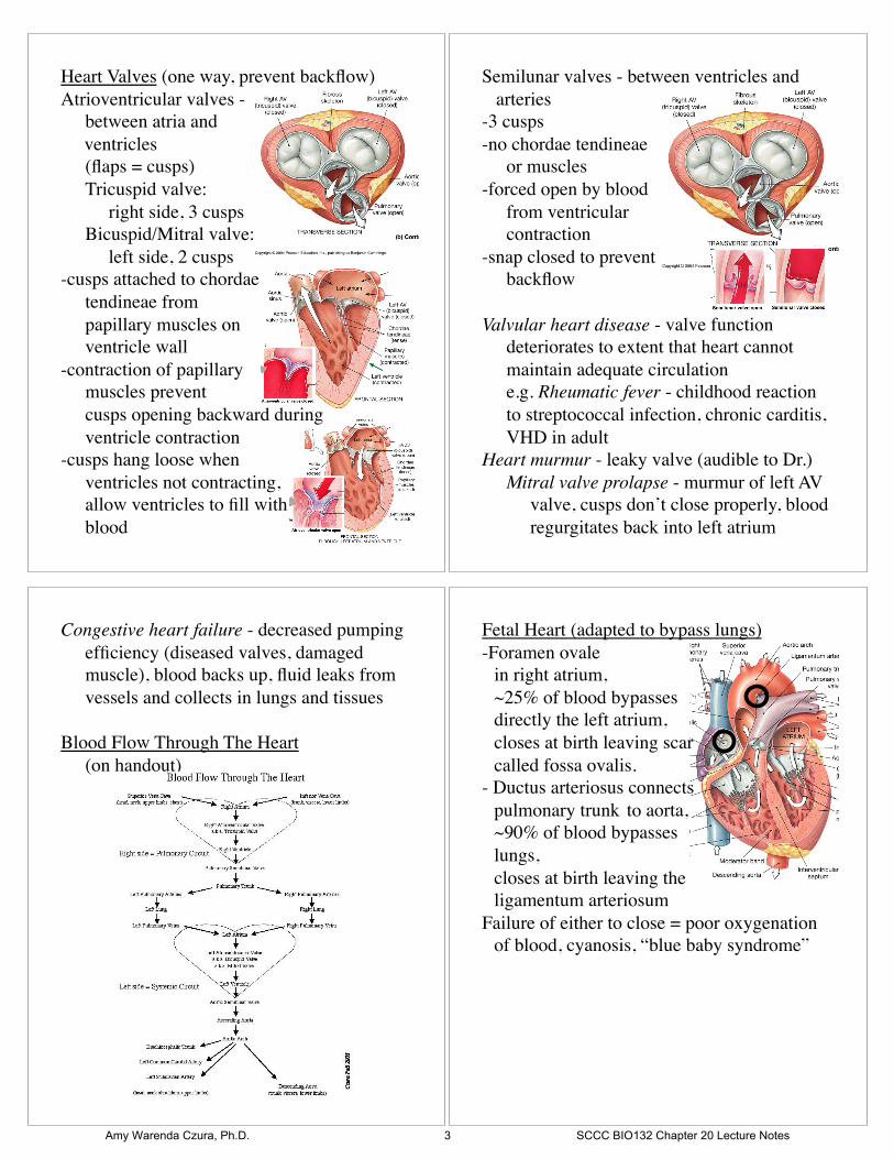
Heart Valves (one way, prevent backflow)!
Atrioventricular valves - !
! between atria and !
ventricles !
! (flaps = cusps)!
! Tricuspid valve: !
! !right side, 3 cusps!
! Bicuspid/Mitral valve: !
! !left side, 2 cusps!
-cusps attached to chordae !
! tendineae from !
! papillary muscles on !
! ventricle wall!
-contraction of papillary !
! muscles prevent !
! cusps opening backward during !
! ventricle contraction!
-cusps hang loose when !
! ventricles not contracting, !
! allow ventricles to fill with !
! blood!
Semilunar valves - between ventricles and !
arteries!
-3 cusps!
-no chordae tendineae !
! or muscles!
-forced open by blood !
! from ventricular !
! contraction!
-snap closed to prevent !
! backflow!
Valvular heart disease - valve function !
! deteriorates to extent that heart cannot !
! maintain adequate circulation!
! e.g. Rheumatic fever - childhood reaction
! to streptococcal infection, chronic carditis,
! VHD in adult!
Heart murmur - leaky valve (audible to Dr.)!
! Mitral valve prolapse - murmur of left AV
! !valve, cusps don’t close properly, blood
! !regurgitates back into left atrium!
Congestive heart failure - decreased pumping
! efficiency (diseased valves, damaged !
! muscle), blood backs up, fluid leaks from
! vessels and collects in lungs and tissues!
Blood Flow Through The Heart !
! (on handout)!
Fetal Heart (adapted to bypass lungs)!
-Foramen ovale !
! in right atrium, !
! ~25% of blood bypasses !
! directly the left atrium, ! !!
! closes at birth leaving scar !
! called fossa ovalis.!
- Ductus arteriosus connects !
! pulmonary trunk !to aorta, !
! ~90% of blood bypasses !
! lungs, !
! closes at birth leaving the !
! ligamentum arteriosum!
Failure of either to close = poor oxygenation
! of blood, cyanosis, “blue baby syndrome” !
Amy Warenda Czura, Ph.D. 3 SCCC BIO132 Chapter 20 Lecture Notes

Coronary circulation!
-heart: <1% body mass, requires 5% of blood!
-too thick for diffusion!
-coronary arteries originate at base of ! !
! ascending aorta, branch to capillary beds !
! for diffusion!
-blood returns via cardiac veins that empty !
! into right atrium!
Coronary artery disease -partial or complete
! block of coronary circulation, results in !
! coronary ischemia !
Can lead to myocardial infarction (heart !
! attack): heart tissue denied oxygen dies.!
Common symptom of CAD: angina pectoralis
! pain in the chest, especially during activity,
! as a result of the ischemia!
Coronary bypass surgery - use healthy veins !
! (from legs) to create anatomoses around !
! blockages!
! Most people have 4 major coronary !
! arteries:!
! “quadruple bypass”!
Heart Beat!
-1% myocardial cells autorhythmic: ! !
! depolarize without neural or endocrine !
! stimulation!
-depolarization transmitted to other ! !
! myocardial cells through cardiac ! !
! conduction system:!
! Sinoatrial (SA) node -!
! !right atrium wall ! ! !!
! !near superior !
! !vena cava!
! Atrioventricular !
! !(AV) node - !
! !inferior portion of !
! !interatrial septum above tricuspid valve!
! Conducting cells: AV bundle, Bundle !
! !branches, and Purkinje fibers: connect
! !nodes and myocardium, run down !
! !interventricular septum and around apex!
-cells of nodes cannot maintain resting !
! membrane potential, drift to depolarization:!
! !SA node 80-100 action potentials/min!
! ! !(“natural pacemaker”)!
! !AV node 40-60 action potentials/min!
-Resting rate (sinus rhythm) ~75bpm set by !
! SA node + parasympathetic stimulation!
Amy Warenda Czura, Ph.D. 4 SCCC BIO132 Chapter 20 Lecture Notes
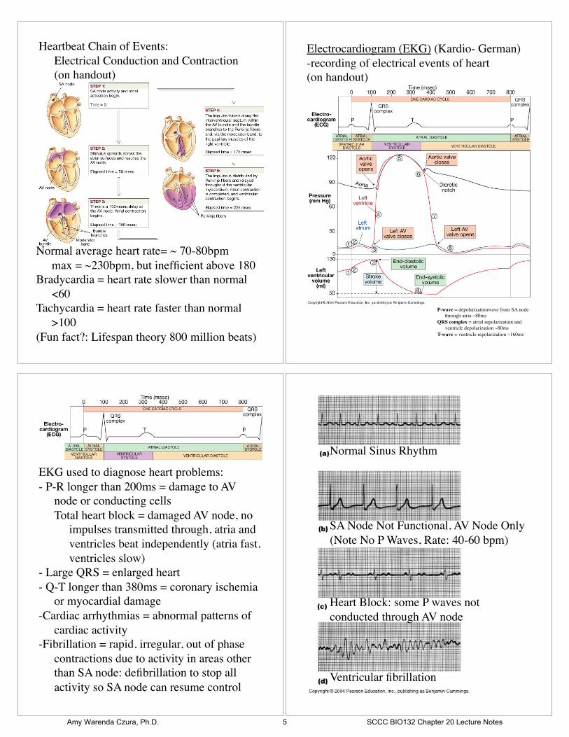
Heartbeat Chain of Events:!
! Electrical Conduction and Contraction!
! (on handout)!
Normal average heart rate= ~ 70-80bpm!
! max = ~230bpm, but inefficient above 180!
Bradycardia = heart rate slower than normal!
! <60!
Tachycardia = heart rate faster than normal!
! >100!
(Fun fact?: Lifespan theory 800 million beats)!
Electrocardiogram (EKG) (Kardio- German)!
-recording of electrical events of heart!
(on handout)!
P-wave = depolarizationwave from SA node
! through atria ~80ms!
QRS complex = atrial repolarization and !
! ventricle depolarization ~80ms!
T-wave = ventricle repolarization ~160ms!
EKG used to diagnose heart problems:!
- P-R longer than 200ms = damage to AV !
! node or conducting cells!
! Total heart block = damaged AV node, no
! !impulses transmitted through, atria and
! !ventricles beat independently (atria fast,
! !ventricles slow)!
- Large QRS = enlarged heart!
- Q-T longer than 380ms = coronary ischemia
! or myocardial damage!
-Cardiac arrhythmias = abnormal patterns of
! cardiac activity!
-Fibrillation = rapid, irregular, out of phase !
! contractions due to activity in areas other
! than SA node: defibrillation to stop all !
! activity so SA node can resume control!
Normal Sinus Rhythm!
SA Node Not Functional, AV Node Only!
(Note No P Waves, Rate: 40-60 bpm)!
Heart Block: some P waves not
conducted through AV node!
Ventricular fibrillation!
Amy Warenda Czura, Ph.D. 5 SCCC BIO132 Chapter 20 Lecture Notes

Cardiac Cycle!
-alternating contraction and relaxation!
Systole = contraction, high pressure, blood !
! gets pushed to next chamber!
Diastole = relaxation, low pressure, chamber
! fills with blood!
(on handout)!
"heart rate = #cycle time, #diastole time = !
! # time to fill !
Atrial contraction only adds ~30% more to !
! ventricles (can live with bad atria)!
Heart Sounds!
“lubb” - S1: AV valves close at start of !
! ventricular systole!
“dupp” - S2: semilunar valves close at start of
! ventricular diastole!
Cardiac Output!
CO = amount of blood pumped by each !
! ventricle in one minute, depends on heart
! rate and stroke volume: CO = HR X SV !
SV = amount of blood pumped by ventricle!
Usually SV constant, change HR to increase !
! CO as needed. !
HR affected by:!
! !1. autonomic nervous input!
! ! !sympathetic = " HR!
! ! !parasympathetic = # HR!
! !2. hormones! !!
! !3. venous return !
! ! !more blood return = " HR (stretch
! ! ! !receptors activate sympathetic)!
! !4. other factors (ions, drugs)!
@160-180 bpm CO at max:!
"HR = #time to fill ventricles, !
! if not full = #SV and #CO!
Conditioning can "SV and #HR!
Fit athletes can " max CO by 700%, & # !
! resting HR by 50% with same CO due to !
! "SV !
Heart Rate Effectors:!
1. Autonomic Innervation!
-SA node, AV node and atrial myocardium !
! innervated by both sympathetic (NE) and
! parasympathetic (Ach) nerve fibers !
! equally. !
-Sympathetic dominates in ventricles.!
-Cardiac centers in medulla oblongata monitor
! BP and gasses to adjust HR:!
A. Cardioacceleratory center -sympathetic!
B. Cardioinhibitory center -parasympathetic!
-Parasympathetic tone reduce rate of SA node: !
! 72-80bpm female!
! 64-72bpm male!
! 40bpm athletes!
2. Hormones!
Epinephrine, Norepinephrine, Thyroxine --all
! increase HR by acting at SA node!
-Beta blockers- drugs to treat hypertension, !
! block $-receptors for E / NE thus ! !
! preventing sympathetic stimulation!
Amy Warenda Czura, Ph.D. 6 SCCC BIO132 Chapter 20 Lecture Notes
3. Other Heart Rate Effectors:!
- Caffeine: rapid depolarization of SA node, !
! increases HR!
- Nicotine: stimulates sympathetic neurons, !
! increases HR!
- Hyperkalemia: high K+, inhibits ! !
! repolarization, beats weak, heart can stop!
- Hypokalemia: low K+, hyperpolarization, !
! cells less responsive, decrease HR!
- Hypercalcemia: high Ca+, muscle cells !
! excitable, increase HR, can cause ! !
! prolonged contraction, heart seizes!
- Hypocalcemia: low Ca+, contractions weak,
! heart can stop!
-Temperature: affects metabolic rate of !
! cardiocytes!
! !high temp = "HR!
! !low temp = #HR!
Amy Warenda Czura, Ph.D. 7 SCCC BIO132 Chapter 20 Lecture Notes