Embed Size (px)
Citation preview
Nervous System: Nervous Tissue!
(Chapter 12)!
Lecture Materials!
for!
Amy Warenda Czura, Ph.D.!
Suffolk County Community College!
Eastern Campus!
Primary Sources for figures and content:!
Marieb, E. N. Human Anatomy & Physiology 6th ed. San Francisco: Pearson Benjamin
Cummings, 2004.!
Martini, F. H. Fundamentals of Anatomy & Physiology 6th ed. San Francisco: Pearson
Benjamin Cummings, 2004.!
Neural Tissue!
-3% of body mass!
-cellular, ~20% extracellular space!
-two categories of cells:!
1. Neurons: conduct nervous impulses!
2. Neuroglia / glial cells: “nerve glue”, !
! supporting cells!
Organization of Nervous System!
1. Central Nervous System (CNS)!
-spinal cord, brain!
-function: integrate, process, coordinate !
! sensory input and motor output!
2. Peripheral Nervous System (PNS)!
-all neural tissue outside CNS!
-function: carry info to/from CNS via nerves!
Nerve = bundle of axons (nerve fibers) with !
! blood vessels and CT!
-cranial nerves " brain!
-spinal nerves " spinal cord!
Divisions of PNS:!
1. Sensory/Afferent Division!
! -sensory receptors ! CNS!
A. Somatic afferent division!
! -from skin, skeletal muscles, joints!
B. Visceral afferent division!
! -from internal organs!
2. Motor/Efferent Division!
! -CNS ! effectors!
A. Somatic Nervous System!
! -“voluntary nervous system”!
! -to skeletal muscles!
B. Autonomic Nervous System (ANS)!
! -“involuntary nervous system”!
! -to smooth & cardiac muscle, glands!
! 1. Sympathetic Division !
! !- “fight or flight”!
! 2. Parasympathetic Division!
! !- “rest and digest”!
! (tend to be antagonistic to each other)!
Histology of Nervous System!
Neuron!
-function:conduct nervous impulses (message)!
-characteristics:!
1. Extreme longevity!
2. Amitotic (exceptions: hippocampus, !
! olfactory receptors)!
3. High metabolic rate: need O2 and glucose!
Amy Warenda Czura, Ph.D. 1 SCCC BIO130 Chapter 12 Lecture Notes
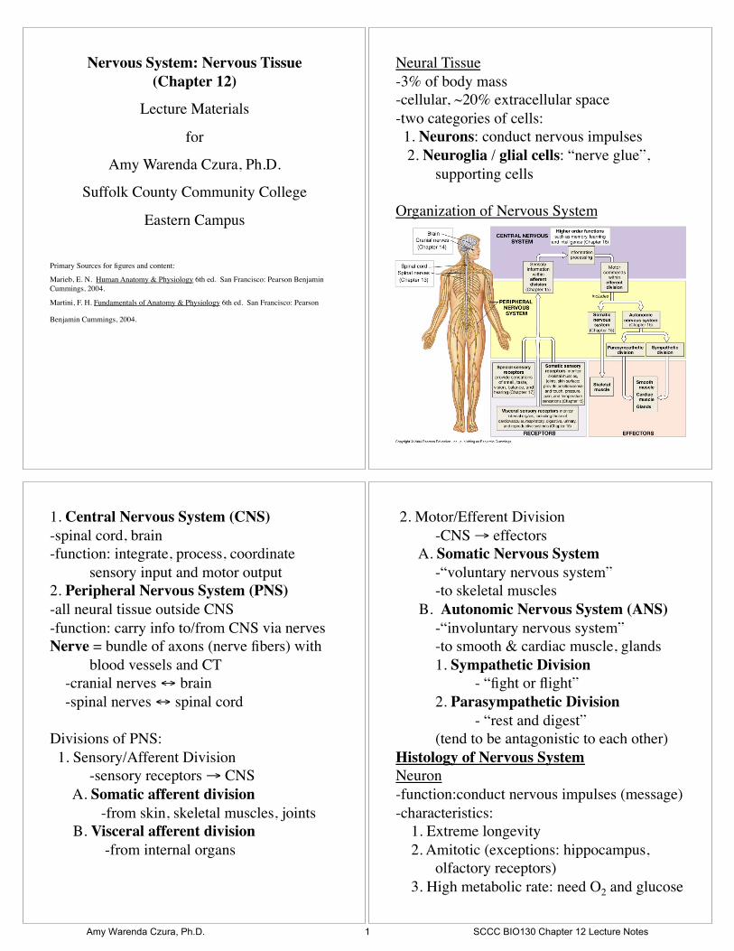
Structure:!
-large soma / perikaryon!
-large nucleus, large nucleolus (rRNA)!
-many mitochondria, ribosomes, RER, Golgi: !
! (#ATP, #protein synthesis to produce !
! !neurotransmitters)!
-Nissl bodies: visible RER & ribosomes, gray!
-neurofilaments = neurofibrils, neurotubules !
! (internal structure)!
-no centrioles!
-2 types of !
! processes:!
(cell extensions)!
1. Dendrites: !
! -receive info !
! -carry a graded potential toward soma!
! -contain same organelles as soma!
! -short, branched!
! -end in dendritic spines!
2. Axon: !
! -single, long !
! -carry an action potential away from soma!
! -release neurotransmitters at end to signal
! !next cell!
! -long ones = “nerve fibers”!
! -contains:!
! !-neurofibrils & neurotubules (abundant)!
! !-vesicles of neurotransmitter!
! !-lysosomes, mitochondria, enzymes!
! !-no Nissl bodies, no Golgi (no protein !
! !! !synthesis in axon)!
! -connects to soma at axon hillock!
! -covered in axolemma (membrane)!
! -may branch: axon collaterals!
! -end in synaptic terminals or knobs!
! -may have myelin sheath: protein+lipid!
! !-protection!
! !-insulation!
! !-increase speed of impulse!
! CNS: myelin from oligodendrocytes!
! PNS: myelin from Schwann cells/!
! !! !neurilemma cells!
Axoplasmic transport!
-move materials between soma and terminal!
-along neurotubules on kinesins!
-Anterograde transport = soma ! terminal!
! (neurotransmitters from soma)!
-Retrograde transport = terminal ! soma!
! (recycle breakdown products from used !
! !neurotransmitters)!
! Some viruses use retrograde transport to !
! !gain access to CNS (Polio, Herpes, !
! !Rabies)!
Amy Warenda Czura, Ph.D. 2 SCCC BIO130 Chapter 12 Lecture Notes
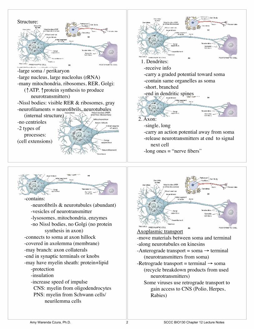
-presynaptic cell sends message along axon to !
! axon terminal!
-postsynaptic cell receives message as !
! neurotransmitter!
Neurotransmitter = chemical, transmits signal !
! from pre- to post- synaptic cell across !
! synaptic cleft !
Synaptic knob = small, round, when !
! postsynaptic cell is neuron, synapse on !
! dendrite or soma!
Synaptic terminal = complex structure, at !
! neuromuscular or neuroglandular junction!
Synapse!
-site where neuron !
communicates with !
another cell: !
neuron or effector!
Structural classification of neurons:!
1. Anaxonic neurons: !
-dendrites and axon look same !
-brain and special sense organs!
2. Bipolar neurons: !
-1 dendrite, 1 axon !
-soma in middle!
-rare: special sense organs, !
! relay from receptor to neuron!
3. Unipolar neurons:!
-1 long axon, dendrites at one !
! end, soma off side (T shape)!
-most sensory neurons!
4. Multipolar neurons:!
-2 or more dendrites!
-1 long axon!
-99% all neurons!
-most CNS!
Functional Classification of Neurons:!
1. Sensory/Afferent neurons!
-transmit info from sensory receptors to CNS!
-most unipolar!
-soma in peripheral sensory ganglia!
Ganglia = collection of cell bodies in PNS!
A. Somatic sensory neurons!
! -receptors monitor outside conditions!
B. Visceral sensory neurons!
! -receptors monitor internal conditions!
2. Motor/Efferent neurons!
-transmit commands from CNS to effectors!
-most multipolar!
A. Somatic motor neurons!
! -innervate skeletal muscle!
! -conscious control or reflexes!
B. Visceral/Autonomic motor neurons!
! -innervate effectors on smooth muscle, !
! cardiac muscle, glands, adipose!
3. Interneurons / Association neurons!
-distribute sensory info and coordinate motor !
! activity!
-between sensory and motor neurons!
-in brain, spinal cord, autonomic ganglia!
-most are multipolar!
Neuroglia =supporting cells!
Neuroglia in CNS!
-outnumber neurons 10:1!
-half mass of brain!
Amy Warenda Czura, Ph.D. 3 SCCC BIO130 Chapter 12 Lecture Notes
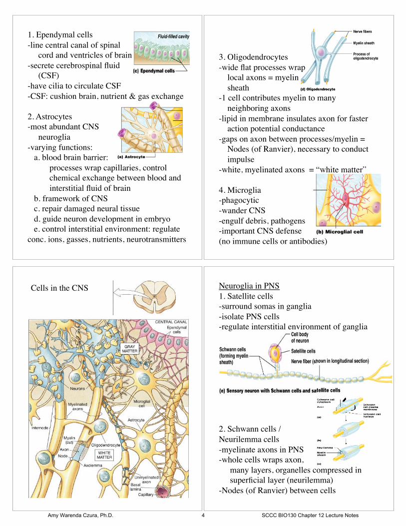
1. Ependymal cells!
-line central canal of spinal !
cord and ventricles of brain!
-secrete cerebrospinal fluid !
(CSF)!
-have cilia to circulate CSF!
-CSF: cushion brain, nutrient & gas exchange!
2. Astrocytes!
-most abundant CNS !
neuroglia!
-varying functions:!
a. blood brain barrier: !
! processes wrap capillaries, control !
! chemical exchange between blood and
! interstitial fluid of brain!
b. framework of CNS!
c. repair damaged neural tissue!
d. guide neuron development in embryo!
e. control interstitial environment: regulate
conc. ions, gasses, nutrients, neurotransmitters!
3. Oligodendrocytes!
-wide flat processes wrap !
local axons = myelin !
sheath!
-1 cell contributes myelin to many !
neighboring axons!
-lipid in membrane insulates axon for faster !
action potential conductance!
-gaps on axon between processes/myelin = !
Nodes (of Ranvier), necessary to conduct !
impulse!
-white, myelinated axons = “white matter”!
4. Microglia!
-phagocytic!
-wander CNS!
-engulf debris, pathogens!
-important CNS defense !
(no immune cells or antibodies)!
Cells in the CNS! Neuroglia in PNS!
1. Satellite cells!
-surround somas in ganglia!
-isolate PNS cells!
-regulate interstitial environment of ganglia!
2. Schwann cells /!
Neurilemma cells!
-myelinate axons in PNS!
-whole cells wraps axon, !
!many layers, organelles compressed in
! superficial layer (neurilemma)!
-Nodes (of Ranvier) between cells!
Amy Warenda Czura, Ph.D. 4 SCCC BIO130 Chapter 12 Lecture Notes
-vital to repair of peripheral never fibers after !
! injury: guide growth to original synapse!
+! +! +! +!
+!
+! +! +! +! +!
+! +! +! +!
+!+!
+!
+!
+!+!+! +! +!
+!+!
+!+!
-!
-!-!-! -!-!
-!-!
-!-! -!-!-!-!-!-!-!
-!-!-!-!
Neurophysiology!
Neurons: conduct electrical impulse!
-requires transmembrane potential = electrical !
! difference across cell membrane!
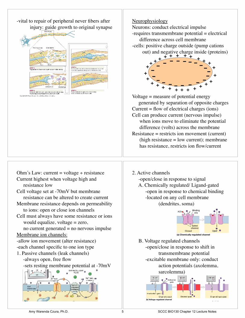
-cells: positive charge outside (pump cations !
out) and negative charge inside (proteins)!
Voltage = measure of potential energy !
generated by separation of opposite charges!
Current = flow of electrical charges (ions)!
Cell can produce current (nervous impulse) !
! when ions move to eliminate the potential
! difference (volts) across the membrane!
Resistance = restricts ion movement (current)!
! (high resistance = low current); membrane
! has resistance, restricts ion flow/current!
Ohm’s Law: current = voltage ÷ resistance!
Current highest when voltage high and !
! resistance low!
Cell voltage set at -70mV but membrane!
! resistance can be altered to create current!
Membrane resistance depends on permeability !
! to ions: open or close ion channels!
Cell must always have some resistance or ions !
! would equalize, voltage = zero, !
! no current generated = no nervous impulse!
Membrane ion channels:!
-allow ion movement (alter resistance)!
-each channel specific to one ion type!
1. Passive channels (leak channels)!
-always open, free flow!
-sets resting membrane potential at -70mV!
2. Active channels!
-open/close in response to signal!
A. Chemically regulated/ Ligand-gated!
! -open in response to chemical binding!
! -located on any cell membrane ! !
! !(dendrites, soma) ! ! !
! !!
B. Voltage regulated channels!
! -open/close in response to shift in !
! !transmembrane potential!
! -excitable membrane only: conduct !
! !action potentials (axolemma, !
! !sarcolemma) !
Amy Warenda Czura, Ph.D. 5 SCCC BIO130 Chapter 12 Lecture Notes

C. Mechanically regulated channels!
! -open in response to membrane !
! !distortion!
! -on dendrites of sensory neurons for !
! !touch, pressure, vibration!
When channel opens, ions flow along ! !
! electrochemical gradient:!
! !-diffusion (high conc. to low)!
! !-electrical attraction/repulsion!
Sodium-Potassium Pump:!
-uses ATP to move 3 Na+ out 2 K+ in !
! (70% of neuron ATP for this)!
-runs anytime cell not conducting impulse!
-creates high [K+] inside and high [Na+]outside!
When Na+ channel opens:!
- Na+ flows into cell:!
!1. Favored by diffusion gradient!
!2. Favored by electrical gradient!
open channel = $resistance = #ion flow/current!
When K+ channel opens:!
- K+ flows out of cell:!
!1. Favored by diffusion gradient only!
!2. Electrical gradient repels K+ exit!
! - Thus less current than Na+ !
Channels open = resistance low = ions move !
! until equilibrium potential: depends on !
! !-diffusion gradient!
! !-electrical gradient!
Equilibrium Potential!
! For K+ = -90mV!
! For Na+ = +66mV!
Open channel ! current ! graded potential!
Graded potential = localized shift in !
! transmembrane potential due to !
! movement of charges in to /out of cell!
Na+ channel opens = Na+ flows in, ! !
! depolarization (cell less negative)!
K+ channel opens = K+ flows out, ! !
!hyperpolarization (cell more negative)!
Graded potentials:!
-occur on any membrane: dendrites and somas!
-can be depolarizing or hyperpolarizing!
-amount of depolarization or hyperpolarization !
! depends on intensity of stimulus: ! !
! # channels open = # voltage change!
-passive spread from site of stimulation over
! short distance!
-effect on membrane potential decreases with !
! distance from stimulation site!
-repolarization occurs as soon as stimulus is !
! removed: leak channels & Na+/K+ pump !
! reset resting potential!
Graded potential = localized change in !
! transmembrane potential, not nervous !
! impulse (message)!
Amy Warenda Czura, Ph.D. 6 SCCC BIO130 Chapter 12 Lecture Notes
If big enough depolarization = action potential !
! = nervous impulse = transmission to !
!next cell!
Action potentials:!
-occur on excitable membranes only !
! (axolemma, sarcolemma)!
-always depolarizing!
-must depolarize to threshold (-55mV) before !
! action potential begins !
! (voltage gated channels on excitable !
! !membrane open at threshold to ! !
! !propagate action potential)!
- “all-or-none” : all stimuli that exceed !
! threshold will produce identical action !
! potentials!
-action potential at one site depolarizes !
! adjacent sites to threshold!
-propagated across entire membrane surface !
! without decrease in strength!
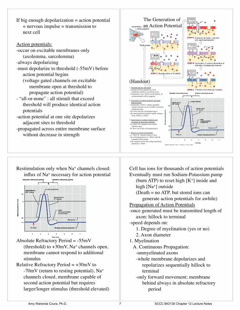
The Generation of
an Action Potential!
-55 mV!
1. Depolarization to threshold:!
- a graded potential depolarizes local !
membrane and flows toward the axon!
- if threshold is met (-55mV) at the hillock, an !
action potential will be triggered!
2. Activation of sodium channels and rapid !
depolarization:!
- at threshold (-55mV), voltage-regulated !
sodium channels on the excitable axolemma !
membrane open!
- Na+ flows into the cell depolarizing it!
- the transmembrane potential rapidly changes !
from -55mV to +30mV!
3. Inactivation of sodium channels and !
activation of potassium channels:!
- at +30mV Na+ channels close and K+ !
channels open!
- K+ flows out of the cell repolarizing it!
4. Return to normal permeability:!
- at -70mV K+ channels begin to close!
- the cell hyperpolarizes to -90mV until all !
channels finish closing!
- leak channels restore the resting membrane !
potential to -70mV!
(Handout)!
Restimulation only when Na+ channels closed: !
influx of Na+ necessary for action potential!
Absolute Refractory Period = -55mV !
! (threshold) to +30mV, Na+ channels open,
! membrane cannot respond to additional !
! stimulus!
Relative Refractory Period = +30mV to !
! -70mV (return to resting potential), Na+ !
! channels closed, membrane capable of !
! second action potential but requires !
! larger/longer stimulus (threshold elevated)!
Cell has ions for thousands of action potentials!
Eventually must run Sodium-Potassium pump !
! (burn ATP) to reset high [K+] inside and !
! high [Na+] outside!
! (Death = no ATP, but stored ions can !
! !generate action potentials for awhile)!
Propagation of Action Potentials!
-once generated must be transmitted length of !
! axon: hillock to terminal!
-speed depends on: !
! 1. Degree of myelination (yes or no)!
! 2. Axon diameter!
1. Myelination!
A. Continuous Propagation: !
! -unmyelinated axons!
! -whole membrane depolarizes and !
! repolarizes sequentially hillock to !
! terminal!
! -only forward movement; membrane !
! behind always in absolute refractory !
period!
Amy Warenda Czura, Ph.D. 7 SCCC BIO130 Chapter 12 Lecture Notes
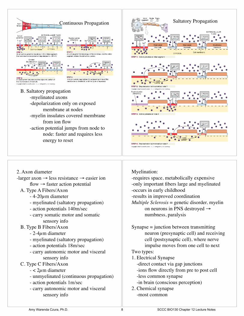
B. Saltatory propagation!
! -myelinated axons!
! -depolarization only on exposed !
! !membrane at nodes!
! -myelin insulates covered membrane !
! !from ion flow!
! -action potential jumps from node to !
! !node: faster and requires less !
! !energy to reset!
Continuous Propagation! Saltatory Propagation!
2. Axon diameter!
-larger axon ! less resistance ! easier ion !
!flow ! faster action potential!
A. Type A Fibers/Axon!
! - 4-20µm diameter !
! - myelinated (saltatory propagation)!
! - action potentials 140m/sec!
! - carry somatic motor and somatic !
! !sensory info!
B. Type B Fibers/Axon!
! - 2-4µm diameter!
! - myelinated (saltatory propagation)!
! - action potentials 18m/sec!
! - carry autonomic motor and visceral !
! !sensory info!
C. Type C Fibers/Axon!
! - < 2µm diameter!
! - unmyelinated (continuous propagation)!
! - action potentials 1m/sec!
! - carry autonomic motor and visceral !
! !sensory info!
Myelination: !
-requires space, metabolically expensive!
-only important fibers large and myelinated!
-occurs in early childhood!
-results in improved coordination!
Multiple Sclerosis = genetic disorder, myelin !
! on neurons in PNS destroyed ! !
! numbness, paralysis!
Synapse = junction between transmitting !
! neuron (presynaptic cell) and receiving !
! cell (postsynaptic cell), where nerve !
! impulse moves from one cell to next!
Two types:!
1. Electrical Synapse!
-direct contact via gap junctions!
-ions flow directly from pre to post cell!
-less common synapse!
-in brain (conscious perception)!
2. Chemical synapse!
-most common!
Amy Warenda Czura, Ph.D. 8 SCCC BIO130 Chapter 12 Lecture Notes
-pre and post cells separated by synaptic cleft!
-presynaptic neuron releases neurotransmitter !
! to trigger effect on post synaptic cell!
-dynamic: facilitate or inhibit transmission, !
! depends on neurotransmitter:!
! 1. Excitatory Neurotransmitters = !
! !-depolarization!
! !-propagate action potential!
! 2. Inhibitory Neurotransmitters = !
! !-hyperpolarization!
! !-suppress action potential!
Propagation across chemical synapse always !
! slow but allows variability!
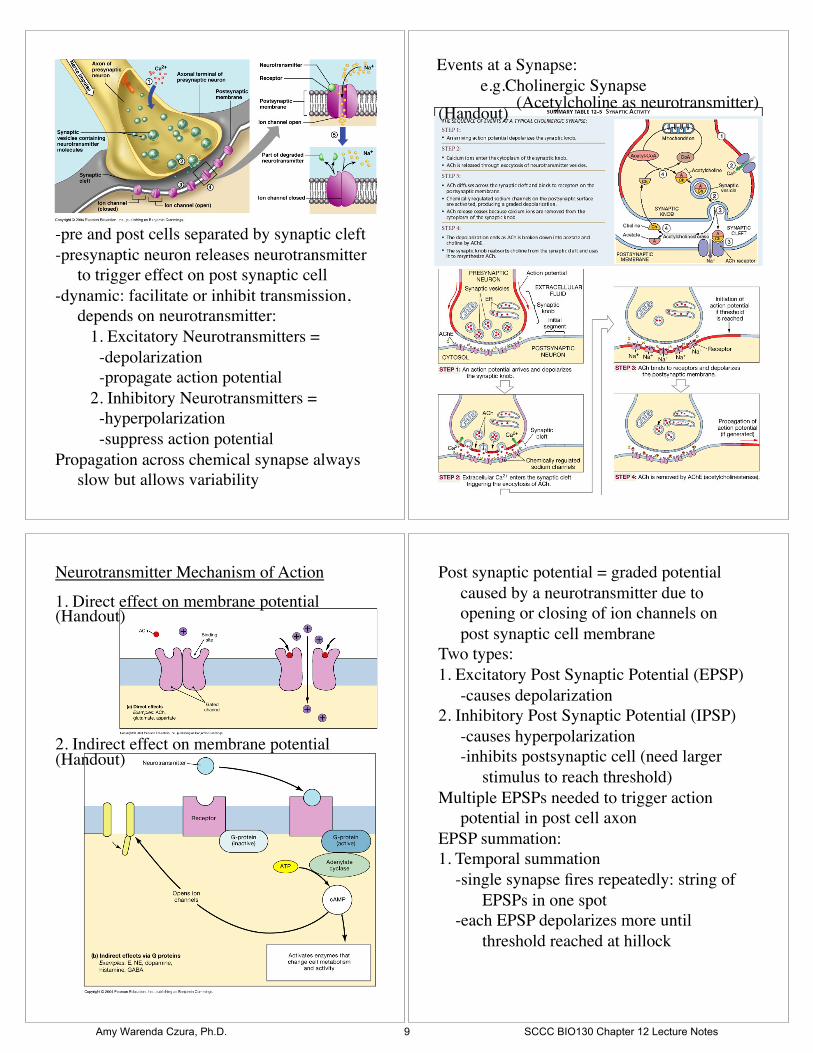
Events at a Synapse:!
! e.g.Cholinergic Synapse!(Acetylcholine as neurotransmitter)!
(Handout)!
Neurotransmitter Mechanism of Action!
1. Direct effect on membrane potential!
2. Indirect effect on membrane potential!
(Handout)!
(Handout)!
Post synaptic potential = graded potential !
! caused by a neurotransmitter due to !
! opening or closing of ion channels on !
! post synaptic cell membrane!
Two types:!
1. Excitatory Post Synaptic Potential (EPSP)!
! -causes depolarization!
2. Inhibitory Post Synaptic Potential (IPSP)!
! -causes hyperpolarization!
! -inhibits postsynaptic cell (need larger !
! !stimulus to reach threshold)!
Multiple EPSPs needed to trigger action !
! potential in post cell axon!
EPSP summation:!
1. Temporal summation!
-single synapse fires repeatedly: string of !
! !EPSPs in one spot!
-each EPSP depolarizes more until !
! !threshold reached at hillock!
Amy Warenda Czura, Ph.D. 9 SCCC BIO130 Chapter 12 Lecture Notes

2. Spatial summation!
-multiple synapses fire simultaneously!
-collective depolarization reaches threshold!
Facilitated = depolarized; brought closer to !
! threshold by some sort of stimulus, less !
! stimulus now required to reach threshold!
! (e.g. caffeine)!
Post Synaptic Potentiation:!
-repeat stimulation of same synapse ! !
! conditions synapse, pre cell more easily !
! stimulates post cell to threshold (repetition)!
Most nervous system activity results from !
! interplay of EPSPs and IPSPs to !
! promote differing degrees of facilitation !
! or inhibition to allow constant fine !
! tuning of response!
Neuromodulators = chemicals that influence !
! synthesis, release, or degradation of !
! neurotransmitters thus altering normal !
! response of the synapse!
Common Neurotransmitters:!
1. Acetycholine- cholinergic synapses!
-excitatory!
-direct effect!
-skeletal neuromuscular junctions, many !
!CNS synapses, all neuron to neuron !
! PNS, all parasympathetic ANS!
2. Norepinephrine- adrenergic synapses!
-excitatory!
-second messengers!
-many brain synapses, all sympathetic ANS !
! effector junctions!
3. Dopamine!
-excitatory or inhibitory !
-second messengers!
-many brain synapses, many functions!
! -responsible for reward feeling!
! !-cocaine: inhibits removal = “high”!
! -Parkinson’s disease: damage neurons = !
! !ticks, jitters!
4. Serotonin!
-inhibitory!
-direct or second messenger!
-brain stem for emotion!
! -anti-depression/ anti-anxiety drugs !
! !block uptake!
5. Gamma aminobutyric acid (GABA)!
-inhibitory!
-direct effect!
-brain: anxiety control, motor coordination!
! -alcohol: augments effects = loss of !
! !coordination!
Factors that disrupt neural function:!
1. pH: normal = 7.4!
@ pH 7.8 ! spontaneous action potentials !
! != convulsions!
@ pH 7.0 ! no action potentials!
! ! = unresponsive!
2. Ion concentrations!
high extracellular [K+] ! depolarize !
! membranes = death, cardiac arrest!
3. Temperature: normal = 37°C!
-higher: neurons more excitable!
! (fever = hallucinations)!
-lower: neurons non-responsive!
! (hypothermia = lethargy, confusion)!
4. Nutrients!
-neurons: no reserves, use a lot of ATP!
-require constant and abundant glucose!
-glucose only!
5. Oxygen!
-aerobic respiration only for ATP!
-no ATP = neuron damage/death!
Amy Warenda Czura, Ph.D. 10 SCCC BIO130 Chapter 12 Lecture Notes