Embed Size (px)
Citation preview
Manual
Cytoskeleton, Inc.
The Protein
Experts
cytoskeleton.com Phone: (303) 322.2254 Fax: (303) 322.2257
Customer Service: [email protected]
Technical Support: [email protected]
V. 5.3
Microtubules/Tubulin
In Vivo Assay Kit
Cat. # BK038

cytoskeleton.com Page 2
cytoskeleton.com Page 3
Section I: Introduction
Overview ............................................................................................. 5
Uses of Kit ........................................................................................... 5
Section II: Purchaser Notification .................................................................... 6
Section III: Kit Contents ................................................................................... 7
Section IV: Reconstitution and Storage of Components ............................... 8
Section V: Important Technical Notes
A) Notes on Updated Version 4.0 ........................................................ 9
B) Assay Temperature ......................................................................... 9
Section VI: Assay Protocol: Detailed Method
Quick View ........................................................................................... 10
Initial Considerations ............................................................................ 10
Detailed Method: Part 1: Assay Preparation ....................................... 11
Detailed Method: Part 2: Lysate Collection & Processing
A) Cells in Suspension ............................................................ 12
B) Adherent Cells .................................................................... 12
C) Tissue Samples .................................................................. 12-13
Detailed Method: Part 3: Western Blot Analysis ..................................... 14
Recommended Controls ........................................................................ 15
Section VII: Troubleshooting .......................................................................... 16
Manual Contents

cytoskeleton.com Page 4
cytoskeleton.com Page 5
Overview
This kit provides the end user with a method of determining the amount of microtubule
content versus free-tubulin content in a cell population. Cells or tissues are lysed in a
microtubule stabilization buffer that preserves the integrity of the microtubule to tubulin
ratio in the cells and forms the basis of the assay. After lysis, a centrifugation step sepa-
rates polymerized microtubules (pellet fraction) from non-polymerized tubulin
(supernatant fraction). The tubulin in supernatant vs pellet fractions are subsequently
analyzed by quantitative western blots. The final result provides a quantitation of the ratio
of tubulin incorporated into the cytoskeleton versus the free-tubulin found in the cytosol.
Uses of the kit
1. To study the effects of pharmaceutical compounds on the ratio of tubulin to microtu-
bules.
2. To study the effects of mutated cell lines versus their parent cell line for the change
in ratio of tubulin to microtubules.
3. To study the effects of physical alterations of environment on the ratio of tubulin to
microtubules.
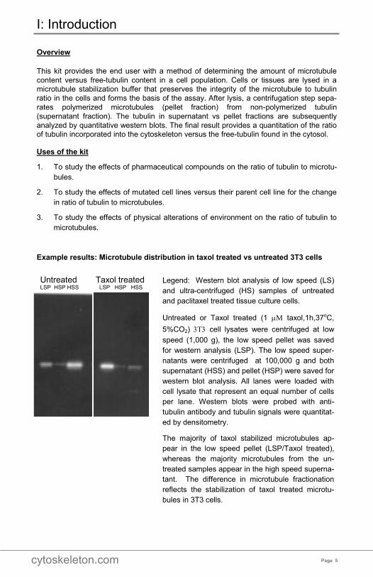
Example results: Microtubule distribution in taxol treated vs untreated 3T3 cells
I: Introduction
Legend: Western blot analysis of low speed (LS)
and ultra-centrifuged (HS) samples of untreated
and paclitaxel treated tissue culture cells.
Untreated or Taxol treated (1 taxol,1h,37oC,
5%CO2) cell lysates were centrifuged at low
speed (1,000 g), the low speed pellet was saved
for western analysis (LSP). The low speed super-
natants were centrifuged at 100,000 g and both
supernatant (HSS) and pellet (HSP) were saved for
western blot analysis. All lanes were loaded with
cell lysate that represent an equal number of cells
per lane. Western blots were probed with anti-
tubulin antibody and tubulin signals were quantitat-
ed by densitometry.
The majority of taxol stabilized microtubules ap-
pear in the low speed pellet (LSP/Taxol treated),
whereas the majority microtubules from the un-
treated samples appear in the high speed superna-
tant. The difference in microtubule fractionation
reflects the stabilization of taxol treated microtu-
bules in 3T3 cells.
Untreated Taxol treated LSP HSP HSS LSP HSP HSS
cytoskeleton.com Page 6
Limited Use Statement
The purchase of this product conveys to the buyer the non-transferable right to use the
purchased amount of product and components of product in research conducted by the
buyer. The buyer cannot sell or otherwise transfer this product or any component thereof
to a third party or otherwise use this product or its components for commercial purposes.
Commercial purposes include, but are not limited to: use of the product or its components
in manufacturing; use of the product or its components to provide a service; resale of the
product or its components.
The terms of this Limited Use Statement apply to all buyers including academic and for-
profit entities. If the purchaser is not willing to accept the conditions of this Limited Use
Statement, Cytoskeleton Inc. is willing to accept return of the unused product with a full
refund.
II: Purchaser Notification
cytoskeleton.com Page 7
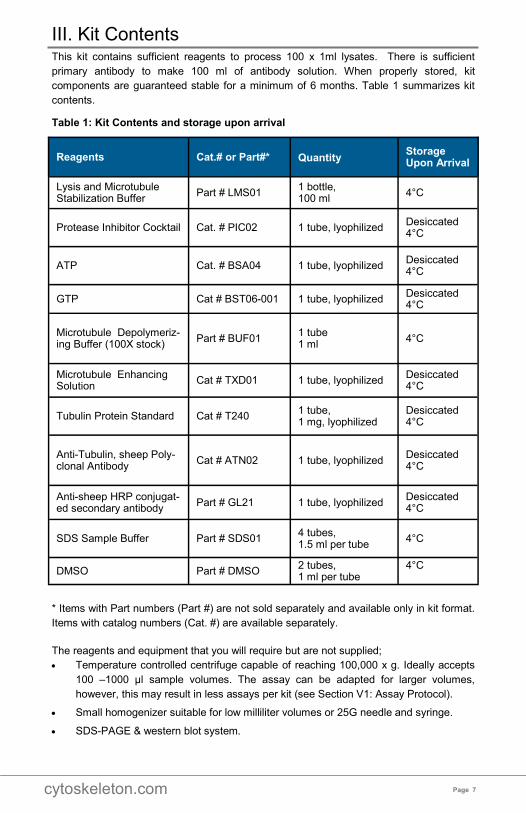
This kit contains sufficient reagents to process 100 x 1ml lysates. There is sufficient
primary antibody to make 100 ml of antibody solution. When properly stored, kit
components are guaranteed stable for a minimum of 6 months. Table 1 summarizes kit
contents.
Table 1: Kit Contents and storage upon arrival
* Items with Part numbers (Part #) are not sold separately and available only in kit format.
Items with catalog numbers (Cat. #) are available separately.
The reagents and equipment that you will require but are not supplied;
• Temperature controlled centrifuge capable of reaching 100,000 x g. Ideally accepts
100 –1000 µl sample volumes. The assay can be adapted for larger volumes,
however, this may result in less assays per kit (see Section V1: Assay Protocol).
• Small homogenizer suitable for low milliliter volumes or 25G needle and syringe.
• SDS-PAGE & western blot system.
III. Kit Contents
Reagents Cat.# or Part#* Quantity
Storage Upon Arrival
Lysis and Microtubule Stabilization Buffer
Part # LMS01 1 bottle, 100 ml
4°C
Protease Inhibitor Cocktail
Cat. # PIC02 1 tube, lyophilized Desiccated 4°C
ATP
Cat. # BSA04 1 tube, lyophilized Desiccated 4°C
GTP Cat # BST06-001 1 tube, lyophilized Desiccated 4°C
Microtubule Depolymeriz-ing Buffer (100X stock)
Part # BUF01 1 tube 1 ml
4°C
Microtubule Enhancing Solution
Cat # TXD01 1 tube, lyophilized Desiccated 4°C
Tubulin Protein Standard
Cat # T240 1 tube, 1 mg, lyophilized
Desiccated 4°C
Anti-Tubulin, sheep Poly-clonal Antibody
Cat # ATN02 1 tube, lyophilized Desiccated 4°C
Anti-sheep HRP conjugat-ed secondary antibody
Part # GL21 1 tube, lyophilized Desiccated 4°C
SDS Sample Buffer
Part # SDS01 4 tubes, 1.5 ml per tube
4°C
DMSO Part # DMSO 2 tubes, 1 ml per tube
4°C
cytoskeleton.com Page 8
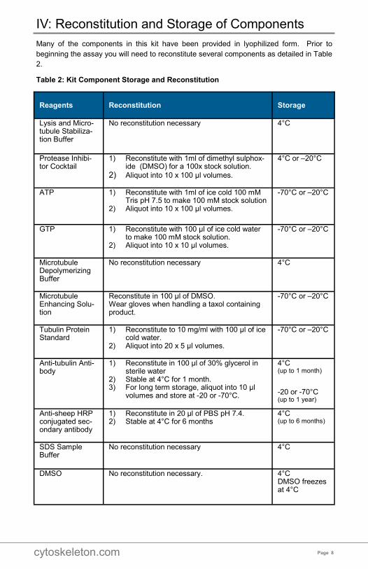
Many of the components in this kit have been provided in lyophilized form. Prior to
beginning the assay you will need to reconstitute several components as detailed in Table
2.
Table 2: Kit Component Storage and Reconstitution
IV: Reconstitution and Storage of Components
Reagents Reconstitution Storage
Lysis and Micro-tubule Stabiliza-tion Buffer
No reconstitution necessary 4°C
Protease Inhibi-tor Cocktail
1) Reconstitute with 1ml of dimethyl sulphox-ide (DMSO) for a 100x stock solution.
2) Aliquot into 10 x 100 µl volumes.
4°C or –20°C
ATP
1) Reconstitute with 1ml of ice cold 100 mM Tris pH 7.5 to make 100 mM stock solution
2) Aliquot into 10 x 100 µl volumes.
-70°C or –20°C
GTP 1) Reconstitute with 100 μl of ice cold water to make 100 mM stock solution.
2) Aliquot into 10 x 10 µl volumes.
-70°C or –20°C
Microtubule Depolymerizing Buffer
No reconstitution necessary 4°C
Microtubule Enhancing Solu-tion
Reconstitute in 100 µl of DMSO. Wear gloves when handling a taxol containing product.
-70°C or –20°C
Tubulin Protein Standard
1) Reconstitute to 10 mg/ml with 100 μl of ice cold water.
2) Aliquot into 20 x 5 µl volumes.
-70°C or –20°C
Anti-tubulin Anti-body
1) Reconstitute in 100 µl of 30% glycerol in sterile water
2) Stable at 4°C for 1 month. 3) For long term storage, aliquot into 10 µl
volumes and store at -20 or -70°C.
4°C (up to 1 month)
-20 or -70°C (up to 1 year)
Anti-sheep HRP conjugated sec-ondary antibody
1) Reconstitute in 20 µl of PBS pH 7.4. 2) Stable at 4°C for 6 months
4°C (up to 6 months)
SDS Sample Buffer
No reconstitution necessary 4°C
DMSO No reconstitution necessary.
4°C DMSO freezes at 4°C
cytoskeleton.com Page 9
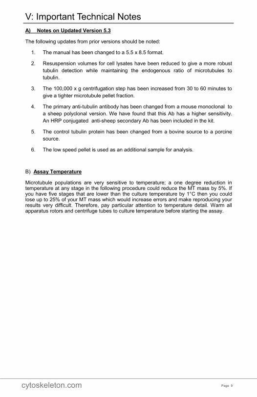
A) Notes on Updated Version 5.3
The following updates from prior versions should be noted:
1. The manual has been changed to a 5.5 x 8.5 format.
2. Resuspension volumes for cell lysates have been reduced to give a more robust
tubulin detection while maintaining the endogenous ratio of microtubules to
tubulin.
3. The 100,000 x g centrifugation step has been increased from 30 to 60 minutes to
give a tighter microtubule pellet fraction.
4. The primary anti-tubulin antibody has been changed from a mouse monoclonal to
a sheep polyclonal version. We have found that this Ab has a higher sensitivity.
An HRP conjugated anti-sheep secondary Ab has been included in the kit.
5. The control tubulin protein has been changed from a bovine source to a porcine
source.
6. The low speed pellet is used as an additional sample for analysis.
B) Assay Temperature
Microtubule populations are very sensitive to temperature; a one degree reduction in temperature at any stage in the following procedure could reduce the MT mass by 5%. If you have five stages that are lower than the culture temperature by 1°C then you could lose up to 25% of your MT mass which would increase errors and make reproducing your results very difficult. Therefore, pay particular attention to temperature detail. Warm all apparatus rotors and centrifuge tubes to culture temperature before starting the assay.
V: Important Technical Notes
cytoskeleton.com Page 10
Assay Quick View: 1. Wash cells with 37°C PBS pH 7.4. 2. Suspend cells or tissue in LMS2. 3. Gently homogenize to lyse cells. 4. Centrifuge lysates at 1,000 g to sediment large complexes of microtubules attached
to nuclei and Golgi (low speed pellet fraction). 5. Centrifuge supernatants at 100,000xg to separate microtubules (high speed pellet
fraction) from soluble tubulin (high speed supernatant fraction). 6. Analyze the high speed supernatant versus high and low speed pellet for tubulin
content using western blotting. 7. Scan tubulin bands by densitometry and calculate the ratio of tubulin in the
microtubules (pellets) versus that present as free tubulin (supernatant).
Initial Considerations: The microtubules / tubulin in vivo assay requires a constant cells to buffer volume ratio. Essentially the lysis step has to dilute the cellular extract so that the free tubulin does not polymerize onto existing microtubules (MTs). This ratio is roughly 10 volumes of buffer to 1 volume of cell pellet, larger volumes of buffer are fine and in this kit the ratio is aiming at 20 - 50 volumes of buffer per volume of cells.
VI: Assay Protocol: Quick Overview
cytoskeleton.com Page 11
Detailed Assay Method
PART 1: Assay Preparation
1. The assay requires a low speed centrifuge (1,000 g) and a high speed centrifuge
(100,000 g) capable of taking small volumes (0.1-1 ml). Rotors should be warmed to
37°C prior to beginning the assay.
2. Determine the total volume of LMS2 buffer you require per experiment using the
volumes given in Table 3 as a guide (see LMS2 recipe below) .
3. Make the required volume of LMS2 buffer as follows;
1 ml LMS01 buffer (Lysis and Microtubule Stabilization Buffer)
1 µl BST06 (100 mM GTP stock solution (remaining stock can be re-frozen)
10 µl BSA04 (100 mM ATP stock solution (remaining stock can be re-frozen)
10 µl PIC02 (100x protease inhibitor cocktail stock (remaining stock can be re-frozen)
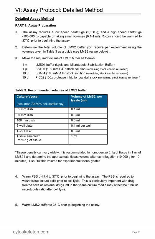
Table 3: Recommended volumes of LMS2 buffer
*Tissue density can vary widely. It is recommended to homogenize 0.1g of tissue in 1 ml of
LMS01 and determine the approximate tissue volume after centrifugation (10,000 g for 10
minutes). Use 20x this volume for experimental tissue lysates.
4. Warm PBS pH 7.4 to 37°C prior to beginning the assay. The PBS is required to
wash tissue culture cells prior to cell lysis. This is particularly important with drug
treated cells as residual drugs left in the tissue culture media may affect the tubulin/
microtubule ratio after cell lysis.
5. Warm LMS2 buffer to 37°C prior to beginning the assay.
VI: Assay Protocol: Detailed Method
Culture Vessel
(assumes 70-80% cell confluency)
Volume of LMS2 per
lysate (ml)
35 mm dish 0.1 ml
60 mm dish 0.3 ml
100 mm dish 0.6 ml
6-well plate 0.1 ml per well
T-25 Flask 0.3 ml
Tissue samples*
Per 0.1g of tissue
1 ml
cytoskeleton.com Page 12
PART 2: Lysate Collection & Processing
1. Lysis methods for suspension cells (A), adherent cells (B) or tissue samples (C) are
given below.
A) Cells in suspension
a) Harvest cells by centrifugation at 1,000 x g for 2 minutes.
b) Remove supernatant and discard.
c) Wash once by gently resuspending the cells in 10 ml of 37˚C PBS pH 7.4.
d) Harvest cells by centrifugation at 1,000 x g for 2 minutes.
e) Resuspend cell pellet in 20 x cell pellet volume of 37˚C LMS2.
f) Proceed to step 2.
B) Adherent Cells
a) Aspirate media from dish. Incline dish to 30° angle to help remove of as much
media as possible.
b) Wash once by gently adding 10 ml of 37˚C PBS pH 7.4.
c) Aspirate PBS from dish. Incline dish to 30° angle to help remove of as much PBS
as possible.
d) Add appropriate volume of 37˚C LMS2 (see Table 3).
e) Harvest cells by scraping thoroughly with cell scraper, again keep the plate at a
30° angle to help collect all of the lysate.
f) Pipette cell lysate into tube and proceed to step 2.
C) Tissue Samples
a) Add 1000 µl of 37˚C LMS2 per 100 mg (0.1g) of tissue sample.
b) Proceed to step 2.
2. Homogenize samples using a small hand held or motorized homogenizer suitable for
low milliliter volumes or a 25G syringe or a 200 µl pipet tip (usually sufficient for cell
culture samples).
3. Immediately centrifuge for 5 minutes at 1,000 x g at 37˚C.
4. Carefully remove the low speed supernatants to fresh tubes and place the low speed
pellets on ice.
5. Remove 100 µl volume of low speed supernatant from each sample into a clean ultra
-centrifuge tube at 37°C. Any remaining lysate can be discarded or used for other
purposes at this point.
NOTE: Larger volumes of lysate can be processed if required.
VI: Assay Protocol: Detailed Method (continued)
cytoskeleton.com Page 13
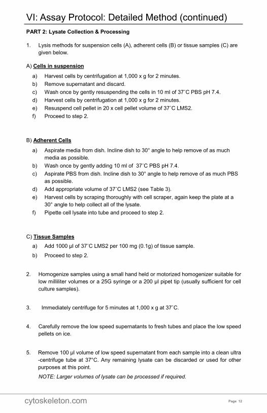
6. Centrifuge the low speed supernatant at 100,000 x g for 60 minutes at 37°C. This
step will pellet microtubules and leave unpolymerized tubulin in the supernatant.
Tip: Before placing the centrifuge tubes in the ultracentrifuge rotor, mark the outer side of
each tube with a sharpie dot to indicate the place where a microtubule pellet will
form.
6. After the ultra-centrifuge step is complete, immediately and gently remove the TOP
80 µl of supernatants to fresh tubes designated as high speed supernatant samples.
Ultra-centrifuge supernatants should be removed gently to avoid disturbing the
microtubule pellet. Only 80 µl of supernatant is removed to avoid disturbing the
microtubule pellet. The microtubule pellet is not likely to be visible. Add 20 ul of 5X
SDS buffer to each 80 µl sample.
7. Very carefully remove the final 20 µl of supernatant, point the pipette away from the
position of the microtubule pellet so as to avoid disturbing the pellet. Discard the 20
µl supernatant.
8. Dilute the 100X stock of Microtubule depolymerization Buffer (BUF01) 1:100 to give
a 1X BUF01 solution (10 µl of BUF01 into 990 µl of ice cold nanopurewater) and
suspend the high speed pellets in a volume equal to the lysate supernatant volume.
e.g. if 100 µl of lysate was centrifuged then suspend the microtubule pellet in 100 µl
of 1X BUF01. Add 25 ul of 5X SDS buffer per 100 ul of resuspended pellet.
9. Leave the tubes at room temperature for 15 minutes to help disperse the microtubule
pellet. Pipette up and down several times with a 200 µl pipette to help shear and
homogenize the microtubule pellet.
10. Resuspend the low speed pellets in a volume of 2X SDS buffer that is equivalent to
1.2 volumes of the original lysis buffer e.g. if you lysed the cells in 200 ul of lysis
buffer then resuspend the low speed pellet in 240 ul of 2X SDS buffer.
11. The samples are now ready for tubulin quantitation by SDS-PAGE and western blot
analysis. Samples can be stored at –20°C prior to moving to PART 3: Tubulin
quantification by SDS-PAGE / Western blot analysis.
VI: Assay Protocol: Detailed Method (continued)
Center of
ultracentrifuge
Red sharpie dot denotes area
where pellet would form after cen-
trifugation. This will help prevent
pellet dislodgement when removing
supernatant.
cytoskeleton.com Page 14
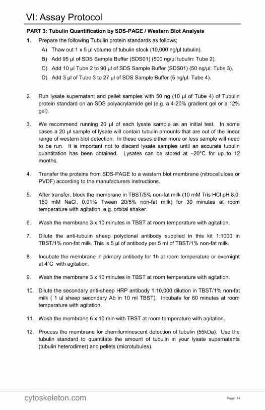
PART 3: Tubulin Quantification by SDS-PAGE / Western Blot Analysis
1. Prepare the following Tubulin protein standards as follows;
A) Thaw out 1 x 5 µl volume of tubulin stock (10,000 ng/µl tubulin).
B) Add 95 µl of SDS Sample Buffer (SDS01) (500 ng/µl tubulin: Tube 2).
C) Add 10 µl Tube 2 to 90 µl of SDS Sample Buffer (SDS01) (50 ng/µl: Tube 3).
D) Add 3 µl of Tube 3 to 27 µl of SDS Sample Buffer (5 ng/µl: Tube 4).
2. Run lysate supernatant and pellet samples with 50 ng (10 µl of Tube 4) of Tubulin
protein standard on an SDS polyacrylamide gel (e.g. a 4-20% gradient gel or a 12%
gel).
3. We recommend running 20 µl of each lysate sample as an initial test. In some
cases a 20 µl sample of lysate will contain tubulin amounts that are out of the linear
range of western blot detection. In these cases either more or less sample will need
to be run. It is important not to discard lysate samples until an accurate tubulin
quantitation has been obtained. Lysates can be stored at –20°C for up to 12
months.
4. Transfer the proteins from SDS-PAGE to a western blot membrane (nitrocellulose or
PVDF) according to the manufacturers instructions.
5. After transfer, block the membrane in TBST/5% non-fat milk (10 mM Tris HCl pH 8.0,
150 mM NaCl, 0.01% Tween 20/5% non-fat milk) for 30 minutes at room
temperature with agitation, e.g. orbital shaker.
6. Wash the membrane 3 x 10 minutes in TBST at room temperature with agitation.
7. Dilute the anti-tubulin sheep polyclonal antibody supplied in this kit 1:1000 in
TBST/1% non-fat milk. This is 5 µl of antibody per 5 ml of TBST/1% non-fat milk.
8. Incubate the membrane in primary antibody for 1h at room temperature or overnight
at 4˚C with agitation.
9. Wash the membrane 3 x 10 minutes in TBST at room temperature with agitation.
10. Dilute the secondary anti-sheep HRP antibody 1:10,000 dilution in TBST/1% non-fat
milk ( 1 ul sheep secondary Ab in 10 ml TBST). Incubate for 60 minutes at room
temperature with agitation.
11. Wash the membrane 6 x 10 min with TBST at room temperature with agitation.
12. Process the membrane for chemiluminescent detection of tubulin (55kDa). Use the
tubulin standard to quantitate the amount of tubulin in your lysate supernatants
(tubulin heterodimer) and pellets (microtubules).
VI: Assay Protocol
cytoskeleton.com Page 15
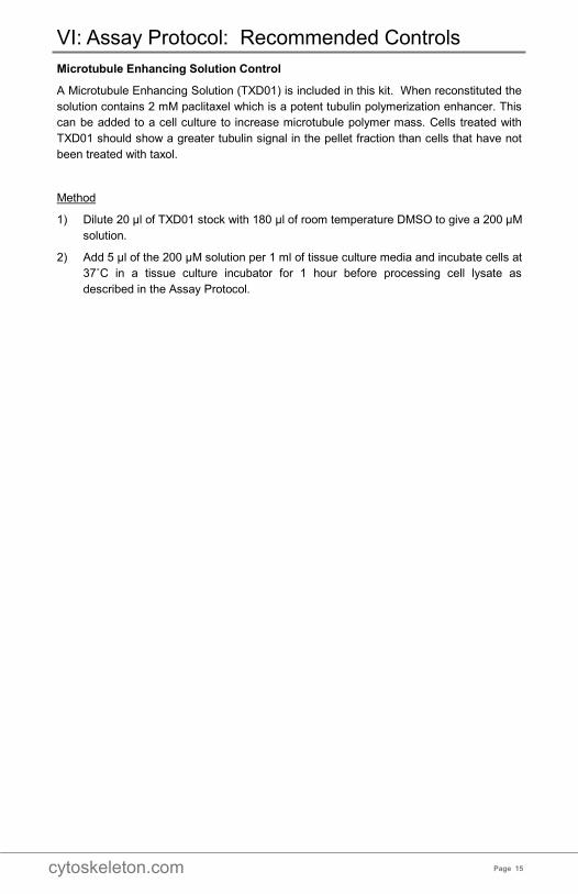
Microtubule Enhancing Solution Control
A Microtubule Enhancing Solution (TXD01) is included in this kit. When reconstituted the
solution contains 2 mM paclitaxel which is a potent tubulin polymerization enhancer. This
can be added to a cell culture to increase microtubule polymer mass. Cells treated with
TXD01 should show a greater tubulin signal in the pellet fraction than cells that have not
been treated with taxol.
Method
1) Dilute 20 μl of TXD01 stock with 180 μl of room temperature DMSO to give a 200 μM
solution.
2) Add 5 μl of the 200 μM solution per 1 ml of tissue culture media and incubate cells at
37˚C in a tissue culture incubator for 1 hour before processing cell lysate as
described in the Assay Protocol.
VI: Assay Protocol: Recommended Controls
cytoskeleton.com Page 16
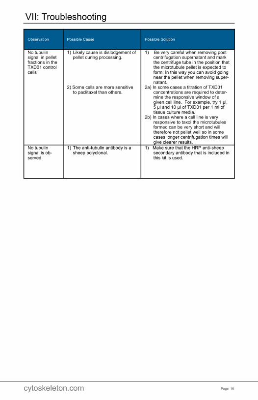
VII: Troubleshooting
Observation
Possible Cause Possible Solution
No tubulin signal in pellet fractions in the TXD01 control cells
1) Likely cause is dislodgement of pellet during processing.
2) Some cells are more sensitive
to paclitaxel than others.
1) Be very careful when removing post centrifugation supernatant and mark the centrifuge tube in the position that the microtubule pellet is expected to form. In this way you can avoid going near the pellet when removing super-natant.
2a) In some cases a titration of TXD01 concentrations are required to deter-mine the responsive window of a given cell line. For example, try 1 μl, 5 μl and 10 μl of TXD01 per 1 ml of tissue culture media.
2b) In cases where a cell line is very responsive to taxol the microtubules formed can be very short and will therefore not pellet well so in some cases longer centrifugation times will give clearer results.
No tubulin signal is ob-served
1) The anti-tubulin antibody is a sheep polyclonal.
1) Make sure that the HRP anti-sheep secondary antibody that is included in this kit is used.

cytoskeleton.com Page 17

cytoskeleton.com Page 18
NOTES:

cytoskeleton.com Page 19
NOTES:

cytoskeleton.com Phone: (303) 322.2254 Fax: (303) 322.2257
Customer Service: [email protected]
Technical Support: [email protected]