Embed Size (px)
Citation preview

MiA
FsibecbfsApp
PaSiwm
srld
D
A
3
anaging Tunnel Malposition and Wideningn Revision Anterior Cruciate Ligament Surgeryndreas H. Gomoll, MD, and Bernard R. Bach, Jr, MD
Tunnel malposition has been identified as the single most common technical error leadingto failure of primary anterior cruciate ligament reconstruction. Revision of malpositioned orwidened tunnels remains a challenging procedure that requires thorough preoperativeplanning. This article will discuss the management of tunnel complications during revisionanterior cruciate ligament reconstruction.Oper Tech Sports Med 14:36-44 © 2006 Elsevier Inc. All rights reserved.
KEYWORDS revision ACL reconstruction, ACL complications, tunnel malposition, tunnelwidening
tcTwatiw
cstnBgarg
saapmttwnteb
ailure rates of 10% to 15%1-3 are being reported afteranterior cruciate ligament (ACL) reconstruction. Malpo-
itioned tibial and/or femoral tunnels are the most commonlydentified technical reasons for failure of ACL reconstructionecause of the resultant abnormal graft isometry. However,ven correctly placed-but-widened tunnels can present ahallenge to the revising surgeon as a result of the associatedone loss, which can compromise fixation. This article willocus on the recognition and management of tunnel malpo-itioning and widening during revision ACL reconstruction.more detailed description of the basic technical aspects of
rimary and revision ACL reconstruction has been providedreviously.4,5
reoperative Workupnd Surgical Decision-Making
urgical decision-making before revision ACL reconstructions based on a thorough history and physical examination, asell as imaging studies to identify associated pathology thatight require concomitant correction at the time of surgery.Radiographic evaluation is essential and should include
tanding anterior–posterior and 45° flexion posterior–ante-ior views, lateral, and Merchant views. Any concerns overimb malalignment should be investigated with single- orouble-stance, full-length alignment films. On the basis of
ivision of Sports Medicine, Department of Orthopedic Surgery, Rush Uni-versity Medical Center, Chicago, IL.
ddress reprint requests to Bernard R. Bach, Jr, MD, Rush University Med-ical Center, 1725 W Harrison St, Suite 1063, Chicago, IL 60612. E-mail:
6 1060-1872/06/$-see front matter © 2006 Elsevier Inc. All rights reserved.doi:10.1053/j.otsm.2006.02.007
hese imaging studies, the tibial and femoral tunnels can belassified as either anatomic or malpositioned or widened.unnel malposition can be further subclassified according tohether anatomically placed revision tunnels would result in
ny overlap. Finally, radiographs will provide information onhe presence, position, and type of fixation devices, such asnterference screws, staples, transfixation pins, or other hard-are that could potentially interfere with revision surgery.If plain radiographs demonstrate tunnel widening, a thin-
ut computed tomography (CT) scan, ideally with 2-dimen-ional image reconstruction in other planes, can be obtainedo assist with preoperative planning and assess the potentialeed for a concomitant or staged bone-grafting procedure.ecause widening has been associated with the use of certainraft types, such as hamstring auto- and allograft, Achillesllograft and synthetic Gore-Tex grafts, a preoperative CT isecommended even if widening is not evident on plain radio-raphs.6
After review of the preoperative examination and imagingtudies, 1 of 3 situations applies: the existing tunnels are in annatomic position; the tunnels are in a nonanatomic position;nd/or significant bone loss because of tunnel widening isresent. Anatomic tunnels usually are easily revised becauseost can simply be reused after hardware removal. Nonana-
omic tunnels that are positioned such as to not interfere withhe new revision tunnels (Fig. 1A) can largely be ignored,ith hardware removal only when needed (Fig. 1B). It is theonanatomic tunnels that overlap with the revision tunnelshat present a challenge because the resultant oval or figure-ight defect can compromise secure fixation and may requireone grafting (Fig. 2).7 Finally, extensive bone loss caused by
ignificant tunnel widening (Fig. 3), although rare, should be
Tunnel malposition and widening in revision ACL 37
Figure 1 (A) Pre-existing nonanatomic femoral tunnel that was by-passed by a new, anatomically positioned tunnel. Theorientation of the initial tunnel might necessitate screw removal during tunnel reaming and subsequent reinsertion.(Reproduced with permission from Bach et al.10 © 2002 American Academy of Orthopaedic Surgeons.) (B) Radiographshowing by-passing of a pre-existing malpositioned tunnel without hardware removal. (1) Index hardware; (2) revision
hardware.Figure 2 Figure-eight defects as the result of new anatomic revision tunnels overlapping with pre-existing malposi-
tioned tunnels. (Reproduced with permission from Bach et al.10 © 2002 American Academy of Orthopaedic Surgeons.)
38 A.H. Gomoll and B.R. Bach
Figure 3 Radiograph (A) and schematic illustration (B) depicting tunnel widening. CT scan should be considered anda staged bone grafting performed before revision reconstruction. (Reproduced with permission from Bach et al.10
© 2002 American Academy of Orthopaedic Surgeons.)

tl
oftec
ippwciflrt
lp9fpfs
Ft
Fs
Fo
F
Tunnel malposition and widening in revision ACL 39
reated with a bone grafting procedure, followed by stagedigament reconstruction after 3 to 4 months.
Clinically significant tunnel malposition is more commonn the femoral side. If encountered in the tibia, the tunnel isrequently placed too anteriorly (Fig. 4), which predisposeso notch impingement and loss of full extension, or is ori-nted too much in the sagittal plane. Although itself notlinically significant, a tibial tunnel that is oriented too much
igure 4 Lateral radiograph showing anterior malpositioning of theibial tunnel.
igure 5 Anteroposterior radiograph demonstrating vertical malpo-
itioning of the femoral footprint and tunnel. bn the sagittal plane (too vertical) forces a femoral startingoint that is high in the notch (Fig. 5), rather than in thereferred 1- or 11-o’clock position, and has been associatedith inadequate restoration of rotational stability. Besides
oronal plane malpositioning, femoral tunnel complicationsnclude anterior tunnel placement (Fig. 6), resulting in loss ofexion, and posterior wall insufficiency (Fig. 7), which oftenequires the use of an alternate fixation device or a 2-incisionechnique to achieve femoral fixation.
The “divergent tunnel” or “funnel” concept (Fig. 8)6,7 al-ows the surgeon to address malpositioned tunnels and avoidre-existing hardware that might be difficult to remove (Fig.). Although the intraarticular entry points of the tibial andemoral tunnels attempt to recreate the anatomic ACL foot-rint, extraarticular tunnel orientation is of less importanceor ACL function. The footprint can be approached fromeveral different extraarticular orientations. For example, al-
igure 6 Lateral radiograph depicting anteriorly malpositioned fem-ral tunnel.
igure 7 Lateral radiograph showing loss of femoral fixation caused
y posterior wall insufficiency.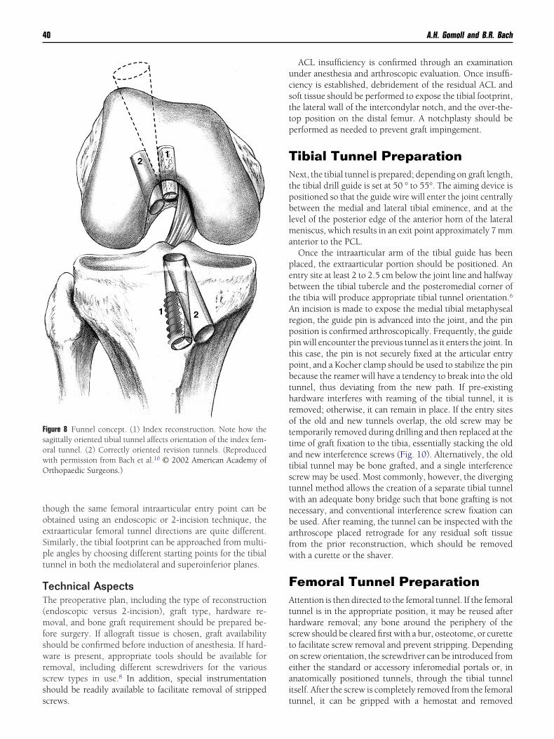
toeSpt
TT(mfswrsss
ucsttp
TNtpblma
pebtArpptpbthrottatstwnbafw
FAthstoeai
FsowO
40 A.H. Gomoll and B.R. Bach
hough the same femoral intraarticular entry point can bebtained using an endoscopic or 2-incision technique, thextraarticular femoral tunnel directions are quite different.imilarly, the tibial footprint can be approached from multi-le angles by choosing different starting points for the tibialunnel in both the mediolateral and superoinferior planes.
echnical Aspectshe preoperative plan, including the type of reconstructionendoscopic versus 2-incision), graft type, hardware re-oval, and bone graft requirement should be prepared be-
ore surgery. If allograft tissue is chosen, graft availabilityhould be confirmed before induction of anesthesia. If hard-are is present, appropriate tools should be available for
emoval, including different screwdrivers for the variouscrew types in use.8 In addition, special instrumentationhould be readily available to facilitate removal of stripped
igure 8 Funnel concept. (1) Index reconstruction. Note how theagittally oriented tibial tunnel affects orientation of the index fem-ral tunnel. (2) Correctly oriented revision tunnels. (Reproducedith permission from Bach et al.10 © 2002 American Academy ofrthopaedic Surgeons.)
crews. t
ACL insufficiency is confirmed through an examinationnder anesthesia and arthroscopic evaluation. Once insuffi-iency is established, debridement of the residual ACL andoft tissue should be performed to expose the tibial footprint,he lateral wall of the intercondylar notch, and the over-the-op position on the distal femur. A notchplasty should beerformed as needed to prevent graft impingement.
ibial Tunnel Preparationext, the tibial tunnel is prepared; depending on graft length,
he tibial drill guide is set at 50 ° to 55°. The aiming device isositioned so that the guide wire will enter the joint centrallyetween the medial and lateral tibial eminence, and at the
evel of the posterior edge of the anterior horn of the lateraleniscus, which results in an exit point approximately 7 mm
nterior to the PCL.Once the intraarticular arm of the tibial guide has been
laced, the extraarticular portion should be positioned. Anntry site at least 2 to 2.5 cm below the joint line and halfwayetween the tibial tubercle and the posteromedial corner ofhe tibia will produce appropriate tibial tunnel orientation.6
n incision is made to expose the medial tibial metaphysealegion, the guide pin is advanced into the joint, and the pinosition is confirmed arthroscopically. Frequently, the guidein will encounter the previous tunnel as it enters the joint. Inhis case, the pin is not securely fixed at the articular entryoint, and a Kocher clamp should be used to stabilize the pinecause the reamer will have a tendency to break into the oldunnel, thus deviating from the new path. If pre-existingardware interferes with reaming of the tibial tunnel, it isemoved; otherwise, it can remain in place. If the entry sitesf the old and new tunnels overlap, the old screw may beemporarily removed during drilling and then replaced at theime of graft fixation to the tibia, essentially stacking the oldnd new interference screws (Fig. 10). Alternatively, the oldibial tunnel may be bone grafted, and a single interferencecrew may be used. Most commonly, however, the divergingunnel method allows the creation of a separate tibial tunnelith an adequate bony bridge such that bone grafting is notecessary, and conventional interference screw fixation cane used. After reaming, the tunnel can be inspected with therthroscope placed retrograde for any residual soft tissuerom the prior reconstruction, which should be removedith a curette or the shaver.
emoral Tunnel Preparationttention is then directed to the femoral tunnel. If the femoral
unnel is in the appropriate position, it may be reused afterardware removal; any bone around the periphery of thecrew should be cleared first with a bur, osteotome, or curetteo facilitate screw removal and prevent stripping. Dependingn screw orientation, the screwdriver can be introduced fromither the standard or accessory inferomedial portals or, innatomically positioned tunnels, through the tibial tunneltself. After the screw is completely removed from the femoral
unnel, it can be gripped with a hemostat and removed
tma
td
fptpt
void ex
Tunnel malposition and widening in revision ACL 41
hrough a portal. If the tunnel is in a nonanatomic position, itay be possible to bypass the hardware altogether and create
n entirely new femoral tunnel.A femoral offset guide is placed through the tibial tunnel in
he over-the-top position on the lateral wall of the intercon-
Figure 9 (A) Pre-revision radiographs demonstrating femposterior soft tissues; (2) second screw aiming medially,use a 9 mm interference screw for improved tibial fixatioreconstruction bypassing preexisting hardware. (4) Revscrew in the posterior soft tissues was not removed to a
ylar notch. Occasionally, the alignment of the tibial tunnel a
orces the femoral offset guide into an unacceptably verticalosition on the femur. In this case, the guide can be placedhrough an accessory inferomedial portal with the knee hy-erflexed (130°) to obtain the correct femoral starting posi-ion.9 A guide pin is then drilled into the femur to a depth of
alpositioning. (1) Interference screw positioned in theiverging from the femoral tunnel; (3) it is preferable toPostoperative radiographs demonstrating revision ACLterference screws in correct position. The interferencetensive dissection close to the neurovascular bundle.
oral mthus dn. (B)
ision in
pproximately 3 to 4 cm, and provisionally over-reamed with

a“fppmtnw
cgiteibanttpttctT
tac
BBsGtviIaandoasml
Ft
Fca
42 A.H. Gomoll and B.R. Bach
cannulated reamer to a depth of 5 to 7 mm to create anendoscopic footprint.” The reamer is then removed, and theootprint is visualized arthroscopically to confirm an intactosterior wall. Once an intact posterior wall is confirmed byrobing, the reamer is advanced into the femur approxi-ately 35 mm. Tunnel integrity can be assessed by placing
he arthroscope retrograde through the tibial tunnel into theewly created femoral tunnel, to ensure an intact posteriorall along the entire course of the tunnel.An anatomic but posterior wall-deficient femoral tunnel
an be addressed by a 2-incision technique to create a diver-ent tunnel (Fig. 11). For the 2-incision technique, a lateralncision is made beginning at the level of the proximal pole ofhe patella and extending 3 to 5 cm further proximally. Thexposure is carried down do the iliotibial band, which isncised in its midsubstance. The vastus lateralis muscle isluntly elevated off the intermuscular septum and retractednteriorly with a Z-retractor. The periosteum is longitudi-ally divided with electrocautery, and subperiosteal dissec-ion is continued with a Cobb elevator such that the “over-he-top” position can be palpated. A J-shaped femoral guideasser is then inserted through the anterolateral portal intohe intercondylar notch, and around the over- the-top posi-ion to exit the joint capsule anterior to the lateral intermus-ular septum. The rear-entry femoral guide is then attachedo the guide passer and directed into the intercondylar notch.
igure 10 Anteroposterior radiograph depicting stacking of screwso allow for secure graft fixation in a mildly expanded tibial tunnel.
he rear-entry guide is placed in the over-the-top position, B
he femoral guide pin is drilled into the distal femur underrthroscopic visualization, and then over-reamed with theannulated reamer.7
one Graftingone grafting may be performed in either a concomitant ortaged fashion to address overlapping or widened tunnels.raft choices include autograft or, more commonly, allograft
o avoid the morbidity associated with iliac-crest graft har-esting. Allograft bone chips or struts, as well as bone result-ng from preparation of the tendon graft is commonly used.n the case of overlapping tibial tunnels, either bone graft orlarger tibial bone plug can be used to fill the defect and stillllow for interference screw fixation. Similarly, if the old andew femoral tunnels overlap, bone graft can be used to fill theefect left by the previous tunnel. To minimize extravasationf bone graft material into the joint, we found it helpful to useclear shoulder arthroscopy cannula or, alternatively, a 3-mLyringe with the front end cut off. The syringe is filled withorcellized bone-graft, introduced through a slightly en-
arged arthroscopy portal, directed into the defect, and the
igure 11 Two-incision technique to address posterior wall defi-iency. Note the different tunnel alignment with an identical intra-rticular entrance position. (Reproduced with permission from
ach et al.10 © 2002 American Academy of Orthopaedic Surgeons.)
bi(fittssfig
twwp
GIWrgp
iitpft
ttihiaBiimthaN
FeA
Fe(
Tunnel malposition and widening in revision ACL 43
one graft delivered by advancing the plunger. Other optionsnclude placing cortical allograft “matchsticks” into the defectFig 12),7 or leaving the graft bone plugs sufficiently large toll the tunnel defects. Alternatively, screws may be “stacked”o enhance bone plug fixation in overlapping or widenedunnels (Fig. 13). Supplemental fixation (eg, Endobutton,taples, post, suture button) on both the femoral and tibialides should be strongly considered whenever secure graftxation may be compromised due to enlarged or bone-rafted tunnels (Fig. 14).
If bone stock is severely compromised because of extensiveunnel widening, primary bone grafting is advisable. All hard-are is removed, and both tibial and femoral tunnels are filledith morcellized bone graft (Fig. 15). After 3 to 4 months, theatient returns for staged ligament reconstruction.
raft Preparation,ntroduction, and Fixatione prefer the use of bone–tendon–bone patellar allograft for
evision ACL reconstructions. This graft type permits the sur-eon to customize the size of the tendon graft and bone plugs,
igure 12 Matchstick bone grafting to address femoral tunnel wid-ning. (Reproduced with permission from Bach et al.10 © 2002merican Academy of Orthopaedic Surgeons.)
rovides additional bone for potential bone grafting, allows for A
nterference screw fixation on both sides of the joint, and resultsn low morbidity to the patient.10 When requesting allograftissue, the patient’s height and a preferred graft length should berovided to prevent a significant graft/host mismatch. Also, in-ormed consent discussing the risks and benefits of allograftissue must be obtained from the patient.
The total graft length is generally 95 to 105 mm, with aendon length between 40 and 50 mm, and bone plugs sizedo approximately 25 mm. The femoral plug/tendon interfaces marked on the cancellous side with a sterile marking pen toelp confirm complete seating of the femoral bone plug dur-
ng fixation. Two drill holes are placed in the tibial bone plugnd #5 braided polyester suture is passed through each hole.ecause we prefer the “push-in” technique to guide the prox-
mal bone plug into the femoral tunnel, no holes are drillednto the femoral bone plug. With the knee flexed approxi-
ately 80°, the femoral bone plug is advanced through theibial tunnel into the joint with a two-pronged pusher. Aemostat is then placed through the medial portal, to graspnd advance the proximal plug into the femoral tunnel. Aitinol wire is inserted through the accessory inferomedial
igure 13 Stacking of screws for improved fixation in mildly wid-ned tunnels, which can be applied to either the femur and tibia.Reproduced with permission from Bach et al.10 © 2002 American
cademy of Orthopaedic Surgeons.)
pwptevt
afatbpgafil
a
diiba
R
1
FteasS
FA©
44 A.H. Gomoll and B.R. Bach
ortal into the femoral tunnel anterior to the femoral plugith the knee hyperflexed to 100° to 110° to assure parallellacement of the femoral interference screw. Femoral fixa-ion is achieved with a 7-mm � 25-mm cannulated interfer-nce screw, and should be performed in hyperflexion to pre-ent divergent screw placement with potential breakouthrough the posterior femur.10
Tibial fixation is then performed. The graft is rotated 180°nd oriented such that the cancellous side of the bone plug isacing posterior and the cortical side of the bone plug is facingnterior. While holding tension on the tibial plug to preventwisting, a tibial interference screw is placed anterior to theone plug with the knee in full extension. Anterior screwlacement is less likely to abrade the tendinous portion of theraft if the screw tip extends past the tibial bone plug. If therere concerns regarding adequate tibial fixation, supplementalxation with a screw and post construct or use of a Hewson
igament button (Richards, Memphis, TN) may be used.7
Once the graft is secured, the knee is cycled multiple timesnd the reconstruction is assessed with Lachman, anterior
igure 14 Supplemental graft fixation with femoral Endobutton andibial post. Note the posterior wall blow-out, which can be managedither with supplemental Endobutton fixation as depicted here, or,lternatively, by a two-incision approach. (Reproduced with permis-ion from Bach et al.10 © 2002 American Academy of Orthopaedic
urgeons.)rawer and pivot-shift tests. The arthroscope is reinserted tonspect the graft, assess for potential graft impingement in thentercondylar notch, and rule out prominent screws or looseone graft. The knee is then copiously irrigated, and woundsre closed in a standard fashion.
eferences1. Bach BR Jr, Jones GT, Sweet FA, et al: Arthroscopy-assisted anterior cruci-
ate ligament reconstruction using patellar tendon substitution. Two- tofour-year follow-up results. Am J Sports Med 22:758-767, 1994
2. Bach BR Jr, Levy ME, Bojchuk J, et al: Single-incision endoscopic anteriorcruciate ligament reconstruction using patellar tendon autograft. Mini-mum two-year follow-up evaluation. Am J Sports Med 26:30-40, 1998
3. Bach BR Jr, Tradonsky S, Bojchuk J, et al: Arthroscopically assistedanterior cruciate ligament reconstruction using patellar tendon au-tograft. Five- to nine-year follow-up evaluation. Am J Sports Med 26:20-29, 1998
4. Bach BR Jr: Patellar tendon autograft for ACL reconstruction, in MillerM, Cole BJ (eds): Textbook of Arthroscopy. Philadelphia, PA, Saunders-Elsevier, 2004, pp 645-655
5. Creighton RA, Bach BR Jr: Revision anterior cruciate ligament recon-struction with patellar tendon allograft. Surgical technique. Sports MedArthrosc Rev 13:38-45, 2005
6. Bach BR Jr: Revision anterior cruciate ligament surgery. Arthroscopy19:14-29, 2003 (suppl 1)
7. Bach BR Jr: Revision ACL reconstruction: indications and technique, inMiller M, Cole BJ (eds): Textbook of Arthroscopy. Philadelphia, PA,Saunders-Elsevier, 2004, pp 675-686
8. Bach BR Jr: Special report: observations on interference screw morphol-ogies. Arthroscopy 16:E10, 2000
9. O’Donnell JB, Scerpella TA: Endoscopic anterior cruciate ligament re-construction: modified technique and radiographic review. Arthros-copy 11:577-584, 1995
0. Bach BR Jr, Mazzocca A, Fox JA: Revision anterior cruciate ligamentsurgery, in Grana WA (ed): Orthopaedic Knowledge Online. Rose-mont, IL, American Academy of Orthopaedic Surgeons, 2002. Avail-
igure 15 Bone grafting of widened tunnels before staged revisionCL reconstruction (Reproduced with permission from Bach et al.10
2002 American Academy of Orthopaedic Surgeons.)
able at: http://www.aaos.org/oko. Accessed March 9, 2006